Vertical Transmission of a Phylogenetically Complex Microbial Consortium in the Viviparous
12
APPLIED AND ENVIRONMENTAL MICROBIOLOGY, Apr. 2007, p. 2067–2078 Vol. 73, No. 7 0099-2240/07/$08.000 doi:10.1128/AEM.01944-06 Copyright © 2007, American Society for Microbiology. All Rights Reserved. Vertical Transmission of a Phylogenetically Complex Microbial Consortium in the Viviparous Sponge Ircinia felix Susanne Schmitt, 1 Jeremy B. Weisz, 2 † Niels Lindquist, 2 and Ute Hentschel 1 * Research Center for Infectious Diseases, University of Wuerzburg, Roentgenring 11, D-97070 Wuerzburg, Germany, 1 and Institute of Marine Sciences, University of North Carolina at Chapel Hill, 3431 Arendell Street, Morehead City, North Carolina 28557 2 Received 16 August 2006/Accepted 21 January 2007 Many marine demosponges contain large amounts of phylogenetically complex yet highly sponge-specific microbial consortia within the mesohyl matrix, but little is known about how these microorganisms are acquired by their hosts. Settlement experiments were performed with the viviparous Caribbean demosponge Ircinia felix to investigate the role of larvae in the vertical transmission of the sponge-associated microbial community. Inspections by electron microscopy revealed large amounts of morphologically diverse microor- ganisms in the center of I. felix larvae, while the outer rim appeared to be devoid of microorganisms. In juveniles, microorganisms were found between densely packed sponge cells. Denaturing gradient gel electro- phoresis (DGGE) was performed to compare the bacterial community profiles of adults, larvae, and juvenile sponges. Adults and larvae were highly similar in DGGE band numbers and banding patterns. Larvae released by the same adult individual contained highly similar DGGE banding patterns, whereas larvae released by different adult individuals showed slightly different DGGE banding patterns. Over 200 bands were excised, sequenced, and phylogenetically analyzed. The bacterial diversity of adult I. felix and its larvae was comparably high, while juveniles showed reduced diversity. In total, 13 vertically transmitted sequence clusters, hereafter termed “IF clusters,” that contained sequences from both the adult sponge and offspring (larvae and/or juveniles) were found. The IF clusters belonged to at least four different eubacterial phyla and one possibly novel eubacterial lineage. In summary, it could be shown that in I. felix, vertical transmission of microorgan- isms through the larvae is an important mechanism for the establishment of the sponge-microbe association. Sponges (Porifera) are evolutionarily ancient metazoa that first appeared in Precambrian times nearly 600 million years ago (28). Today, an estimated 13,000 species, classified in three classes (Demospongiae, Calcarea, and Hexactinellida) populate virtually all aquatic habitats from shallow tropical reefs to the polar seas and the deep ocean and even freshwater lakes and rivers (20). A great diversity of sponges occurs on coral reefs, where they exhibit a wide range of shapes and colors. Sponges have a primitive morphology, as they lack true organs or tis- sues. Instead, sponges possess totipotent, mobile cells freely scattered in an extracellular matrix called the mesohyl, which is covered by a single cell layer, the pinacoderm. Inhalant and exhalant canals in the mesohyl build an aquiferous system through which water is pumped actively by flagellated choano- cytes (3). As filter feeders, sponges efficiently take up nutrients like organic particles and microorganisms from the seawater, leaving the expelled water essentially sterile (37, 39, 57). Re- production occurs either oviparously or viviparously. In ovip- arous sponges, gametes are released into the water during large, synchronized spawning events, with fertilization occur- ring outside the sponge body. Eggs of viviparous sponges are fertilized internally, and the brooding of embryos occurs within the mesohyl, often in specialized brooding chambers. After their release, larvae of viviparous sponges actively swim for a relatively short period of time (30) or creep over the substrate in search of suitable settlement sites. Upon settlement, they undergo a rapid metamorphosis to an early juvenile stage. Additionally, many sponges also reproduce asexually via bud- ding or the formation of gemmulae. The association of many demosponges with large microbial consortia is well recognized (16, 18, 19, 21). This complex microbial community differs significantly from seawater and sediments in both concentration and diversity (12, 17, 56). Approximately 40 to 60% of sponge biomass can consist of microorganisms, which are located mostly extracellularly in the mesohyl. This equals 10 8 to 10 10 bacteria g 1 tissue and ex- ceeds seawater concentrations by 2 to 4 orders of magnitude (11, 55). Morphological diversity of sponge-associated micro- organisms was shown by electron microscopy. Several different morphotypes with unusual membrane features were described for various sponge species (12, 13, 53, 54, 58). More recently, classical cultivation as well as molecular studies based on 16S rRNA gene sequence information revealed a high phyloge- netic diversity of sponge-associated microbial consortia. Mem- bers of eight phyla within the domain Bacteria (Proteobacteria [Alphaproteobacteria, Gammaproteobacteria, and Deltapro- teobacteria], Acidobacteria, Actinobacteria, Bacteroidetes, Chlo- roflexi, Cyanobacteria, Gemmatimonadetes, and Nitrospira) and one archaeal phylum (Crenarchaeota) have been detected (17, 34, 38, 49, 51, 56). Furthermore, a new candidate phylum, termed “Poribacteria,” has been detected in several high-mi- crobial-abundance sponges (9, 10). This phylogenetically com- plex microbial consortium is highly sponge specific in that the corresponding phylogenetic lineages have been found in taxo- * Corresponding author. Mailing address: Research Center for In- fectious Diseases, University of Wuerzburg, Roentgenring 11, D-97070 Wuerzburg, Germany. Phone: 49-931-312581. Fax: 49-931-312578. E-mail: [email protected]. † Present address: Department of Biological Sciences, Old Dominion University, Norfolk, VA 23529. Published ahead of print on 2 February 2007. 2067 Downloaded from https://journals.asm.org/journal/aem on 05 February 2022 by 183.76.6.220.
Transcript of Vertical Transmission of a Phylogenetically Complex Microbial Consortium in the Viviparous

APPLIED AND ENVIRONMENTAL MICROBIOLOGY, Apr. 2007, p. 2067–2078 Vol. 73, No. 70099-2240/07/$08.00�0 doi:10.1128/AEM.01944-06Copyright © 2007, American Society for Microbiology. All Rights Reserved.
Vertical Transmission of a Phylogenetically Complex MicrobialConsortium in the Viviparous Sponge Ircinia felix�
Susanne Schmitt,1 Jeremy B. Weisz,2† Niels Lindquist,2 and Ute Hentschel1*Research Center for Infectious Diseases, University of Wuerzburg, Roentgenring 11, D-97070 Wuerzburg, Germany,1 and Institute of
Marine Sciences, University of North Carolina at Chapel Hill, 3431 Arendell Street, Morehead City, North Carolina 285572
Received 16 August 2006/Accepted 21 January 2007
Many marine demosponges contain large amounts of phylogenetically complex yet highly sponge-specificmicrobial consortia within the mesohyl matrix, but little is known about how these microorganisms areacquired by their hosts. Settlement experiments were performed with the viviparous Caribbean demospongeIrcinia felix to investigate the role of larvae in the vertical transmission of the sponge-associated microbialcommunity. Inspections by electron microscopy revealed large amounts of morphologically diverse microor-ganisms in the center of I. felix larvae, while the outer rim appeared to be devoid of microorganisms. Injuveniles, microorganisms were found between densely packed sponge cells. Denaturing gradient gel electro-phoresis (DGGE) was performed to compare the bacterial community profiles of adults, larvae, and juvenilesponges. Adults and larvae were highly similar in DGGE band numbers and banding patterns. Larvae releasedby the same adult individual contained highly similar DGGE banding patterns, whereas larvae released bydifferent adult individuals showed slightly different DGGE banding patterns. Over 200 bands were excised,sequenced, and phylogenetically analyzed. The bacterial diversity of adult I. felix and its larvae was comparablyhigh, while juveniles showed reduced diversity. In total, 13 vertically transmitted sequence clusters, hereaftertermed “IF clusters,” that contained sequences from both the adult sponge and offspring (larvae and/orjuveniles) were found. The IF clusters belonged to at least four different eubacterial phyla and one possiblynovel eubacterial lineage. In summary, it could be shown that in I. felix, vertical transmission of microorgan-isms through the larvae is an important mechanism for the establishment of the sponge-microbe association.
Sponges (Porifera) are evolutionarily ancient metazoa thatfirst appeared in Precambrian times nearly 600 million yearsago (28). Today, an estimated 13,000 species, classified in threeclasses (Demospongiae, Calcarea, and Hexactinellida) populatevirtually all aquatic habitats from shallow tropical reefs to thepolar seas and the deep ocean and even freshwater lakes andrivers (20). A great diversity of sponges occurs on coral reefs,where they exhibit a wide range of shapes and colors. Spongeshave a primitive morphology, as they lack true organs or tis-sues. Instead, sponges possess totipotent, mobile cells freelyscattered in an extracellular matrix called the mesohyl, which iscovered by a single cell layer, the pinacoderm. Inhalant andexhalant canals in the mesohyl build an aquiferous systemthrough which water is pumped actively by flagellated choano-cytes (3). As filter feeders, sponges efficiently take up nutrientslike organic particles and microorganisms from the seawater,leaving the expelled water essentially sterile (37, 39, 57). Re-production occurs either oviparously or viviparously. In ovip-arous sponges, gametes are released into the water duringlarge, synchronized spawning events, with fertilization occur-ring outside the sponge body. Eggs of viviparous sponges arefertilized internally, and the brooding of embryos occurs withinthe mesohyl, often in specialized brooding chambers. After
their release, larvae of viviparous sponges actively swim for arelatively short period of time (30) or creep over the substratein search of suitable settlement sites. Upon settlement, theyundergo a rapid metamorphosis to an early juvenile stage.Additionally, many sponges also reproduce asexually via bud-ding or the formation of gemmulae.
The association of many demosponges with large microbialconsortia is well recognized (16, 18, 19, 21). This complexmicrobial community differs significantly from seawater andsediments in both concentration and diversity (12, 17, 56).Approximately 40 to 60% of sponge biomass can consist ofmicroorganisms, which are located mostly extracellularly in themesohyl. This equals 108 to 1010 bacteria g�1 tissue and ex-ceeds seawater concentrations by 2 to 4 orders of magnitude(11, 55). Morphological diversity of sponge-associated micro-organisms was shown by electron microscopy. Several differentmorphotypes with unusual membrane features were describedfor various sponge species (12, 13, 53, 54, 58). More recently,classical cultivation as well as molecular studies based on 16SrRNA gene sequence information revealed a high phyloge-netic diversity of sponge-associated microbial consortia. Mem-bers of eight phyla within the domain Bacteria (Proteobacteria[Alphaproteobacteria, Gammaproteobacteria, and Deltapro-teobacteria], Acidobacteria, Actinobacteria, Bacteroidetes, Chlo-roflexi, Cyanobacteria, Gemmatimonadetes, and Nitrospira) andone archaeal phylum (Crenarchaeota) have been detected (17,34, 38, 49, 51, 56). Furthermore, a new candidate phylum,termed “Poribacteria,” has been detected in several high-mi-crobial-abundance sponges (9, 10). This phylogenetically com-plex microbial consortium is highly sponge specific in that thecorresponding phylogenetic lineages have been found in taxo-
* Corresponding author. Mailing address: Research Center for In-fectious Diseases, University of Wuerzburg, Roentgenring 11, D-97070Wuerzburg, Germany. Phone: 49-931-312581. Fax: 49-931-312578.E-mail: [email protected].
† Present address: Department of Biological Sciences, Old DominionUniversity, Norfolk, VA 23529.
� Published ahead of print on 2 February 2007.
2067
Dow
nloa
ded
from
http
s://j
ourn
als.
asm
.org
/jour
nal/a
em o
n 05
Feb
ruar
y 20
22 b
y 18
3.76
.6.2
20.

nomically and geographically different marine demospongesbut have not been detected in seawater, sediments, other ma-rine invertebrates, or a freshwater sponge (14).
Presently, little is known about how the unique and appar-ently stable sponge-microbe associations are established andmaintained over time. Vertical transmission as a mechanismfor bacterial passage between sponge generations was pro-posed by Levi and Porte in 1962 (26). Electron microscopystudies revealed the presence of microorganisms in oocytes ofoviparous sponges and embryos/larvae of viviparous sponges(see reference 7 and references cited therein; 23–25, 40, 45).Vertical transmission of Cyanobacteria in developing eggs andsperm of the sponge Chondrilla australiensis (52) and transmis-sion of a spiral bacterium through all stages of embryonicdevelopment in the sponge Halisarca dujardini (7) were re-ported. Similarly, Maldonado and coworkers (32) used elec-tron microscopy to document the transmission of yeast inChondrilla species. Enticknap et al. (6) were the first to phy-logenetically identify alphaproteobacteria associated with lar-vae of the sponge Mycale laxissima and to confirm their pres-ence by fluorescence in situ hybridization. Isolates JE061 toJE065 were most closely related to strain MBIC3368 that hadpreviously been isolated from at least eight other demosponges(43). Sharp et al. (46) reported the vertical transmission ofcomplex bacterial and archaeal consortia in the tropical spongeCorticium sp. Using fluorescently labeled 16S rRNA probes, itwas possible to localize specific microbial lineages in the adultand in early and late stages of embryonic development.
For this study, the ball-shaped sponge Ircinia felix Duchas-saing and Michelotti 1864 (order Dictyoceratida) (5) was cho-sen because it is a common and accessible species on shallowCaribbean coral reefs, grassbeds, and mangroves. It is easilyidentified by its gray to light brown color and hexagonallyoriented, low white knobs that are interconnected by whitelines. Ircinia species produce an array of low-molecular-weightvolatile compounds that give them a strong, pungent smell(35). Ircinia sponges are viviparous and produce relativelylarge parenchymella-type larvae. In the Florida Keys, they arereleased in the early morning hours (0700 to 1000 h) in syn-chronized spawning events 1 to 2 days following the full moon,typically in May and June (N. Lindquist, personal observation).Larvae swim briefly after spawning and settle within minutes toseveral hours and then metamorphose to a flattened earlyjuvenile stage within a day. The aim of this study was to iden-tify, localize, and phylogenetically characterize the bacterialcommunity in adults, larvae, and early-stage juveniles of I. felix.The different developmental stages were analyzed by a combi-nation of molecular (denaturing gradient gel electrophoresis[DGGE]) and visual (electron microscopy) techniques.
MATERIALS AND METHODS
Larva settlement experiments. Experiments were performed on a shallowtropical patch reef (25°01.610�N, 80°23.671�W) offshore of Key Largo, FL, inJune and August 2004 using the NOAA’s National Undersea Research Centerfacilities and vessels. Sponge larvae were collected in situ from four individualsponges (sponges 2, 3, 4, and 5) using methodology described previously byLindquist et al. (30) and transferred into 3-liter holding trays containing naturalseawater. Additionally, tissue samples were taken from the corresponding adultspecimens from which larvae had been collected. Individual larvae were capturedfrom the holding trays with glass Pasteur pipettes and were either transferredinto 150 �l sterile filtered seawater for DNA extraction and stored at �80°C or
fixed in 2.5% glutaraldehyde–double-distilled water for transmission electronmicroscopy and stored at 4°C. Larvae not used for microscopy or DNA extractionwere settled by placing them into sealed plastic containers (�75-ml volume) withNITEX (100-�m) “windows” to allow the flow of ambient seawater into thecontainers. I. felix larvae settled onto the nylon mesh (i.e., pantyhose) that heldthe larval collection containers on a rack at a depth of 9 m. During this setupprocess, larval and juvenile contact with air was avoided. Juveniles were recov-ered 1 to 3 days postsettlement, along with control pieces of mesh, which did notcontain settled sponges. These samples were preserved as described above forDNA and microscopic analysis. Fluorescence in situ hybridization experimentscould not be reliably performed because of the small larval size and limitedjuvenile biomass.
Transmission electron microscopy. The adult sponge samples that had beenpreserved in 2.5% glutaraldehyde–double-distilled water were cut into smallpieces of about 1 mm3. All samples (adults, larvae, and juveniles) were thenwashed five times in cacodylate buffer (50 mM, pH 7.2), fixed in 2% osmiumtetroxide for 90 min, washed again five times in double-distilled water, andincubated overnight in 0.5% uranyl acetate. After dehydration in an ethanolseries (30, 50, 70, 90, 96, and three times at 100% for 30 min, respectively),samples were incubated three times for 30 min in 1� propylene oxide, main-tained overnight in 1:1 (vol/vol) propylene oxide-Epon 812 (Serva), incubatedtwice for 2 h in Epon 812, and finally embedded in Epon 812 for 48 h at 60°C.Samples were then sectioned with an ultramicrotome (OM U3; C. Reichert,Austria) and examined by transmission electron microscopy (EM 10; Zeiss,Germany).
DGGE. Frozen pieces of adult sponge tissue were ground in liquid nitrogenwith a mortar and pestle, and DNA was extracted using the Fast DNA Spin kitfor soil (Q-Biogene, Heidelberg, Germany) in accordance with the manufactur-er’s instructions. Larva and juvenile DNA were extracted from three, five, orseven individual larvae or juveniles, which had been pooled immediately aftercollection, by heating in 150 �l of double-distilled water in a water bath for 10min at 100°C. The solution was used as a template for PCR. The juvenile DNAwas extracted together with a small piece of nylon on which the sponge hadgrown because it was impossible to remove the tiny sponge from its substratumwithout losing too much biomass. The universal primers 341f, with a GC clamp,and 907r (33) were used for PCR amplification of bacterial 16S rRNA genes.Cycling conditions on a Mastercycler gradient (Eppendorf, Hamburg, Germany)were as follows: an initial denaturing step at 95°C for 5 min and 30 cycles ofdenaturing at 95°C for 1 min, primer annealing at 54°C for 1 min, and elongationat 72°C for 45 s, followed by a final extension step at 72°C for 10 min. DGGE wasperformed with a Bio-Rad (Munchen, Germany) DCode Universal mutationdetection system on a 10% (wt/vol) polyacrylamide gel in 1� Tris-acetate-EDTAand using a 0 to 90% denaturing gradient; 100% denaturant corresponded to 7M urea and 40% (vol/vol) formamide. Electrophoresis was performed for 6 h at150 V and 60°C. Gels were stained for 30 min with SYBR gold (MolecularProbes) and scanned on a Typhoon 8600 scanner (Amersham Biosciences).DGGE banding pattern similarities were determined by cluster analysis usingQuantity One (Bio-Rad, Munchen, Germany). Selected bands were excised withan ethyl alcohol-sterilized scalpel and incubated in 25 �l double-distilled waterovernight at 4°C. Four microliters of eluted DNA was subsequently used forreamplification with primers 341f and 907r under the PCR conditions describedabove. PCR products were ligated into the pGEM-T Easy vector (Promega) andtransformed by electroporation into competent Escherichia coli XL1-Blue cells.Plasmid DNA of up to four different clones per excised band was isolated bystandard miniprep procedures, and the correct insert size was verified by usingagarose gel electrophoresis following restriction digestion (41). Sequencing wasperformed using an ABI 377XL automated sequencer (Applied Biosystems).Sequences were edited with the ContigExpress Tool in Vector NTI suite 6.0(InforMax, Inc.).
Phylogenetic sequence analysis. Sequences were checked for chimeras withthe program Pintail (1) and for other amplification and sequencing artifacts.Following the removal of chimeras from the data set, percent similarities (pdistances) between sequences from the same source (adult, larva, or juvenile)were determined with the editor Align (Multicolor Sequence Alignment Editor;D. Hepperle [http://wwwuser.gwdg.de/�dheppner/]), and those with identitiesabove 99% were grouped together in operational taxonomic units (OTUs). Onlyone randomly chosen sequence per OTU was used for further analysis. As a firstapproximation, the phylogenetic affiliation was determined for each OTU bycomparison against sequences available in GenBank using BLAST (http://www.ncbi.nlm.nih.gov/BLAST). Sequences obtained in this study together with ref-erence sequences (all nearest BLAST matches and, moreover, representatives ofthe respective phylum) were aligned automatically with ClustalX (50), and thealignment was subsequently corrected manually using Align (Multicolor Se-
2068 SCHMITT ET AL. APPL. ENVIRON. MICROBIOL.
Dow
nloa
ded
from
http
s://j
ourn
als.
asm
.org
/jour
nal/a
em o
n 05
Feb
ruar
y 20
22 b
y 18
3.76
.6.2
20.

quence Alignment Editor; D. Hepperle [http://wwwuser.gwdg.de/�dheppner/]).Phylogenetic trees were constructed with the ARB software package (31). Ini-tially, neighbor-joining (Jukes-Cantor correction) and maximum parsimony treeswere calculated with nearly full-length sequences (�1,250 bp) and 100 pseu-doreplicates. Subsequently, partial sequences were added to the trees withoutchanging the topology by the use of the parsimony-interactive method in ARB.Finally, 50% majority rule consensus trees were constructed.
Nucleotide sequence accession numbers. The 16S rRNA gene sequences ob-tained in this study were deposited in the EMBL/GenBank/DDBJ databaseunder accession numbers DQ661746 to DQ661857.
RESULTS
Transmission electron microscopy. Electron microscopy ofadult I. felix specimens 4 and 5 revealed large and complexmicrobial communities (Fig. 1). Microorganisms are embed-ded extracellularly and are scattered equally in the collagenmatrix of the mesohyl. Bacterial sizes were between 1 and 2.5�m in length and 0.5 to 1 �m in diameter and showed differentshapes like cocci, rods, and other, irregular forms. Cyanobac-teria are easily identified by their characteristic internal thyla-koid membrane stacks (Fig. 1). The different morphotypesresemble subtypes from other sponge species described previ-ously by Vacelet (53), Wilkinson (58), and Friedrich et al. (12).Previously described prokaryotic cells with nucleoid-like struc-tures (9, 13) could also be detected in I. felix.
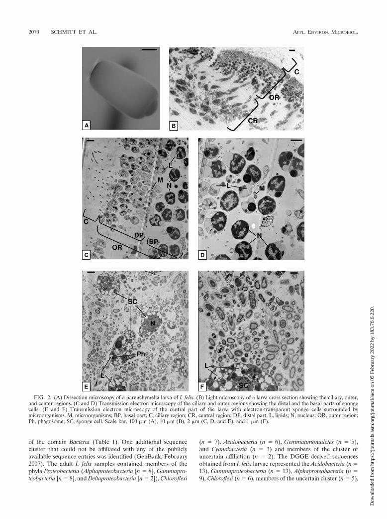
I. felix individuals 4 and 5 released parenchymella-type lar-vae that were light gray, about 500 �m in length (Fig. 2A), andcompletely covered with a carpet of small, equally long cilia asseen under the dissecting microscope. Much longer cilia werepresent at the dark pigmented posterior pole. Figure 2B givesan overview of a larval cross section. The ciliary region, theouter region, and the central region form three distinct layers.The ciliary region is followed by seemingly two layers of smallsponge cells (Fig. 2C and D). In fact, because of diagonalsectioning, the two layers represent the distal and basal parts ofthe same elongated, pear-shaped sponge cells, which build an
external ciliated epithelium. The distal part of these cells con-tains cell organelles like the Golgi apparatus and mitochondria(data not shown). The enlarged basal part contains the nucleuswith condensed chromatin (Fig. 2D). These cells contain largeamounts of lipids (A. Ereskovsky, St. Petersburg State Univer-sity, personal communication). In the central region of thelarvae, electron-transparent amoeboid sponge cells are looselyembedded in the extracellular matrix (Fig. 2E and F). Theycontain nuclei, phagosomes, and many lipids. Visual inspectionof at least five different individuals revealed that the outerregion of the larvae is almost free of bacteria, whereas thecentral region of the larva contains numerous microorganisms.These microorganisms look similar in size, shape, and mem-brane structure to those found in adult samples. Some electronmicrographs also show phagosomes containing digested bacte-rial remnants (Fig. 2E) and bacterial cell division (data notshown).
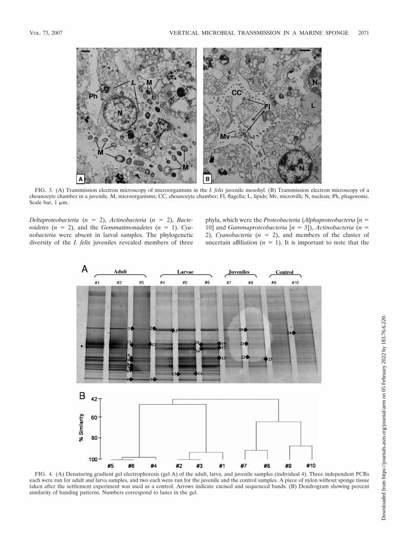
The juveniles of sponges 4 and 5 showed densely packedelectron-transparent amoeboid sponge cells (Fig. 3). In con-trast to the sponge cells of the larvae, juvenile cells containfewer lipids but more phagosomes and vesicles (data notshown). Microorganisms in juveniles are located betweensponge cells (Fig. 3A), which show phagosomes, indicatingmicrobial phagocytosis. The presence of choanocyte chambersindicates that a functional aquiferous system has already beenestablished (Fig. 3B).
DGGE. DGGE was used to fingerprint the bacterial com-munity of I. felix adult, larval, and juvenile stages. Each lane ona gel represents an independent PCR. Banding patterns of thesame sample were highly similar, indicating little or no PCRbias between PCRs of the same DNA extraction. Total bandnumbers were similar for adult 4 (average number of 21) andits larvae (average number of 22.7) but increased in juveniles(average number of 27.5) and the nylon control (average num-ber of 27) (Fig. 4A). Cluster analysis of DGGE banding pat-terns placed the adult and larva samples together, whereas thejuvenile samples clustered with the control sample (Fig. 4B).DGGE band analysis of different larval pools released by thesame adult individual revealed highly similar banding patternsthat corresponded to that of the adult (Fig. 5A and B). Incontrast, comparison of DGGE bands of larvae released bydifferent adult individuals revealed a higher variability (Fig. 6Aand B). The similarity of DGGE banding patterns from differ-ent larva pools from the same individual ranged down to 70%,whereas that of larva pools from different individuals was evenlower (Fig. 5B and 6B). On the gel in Fig. 6, some bands werepresent (e.g., DGGE band 25) or absent (e.g., DGGE bands 24and 43 are missing in larva pool 4) only in a given sample, yetthe majority of bands were still shared between the differentsamples (e.g., DGGE bands 1, 13, 26, and 32).
Phylogenetic sequence analysis. In total, 218 sequences werederived from excised DGGE bands. After the removal of 54chimeras (Pintail) and five sequences with undefined sequenc-ing artifacts, the remaining 159 sequences were maintainedseparately according to their source (adult, larva, or juvenile).Each set of sequences was grouped into OTUs based on a�99% similarity cutoff. One sequence of each of the altogether112 unique OTUs was chosen, and accordingly, 41 adult, 53larval, and 18 juvenile sequences were used for further phylo-genetic analysis. The sequences fell into seven different phyla
FIG. 1. Transmission electron microscopy of microorganisms in theI. felix adult mesohyl. Cy, Cyanobacteria; M, microorganisms. Scale bar,1 �m.
VOL. 73, 2007 VERTICAL MICROBIAL TRANSMISSION IN A MARINE SPONGE 2069
Dow
nloa
ded
from
http
s://j
ourn
als.
asm
.org
/jour
nal/a
em o
n 05
Feb
ruar
y 20
22 b
y 18
3.76
.6.2
20.
of the domain Bacteria (Table 1). One additional sequencecluster that could not be affiliated with any of the publiclyavailable sequence entries was identified (GenBank, February2007). The adult I. felix samples contained members of thephyla Proteobacteria (Alphaproteobacteria [n � 8], Gammapro-teobacteria [n � 8], and Deltaproteobacteria [n � 2]), Chloroflexi
(n � 7), Acidobacteria (n � 6), Gemmatimonadetes (n � 5),and Cyanobacteria (n � 3) and members of the cluster ofuncertain affiliation (n � 2). The DGGE-derived sequencesobtained from I. felix larvae represented the Acidobacteria (n �13), Gammaproteobacteria (n � 13), Alphaproteobacteria (n �9), Chloroflexi (n � 6), members of the uncertain cluster (n � 5),
FIG. 2. (A) Dissection microscopy of a parenchymella larva of I. felix. (B) Light microscopy of a larva cross section showing the ciliary, outer,and center regions. (C and D) Transmission electron microscopy of the ciliary and outer regions showing the distal and the basal parts of spongecells. (E and F) Transmission electron microscopy of the central part of the larva with electron-transparent sponge cells surrounded bymicroorganisms. M, microorganisms; BP, basal part; C, ciliary region; CR, central region; DP, distal part; L, lipids; N, nucleus; OR, outer region;Ph, phagosome; SC, sponge cell. Scale bar, 100 �m (A), 10 �m (B), 2 �m (C, D, and E), and 1 �m (F).
2070 SCHMITT ET AL. APPL. ENVIRON. MICROBIOL.
Dow
nloa
ded
from
http
s://j
ourn
als.
asm
.org
/jour
nal/a
em o
n 05
Feb
ruar
y 20
22 b
y 18
3.76
.6.2
20.
Deltaproteobacteria (n � 2), Actinobacteria (n � 2), Bacte-roidetes (n � 2), and the Gemmatimonadetes (n � 1). Cya-nobacteria were absent in larval samples. The phylogeneticdiversity of the I. felix juveniles revealed members of three
phyla, which were the Proteobacteria (Alphaproteobacteria [n �10] and Gammaproteobacteria [n � 3]), Actinobacteria (n �2), Cyanobacteria (n � 2), and members of the cluster ofuncertain affiliation (n � 1). It is important to note that the
FIG. 3. (A) Transmission electron microscopy of microorganisms in the I. felix juvenile mesohyl. (B) Transmission electron microscopy of achoanocyte chamber in a juvenile. M, microorganisms; CC, choanocyte chamber; Fl, flagella; L, lipids; Mv, microvilli; N, nucleus; Ph, phagosome.Scale bar, 1 �m.
FIG. 4. (A) Denaturing gradient gel electrophoresis (gel A) of the adult, larva, and juvenile samples (individual 4). Three independent PCRseach were run for adult and larva samples, and two each were run for the juvenile and the control samples. A piece of nylon without sponge tissuetaken after the settlement experiment was used as a control. Arrows indicate excised and sequenced bands. (B) Dendrogram showing percentsimilarity of banding patterns. Numbers correspond to lanes in the gel.
VOL. 73, 2007 VERTICAL MICROBIAL TRANSMISSION IN A MARINE SPONGE 2071
Dow
nloa
ded
from
http
s://j
ourn
als.
asm
.org
/jour
nal/a
em o
n 05
Feb
ruar
y 20
22 b
y 18
3.76
.6.2
20.

numbers do not reflect the in vivo abundances in the sponge. Atotal of 63%, 55%, and 28% of all sequences obtained fromadult, larva, and juvenile samples, respectively, had a sponge-derived sequence as their closest relative (BLAST analysis, asa first approximation).
Clusters of vertically transmitted bacterial groups. Verti-cally transmitted phylotypes are defined as monophyletic clus-ters of two or more sequences that were recovered from boththe adult sponge and offspring (larvae and/or juveniles) of I.felix (hereafter termed IF clusters). Altogether, 13 monophy-letic sequence clusters were identified (Fig. 7 and 8), whichbelonged to four different bacterial phyla and one additionallineage of uncertain affiliation. More than 60% of all I. felix-derived DGGE sequences fell into these clusters. The in-clus-ter similarity was above 97% for eight clusters and above95.5% for 12 clusters. Only cluster IF-Alpha-2 had a relativelylow in-cluster similarity of 93.9%. Two clusters (IF-Alpha-2and IF-Alpha-4) contained sequences of all three developmen-tal stages (adults, larvae, and juveniles).
DISCUSSION
Vertical transmission, the passage of microbial symbionts tothe next host generation through the reproductive cell lines, isa hallmark of evolutionarily ancient symbioses. Vertical trans-mission has been documented in terrestrial insects such as ants(42) and aphids (2) as well as in marine invertebrates such asbryozoans (29) and bivalves (4, 47). Since the passage of sym-
bionts via the reproductive stages is highly selective, this pro-cess frequently leads to cospeciation between the host andsymbiotic lineages, resulting in congruent phylogenetic trees(36). The presence of bacteria in the reproductive stages hasalso been demonstrated in representatives of all three spongeclasses by electron microscopy, suggesting that vertical trans-mission is a widespread mechanism in this phylum (see refer-ence 7 and references cited therein). In comparison to thewell-studied symbioses mentioned above, the microbial associ-ations of sponges are different in several respects. Rather thanthe known one (few)-symbiont-one-host types of associations,the microbial consortia of sponges are exceedingly complex,containing sponge-specific representatives of at least eight dif-ferent bacterial phyla and one archaeal phylum (18). Second,the microbial biomass within the bacteriosponges (high-micro-bial-abundance sponges) is massive, contributing up to 40 to60% of the animal’s biomass (53, 59). To our knowledge, noother animal phylum tolerates such amounts of internal, freelydispersed microorganisms. The enormous microbial biomass invertebrate intestines is also located internally but is containedwithin specialized organs such as the rumen. Third, because ofthe characteristic anatomy of sponges, there are no physicalbarriers such as organs or tissues that separate the sites ofreproduction from the mesohyl microbiota. In other symbio-ses, symbionts are frequently contained in specialized cells(bacteriocytes) or modified host organs (i.e., the trophosomeof the tubeworm Riftia pachyptila and the glands of Deshayes
FIG. 5. (A) Denaturing gradient gel electrophoresis (gel E) of I. felix adult 4 compared to different larva pools released by the same adultspecimen. Three independent PCRs were run for the adult, and two each were run for pooled larvae. Arrows indicate excised and sequenced bands.(B) Dendrogram showing percent similarity of banding patterns. Numbers correspond to lanes in the gel.
2072 SCHMITT ET AL. APPL. ENVIRON. MICROBIOL.
Dow
nloa
ded
from
http
s://j
ourn
als.
asm
.org
/jour
nal/a
em o
n 05
Feb
ruar
y 20
22 b
y 18
3.76
.6.2
20.

of the shipworm Bankia setacea). It was therefore of interest todocument the passage of microorganisms via the reproductivestages of a Caribbean demosponge and to phylogeneticallycharacterize the bacterial consortia within them. A combina-tion of visual (electron microscopy) and molecular (DGGE)techniques was used towards this goal.
Electron microscopy studies provided the first insights intothe presence of microorganisms within the larvae of I. felix(Fig. 2). High amounts of morphologically diverse microorgan-isms were located extracellularly in the central part of thelarvae, while the outer rim appeared to be almost free of
bacteria. This is in agreement with the microscopic descriptionof the Mediterranean species Ircinia oros, which also containednumerous bacteria in the inner part of the larvae but not in theperipheral region (8). In general, the larvae of both Irciniaspecies have a similar morphology including dark-pigmentedcells and a ring of elongated cilia at the posterior pole, whichmight be important in the response to external stimuli duringthe planktonic life stage (27). Both Ircinia species have a cili-ated epithelium consisting of densely packed sponge cells. Thispear-shaped cell form and the location of the nucleus in thebasal part are unusual, as the nucleus is located more distallyin most parenchymella larvae (8). The central part of the I. felixlarva contains only one loosely associated cell type, which hasbeen identified as being archaeocytes in Ircinia oros (8) andother dictyoceratid species (24). Finally, both Ircinia larvaecontain unusually high amounts of lipids, which is consistentwith the lecithotrophic (nonfeeding) nature of this larvae. Thisreserve substance may allow for a long pelagic life phase andwould increase the chance of survival during metamorphosis.
The bacterial community profile of the adult I. felix spongestrikingly resembles that of other high-microbial-abundancesponges that have been subject to molecular analysis of micro-bially diverse populations (i.e., see references 17, 44, 48, 51,and 56). I. felix contained sponge-specific sequences fromseven of the eight previously reported bacterial phyla (Table1). These are the Proteobacteria (Alphaproteobacteria, Gamma-proteobacteria, and Deltaproteobacteria), Acidobacteria, Acti-
FIG. 6. (A) Denaturing gradient gel electrophoresis (gel F) of four larva pools released by four different adult specimens (specimens 2, 3, 4,and 5). Two independent PCRs each were run. Arrows indicate excised and sequenced bands. (B) Dendrogram showing the percent similarity ofbanding patterns. Numbers correspond to lanes in the gel.
TABLE 1. Phylogenetic diversity of bacteria associated with adult,larval, and juvenile I. felix
Bacterial phylumResulta
Adult Larvae Juveniles
Acidobacteria � � �Actinobacteria � � �Bacteroidetes � � �Chloroflexi � � �Cyanobacteria � � �Gemmatimonadetes � � �Alphaproteobacteria � � �Gammaproteobacteria � � �Deltaproteobacteria � � �Uncertain affiliation � � �
a �, present; �, absent.
VOL. 73, 2007 VERTICAL MICROBIAL TRANSMISSION IN A MARINE SPONGE 2073
Dow
nloa
ded
from
http
s://j
ourn
als.
asm
.org
/jour
nal/a
em o
n 05
Feb
ruar
y 20
22 b
y 18
3.76
.6.2
20.

FIG. 7. Phylogenetic distance tree calculated with 16S rRNA gene proteobacterial DGGE sequences recovered from I. felix. Neighbor-joiningand maximum parsimony (100 pseudoreplicates) bootstrap values are provided. Sequences obtained in this study are in boldface type. The clonesare coded as follows: DGGE gel (capital letter)-excised band (first number)-clone (second number). Gray boxes depict monophyletic clusters ofsequences that originated from both adults and offspring (larvae and/or juveniles) of I. felix. The scale bar indicates 10% divergence. Arrow, tooutgroup (Geothrix fermentans U41563, Holophaga foetidae X77215, and Acidobacterium capsulatum D26171). GenBank accession numbers areshown in parentheses.
2074 SCHMITT ET AL. APPL. ENVIRON. MICROBIOL.
Dow
nloa
ded
from
http
s://j
ourn
als.
asm
.org
/jour
nal/a
em o
n 05
Feb
ruar
y 20
22 b
y 18
3.76
.6.2
20.
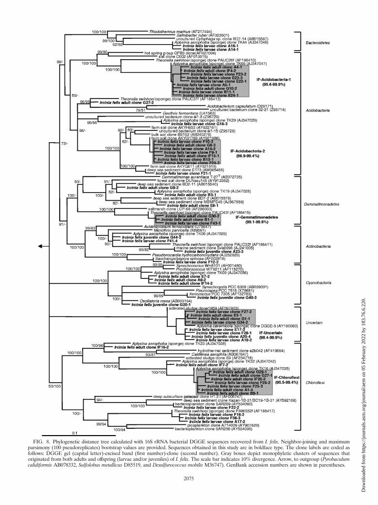
FIG. 8. Phylogenetic distance tree calculated with 16S rRNA bacterial DGGE sequences recovered from I. felix. Neighbor-joining and maximumparsimony (100 pseudoreplicates) bootstrap values are provided. Sequences obtained in this study are in boldface type. The clone labels are coded asfollows: DGGE gel (capital letter)-excised band (first number)-clone (second number). Gray boxes depict monophyletic clusters of sequences thatoriginated from both adults and offspring (larvae and/or juveniles) of I. felix. The scale bar indicates 10% divergence. Arrow, to outgroup (Pyrobaculumcalidiformis AB078332, Sulfolobus metallicus D85519, and Desulfurococcus mobilis M36747). GenBank accession numbers are shown in parentheses.
2075
Dow
nloa
ded
from
http
s://j
ourn
als.
asm
.org
/jour
nal/a
em o
n 05
Feb
ruar
y 20
22 b
y 18
3.76
.6.2
20.
nobacteria, Bacteroidetes, Chloroflexi, and Cyanobacteria. Mem-bers of two sequence clusters previously reported as “uncertainaffiliation-I and -II” (17), now known to belong to the phylumGemmatimonadetes (60; this study), were also recovered fromI. felix. Furthermore, a deeply routing sequence cluster thatcould not be affiliated with any of the known sequences avail-able in the public libraries was identified in this study. Theclosest relatives are an unidentified clone from the Mediterra-nean sponge Aplysina cavernicola (GenBank accession numberAY180080) (�97% similarity) (51) and an unidentified acti-vated-sludge clone (GenBank accession number AF097803)(94.4% similarity) (Fig. 8). This sequence cluster might repre-sent a novel clade of sponge-specific bacteria. Additionally, agammaproteobacterial cluster (IF-Gamma-3) is noteworthy, asit forms a coherent clade with a number of 16S rRNA genesequences from chemoautotrophic symbionts of deep-sea in-vertebrates including gastropods, bivalves, and tubeworms.
Interestingly, several previously reported sponge-specificclusters (17) could not be detected in this study (Fig. 7 and 8).These belong to the “Nitrospira-I” cluster of the phylum Ni-trospira that contains exclusively nitrite-oxidizing bacteria. Thegammaproteobacterial “Gamma-I” cluster that is most closelyrelated to ammonia-oxidizing Nitrosococcus species was alsonot identified. If the coordinated metabolism of ammonia-oxidizing bacteria and nitrite-oxidizing bacteria is responsiblefor the process of nitrification in sponges, then the conspicuousabsence of both clades would suggest that eubacterial nitrifi-cation is not an important process in I. felix. Nevertheless,nitrification mediated by Archaea, such as, for example, theCenarchaeum symbiosum clade of sponge symbionts, remains adistinct possibility (15, 38). Members of the recently discoveredcandidate phylum “Poribacteria” and of the domain Archaeawould not have been expected in this study because of mis-matches in the PCR primer regions. Several lineages have sofar exclusively been found in I. felix. These belong to the IF-Alpha-3 cluster related to a coral associated clone (GenBankaccession number DQ200432), the IF-Alpha-4 cluster related toRhodovulum species (GenBank accession number AM180953),and the IF-Acido-2 cluster related to acidobacterial clonesfrom soil (GenBank accession numbers AY921986, AB240276,and AY922161). Whether these lineages are specific to thesponge I. felix remains to be seen as more sequences fromother Ircinia sponges become available.
The bacterial diversity of I. felix larvae is comparable to thatof the adult sponge with respect to the DGGE band numbers,patterns, and phylotypes recovered (Fig. 4, 7, and 8). Thebanding patterns of different larval pools from the same indi-vidual were more similar than those from different individuals(Fig. 5A and 6A), a fact that is also reflected in the clusteranalysis (Fig. 5B and 6B). Deciphering the degree of fine-scaleintraspecies variation poses a challenge for further studies.Sequencing and phylogenetic analysis of the DGGE bandsshowed the presence of all previously identified, sponge-spe-cific bacterial phyla (18, 19, 21) in the larvae, with the excep-tion of the Cyanobacteria. Since DGGE bands of the appro-priate migration distance were also identified in the larvalsample (Fig. 4), it is possible that the cyanobacterial phylotypesare present in the larvae but that the corresponding bands werenot chosen for sequencing. Alternatively, since the larvae arebrooded in the mesohyl and the Cyanobacteria are found pre-
dominantly on the outer surface, they might be incorporatedinto the larvae less efficiently. In fact, Usher et al. showed thatonly 25% of all larvae from Chondrilla australiensis containedCyanobacteria (52).
The DGGE banding pattern of the I. felix juvenile samplecontained more bands than the corresponding adult and larvasamples. The dendrograms clustered the adult and larval sam-ples together, while the juvenile formed one cluster with thecontrol. The higher number of DGGE bands is probably anartifact from the extraction protocol, as the newly grownsponges were still attached to the nylon substrate at the time ofDNA extraction. Hence, the DGGE banding pattern is a mix-ture of sponge-specific phylotypes and colonizers from seawa-ter. Altogether, representatives of three bacterial phyla couldbe identified, including sponge-specific lineages of the phylaActinobacteria and Alphaproteobacteria and the clade of uncer-tain affiliation. The reduced bacterial diversity might be due tomethodological constraints, as more than twice as many se-quences were gained from adult samples (n � 41) and fromlarvae (n � 53) than from juveniles (n � 18), which showed agenerally more faint banding pattern. If more sequences wouldhave been obtained from juveniles, the number of identifiedphyla that contain sponge-specific representatives might havebeen higher.
Alternatively, the reduced diversity in juveniles may be cor-related to the feeding behavior of sponge larvae. Free-swim-ming larvae are unable to take up food particles from the watercolumn (22). If the internal sponge symbionts would serve asfood during the nonfeeding planktonic phase, then the micro-bial community within the larvae would be naturally reduced.This hypothesis is supported by electron microscopical obser-vations that show evidence of phagocytosis in the larvae (Fig.2E). Further experiments focusing on the juvenile stages arenecessary to determine whether microbially diverse popula-tions are truly reduced or whether the sponge-specific lineagesare present in the larvae, albeit below the limit of detection.Conceivably, only a single bacterium of each lineage would beneeded to inoculate the newly grown sponge.
In summary, it could be shown that in the sponge I. felix, theentire microbial consortium rather than individual phyloge-netic lineages is passed on to the next generation via the re-productive stages. Accordingly, vertical transmission is specificin that the microorganisms of I. felix, but not those from sea-water, are passed on but unselective in that there appears to beno differentiation between individual sponge-specific lineages.These data are congruent with those described previously bySharp et al. (46), who reported on the vertical transmission ofsimilarly complex phylogenetic lineages throughout the embry-onic development of Corticium sp. This passage of diversemicroorganisms to the next generation is probably due to thecharacteristic anatomy of sponges, where the reproductiveelements are exposed to microorganisms in the mesohyl,whereas in higher animals, the reproductive elements are con-tained in specialized, bacterium-free reproductive organs. Inconclusion, the ancient and widespread mechanism of verticaltransmission is clearly important for the formation and main-tenance of the phylogenetically complex yet highly sponge-specific microbial communities of many marine demosponges.
2076 SCHMITT ET AL. APPL. ENVIRON. MICROBIOL.
Dow
nloa
ded
from
http
s://j
ourn
als.
asm
.org
/jour
nal/a
em o
n 05
Feb
ruar
y 20
22 b
y 18
3.76
.6.2
20.
ACKNOWLEDGMENTS
We gratefully acknowledge the staff of NOAA’s National UnderseaResearch Center at Key Largo, FL, for professional work during fieldtrips. We thank Melissa Southwell and Channing Jones (University ofNorth Carolina at Chapel Hill) for help during sponge collection andsettlement experiments and Andrey Vishnyakov and Alexander Eres-kovsky (St. Petersburg State University) for helpful discussion of elec-tron micrographs.
This research was supported by Deutsche Forschungsgemeinschaftgrants HE3299/1-1 and HE3299/1-2 to U.H., UNCW/NURC(NA03OAR4300088) and NSF Chemical Oceanography Program(OCE 0351893) grants to C. S. Martens and N.L., and an NSF grad-uate fellowship to J.B.W.
REFERENCES
1. Ashelford, K. E., N. A. Chuzhanova, J. C. Fry, A. J. Jones, and A. J.Weightman. 2005. At least 1 in 20 16S rRNA sequence records currently heldin public repositories is estimated to contain substantial anomalies. Appl.Environ. Microbiol. 71:7724–7736.
2. Baumann, P., L. Baumann, C. Y. Lai, D. Rouhbakhsh, N. A. Moran, andM. A. Clark. 1995. Genetics, physiology, and evolutionary relationships ofthe genus Buchnera: intracellular symbionts of aphids. Annu. Rev. Micro-biol. 49:55–94.
3. Brusca, R. C., and G. J. Brusca. 1990. Phylum Porifera: the sponges, p.181–210. In A. D. Sinauer (ed.), Invertebrates. Sinauer Press, Sunderland,MA.
4. Cary, S., and S. Giovannoni. 1993. Transovarial inheritance of endosymbi-otic bacteria in clams inhabiting deep-sea hydrothermal vents and cold seeps.Proc. Natl. Acad. Sci. USA 90:5695–5699.
5. Duchassaing De Fonbressin, P., and G. Michelotti. 1864. Spongiaires de lamer Caraibe. Natuurk. Verh. Holl. Mij. Venetsch. Haarlem 21:1–124.
6. Enticknap, J. J., M. Kelly, O. Peraud, and R. T. Hill. 2006. Characterizationof a culturable alphaproteobacterial symbiont common to many marinesponges and evidence for vertical transmission via sponge larvae. Appl.Environ. Microbiol. 72:3724–3732.
7. Ereskovsky, A. V., E. Gonobobleva, and A. Vishnyakov. 2005. Morphologicalevidence for vertical transmission of symbiotic bacteria in the viviparoussponge Halisarca dujardini Johnston (Porifera, Demospongiae, Halisarca).Mar. Biol. 146:869–875.
8. Ereskovsky, A. V., and D. Tokina. 2004. Morphology and fine structure of theswimming larvae of Ircinia oros (Porifera, Demospongiae, Dictyoceratida).Invertebr. Reprod. Dev. 45:137–150.
9. Fieseler, L., M. Horn, M. Wagner, and U. Hentschel. 2004. Discovery of thenovel candidate phylum “Poribacteria” in marine sponges. Appl. Environ.Microbiol. 70:3724–3732.
10. Fieseler, L., A. Quaiser, C. Schleper, and U. Hentschel. 2006. Analysis of thefirst genome fragment from the marine sponge-associated, novel candidatephylum Poribacteria by environmental genomics. Environ. Microbiol. 8:612–624.
11. Friedrich, A. B., I. Fischer, P. Proksch, J. Hacker, and U. Hentschel. 2001.Temporal variation of the microbial community associated with the Medi-terranean sponge Aplysina aerophoba. FEMS Microbiol. Ecol. 38:105–113.
12. Friedrich, A. B., H. Merkert, T. Fendert, J. Hacker, P. Proksch, and U.Hentschel. 1999. Microbial diversity in the marine sponge Aplysina caverni-cola (formerly Verongia cavernicola) analyzed by fluorescence in situ hybri-disation (FISH). Mar. Biol. 134:461–470.
13. Fuerst, J. A., R. I. Webb, M. J. Garson, L. Hardy, and H. M. Reiswig. 1998.Membrane-bounded nucleoids in microbial symbionts of marine sponges.FEMS Microbiol. Lett. 166:29–34.
14. Gernert, C., F. O. Glockner, G. Krohne, and U. Hentschel. 2005. Microbialdiversity of the freshwater sponge Spongilla lacustris. Microb. Ecol. 50:206–212.
15. Hallam, S. J., T. J. Mincer, C. Schleper, C. M. Preston, K. Roberts, P. M.Richardson, and E. F. DeLong. 2006. Pathways of carbon assimilation andammonia oxidation suggested by environmental genomic analyses of marineCrenarchaeota. PLoS Biol. 4:520–536.
16. Hentschel, U., L. Fieseler, M. Wehrl, C. Gernert, M. Steinert, M. Horn,and J. Hacker. 2003. Microbial diversity of marine sponges, p. 59–88. InW. E. G. Muller (ed.), Molecular marine biology of sponges. Springer-Verlag, Heidelberg, Germany.
17. Hentschel, U., J. Hopke, M. Horn, A. B. Friedrich, M. Wagner, J. Hacker,and B. S. Moore. 2002. Molecular evidence for a uniform microbial commu-nity in sponges from different oceans. Appl. Environ. Microbiol. 68:4431–4440.
18. Hentschel, U., K. M. Usher, and M. W. Taylor. 2006. Marine sponges asmicrobial fermenters. FEMS Microbiol. Ecol. 55:167–177.
19. Hill, R. T. 2004. Microbes from marine sponges: a treasure trove of biodi-versity for natural products discovery, p. 177–190. In A. T. Bull (ed.), Mi-crobial diversity and bioprospecting. ASM Press, Washington, DC.
20. Hooper, J. N. A., and R. W. M. van Soest. 2002. Systema Porifera. A guide
to the classification of sponges, volume 1. Plenum Publishers, New York,NY.
21. Imhoff, J. F., and R. Stohr. 2003. Sponge-associated bacteria: general over-view and special aspects of bacteria associated with Halichondria panicea, p.35–56. In W. E. G. Muller (ed.), Molecular marine biology of sponges.Springer-Verlag, Heidelberg, Germany.
22. Jaeckle, W. 1995. Transport and metabolism of alanine and palmitic acid byfield-collected larvae of Tedania ignis (Porifera, Demospongiae): estimatedconsequences of limited label translocation. Biol. Bull. 189:159–167.
23. Kaye, H. 1991. Sexual reproduction in four Caribbean commercial sponges.II. Oogenesis and transfer of bacterial symbionts. Invertebr. Reprod. Dev.19:13–24.
24. Kaye, H. R., and H. M. Reiswig. 1991. Sexual reproduction in four Caribbeancommercial sponges. III. Larval behaviour, settlement and metamorphosis.Invertebr. Reprod. Dev. 19:25–35.
25. Levi, C., and P. Levi. 1976. Embryogenese de Chondrosia reniformis (Nardo),demosponge ovipare, et transmission des bacteries symbiotiques. Ann. Sci.Nat. Zool. 18:367–380.
26. Levi, C., and A. Porte. 1962. Etude au microscope electronique de l’epongeOscarella lobularis Schmidt et de sa larve amphiblastula. Cah. Biol. Mar.3:307–315.
27. Leys, S., and B. Degnan. 2001. Cytological basis of photoresponsive behaviorin a sponge larva. Biol. Bull. 201:323–338.
28. Li, C. W., J. Y. Chen, and T. E. Hua. 1998. Precambrian sponges with cellularstructures. Science 279:879–882.
29. Lim, G., and M. Haygood. 2004. “Candidatus Endobugula glebosa,” a specificbacterial symbiont of the marine bryozoan Bugula simplex. Appl. Environ.Microbiol. 70:4921–4929.
30. Lindquist, N., R. Bolser, and K. Laing. 1997. Timing of larval release by twoCaribbean demosponges. Mar. Ecol. Prog. Ser. 155:309–313.
31. Ludwig, W., O. Strunk, R. Westram, L. Richter, H. Meier, Yadhukumar, A.Buchner, T. Lai, S. Steppi, G. Jobb, W. Forster, I. Brettske, S. Gerber, A. W.Ginhart, O. Gross, S. Grumann, S. Hermann, R. Jost, A. Konig, T. Liss, R.Lussmann, M. May, B. Nonhoff, B. Reichel, R. Strehlow, A. Stamatakis, N.Stuckmann, A. Vilbig, M. Lenke, T. Ludwig, A. Bode, and K. H. Schleifer.2004. ARB: a software environment for sequence data. Nucleic Acids Res.32:1363–1371.
32. Maldonado, M., N. Cortadellas, M. I. Trillas, and K. Rutzler. 2005. Endo-symbiotic yeast maternally transmitted in a marine sponge. Biol. Bull. 209:94–106.
33. Muyzer, G., T. Brinkhoff, U. Nubel, C. Santegoeds, H. Schafer, and C.Wawer. 1998. Denaturing gradient gel electrophoresis (DGGE) in microbialecology, p. 1–27. In A. D. L. Akkermans, J. D. van Elsas, and F. J. de Bruijn(ed.), Molecular microbial ecology, manual 3.4.4. Kluwer, Dordrecht, TheNetherlands.
34. Olson, J. B., and P. J. McCarthy. 2005. Associated bacterial communities oftwo deep-water sponges. Aquat. Microb. Ecol. 39:47–55.
35. Pawlik, J. R., G. McFall, and S. Zea. 2002. Does the odor from sponges ofthe genus Ircinia protect them from fish predators? J. Chem. Ecol. 28:1103–1115.
36. Peek, A. S., R. A. Feldman, R. A. Lutz, and R. C. Vrijenhoek. 1998. Cospe-ciation of chemoautotrophic bacteria and deep sea clams. Proc. Natl. Acad.Sci. USA 95:9962–9966.
37. Pile, A. J. 1997. Finding Reiswig’s missing carbon: quantification of spongefeeding using dual-beam flow cytometry, p. 1403–1410. In H. A. Lessios andI. G. Macintyre (ed.), Proceedings of the 8th International Coral ReefSymposium, vol. 2. Smithsonian Tropical Research Institute, Balboa, Panama.
38. Preston, C. M., K. Y. Wu, T. F. Molinski, and E. F. DeLong. 1996. Apsychrophilic crenarchaeon inhabits a marine sponge: Cenarchaeum symbio-sum gen. nov., sp. nov. Proc. Natl. Acad. Sci. USA 93:6241–6246.
39. Reiswig, H. 1974. Water transport, respiration and energetics of three trop-ical marine sponges. J. Exp. Mar. Biol. Ecol. 14:231–249.
40. Rutzler, K., R. W. M. van Soest, and B. Alvarez. 2003. Swenzea zeai, aCaribbean reef sponge with a giant larva, and Scopalina ruetzleri: a compar-ative fine-structural approach to classification (Demospongiae, Halichon-drida, Dictyonellidae). Invertebr. Biol. 122:203–222.
41. Sambrook, J., and D. W. Russell. 2001. Molecular cloning: a laboratorymanual, 3rd ed. Cold Spring Harbor Laboratory Press, Cold Spring Harbor, NY.
42. Sauer, C., D. Dudaczek, B. Holldobler, and R. Gross. 2002. Tissue localiza-tion of the endosymbiotic bacterium “Candidatus Blochmannia floridanus”in adults and larvae of the carpenter ant Camponotus floridanus. Appl.Environ. Microbiol. 68:4187–4193.
43. Scheuermayer, M., S. M. Pimentel-Elardo, L. Fieseler, L. Grozdanov, and U.Hentschel. 2006. Microorganisms of sponges: phylogenetic diversity andbiotechnological potential, p. 289–312. In P. Proksch and W. E. G. Muller(ed.), Frontiers in marine biotechnology, Horizon Scientific Press, London,United Kingdom.
44. Schirmer, A., R. Gadkari, C. Reeves, F. Ibrahim, E. DeLong, and C.Hutchinson. 2005. Metagenomic analysis reveals diverse polyketide synthasegene clusters in microorganisms associated with the marine sponge Disco-dermia dissoluta. Appl. Environ. Microbiol. 71:4840–4849.
45. Sciscioli, M., E. Lepore, M. Gherardi, and L. S. Liaci. 1994. Transfer of
VOL. 73, 2007 VERTICAL MICROBIAL TRANSMISSION IN A MARINE SPONGE 2077
Dow
nloa
ded
from
http
s://j
ourn
als.
asm
.org
/jour
nal/a
em o
n 05
Feb
ruar
y 20
22 b
y 18
3.76
.6.2
20.
symbiotic bacteria in the mature oocyte of Geodia sydonium (Porifera,Demospongiae): an ultrastructural study. Cah. Biol. Mar. 35:471–478.
46. Sharp, K. H., B. Eam, D. J. Faulkner, and M. G. Haygood. 2007. Verticaltransmission of diverse microbes in the tropical sponge Corticium sp. Appl.Environ. Microbiol. 73:622–629.
47. Sipe, A., A. Wilbur, and S. Cary. 2000. Bacterial symbiont transmission in thewood-boring shipworm Bankia setacea (Bivalvia: Teredinidae). Appl. Envi-ron. Microbiol. 66:1685–1691.
48. Taylor, M. W., P. J. Schupp, I. Dahllof, S. Kjelleberg, and P. D. Steinberg.2004. Host specificity in marine sponge-associated bacteria, and potentialimplications for marine microbial diversity. Environ. Microbiol. 6:121–130.
49. Taylor, M. W., P. J. Schupp, R. de Nys, S. Kjelleberg, and P. D. Steinberg.2005. Biogeography of bacteria associated with the marine sponge Cymbas-tela concentrica. Environ. Microbiol. 7:419–433.
50. Thompson, J. D., T. J. Gibson, F. Plewniak, F. Jeanmougin, and D. G.Higgins. 1997. The CLUSTAL_X windows interface: flexible strategies formultiple sequence alignment aided by quality analysis tools. Nucleic AcidsRes. 25:4876–4882.
51. Thoms, C., M. Horn, M. Wagner, U. Hentschel, and P. Proksch. 2003.Monitoring microbial diversity and natural products profiles of the spongeAplysina cavernicola following transplantation. Mar. Biol. 142:685–692.
52. Usher, K. M., J. Kuo, J. Fromont, and D. C. Sutton. 2001. Vertical trans-mission of cyanobacterial symbionts in the marine sponge Chondrilla aus-traliensis (Demospongiae). Hydrobiologia 461:15–23.
53. Vacelet, J. 1975. Etude en microscopie electronique de l’association entrebacteries et spongiaires du genre Verongia (Dictyoceratida). J. Microsc. Biol.Cell. 23:271–288.
54. Vacelet, J., and C. Donadey. 1977. Electron microscope study of the associ-ation between some sponges and bacteria. J. Exp. Mar. Biol. Ecol. 30:301–314.
55. Webster, N., and R. T. Hill. 2001. The culturable microbial community of theGreat Barrier Reef sponge Rhopaloeides odorabile is dominated by an -pro-teobacterium. Mar. Biol. 138:843–851.
56. Webster, N. S., K. J. Wilson, L. L. Blackall, and R. T. Hill. 2001. Phyloge-netic diversity of bacteria associated with the marine sponge Rhopaloeidesodorabile. Appl. Environ. Microbiol. 67:434–444.
57. Wehrl, M., M. Steinert, and U. Hentschel. Bacterial uptake by the marinesponge Aplysina aerophoba. Microb. Ecol., in press.
58. Wilkinson, C. R. 1978. Microbial associations in sponges. III. Ultrastructureof the in situ associations in coral reef sponges. Mar. Biol. 49:169–176.
59. Willenz, P., and W. D. Hartman. 1989. Micromorphology and ultrastructureof Caribbean sclerosponges. I. Ceratoporella nicholsoni and Stromatospongianorae (Ceratoporellidae-Porifera). Mar. Biol. 103:387–402.
60. Zhang, H., Y. Sekiguchi, S. Hanada, P. Hugenholtz, H. Kim, Y. Kamagata,and K. Nakamura. 2003. Gemmatimonas aurantiaca gen. nov., sp. nov., agram-negative, aerobic, polyphosphate-accumulating microorganism, thefirst cultured representative of the new bacterial phylum Gemmatimon-adetes phyl. nov. Int. J. Syst. Evol. Microbiol. 53:1155–1163.
2078 SCHMITT ET AL. APPL. ENVIRON. MICROBIOL.
Dow
nloa
ded
from
http
s://j
ourn
als.
asm
.org
/jour
nal/a
em o
n 05
Feb
ruar
y 20
22 b
y 18
3.76
.6.2
20.