Vertical and Horizontal Ridge Augmentation of a Severely ... · Vertical and Horizontal Ridge...
21
Clinical Advances in Periodontics; Copyright 2012 DOI: 10.1902/cap.2012.120068 1 Vertical and Horizontal Ridge Augmentation of a Severely Resorbed Ridge in the Anterior Maxilla Alberto Monje, D.D.S.*, Florencio Monje, MD, PhD†, Fernando Suarez, D.D.S.*, Raúl González-García, MD, PhD†, Laura Villanueva-Alcojol, MD†, Carlos Moreno, MD†, Pablo Galindo-Moreno, DDS, PhD‡ and Hom-Lay Wang, D.D.S., M.S., PhD* *Department of Periodontics and Oral Medicine, University of Michigan, School of Dentistry, Ann Arbor, MI, USA. †Department of Oral and Maxillofacial Surgery, CICOM, Centro de Implantologia y Cirugia Oral y Maxillofacial, Badajoz, Spain. ‡Department of Oral Surgery, University of Granada, Granada, Spain. Introduction: Achieving esthetically pleasing therapeutic outcomes in the rehabilitation of a deficient anterior maxilla with dental implants, can be very challenging. This case report aims to present the use of platelet rich plasma (PRP) and guided bone regeneration (GBR) in combination with autogenous block grafts in the three dimensional bone augmentation of a severely deficient anterior maxillary ridge. Case presentation: A patient with a missing maxillary left lateral incisor received a cone beam computed tomography (CBCT) scan, which revealed a horizontally and vertically deficient residual ridge. Horizontally, the residual had no buccal plate and a thin palatal plate. Vertically, the defect was 14.5mm as measured from the cement-enamel junction (CEJ) of adjacent teeth to the most apical point. Both buccal and palatal sites were treated with autogenous ramus block grafts, GBR and PRP in attempt to gain adequate ridge height and width for future implant placement. The surgical site was re-entered 6-9 months after the bone augmentation procedure and a dental implant, with no additional bone grafting, was placed with adequate primary implant stability. As a result of the successful gain in bone width and height through the augmentation procedure, an esthetic outcome was achieved with the implant supported restoration. In addition, no signs of inflammation, peri-implant bone loss or implant mobility was reported. Conclusion: Vertical and horizontal bone augmentation can be successfully performed to gain bone height and width that is essential for ideal implant positioning and esthetic outcomes. KEY WORDS: Dental Implants, single-tooth, bone graft(s), bone transplantation. BACKGROUND One of the most important factors that influence the esthetic therapeutic outcome of implant treatment is the presence of the buccal alveolar bone. 1 Periodontal disease, traumatic extractions, aberrant bony tomography and physiological resorption due to tooth extraction, can lead to vertical and horizontal alveolar bone loss 2 thereby adversely compromising implant treatment. 1 Generally, due to the loss of bone volume, it is a challenge to place the implant in a proper three- dimensional position, without which, long term implant stability and esthetic outcomes are compromised. Hence, to achieve a successful functional and esthetic implant supported restoration, atrophic ridges often require hard and/or soft tissue grafting prior to implant placement.
Transcript of Vertical and Horizontal Ridge Augmentation of a Severely ... · Vertical and Horizontal Ridge...
Clinical Advances in Periodontics; Copyright 2012 DOI: 10.1902/cap.2012.120068
1
Vertical and Horizontal Ridge Augmentation of a Severely Resorbed Ridge in the Anterior Maxilla
Alberto Monje, D.D.S.*, Florencio Monje, MD, PhD†, Fernando Suarez, D.D.S.*, Raúl González-García, MD, PhD†, Laura Villanueva-Alcojol, MD†, Carlos Moreno, MD†, Pablo
Galindo-Moreno, DDS, PhD‡ and Hom-Lay Wang, D.D.S., M.S., PhD*
*Department of Periodontics and Oral Medicine, University of Michigan, School of Dentistry, Ann Arbor, MI, USA.
†Department of Oral and Maxillofacial Surgery, CICOM, Centro de Implantologia y Cirugia Oral y Maxillofacial, Badajoz, Spain.
‡Department of Oral Surgery, University of Granada, Granada, Spain.
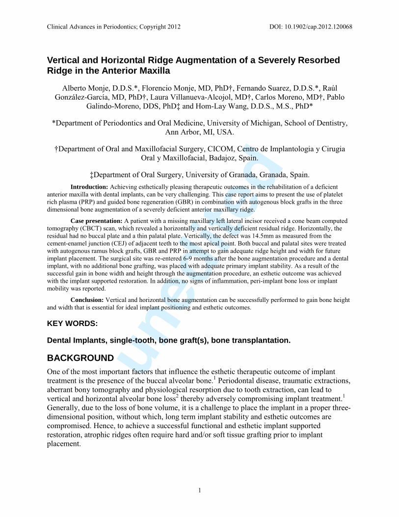
Introduction: Achieving esthetically pleasing therapeutic outcomes in the rehabilitation of a deficient anterior maxilla with dental implants, can be very challenging. This case report aims to present the use of platelet rich plasma (PRP) and guided bone regeneration (GBR) in combination with autogenous block grafts in the three dimensional bone augmentation of a severely deficient anterior maxillary ridge.
Case presentation: A patient with a missing maxillary left lateral incisor received a cone beam computed tomography (CBCT) scan, which revealed a horizontally and vertically deficient residual ridge. Horizontally, the residual had no buccal plate and a thin palatal plate. Vertically, the defect was 14.5mm as measured from the cement-enamel junction (CEJ) of adjacent teeth to the most apical point. Both buccal and palatal sites were treated with autogenous ramus block grafts, GBR and PRP in attempt to gain adequate ridge height and width for future implant placement. The surgical site was re-entered 6-9 months after the bone augmentation procedure and a dental implant, with no additional bone grafting, was placed with adequate primary implant stability. As a result of the successful gain in bone width and height through the augmentation procedure, an esthetic outcome was achieved with the implant supported restoration. In addition, no signs of inflammation, peri-implant bone loss or implant mobility was reported.
Conclusion: Vertical and horizontal bone augmentation can be successfully performed to gain bone height and width that is essential for ideal implant positioning and esthetic outcomes.
KEY WORDS:
Dental Implants, single-tooth, bone graft(s), bone transplantation.
BACKGROUND One of the most important factors that influence the esthetic therapeutic outcome of implant treatment is the presence of the buccal alveolar bone.1 Periodontal disease, traumatic extractions, aberrant bony tomography and physiological resorption due to tooth extraction, can lead to vertical and horizontal alveolar bone loss2 thereby adversely compromising implant treatment.1 Generally, due to the loss of bone volume, it is a challenge to place the implant in a proper three-dimensional position, without which, long term implant stability and esthetic outcomes are compromised. Hence, to achieve a successful functional and esthetic implant supported restoration, atrophic ridges often require hard and/or soft tissue grafting prior to implant placement.
Clinical Advances in Periodontics; Copyright 2012 DOI: 10.1902/cap.2012.120068
2
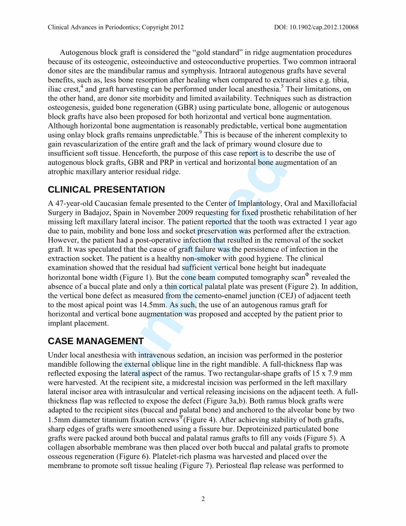
Autogenous block graft is considered the “gold standard” in ridge augmentation procedures because of its osteogenic, osteoinductive and osteoconductive properties. Two common intraoral donor sites are the mandibular ramus and symphysis. Intraoral autogenous grafts have several benefits, such as, less bone resorption after healing when compared to extraoral sites e.g. tibia, iliac crest,4 and graft harvesting can be performed under local anesthesia.5 Their limitations, on the other hand, are donor site morbidity and limited availability. Techniques such as distraction osteogenesis, guided bone regeneration (GBR) using particulate bone, allogenic or autogenous block grafts have also been proposed for both horizontal and vertical bone augmentation. Although horizontal bone augmentation is reasonably predictable, vertical bone augmentation using onlay block grafts remains unpredictable.9 This is because of the inherent complexity to gain revascularization of the entire graft and the lack of primary wound closure due to insufficient soft tissue. Henceforth, the purpose of this case report is to describe the use of autogenous block grafts, GBR and PRP in vertical and horizontal bone augmentation of an atrophic maxillary anterior residual ridge.
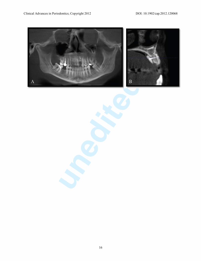
CLINICAL PRESENTATION A 47-year-old Caucasian female presented to the Center of Implantology, Oral and Maxillofacial Surgery in Badajoz, Spain in November 2009 requesting for fixed prosthetic rehabilitation of her missing left maxillary lateral incisor. The patient reported that the tooth was extracted 1 year ago due to pain, mobility and bone loss and socket preservation was performed after the extraction. However, the patient had a post-operative infection that resulted in the removal of the socket graft. It was speculated that the cause of graft failure was the persistence of infection in the extraction socket. The patient is a healthy non-smoker with good hygiene. The clinical examination showed that the residual had sufficient vertical bone height but inadequate horizontal bone width (Figure 1). But the cone beam computed tomography scanΦ revealed the absence of a buccal plate and only a thin cortical palatal plate was present (Figure 2). In addition, the vertical bone defect as measured from the cemento-enamel junction (CEJ) of adjacent teeth to the most apical point was 14.5mm. As such, the use of an autogenous ramus graft for horizontal and vertical bone augmentation was proposed and accepted by the patient prior to implant placement.
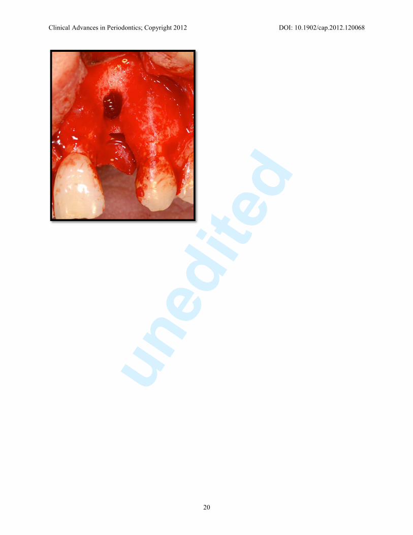
CASE MANAGEMENT Under local anesthesia with intravenous sedation, an incision was performed in the posterior mandible following the external oblique line in the right mandible. A full-thickness flap was reflected exposing the lateral aspect of the ramus. Two rectangular-shape grafts of 15 x 7.9 mm were harvested. At the recipient site, a midcrestal incision was performed in the left maxillary lateral incisor area with intrasulcular and vertical releasing incisions on the adjacent teeth. A full-thickness flap was reflected to expose the defect (Figure 3a,b). Both ramus block grafts were adapted to the recipient sites (buccal and palatal bone) and anchored to the alveolar bone by two 1.5mm diameter titanium fixation screwsΨ(Figure 4). After achieving stability of both grafts, sharp edges of grafts were smoothened using a fissure bur. Deproteinized particulated bone grafts were packed around both buccal and palatal ramus grafts to fill any voids (Figure 5). A collagen absorbable membrane was then placed over both buccal and palatal grafts to promote osseous regeneration (Figure 6). Platelet-rich plasma was harvested and placed over the membrane to promote soft tissue healing (Figure 7). Periosteal flap release was performed to
Clinical Advances in Periodontics; Copyright 2012 DOI: 10.1902/cap.2012.120068
3
achieve tension-free primary wound closure. The donor and recipient sites were subsequently closed with both resorbable and non-resorbable suturesβ. At the 2-week post-operative review, the recipient site had a wound healing index score of 2, which indicated slight gingival edema, erythema and patient discomfort. Healing of the recipient site slowly improved over time without any complications. Radiographic bone formation was observed at the 8-month post-operative review (Figure 9).
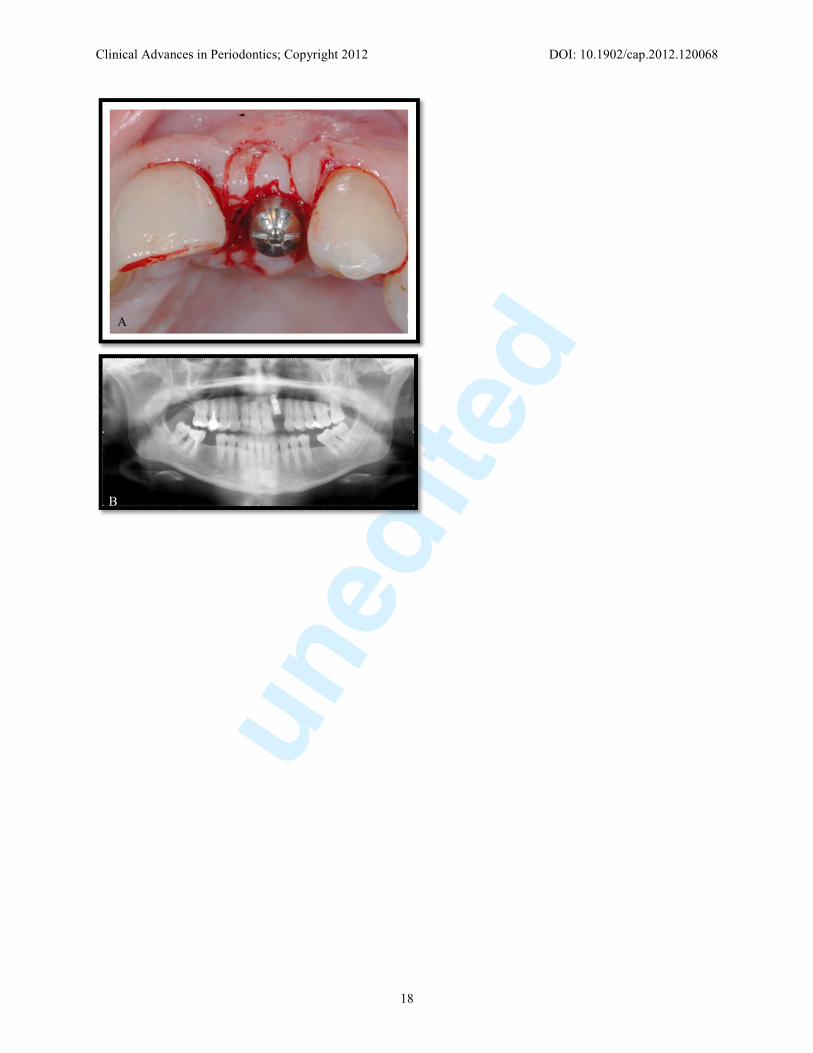
Implant placement was thus performed under local anesthesia. Upon flap reflection, bone formation was observed at the surgical site. One 3.75 x 11.5mm implantζ was placed with good implant stabilityξ (ISQ 74) (Figure 10a,b). Three months later, a healing abutment was placed with ISQ 82 (Figure 11a,b). A final ceramic-metal screw-retained crown was installed 6 months after implant placement (Figure 12a,b).
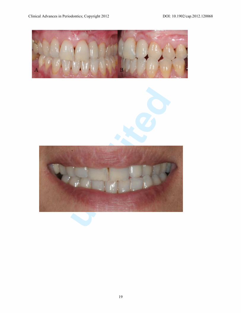
CLINICAL OUTCOME One year after, no peri-implant inflammation and/or bone loss or implant mobility were reported. In addition, patient was very pleased with the esthetic outcome (Figure 12c)
DISCUSSION To achieve a satisfactory esthetic outcome with dental implants in a deficient anterior maxilla, augmentation is often required. Autogenous bone is the “gold standard” for bone augmentation because of its osteogenic ability. As intraoral grafts have a lesser resorption, higher revascularization and better graft incorporation compared to extraoral grafts,4 they are thus the best material available to reconstruct isolated horizontal and vertical defects. Symphyseal and ramus block grafts are the most common intraoral donor sites. While symphyseal block grafts provide a thicker graft, ramus grafts have been shown to have a lower incidence of postoperative sensory disturbances.5 In addition, ramus grafts have a higher acceptance rate than symphyseal grafts.5 Furthermore, bone substitute around the block graft used in treating this patient will provide the space maintenance for bone to ingrowth 11.
Barrier membranes were generally used to exclude unwanted cells (fibroblasts) from populating the surgical site thus promoting bone regeneration. Non-resorbable barrier membranes provide the grafted area with more isolation, rigidity and protection from mechanical forces. However, non-resorbable membranes have shown higher membrane exposure rate associated with higher incidence of infection, incomplete healing and connective tissue formation below the membrane due to the micromovement of the barrier.12 On the contrary, collagen absorbable membranes have some biologic advantages such as the higher stimulation of DNA synthesis over non-resorbables membranes.13 It seems that barrier membranes are not required when block grafts are used in ridge augmentation because of the space maintaining property of the graft.14 However, a collagen membrane was used in this case to contain and protect the particulated graft particles placed around the block graft and also the membrane could potentially retard graft resorption.
Platelet-rich plasma (PRP) is an autologous platelet concentrate suspended in plasma. These platelets contain growth factors such as epidermal growth factor (EGF), vascular endothelial growth factor (VEGF), transforming growth factor-β1 (TGF-β1), and platelet derived growth factor–BB (PDGF-BB), which initiate and modulate both soft and hard tissues wound healing.15 Even though PRP was often used to manage chronic non-healing wounds, it is also employed in

Clinical Advances in Periodontics; Copyright 2012 DOI: 10.1902/cap.2012.120068
4
oral and maxillofacial surgery to enhance wound healing and bone growth.16 A recent publication attempting to review the literature concerning PRP and its use for hard and soft tissue healing, concluded that despite the great variability in study designs, autologous platelets constitute a safe, reproducible, and effective mean of reproducing the process of wound healing.16 The rationale of adding PRP on top of the membrane was to promote wound healing as platelets aggregate and release cytokines, growth factors and hemostatic factors during the first phase of wound healing.16
In summary, an autogenous block graft, xenogenic particulated graft, absorbable collagen plus PRP were successfully incorporated in augmenting a significant deficient horizontal and vertical ridge in the maxillary anterior region
SUMMARY
Why is this case new information? • Ramus block grafts were placed on both buccal and palatal sides in the maxillary anterior
region to augment a horizontally and vertically deficient ridge.
• Vertical augmentation could be achieved with a ramus block graft.
What are the keys to successful management of these cases? • Proper treatment plan
• Carefully executed surgical plan, which follows the “PASS” principle, the key to achieving predictable bone regeneration.17 The PASS principle is described as: “primary wound closure to ensure undisturbed and uninterrupted wound healing, angiogenesis to provide necessary blood supply and undifferentiated mesenchymal cells, space maintenance/creation to facilitate adequate space for bone ingrowth, and stability of wound and implant to induce blood clot formation and uneventful healing events.”
• Understand the biological properties of each material used in the surgery.
• Ensure that there is no mobility of the block graft and no dead-space between the graft and the recipient bed
What are the primary limitations to success in this case? • Lack of primary wound coverage
• Lack of graft stability
• Poor oral hygiene and premature loading.
ACKNOWLEDGEMENTS The authors would like to thank FEDICOM (Foundation for the study of Implantology, Oral and Maxillofacial Surgery), Badajoz, Spain for providing the financial support and Dr. Jia-Hui Fu (Assistant Professor, Department of Periodontics, Faculty of Dentistry, National University of Singapore, Singapore) for editing this manuscript.

Clinical Advances in Periodontics; Copyright 2012 DOI: 10.1902/cap.2012.120068
5
Disclaimer: The authors do not have any financial interests, either directly or indirectly, in the products or information listed in the paper.
REFERENCES 1. McCarthy C, Patel RR, Wragg PF, Brook IM. Dental implants and onlay bone grafts in the anterior maxilla:
analysis of clinical outcome. Int J Oral Maxillofac Implants 2003;18:238-241.
2. Garber DA, Rosenberg ES. The edentulous ridge in fixed prosthodontics. Compend Contin Educ Dent 1981;2:212-223.
3. Burchardt H. The biology of bone graft repair. Clin Orthop Relat Res 1983:28-42.
4. Zins JE, Whitaker LA. Membranous versus endochondral bone: implications for craniofacial reconstruction. Plast Reconstr Surg 1983;72:778-785.
5. Schwartz-Arad D, Levin L. Intraoral autogenous block onlay bone grafting for extensive reconstruction of atrophic maxillary alveolar ridges. J Periodontol 2005;76:636-641.
6. Tolman DE. Reconstructive procedures with endosseous implants in grafted bone: a review of the literature. Int J Oral Maxillofac Implants 1995;10:275-294.
7. Wang HL, Al-Shammari K. HVC ridge deficiency classification: a therapeutically oriented classification. Int J Periodontics Restorative Dent 2002;22:335-343.
8. Hurzeler MB, Zuhr O, Schenk G, Schoberer U, Wachtel H, Bolz W. Distraction osteogenesis: a treatment tool to improve baseline conditions for esthetic restorations on immediately placed dental implants--a case report. Int J Periodontics Restorative Dent 2002;22:451-461.
9. Felice P, Pistilli R, Lizio G, Pellegrino G, Nisii A, Marchetti C. Inlay versus onlay iliac bone grafting in atrophic posterior mandible: a prospective controlled clinical trial for the comparison of two techniques. Clin Implant Dent Relat Res 2009;11 Suppl 1:e69-82.
10. Wang HL, Misch C, Neiva RF. "Sandwich" bone augmentation technique: rationale and report of pilot cases. Int J Periodontics Restorative Dent 2004;24:232-245.
11. McAllister BS, Haghighat K. Bone augmentation techniques. J Periodontol 2007;78:377-396.
12. Doblin JM, Salkin LM, Mellado JR, Freedman AL, Stein MD. A histologic evaluation of localized ridge augmentation utilizing DFDBA in combination with e-PTFE membranes and stainless steel bone pins in humans. Int J Periodontics Restorative Dent 1996;16:120-129.
13. Marinucci L, Lilli C, Baroni T, et al. In vitro comparison of bioabsorbable and non-resorbable membranes in bone regeneration. J Periodontol 2001;72:753-759.
14. Proussaefs P, Lozada J. The use of intraorally harvested autogenous block grafts for vertical alveolar ridge augmentation: a human study. Int J Periodontics Restorative Dent 2005;25:351-363.
15. Eppley BL, Woodell JE, Higgins J. Platelet quantification and growth factor analysis from platelet-rich plasma: implications for wound healing. Plast Reconstr Surg 2004;114:1502-1508.
16. Roukis TS, Zgonis T, Tiernan B. Autologous platelet-rich plasma for wound and osseous healing: a review of the literature and commercially available products. Adv Ther 2006;23:218-237.
17. Wang HL, Boyapati L. "PASS" principles for predictable bone regeneration. Implant Dent 2006;15:8-17.
Corresponding author: Florencio Monje Gil Calle Juan Miró s/n, local 16-17 06010 Badajoz (Spain) Tel: 0034 924 205 235 Fax: 0034 924 260 773 E-mail address:
Submitted June 3, 2012; accepted for publication July 12, 2012.

Clinical Advances in Periodontics; Copyright 2012 DOI: 10.1902/cap.2012.120068
6
Figure 1.
Initial clinical view of the deficiency. A slight vertical defect is observed indicating an esthetic challenge from the restorative perspective.
Figure 2.
Preoperative sagittal views of the defect. The buccal plate was absent and only a thin palatal plate was present.
Figure 3.
Clinical views of the hard tissue defect.
3a. Intraoral frontal view of the defect. (to be included on the first page)
3b. Intraoral occlusal view of the defect.
Figure 4.
Adaptation of one of the monocortical grafts to the recipient site anchored to the residual ridge by two 1.5mm titanium fixation screws.
Figure 5.
Placement of particulated xenograft between and around the monocortical grafts.
Figure 6.
Absorbable membrane placed over the particles to promote bone regeneration
Figure 7.
Platelet-rich plasma placed over the membrane to promote soft tissue healing
Figure 8.
Extraoral view of the grafted area 8 months after bone augmentation.
Figure 9.
Postoperative radiographic examination.
9A. Panoramic radiograph
9B. Sagittal view of the grafted area. Both grafts were anchored to each other and the recipient site by the fixation screws. (to be included on the first page)
Figure 10.
Implant placement surgery 8 months after bone augmentation.
10a. Intraoral occlusal view of ridge before implant placement.
10b. Intraoral frontal view of implant placed.
Figure 11.
Abutment placement 3 months after implant placement
11a. Coronal intraoral view of the abutment placed.
11b. Radiographic view of the implant placed just before abutment placement.
Clinical Advances in Periodontics; Copyright 2012 DOI: 10.1902/cap.2012.120068
7
Figure 12.
Final clinical extraoral view of the restoration. A harmonious gingival margin preserving a convex contour can be observed.
12A. Frontal view of the restoration
12B. Lateral view of the restoration
12C. Smile of the patient once was restored. Φ i-CAT (Imaging Sciences International, Hatfield,PA, USA)
Ψ Level One 1.5 Neuro (KLS Martin LP, FL, USA)
β Cytoplast™ Suture (Osteogenics Biomedical Inc, Lubbock, TX, USA)
ζ Speedy Groovy Nobel Biocare Implants (Nobel Biocare AB, Göteborg, Sweden)
ξ Osstell™ (Osstell AB, Göteborg, Sweden)

Clinical Advances in Periodontics; Copyright 2012 DOI: 10.1902/cap.2012.120068
8

Clinical Advances in Periodontics; Copyright 2012 DOI: 10.1902/cap.2012.120068
9

Clinical Advances in Periodontics; Copyright 2012 DOI: 10.1902/cap.2012.120068
10
A B

Clinical Advances in Periodontics; Copyright 2012 DOI: 10.1902/cap.2012.120068
11

Clinical Advances in Periodontics; Copyright 2012 DOI: 10.1902/cap.2012.120068
12

Clinical Advances in Periodontics; Copyright 2012 DOI: 10.1902/cap.2012.120068
13

Clinical Advances in Periodontics; Copyright 2012 DOI: 10.1902/cap.2012.120068
14

Clinical Advances in Periodontics; Copyright 2012 DOI: 10.1902/cap.2012.120068
15
Clinical Advances in Periodontics; Copyright 2012 DOI: 10.1902/cap.2012.120068
16

Clinical Advances in Periodontics; Copyright 2012 DOI: 10.1902/cap.2012.120068
17
A B
Clinical Advances in Periodontics; Copyright 2012 DOI: 10.1902/cap.2012.120068
18
A
B
Clinical Advances in Periodontics; Copyright 2012 DOI: 10.1902/cap.2012.120068
19
Clinical Advances in Periodontics; Copyright 2012 DOI: 10.1902/cap.2012.120068
20

Clinical Advances in Periodontics; Copyright 2012 DOI: 10.1902/cap.2012.120068
21