Vertebral column Supports Skull Pectoral girdle Upper limb Thoracic cage Protects Spinal cord and...
16
Vertebral column Supports Skull Pectoral girdle Upper limb Thoracic cage Protects Spinal cord and the spinal nerve roots 1
-
Upload
shana-shaw -
Category
Documents
-
view
219 -
download
1
Transcript of Vertebral column Supports Skull Pectoral girdle Upper limb Thoracic cage Protects Spinal cord and...
Vertebral column Supports SkullPectoral girdleUpper limbThoracic cage
ProtectsSpinal cord and the spinal nerve roots
1
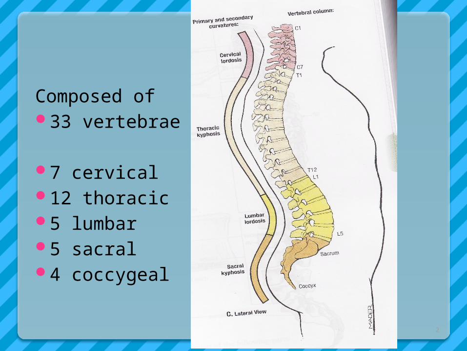
Composed of 33 vertebrae
7 cervical12 thoracic5 lumbar5 sacral4 coccygeal
2
Because it is segmented, made of vertebrae, pads of fibrocartilage, the intervertebral discs join each vertebra
The intervertebral discs form about 1/4th of the length of column
3
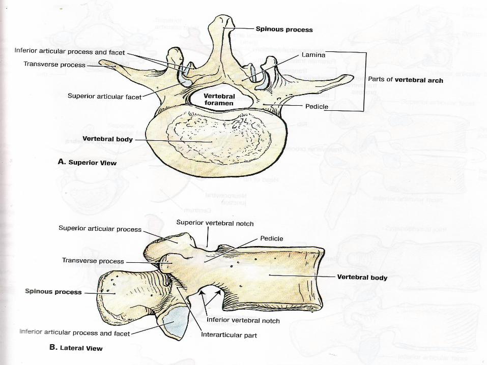
Typical vertebra consists of rounded body, anteriorly
Vertebral arch posteriorly.These enclose vertebral foramen, through
which spinal cord runs.Arch consists of 7 processesSpinous process(1)Transverse process(2)Articular processes(4)
4
5
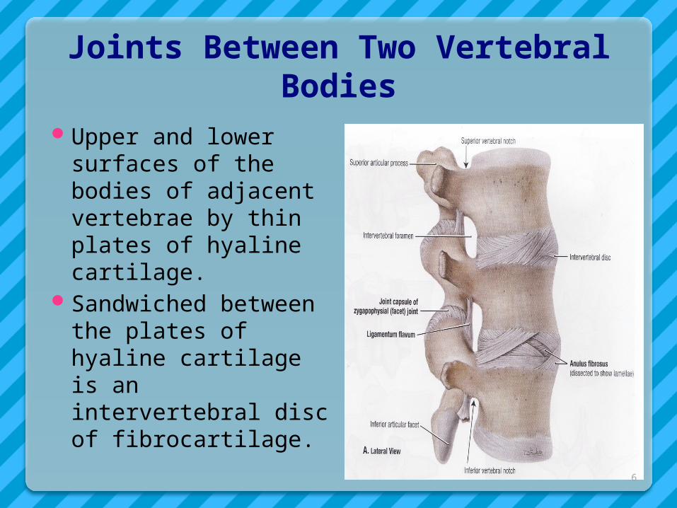
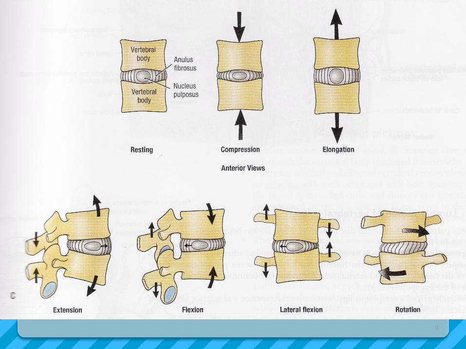
Joints Between Two Vertebral Bodies
Upper and lower surfaces of the bodies of adjacent vertebrae by thin plates of hyaline cartilage.
Sandwiched between the plates of hyaline cartilage is an intervertebral disc of fibrocartilage.
6
7
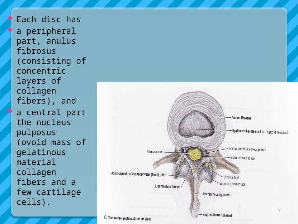
Each disc has a peripheral
part, anulus fibrosus (consisting of concentric layers of collagen fibers), and
a central part the nucleus pulposus (ovoid mass of gelatinous material collagen fibers and a few cartilage cells).
A sudden increase in compression load on vertebral column causes nucleus pulposus to thrust outward, where it may press on the spinal cord or the nerve roots causing pain.
This mainly happens with advancing age collagen fibers of the anulus degenrates and as a result the anulus cannot contain the nucleus pulposus under stress.
The 1st, 2nd cervical verterbrae, sacrum, and coccyx lacks intervertebral discs
8
9
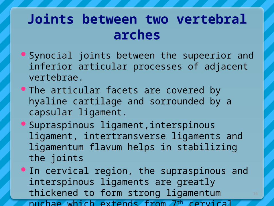
Joints between two vertebral arches
Synocial joints between the supeerior and inferior articular processes of adjacent vertebrae.
The articular facets are covered by hyaline cartilage and sorrounded by a capsular ligament.
Supraspinous ligament,interspinous ligament, intertransverse ligaments and ligamentum flavum helps in stabilizing the joints
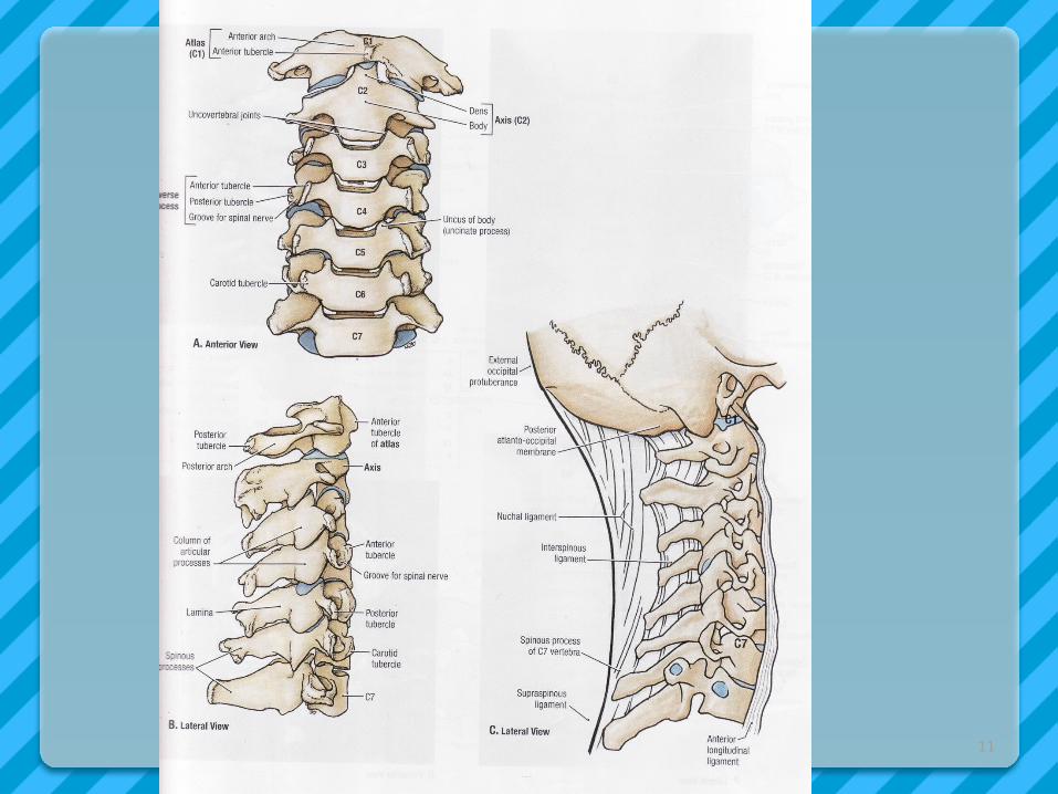
In cervical region, the supraspinous and interspinous ligaments are greatly thickened to form strong ligamentum nuchae,which extends from 7th cervical spine to the external occipital protuberance
10
11
Atlanto-Occipital joints
Synovial joints Between occipital condyles(present on
either side of foramen magnum) and facets on the superior surfaces of the lateral masses of the atlas
Capable of flexion , extension and lateral flexion. They do not rotate
12
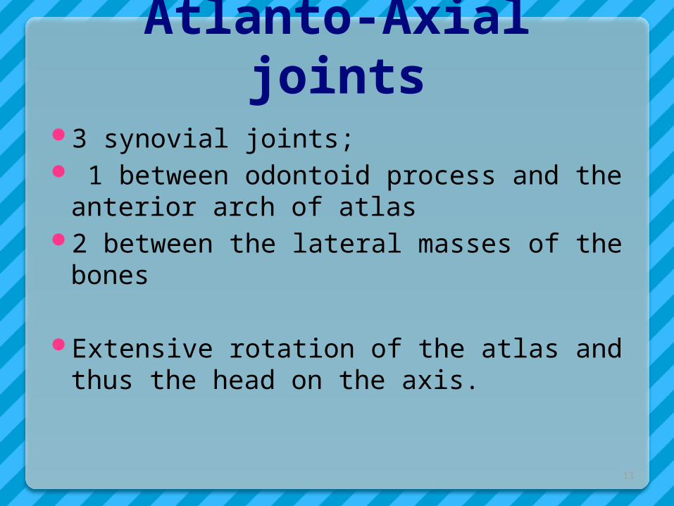
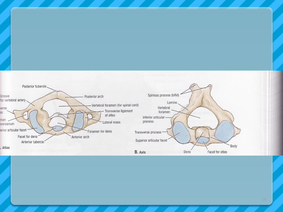
Atlanto-Axial joints3 synovial joints; 1 between odontoid process and the
anterior arch of atlas2 between the lateral masses of the bones
Extensive rotation of the atlas and thus the head on the axis.
13
14
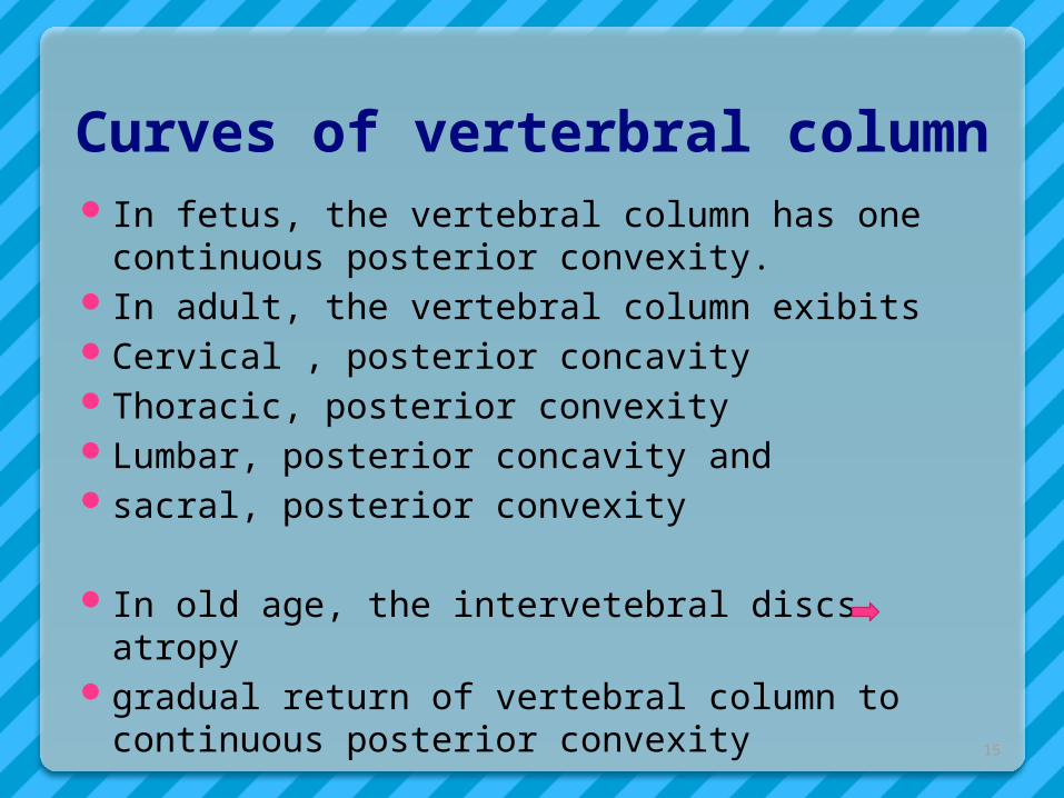
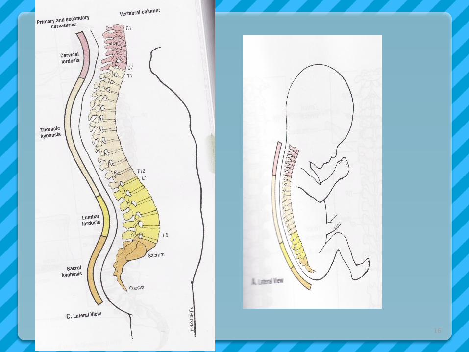
Curves of verterbral column In fetus, the vertebral column has one
continuous posterior convexity. In adult, the vertebral column exibitsCervical , posterior concavityThoracic, posterior convexityLumbar, posterior concavity and sacral, posterior convexity
In old age, the intervetebral discs atropygradual return of vertebral column to continuous
posterior convexity15
16