![Dr Sharan Hiremath’s Preauricular flag flap for temple and ...€¦ · the technique of choice for the reconstruction of small-sized defects on the lateral forehead [1]. These flaps](https://static.fdocuments.net/doc/165x107/5fbe5be53c273e5ced393610/dr-sharan-hiremathas-preauricular-flag-flap-for-temple-and-the-technique-of.jpg)
Versatility of the Forehead Flaps as a Reconstructive Tool
66
The Versatility Of Forehead Flaps As A Reconstructive Tool Dissertation submitted in partial fulfillment of the requirements for the degree of M.Ch. (Plastic Surgery) - Branch III THE TAMILNADU DR.M.G.R.MEDICAL UNIVERSITY CHENNAI AUGUST 2008
Transcript of Versatility of the Forehead Flaps as a Reconstructive Tool

The Versatility Of Forehead Flaps As A Reconstructive Tool
Dissertation submitted in partial fulfillment of the requirements for the degree of
M.Ch. (Plastic Surgery) - Branch III
THE TAMILNADU DR.M.G.R.MEDICAL UNIVERSITY CHENNAI
AUGUST 2008

CERTIFICATE
This is to certify that, this dissertation titled “ The Versatility Of Forehead
Flaps As A Reconstructive Tool ”, submitted by Dr. Dinesh Kumar .S appearing for
M.Ch. (Plastic Surgery) degree examination in August 2008 is a bonafide record of
work done by him under my guidance and supervision.
Prof. V. Alamelu M.S., M.Ch., FICS, FMMSProfessor and Head of the Department,Department of Plastic, Reconstructive and Facio-Maxillary Surgery,Madras Medical College,Chennai- 600003.
The Dean,Madras Medical College,Chennai- 600003.

DECLARATION
I solemnly declare that this dissertation “The Versatility of the Forehead
Flaps as a Reconstructive Tool” was prepared by me in the Department of Plastic,
Reconstructive and Maxillofacial Surgery, Madras Medical College and Government
General Hospital, Chennai under the guidance and supervision of Professor & HOD
Department of Plastic, Reconstructive and Maxillofacial Surgery, Madras Medical
College and Government General Hospital, Chennai between 2005 and 2008.
This dissertation is submitted to the TamilNadu Dr. MGR Medical University,
Chennai in partial fulfillment of the University requirements for the award of degree
of MCh Plastic Surgery.
Place: Chennai
Date : 25-05-08

ACKNOWLEDGEMENT
I gratefully acknowledge and sincerely thank The Dean, Madras Medical
College, Chennai, for granting me permission to utilize the facilities of the institution
for my study.
I am extremely grateful to my teacher and guide Prof. V. Alamelu M.S, M. Ch.,
FICS, FMMS Professor and Head Of the Department of Plastic, Reconstructive and
Facio-Maxillary Surgery, Madras Medical College, Chennai, who helped me in all
stages of my study. I am thankful to her for her timely suggestions, unending
patience, constant encouragement and scholarly guidance.
I am extremely grateful to and Prof. K.V. Alalasundaram M.S, M. Ch., for his
guidance and support.
I also thank retired Prof. M. P. Namasivayam M.S., M.Ch. for his initial effort in
the preparation of this dissertation.
I express my thanks to Dr. Anand Subramaniam, D.A., M.S, M. Ch.,
Dr.K.Gopalakrishnan, M.S, M. Ch., Dr.Jeyavel Rajkumar, M.S, M. Ch., Dr. T. M.
Balakrishnan,M.S, M. Ch., and Dr. Udesh Ganapathy M.S., M.Ch., for their sustained
encouragement in my work and study.
Last, but not the least, I thank all my patients, without their help and co-
operation this study would not have been possible.

CONTENTS
1. Introduction 1
2. Aim of the study 3
3. Review of Literature 4
4. Surgical Anatomy of the Forehead 7
5. Surgical Techniques 13
6. Materials & Methods 41
8. Observations & Results 43
9. Discussion 46
10. Conclusion 51
Bibliography
Appendix
- Proforma
- Master chart

INTRODUCTION
Forehead Flaps have been in use since ancient times as has been depicted in the
classical paintings of the great Susrutha at work repairing the nose of those
unfortunate to have their noses cut off as a punishment. The forehead flaps hold a
closer relation to us Indians as we seem not only to be pioneers in utilizing this area
for reconstruction but also contribute to the further development of their uses. The
bipolar Narayanan flap is a point in case.
The forehead flaps have waxed and waned in their popularity at various periods
in the long history of plastic surgery. Although it has proved its reliability time and
again there seems to be a continued reluctance among the younger generations of
plastic surgeons in using this virtual treasure trove of tissue for reconstructive options
in the head and neck region due to concerns about the donor area scarring.
Probably the easy accessibility to technology and the ever increasing popularity
of free flaps with their promise of a single-stage procedure – where the tissue can be
sculpted to exactly fit the defect and thereby allowing the patient to return to near-
normal life as early as possible – as compared to the multiple stages generally
required when the forehead flap is used, distracts surgeons from these age-old and
time-tested options.
It is also widely accepted that local tissues give the best colour and texture
match to any reconstructed area. This is especially applicable to the face. Thus,
currently the central forehead flaps are favoured in the nasal reconstruction.
The forehead flaps have lent themselves to such a wide variety of uses that a

revisit to their different clinical applications is well justified, if only to serve to bring
the spotlight back to the most versatile donor area in the face.

AIM OF THE STUDY
This study on the versatility of the forehead flaps as a reconstructive tool was
done with following aims.
1) To review the various flaps used from the forehead region in Department of
Plastic and Reconstructive Surgery, Government Medical College, Chennai.
2) To study the effectiveness of these flaps in various clinical situations.
3) To report our experiences along with the other uses of forehead flaps reported
in the literature.

REVIEW OF LITERATURE
The earliest accounts of forehead flaps appear in the Susrutha Samhitha where
skin from the centre of the forehead was utilised to reconstruct noses as far back as
600 – 700 B.C.3 This knowledge was transferred to a family of potters (the Koomas)
who continued to use it even during the time of British rule in India. 6
Antonio Branca, of Italy, is credited with the first reported use of the mid-
forehead flap outside of India.4 Based on an arabic translation of the Susrutha
Samhitha, Branca performed the operation in the 15th century. He was persecuted for
“interfering in God's work” and so the technique was still remained unknown to the
Western World until J. C. Carpue read an editorial description of the technique that
was published in the Gentlemen’s Gazette in 1794.5 Carpue practised the technique
on cadavers for 20 years, until he found the right patients. He then described 2 cases
in a monograph entitled “An Account of Two Successful Operations for Restoring a
Lost Nose.”. 6
Further modifications of the forehead flaps for nasal reconstruction continued
by Millard,9,11,12,13 Burget,14,15,16 Menick14,15,17 and others. The preference kept shifting
from midline to paramedian to oblique and back to midline flaps in the attempt to
increase the reach while preserving the blood supply. Elucidation of the vascular
anatomy of these flaps was done by Kazanjian (1946)10 and Shumrick KA, Smith TL
(1993).5 allowing for further refinements of these flaps. Converse described the
scalping technique for using the forehead skin for reconstruction of near-total loss of
nose (1942).74,75,76,77

In 1893 Dunham described a laterally based forehead flap which 'was so cut as
to contain, traversing its pedicle and ramifying in it, the anterior temporal artery. It
had a rather long pedicle, about an inch wide, attached in front of the ear, and the
mass of the flap was from the upper forehead where it slightly crossed the median
line'.58 He then dissected out the vessels and returned it to the forehead along with the
pedicle at the time of division leaving the skin for reconstruction of the defect in the
face.
The ability of a narrow pedicle to support a forehead flap was further
demonstrated clinically by Monks (lower eyelid reconstruction 1989),68 Horsley
(cheek reconstruction 1916),69 Esser (1917,1931), Gillies (1949) and many others.
Wilson advocated a 2-centimetre pedicle for the forehead flap (1967).70 A thorough
understanding of the vascular anatomy underlying the forehead flaps was provided by
the cadaver injection studies of Conway et al,59 Corso60 and Behan & Wilson.61 They
showed the 4 major vascular territories supplied by the 6 named vessels with
extensive anastomoses between each other.
Use of the flap for intra-oral lining was described by Blair in 1941.62 Four
methods for its use have been described for passing the flap inside the mouth –
McGregor passed the flap through an incision in the cheek about 1.5cm below the
zygomatic arch,71 Millard used a lower route (a submandibular incision),65 Hoopes &
Edgerton described a subcutaneous tunnelling procedure66 and subsequently Davis &
Hoopes suggested passing the flap deep to the zygomatic arch.67
The folded forehead flap was used for reconstruction of full-thickness defects
of the cheek by Champion in 196064 and Naozer M. Kavarana from Tata Memorial

Hospital described its use for large full-thickness cheek defects in 1975.55 They found
that it decreased the morbidity for the patient and decreased the need for separate flap
for lining/cover.
Narayanan described the bilobar forehead and scalp flap for reconstruction of
very wide full-thickness cheek defects.24
Smalley and Cunningham reported using the forehead flap to cover the defect
over the carotid artery over the upper neck region in 1972.56
Galeo-frontalis flaps were devised for repair of dural leaks, reconstruction of
anterior cranial fossa defects and other craniodacial defects.80,81,82,83,84,85
Worthen described the forehead rotation flap for reconstruction of forehead
defects (1976).73 He used various rotation flaps to cover the defects in the forehead.
But most significantly, he used the remnant forehead skin as a single unit to
reconstruct losses upto 40% of the forehead skin.
Mustarde described the glabellar V-Y skin flap for medial canthal
reconstruction (1980).78
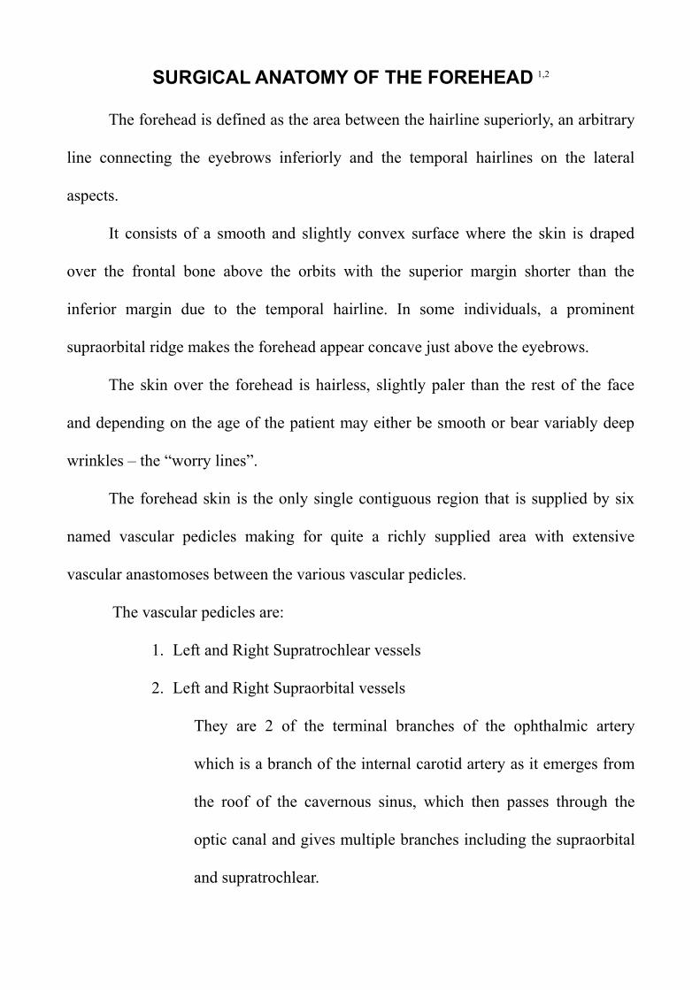
SURGICAL ANATOMY OF THE FOREHEAD 1,2
The forehead is defined as the area between the hairline superiorly, an arbitrary
line connecting the eyebrows inferiorly and the temporal hairlines on the lateral
aspects.
It consists of a smooth and slightly convex surface where the skin is draped
over the frontal bone above the orbits with the superior margin shorter than the
inferior margin due to the temporal hairline. In some individuals, a prominent
supraorbital ridge makes the forehead appear concave just above the eyebrows.
The skin over the forehead is hairless, slightly paler than the rest of the face
and depending on the age of the patient may either be smooth or bear variably deep
wrinkles – the “worry lines”.
The forehead skin is the only single contiguous region that is supplied by six
named vascular pedicles making for quite a richly supplied area with extensive
vascular anastomoses between the various vascular pedicles.
The vascular pedicles are:
1. Left and Right Supratrochlear vessels
2. Left and Right Supraorbital vessels
They are 2 of the terminal branches of the ophthalmic artery
which is a branch of the internal carotid artery as it emerges from
the roof of the cavernous sinus, which then passes through the
optic canal and gives multiple branches including the supraorbital
and supratrochlear.

It is commonly believed that the supraorbital artery is the bigger
of the two and that it is the dominant vessel. But recent studies
show that it is not always so, and that in many cases, the
supratrochlear arteries are the dominant vessels.
3. Left and Right Superficial Temporal vessels
The superficial temporal artery is one of the terminal branch of the
external carotid artery. Running up behind the temporo-
mandibular joint and in front of the ear and the auriculotemporal
nerve, it crosses the zygomatic arch, where its pulsations can be
felt, and branches out widely into the skin that overlies the
temporal fascia. One branch, the middle temporal artery, pierces
the fascia, supplies temporalis and anastomoses with the deep
temporal branches of the maxillary artery.
The anastomoses between the supraorbital and the superficial temporal arteries
provide a link between the internal and external carotid arterial circulations.
The vessels supplying the forehead are further bolstered by anastomoses with
the terminal branches of the facial artery – the angular artery – and the vessels
supplying the scalp – mainly the occipital and the posterior auricular arteries.
Fig.1 depicts the vascular anatomy of the forehead and scalp and the pattern of
anastomoses between the various vessels.

The supratrochlear and supraorbital arteries exit the orbit at the medial end of
the orbit and the supraorbital foramen/notch respectively. At this level they are
closely applied to the periosteum of the orbital rim. They then proceed deep to and
rapidly pierce the frontalis muscle to enter the subcutaneous layer as they ascend into
the forehead. In the upper half of the forehead, many fine branches of the arteries lie
in the subcutaneous fat close to the dermis. So the entire frontalis muscle and some of
the subcutaneous fat may be removed from the distal end of a paramedian forehead
flap without injuring its axial arteries.
Fig 1: The vascular supply of the forehead and scalp.

The superficial temporal artery comes to lie in the superficial plane just
anterior to the tragus of the ear. Proximal to this, it lies deep in the parotid gland
which prevents its further mobilization. The superficial temporal artery ascends
superficial to the zygomatic arch and soon divides into an anterior branch and
posterior branch. A zygomatic branch arises from the trunk of the superficial temporal
artery in about 80% of cadaveric studies and in the remaining 20% was seen arising
from the anterior branch of the superficial temporal artery. Failure to include this
dominant branch in the flap is thought to be the reason for the reported cases of
failure of the transverse forehead flaps.
Fig 2: Pattern of branching of vessels allowing laminar flap harvest

The surface markings for the main vessels are as follows:
1. Supratrochlear vessels :
The vessels are found between 3mm medial or lateral to the medial
canthal region on the supraorbital margin.
2. Supraorbital vessels :
These vessels are usually found at the point where the mid-pupillary line
at straight gaze meets the supraorbital margin.
3. Ant. branch of the superficial temporal vessels :
The superficial temporal artery divides into an anterior and posterior
branches at the superior border of the ear just in front of the external ear.
The anterior branch then follows a curvilinear course more or less
parallel to the hairline giving perpendicular branches to the scalp and
corresponding branches to the forehead skin.
The supratrochlear and supraorbital vessels are accompanied by the
supratrochlear and supraorbital nerves respectively. These nerves carry the sensations
from the forehead as well as the frontal scalp as far as the vertex.
As part of the face and the scalp, the forehead skin also maintains the special
layered structure that is present elsewhere on the face, namely, skin, subcutaneous fat,
superficial muscles and its fascia, a thin layer of fibrofatty tissue, the galeo-frontalis,
a layer of loose areolar tissue and periosteum (from superficial to deep). Special
pattern of blood supply in the face also allows the same area to contribute two layers
of flap based on a single common vascular pedicle, especially in the forehead region.

Due to this rich vascular supply of the forehead region, it allows mobilization
of tissue in a variety of ways giving us paramedian, median, oblique, transverse
forehead flaps as axial pattern flaps with skin pedicles or as islanded flaps, rotation
flaps, V-Y advancement flaps and the scalping flap.

SURGICAL TECHNIQUES 2
1. Forehead Rotation Flap :
The entire forehead is available as an arterialized flap based on the superficial
temporal artery to reconstruct forehead defects involving up to 40% of the surface
area. The superior border of a forehead rotation flap is developed in the hair-bearing
scalp and traverses the frontal scalp to the pre-auricular crease.
Elevation of the flap down to the supraorbital rim, results in a “free-swinging”
extensible segment of tissue. After mobilisation is complete, back-cutting of the base
is permissible but rarely necessary. Once the flap has been rotated to the desired
position, the hairline on the distal margin of the flap is aligned with and sutured to the
temporal hairline on the lateral margin of the defect without tension. The suture line
is oriented transversely to minimise the only portion of scar that will be visible on the
exposed forehead.
The secondary frontal scalp defect extends across the entire width of the frontal
scalp but it is relatively narrow and amenable to primary closure.

Fig 3: Forehead Defect and Flap Raised
Fig 6: Forehead Flap Inset Fig 5: Secondary Defect Closed Primarily
Fig 4: Diagrammatic representation

2. Paramedian Forehead Flap :
This flap has been classically used for reconstruction of nasal defects. It can be
used to reconstruct the entire nose if needed. There are many variations of this flap
made to increase its length using non-vertical extensions such as New's sickle flap
and Gillie's up-and-down flap.
Principles of flap design and dimensions:
1. The flap is designed vertically and axially along the supratrochlear
vessels. This makes its vascularity robust, so that it may be radically
thinned and depilated.
2. The base of the flap is made no wider than 1.5 cm for easy mobility
without strangulation.
3. The base of the flap is positioned to include a branch of the angular-
supratrochlear arteries, which may be located with a Doppler flowmeter
though usually not required.
4. Additional length is obtained by extending the flap's proximal end across
the orbital rim or it distal end into the hair-bearing scalp.
5. The flap is not designed to fit the defect exactly – it is made slightly
larger to allow for the edema at the recipient area and for the tissue
shrinkage of the flap.
6. An exact three-dimensional pattern of the defect is used as a template for
the flap's design.
7. The skin incision is made on the inner aspect of the flap markings to
minimise the centripetal flap contraction, which obliterates surface

contour.
8. Distal portion of the flap is thinned to the required thickness to cover the
defect and depilated where required.
9. The base of the flap is excised and discarded, not replaced on the
forehead.
10. If primary closure is not possible, the upper half of the donor defect is
allowed to close by biologic wound contraction and forehead skin auto-
expansion. There is no necessity for skin grafts, local flaps, or
mechanical tissue expansion to close this secondary defect.
It requires the talent of a tailor to fashion the forehead flap. A flap cut too small
will collapse and distort the features at the site of inset. A flap cut too large allows
centripetal contraction to pull the excess skin into a blob, obscuring the surface
contour.
The incisions are made at the inside margins of the flap markings so as to not
waste even half a millimetre of forehead skin. In the distal end of the flap (upper half
of the forehead) the flap is raised superficial to the frontalis muscle. Proximally, the
frontalis muscle is included with the pedicle to protect the axial vessels. If extra
length is needed, corrugator muscle fibres are divided using magnifying loupes so
that the vascular branches are preserved while restricting bands of muscle are
released. If the flap still proves short, the the pedicle is extended across the orbital
rim, including a bit of the eyebrow if necessary dividing corrugator muscle fibres
while preserving the vascular branches.

After the flap is raised, the distal end and the flap borders are thinned as
required using curved Joseph's scissors. Hair follicles remaining in the distal part of
the flap are clipped off with fine scissors under 2.5X magnification. During the
thinning, axial branches of the supratrochlear-angular arteries visible in the
subcutaneous tissue very close to the dermis are preserved.
A right-sided flap usually rotates clockwise and a left-sided flap usually rotates
anticlockwise. When required the flaps may be rotated in the opposite direction too.
Key sutures are used to hold the flap in place and fine sutures are used to adjust its
edges to the defect.
The secondary defect is usually closed primarily. But when the defect is too
large for primary closure grafts and local flaps have all been tried to close the defect
primarily. This usually compounds the deformity and increases the patient morbidity.
The forehead is usually very forgiving and heals by wound contraction and secondary
intention healing (described as auto-expansion of forehead skin). This process usually
is not apparent until 3 weeks of healing when only granulation tissue is seen. Later it
rapidly contracts and epithelialises within 1-2 weeks. So, only a layer on non-
adherent dressing (e.g. Vaseline gauze) is placed over the defect and absorbent
dressing done.
The raw posterior surface of the pedicle may be left open. However, a thin
split-thickness skin graft applied to the back of the flap minimizes the problems of an
open wound and the need for frequent post-operative visits.

Nearly all-sutures are removed by 4 - 5 days and the skin edges supported by
skin tapes to avoid suture marks. The dressing on the donor defect is removed by the
7th post-operative day and then followed up with light bandaging/dressings until
wound healing occurs.
Twenty one days later, the skin pedicle is excised and discarded as it will
remain as a visible bump if replaced on the forehead. Because of the narrow design of
the pedicle the eyebrows are not pulled closer.
Fig 7: Paramedian Flap
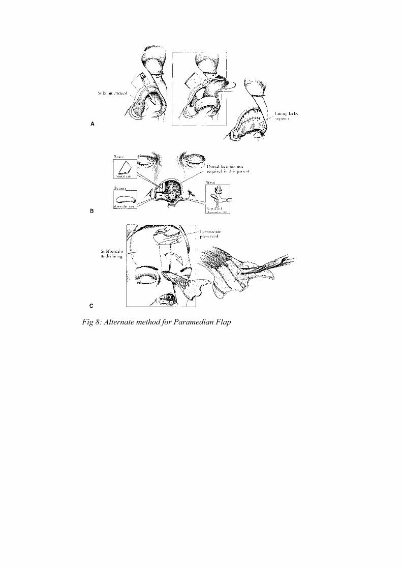
Fig 8: Alternate method for Paramedian Flap

Paramedian Forehead Flap
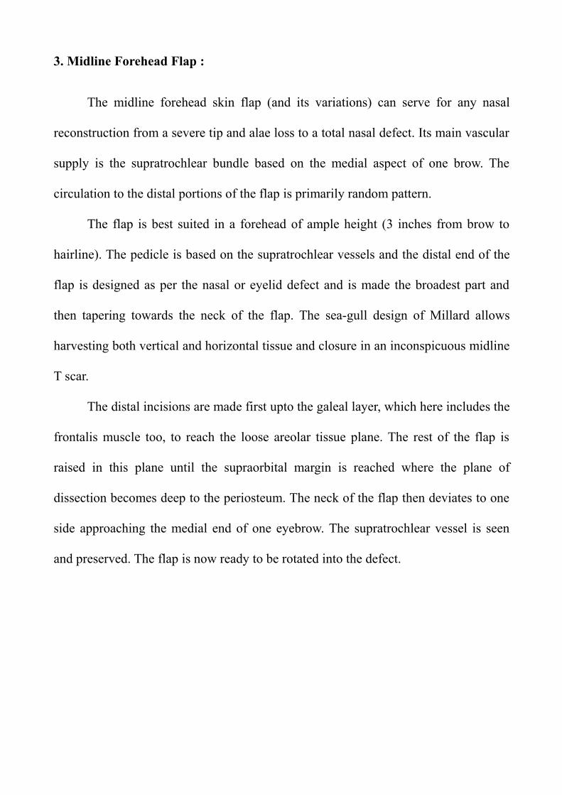
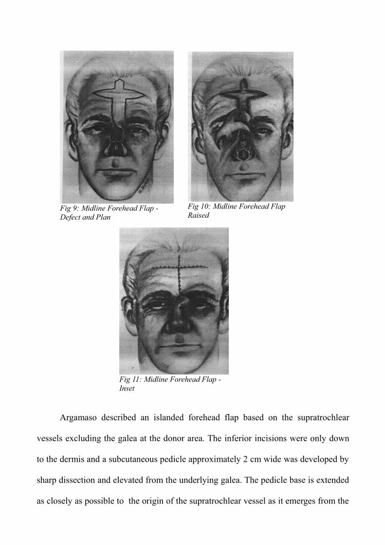
3. Midline Forehead Flap :
The midline forehead skin flap (and its variations) can serve for any nasal
reconstruction from a severe tip and alae loss to a total nasal defect. Its main vascular
supply is the supratrochlear bundle based on the medial aspect of one brow. The
circulation to the distal portions of the flap is primarily random pattern.
The flap is best suited in a forehead of ample height (3 inches from brow to
hairline). The pedicle is based on the supratrochlear vessels and the distal end of the
flap is designed as per the nasal or eyelid defect and is made the broadest part and
then tapering towards the neck of the flap. The sea-gull design of Millard allows
harvesting both vertical and horizontal tissue and closure in an inconspicuous midline
T scar.
The distal incisions are made first upto the galeal layer, which here includes the
frontalis muscle too, to reach the loose areolar tissue plane. The rest of the flap is
raised in this plane until the supraorbital margin is reached where the plane of
dissection becomes deep to the periosteum. The neck of the flap then deviates to one
side approaching the medial end of one eyebrow. The supratrochlear vessel is seen
and preserved. The flap is now ready to be rotated into the defect.
Argamaso described an islanded forehead flap based on the supratrochlear
vessels excluding the galea at the donor area. The inferior incisions were only down
to the dermis and a subcutaneous pedicle approximately 2 cm wide was developed by
sharp dissection and elevated from the underlying galea. The pedicle base is extended
as closely as possible to the origin of the supratrochlear vessel as it emerges from the
Fig 9: Midline Forehead Flap - Defect and Plan
Fig 10: Midline Forehead Flap Raised
Fig 11: Midline Forehead Flap - Inset

rim of the orbit. He advised closure of the donor defect with an advancement flap
from one side of the secondary defect and a rotation flap from the other side of the
defect.
Fig 13: Islanded Forehead Flap - Defect and Plan
Fig 12: Islanded Forehead Flap - Flap Raised
Fig 14: Islanded Forehead Flap Inset

4. Oblique Forehead Flap :
This flap is very useful when the forehead width is not adequate for a median
or a paramedian flap to be used. The flap is located to one side of the midline and
extends to the hairline recess. It is based on the supratrochlear vessels and its
anastomoses with the supraorbital vessel branches.
The flap is based at the medial end of the eyebrow with a pedicle width (of the
carrier segment) of about 0.5 to 1.0 cm. The length of the flap depends on the height
of the browline and the depth of the hairline recess. The paddle is designed to fit the
defect to be covered and can be “flagged” to one side of the carrier segment.
The supratrochlear artery is marked pre-operatively either by palpation or by
use of a Doppler flowmeter. The flap is raised above the periosteum in the loose
areolar plane down to the orbital rim, taking care to protect the vessels at the base.
The defect left by the carrier segment is undermined and closed in 2 layers and that
left by the paddle, if large, is closed by a post-auricular full-thickness skin graft.
Fig 15: Oblique Forehead Flap A,B - showing different type of paddles

The pedicle cannot be tubed as it is too narrow and maybe be either split skin
grafted or left open. The pedicle is divided by 18 - 21 days reinserting only the
proximal part of the pedicle as a triangle to restore the eyebrow alignment.
Fig 16: Oblique Forehead Flap C,D - showing transfer and division

5. Transverse Forehead Flap : (Total/Half)
This is one of the historic flaps and one of the first few axial pattern flaps
described. It achieved enormous importance in head and neck reconstructions. But
later lost its place to other options due the major drawback of the secondary defect
and unappealing esthetics. Nevertheless, because of the reliability of its vascular
supply, the excellent colour match (esp. for the cheek and upper lips) and its
proximity to the face, it is always given a consideration and sometimes kept as the
backup plan.
A forehead flap is long enough to cover any part of the ipsilateral face and can
even cover the carotid artery in the upper neck on the same side. If the flap is
tunnelled under the malar prominence, it can be used to line the entire cheek mucosa
or even used to reconstruct the oropharynx. This flap also has the advantages of not
causing any restriction of movement of the neck or limbs especially in the elderly
patients in whom the majority of these flaps are required.
This flap is unique in the entire body in the fact that it is able to recruit 6
vascular territories – those of the supraorbital, supratrochlear and the anterior branch
of the superficial temporal arteries – on a single pedicle (the ipsilateral superficial
temporal artery) without delay procedures.
The flap vascular dynamics allows the harvest of the forehead skin from one
malar bone to the contralateral malar bone. An average flap measures 25cm and can
be safely carried on a pedicle no wider than 2 cm at its base. This pedicle is based just
anterior to the tragus of the ear and contains the superficial temporal artery. It is also
imperative that the superficial temporal vein that accompanies the artery is kept intact

along with its small accompanying veins.
The superior incision is kept just below the hairline and the lower incision just
above the eyebrows. In the midline, it is carried down into the glabellar region of the
nose. The temporal hairline is preserved if possible. The most satisfying cosmetic
result is obtained by elevating the entire forehead as a cosmetic unit with edges
bevelled at 45 degrees to avoid the marginal “step” deformity.
The plane of dissection is kept at the loose areolar tissue plane, ensuring that
the periosteum over the frontal bone is not lifted off along with the flap. Use of
diathermy for hemostasis is also to be minimized. This allows for uniform 'take' of
the sheet skin graft placed on the secondary defect. A single sheet graft applied onto
the entire forehead gives the best cosmetic result. This may either be applied
immediately and immobilised with a tie-over dressing or applied as an exposed graft
after 48 hrs.
When used as a skin pedicled flap, the pedicle can be divided at 21 days and
the proximal portion the flap that bears the temporal hairline can be replaced.
Otherwise, the excess flap is either used for another part of the reconstruction or
discarded, esp. when the forehead skin grafted area has healed well.
When used for intraoral reconstructions, immediate reconstruction can be
performed by de-epithelialising the pedicle to allow for a single-stage inset of the
flap. The pedicle can then be buried in the tunnel under the skin. This technique
avoids the risk of second operation in the elderly, poor-risk patient but may result in
some additional bulk or eventually the occasional development of epithelial cysts..
Introduction of the flap deep to the zygomatic arch increases the versatility of the

flap. This may require the division of the coronoid process of the mandible to allow
for an adequate sized tunnel for the flap to pass through without compression of its
pedicle.
A true island flap may be used for a single-stage reconstruction. The course of
the superficial temporal artery is carefully defined, and an incision is made 1 cm
medial to this. The skin then is elevated to expose the vessel. A 1cm pedicle of
vessels and subcutaneous tissue of sufficient length to carry the flap to the defect
without tension is then carefully elevated. A subcutaneous tunnel between the base of
the pedicle and the edge of the defect is created by blunt dissection, and the island
flap is passed through the tunnel and sutured to the defect.
If the flap blanches, it should be returned to the forehead to allow the relief of
the vessel spasm that is often caused by excessive stretching of the vessels not
supported by a skin pedicle. Strict hemostasis should be achieved as any hematoma in
Fig 17: Classical Transverse Forehead Flap

the tunnel will cause compression of the pedicle and will rapidly lead to necrosis of
the flap.
Modifications of the flap
Axial Fold
This is the safest method of folding, as the fold is along the axis of the vessels.
It is most useful when the defect is along a free margin, such as in the full-thickness
reconstruction of a lip.
Contraxial Fold
This flap provides for both the lining and cover, but folding the flap 180
degrees causes an acute angulation of the vessels. On occasion, it is possible to de-
epithelialise the fold and obtain an immediate 90% inset; however, if the flap is first
folded and delayed, the technique is usually free of complications. Alternatively, the
Fig 18: Variations of the pedicle

fold can be inset later, after laving a temporary fistula. The flap can then be safely
inset after 14 days.
Lined Flap
Two weeks before the flap is required, it is lined by a split-thickness skin graft
mounted on a stent mold that is inserted by means of an incision in the hairline into a
pocket under the forehead flap. Thus, preliminary grafting of the forehead is
achieved, and a lined flap is produced for the repair of full-thickness defects of the
face.
Forked Flap or Split Flap
The forehead flap may be split to provide lining and cover to two separate
cosmetic units. The position of the vessels is studied on the undersurface of the flap,
and the flap is divided into two parts, each containing a branch of the superficial
temporal artery. This is especially useful in the patient requiring reconstruction of
both the upper and lower lips with the adjacent tissue in the nasolabial region. The
two flaps are folded axially to provide lining and cover to the lip.

Extended Transverse Forehead Flap

6. Bilobar Forehead and Scalp Flap :
This flap can be used to reconstruct large full-thickness defects of the cheek
and is especially used for reconstruction of post-excisional defects in patients with
advanced malignancy. It is worthwhile to note that these patients need to undergo
irradiation to ensure maximal disease free period and that this flap allows for that.
This flap uses both the terminal branches of the superficial temporal artery to
harvest the forehead skin and a scalp flap on a single pedicle. The folding of the flap
gives a well-matched thickness to the flap for reconstructing cheek defects.
The branches of the superficial temporal artery are identified either by
palpation or by Doppler. The forehead part of the flap is raised as described above for
the transverse forehead flap, but deviates when the superior incision reaches about
5cm near the point of branching of the superficial temporal artery. From there the
incision is carried into the parietal scalp posteriorly and superiorly to harvest a flap
measured to the exact skin defect of the cheek – which can be marked by planning-in-
reverse. The parietal flap is raised again in the sub-galeal plane leaving the
periosteum and the deep temporal fascia intact.
The pedicle of the bilobar flap is just 2.5cm wide above the zygomatic arch.
The whole flap is everted and turned down. The forehead flap at this stage fits snugly
into the oral mucosal defect, where it is sutured. The parietal scalp flap now is folded
forward across the face to fit exactly into the skin defect of the cheek/chin/lips as
necessary. The inset is given except for the posterior folded border. The secondary
defect is covered by split skin grafts.
This obviously leaves a superiorly based fistula just in front of the ear which is

closed when the pedicle is divided at end of 21 days and the pedicle tissue is returned
to the scalp.
The island bilobar flap overcomes this problem. The superficial temporal artery
is isolated by raising a rectangular scalp flap of skin only overlying the branching
point of the artery and above. Once the vessel comes into view, it is preserved along
with the surrounding dense fibrous tissue to form the vascular pedicle. The
rectangular flap overlying the pedicle is then cut off and discarded. The pedicle is
then retracted posteriorly and a vertical incision is made in the zygomatic skin bridge.
The edges of this skin bridge are turned back and the incision is deepened and
connected with the defect in the cheek preserving the facial nerve branches carefully
the flap is then turned into the defect as detailed earlier and inset given completely.
Fig 19: Bilobar Flap - Operative Steps

The zygomatic skin bridge is returned to complete the inset.
A trilobar flap has also been described with a third lobe being designed on the
frontal part of the scalp. This allows for the third lobe to cover the neck or the region
posterior to the ear. The third lobe may either be based on a specific branch of the
superficial temporal artery or may be based on either the forehead flap or on the
parietal scalp flap to suit the requirements.

7. Scalping Flap :
This flap described by Converse in 1942 was mainly designed primarily for
nasal reconstruction. It is a reliable technique for reconstructing large nasal defects.
The flap skin is supple enough to be folded to re-create the lobular portion of the
nose. The flap design provides the desired length for reconstruction of the columella
and the reconstructed nose thus has adequate size and projection.
The flap includes the forehead skin, the scalp and galea, and a major portion of
the vasculature of the forehead and anterior portion of the scalp. The flap is based on
the the rich vascular anastomoses between the supraorbital, supratrochlear and
superficial temporal artery of the contralateral side with the supraorbital and
supratrochlear vessels on the ipsilateral side.
Fig 20: Operative steps of a scalping flap (Converse 1942)

A rectangular flap is designed on the lateral part of the forehead based on the
hairline. The carrier scalp flap will then be designed by a semicircular incision from
the lateral edge of the flap running over the vertex and back down to the temporal
region on the opposite side. When tissue for lining is also required, this flap may be
combined with a central forehead flap (e.g. paramedian forehead flap).
After making an outline of the flap on the forehead and scalp, the flap is raised
at the subcutaneous plane sparing the frontalis muscle. When the junction between
the frontalis muscle and the galea is reached, the galea is incised and the remainder of
the flap is raised in the subgaleal plane as for any other scalp flap. The flap incision
continues posteriorly from the lateral margin of the flap to the level of a line
extending across the scalp from the tip of one auricle to other – thereby preserving the
superficial temporal vessels of the contralateral side (base of the flap). The flap is
raised in this plane upto the supraorbital arches preserving the supraorbital and
supratrochlear vessels as well as the respective nerves. The flap is now folded over
itself (the scalp portion running over the everted and turned down forehead portion.
The flap now is seen to easily reach the nasal defect and is inset as required. The
donor area is resurfaced with a split thickness skin graft. The flap can be pre-
fabricated with a cartilage graft which is inserted into the “business” area of the flap
for reconstruction of the nasal support when used for columellar and alar
reconstruction.
The flap is divided in a second stage between the fourteenth and eighteenth
days. The division is done so as to leave enough skin for covering the superior
portion of the nose if required. The scalp flap is returned after excising the skin graft.

Scalping Flap

8. Glabellar V-Y Advancement Flap :
This flap was primarily designed for reconstruction of medial canthal region
defects involving the subcutaneous structures and/or bone when a skin graft will not
be sufficient.
An inverted 'V' is designed over the glabellar skin which can then be advanced
in V-Y fashion to cover the medial canthal defects.
The incision is carried down to the galeal layer and undermining of the flap is
done in the loose areolar plane. The tissue is advanced into the defect and inset. The
donor defect is closed as an inverted 'Y'. The flap is based on the angular vessels on
the opposite side and its anastomoses with the vessels supplying the glabellar region.
Fig 21: Medial Canthal Defect

If the defect extends into the eyelids, other flaps need to be used along with the
glabellar flap for complete reconstruction.
9. Fricke Flap :
This is simply an interpolation flap based over the lateral canthal region
utilising the supraorbital skin for reconstruction of the upper or lower eyelids.
Fig 22: Glabellar V-Y Advancement Flap

MATERIALS AND METHODS
This study was conducted in the Department of Plastic Surgery, Government
General Hospital and Madras Medical College over a period of 32 months September
2005 to April 2008.
All cases where a forehead flap was used in the reconstruction of a soft tissue
defect in the face – either primarily or secondarily – were included in the study. The
flaps were loosely classified as central forehead flaps (paramedian, median, oblique
forehead flaps and the glabellar V-Y advancement flap) and laterally based forehead
flaps (transverse forehead flap, bilobar Narayanan flap, scalping flap, forehead
rotation flap, Fricke flap, etc., ).
Forehead flaps were done for a total of 29 patients for various indications. One
patient died during post-operative period due to anesthetic complications and was not
included in the study. So a total of 28 patients accounting for 28 flaps were enrolled
for the study after getting their informed written consent.
The proforma for the collection of data was made. All the relevant details of the
patient during preoperative, surgical, postoperative and follow up periods were
collected and analysed.
Indications for forehead flap cover in our study included :
1) Post-excision defects in patients with malignancies.
2) Soft tissue defects in patients acute trauma.
3) Patients presenting with post traumatic soft tissue defects at a later date.
4) Patient presenting with a post-surgical defect.

The regions where defects were covered with forehead flaps included :
1) Nose
2) Cheek
3) Forehead
4) Eyelid/Periorbita
The age range of the patients was from 3 - 80 yr (average age – 45.21 yr). The
study included 2 children (ages 3 and 4 yr) and 2 senior citizens (ages 73 and 80 yr).
The study included 18 male patients and 10 female patients.
The defects around the medial canthus, eyelids and nose were favoured with
one of the central forehead flaps whereas cheek, lateral canthus and lower face and
neck defects were favoured with the transverse forehead flaps.
Among the central forehead flaps the paramedian forehead flap was the most
favoured (5 out of 8). Similarly, among the superficial temporal artery based flaps,
the folded transverse forehead flap was the most commonly used (12 out of 20).
All patients were kept in post-operative wards for a minimum of 5 days.
Patients with good general condition were then discharged and reviewed twice a
week – if the patients lived nearby to the hospital – or once weekly – if the patients
were from a longer distance.
All patients with skin-pedicled forehead flaps underwent flap division at or
around 21 days for laterally based flaps and at end of 14 days for central forehead
flaps. All procedures were done after admitting the patient and with formal anesthetic
assessment under adequate anesthesia. Patients were discharged on 4th / 5th post-
operative day, after suture removal and advised to come for review to the OPD

regularly.
Follow-up visits were scheduled at weekly intervals for the first one month,
then once a month for the next six months and then finally once a year. All patients
who completed up to the 1 month review have been included in this study.
Any further flap adjustments, thinning and/or reshaping were undertaken only
after a minimum of 3 months of complication-free healing period. Among the patients
with central forehead flaps, only 2 patients had undergone a 3rd stage for flap thinning
as compared to 8 out of 15 patients with a laterally based forehead flap.
Despite the gross inequalities between these essentially different flap types,
they have been included in a single study as this study does not aim to assess the
efficacy of any single flap. The study was only designed to highlight the versatility of
the forehead region as a donor of good quality and highly color and texture matched
tissue for reconstruction of defects in the head and region.
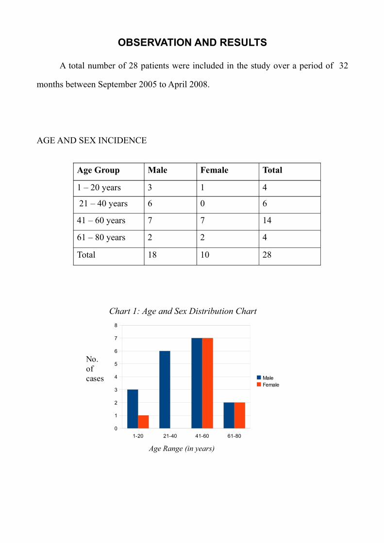
OBSERVATION AND RESULTS
A total number of 28 patients were included in the study over a period of 32
months between September 2005 to April 2008.
AGE AND SEX INCIDENCE
Age Group Male Female Total
1 – 20 years 3 1 4
21 – 40 years 6 0 6
41 – 60 years 7 7 14
61 – 80 years 2 2 4
Total 18 10 28
Chart 1: Age and Sex Distribution Chart
Age Range (in years)
No. of cases
1-20 21-40 41-60 61-80
0
1
2
3
4
5
6
7
8
MaleFemale

ETIOLOGICAL INCIDENCE
S.No. Cause Number
1. Acute Trauma 5
2. Post-Trauma Secondary Defects 6
3. Malignancy – Excision 16
4. Post-Surgical Defect 1
Total no. of flaps = 28
Total no. of central forehead flaps = 9
Total no. of laterally based forehead flaps = 19
Chart 2: Etiology of Defects
Acute Trauma
Post Trauma Defects
Post-surgical Defects
Post excision – Malignancy

TYPES OF FLAP USED
S.No. Type of Flap Number
1 Central Forehead Flaps 9
Paramedian Forehead Flap 5
Median Forehead Flap 2
Oblique Forehead Flap 1
Glabellar V-Y Advancement 1
2 Laterally-based Forehead Flaps 19
Transverse Forehead Flap 12
Forehead Rotation Flap 3
Scalping Flap 1
Bipolar Narayanan Flap 2
Fricke Flap 1
Chart 3: Break-up of types of Flaps used

OUTCOMES
No. of completely healed flaps 20
No. of flaps with wound dehiscence 2
No. of flaps with partial necrosis 5
No. of flaps with complete flap loss 1
Total 28
No. of complications in central forehead flaps = 3
No. of complications in lateral forehead flaps = 5
Total no. of flap complications = 8
Chart 4: Pie-chart of various outcomes
Complete Healing
Wound Infection/ Dehiscence
Partial Necrosis
Complete Necrosis
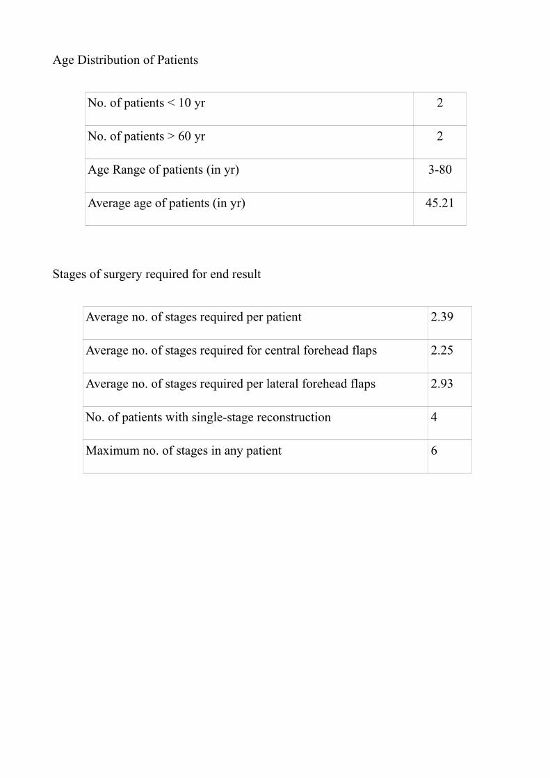
Age Distribution of Patients
No. of patients < 10 yr 2
No. of patients > 60 yr 2
Age Range of patients (in yr) 3-80
Average age of patients (in yr) 45.21
Stages of surgery required for end result
Average no. of stages required per patient 2.39
Average no. of stages required for central forehead flaps 2.25
Average no. of stages required per lateral forehead flaps 2.93
No. of patients with single-stage reconstruction 4
Maximum no. of stages in any patient 6

DISCUSSION
The forehead is a special area in the context of head and neck reconstructive
surgery. It is unique in that it shares characteristics with both the face and the scalp.
The forehead region has a rich vascular supply from 6 major vascular pedicles and is
augmented by anastomoses with the arteries of the face and those of the scalp.
Anatomic studies have proven the predictable vascularity of this region and reliable
anastomoses between 4 vascular territories.79 The forehead allows mobilization of
tissue in a variety of ways with robust and reliable vascularity. It's proximity to the
face and the qualities of the skin of the forehead makes it an indispensable tool in the
reconstruction of soft tissue defects in the face and neck.
Various types of flaps have been described. The transverse forehead flap is one
of the earliest axial flaps described. The paramedian/midline forehead flap has been
practised from ancient times by early Indian practitioners. The major drawback as
cited against the forehead flap is the cosmetic deformity associated with the donor
defect.
Among the 28 patients who underwent various forehead flaps, 15 patients had
primary or delayed primary cover for post-excisional defects. The majority of these
were transverse forehead flaps with or without modifications. The majority of the
flaps healed well although they invariably were staged procedures. Transverse
forehead flaps seem to require a minimum of 3 stages – flap elevation and inset, flap
division and flap thinning/commissuroplasty with or without reanimation of the oral
sphincter. This is also the group that had maximum post-operative complications
(partial/complete flap necrosis).

Although an objective assessment was not made regarding the patient
satisfaction with the esthetic outcome, most of the patients were not happy with the
end result in terms of appearance in the immediate post-op period. On further follow-
up, however, most of the patients seem to be satisfied with the end result.
The patients who underwent nasal reconstruction with different types of central
forehead skin flaps either for post traumatic defects or for post excision defects for
malignancy were the most satisfied with the results. They also underwent lesser no. of
procedures. Therefore, the central forehead flaps are the first choice reconstructive
options for nasal reconstruction.
The forehead rotation flap for forehead skin defects was as a single-stage
operation and achieved acceptable esthetic results. There was only one case of wound
dehiscence which was noted in a patient with extensive injuries to the forehead and
scalp for whom the flap cover was done in the emergency operation theatre after
wound debridement.
The glabellar V-Y advancement procedure was done for a patient with a Tessier
0 cleft who had undergone multiple procedures for nasal reconstruction and presented
with extrusion of rib graft. The flap cover was completed in a single-stage and healed
well.
Usual Indications :
1. Nasal Reconstruction
i. Nasal ala
ii. Columella/Nasal tip defects

iii. Sub-total/total nose defects
2. Eyelid and periorbital reconstruction
i. Upper Eyelid
ii. Lower Eyelid
iii. Medial Canthal region
iv. Lateral Canthal region
3. Reconstruction of lip and cheek defects
i. Combined Upper Lip and Lower Lip defects
ii. Upper Lip and Nasal/nasolabial region defects
iii. Combined Lip and Cheek defects
iv. Full-thickness cheek defects
4. Reconstruction of chin and upper neck defects
Advantages :
1) Colour match
2) Texture match
3) Proximity to the defects
4) Pliability of the flap

5) Reliability of blood supply
6) Versatility of the flap in terms of flap design
Disadvantages :
1) Donor area scar
2) Multiple stages
Specialised Indications (reported in literature):
1. The vertical median forehead flap has been used for repairing dural defects
in the anterior cranial base.31,80,81
2. It has been combined with other local or distant flaps for reconstruction of
tongue,37 oronasal fistulae,39 recurrent oro-cutaneous fistulae, floor of
mouth57 etc.,.
3. Galeo-frontalis flaps have been similarly used to line anterior cranial fossa
CSF leaks, and craniofacial defects80,81,82,83,84,85, . Large defects of the
cribriform fossa have been closed with forehead flaps.36
4. Vascularised calvarial bone has been used in conjuction with the galea-
periosteal flaps for nasal, maxillary and orbit reconstructions.15
5. Bipedicled forehead flaps have been used for reconstruction of soft tissue
defects in the face.38
6. The paramedian forehead flap has also been used as a bilaminar flap – one
layer consisting of skin and subcutaneous fat and the second layer

consisting of the galeo-frontalis.86
Thus it seems that the versatility of the forehead region as a donor for various
reconstructive purposes is only limited by the imagination and skill of the surgeon.
The current body of literature also reaffirms the applicability of the forehead flaps in
the current practice of “esthetic” reconstructive surgery.
Notwithstanding the objections raised to the use of forehead flaps, the
reliability and robustness of the flaps and their versatility in terms of amount of
tissue availability and freedom of fabricating the flaps should make them one of the
main reconstructive options in head and neck reconstructions. The younger
generation of plastic surgeons who seem fascinated with the latest advances like free
flaps would do well to remember the forehead flaps as an important option and not
just use it as a fall-back option.

CONCLUSION
The following conclusions can be made from this study
1. The forehead is a versatile donor area for head and neck reconstruction.
2. The forehead flaps have a robust vascularity and are reliable.
3. Properly planned forehead flaps are adequate and effective for most of the
nasal defects and many of the post-excision defects in the face and neck.
4. The uses of the forehead flaps are only limited by the imagination and skill
of the surgeon.

BIBLIOGRAPHY
1. Fundamental Techniques of Plastic Surgery and their surgical applications 10th EditionFlaps Page 63 Alan D. McGregor, Ian A. McGregor
2. Grabb's Encyclopedia of Flaps 2nd Edition Volume1 Pages 5-10, 35-36, 37-41, 108-114, 195-219, 378-397 Ed. Berisch Strauch, Luis O. Vasconez, Elizabeth J. Hall-Findlay
3. The Forehead Flap for Nasal Reconstruction. Charles M. Boyd, MD; Shan R. Baker, MD; Darrell J. Fader, MD; Timothy S. Wang, MD; Timothy M. Johnson, MD Arch Dermatol. 2000;136:1365-1370
4. Johnson TM, Baker SR, Swanson N. Concepts of sliding and lifting tissue movement in flap reconstruction. Dermatol Surg. 2000;26:274-278.
5. Shumrick KA, Smith TL. The anatomic basis for the design of forehead flaps in nasal reconstruction. Arch Otolaryngol Head Neck Surg. 1992;118:373-379.
6. Mazzola RF, Marcus S. History of total nasal reconstruction with particular emphasis on the folded forehead flap technique. Plast Reconstr Surg. 1983; 72:408-414.
7. McDowell F. The “B.L.” bomb-shell (B. Lucas). Plast Reconstr Surg. 1969;44: 67-73. 8. Carpue JC. An account of two successful operations for restoring a lost nose. Plast Reconstr
Surg. 1969;44:175-182. 9. Millard DR. Total reconstructive rhinoplasty and a missing link. Plast Reconstr Surg.
1966;37:167-183. 10. Kazanjian VH. The repair of nasal defects with the median forehead flap: primary closure of
the forehead wound. Surg Gynecol Obstet. 1946;83:37-42. 11. Millard DR. Reconstructive rhinoplasty for the lower half of a nose. Plast Reconstr Surg.
1974;53:133-139. 12. Millard DR. Reconstructive rhinoplasty for the lower two-thirds of the nose. Plast Reconstr
Surg. 1976;57:722-728. 13. Millard DR. Hemirhinoplasty. Plast Reconstr Surg. 1967;40:440-445. 14. Burget GC, Menick FJ. Nasal reconstruction: seeking a fourth dimension. Plast Reconstr
Surg. 1986;78:145-157. 15. Burget GC, Menick FJ. Nasal support and lining: the marriage of beauty and blood supply.
Plast Reconstr Surg. 1989;84:189-203. 16. Burget GC. Aesthetic restoration of the nose. Clin Plast Surg. 1985;12:463-480. 17. Menick FJ. Aesthetic refinements in use of forehead for nasal reconstruction: The
paramedian forehead flap. Clin Plast Surg. 1990;17:607-622. 18. Burget GC, Menick FJ. Aesthetic Reconstruction of the Nose. St Louis, Mo: Mosby - Year
Book Inc; 1994. 19. Mangold U, Lierse W, Pfeifer G. The arteries of the forehead as the basis of nasal
reconstruction with forehead flaps. Acta Anat. 1980;107:18-25. 20. McCarthy JG, Lorenc ZP, Cutting L, Rachesky M. The median forehead flap revisited: the
blood supply. Plast Reconstr Surg. 1985;76:866-869. 21. McCarthy JG. Acquired deformities of the nose. In: Plastic Surgery. 3rd ed. Philadelphia,
Pa: WB Saunders Co; 1990:1925-2005. 22. Baker SR, Swanson NA. Local Flaps in Facial Reconstruction. St Louis, Mo: Mosby - Year
Book Inc; 1995. 23. Burget GC. Surgical restoration of the nose. In: Mansor M, Marsh JL, eds. Current Therapy
in Plastic and Reconstructive Surgery. Phildelphia, Pa: BC Decker;1989:400-412. 24. Narayanan M.: Immediate Reconstructions with bipolar scalp flap after excisions of
extensive cheek cancers. Plast. & Reconstr. Surg., 46:548, 1970, and 48: 274,1971.25. Surinder Makkar, Atul Parashar, Ramesh K. Sharma, Kanwaldeep S. Aneja. ‘Turbaned’
forehead flap. Journal of Plastic, Reconstructive & Aesthetic Surgery, Vol 61, Issue 5, May 2008, Pages 594-595

26. Othon Papadopoulos, Dimitrios Karypidis, Chrisostomos Chrisostomidis, Petros Konofaos, Grigorios Champsas, George Kazdaglis. Use of the hemifrontal flap in reconstruction of the forehead. British Journal of Oral and Maxillofacial Surgery, In Press, Corrected Proof, Available online 23 April 2008.
27. J. Benito-Ruiz, J. Monner, J. Fontdevila, J. M. Serra-Renom. Forehead flag flap. British Journal of Plastic Surgery, Volume 57, Issue 3, April 2004, Pages 270-272
28. K. Agrawal, K. N. Panda Moustache reconstruction using an extended midline forehead flap. British Journal of Plastic Surgery, Volume 54, Issue 2, March 2001, Pages 159-161
29. P.H. Simon Bennett, Bruce M. Richard, Kenneth E. Graham. Median forehead flaps for eyelid reconstruction. British Journal of Plastic Surgery, Vol. 54, Issue 8, Dec 2001, Pg 733-734
30. Gady Har-El. Single-stage paramedian forehead flap for nasal reconstruction. Operative Tech. in Otolaryngology-Head and Neck Surgery, Vol 10, Issue 2, Jun 1999, Pg 127-130
31. Mitsuhiro Kawaura, Hideo Nameki, Masato Fujii, Jin Kanzaki..Use of vertical median forehead flap in the reconstruction of the anterior skull base: Report of two cases Auris Nasus Larynx, Volume 24, Issue 4, October 1997, Pages 379-383
32. Berend Van der Lei. Tissue expansion of a forehead flap for nasal reconstruction. British Journal of Plastic Surgery, Volume 50, Issue 3, April 1997, Pages 217-218
33. Z. Potpari´c, K. Fukuta, L.B. Colen, I.T. Jackson, J.H. Carraway Galeo-pericranial flaps in the forehead: a study of blood supply and volumes. British Journal of Plastic Surgery, Volume 49, Issue 8, 1996, Pages 519-528
34. Hisashi Ohtsuka, Yoshiharu Miki, Nobuyuki Shioya, Kitasato Trilobed flap in facial reconstruction British Journal of Plastic Surgery, Volume 35, Issue 4, October 1982, Pages 493-497
35. Neil R. McLean, Anthony C.H. Watson Reconstruction of a defect of the ala nasi following trigeminal anaesthesia with an innervated forehead flap British Journal of Plastic Surgery, Volume 35, Issue 2, April 1982, Pages 201-203
36. Douglas K. Ousterhout, Paul Tessier. Closure of large cribriform defects with a forehead flap. Journal of Maxillofacial Surgery, Volume 9, 1981, Pages 7-9.
37. Zlatko Matulic´, Mladen Barlovic´, Vladimir Mikolji, Misˇo Virag Tongue reconstruction by means of the sternocleidomastoid muscle and a forehead flap British Journal of Plastic Surgery, Volume 31, Issue 2, April 1978, Pages 147-151
38. S.S. Rawat. Bipedicled (vascular) forehead flap British Journal of Plastic Surgery, Volume 30, Issue 1, January 1977, Pages 42-43
39. Oscar Contreras, Miguel Gonzales, Rodrigo A. Villalobos. The tongue and forehead flap in the closure of residual oronasal fistulae Journal of Cranio-Maxillofacial Surgery, Volume 17, Supplement 1, December 1989, Pages 39-41
40. Igor V. Reshetov, Valery I. Chissov, Alexander M. Sdvizkov, Sergey A. Kravtzov, Oleg V. Matorin, Sergey V. Tanyashin, Vasiliy A. Tcherekaev. Reconstructive surgery of skull base region after malignant tumor excision. Clinical Neurology and Neurosurgery, Volume 99, Supplement 1, July 1997, Pages S23-S24
41. Frederick J. Menick. Lining options in nasal reconstruction. Operative Techniques in Plastic and Reconstructive Surgery, Volume 5, Issue 1, February 1998, Pages 65-75
42. Calhoun KH, Tan LKS..Skin cancer. In: Gates GA, ed. Current Therapies in Otolaryngology - Head and Neck Surgery..6th ed. St. Louis, Mo: Mosby; 1998.
43. Cook TA, Park SS..Locoregional flaps for facial resurfacing. In: Myers EN, ed. Advances in Otolaryngology-Head and Neck Surgery..St. Louis, Mo: Mosby; 1995:..1-28.
44. Park SS, Cook TA..Reconstructive rhinoplasty..Facial Plast Surg..Oct.1997;13(4):309-16.
45. Park SS..Nasal restoration with flaps and grafts. In: Bailey JB, ed. Head and Neck Surgery-Otolaryngology..Philadelphia, Pa: Lippincott-Raven; 2006.
46. Park SS..Local and regional cutaneous flaps. In: Papel ID, ed. Facial Plastic and

Reconstructive Surgery 2nd ed..New York, NY: Thieme; 2002.
47. Park SS..Cutaneous lesions and local flaps. In Park SS, ed. Facial Plastic Surgery - an Essential Guide..New York, NY: Thieme; 2005.
48. Quatela VC, Sherris DA, Rounds MF..Esthetic refinements in forehead flap nasal reconstruction..Arch Otolaryngol Head Neck Surg..Oct.1995;121(10):1106-13.
49. Samhita S..English translation of the Sushruta Samhita based on original Sanskrit text..Bose, Calcutta, India: Kaviraj Kunja Lal Bhishagratna; 1907-1916
50. Shumrick KA, Campbell A..Improvements in forehead flap design for nasal reconstruction..Facial Plast Surg..1998;14(2):165-71.
51. Tardy ME, Brown RJ..Surgical Anatomy of the Nose..New York, NY: Raven Press; 1990:..25.
52. Tardy ME Jr, Sykes J, Kron T..The precise midline forehead flap in reconstruction of the nose..Clin Plast Surg..Jul.1985;12(3):481-94.
53. Thomas JR, Griner N, Cook TA..The precise midline forehead flap as a musculocutaneous flap..Arch Otolaryngol Head Neck Surg..Jan.1988;114(1):79-84.
54. Tollefson TT, Kriet JD..Complex nasal defects. In Park SS, ed. Facial Plastic Surgery Clinics of North America..Philadelphia, PA: Elsevier Inc; 2005.
55. Naozer M. Kavarana. The Use of folded forehead flap for reconstruction of a large defect after excision of full-thickness of cheek. Plast & Reconstr. Surg 1975; Vol 56, No. 6: 629-632.
56. John J.Smalley MD and Myles P. Cunningham MD. Forehead Flap Rotation to Protect the Carotid Artery. Plat. & Reconstr. Surg. 1972; Vol.49, No.1. 96-97.
57. Toomey J. M. Forehead flap reconstruction of floor of mouth. Ann. Oto. Rhin. Laryng. 1968; 77:94 1968.
58. Dunham M T 1893 A method for obtaining a skin flap from the scalp and a permanent buried vascular pedicle for covering defects of the face. Annals of Surgery 17:677-679
59. Conway H, Stark B, Kavanagh J D 1952 Variations of the temporal flap. Plastic and Reconstructive Surgery 9: 410-423
60. Corso P F 1961 Variations of the arterial, venoua and capillary circulation of the soft tissues of the head by decades as demonstrated by the methyl methacrolate injection technique, and their application to the construction of flaps and pedicles. Plast and Reconstr Surg 27: 160-184
61. Behan F C, Wilson J S P 1973 The vascular basis of laterally based forehead island flaps, and their clinical application. Presented at the 2nd Congress of the European Section of the International Confederation of Plast and Reconstr Surg, Madrid. Royal college of Surgeons of England, London.
62. Cramer L R, Culf N K 1969 Use of pedicle flap tissues in conjunction with a neck dissection. In:Gaisford J C (ed) Symposium on Cancer of the Head and Neck. C V Mosby Company, St. Loius.
63. Gillies L D, Millard D R Jr 1957 Principles and Art of Plastic Surgery. Little Brown, Boston.
64. Champion R 1960 Closure of full-thickness cheek loss by forehead flap. British Journal of Plastic Surgery 13: 76-78
65. Millard DR 1964 A new approach to immediate mandibular repair. Annals of Surgery 160: 306-313

66. Hoopes J E, Edgeron M T 1966 Immediate forehead flap repair in resectionfor oro-pharyngeal cancer. American Journal of Surgery 112: 527-533
67. Davis G N, Hoopes J E 1971 New route for passage of forehead flap to inside of mouth. Plastic and Reconstructive Surgery 47:390-392
68. Monks G H 1898 The restoration of a lower eyelid by a new method. Boston Medical and Surgical Journal 139:385-387
69. Horsley J S 1916 Transplantation of the anterior temporal artery. Clinical Jounal, London 45: 193-196
70. Wilson J S 1967 The application of the 2-centimetre pedicle flap in plastic surgery. British Journal of Plastic Surgery 20: 278-296
71. McGregor I.A., Reid W.H.: The use of the temporal flap in the primary repair of full-thickness defects of the cheek. Plastic and Reconstructive Surgery 38: 1, 1967
72. R. Ozdemir and N. Sungur, Reconstruction of facial defects with superficial temporal artery island flaps:a donor site with various alternatives, Plast & Reconstr Surg 109 (2002), p. 1528.
73. Worthen E F. Repair of forehead defects by rotation flaps. Plast and Reconstructive Surgery 1976; 57: 204.
74. Converse J M. New forehead flap for nasal reconstruction. Proc R Soc Med 1942;35:811.
75. Converse J M. Reconstruction of the nose by the scalping technique. Surg Clin North Am 1959;39:335
76. Converse J M. Clinical applications of the scalping flap in reconstructive surgery of the nose. Plastic and Reconstructive Surgery 1969;43:247
77. Converse J M, McCarthy JG. The scalping forehead flap revisited. Clin Plast Surg 1981;8:413.
78. Mustarde J C. Repair and reconstruction of the orbital region, 2nd Ed. Edinburgh: Churchill Livingstone, 1980. Chap. 11.
79. Cormack G.C. & Lamberty B.G.H. The Arterial Anatomy of Skin Flaps.Edinburgh: Churchill Livingstone, 1986.Chap.7 Pg 383-385.
80. Steinberg CM, Bailey BJ, Weiner RL.Calhoun KH, Quint FB. Reconstruction of the anterior skull base following craniofacial resection. Arch Otolaryngol Head Neck Surg 1987; 113: 710-12.
81. Snyderman CH, Janecka IP, Sekhar LN, Sen CN, Eibling DE. Anterior cranial base reconstruction: role of galeal and pericranial flaps. Laryngoscope 1990; 100: 607-13.
82. Johns ME, Winn HR, McLean WC, Cantrell WR. Pericranial flap for the closure of defects of craniofacial resections, Laryngoscope 1981; 91: 952-9.
83. Avelar MJ, Psillakis JM. The use of galea flaps in craniofacial deformities, Ann Plast Surg 1981; 6: 46449.
84. Jackson IT, Adham MN, Marsh WR. Use of the galeal frontalis myofascial flap in craniofacial surgery. Plast Reconstr Surg 1986; 77: 905510.
85. Costa H, Cerejo A, Baptista A et al. The galea frontalis myofascial flap in anterior fossa CSF leaks. Br J Plast Surg 1993; 46: 503-7.
86. Qing Feng Li et al. Nasal Reconstruction Using a Split Forehead Flap . Plastic and Reconstructive Surgery December 2006 Vol.118, No.7 Pg 1544-1550.

APPENDIX
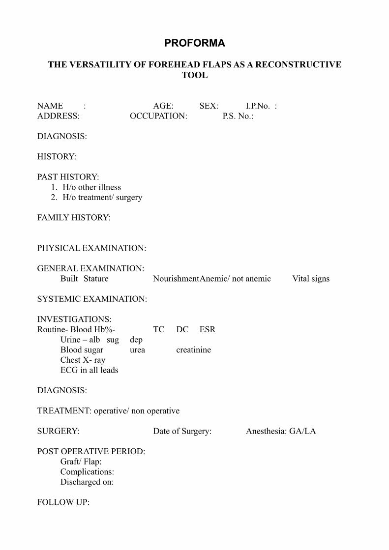
PROFORMA
THE VERSATILITY OF FOREHEAD FLAPS AS A RECONSTRUCTIVE TOOL
NAME : AGE: SEX: I.P.No. :ADDRESS: OCCUPATION: P.S. No.:
DIAGNOSIS:
HISTORY:
PAST HISTORY:1. H/o other illness2. H/o treatment/ surgery
FAMILY HISTORY:
PHYSICAL EXAMINATION:
GENERAL EXAMINATION:Built Stature NourishmentAnemic/ not anemic Vital signs
SYSTEMIC EXAMINATION:
INVESTIGATIONS:Routine- Blood Hb%- TC DC ESR
Urine – alb sug depBlood sugar urea creatinineChest X- rayECG in all leads
DIAGNOSIS:
TREATMENT: operative/ non operative
SURGERY: Date of Surgery: Anesthesia: GA/LA
POST OPERATIVE PERIOD:Graft/ Flap:Complications:Discharged on:
FOLLOW UP:

S.No. Name Age Sex I.P. No. P.S. No. DO A DO D Diagnosis Surge ry O utcome
1 Umashankar 40 M 761271 /06 210 /05 24/10/05 12/11/05 PT Defect Lt side Nose & Upper Lip T ransverse Folded Forehead Flap Cover Complete healing 4
2 Valliammal 50 F 769703 /06 726 /06 05/12/05 10/12/06 Ca Lt Buccal Mucosa Complete healing 3
3 Amirtham 60 F 786760 /06 1153 /05 16/02/06 14/03/06 T ransverse Forehead Flap Cover Complete healing 2
4 Chinnaponnu 55 F 790302 /06 390 /06 09/03/06 25/03/06 Ca Lt Buccal Mucosa Complete healing 3
5 Swaminathan 54 M 794122 /06 590 /06 18/03/06 20/04/06 Ca Rt Cheek Complete healing 2
6 Baskar 32 M 799203 /06 273 /06 08/04/06 20/04/06 PS Defect Rt Medial Canthal Region Oblique Forehead Flap Cover Complete healing 3
7 Rukkumani 55 F 835994 /06 06/09/06 26/10/06 Ca Rt Buccal mucosa Complete healing 2
8 Balaraman 44 M 842351 /06 02/10/06 23/11/06 PT ST Defect Rt Cheek, Panfacial # T ransverse Folded Forehead Flap Cover Complete healing 6
9 Kumaran 36 M 852368 /06 3037 /06 15/11/06 01/12/06 Forehead Rotation Flap cover 1
10 Chandra 55 F 852860 /06 2866 /06 17/11/06 01/12/06 BCC Nose & Rt Cheek 2
11 Nallan 55 M 860536 /06 19/12/06 26/12/06 PT Fistula Lt Medial Canthal Area Paramedian Forehead Flap Cover 2
12 Premkumar 4 M 006377 /07 297 /07 29/01/07 08/03/07 PT Skin Loss Lt Forehead, Scalp Forehead Rotation Flap cover Complete healing 1
13 Arunkumar 15 M 009886 /07 5355 /07 13/02/07 23/02/07 Glabellar V-Y Flap Cover Complete healing 1
14 Rajam 63 F 023189 /07 3612 /07 10/04/07 24/04/07 BCC Rt Upper Lip & Ala Complete healing 3
15 Murugan 28 M 024088 /07 1194 /07 14/04/07 26/04/07 ST Injury Face – Lower Lid Defect Complete healing 2
16 Veeramma 49 F 035491 /07 1759 /07 29/05/07 14/06/07 PT Deformity Dorsum & T ip of Nose Paramedian Forehead Flap Cover Complete healing 3
17 Jeyakumar 25 M 036424 /07 1390 /07 02/06/07 08/06/07 Median Forehead Flap Cover Complete healing 2
18 Rathinavel 62 M 053262 /07 2443 /07 07/08/07 24/08/07 Ca Lt Lower Alveolus 3
19 Pandian 54 M 062800 /07 2679 /07 14/09/07 02/10/07 Recurrent Ca Rt Alveolus 3
20 Baby 60 F 066734 /07 29/09/07 23/10/07 Recurrent BCC Lt Lat wall of Nose WLE with Paramedian Flap Cover Complete healing 2
21 Chithra 3 F 066845 /07 30/09/07 02/10/07 RT A with ST Defect Rt Forehead Forehead Rotation Flap cover Complete healing 1
22 Jayavel 52 M 070966 /07 3237 /07 16/10/07 23/10/07 Meibomian gland Ca Lt Upper Eyelid 3
23 Manikandan 19 M 088697 /07 3942 /07 29/12/07 03/01/08 2
24 35 M 005128 /08 110 /08 22/01/08 31/01/08 Post-Human Bite Nasal Deformity Scalping Flap for Nasal Reconstruction Complete healing 2
25 Raji 50 M 018967 /08 664 / 08 13/03/08 02/02/08 Ca Rt Upper Lip Complete healing 3
26 Kuppammal 73 F 020147 /08 18/03/08 08/04/08 BCC Lt Nostril with DM WLE with Median Forehead Flap Cover Complete healing 2
27 Raji 80 M 023108 /08 725 /08 29/03/08 20/05/08 Ca Lt Lower Alveolus Complete healing 2
28 Muthu 58 M 025026 /08 05/04/08 18/05/08 Ca Rt Buccal mucosa 2
Total No. of Stages
WLE with SOND with Folded T rans-verse Forehead Flap
PS Defect Lt Buccal Mucosa & Lower Lip- Ca Cheek excision done
WLE with SOND with Folded T rans-verse Forehead FlapWLE with SOND with Bipolar Naray-anan Forehead Flap
WLE with Rt RND with Folded Fore-head flap
PT Skin Defect Rt Forehead and Frontal Scalp
Wound Dehiscence – Healing by Secondary Intent ion
WLE with Paramedian Forehead Flap Cover
Part ial Flap Loss – Flap Adjustment
Part ial Flap Loss – Flap Adjustment
T essier 0 Cleft with Augmentation Rhinoplasty with exposed cart ilage – status
WLE with T ransverse Forehead Flap CoverIslanded T ransverse Forehead Flap Cov-er
PT Nose Deformity – Columella de-fect
WLE with Lt Hemimandibulectomy with T ransverse Folded Forehead Flap Cover
Part ial Flap Loss – DP Flap Cover
WLE with T ransverse Folded Forehead Flap Cover
Part ial Flap Loss – Flap Adjustment & DP Flap Cover
WLE with Excision of Lt Upper Eyelid with Fricke's Flap Cover
Near-T otal Flap Loss – Lid Switch and Cheek Rotat ion Flap
Compound # Nasal bone with Skin Loss
Open Reduction and Paramedian Fore-head Flap Cover
Wound Dehiscence – Healing by Secondary Intent ion
Sulthan Basheer
WLE with Rt RND with Folded Fore-head flap
WLE with Lt Hemimandibulectomy with Bipolar Narayanan Flap Cover
WLE with Rt RND with Forehead flap for lining & PMMC for cover
Part ial Necrosis of forehead flap – SSG
MASTER CHART