Vernal keratoconjunctivitis
54
TITLE TEXT DR C V GOPALA RAJU, VISAKHA EYE HOSPITAL VISAKHAPATNAM Vernal Keratoconjunctivitis New Drugs
-
Upload
vijaygopalraju-ch -
Category
Healthcare
-
view
698 -
download
1
Transcript of Vernal keratoconjunctivitis

TITLE TEXTDR C V GOPALA RAJU,
VISAKHA EYE HOSPITAL VISAKHAPATNAM
Vernal Keratoconjunctivitis
New Drugs
stage. Limbal papillae tend to begelatinous and confluent (Fig. 3).
Bonini et al. (2000) graded the papil-lae on the upper tarsal conjunctiva orat the corneoscleral limbus as follows:
(1) Grade 0: no papillary reaction.(2) Grade 1+: few papillae, 0.2 mmwidespread over the tarsal conjunctivaor around the limbus.
(3) Grade 2+: papillae of 0.3–1 mmover the tarsal conjunctiva or at thelimbus.(4) Grade 3+: papillae of 1–3 mm allover the tarsal conjunctiva or for 360!around the limbus.(5) Grade 4+: papillae of more than3 mm over the tarsal conjunctiva orgelatinous appearance at the limbuscovering the peripheral cornea.
Based on the predominant involvementof either tarsal or limbal conjunctiva,VKC can be divided into palpebral,bulbar or mixed type with limbal formsbeing prevalent in non-White patients(Verin et al. 1999).
Photophobia, pain and foreignbody sensation are caused by involve-ment of the cornea. Corneal changesinclude punctate epithelial keratitis,epithelial macro-erosions, shield ulcer,plaque formation and late cornealvascularization (Allansmith & Ross1988; Buckley 1988). Coalescence ofpunctate epithelial keratitis areas leadsto frank corneal epithelial erosion,leaving Bowman’s membrane intact.If untreated, a plaque containingfibrin and mucus deposits over theepithelial defect (Rahi et al. 1985).Epithelial healing is then impaired,and new vessel growth is encouraged.The oval-shaped epithelial defects,known as shield ulcers (Fig. 4), usu-ally have their lower border in theupper half of the visual axis. Healedshield ulcers may leave a subepithelialring-like scar (Fig. 5). Corneal ulcer isreported to occur in 3–11% ofpatients. Corneal changes cause per-manent reduction in visual acuity in6% of patients suffering from VKC(Neumann et al. 1959; Cameron 1995a;Tabbara 1999). Pseudogerontoxon,which resembles arcus senilis, is a wax-ing and waning grey-white lipid deposi-tion in the superficial stroma of theperipheral cornea.
Signs of VKC are confined mostlyto the conjunctiva and cornea. Theskin of the lid and lid margin are rela-tively uninvolved. The conjunctiva ofthe fornices does not usually showforeshortening and symblepharonformation. Iritis is not reported tooccur in VKC. Ocular complicationsof VKC have been reported toinclude steroid-induced cataract andglaucoma, corneal scarring, microbialkeratitis and limbal tissue hyperplasia(Sridhar et al. 2003). Amblyopia seenamong VKC may be caused by cor-neal opacity, irregular astigmatismand keratoconus. Dry eye syndrome,reported in patients suffering fromVKC, may be caused by unsuperviseduse of topical corticosteroids (Tabbara1999).
No precise diagnostic criteria havebeen established for this disease.Hyperaemia, itching, photophobia,tearing and mucus discharge are
Fig. 1. Horner-Tranta’s dots.
Fig. 2. Cobble stone papillae.
Acta Ophthalmologica 2009
135

INTRODUCTION : DEFINITION
VERNAL KERATOCONJUNCTIVITIS
‣ Vernal keratoconjunctivitis (VKC) is a chronic, bilateral, at times asymmetrical, seasonally exacerbated, allergic inflammation of the ocular surface, involving tarsal and ⁄ or bulbar conjunctiva.
‣ Initially called as conjunctiva lymphatica - 150 years ago. ‣ spring catarrh, phlyctenula pallida, circumcorneal hypertrophy,
recurrent vegetative conjunctiva, verrucosa conjunctiva and aestivale conjunctiva
‣ Although the allergic nature of this entity has been accepted for a long time, its exact aetiology and pathogenesis is still unclear.

Geographical distribution
PREVALENCE
‣ It is more common in temperate zones of Mediterranean areas, central and west Africa, the Middle East, Japan, the Indian subcontinent and South America.
‣ VKC cases are also seen in Western Europe (including the UK and Sweden), Australia and North America.
‣ After the recent decline of endemic trachoma, VKC is a leading cause of outpatient ophthalmic morbidity.

Demography
AGE, GENDER, GENETICS AND ASSOCIATED DISEASES
‣ Starts before 10 years. (The earliest reported age is 5 months. Resolves after puberty, usually around 4–10 years after onset (Bielory 2000; Leonardi 2002a).
‣ male to female ratio reported in the literature varying from 4:1 to 2:1 (Neumann et al. 1959; Beigelman 1965; Bonini et al. 2000).
‣ Atopic allergy, Nasal Allergy and Asthma. One third of VKC patients have multiple atopic diseases (Bonini et al. 2000).
‣ In a gender- and age-matched study, a positive correla- tion between eyelash length and severity of VKC has been reported.
‣ long lashes may represent the protective mechanism against physical agents that might have an important role in the aetiopathogenesis of VKC, although the chemical mediator responsible for lash growth was not identified (Pucci et al. 2005).

Clinical features and diagnosis
SYMPTOMS
‣ Pruritus, hyperaemia, photophobia and watering. ‣ Bilateral in - 98% ‣ Minor differences in severity between the eyes are common
(Bonini et al. 2000). ‣ Several episodes of active inflammation throughout the year.
Initially seasonal may become perennial after a few years. ‣ Exacerbated by exposure to wind, dust, bright light, hot weather
or physical exertion associated with sweating. ‣ Tabuchi et al. (2004) reported Staphylococcus aureus to be one
of the exacerbating factors in VKC.

Clinical features and diagnosis
SIGNS
‣ Thick mucus hyper-secretion with sticky mucous filaments, called ‘ropy discharge’, is a characteristic of VKC.
‣ Transient limbal or conjunctival yellow-white points or deposits, known as Horner–Trantas’s dots are degenerating eosinophils and epithelial cell debris.
‣ Asians show, perilimbal conjunctival pigmentation The extent of pigmentation did not correlate with the severity of symptoms and signs of VKC. The pigmentation persisted when the disease was inactive (Rao et al. 2004).

Clinical features and diagnosis
SIGNS
‣ Limbal Papillae, Horner-Tranta’s Dots, Cobble stone Papillae
stage. Limbal papillae tend to begelatinous and confluent (Fig. 3).
Bonini et al. (2000) graded the papil-lae on the upper tarsal conjunctiva orat the corneoscleral limbus as follows:
(1) Grade 0: no papillary reaction.(2) Grade 1+: few papillae, 0.2 mmwidespread over the tarsal conjunctivaor around the limbus.
(3) Grade 2+: papillae of 0.3–1 mmover the tarsal conjunctiva or at thelimbus.(4) Grade 3+: papillae of 1–3 mm allover the tarsal conjunctiva or for 360!around the limbus.(5) Grade 4+: papillae of more than3 mm over the tarsal conjunctiva orgelatinous appearance at the limbuscovering the peripheral cornea.
Based on the predominant involvementof either tarsal or limbal conjunctiva,VKC can be divided into palpebral,bulbar or mixed type with limbal formsbeing prevalent in non-White patients(Verin et al. 1999).
Photophobia, pain and foreignbody sensation are caused by involve-ment of the cornea. Corneal changesinclude punctate epithelial keratitis,epithelial macro-erosions, shield ulcer,plaque formation and late cornealvascularization (Allansmith & Ross1988; Buckley 1988). Coalescence ofpunctate epithelial keratitis areas leadsto frank corneal epithelial erosion,leaving Bowman’s membrane intact.If untreated, a plaque containingfibrin and mucus deposits over theepithelial defect (Rahi et al. 1985).Epithelial healing is then impaired,and new vessel growth is encouraged.The oval-shaped epithelial defects,known as shield ulcers (Fig. 4), usu-ally have their lower border in theupper half of the visual axis. Healedshield ulcers may leave a subepithelialring-like scar (Fig. 5). Corneal ulcer isreported to occur in 3–11% ofpatients. Corneal changes cause per-manent reduction in visual acuity in6% of patients suffering from VKC(Neumann et al. 1959; Cameron 1995a;Tabbara 1999). Pseudogerontoxon,which resembles arcus senilis, is a wax-ing and waning grey-white lipid deposi-tion in the superficial stroma of theperipheral cornea.
Signs of VKC are confined mostlyto the conjunctiva and cornea. Theskin of the lid and lid margin are rela-tively uninvolved. The conjunctiva ofthe fornices does not usually showforeshortening and symblepharonformation. Iritis is not reported tooccur in VKC. Ocular complicationsof VKC have been reported toinclude steroid-induced cataract andglaucoma, corneal scarring, microbialkeratitis and limbal tissue hyperplasia(Sridhar et al. 2003). Amblyopia seenamong VKC may be caused by cor-neal opacity, irregular astigmatismand keratoconus. Dry eye syndrome,reported in patients suffering fromVKC, may be caused by unsuperviseduse of topical corticosteroids (Tabbara1999).
No precise diagnostic criteria havebeen established for this disease.Hyperaemia, itching, photophobia,tearing and mucus discharge are
Fig. 1. Horner-Tranta’s dots.
Fig. 2. Cobble stone papillae.
Acta Ophthalmologica 2009
135
typical symptoms of VKC. Largepapilla on the upper tarsal conjunctivaand corneoscleral junction are hall-marks of VKC. Diagnosis is based ontypical clinical signs and symptoms,thus many mild or atypical cases mayescape diagnosis. The lack of stan-dardized diagnostic criteria and lackof common language among physi-cians regarding the severity of VKCrenders this disease more difficult todiagnose and treat. Bonini et al.(2007) proposed a clinical grading
according to clinical phases of VKCto help physicians use a common lan-guage in the diagnosis and manage-ment of VKC and to allow a morehomogenous selection of patients forclinical trials. Despite mounting datasuggesting the role of both IgE andnon-IgE mediated immune responsesin the pathogenesis of VKC, no clini-cal or laboratory test has evolved tosupport the diagnosis in atypical casesor predict the course of this disease(Bonini et al. 2000).
PathophysiologyNumerous cytological, immunohisto-logical and molecular biological stud-ies of allergic inflammation havefacilitated our understanding of thepathophysiology of VKC. The abilityto measure cytokines in tears, alongwith in-vitro analysis of the individualor combined effects of these cytokineson conjunctival mast cells, epithelialcells and fibroblasts has facilitated ourunderstanding of specific processescontributing to the pathogenesis ofVKC. The clear abundance of Th2cytokines, upregulated expression oftheir receptors and conspicuous pau-city of T helper cell type 1 (Th1) cyto-kines in tear and serum of VKCpatients confirm the crucial roleplayed by these factors in the onsetand perpetuation of the chronic aller-gic inflammation observed in VKC. Avariety of cells, normally present orinfiltrating the ocular surface, areresponsible for the profound expres-sion of these cytokines in VKC. Thefactors stimulating these varied cellsto increase their cytokine productionare still poorly defined. The immune,nervous and endocrine systems seemto interact with each other in thepathogenesis of VKC (Bonini et al.2004). Infective factors were thoughtto contribute to the pathogenesis ofVKC but respiratory syncytial virusand chlamydia were not detected inconjunctival biopsies from patientswith VKC (Koulikovska et al. 2001).Furthermore, serology for ocular chla-mydial disease was also negative(Montan et al. 1999).
Personal or family history of atopy,elevated serum level of total and spe-cific IgE, higher number of mast cellsand eosinophils, increased level ofmediators and favourable response toanti-allergic therapy – the features ofallergic diseases – are also observed inVKC (Allansmith 1982; Bielory &Frohman 1992; Abelson & Schaefer1993).
Conventionally, VKC was consid-ered primarily a type 1 hypersensitivityreaction but the IgE-mast-cell-medi-ated process is not enough to explainthe various clinical and histopathologi-cal changes associated with VKC(Coombs & Gell 1962; Allansmith1982; Johansson et al. 2001). Negativeresponse to skin tests or radioallergo-sorbent test for common allergens in
Fig. 3. Limbal papillae.
Fig. 4. Shield ulcer.
Acta Ophthalmologica 2009
136

Clinical features and diagnosis
SIGNS : PAPILLAE
‣ Large (> 1 mm) papillae in VKC occur at the upper tarsus. ‣ Size of 7–8 mm are known as cobble- stone papillae. ‣ Papillae size correlate positively with the persistence or
worsening of symptoms over long-term follow-up (Bonini et al. 2000).
‣ These papillae become quite swollen during the active stage but persist even during the quiescent stage.
‣ Limbal papillae tend to be gelatinous and confluent.

Clinical features and diagnosis
SIGNS : PAPILLAE GRADING - BONINI ET AL (2000)
‣ Grade 0: no papillary reaction. ‣ Grade 1+: few papillae, 0.2 mm widespread over the tarsal
conjunctiva or around the limbus. ‣ Grade 2+: papillae of 0.3–1 mm over the tarsal conjunctiva or at
the limbus. ‣ Grade 3+: papillae of 1–3 mm all over the tarsal conjunctiva or for
360° around the limbus. ‣ Grade 4+: papillae of more than 3 mm over the tarsal conjunctiva
or gelatinous appearance at the limbus covering the peripheral cornea

Clinical features and diagnosis
CLASSIFICATION AND CORNEAL CHANGES OF VKC
‣ Palpebral, bulbar or mixed type ‣ Limbal forms prevalent in non-White patients (Verin et al. 1999). ‣ Photophobia, pain and foreign body sensation are caused by
involve- ment of the cornea. ‣ Corneal changes: punctate epithelial keratitis, epithelial macro-
erosions, shield ulcer, plaque formation and late corneal vascularization (Allansmith & Ross 1988; Buckley 1988).
‣ Coalescence of punctate epithelial keratitis areas leads to frank corneal epithelial erosion, leaving Bowman’s membrane intact.

Clinical features and diagnosis
SHIELD ULCER IN VKC
‣ If untreated, a plaque containing fibrin and mucus deposits over the epithelial defect (Rahi et al. 1985). Epithelial healing is then impaired, and new vessel growth is encouraged.
‣ The oval-shaped epithelial defects, known as shield ulcers, usually have their lower border in the upper half of the visual axis. Healed shield ulcers may leave a subepithelial ring-like scar. Corneal ulcer is reported to occur in 3–11% of patients.
typical symptoms of VKC. Largepapilla on the upper tarsal conjunctivaand corneoscleral junction are hall-marks of VKC. Diagnosis is based ontypical clinical signs and symptoms,thus many mild or atypical cases mayescape diagnosis. The lack of stan-dardized diagnostic criteria and lackof common language among physi-cians regarding the severity of VKCrenders this disease more difficult todiagnose and treat. Bonini et al.(2007) proposed a clinical grading
according to clinical phases of VKCto help physicians use a common lan-guage in the diagnosis and manage-ment of VKC and to allow a morehomogenous selection of patients forclinical trials. Despite mounting datasuggesting the role of both IgE andnon-IgE mediated immune responsesin the pathogenesis of VKC, no clini-cal or laboratory test has evolved tosupport the diagnosis in atypical casesor predict the course of this disease(Bonini et al. 2000).
PathophysiologyNumerous cytological, immunohisto-logical and molecular biological stud-ies of allergic inflammation havefacilitated our understanding of thepathophysiology of VKC. The abilityto measure cytokines in tears, alongwith in-vitro analysis of the individualor combined effects of these cytokineson conjunctival mast cells, epithelialcells and fibroblasts has facilitated ourunderstanding of specific processescontributing to the pathogenesis ofVKC. The clear abundance of Th2cytokines, upregulated expression oftheir receptors and conspicuous pau-city of T helper cell type 1 (Th1) cyto-kines in tear and serum of VKCpatients confirm the crucial roleplayed by these factors in the onsetand perpetuation of the chronic aller-gic inflammation observed in VKC. Avariety of cells, normally present orinfiltrating the ocular surface, areresponsible for the profound expres-sion of these cytokines in VKC. Thefactors stimulating these varied cellsto increase their cytokine productionare still poorly defined. The immune,nervous and endocrine systems seemto interact with each other in thepathogenesis of VKC (Bonini et al.2004). Infective factors were thoughtto contribute to the pathogenesis ofVKC but respiratory syncytial virusand chlamydia were not detected inconjunctival biopsies from patientswith VKC (Koulikovska et al. 2001).Furthermore, serology for ocular chla-mydial disease was also negative(Montan et al. 1999).
Personal or family history of atopy,elevated serum level of total and spe-cific IgE, higher number of mast cellsand eosinophils, increased level ofmediators and favourable response toanti-allergic therapy – the features ofallergic diseases – are also observed inVKC (Allansmith 1982; Bielory &Frohman 1992; Abelson & Schaefer1993).
Conventionally, VKC was consid-ered primarily a type 1 hypersensitivityreaction but the IgE-mast-cell-medi-ated process is not enough to explainthe various clinical and histopathologi-cal changes associated with VKC(Coombs & Gell 1962; Allansmith1982; Johansson et al. 2001). Negativeresponse to skin tests or radioallergo-sorbent test for common allergens in
Fig. 3. Limbal papillae.
Fig. 4. Shield ulcer.
Acta Ophthalmologica 2009
136
almost 50% of patients, along with thepresence of a multitude of cells andmediators in the conjunctiva, tears andserum of VKC patients, suggest amore complex pathophysiology than amere type 1 hypersensitivity reaction(Bonini et al. 2000). The prevalence ofIgE sensitization was significantlylower in bulbar than in tarsal andmixed VKC. Higher serum eosinophilcationic protein (s-ECP), total serumIgE (s-total IgE) and peripheral bloodeosinophil counts have been reportedin IgE-sensitized than in non-IgE-sen-sitized VKC patients (Pucci et al.2003). Interestingly, Montan et al.(2002) found that the amount ofmRNA encoding Th2-type cytokinesand inflammatory cell markers inVKC remain the same irrespective ofIgE sensitization.
Mediators in VKC
The plethora of mediators and cyto-kines in VKC compared to controls,seasonal allergic conjunctivitis andgiant papillary conjunctivitis providesa new perspective on the complexinflammatory processes occurring onthe ocular surface in this chronicdisease (Cook 2004).
CytokinesCytokines are small secreted proteinsthat mediate and regulate immunityand inflammation. Unlike hormones,
these are not stored as preformedmolecules but must be produced,de novo, in response to a stimulus.Different cell types may secrete thesame cytokine or a single cytokinemay act on several different cell types.Similar functions can be stimulated bydifferent cytokines. ILs are the cyto-kines that are made by leukocytes andact on other leukocytes.
Activated helper T cells (CD4),mast cells and eosinophils are themain cytokine-producing cell typesinfiltrating the conjunctiva duringchronic allergic eye diseases. Two dis-tinct subtypes of helper T cells pro-duce different cytokines. T cellsisolated from conjunctiva of VKCpatients and expanded into cell linesshowed a Th2-like cytokine profile(Calder et al. 1999).
Th2 cytokines, i.e. IL-4 and -5,were high among VKC patients(Metz et al. 1997; Calder et al. 1999;Leonardi et al. 1999a). Increasedexpression of mRNA encoding Th2-type ILs was observed in allergic tis-sue from VKC (Metz et al. 1997;Mori et al. 2002). The serum levelsof IL-4 and tear levels of IL-4,-5were higher in patients with VKCcompared to controls. Interestingly,IL-2, interferon (IFN)-gamma andtumour necrosis factor (TNF)-b, themajor cytokines secreted by Th1, werenot increased in VKC (Leonardi 2002a;
Mori et al. 2002). These findingsconfirm that VKC has a mainly Th2profile (Uchio et al.2000; Fujishimaet al. 2002).
ChemokinesChemokines, a short term for chemo-tactic cytokine (CC), are potent activa-tors and chemoattractants. Chemo-kines are produced not only by inflam-matory cells but also by stimulatedepithelial cells, fibroblasts and vascularendothelial cells in the conjunctiva.Chemokines are involved in normaltrafficking of leukocytes and recruit-ment during inflammation but theirrole is not restricted to cell attraction.These multipotent cytokines localizeand enhance inflammation by inducingchemotaxis and cell activation of dif-ferent types of inflammatory cells pres-ent at sites of inflammation.Chemokines bind to transmembraneG-protein coupled receptors (CCRs),which then signal the cells via second-ary messengers to alter its behaviour.
Chemokines are grouped into theCXC, CC, C and CX3C subfamilies(Murphy et al. 2000). The CCchemokines – monocyte chemotacticprotein (MCP), regulated upon acti-vation, normal T cells expressed andsecreted (RANTES), macrophageinhibitory protein (MIP), thymusand activation-regulated chemokine(TARC) and eotaxin – act on eosin-ophils, basophils, monocytes andlymphocytes, suggesting their impor-tant role in allergic eye diseases(Abu El-Asrar et al. 2000).
High levels of eotaxin were foundin mucus from VKC patients.Eotaxin levels correlate significantlywith the percentage of eosinophils intears and may be responsible for theeosinophil recruitment in VKC (Leo-nardi et al. 2003a). Eotaxin, alongwith MCP and RANTES, werehighly expressed in limbal tissues(primarily by macrophages) and wereresponsible for the massive eosino-phil infiltration in this tissue (AbuEl-Asrar et al. 2000).
IL-8 and the CXC chemokine,monokine induced by interferongamma (Mig), seem to play animportant role in the pathogenesis ofVKC. The chemokine IL-8 activelysecreted by macrophages and epithelialcells in VKC is a chemoattractant aswell as an activator of polymorpho-nuclear cells. It plays a crucial role in
Fig. 5. Ring-shaped corneal opacity after healed shield ulcer.
Acta Ophthalmologica 2009
137

Clinical features and diagnosis
OTHER SIGNS IN VKC
‣ Pseudogerontoxon, which resembles arcus senilis, is a waxing and waning grey-white lipid deposition in the superficial stroma of the peripheral cornea.
‣ The skin of the lid and lid margin may be thick and lax. ‣ Amblyopia seen among VKC may be caused by corneal opacity,
irregular astigmatism. ‣ Keratoconus. ‣ Dry eye syndrome, ‣ Cataract and glaucoma caused by unsupervised use of topical
corticosteroids (Tabbara 1999).

Pathophysiology
ALLERGEN AND MAST CELL REACTION,
‣ Starts before 10 years..

Pathophysiology
ALLERGEN AND MAST CELL REACTION, The immune, nervous and endocrine systems seem to interact with each other in the pathogenesis of VKC (Bonini et al. 2004)
‣ Immune, nervous and endocrine systems interact with each other in the pathogenesis of VKC (Bonini et al. 2004)
‣ VKC was primarily a type 1 hypersensitivity reaction ‣ ‣ Abundance of Th2 cytokines ‣ Paucity of T helper cell type 1 (Th1) cytokines in tear and serum ‣ .

Pathophysiology
ALLERGEN AND MAST CELL REACTION,

Pathophysiology
DEGRANULATION PROCESS‣ Degranulation process in allergy. Second exposure to allergen. 1 – antigen; 2 – IgE antibody; 3 –
FcεRI receptor; 4 – preformed mediators (histamine, proteases, chemokines, heparine); 5 – granules; 6 – mast cell; 7 – newly formed mediators (prostaglandins, leukotrienes, thromboxanes, PAF)

Pathophysiology
MEDIATORS
‣ Cytokines are small secreted proteins that mediate and regulate immunity and inflammation.
‣ Chemokines, a short term for chemo- tactic cytokine (CC), are potent activa- tors and chemoattractants.
‣ Histamine, an important inflamma- tory mediator in allergic eye disease, is released by activated mast cells and basophils.
‣ Metalloproteinases (MMPs) are extra- cellular endopeptidases that selectively degrade components of the extra- cellular matrix. Inflammatory cells, particularly eosinophils, and structural cells like epithelial cells and conjuncti- val fibroblasts are the probable cellu- lar source of these enzymes.
‣ Several growth factors, such as epider- mal growth factor, fibroblast growth factor and transforming growth factor beta-1 (TGFb-1), were increased in VKC.

Pathophysiology
MEDIATORS - CYTOKINES AND CHEMOKINES
‣ Cytokines are small secreted proteins that mediate and regulate immunity and inflammation.
‣ Activated helper T cells (CD4), mast cells and eosinophils are the main cytokine-producing cell types infiltrating the conjunctiva during chronic allergic eye diseases.
‣ Chemokines, a short term for chemotactic cytokine (CC), are potent activators and chemoattractants.
‣ Chemokines are produced not only by inflammatory cells but also by stimulated epithelial cells, fibroblasts and vascular endothelial cells in the conjunctiva.
‣

Pathophysiology
MEDIATORS - HISTAMINE, MMP AND GROWTH FACTOR
‣ Histamine exerts its biological effects by interacting with four G- protein coupled receptors, classified as H1–H4. Vasodilatation, chemosis and itching of eye are caused by histamine interaction with H1 receptors.
‣ Released by activated mast cells and basophils. ‣ Reduced inactivation by histaminase and increased production by specific or non-
specific activation of mast cells and basophils (Abelson et al. 1995). ‣ MMP: Increased production and activation of MMPs or imbalance between MMPs
and their natural tissue inhibitors (TIMPs) are all probably involved in the pathogenesis of conjunctival inflammation, remodel- ling and corneal changes in VKC.
‣ Growth factors, such as epidermal growth factor, fibroblast growth factor and transforming growth factor beta-1 (TGFb-1), were increased in VKC. These factors induce fibroblast growth and procollagen production (Leonardi et al. 1998).

Pathophysiology
CELLS IN VKC ,
‣ Mast cells ‣ Eosinophils ‣ T cells ‣ B cells ‣ Epithelial Cells ‣ Fibroblasts ‣ Natural Killer Cells

Pathophysiology
CELLS - MAST CELLS AND EOSINOPHILS
‣ Mast cells bind IgE on its surface. cross-linkage of this IgE by specific allergens results in the release of pro-inflammatory mediators, including histamine, proteases, prostaglandin D2 and leukotriene C4, into the local extracellular environment.
‣ These mediators are responsible for causing ocular itching, hyperaemia, lacrimation and chemosis in allergic conjunctivitis (McGill et al. 1998; Church & McGill 2002).
‣ Eosinophils : 50–90% of cells in the tears during the active phase of VKC are eosinophils (Leonardi 2002a).
‣ Activated eosinophils release cytokines, chemokines, leukotrienes and epithelio- toxic proteins such as MBP, ECP, eosinophil peroxidase (EPO) and eosinophil protein X ⁄ neurotoxin (EPX) (Tomassini et al. 1994; Leonardi et al. 1995; Leonardi 2002a).

Pathophysiology
CELLS - T CELLS AND B CELLS
‣ T cell are mainly Th2 type (Maggi et al. 1991). Cytokine flow cytometry has shown that 67% of VKC patients have Th2 cells in tears, while Th1 cells are seen in the tears of only 8% (Leo- nardi et al. 1999a).
‣ The predominance of Th2-like cells in tears and conjuncti- val biopsy suggests a local Th2 response in VKC. Th2 lymphocytes, by virtue of their cytokine profile, are responsible for increased production of IgE, recruitment and activation of mast cells and eosinophils (Umetsu & De Kruyff 1997; Bielory et al. 2002a).
‣ B lymphocytes expressing the ligands CD23, 21 and 40 in conjunctiva speci- men from VKC may be a precursor of IgE producing B cells (Abu El-Asrar et al. 2001d).
Pathophysiology
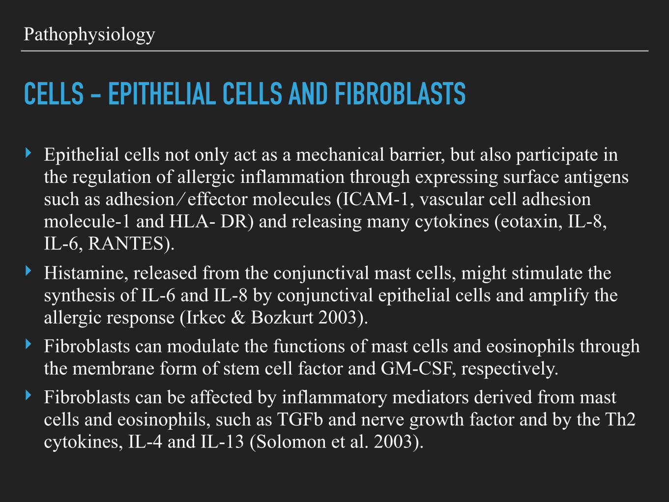
CELLS - EPITHELIAL CELLS AND FIBROBLASTS
‣ Epithelial cells not only act as a mechanical barrier, but also participate in the regulation of allergic inflammation through expressing surface antigens such as adhesion ⁄ effector molecules (ICAM-1, vascular cell adhesion molecule-1 and HLA- DR) and releasing many cytokines (eotaxin, IL-8, IL-6, RANTES).
‣ Histamine, released from the conjunctival mast cells, might stimulate the synthesis of IL-6 and IL-8 by conjunctival epithelial cells and amplify the allergic response (Irkec & Bozkurt 2003).
‣ Fibroblasts can modulate the functions of mast cells and eosinophils through the membrane form of stem cell factor and GM-CSF, respectively.
‣ Fibroblasts can be affected by inflammatory mediators derived from mast cells and eosinophils, such as TGFb and nerve growth factor and by the Th2 cytokines, IL-4 and IL-13 (Solomon et al. 2003).

Pathophysiology
CASCADE OF REACTIONS

Histopathology and Immunohistochemistry
TISSUE INFLAMMATION AND REMODELLING
‣ Histopathological studies of conjunctival tissue from VKC patients show a prominent inflammatory cellular infiltration in the epithelium and substantia propria and post-inflammatory tissue remodelling.
‣ Tissue remodelling is more marked in tarsal than in bulbar conjunctiva.
‣ Tissue inflammation ‣ Tissue remodelling

Histopathology and Immunohistochemistry
SUMMARY OF TISSUE REACTION IN VKC ,
‣ Infiltration of the conjunctiva by eosinophils, basophils, mast cells, Th2 cells, monocyte ⁄ macrophages, dendritic cells, plasma cells and B lymphocytes, frequently organized as small lymphoid follicles without a germinative centre (Abu El-Asrar et al. 2001d; Leonardi 2002a).
‣ Conjunctival thickening, subepithelial fibrosis, mucus metaplasia, neovascularization and scarring are typical of chronic VKC. Epithelial changes, connective tissue deposition, oedema, inflammatory cell infiltration and glandular hypertrophy all contribute in the tissue remodelling observed in VKC (Leonardi 2002a).

Pathophysiology
CASCADE OF REACTIONS

End of part I
PART II
‣ Treatment ‣ New drugs

Treatment
PREVENTIVE MEASURES AND PATIENT EDUCATION
‣ Avoidance of sun, wind and salt water helpful but not enough. ‣ Contact with plants and flowers should be avoided. ‣ Use of sunglasses or any shading measures useful. ‣ Application of cold compresses and use of artificial tears
effective in the relief of symptoms by direct removal and dilution of allergen from the ocular surface (Bielory 2002b).
‣ Cold compresses provide symptomatic relief, from ocular pruritus.
‣ Frequent hand, face and hair washing – before going to bed may be helpful (Leonardi 2002a).

Treatment
PHARMACOLOGICAL THERAPY

Treatment
PHARMACOLOGICAL THERAPY
‣ Anti-histamines, ‣ Mast-cell stabilizers, ‣ Dual acting agents, ‣ Corticosteroids ‣ Immunomodulators ‣ None is enough to treat all aspects of multifaceted
pathophysiology of VKC.

Treatment
PHARMACOLOGICAL THERAPY
‣ Why disease is regressing after 2 to 10 years ? ‣ Why disease is regressing after puberty? ‣ Why some cases are resistant to treatment ? ‣ Why recurrence of disease when the therapy is discontinued?
‣ Humoral, Endocrinal and Neurological elements in the disease which we couldn't understand.
‣ Palliative and do not eliminate the complex immune response
Treatment - Pharmacotherapy
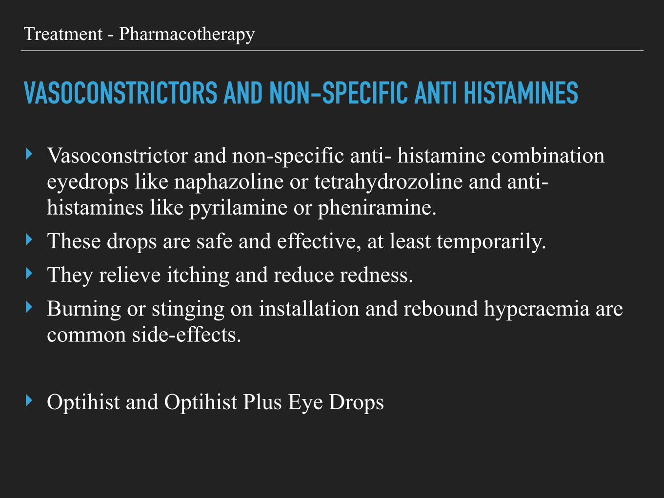
VASOCONSTRICTORS AND NON-SPECIFIC ANTI HISTAMINES
‣ Vasoconstrictor and non-specific anti- histamine combination eyedrops like naphazoline or tetrahydrozoline and anti-histamines like pyrilamine or pheniramine.
‣ These drops are safe and effective, at least temporarily. ‣ They relieve itching and reduce redness. ‣ Burning or stinging on installation and rebound hyperaemia are
common side-effects.
‣ Optihist and Optihist Plus Eye Drops

Treatment - Pharmacotherapy
MAST CELL STABILISERS
‣ Stabilize mast cells and prevent degranulation. ‣ Sodium cromoglycate (qid), ‣ Lodoxamide (qid), ‣ Nedocromil (bid) ‣ Pemirolast (qid)
‣ Cromal and Cromal Forte Eye Drops

Treatment - Pharmacotherapy
ANTI HISTAMINES
‣ Oral anti-histamines are a good choice when allergy involves the eyes, nose or pharynx simultaneously.
‣ When allergic complaints are limited to eyes, focused therapy with topical anti-histamines is efficacious and free of untoward effects related to oral anti-histamines.

Treatment - Pharmacotherapy
SPECIFIC ANTI HISTAMINES - H1 RECEPTOR BLOCKERS
‣ Topical selective H1 blocker, emedastine and levocabasitne, are better than vasoconstrictor ⁄ non-specific anti-histamine combination eyedrops in controlling the signs and symptoms of VKC.
‣ The anti-inflammatory effect seen with pure anti-histamines like levocabastine and emedastine is attributed to the blocking of histamine receptors, thus downregulating the expression of ICAM-1 and limiting che- motaxis of inflammatory cells (Bielory et al. 2005).

Treatment - Pharmacotherapy
DUAL ACTION - MAST CELL STABILISATION & ANTIHISTAMINE
‣ New generation of drugs such as olopatadine, epinastine, ketot- ifen and azelastine has shown dual activity of mast-cell stability and H1 receptor antagonism.
‣ The action of these drugs is not limited to mast-cell stabilization and H1 receptor antagonism, they also exert anti-inflammatory effects through several different mechanisms.
‣

Treatment - Pharmacotherapy
NON-STEROIDAL ANTI-INFLAMMATORY DRUGS
‣ Topical formulations of ketorolac and diclofenac have been shown to diminish ocular pruritus and conjunctival hyperaemia.
‣ Prostaglandin E2 and I2 has been shown to be pruritogenic. ‣ Ketorolac may be a good alternative to topical steroid because it
reduces itching by inhibit- ing the synthesis of prostaglandins (Sharma 1997).
‣ Stinging and burning sensation is the disadvantage.

Treatment - Pharmacotherapy
CORTICOSTEROIDS
‣ Loteprednol is highly effective in the acute and prophylactic treatment of allergic conjunctivitis.
‣ In a retro- spective study, loteprednol etabonate was shown to be safe and effective when used for 12 months or more for the treatment of seasonal or perennial allergic conjunctivitis (Ilyas et al. 2004).
‣ Fluorometholone is a soft cortico- steroid and is effective in controlling the signs and symptoms of VKC.
‣ Supratarsal injection of dexamethasone sodium succinate, triamcinolone acetonide and hydrocortisone sodium succinate is effective in the temporary suppression of inflammation associated with VKC (Saini et al. 1999; Singh et al. 2002; Lisanework 2003).
‣ Although corticosteroids are the most efficacious drugs, steroid-resistant forms of VKC are not unusual and may necessitate an alternative therapy.

Treatment - Pharmacotherapy
IMMUNE MODULATORS - CYCLOSPORINE
‣ Cyclosporine (2%) eyedrops were effective and safe in the treat- ment of severe VKC (Pucci et al. 2002; Kilic & Gurler 2006).
‣ Most of the therapeutic effect was achieved 2 weeks after commencing the treatment and was maintained during the next 3 months by continuous medication (Pucci et al. 2002).
‣ Main inhibition - cost ‣ Main confusion - preparation and concentration

Treatment - Pharmacotherapy
IMMUNE MODULATORS - CYCLOSPORINE
‣ Cyclosporine, a fungal metabolite, decreases the signs and symptoms of VKC. BenEzra et al. (1986)
‣ Cyclosporine 0.05% eyedrops in oil solution to treat severe VKC. ‣ Cyclosporine is lipophilic so it must be dissolved in an alcohol–oil
base, which causes ocular irritation. ‣ Immunomodulating effect on components of cell-mediated and
humoral immune response. (Abu El-Asrar et al. 1996). ‣ Cyclosporine blocks Th2 lymphocyte proliferation and IL-2
production. Furthermore, it inhibits histamine release through a reduction in IL-5 production (Ben- Ezra et al. 1988; Secchi et al. 1990). It accelerates apoptosis in fibroblast.
Treatment - Pharmacotherapy
CYCLOSPORINE - DRUG DELIVERY IS A CHALLENGE
‣ Cyclosporine is a Lipophillic drug ‣ Currently available systems using oils to deliver CsA topically are
poorly tolerated and provide a low bioavailability. ‣ CsA water solubility (e.g. cyclodextrins), or those designed to
facilitate tissue drug penetration using penetration enhancers. ‣ The use of colloidal carriers (micelles, emulsions, liposomes and
nanoparticles) as well as the approach using hydrosoluble prodrugs of CsA have shown promising results. Solid devices such as shields and particles of collagen have been investigated to enhance retention time on the eye surface.
‣ None of these formulations are ideal.
Treatment - Pharmacotherapy
CYCLOSPORINE EYE DROPS - DRUG INTERACTIONS
‣ Pregnancy or planning to become or breast feeding ‣ Herpes Simplex corneal infection ‣ Patients with punctal plugs ‣ Topical Anti-inflammatory drugs like Ketorolac reduces the
effect of cyclosporine.

Treatment - Pharmacotherapy
CYCLOSPORINE EYE DROPS - META ANALYSIS
29/10/15 11:25 pmTopical cyclosporine in the treatment of allergic conjunctivitis: a meta-analysis. - PubMed - NCBI
Page 1 of 2http://www.ncbi.nlm.nih.gov/pubmed/23743438
PURPOSE:
DESIGN:
PARTICIPANTS:
METHODS:
MAIN OUTCOME MEASURES:
RESULTS:
CONCLUSIONS:
Ophthalmology. 2013 Nov;120(11):2197-203. doi: 10.1016/j.ophtha.2013.03.044. Epub 2013 Jun 3.
Topical cyclosporine in the treatment of allergic conjunctivitis: a meta-analysis.Wan KH , Chen LJ, Rong SS, Pang CP, Young AL.
AbstractTo assess the efficacy and safety of topical cyclosporine versus placebo in the
treatment of allergic conjunctivitis.
Systematic review and meta-analysis.
Seven qualified studies incorporating 306 eyes of 153 patients were analyzed.
Searches of randomized controlled trials were conducted in MEDLINE, EMBASE, theCochrane Central Register of Controlled Trials, ClinicalTrials.gov, and the World HealthOrganization International Clinical Trials Registry Platform.
We assessed the methodologic quality of individual included trialsand performed meta-analyses using the random effects model if P<0.1 in the test forheterogeneity, or otherwise used the fixed effects model. We assessed scores of composite signsand symptoms, reduction in steroid eye drop use in steroid-dependent patients, and safetyoutcomes (i.e., stinging or burning sensation).
At 2 weeks of follow-up or longer, evidence suggests a statistically significantimprovement in the composite signs (standardized mean difference [SMD], -1.21; 95% confidenceinterval [CI], -1.80 to -0.62; I(2) = 71%) and symptoms (SMD, -0.84; 95% CI, -1.51 to -0.16; I(2) =80%) after topical cyclosporine treatment for allergic conjunctivitis regardless of the dosage oftreatment. There was a significant reduction (mean difference, -61.16; 95% CI, -101.61 to -20.72;I(2) = 58%) in the use of steroid eye drops in patients with steroid-dependent allergicconjunctivitis. Stinging or burning sensation (odds ratio, 2.56; 95% CI, 0.19-35.06; I(2) = 73%) wascommon in both the cyclosporine and placebo groups.
This systematic review and meta-analysis suggests topical cyclosporine couldbe an effective and safe treatment method for allergic conjunctivitis. Further randomized controlledtrials with larger sample sizes and standardized outcome measurements, follow-up periods, andcyclosporine concentrations are warranted to determine the short- and long-term efficacy andsafety and the minimal effective dosage of topical cyclosporine for allergic conjunctivitis.
Copyright © 2013 American Academy of Ophthalmology. Published by Elsevier Inc. All rights
Abstract
1
Author information
Full text links
PubMed
Treatment - Pharmacotherapy
IMMUNE MODULATORS - MYTOMYCIN-C
‣ Mitomycin-C is an inhibitor of fibroblast proliferation. ‣ Mitomycin-C (0.01%) eyedrops were shown to decrease the
mucous discharge, conjunctival hyperaemia and limbal oedema in VKC patients refractory to topical steroids and mast-cell stabilizers (Akpek et al. 2000; Jain & Sukhija 2006).
‣ ‣

Treatment - Pharmacotherapy
IMMUNE MODULATORS - TACROLIMUS
‣ Used in Sever atopic dermatitis. ‣ Topical Immunosuppressant ‣ VKC and KCS - Ophthalmic use - Ointment form ‣ Emulsion, Solution or Suspension or gel form are also available ‣ Strength - 0.01% to 0.03% ‣ In Severe cases and as a loading dose BD and maintenance OD

Treatment - Pharmacotherapy
IMMUNE MODULATORS - TACROLIMUS - REVIEW OF LITERATURE
31/10/15 6:30 amLong-term follow-up of tacrolimus ointment for treatment of atopic keratoconjunctivitis. - PubMed - NCBI
Page 1 of 2http://www.ncbi.nlm.nih.gov/pubmed/24439439
PURPOSE:
DESIGN:
METHODS:
RESULTS:
CONCLUSIONS:
Am J Ophthalmol. 2014 Feb;157(2):280-6. doi: 10.1016/j.ajo.2013.10.006. Epub 2013 Oct 19.
Long-term follow-up of tacrolimus ointment for treatment of atopickeratoconjunctivitis.Al-Amri AM .
AbstractTo evaluate the long-term clinical outcomes of 0.1% tacrolimus dermatologic ointment
(Protopic) in cases of refractory atopic keratoconjunctivitis (AKC).
Prospective, nonrandomized, noncontrolled case series.
Twenty-two eyes from 11 patients with severe AKC who were treated with 0.1%tacrolimus ointment were followed prospectively. The mean age of the patients was 32.27 ± 12.7years (range, 19-61 years). Each patient completed a follow-up period of at least 48 months,during which the signs and symptoms of AKC were assessed. Changes in the total scores of signsand symptoms from baseline were recorded at each visit, and the main outcome measure was theclinical response to topical tacrolimus treatment.
Dramatic improvements in clinical signs and symptoms were achieved 1 week afterstarting topical tacrolimus treatment, and complete clinical resolution was observed in almost allpatients 6 weeks after starting treatment. Treatment was gradually reduced, with increasingintervals between applications. Eight patients remained asymptomatic for up to 3 years, althoughrecurrence occurred in 3 patients who attempted to discontinue treatment. All patients complainedof a mild burning sensation upon application of the ointment. No additional medications wererequired to provide relief, and no patient discontinued treatment because of adverse drug effects.No drug-related ocular complications were encountered, and no significant changes in visualacuity or refraction were documented.
Tacrolimus dermatologic ointment is a potentially safe and effective treatment forAKC cases refractory to standard treatment and may substitute for steroid treatments aimed atcontrolling disease activity.
Copyright © 2014 Elsevier Inc. All rights reserved.
PMID: 24439439 [PubMed - indexed for MEDLINE]
Abstract
1
Author information
Full text links
PubMed

Treatment - Pharmacotherapy
IMMUNE MODULATORS - TACROLIMUS - DRUG INTERACTION
‣ Ultraviolet light therapy on skin ‣ Herpes or Chickenpox ‣ Precancerous skin conditions ‣ weakened Immunesystem
‣ Avoid in ‣ Lymphoma or skin cancers ‣ Pregnant or planning to become, breast feeding ‣ Swollen Lymph nodes or Mononucleosis

Treatment - Pharmacotherapy
IMMUNE MODULATORS - CYCLOSPORINE & TACROLIMUS 31/10/15 6:36 amTopical immunomodulators in the management of allergic eye diseases. - PubMed - NCBI
Page 1 of 2http://www.ncbi.nlm.nih.gov/pubmed/25054831
PURPOSE OF REVIEW:
RECENT FINDINGS:
SUMMARY:
PubMed Commons home
Curr Opin Allergy Clin Immunol. 2014 Oct;14(5):457-63. doi: 10.1097/ACI.0000000000000089.
Topical immunomodulators in the management of allergic eye diseases.Erdinest N , Solomon A.
AbstractAllergic eye diseases comprise a spectrum of diseases, with each
condition being characterized by a complex immunopathology. The more severe and chronicconditions, such as vernal keratoconjunctivitis and atopic keratoconjunctivitis, involvepredominantly mast cells and eosinophils, while also being associated with a preponderance of Tcells. Treatment with topical antihistamines or mast cell stabilizers is often unsatisfactory, andtherapy depends on topical corticosteroids. Corticosteroids have significant side-effects with long-term use; therefore, they appear to be more appropriate for short-term pulse therapy.Immunomodulatory agents can also be used to inhibit T-cell activation and show encouragingresults among patients with severe allergic eye conditions. The present review is an attempt topresent a coherent picture of the recent investigations of topical immunomodulatory agents'therapy in severe allergic eye diseases, especially cyclosporine A and tacrolimus, and theirmechanisms of action.
Immunomodulatory agents are commonly indicated for the treatment ofsevere and prolonged allergic conjunctivitis. This article reviews the recent studies of these drugsand the development of immunomodulatory treatments for severe allergic eye diseases.
Cyclosporine A and tacrolimus are currently available for the treatment of severeallergic conjunctivitis. These agents have led to improved therapeutic results for patients withsevere and chronic allergic eye diseases.
PMID: 25054831 [PubMed - indexed for MEDLINE]
PubMed Commons
Abstract
1
Author information
Publication Types, MeSH Terms, Substances
LinkOut - more resources
Full text links
PubMed

Treatment - Pharmacotherapy
IMMUNE MODULATORS - CYCLOSPORINE & TACROLIMUS
31/10/15 6:36 amTopical immunomodulators in the management of allergic eye diseases. - PubMed - NCBI
Page 1 of 2http://www.ncbi.nlm.nih.gov/pubmed/25054831
PURPOSE OF REVIEW:
RECENT FINDINGS:
SUMMARY:
PubMed Commons home
Curr Opin Allergy Clin Immunol. 2014 Oct;14(5):457-63. doi: 10.1097/ACI.0000000000000089.
Topical immunomodulators in the management of allergic eye diseases.Erdinest N , Solomon A.
AbstractAllergic eye diseases comprise a spectrum of diseases, with each
condition being characterized by a complex immunopathology. The more severe and chronicconditions, such as vernal keratoconjunctivitis and atopic keratoconjunctivitis, involvepredominantly mast cells and eosinophils, while also being associated with a preponderance of Tcells. Treatment with topical antihistamines or mast cell stabilizers is often unsatisfactory, andtherapy depends on topical corticosteroids. Corticosteroids have significant side-effects with long-term use; therefore, they appear to be more appropriate for short-term pulse therapy.Immunomodulatory agents can also be used to inhibit T-cell activation and show encouragingresults among patients with severe allergic eye conditions. The present review is an attempt topresent a coherent picture of the recent investigations of topical immunomodulatory agents'therapy in severe allergic eye diseases, especially cyclosporine A and tacrolimus, and theirmechanisms of action.
Immunomodulatory agents are commonly indicated for the treatment ofsevere and prolonged allergic conjunctivitis. This article reviews the recent studies of these drugsand the development of immunomodulatory treatments for severe allergic eye diseases.
Cyclosporine A and tacrolimus are currently available for the treatment of severeallergic conjunctivitis. These agents have led to improved therapeutic results for patients withsevere and chronic allergic eye diseases.
PMID: 25054831 [PubMed - indexed for MEDLINE]
PubMed Commons
Abstract
1
Author information
Publication Types, MeSH Terms, Substances
LinkOut - more resources
Full text links
PubMed

Treatment
SURGICAL TREATMENT
‣ Surgical excision of giant papillae is recommended if they cause corneal lesions.
‣ Excision or cryocoagulation of large papillae helps in the early resolution of corneal epitheliopathy or ulcer, although papillae regrow in most patients.
‣ Debridement of ulcer base, surgical removal of plaque or excimer laser phototherapeutic keratectomy helps in early re-epithelialization of vernal shield ulcer refractory to medical treatment. (Cameron et al. 1995b; Sol- omon et al. 2004; Ozbek et al. 2006).
‣ Amniotic membrane implantation leads to complete re-epithelialization of persistent corneal epithelial defects and vernal plaques recalcitrant to con- ventional medical treatment (Rouher et al. 2004; Pelegrin et al. 2008).

Treatment
FUTURE CHALLENGES
‣ Despite the development of newer drugs in the last decade, the statement of Professor Lightman – ‘at present however, the current situation for those with severe VKC remains a disturbing dependence upon topical steroids, with all the attendant risks’, emphasizing the need for more selective drugs for better and long-lasting control of VKC – is still appropriate (Hingorani & Lightman 1995)

Thank you for your patient hearing
THANK YOU