
Vergence eye movements in bipolar disorder
19
Psychiatr. Pol. ONLINE FIRST Nr 134: 1–19 Published ahead of print 15 April 2019 www.psychiatriapolska.pl ISSN 0033-2674 (PRINT), ISSN 2391-5854 (ONLINE) DOI: https://doi.org/10.12740/PP/OnlineFirst/105229 Vergence eye movements in bipolar disorder Adrian Andrzej Chrobak 1 , Janusz Kazimierz Rybakowski 2 , Maria Abramowicz 2 , Maciej Perdziak 3,4 , Wojciech Gryncewicz 3 , Anna Tereszko 5 , Marta Włodarczyk 2 , Sebastian Dziuda 2 , Magdalena Fafrowicz 6,9 , Paweł Czarnecki 3 , Zbigniew Soltys 7 , Marcin Siwek 8 , Jan Krzysztof O b e r 3 , Tadeusz Marek 9 , Dominika D u d e k 1 1 Jagiellonian University Medical College, Department of Adult Psychiatry 2 Poznan University of Medical Sciences, Chair of Psychiatry, Department of Adult Psychiatry 3 Polish Academy of Sciences, Nałęcz Institute of Biocybernetics and Biomedical Engineering 4 Adam Mickiewicz University in Poznan, Faculty of Physics, Laboratory of Vision Science and Optometry 5 Jagiellonian University Medical College, Chair of Psychiatry 6 Jagiellonian University, Malopolska Centre of Biotechnology, Neuroimaging Group 7 Jagiellonian University, Institute of Zoology and Biomedical Research, Department of Neuroanatomy 8 Jagiellonian University Medical College, Chair of Psychiatry, Department of Affective Disorders 9 Jagiellonian University, Institute of Applied Psychology, Department of Cognitive Neuroscience and Neuroergonomics Summary Aim: With respect to bipolar disorder (BD), previous research have demonstrated saccadic eye movements abnormalities, manifested mainly as an increase in reaction time (latency) in both prosaccadic and antisaccadic task. So far, there were no studies related to vergence eye movements in subjects with BD. Our primary aim was to evaluate vergence tracking performance in this clinical group. Methods: 30 patients with BD in remission and 23 healthy controls were enrolled. Subjects underwent optometric examination where near point of convergence was measured by the use of Wolff Wand. Instrumented convergence measurements were performed using infrared eye tracker and dedicated vergence stimuli generator. Results: BD patients presented significantly higher average error between eyes’ conver- gence and convergence required to fixate the target and higher number of saccadic intrusions compared with healthy controls group. Principal component analysis performed on oculometric parameters revealed differences between BD patients and healthy controls. Significant cor- relations between the vergence disturbances and saccadic intrusions were found.
Transcript of Vergence eye movements in bipolar disorder

Psychiatr. Pol. ONLINE FIRST Nr 134: 1–19Published ahead of print 15 April 2019
www.psychiatriapolska.plISSN 0033-2674 (PRINT), ISSN 2391-5854 (ONLINE)
DOI: https://doi.org/10.12740/PP/OnlineFirst/105229
Vergence eye movements in bipolar disorder
Adrian Andrzej Chrobak 1, Janusz Kazimierz Rybakowski 2, Maria Abramowicz 2, Maciej Perdziak 3,4, Wojciech Gryncewicz 3,
Anna Tereszko 5, Marta Włodarczyk 2, Sebastian Dziuda 2, Magdalena Fafrowicz 6,9, Paweł Czarnecki 3, Zbigniew Sol tys 7,
Marcin Siwek 8, Jan Krzysztof Ober 3, Tadeusz Marek 9, Dominika Dudek 1
1 Jagiellonian University Medical College, Department of Adult Psychiatry2 Poznan University of Medical Sciences, Chair of Psychiatry, Department of Adult Psychiatry3 Polish Academy of Sciences, Nałęcz Institute of Biocybernetics and Biomedical Engineering
4 Adam Mickiewicz University in Poznan, Faculty of Physics, Laboratory of Vision Science and Optometry
5 Jagiellonian University Medical College, Chair of Psychiatry6 Jagiellonian University, Malopolska Centre of Biotechnology, Neuroimaging Group
7 Jagiellonian University, Institute of Zoology and Biomedical Research, Department of Neuroanatomy
8 Jagiellonian University Medical College, Chair of Psychiatry, Department of Affective Disorders9 Jagiellonian University, Institute of Applied Psychology,
Department of Cognitive Neuroscience and Neuroergonomics
SummaryAim: With respect to bipolar disorder (BD), previous research have demonstrated saccadic
eye movements abnormalities, manifested mainly as an increase in reaction time (latency) in both prosaccadic and antisaccadic task. So far, there were no studies related to vergence eye movements in subjects with BD. Our primary aim was to evaluate vergence tracking performance in this clinical group.
Methods: 30 patients with BD in remission and 23 healthy controls were enrolled. Subjects underwent optometric examination where near point of convergence was measured by the use of Wolff Wand. Instrumented convergence measurements were performed using infrared eye tracker and dedicated vergence stimuli generator.
Results: BD patients presented significantly higher average error between eyes’ conver-gence and convergence required to fixate the target and higher number of saccadic intrusions compared with healthy controls group. Principal component analysis performed on oculometric parameters revealed differences between BD patients and healthy controls. Significant cor-relations between the vergence disturbances and saccadic intrusions were found.
Adrian Andrzej Chrobak et al.2
Conclusions: BD patients showed the alterations of the vergence eye movements similar to the disturbances of eye movements in the fronto-parallel plane. While the abnormalities of vergence eye movements in some mental disorders have been reported, we have for the first time objectively measured this phenomenon in BD.
Key words: oculometry, optometry, affective disorders
Introduction
Biological, in particular the neuroanatomical basis of bipolar disorder (BD) has been widely recognized and implicated in the literature [1–3]. While the number of studies on cognitive dysfunctions in BD continues to increase [4–6], the motor functions in this disorder are still not comprehensively described. One of the emerg-ing issues would be neurological soft signs (NSS) – minor neurological anomalies indicating non-specific cerebral dysfunction [7]. They include abnormalities such as motor incoordination (e.g., in tandem walk, finger to nose, finger to thumb opposition, dysdiadochokinesis), integrative function deficits (e.g., right/left confusion, bilateral extinction, agraphesthesia, astereognosis, and impaired audiovisual integration) and impaired sequencing of complex motor acts (demonstrated, e.g., in the fist-ring, fist-edge-palm, and Ozeretski tests) [8]. It is generally recognized that NSS are prevalent in patients with schizophrenia but recent studies suggest that BD patients may also exhibit similar dysfunctions [9].
Especially noteworthy among NSS in BD are eye movement deficiencies. It has been shown that BD patients reveal smooth pursuit eye movement (SPEM) deficits, i.e., decreased ratio of eye velocity to target velocity during pursuit of continuously visible targets, higher number of catch-up saccades (initiated when the gaze position lags behind the target it follows in order to increase velocity to catch up with the tar-get) [10]. Up until now, most research focused on tracking object moving only in the fronto-parallel plane, but not in depth. Vergence is defined as disconjugate movements of the eyes in the horizontal plane and include convergence or divergence [11]. These movements are guided primarily by binocular retinal disparity (fusional vergence) and retinal blur (accommodative vergence) but other cues such as awareness of the target proximity (proximal convergence) may evoke vergence movements [12, 13]. Recent neuroimaging studies on healthy participants identified neural structures responsible for vergence movements: frontal eye fields (FEF), posterior parietal cortex (PPC) and cerebellar vermis (CV) [14, 15]. Some of those structures present structural and func-tional alterations in BD patients. Recent longitudinal investigation reveals that patients present white matter reduction in the parietal lobe [16]. fMRI study of SPEM indicates that subjects with BD are characterized by increased activity in the cerebellar vermis during the performance when compared to healthy controls [17]. A growing number of studies indicate that BD patients present disrupted cerebellar structure and activity [18],
Bolding et al. [19] observed disturbances of vergence movements in the form of reduced SPEM in patients diagnosed with schizophrenia. An increasing number of
3Vergence eye movements in bipolar disorder
table continued on the next page
studies in the area of genetics, neurophysiology, eye tracking, and imaging points out wide fields on which schizophrenia and bipolar disorder overlap [20]. Thus, as available literature suggests that both disorders share similar impairment of SPEM [21], and disruption of mentioned brain structures associated with vergence eye movements in BD [18, 22], we hypothesize that such similarities may be observed in vergence eye movements as well. In this study, we aimed to comprehensively evaluate vergence tracking performance (especially convergence) in patients with BD. The study was carried out with the use of a new appliance, the oculometer, specially designed for this purpose.
Methods
Participants
Fifty-three participants were enrolled to this study. Patients were recruited from the Department of Adult Psychiatry, Poznan University of Medical Sciences, Poznan, and the Department of Affective Disorders, Jagiellonian University Medical College, Krakow. 30 patients with BD (14 males and 16 females) were included. Their mean age was 41 ± 9 years. There were 19 bipolar I patients and 11 bipolar II patients. The mean duration of the illness was 11 ± 11 years. A consensus diagnosis by at least two psychiatrists, according to the International Classification of Diseases (ICD-10) and Diagnostic and Statistical Manual of Mental Disorders (DSM-5). The assessment was performed on euthymic patients during a stable remission period. The criterion for remission was obtaining ≤ 7 points on the 17-item Hamilton Depression Rating Scale and/or on the Young Mania Rating Scale. At the time of the assessment, all patients were on first-generation (lithium, valproic acid) and/or second-generation (olanzapine, quetiapine, aripiprazole, lamotrigine) mood-stabilizing drugs [23]. Seven patients were taking antidepressants: venlafaxine, paroxetine, escitalopram, duloxetine or agomelatine. Examination was performed between 8 am and 8 pm. Description of studied groups is presented in Table 1.
Table 1. The description of studied groups
BD group Control groupAge (years, mean (SD))a 41 (9) 36 (13)Sex (men/women)b 14/16 14/9Duration of the illness (years, mean (SD)) 11 (11) -
MedicationEquivalent of olanzapine daily dosage (mg/day, (SD)) 10.84 (11.32) -
Number of patients (%)Quetiapine 10 (33%) -Olanzapine 8 (27%) -

Adrian Andrzej Chrobak et al.4
Lithium 7 (23%) -Lamotrigine 9 (30%) -Valproic acid 10 (33%) -Aripiprazol 3 (10%) -Venlafaxine 3 (10%) -Duloxetine 1 (3%) -Agomelatine 1 (3%) -Paroxetine 1 (3%) -Escitalopram 1 (3%) -
BD – bipolar disorder; SD – standard deviationa Student’s T-test, ns; b Chi-square test, ns
Exclusion criteria included any other psychiatric comorbidity, a history of alcohol or drug abuse (according to DSM-5 criteria for substance use disorder), severe, acute or chronic neurological and somatic diseases, taking typical antipsychotics. Since many visual and oculomotor disturbances may affect vergence movement parameters (including near point of convergence – NPC), we decided additionally to implement the following exclusion criteria: constant strabismus or history of strabismus surgery, strabismic and/or anisometropic amblyopia, visual acuity less than 0.8 (20/25) in either eye, nystagmus, vertical phoria greater than 1.0 prism diopters, a history of chronic disease that may affect accommodation and binocular vision. Moreover, subjects with high and uncorrected refractive errors were excluded from further analysis.
The control group consisted of 23 subjects (14 males and 9 females) with a mean age of 36 ± 13 years, matched in terms of sex and age with the group of BD patients. Control group was examined by experienced psychiatrist and screened for the presence of mental diseases with the use of the Mini-International Neuropsychiatric Interview. They did not report any psychiatric disturbance in themselves or first-degree relatives. None of the healthy controls took antipsychotics or antidepressants. None of them had any serious somatic condition. All participants signed a written informed consent before the assessment. The study was approved by the Jagiellonian University Bioeth-ics Committee and Bioethics Committee of Poznan University of Medical Sciences.
Optometric examination
NPC is usually defined as the point of the line of sight intersection when the maxi-mum fusional convergence at near is used [24]. That is, NPC is a basic parameter of the visual system and represents the amplitude of convergence, or the nearest point space (measured from the eye center of rotation to the observed target) where the subject can hold binocular fusion [25, 26]. All subjects underwent optometric examination (with special emphasis on binocular vision function at near) carried out by experienced op-

5Vergence eye movements in bipolar disorder
tometrist, which included the measurement of Snellen visual acuity (at far and at near), ocular dominance (hole-in-the-hands test), objective refraction examination (static retinoscopy), Worth four-dot test at near, stereopsis (Stereo Fly test), prism cover test at far and near (measurement of heterophoria), fusional convergence reserve at near (measured using prism bar), vertical phoria examination – Maddox rod test. NPC was measured by the use of Wolff Wand.
The measurement of NPC and fusional convergence was done with optimal distance refractive correction (glasses or contact lenses), in a sedentary position (for presbyopic individuals – through the appropriate reading glasses) [26]. The NPC was measured using Wolff Wand fixation target placed along the subject’s midline, at a distance of about 50 cm from the base of the nose along the midline of the subject. The target was moved slowly (1 to 2 cm/s) toward them, until they reported double vision (diplopia) or the examiner observed a loss of binocularity – one eye was seen to deviate outward from the fixation position (break point of fusion). Patients were asked to keep the target moving towards them single (Wolff Wand) as long as possible. The break point of fusion was measured from the outer corner of the eye (which corresponds approximately to the eye center of rotation) with a 50 cm long ruler to the nearest centimeter [25]. The Wolff Wand was then moved away until either one target was again reported, or until the eyes were seen to make a fusional movement to regain binocularity (recovery point of fusion). This distance was also measured with a ruler to the nearest centimeter. If the break of fusion point was < 1 cm, it was recorded as ‛nose’. Five measurements were done in order to improve sensitivity and the mean values of break and recovery points of fusion were recorded as subject’s point of convergence break and recovery, respectively [27].
Oculometric measurement
Instrumented vergence measurements were performed using infrared eye tracker and dedicated vergence stimuli generator. The system used for eye movement acqui-sition, Jazz Integra Binocular, measures the relative rotation of each eye in horizontal and vertical axis, by means of infrared reflection (near infrared – 940 nm) from corneal bulge (photoelectric method) [28, 29]. This method provides robust measurement with high temporal resolution (1000 Hz) and spatial sensitivity (2–5 minarc).
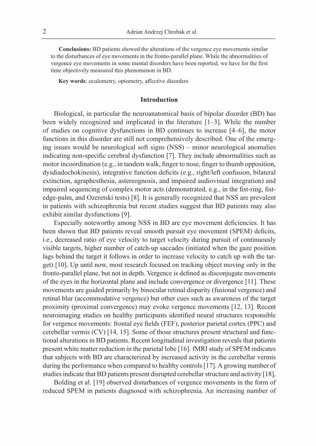
Stimuli were generated by custom design display, consisting of 128 LEDs, forming continuous 64 cm line, and three calibration LEDs placed on perpendicular display on one of the line’s ends (Figure 1). The angles of convergence for the closest and farthest LED were 66° and 5.6°, respectively. Stimuli display was synchronized with eye movement measurement by the optical connection.
Adrian Andrzej Chrobak et al.6
Calibration LEDs (3)
Vergence stimuli LEDs (128)
Eye movementsensor
Headrest
SubjectOptical
synchronization link
Calibration procedure (x3)
Time [
s]
Calibration stimulus [deg]
Stim
ulus –
dista
nce t
o the
eyes
[mm]
Time [s]
Convergence / divergence pocedure [x5]
Figure 1. Oculometric procedure. Left: picture presents vergence eye movement stimuli generator setup during the oculometric procedure. Upper right: calibration procedure.
Lower right: eye vergence stimulation
Oculometric procedure
The subject was seated in front of stimuli generator setup, with the head placed on a headrest. Eye movement sensor was placed on subject’s head, tested and adjust-ed. A position of stimuli setup was adjusted so the line of LEDs was visible with no obtrusions and placed perpendicularly to subject’s face with its tip touching subject’s nose. First LED position was 14 mm from the subject’s nose, while the actual distance to the eyes was measured. The subject was asked to fixate eyes on LED which was currently on, and to try to keep it as a single image. The stimuli started with three-point calibration procedure. Each of calibration LEDs, placed in horizontal axis on positions (0, ±12°), was lit for 3 seconds, and the cycle was repeated 3 times. Eye convergence stimulation started from the most distant LED of the line and proceeded with moving the light point along the line, with constant linear speed 37.6 mm/s. At the most extreme position light target stopped for 3.13 s and proceeded to move in opposite direction. Such stimulation cycle, consisting of convergence and divergence segments, was repeated five times.
7Vergence eye movements in bipolar disorder
Oculometric data processing
Horizontal eye position was calibrated according to three point calibration. Oculomet-ric parameters were calculated for 20 cm segments (60 to 260 mm from the eye surface) of target movement of each segment of the procedure. Every parameter was calculated separately for convergence (target moving towards the eyes) and divergence (target moving away from the eyes) segments. The calculations were made by custom software script.
Eye vergence signal
Slow eye vergence signal was calculated as a difference between the horizontal rotation of the left and right eye, passed through the low-pass filter. Blinks, saccades and fast vergence eye movements were detected automatically and excluded from calculation of the vergence signal parameters. Following parameters were calculated for slow vergence component:
ZPOSt – maximal eye convergence achieved;ZPOSv – maximal eye divergence achieved.ERRt – average error (difference) between eye convergence and convergence
required to fixate the target;ERRf – average error (difference) between eye divergence and divergence required
to fixate the target;GAINt – average ratio between eye convergence velocity and convergence velocity
required by target movement;GAINf – average ratio between eye divergence velocity and divergence velocity
required by target movement;GAINPt – average ratio between eye and target convergence velocity, excluding
fragments when eye vergence did not change according to stimuli change (fragments with average ratio between eye vergence velocity and vergence velocity required by target movement <0 excluded);
GAINPf – average ratio between eye and target divergence velocity, excluding fragments when eye vergence did not change according to stimuli change (fragments with average ratio between eye vergence velocity and vergence velocity required by target movement <0 excluded);
VERDt – average distance from the eye for which eye convergence changed ac-cordingly to stimuli change (average ratio between eye vergence velocity and vergence velocity required by target movement >0);
VERDf – average distance from the eye for which eye divergence changed accord-ingly to stimuli change (average ratio between eye vergence velocity and vergence velocity required by target movement >0);
VERCt – cumulative time during experiment segments for which eye convergence changed accordingly to stimuli change (average ratio between eye vergence velocity and vergence velocity required by target movement >0);
Adrian Andrzej Chrobak et al.8
VERCf – Cumulative time during experiment segments for which eye divergence changed accordingly to stimuli change (average ratio between eye vergence velocity and vergence velocity required by target movement >0);
Saccades and fast vergence movements
Fast eye movements and blinks were detected on the basis of velocity threshold (15°/s) and peak velocity (80°/s) criteria. Segments of oculomotor signal spanned between sub-threshold, local signal velocity minimum and exceeding peak velocity criteria were classified depending on amplitude, time and conjugacy between left and right eye signal. Fast conjugate, monotonic movements were considered as saccades, and non-conjugate, monotonic movements as fast vergence movements. Other fast signal changes were considered as artifacts (blinks). Saccades larger than 10 degrees were excluded from analysis, as associated with distractors, rather than visual pursuit task. Following parameters were calculated for fast saccades:
SACnAt – average amplitude of saccadic intrusions during convergence;SACnAf – average amplitude of saccadic intrusions during divergence;SACnCt – number of fast vergence movement intrusions during convergence;SACnCf – number of fast vergence movement intrusions during divergence;SACvAt – average amplitude of fast vergence intrusions during convergence;SACvAf – average amplitude of fast divergence intrusions during divergence;SACvCt – number of saccadic intrusions during tracking the target in convergence;SACvCf – number of saccadic intrusions during tracking the target in divergence;
Statistical analysis
All statistical analyses were performed with R software [30]. Demographical characteristics of examined groups were compared with Student’s T and Chi-square tests as appropriate. Normality and homogeneity of variance were examined using Shapiro-Wilk and Levene tests, respectively. Parameters from optometric and ocu-lometric examination were compared between the groups with the use of Student’s T-test. Power of T-test and effect size (Cohen’s d) were also calculated.
Additionally, these tests were used to investigate differences between BD patients with and without lithium, lamotrigine, valproic acid or antidepressant treatment. Daily dosages of antipsychotics were converted to equivalent of olanzapine according to Leucht et al. [31].
Principal component analysis (PCA) was performed on oculometric parameters which received at least medium effect size (Cohen’s d >0.5) in comparisons between BD and control groups: ERRt, ERRf, GAINPt, SACvCt, SACvCf, VERDt, ZPOSt.
The differences in PCA components between groups were analyzed with two-way ANOVA, with the factor of group (control group, BD group), factor of sex and group-sex interactions. In the case of an insignificant interaction effect, two-way ANOVA

9Vergence eye movements in bipolar disorder
ERRf
ERRt
GAINf
GAINPf
GAINPt
GAINt
MBF
MRF
ZPOSv
ZPOSt
VERDt
VERDf
VERCt
SACvCt
SACnCt
SACvAt
SACvCf
SACvAf
SACnAt
SACnCf
SACnAf
VERCf
HC
* power = 0.64
* power = 0.54
* power = 0.51
BD
mediumsmall smallnegligiblemediumEffect size (Cohen’s d)
Para
meter
Figure 2. Results of optometric and oculometric parameters comparison between healthy controls (HC) and bipolar disorder (BD) groups
Bars’ directions present means differences. Bars on the left side present parameters for which mean values were higher in HC group, and bars on the right side present those higher in BD group.ERRt – average error between eye convergence and convergence required to fixate the target; ERRf – average error between eye divergence and divergence required to fixate the target; GAINt – average ratio between eye convergence velocity and convergence velocity required by target movement; GAINf – average ratio between eye divergence velocity and divergence velocity required by target movement; GAINPt – average ratio between eye and target convergence velocity, excluding fragments when eye vergence did not change according to stimuli change (fragments with average ratio between eye vergence velocity and vergence velocity required by target movement <0 excluded); GAINPf – average ratio between eye and target divergence velocity, excluding fragments when eye vergence did not change according to stimuli change (fragments with average ratio between eye vergence velocity and vergence velocity required by target movement <0 excluded); VERDt – average distance from the eye for which eye convergence changed accordingly to stimuli change (average ratio between eye vergence velocity and vergence velocity required by target movement >0); VERDf – average distance from the eye for which eye divergence changed accordingly to stimuli change (average ratio between eye vergence velocity and vergence velocity required
Adrian Andrzej Chrobak et al.10
by target movement >0); VERCt – cumulative time during experiment segments for which eye convergence changed accordingly to stimuli change (average ratio between eye vergence velocity and vergence velocity required by target movement >0); VERCf – Cumulative time during experiment segments. for which eye divergence changed accordingly to stimuli change (average ratio between eye vergence velocity and vergence velocity required by target movement >0); SACnAt – average amplitude of saccadic intrusions during convergence; SACnAf – average amplitude of saccadic intrusions during divergence; SACnCt – number of fast vergence movement intrusions during convergence; SACnCf – number of fast vergence movement intrusions during divergence; SACvAt – average amplitude of fast vergence intrusions during convergence; SACvAf – average amplitude of fast divergence intrusions during divergence; SACvCt – number of saccadic intrusions during tracking the target in convergence; SACvCf – number of saccadic intrusions during tracking the target in divergence; ZPOSt – maximal eye convergence achieved; ZPOSv – maximal eye divergence achieved
model without group-sex interaction was analyzed. Pearson’s r correlation coefficients were used to evaluate associations between equivalent olanzapine daily doses and oculometric parameters
Results
Comparisons between control and BD groups performed on all optometric and oculometric measures revealed significant differences, with at least medium effect size and power of test greater than 0.5 in three parameters: (1) average error (difference) between eyes’ convergence and convergence required to fixate the target (Student’s T-test, p < 0.05; Cohen’s d = 0.65; power = 0.63); (2) number of saccadic intrusions during tracking the target in convergence (p < 0.05; Cohen’s d = 0.56; power = 0.51); (3) number of saccadic intrusions during tracking the target in divergence (p < 0.05; Cohen’s d = 0.58; power = 0.54) (Figure 2).
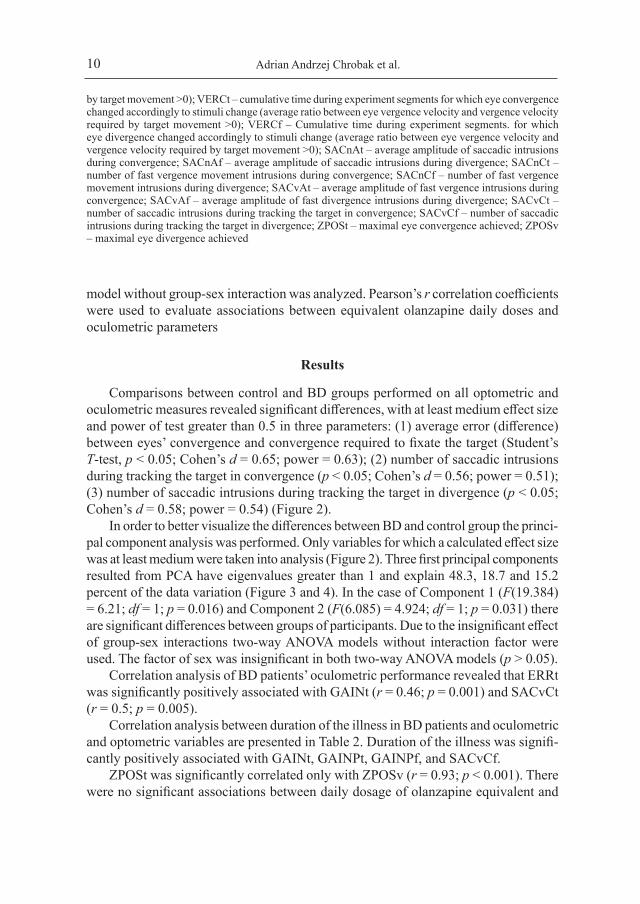
In order to better visualize the differences between BD and control group the princi-pal component analysis was performed. Only variables for which a calculated effect size was at least medium were taken into analysis (Figure 2). Three first principal components resulted from PCA have eigenvalues greater than 1 and explain 48.3, 18.7 and 15.2 percent of the data variation (Figure 3 and 4). In the case of Component 1 (F(19.384) = 6.21; df = 1; p = 0.016) and Component 2 (F(6.085) = 4.924; df = 1; p = 0.031) there are significant differences between groups of participants. Due to the insignificant effect of group-sex interactions two-way ANOVA models without interaction factor were used. The factor of sex was insignificant in both two-way ANOVA models (p > 0.05).
Correlation analysis of BD patients’ oculometric performance revealed that ERRt was significantly positively associated with GAINt (r = 0.46; p = 0.001) and SACvCt (r = 0.5; p = 0.005).
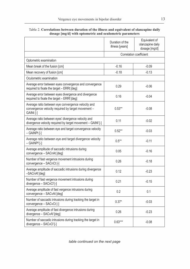
Correlation analysis between duration of the illness in BD patients and oculometric and optometric variables are presented in Table 2. Duration of the illness was signifi-cantly positively associated with GAINt, GAINPt, GAINPf, and SACvCf.
ZPOSt was significantly correlated only with ZPOSv (r = 0.93; p < 0.001). There were no significant associations between daily dosage of olanzapine equivalent and
11Vergence eye movements in bipolar disorder
1.0
1.0
0.5
0.5
–0.5
–0.5–1.0
–1.0
0.0
0.0
Comp. 1, 48.3%
Comp
. 2, 1
8.7%
ZPOSt
VERDt
ERRf
ERRt
GAINPt
SACvCt
SACvCf
Figure 3. Principal component analysis: relations between loadings and first two principal components
ERRt – average error between eye convergence and convergence required to fixate the target; ERRf – average error between eye divergence and divergence required to fixate the target; GAINPt – average ratio between eye and target convergence velocity, excluding fragments when eye vergence did not change according to stimuli change (fragments with average ratio between eye vergence velocity and vergence velocity required by target movement <0 excluded); GAINPf – average ratio between eye and target divergence velocity, excluding fragments when eye vergence did not change according to stimuli change (fragments with average ratio between eye vergence velocity and vergence velocity required by target movement <0 excluded); ZPOSt – maximal eye convergence achieved; VERDt – average distance from the eye for which eye convergence changed accordingly to stimuli change (average ratio between eye vergence velocity and vergence velocity required by target movement >0); SACvCt – number of saccadic intrusions during tracking the target in convergence; SACvCf – number of saccadic intrusions during tracking the target in divergence

Adrian Andrzej Chrobak et al.12
HC BD
2
0
-2
-2.5 0.0 2.5 5.0
Comp
2 (1
8.7%
)
Comp 1 (48.3%)
Figure 4. Scatterplot of two first components of the principal component analysis performed on the selected oculometric parameters
Ellipses show 95% confidence interval per each groupBD – bipolar disorderHC – healthy controls
any of the optometric or oculometric parameter. Comparison of evaluated parameters between BD patients with and without lithium, lamotrigine, valproic acid or antide-pressant treatment revealed no statistically significant differences.
13Vergence eye movements in bipolar disorder
table continued on the next page
Table 2. Correlations between duration of the illness and equivalent of olanzapine daily dosage [mg/d] with optometric and oculometric parameters
Duration of the illness [years]
Equivalent of olanzapine daily dosage [mg/d]
Correlation coefficientOptometric examinationMean break of the fusion [cm] -0.16 -0.09Mean recovery of fusion [cm] -0.18 -0.13Oculometric examinationAverage error between eyes convergence and convergence required to fixate the target – ERRt [deg] 0.29 -0.06
Average error between eyes divergence and divergence required to fixate the target – ERRf [deg] 0.16 -0.04
Average ratio between eye convergence velocity and convergence velocity required by target movement – GAINt [-]
0.53** -0.08
Average ratio between eyes’ divergence velocity and divergence velocity required by target movement – GAINf [-] 0.11 -0.02
Average ratio between eye and target convergence velocity – GAINPt [-] 0.52** -0.03
Average ratio between eye and target divergence velocity – GAINPf [-] 0.5** -0.11
Average amplitude of saccadic intrusions during convergence – SACnAt [deg] 0.05 -0.16
Number of fast vergence movement intrusions during convergence – SACnCt [-] 0.26 -0.18
Average amplitude of saccadic intrusions during divergence –SACnAf [deg] 0.12 -0.23
Number of fast vergence movement intrusions during divergence – SACnCf [-] 0.21 -0.15
Average amplitude of fast vergence intrusions during convergence – SACvAt [deg] 0.2 0.1
Number of saccadic intrusions during tracking the target in convergence – SACvCt [-] 0.37* -0.03
Average amplitude of fast divergence intrusions during divergence – SACvAf [deg] 0.26 -0.23
Number of saccadic intrusions during tracking the target in divergence – SACvCf [-] 0.63*** -0.08
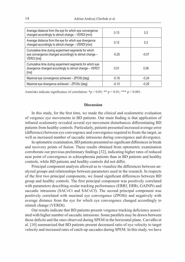
Adrian Andrzej Chrobak et al.14
Average distance from the eye for which eye convergence changed accordingly to stimuli change – VERDt [mm] 0.13 0.2
Average distance from the eye for which eye divergence changed accordingly to stimuli change – VERDf [mm] 0.12 0.2
Cumulative time during experiment segments for which eye convergence changed accordingly to stimuli change – VERCt [ms]
-0.25 -0.07
Cumulative time during experiment segments for which eye divergence changed accordingly to stimuli change – VERCf [ms]
0.01 0.06
Maximal eye convergence achieved – ZPOSt [deg] -0.16 -0.24Maximal eye divergence achieved – ZPOSv [deg] -0.15 -0.29
Asterisks indicate significance of correlation: *p < 0.05; ** p < 0.01; *** p < 0.001.
Discussion
In this study, for the first time, we made the clinical and oculometric evaluation of vergence eye movements in BD patients. Our main finding is that application of infrared oculometry revealed several eye movement disturbances differentiating BD patients from healthy controls. Particularly, patients presented increased average error (difference) between eye convergence and convergence required to fixate the target, as well as increased number of saccadic intrusions during convergence and divergence.
In optometric examination, BD patients presented no significant differences in break and recovery points of fusion. These results obtained from optometric examination corroborate our previous preliminary findings [32], indicating higher rates of reduced near point of convergence in schizophrenia patients than in BD patients and healthy controls, while BD patients and healthy controls did not differ.
Principal component analysis allowed us to visualize the differences between an-alyzed groups and relationships between parameters used in the research. In respects of the first two principal components, we found significant differences between BD group and healthy controls. The first principal component was positively correlated with parameters describing ocular tracking performance (ERRf, ERRt, GAINPt) and saccadic intrusions (SACvCt and SACvCf). The second principal component was positively correlated with maximal eye convergence (ZPOSt) and negatively with average distance from the eye for which eye convergence changed accordingly to stimuli change (VERDt).
Our results indicate that BD patients present vergence tracking deficiency associ-ated with higher number of saccadic intrusions. Some parallels may be drawn between those deficits and the ones observed during SPEM in the horizontal plane. Carvalho et al. [10] summarized that BD patients present decreased ratio of eye velocity to target velocity and increased rates of catch-up saccades during SPEM. In this study, we have

15Vergence eye movements in bipolar disorder
shown that the number of saccadic intrusions is correlated with ERRt, as well as with GAINPt and GAINPf. Thus, we believe that higher number of saccadic intrusions during vergence tracking is equivalent to catch-up saccades during SPEM. As the catch-up saccades are used to compensate for poor smooth pursuit gain [19] both processes are intertwined, therefore it may be the reason they have a similar influence on the first principal component. An observation that these components differentiate BD patients from the healthy controls indicates that deficits observed during SPEM in BD patients are not specific to horizontal but are also present in fronto-parallel plane.
Neuroimaging studies are required to evaluate which brain structures are associated with vergence eye movement dysfunction in BD. However, with all the data available, it is possible that cerebellar vermis, due to its role in vergence tasks [14, 15, 33, 34] and its abnormal functioning during SPEM in BD patients [17], may also play a role in vergence eye movement dysfunctions in BD. Future studies should address asso-ciations between SPEM deficits during vergence tracking in BD and structural and functional deficits of brain regions responsible for vergence tasks: frontal eye fields (FEF), posterior parietal cortex (PPC) and cerebellar vermis [14, 15].
For every difference in vergence eye movements between BD patients and healthy controls, it must be noted that the examined patients were under the effect of psy-chopharmacological treatment, receiving atypical antipsychotics, mood stabilizers and/or antidepressants. To our best knowledge, there are no studies evaluating the effect of this treatment on vergence eye movements in BD. In the case of patients with schizophrenia, Bolding et al. [19] showed no differences in convergence eye movements between patients taking antidepressants or anticholinergic medication and patients without those medications. Flechtner et al. [35] showed no impact of antipsychotic medication on SPEM performance of patients with major depressive disorder. In schizophrenia patients, long-term antipsychotic medication use has been associated with impaired SPEM gain [36, 37]. Thus, we cannot rule out the possible effect of medication on vergence eye movements. In our study, we have found no statistically significant associations between daily doses of olanzapine equivalents and any of assessed oculometric and optometric parameters. We suppose that atypical neuroleptics chosen in our study may have minimal effect on patients’ oculomotor performance. Due to the potential effect of lithium on SPEM [10] we have compared the performance of BD patients taking and not taking lithium. We have found no difference between these two groups in any of the parameters evaluated in the study. Some studies have shown an effect of antidepressants on oculomotor performance [38, 39], while other studies have reported no effect of this treatment on reaction times or error rates in depression [10, 35, 40]. In our study, we have shown no significant differences between BD patients taking antidepressant medication and those not treated with such medication. Neither we have found any differences between BD patients treated ant not treated with lamotrigine or valproic acid treatment. However additional studies are needed to investigate the effect of psychiatric treatments on vergence eye movements and binocular vision.
Adrian Andrzej Chrobak et al.16
We have shown that there are positive associations between duration of the illness and the disturbances of vergence eye movements (GAINt, GAINPt, GAINPf, SACvCt, SACvCf). One possible explanation is that those deficits are associated with the progres-sion of the illness. The growing number of studies revealed that BD patients develop neuroanatomic abnormalities such as ventricular enlargement and grey matter loss. It has been shown that BD patients present a progressive decline of cerebellar gray matter density [41]. Brambilla et al. [42] showed the inverse correlation between cer-ebellar vermis volume and number of BD episodes. Longitudinal investigation reveals that patients present white matter reductions in the parietal lobe [16]. Both mentioned structures, declining in the course of BD, are associated with vergence eye movements.
Second possible explanation is the effect of chronic medication. Hutton et al. showed that SPEM gain is unaffected by short-term antipsychotic medication but it is worsened by chronic treatment in the group of patients with schizophrenia. However, other oculometric parameters, like catch-up saccades, are adversely influenced by ill-ness duration rather than medication. Thus, further studies are necessary to disseminate effects of the illness progression and treatment on vergence eye movements in BD.
We are aware of limitations of this study, such as relatively small subject sample, performing examination at different times of the day (between 8 am and 8 pm) and the fact that the group of patients was not drug naïve. On the other hand, the strength of our study is thorough optometric examination which enabled us to evaluate vergence eye movements in the group of participants without visual and oculomotor disturbances, which could significantly affect our results. Conversion of atypical antipsychotics’ doses to olanzapine equivalents enabled us to partially evaluate the effect of treatment on vergence eye movements.
Conclusions
In our study, we have confronted the optometric examination of vergence eye movements with oculometric examination of SPEM during vergence in the group of BD patients and healthy controls. While in the case of optometric parameters patients seem not to differ from healthy controls, application of oculometry reveal several alterations of vergence eye movements, namely: increased average error (difference) between eye convergence and convergence required to fixate the target and increased number of saccadic intrusions during tracking the target in convergence and divergence. Application of multivariate analysis revealed the presence of the components differ-entiating BD patients from the healthy controls, indicating disrupted ocular-tracking performance and increased saccadic intrusions in the group of patients.
AcknowledgementThe study financed from budget funds for science in years 2015–2019, as a research project being a part of “Diamond Grant” (0112/DIA/2015/44) sponsored by the Ministry of Science and Higher Education, Republic of Poland.
17Vergence eye movements in bipolar disorder
References
1. Strakowski SM, Delbello MP, Adler CM. The functional neuroanatomy of bipolar disorder: A review of neuroimaging findings. Mol. Psychiatry. 2005; 10(1): 105–116.
2. Strakowski SM, Adler CM, Almeida J, Altshuler LL, Blumberg HP, Chang KD et al. The function-al neuroanatomy of bipolar disorder: A consensus model. Bipolar Disord. 2012; 14(4): 313–325.
3. Wilczyńska K, Simonienko K, Konarzewska B, Szajda SD, Waszkiewicz N. Morphological changes of the brain in mood disorders. Psychiatr. Pol. 2018; 52(5): 797–805.
4. Tsitsipa E, Fountoulakis KN. The neurocognitive functioning in bipolar disorder: A systematic review of data. Ann. Gen. Psychiatry. 2015; 14(1): 1–29.
5. Henin A, Micco JA, Wozniak J, Briesch JM, Narayan AJ, Hirshfeld-Becker DR et al. Neurocog-nitive functioning in bipolar disorder. Clin. Psychol. Pract. 2009; 16(2): 231–250.
6. Chrobak AA, Jeziorko S, Tereszko A, Janeczko W, Arciszewska A, Siuda-Krzywicka K et al. Mental rotation task in bipolar disorder. Psychiatr. Pol. 2018; 52(5): 807–817.
7. Dimitri-Valente G, Rigucci S, Manfredi G, Girardi P, Ferracuti S. Neurological soft signs: sig-nificato e rilevanza nel corsodella patologia psichiatrica. Uno screening obiettivo veloce per psicosi? Riv. Psichiatr. 2012; 47(6): 465–478.
8. Buchanan RW, Heinrichs DW. The Neurological Evaluation Scale (NES): A structured in-strument for the assessment of neurological signs in schizophrenia. Psychiatry. Res. 1989; 27(3): 335–350.
9. Chrobak AA, Siwek GP, Siuda-Krzywicka K, Arciszewska A, Starowicz-Filip A, Siwek M et al. Neurological and Cerebellar Soft Signs do not discriminate schizophrenia from bipolar disorder patients. Prog. Neuropsychopharmacology Biol. Psychiatry. 2015; 64: 96–101.
10. Carvalho N, Laurent E, Noiret N, Chopard G, Haffen E, Bennabi D et al. Eye movement in unipo-lar and bipolar depression: A systematic review of the literature. Front. Psychol. 2015; 6: 1–19.
11. Purves D, Augustine GJ, Fitzpatrick D, Hall W, LaMantia A-S, McNamara J et al. Neural con-trol of vergence movements. In: Purves D, Augustine GJ, Fitzpatrick D, Hall W, LaMantia A-S, McNamara J et al., editors. Neuroscience. Sunderland, MA: Sinauer Associates; 2001. P. 466.
12. Frey J, Ringach DL. Binocular eye movements evoked by self-induced motion parallax. J. Neurosci. 2011; 31(47): 17069–17073.
13. Leigh RJ, Zee DS. Vergence eye movements. In: Leigh RJ, Zee DS. The neurology of eye move-ments, 5th ed. Oxford–New York: Oxford University Press; 2015. p. 520-568.
14. Alvarez TL, Alkan Y, Gohel S, Douglas Ward B, Biswal BB. Functional anatomy of predictive vergence and saccade eye movements in humans: A functional MRI investigation. Vision Res. 2010; 50(21): 2163–2175.
15. Alvarez TL, Jaswal R, Gohel S, Biswal BB. Functional activity within the frontal eye fields, posterior parietal cortex, and cerebellar vermis significantly correlates to symmetrical ver-gence peak velocity: An ROI-based, fMRI study of vergence training. Front. Integr. Neurosci. 2014; 8: 50.
16. Ferro A, Bonivento C, Delvecchio G, Bellani M, Perlini C, Dusi N et al. Longitudinal investiga-tion of the parietal lobe anatomy in bipolar disorder and its association with general functioning. Psychiatry Res. Neuroimaging. 2017; 267: 22–31.
17. Martin LF, Olincy A, Ross RG, Du YP, Singel D, Shatti S et al. Cerebellar hyperactivity during smooth pursuit eye movements in bipolar disorder. J. Psychiatr Res. 2011; 45(5): 670–677.
18. Chrobak AA, Siuda K, Tereszko A, Siwek M, Dudek D. Zaburzenia psychiczne a struktura i funkcje móżdżku – przegląd najnowszych badań. Psychiatria. 2014; 11(1): 1–8.

Adrian Andrzej Chrobak et al.18
19. Bolding MS, Lahti AC, White D, Moore C, Gurler D, Gawne TJ et al. Vergence eye movements in patients with schizophrenia. Vision Res. 2014; 102: 64–70.
20. Ivleva EI, Morris DW, Moates AF, Suppes T, Thaker GK, Tamminga CA. Genetics and inter-mediate phenotypes of the schizophrenia – bipolar disorder boundary. Neurosci. Biobehav. Rev. 2010; 34(6): 897–921.
21. Ivleva EI, Moates AF, Hamm JP, Bernstein IH, O’Neill HB, Cole D et al. Smooth pursuit eye movement, prepulse inhibition, and auditory paired stimuli processing endophenotypes across the schizophrenia-bipolar disorder psychosis dimension. Schizophr. Bull. 2014; 40(3): 642–652.
22. Birur B, Kraguljac NV, Shelton RC, Lahti AC. Brain structure, function, and neurochemistry in schizophrenia and bipolar disorder – A systematic review of the magnetic resonance neuro-imaging literature. NPJ Schizophr. 2017; 3(1): 15.
23. Rybakowski JK. Meaningful aspects of the term ‘mood stabilizer’. Bipolar Disord. 2018; 20(4): 391–392.
24. Benjamin WJ. Borish’s clinical refraction. St Louis, Mo: Butterworth-Heinemann, Elsevier; 2006. P. 1694.
25. Maples WC, Hoenes R. Near point of convergence norms measured in elementary school chil-dren. Optom. Vis. Sci. 2007; 84(3): 224–228.
26. Ostadimoghaddam H, Hashemi H, Nabovati P, Yekta A, Khabazkhoob M. The distribution of near point of convergence and its association with age, gender and refractive error: A popula-tion-based study. Clin. Exp. Optom. 2017; 100(3): 255–259.
27. Scheiman M, Gallaway M, Frantz KA, Peters RJ, Hatch S, Cuff M et al. Nearpoint of convergence: Test procedure, target selection, and normative data. Optom. Vis. Sci. 2003; 80(3): 214–225.
28. Ober J. Infra-red reflection technique. In: Ygge J, Lennerstrand G, editors. Eye movements in reading. Oxford, UK–Tarrytown, NY, USA: Pergamon Press; 1994. P. 9–21.
29. Eggert T. Eye movement recordings: Methods. In: Straube A, Büttner U, editors. Neuro-ophthal-mology: Neuronal control of eye movements. Basel–New York: Karger Medical and Scientific Publishers; 2007. P. 15–34.
30. RCoreTeam. R: A language and environment for statistical computing [Internet]. Vienna, Austria: R Foundation for Statistical Computing; 2017.
31. Leucht S, Samara M, Heres S, Patel MX, Furukawa T, Cipriani A et al. Dose equivalents for second-generation antipsychotic drugs: The classical mean dose method. Schizophr. Bull. 2015; 41(6): 1397–1402.
32. Chrobak AA, Siuda K, Biela M, Arciszewska A, Siwek M, Pilecki MW et al. Convergence insufficiency with unilateral exophoria at near in schizophrenia and bipolar disorder – a pre-liminary study. Psychiatr. Pol. 2014; 48(6): 1143–1154.
33. Alkan Y, Biswal B, Han SJ, Alvarez TL. Cortical location of saccadic and vergence oculomotor learning revealed using fMRI. EngineeringNjitEdu.: 7–8.
34. Alkan Y, Biswal BB, Taylor PA, Alvarez TL. Segregation of frontoparietal and cerebellar components within saccade and vergence networks using hierarchical independent component analysis of fMRI. Vis. Neurosci. 2011; 28(3): 247–261.
35. Flechtner K-M, Steinacher B, Sauer R, Mackert A. Smooth pursuit eye movements of patients with schizophrenia and affective disorder during clinical treatment. Eur. Arch. Psychiatry Clin Neurosci. 2002; 252(2): 49–53.
36. Hutton S, Crawford T, Gibbins H, Cuthbert I, Barnes T, Kennard C et al. Short and long term effects of antipsychotic medication on smooth pursuit eye tracking in schizophrenia. Psycho-pharmacology (Berl). 2001; 157(3): 284–291.
19Vergence eye movements in bipolar disorder
37. Lencer R, Sprenger A, Harris MSH, Reilly JL, Keshavan MS, Sweeney JA. Effects of sec-ond-generation antipsychotic medication on smooth pursuit performance in antipsychotic-naïve schizophrenia. Arch. Gen. Psychiatry. 2008; 65(10): 1146–1154.
38. Green JF, King DJ, Trimble KM. Antisaccade and smooth pursuit eye movements in healthy subjects receiving sertraline and lorazepam. J. Psychopharmacol. 2000; 14(1): 30–36.
39. Morrens M, Wezenberg E, Verkes RJ, Hulstijn W, Ruigt GSF, Sabbe BGC. Psychomotor and memory effects of haloperidol, olanzapine, and paroxetine in healthy subjects after short-term administration. J. Clin. Psychopharmacol. 2007; 27(1): 15–21.
40. Katsanis J, Kortenkamp S, Iacono WG, Grove WM. Antisaccade performance in patients with schizophrenia and affective disorder. J. Abnorm. Psychol. 1997; 106(3): 468–472.
41. Moorhead TWJ, McKirdy J, Sussmann JED, Hall J, Lawrie SM, Johnstone EC et al. Progres-sive gray matter loss in patients with bipolar disorder. Biol. Psychiatry. 2007; 62(8): 894–900.
42. Brambilla P, Harenski K, Nicoletti M, Mallinger AG, Frank E, Kupfer DJ et al. MRI study of posterior fossa structures and brain ventricles in bipolar patients. J. Psychiatr. Res. 2001; 35(6): 313–322.
Address: Adrian Andrzej ChrobakDepartment of Adult PsychiatryJagiellonian University Medical College31-501 Kraków, Kopernika Street 21Ae-mail: [email protected]


![Learning of Active Binocular Vision in a Biomechanical ... · in vergence eye movements obeying Sherrington's law of reciprocal innervation [12]: as one eye muscle contracts, its](https://static.fdocuments.net/doc/165x107/6043d13a7e683d066b3fc5fa/learning-of-active-binocular-vision-in-a-biomechanical-in-vergence-eye-movements.jpg)
