Penanganan Ventricular Tachycardia & Ventricular Fibrillation

Brit. Heart J., 1967, 29, 533.
Ventricular Structure and Function After RadicalCorrection of the Tetralogy of Fallot
K. A. HALLIDIE-SMITH, M. DULAKE, M. WONG*, C. M. OAKLEYt, AND J. F. GOODWIN
From the Department of Medicine (Unit of Clinical Cardiology), Postgraduate Medical School of London, and Ham-mersmith Hospital, London W.12
Total, or radical, surgical correction of the tetra-logy of Fallot has now become standard practice.Mortality figures differ widely and vary from nonein a series of 41 patients (Malm et al., 1963) tobetween 30 and 40 per cent in other hands. In1960 Kirklin et al., reported a 17 per cent mortalityin 91 patients, and in 1961 Barnard and Schrirepublished a similar mortality in 42 patients, thoughBahnson et al. (1962) recorded a 30 per cent mortal-ity in 147 patients. In experienced hands todaythe mortality should be under 20 per cent.The main hazards of the operation relate to un-
usual intracardiac deformity, complete heart block,post-operative pulmonary cedema and insufficiency,htmorrhagic problems, and incomplete correctionof the lesions. Although complete heart block hasbecome much less common with increased surgicalexpertise, the other hazards remain, and are appreci-ably greater in severely cyanotic patients with highheematocrit values, and particularly in those with ahypoplastic right ventricular outflow tract, smallpulmonary valve ring, or absent crista supra-ventricularis (bulboventricular defect).The object of this paper is to present in detail the
results of investigation in 24 patients following radi-cal correction. No attempt will be made to analysethe results of operation in detail in the entire series,for these results will subsequently be published infull, but brief comments will be made in order toprovide a background for the detailed studies onthis smaller group.
Since the operation for radical correction is acomplex one, and since it involves a large ventricu-lotomy, we felt it important to investigate fully the
Received Se tember 28, 1966.* On study ieave from Cardiovascular Research Institute,
San Francisco, California, U.S.A.t Supported by the British Heart Foundation.
5
subsequent structure and function of the heart inpatients who survived the operation, with specialreference to long-term prognosis.
PATIENTS STUDIED AND TECHNIQUE USEDBetween 1958 and 1965, 133 patients were treated by
radical correction, by Mr. W. P. Cleland and ProfessorH. H. Bentall, under cardiopulmonary bypass directedby Dr. Denis Melrose. Between 1962 and 1965, 32patients were treated by closed infundibular resection,as a preliminary to radical correction at a subsequentlater stage.
In view ofthe greater risks in patients with high haema-tocrit (over 65%), unusual anatomy (bulboventriculardefect, or double outlet right ventricle), or small bodysize, a policy of two-stage correction for such patientshas been evolved, closed infundibular resection beingfollowed 6 months to 2 years later by closure of theventricular septal defect.
All patients have been investigated before operationby selective right ventricular angiocardiography and themajority by full right heart catheterization also. Specialattention has been paid to the position of the aorticroot and pulmonary artery, the site and size of the ven-tricular septal defect, the presence of the crista in lateraland frontal projections, and the size of the pulmonaryvalve ring. The detailed analysis and the correlationof angiographic and surgical anatomy have been reportedby Smith et al. (1965).
Studies were made on 24 patients 1 month to 6 yearsafter radical correction; 21 of these had been treated inone stage, and 3 in two stages.The indications for investigation were varied. Four
patients were studied because their post-operative con-dition caused anxiety and suggested pertsisence of theventricular septal defect, severe pulmonary incompe-tence, or considerable residual pulmonary stenosis.The other patients, who were dramatically improved,did not give rise to anxiety, and were investigated inorder to assess long-term prognosis.
533
Hallidie-Smith, Dulake, Wong, Oakley, and Goodwin
Right heart catheterization was performed in all 24patients, and in 17 selective right ventricular angio-cardiography was carried out. In 10 patients, all ofwhom had a strictly intact ventricular septum, ventricu-lar volumes were estimated by the thermodilution tech-nique (Rapaport et al., 1965). In 5 patients, studies weremade before and after isoprenaline infusion, to assessthe performance of the right ventricle under stress.
RESULTS
Over-all Results of OperationsThe over-all results of surgical treatment may be
divided into two phases. Phase 1, between 1958and 1962, was characterized by one-stage proceduresonly, and included early inexperience before thepresent skill in surgical and pre- and post-operativemanagement had been acquired. Not surprisingly,the mortality was 38 per cent in the first 65 patients,deaths occurring most frequently in the severelycyanotic patients. In the second phase, whenseverely cyanosed patients and those with ana-tomical variants (as defined above) were rejectedfor one-stage correction, the mortality of one-stagecorrection was only 19 per cent in 56 patients.
In all, there were 36 deaths in 133 one-stageradical corrections. Of these 36, 7 had open(persistent) ventricular septal defects, and deathoccurred in the early post-operative period; 2 latedeaths occurred, one after a second operation toclose the defect, and one from bacterial infectionof the heart.Of the survivors, all had improvement in effort
tolerance and reduction in cyanosis, both of whichwere usually dramatic. Persistence of the ventri-cular septal defect was suspected in 25 patients, butwas clinically unimportant in all except 4 patients.Some degree of pulmonary valve incompetence wassuspected in about half the patients on the basis ofan early diastolic murmur at the left sternal edge,but it was clinically important in only three.
Clinically satisfactory signs were the disappear-ance of cyanosis and dyspncea (hyperventilation);a soft short pulmonary systolic murmur, audible(though delayed) pulmonary valve closure, and asoft or absent pulmonary diastolic murmur. Thejugular venous pressure was raised in at least 50per cent ofpatients for up to one year after operation,and this will be discussed fully later.
Full data on causes of death, incidence of heartblock, and clinical status of all surviving patientswill be published elsewhere. The foregoing figuresare given to set the stage for the analysis of theresults of special investigation in the 24 patientsdescribed here.The results of infundibular resection (first stage
of two-stage procedure) will also be reported in fullelsewhere. Of 32 patients, 3 died, 2 during sub-sequent second stage procedures; 8 other patientsdid well after the second stage and the remainderhave not yet had the second stage.
Results of Investigation in 24 Patientsafter Radical Correction
Surgical Anatomy. Stenosis of the outflow tractof the right ventricle was present in all patients andwas solely valvar in 4, solely infundibular in 9, andcombined in 11. Of the 4 patients with lonevalvar stenosis, 3 had an uncommonly small valvering; 2 had a bulboventricular defect, and one had asupracristal ventricular septal defect.The ventricular septal defect was large and occu-
pied the usual site extending from the crista supra-ventricularis to the septal cusp of the tricuspidvalve in 18 patients. The size ranged from 4 x 2cm. to 15 x 2 cm. Six patients had unusual de-fects which were subcristal in 2 and bulboventricu-lar (with absent or split crista) in 3, while the re-maming patient had an infracristal defect with anadditional left ventricular-right atrial shunt and acleft tricuspid valve suggesting a partial form ofatrio-ventricular canal (Case 5).
Clinical Status after Operation. Dyspncea wasmuch less severe than before operation and, in allexcept 3 patients with open defects or severe pul-monary incompetence, disappeared entirely. Cyan-osis was abolished in all patients except one inwhom it appeared only on effort.
After operation, the systolic murmur due to pul-monary stenosis was grade 4 (out of 4) in 6 patients,grade 3 in 15, grade 2 in 2, and grade 1 in onepatient. The last of these had virtual pulmonaryatresia and was operated upon in 2 stages: after the -first stage the systolic murmur increased. Themurmur was accompanied by a thrill in 11 patients;it tended to diminish in intensity near the secondheart sound, and never extended past aortic valveclosure.
After operation the murmur was grade 4 in 4patients, grade 3 in 13 patients, grade 2 in 6 patients,and grade 1 in one patient (Fig. 1). A thrill waspresent in only 4 patients, and in 2 patients theinitially loud murmur became softer with the pas-sage of several months. The systolic murmur wasfull length in 2 patients who had a left-to-rightshunt through a persistent ventricular septal defect(Fig. 2). Of the 4 patients with a grade 4 murmur,3 had left-to-right shunts, and 2 had residualobstruction to right ventricular outflow, (76, 68,and 45 mm. Hg gradients, respectively); 3 of these
534

Radical Correction of Tetralogy of Fallot
Pre-op. 18.9.61 Post-op. 10.5.65
A2PA A2 P2HF PA
A%szS 4 *$ i',, 0 f _
MF
MA
HF
P2 0C:6 sec. after A2
Fully satistactory result
FIG. 1.-Phonocardiogram before and after radical correction in Case 2. The systolic murmur had almostdisappeared and pulmonary closure was audible after operation. PA= pulmonary area; MF = medium fre-quency; HF = high frequency; MA= mitral area; P2 = pulmonary valve closure; A2 = aortic valve closure.
Pre-op. 8.8.60 Post-op. 1.2.61
*I :
}F -t- F PA HF
LSE HE ,
Open defect; residual obstruction; pulmonary incompetence
FIG. 2.-Phonocardiogram of Case 12 with open defect, residual obstruction, and pulmonary incompetence.After operation the pansystolic murmur due to the left-to-right shunt at ventricular level and the faint earlydiastolic murmur were recorded. Pulmonary closure was not seen. PA= pulmonary artery; LSE = left
sternal edge; HF=high frequency.
535

Hallidie-Smith, Dulake, Wong, Oakley, and Goodwin
TABLE IPOST-OPETRATIVE JUGULAR VENOUS PRESSURE
Case No. Age Jugular Rt. vent. Pulmonary Open VSD Jugular venous pressure(yr.) venous outflow tract incompetence Pulmonary
pressure (cm. gradient systemic flowabove S.A.) (mm. Hg) ratio
1 11 +6 0 0 0 Later normal2 9 + 6 0 0 0 Later normal3 12 +8 85 0 0 Later normal
2-stage correction4 19 +6 23 + 0 Later normal
2-stage correction5 11 +5 0 + 1-3:1 Later normal6 13 +6 0 + 0 Later fell to + 4 cm.7 34 +8 0 + + 0 Retained + 8 cm.8 12 +4 0 + 0 Later normal9 11 + 8 76 0 R-'-L Retained high10 11 +6 0 0 0 Later normal11 7 + 6 0 + + 0 Retained high12 7 + 8 45 + 1-6:1 Retained high13 11 +7 80 + 0 Retained high14 13 + 5 50 0 1-3:1 Later normal15 11 + 5 68 0 "'Small" Retained normal16 13 0 47 0 017 14 0 54 + 0 2-stage correction18 8 0 0 + 019 12 0 23 + 020 12 0 0 + 021 13 0 23 + 022 9 0 23 + 023 10 0 35 + 024 13 0 30 + 0
patients had pulmonary incompetence which wouldbe expected to increase the stroke output of theventricle and thus enhance the systolic murmur.Of the 13 patients with a grade 3 murmur, 6 hadpulmonary incompetence which was severe in one,one had a left-to-right shunt, and one a right-to-left shunt. A gradient over 20 mm. Hg across theright ventricular outflow tract was present in 7 of the13 patients, but was appreciable in only 4 (47, 50,76, 80 mm. Hg gradients, respectively). Thegradient of 76 mm. Hg was associated with a right-to-left shunt. Two patients had neither pulmonaryincompetence, a gradient, nor a shunt. Thus aloud residual systolic murmur usually indicated somedegree of residual obstruction, and was exaggeratedby pulmonary incompetence and a persistent shunt,but was compatible with a fully satisfactory result.The significance of the murmur could not be reli-ably assessed in the presence of pulmonary incom-petence. A loud murmur immediately after opera-tion was compatible with residual obstruction whichmight resolve, as suggested in the two patientswithout a shunt or pulmonary incompetence inwhom subsequent catheterization revealed nogradient.An early diastolic murmur of pulmonary incom-
petence was present in 15 patients and was grade2 or less in 14 (Fig. 2). A mid-diastolic murmur atthe apex was present in only one patient who had aleft-to-right shunt at ventricular level. Pulmonary
valve closure was audible in 17 patients, and wasdelayed in all.
The jugular venous pressure was raised more than+3 cm. above the sternal angle in 15 patients afterthe operation, and ranged from + 8 to + 4 cm. Itfell to normal within 6-9 months in all but 3 patientswho had persistent shunts or severe pulmonaryincompetence. Table I relates the jugular venouspressure to the residual gradient, to pulmonaryincompetence, and to a persistent ventricular septaldefect. The relation of the venous pressure to rightatrial and right ventricular pressures will be des-cribed later.
It will be seen that a persistent gradient andpulmonary incompetence were associated with botha raised and a normal venous pressure, though therewas a slight tendency for the venous pressure to beraised more frequently in association with ratherhigher gradients, but the differences are not sig-nificant. It is noteworthy, however, that, of the 6patients who retained a high jugular venous pres-sure one year after operation, 4 had an open defectand one had severe pulmonary incompetence.Cases 9 and 14 (both with considerable residualobstruction) were investigated within 2 months ofoperation and no comment can be made on thepersistence of their raised pressures, but it seems
possible that in Case 14 with no shunt the raisedpressure may return to normal.A raised jugular venous pressure therefore may
536

Radical Correction of Tetralogy of Fallot
(a) (b)FIG. 3.-Six-foot postero-anterior radiographs of the chest of Case 11 before (a) and after (b) operation
showing considerable enlargement due to pulmonary incompetence.
be expected in more than 50 per cent of cases afteroperation, and is likely to fall to normal within oneyear unless there is a persistent shunt or severe pul-monary incompetence. The presence of a raisedpressure after one year points to one or other ofthese complications, perhaps with added appreci-able residual obstruction. The cause of the raisedpressure in satisfactory patients is uncertain and willbe discussed further in the section on hemodynamicdata. However, the fact that in many patients theinvestigations were performed after the venouspressure had become normal makes correlationbetween right ventricular and venous pressuresdifficult in the immediate post-operative period.We believe that a raised venous pressure should
not be regarded as a sign of congestive heart failure(Bristow et al., 1961) unless other signs (especiallyan enlarged liver) are present also, and this view isborne out by the finding of hepatomegaly only inthose patients with left-to-right shunts or severepulmonary incompetence.
Haematocrit and Hemoglobin Values. Beforeoperation the hematocrit ranged from 84 to 40per cent and the haemoglobin from 24 to 13-6g./100 ml. After operation both hematocrit andhemoglobin were normal or subnormal, thehighest hematocrit being 46 per cent and the high-est hemoglobin being 15 g./100 ml. Subnormalvalues could be attributed to post-operativean.emia in the early post-operative phase.
Electrocardiogram. Before operation 23 patientshad right ventricular hypertrophy, and one had
right bundle-branch block. Right ventricularhypertrophy was grade 2 in 7, grade 3 in 12, andgrade 4 in 4 patients (Goodwin and Abdin, 1959).Right atrial enlargement shown by pointed Pwaves 2-5 mm. or more in amplitude was presentin 9 patients. All had right axis deviation whichwas between + 90° and + 150° in 22, and exceeded+ 1500 in 2 patients.After operation 22 patients showed complete, and
one patient incomplete, right bundle-branch block.The cardiac axis was between +30° and + 1500
in 22 patients, and left axis deviation (- 1100 and- 20°) was present in 2. One of these (Case 5, axis-1110) had an atrio-ventricular canal type of defect.One patient (Case 14) had left ventricular hyper-
trophy, because of an open supracristal defect.No patient had complete heart block.
Plain Chest Radiograph. Slight cardiac en-largement was usual, but considerable enlargementoccurred when severe pulmonary incompetenceresulted, or when there was a left-to-right shunt.Thus in Case 11 the cardiothoracic ratio increasedfrom 49 to 67 per cent (Fig. 3).
Hacnodynamics (Table II). Pre-operative hamo-dynamic data were available in 18 patients, but werenot always complete for technical or other reasons.Post-operative data were complete on all 24 casesexcept for Case 24 where right atrial pressure wasnot measured.
Before operation the hemodynamics were typicalof tetralogy. Systolic right ventricular pressurewas at systemic level, varying between 100 and
537

538 Hallidie-Smith, Dulake, Wong, Oakley, and Goodwin
TABLE IIH&MODYNAMICS
Case Pre-operative catheter Post-operative catheterNo.
Mean Rt. Pulm. to Left-to-I Resting Cardiac Mean Rt. Pulm. be Left-to- Resting Cardiac RemarksRt. vent. art. = right art. 02 output Rt. vent. art. t right art. 02 output
atrial pressure pressure :3 I shunt (%) (1./min.) atrial pressure pressure * shunt (%) (1./min.)pressure (mm.Hg) (mm.Hg) pressure (mm.Hg) (mm.Hg)(mm. Hg.) 0 (mm. Hg)
1 - 100/10 20/15 80 0 78-85 - 4 38/6 28/12 10 0 98 4-32 5 110-130 - - 0 80 4-7 5 28/8 17/9 11 0 93 5
03 - 99/4 17/4 82 + 86 after 3 7 102/13 17/6 85 0 90 6-4
closed in-fundibularresection
4 - 120/7 - - 0 68 2-7 4 50/18 31/8 19 0 94 6-85 6 112/11 22/11 90 1-6:1 97 - 7 57/10 42/10 15 1-3:1 94 -
6 5 120/10 14/9 106 0 70 4 28/8 22/8 6 0 96 97 - - - - - - 9 34/10 24/7 10 0 97 48 - - - - - - 9 35/8 18/6 17 0 94 2-89 8 125/12 - - 0 78 3 12 110/18 34/12 76 R to L 85 3-410 - - - - - - 8 39/0 25/10 14 0 98 3-711 - - - - - - 8 59/9 39/6 20 0 98 312 3 124/4 15/8 109 0 73 12 95/20 50/18 45 1-6:1 90 - PCV
wedge20
13 3 125/7 20/10 105 1-6:1 91 4-5 11 105/13 25/12 80 0 95 2-4 PCVwedge4
14 - 120/12 22/12 98 + 94 3-3 3 87/0 37/15 50 1-3:1 99 815 - 108/4 - - 0 90 3-1 7 87/9 19/10 68 Small 94 -
16 - 119/5 19/8 100 + 92 4 68/5 21/5 47 0 96 -
17 5 107/0 20/6 87 0 79 4-6 4 79/8 25/6 54 0 83 718 3 100/2 14/9 86 0 87 3-8 6 30/7 27/8 9 0 93 4-219 - - - - - - - 4 50/6 27/8 23 0 95 3-620 - 90/5 - - - - - 5 34/8 23/6 17 0 95 6-421 4 100/2 - - 0 74 8-6 4 50/6 27/10 23 0 98 7-322 - 150/12 12/7 138 Small 85 4 3 43/5 20/7 23 0 95 4-223 - - - - 0 80 - 5 59/8 24/7 35 0 96 7-124 - 100/0 12/6 88 Small 77 - - 56/10 26/8 30 0 95 5
150 mm. Hg, and the pulmonary artery pressurefrom 12/6 to 22/11 mm. Hg. Six patients had asmall left-to-right shunt at ventricular level, theratio of pulmonary to systemic flow not exceeding1-6: 1. The arterial oxygen saturation ranged from70 to 95 per cent at rest, and always fell after effortby at least 10 per cent and sometimes by as much as25 per cent.A systolic gradient between the right ventricle and
pulmonary artery was present in all patients afteroperation, but was 20-25 mm. Hg or less in 10(Fig. 4) and between 25 and 50 mm. Hg in 7.
A.I/A1tko ty I
Ii IL I
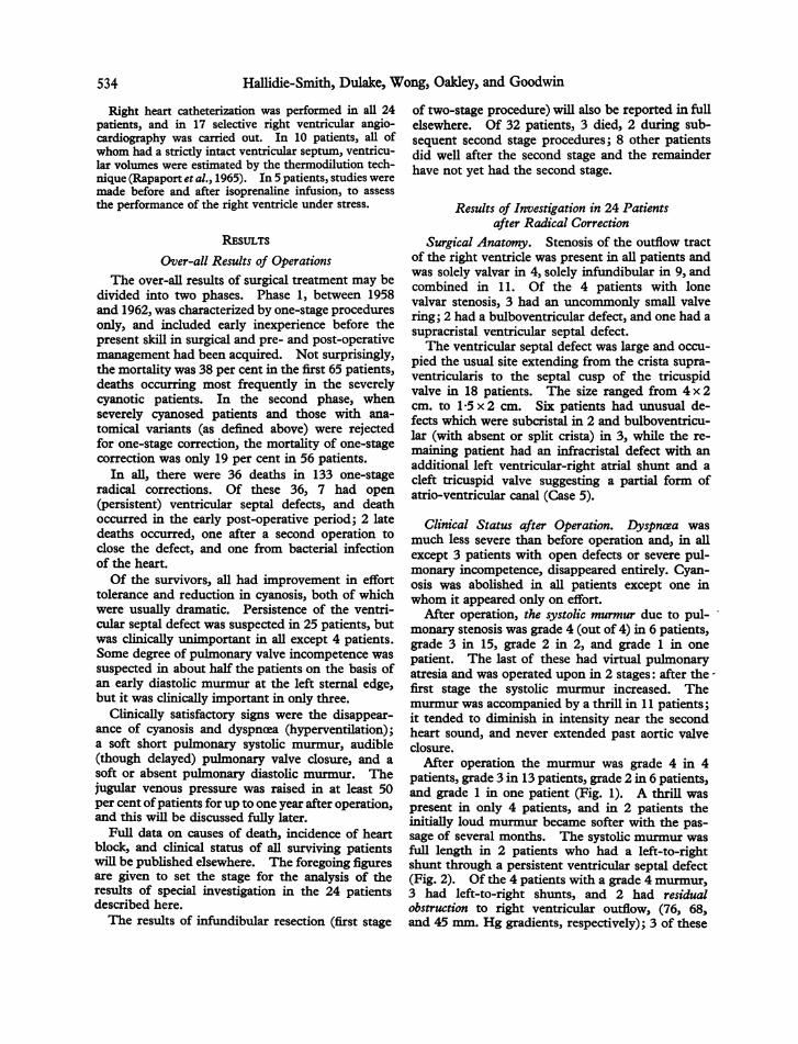
Gradients in excess of 50 mm. Hg were present inthe remaining 5 patients: 4 had pulmonary incom-petence, and 2 had persistence of the ventricular sep-tal defect (Fig. 5 and 6).
Table III shows the association of residual lesions.It will be seen that 5 patients had a residual ventricu-lar septal defect, and that this was always associatedwith either pulmonary incompetence or outflowtract obstruction.
Applying the very rigorous requirement of mini-mal outflow tract obstruction (less than 25 mm.Hg) without any pulmonary incompetence, only 4
9**I
6.t .!i 'I't;'!4 " "
PA= 15/10 RV INF.= 20/8 RV body=27/5
Fully satisfactory result
FIG. 4.-Withdrawal tracing from pulmonary artery to right ventricle in Case 2, showing normal right ven-tricular pressures and relief of outflow obstruction. PA=pulmonary artery; RV=right ventricle; INF=
infundibulum.
}7
Radical Correction of Tetralogy of Fallot
PA 14/4
J .: ....Avt (A 7>,
V on I1KA
fi% q V9 ir
cvu/4 - 25n..' z mm.Hg
i rI
f \.,^ _
Lone pulmonary incompetence
FIG. 5.-Withdrawal tracing from pulmonary artery (PA) to right ventricle (RV) in Case 7 with lone pul-monary incompetence, showing low pulmonary diastolic pressure equal to that of the right ventricle.
mm. Hg100
n
PA 55/20 RV 95120
a=20, v=20,
5O _1 >>t/ \ 2 ~ ~~~I 2S~~~~~~~~~~~~X= 1
so~~ ~ ~ ~ ~ ~ ~
a v
Open defect; residual obstruction; pulmonary incompetence
FIG. 6.-Pressure pulses in pulmonary artery (PA), right ventricle (RV), and right atrium (RA) in Case 12,showing high right ventricular diastolic pressure, equal to pulmonary artery diastolic pressure, high rightventricular systolic pressure, and raised right atrial pressure, with poor "x" descent suggesting tricuspid
incompetence. Note pulmonary hypertension.
TABLE IIIRESIDUAL LESIONS AFTER OPERATION
Patients
Lone residual obstruction (gradient greater than25 mm. Hg) 2
Lone pulmonary infundibular incompetence (definedclinically) 9
Pulmonary incompetence plus residual obstruction 4Open defect plus pulmonary incompetence 1Open defect plus residual obstruction 3Open defect plus pulmonary incompetence plus
obstruction 1
539
w

Hallidie-Smith, Dulake, Wong, Oakley, and Goodwin
patients can be regarded as 'fully satisfactory'(Fig. 4). However, in only 2 patients was pul-monary incompetence of appreciable severity, whileonly 5 patients had gradients of over 50 mm. Hg,and these may be expected to resolve with time, sothat a highly satisfactory result was achieved inmost. Anxiety must be felt, however, for the 5patients with residual defects, even though sympto-matically they have been greatly improved. In theother 19 patients, results have been excellent fromthe hemodynamic aspect.
Relation of Results of Operation to Anatomy Found atOperation and Operative Procedure
Tables IV and V relate the results to the surgicalanatomy, and to the presence or absence of outflowtract or septal patches applied by the surgeons,
respectively. No clear correlation emerges, but it isperhaps significant that 3 of the 5 patients with per-
sistent shunts had complex or multiple ventricularseptal defects, while only 1 of the 4 fully satisfactorypatients had such defects. Of the 4 fully successfulpatients, 3 had lone infundibular stenosis, whichwas present in only 1 ofthe 5 patients with persistentshunts. The numbers are naturally too small forany definite conclusions to be drawn, but it seems
reasonable to suppose that the more complex ana-
tomies such as combined valvar and infundibular
stenosis, and unduly complicated forms of ventri-cular septal defects, are more likely to be associatedwith residual defects after operation. Lone valvarstenosis, a small valve ring, and an atypical ventri-cular septal defect were compatible with a fullysatisfactory result, though combined valvar andinfundibular stenosis militated against completerelief of obstruction and prevented complete com-
petence of the pulmonary valve. The combinedstenosis, however, did not prevent complete closureof the ventricular septal defect, which was achievedin 9 out of the 11 patients with combined stenosis.
Table V shows that only 7 of the 24 patients were
treated by direct suture of the ventricular septaldefect, without any form of patch of prosthesis,though 4 patients had direct suture over teflonpledgets. In those requiring a patch repair of theventricular septal defect, or an outflow prosthesis torelieve severe narrowing, residual abnormalitiesseemed more common. In this small group itwould be unwise to draw firm conclusions, butthis trend is suggested by our results, for 6 of the 9with lone pulmonary incompetence, all patientswith incompetence and residual obstruction, and3 of the 5 patients with open defects, all had a
prosthesis to the ventricular septal defect or out-flow tract, which appears to favour residual ob-struction. Again, numbers are too small to per-mit accurate conclusions.
LE IV
RELATION OF SURGICAL ANATOMY TO RESULTS
Anatomy Fully Lone residual Lone pulmonary Pulmonary incompe- Open defect Totalsatisfactory obstruction incompetence tence plus obstruction
Combined infundibular andvalvar stenosis 0 1 6 2 2 11
Lone valvar stenosis 1 1 0 1* 2 4Small valve ring 1 1 1 0 it 4Lone infundibular stenosis 3 0 3 2 1 9Atypical ventricular septal defect 1 1 1 0 3 6Supracristal 2Bulboventricular 3Multiple 1
_
* Also open defect. t Also pulmonary incompetence and obstruction.
TABLE VRELATION OF SURGICAL PROSTHESIS TO RESULT
Procedure Fully Lone residual Lone pulmonary Pulmonary incompetence Open defect Totalsatisfactory obstruction incompetence and obstruction
Direct suture of ventricularseptal defect 2 0 3 0 2 7
Patch for ventricular septaldefect and/or for outflow tract 2 2 6 4 3 17
Total 4 2 9 4 5 24
540

Radical Correction of Tetralogy of Fallot
TABLE VI
RIGHT ATRIAL MEAN, RIGHT VENTRICULAR, AND PULMONARY ARTERY DIASTOLIC PRESSURES,BEFORE AND AFTER OPERATION
Mean rightatrial pressure(mm. Hg)
Rightventricular
diast. pressure
Before After Before After
5
65
8
33
3
53
4
9912812121137
44645435
10 60 84 137 1811 1010 8- 10- 812 18- 0- 94 207 1312 04 9
5 50 82 7- 60 82 612 5- 80 0
Pulmonaryartery diast.pressure
Before After
15
4
119
81012
869
7
6
12968108761010618121510
5688610778
Pulmonaryincompetence
after
Post-operative clinicaljugular venous
pressure > + 3 cm.above sternal angle
+
+
+
+
+
+
+
+
Remarks
Fully satisfactoryFully satisfactory
Pulmonary atresiaPersistent VSD; left-to-right shunt
Persistent VSD; right-to-left shunt;Fully satisfactory
Persistent VSD; left-to-right shunt
Persistent VSD; left-to-right shuntPersistent VSD; small left-to-right
shunt
Fully satisfactory
Right Ventricular End-diastolic Pressure, MeanRight Atrial Pressure, and Jugular Venous Pressureand Pulmonary Incompetence, after Operation.Table VI shows right ventricular and pullmonaryartery diastolic pressures and mean right atrialpressures before and after operation, in relation tothe jugular venous pressure and the presence ofpulmonary incompetence after operation.
Before operation the mean right atrial pressurewas more than 5 mm. Hg in only 2 of 10 patientsin whom it was measured. After operation it wasraised in 11 of 23 patients. In the 5 patients withpersistence of the ventricular septal defect it was7, 3, 12, 12, 7 mm. Hg, respectively. A level ashigh as 12 mm. Hg was not encountered in anyother patients.The mean right atrial pressure after operation
was above 5 mm. Hg in 10 of 15 patients with araised jugular venous pressure, with a range of3-12 mm. Hg, the average being 7-3 mm. Hg. Themean right atrial pressure was above 5 mm. Hgin 6 of 9 patients with a normal post-operativejugular venous pressure, the range being 3-6 andthe average 4 mm. Hg.The right ventricular end-diastolic pressure
before operation ranged from 0-12 mm. Hg, andwas above 5 (maximum normal) in 8 patients. Ofthe 15 patients with a raised jugular venous pres-sure after operation, the end-diastolic pressure was
below 5 mm. in only 2 patients, the average being6
10 mm. Hg and the range 0-20 mm. Hg. Of the9 patients without post-operative increases in jugu-lar venous pressure, the right ventricular end-diastolic pressure was above 5 mm. Hg in 5. Theaverage was 6 and the range 0-8 mm. Hg.Though the catheterization data were often
obtained at a time when the jugular venous pres-sure had fallen to normal, there appeared to be atendency for a clinically raised jugular venouspressure to be associated with raised right atrialmean and right ventricular end-diastolic pressuresat catheterization.The right ventricular end-diastolic pressure in
patients with and without post-operative pulmonaryincompetence ranged from 0-18 mm. Hg, with a
mean of 9 mm. Hg.The pulmonary artery diastolic pressure ranged
from 5 to 18 mm. Hg after operation, with a mean
of 8 mm. Hg. In patients with pulmonary in-competence it ranged from 6 to 10, with a mean of8A4 mm. Hg. In those without pulmonary incom-petence it ranged from 6 to 15, with a mean of 9.4mm. Hg.The pulmonary artery pulse pressure after opera-
tion ranged from 14 to 33 mm. Hg, with a mean of19-6 mm. Hg in patients with post-operative pul-monary incompetence, and from 8 to 24 mm. Hg inthose without, with a mean of 15-5 mm. Hg.Thus while the diastolic right ventricular-pul-
monary artery gradient did not show any trend,
541
CaseNo.
123456789101112131415
161718192021222324
542 Hallidie-Smith, Dulake, Wong, Oakley, and Goodwin
a b
c d
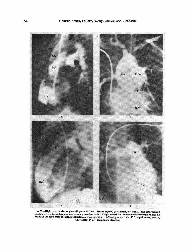
FIG. 7.-Right ventricular angiocardiogram of Case 2 before (upper) (a= lateral, b = frontal) and after (lower)(c=lateral, d= frontal) operation, showing excellent relief of right ventricular outflow tract obstruction and nofilling of the aorta from the right ventricle following operation. R.V.= right ventricle; P.A. = pulmonary artery;
Ao. =aorta; P.S. =pulmonary stenosis.

Radical Correction of Tetralogy of Fallot
there was a tendency towards a higher post-operativepulmonary artery pulse pressure when pulmonaryincompetence was diagnosed.Pulmonary hypertension, defined as a pressure
exceeding 30/15 mm. Hg, was present in 5 cases(Cases 5, 9, 11, 12, and 14), the pressures being42/10, 34/12, 39/6, 50/18, and 37/15 mm. Hg,respectively. Diastolic hypertension did not occur.
Four of them had a residual shunt, and the fifth(Case 11), had severe pulmonary incompetence(Table II).
Thermodilution Studies. These were carried outin 7 patients, 4 of whom had pulmonary incompe-tence, 6 had a raised jugular venous pressure, andonly one had residual obstruction.
End-diastolic volume and the ratio of forwardstroke volume to end-diastolic volume were withinthe normal range at rest in all. Measurements were
made after infusion of isoprenaline in 4, and allsave one were normal. This patient (Case 6) hada normal ratio, but slightly increased end-diastolicvolume (141 ml./m.2 - normal 103 + 34.4 ml./m.2).The validity of the method and the significance ofthese studies require confirmation and further as-
sessment. These results have been reported in fullelsewhere (Wong and Dulake, 1966).
Angiocardiography. Selective right ventricularangiocardiograms were analysed in 17 patients. In3 of the 5 patients who had a shunt at ventricularlevel demonstrated by cardiac catheterization, theangiocardiogram revealed it. Residual obstruc-tion was obvious in only 4 patients. Thirteenshowed an excellent wide outflow tract (Fig. 7),
but in 4 a pouch or diverticulum iu the outflowtract near the ventriculotomy could be seen, similarto that described by Anderson, Newman, andUrquhart (1965) (Fig. 8). Case 7 showed massivedilatation of the right ventricle (Fig. 9), presumablydue to severe pulmonary incompetence. Case 9showed residual pulmonary stenosis and right-to-left shunt (Fig. 10).
DIscussIoNThe results of radical correction of Fallot's tetra-
logy show that despite the extent of the operationthe integrity of the right ventricle and its outflowtract is usually preserved. The operation is a com-plex and difficult one and it is gratifying to note thatof the 24 patients only 5 had residual ventricularseptal defects. All but one had left-to-right shuntswith pulmonary to systemic flow ratios of 1-3: 1 to1-6:1 and only one had a right-to-left shunt. Allsave one had balanced right ventricular and arterialpressures. Only five patients had serious outflowtract gradients over 50 mm. Hg, and these may re-
solve with time. Pulmonary incompetence was
severe in only 2 patients. Thus 10 patients must beconsidered less than satisfactory on account of con-
siderable gradients, severe pulmonary incompetence,or a persistent septal defect. Table VII analyses thesurgical anatomy and procedure in the 10 patients,and it will be seen that no single factor emerges toexplain these less satisfactory results. Localanatomical problems, which cannot be exactlydelineated, probably account for the difficulty inobtaining more satisfactory results. Four patients,by contrast, had remarkably full restoration of
LE VIIDATA IN 10 PATIENTS IN WHOM OPERATION RESULTS WERE LESS THAN SATISFACTORY
Severe residual Pulmonary Right ventricular Type of Type of Small PatchCase Open obstruction incom- end-diastolic ventricular pulmonary valve Direct sutureNo. defect (gradient petence pressure (mim. Hg) defect stenoaia ring of VSD
mm. Hg) ____
3 0 85 0 13 Infracristal Infund. and 0 + 0valvar
5 + 0 + 10 Infracristal Infund. only 0 + Direct sutureL-+R and LV-RA over teflon
shunt defect pledgets7 0 0 + + 10 Infracristal Infund.and 0 0 +
valvar9 + 76 0 18 Infracristal Infund. and 0 + 0
R--*L ~~~~~~~~~~~~~~~~~valvar11 0 0 + + 9 Infracristal Infund. and 0 + 0
valvar12 + + 20 Infracristal Valvar only + 0
L-+.R13 0 80 + 13 Infracristal Infund. and 0 + 0
valvar14 + 0 0 Supracristal Valvar only 0 0 +
15 + 68 0 9 Bulboventricular Infund. and 0 0 +17 RO 54Infracristal Infund.only0valvar17 0 54 + 8 Infracristal Infund. only 0 + 0
543

544 Hallidie-Smith, Dulake, Wong, Oakley, and Goodwin
a
c d
FIG. 8.-Right ventricular angiocardiogram of Case 16 before (upper) (a = lateral, b = frontal) and after (lower)(c= lateral, d= frontal) operation, showing early filling of aorta from right ventricle and severe pulmonarystenosis before, but wide outflow tract and no early aortic filling after operation. There is a small diverticu-lum (D) in the right ventricular outflow tract at the site of the ventriculotomy (both on left). (The pul-
monary valve is closed.)

Radical Correction of Tetralogy of Fallot
FIG. 9.-Right ventricular angiocardiogram (lateral projec- FIG. 10.-Right ventricular angiocardiogram of Case 9, show-tion) of Case 7, showing enormous dilatation of the right ven- ing residual pulmonary valve stenosis (PS) and outline of the
tricle due to pulmonary incompetence. aortic valve cusp from the right ventricle, after operation.
abnormality, with gradients less than 20-25 mm.Hg, no pulmonary incompetence, and no shunts.The remaning 10 patients may be regarded as verysatisfactory and unlikely to give rise to anxiety in thefuture. These results are in general agreement withthose of Gotsman (1966) who studied 11 patientsafter radical correction and found pulmonary in-competence in 6 and residual obstruction in 2.The angiographic appearances indicate that ex-
cellent anatomical results are achieved in the majorityof patients (Gotsman, 1966). The appearances ofresidual obstruction and patency of the ventricularseptum in the relevant patients were predictablefrom the heemodynamics or physical signs, but thediverticulum-like lesions near the ventriculotomycannot be diagnosed without angiography, and maybe present in a number of patients after an operationof this type. Their significance is uncertain, but itseems improbable that they will cause trouble in thefuture.The raised right ventricular end-diastolic pres-
sures might be regarded as evidence of impairedfunction, but diastolic pressures alone do not give areliable index of function. It is possible that somereduction in compliance follows the healing of theventriculotomy scar, but the normal ventriculardiastolic volumes in the few cases studied encourage
the belief that ventricular function is likely to beadequate and to remain so, though the thermodilu-tion method still remains to be fully validated.However, end-diastolic pressures in the right ven-tricle were often raised before operation. Pulmon-ary incompetence does not seem to contribute to araised end-diastolic pressure, which was increasedin 9 of the 10 patients with unsatisfactory results(Table VII).The presence of right bundle-branch block after
operation has not so far appeared to contribute anydisability. The development of left axis deviationis interesting and may be related to interference witharborization conduction bundles. Similar changesare often seen after closure of simple ventricularseptal defects, in our experience.From the clinical aspect these patients have all
been greatly improved, and even those with shuntsor severe pulmonary incompetence have strikingimprovement in symptoms. The results obtainedby special investigations are helpful in assessing thesignificance of clinical signs after operation.A systolic murmur of medium intensity and
length, with audible pulmonary closure, denotes anexcellent result with little or no residual obstruction.Even a loud murmur immediately after operationmay be compatible with an excellent final result,
.545

Hallidie-Smith, Dulake, Wong, Oakley, and Goodwin
for it may diminish gradually as the obstruction re-gresses. It is unusual for the murmur to becomefaint, as it does after resection of lone valvar stenosiswithout ventricular septal defect (Oakley et al.,1964), in view of the more extensive outflow tractdisorders in most cases. The intensity of the mur-mur is increased by the presence of appreciablepulmonary incompetence because of the increasedstroke volume of the right ventricle.A soft or medium intensity early diastolic mur-
mur denotes slight or moderate pulmonary incom-petence which is unlikely to be of future significance.A high jugular venous pressure up to 6 or 8 mm.
above the sternal angle is a frequent occurrence, andmay be expected to fall to normal in 6-12 months.It is not specifically related to any one factor, but isprobably mainly due to residual obstruction, andpossibly to some reduction in ventricular compliancefollowing the ventriculotomy. Appreciable pul-monary incompetence, or a left-to-right shunt, willtend to maintain a high venous pressure. A raisedvenous pressure in no way precludes an excellentresult and need not cause anxiety unless it persistsfor more than 12 months, or is associated with evi-dence of congestive heart failure, notably hepato-megaly, or with signs of severe pulmonary incom-petence or a persistent shunt.The development of a systolic wave of tricuspid
incompetence should arouse suspicion of damage tothe tricuspid valve at the time of operation or byendocarditis.A pansystolic murmur, active ventricular pulsa-
tion, and apical diastolic rumble all suggest that aleft-to-right shunt is present at ventricular level.The long-term prognosis of most of these patients
seems to be excellent; that of the patients withsevere pulmonary incompetence and persistentshunts is less certain. Decision regarding furtheroperation is difficult, for all the patients have beenconsiderably improved, and the risks of furtheroperation must be weighed carefully against those ofconservative treatment. It is probable that Cases 5,9, and 12 will need re-operation. Case 5 has hadbacterial endocarditis since operation, with damageto the tricuspid valve, Case 9 still has a right-to-leftshunt, and Case 12 is in heart failure controlled bydiuretics.We feel that the results in this small pilot series
encourage the belief that ventricular structure andfunction are satisfactory in the majority of patientsafter radical correction, and that the great improve-ment in ail but a very small minority of those whosurvive the operation fully justifies a difficult opera-tion to relieve a hazardous anomaly which seriouslylimits normal life both symptomatically and prog-nostically.
SUMMARYThe over-all results of radical correction of the
tetralogy of Fallot in 133 patients are presentedbriefly. Although initial mortality was high, itsubsequently fell to 19 per cent in 56 patientsoperated upon.Of the 36 patients who died, 7 had persistence of
the ventricular septal defect, and died in the earlypost-operative period. There were 2 late deaths.Improvement in the survivors was dramatic, with
relief of dyspnoea and cyanosis to a degree that fullyjustified the operation.The detailed over-all results in the 133 patients
will be published elsewhere. This paper presentsthe results of detailed clinical, haemodynamic, andangiographic studies of ventricular function andstructure in 24 patients who have been treated byradical correction of the tetralogy of Fallot. Thepatients were selected for special study, or because itwas felt advisable to check on ventricular functionfrom the aspect of long-term prognosis. The im-provement in all patients was dramatic, with relief ofcyanosis and dyspncea. However, in order to testlong-term prognosis, deliberately over-rigid criteriafor a fully satisfactory result were set.Of the 24 patients, 10 had appreciable systolic
gradients (more than 50 mm. Hg) across the rightventricular outflow tract, a persistent shunt atventricular level, or severe pulmonary incompetence,or a combination of these lesions. Ten patients hadminor degrees of outflow tract obstruction and mildpulmonary incompetence, and were consideredsatisfactory. Four patients had well-nigh perfectrestoration of normal function with minimal orabsent outflow tract gradients, no pulmonary in-competence and no shunts.A raised right ventricular end-diastolic pressure
after operation was present in many and was oftenrelated to a raised jugular venous pressure. Thejugular venous pressure was raised after operation in15 patients, who were denied an excellent result, andfell to normal in 6-12 months. It did not appearto be related specifically to any one factor but wasprobably connected with the high right ven-tricular end-diastolic pressure, which in turn waspossibly due to a reduction in compliance of theright ventricle following the ventriculotomy. Severepulmonary incompetence or a left-to-right shuntexaggerated and perpetuated the raised jugularvenous pressure.
Angiocardiography revealed unexpected dilata-tion lesions of the right ventricle in the region of theventriculotomy in 4 patients; the significance ofthis is discussed.The occurrence of right bundle-branch block in
23 of the 24 patients after operation is noted.
546

Radical Correction of Tetralogy of Fallot
It is concluded that the long-term prognosis inthe majority of patients following radical correctionof the tetralogy of Fallot is excellent despite thepresence of the ventriculotomy scar and rightbundle-branch block. The prognosis is less cer-
tain for the minority who are left with importantresidual lesions.
We are grateful to our surgical colleagues, Mr. W. P.Cleland and Professor H. H. Bentall, for providing dataand for their advice in the preparation of this paper, andto Mr. Gerald Rainbow and his technical staff for assist-ance with the investigations. We are indebted toProfessor R. E. Steiner for help with the angiographicstudies.
REFERENCESAnderson, I. M., Newman, C. G. H., and Urquhart, W. (1965).
Fallot's tetralogy-Some radiological and other findingsin the first few years after total correction. Brit. J.
Radiol., 38, 81.Bahnson, H. T., Spencer, F. C., Landtman, B., Wolf, M. D.,
Neill, C. A., and Taussig, H. B. (1962). Surgicaltreatment and follow-up of 147 cases of tetralogy ofFallot treated by correction. J. thorac. cardiovasc.Surg., 44, 419.
Barnard, C. N., and Schrire, V. (1961). The surgical treat-ment of the tetralogy of Fallot. Thorax, 16, 346.
Bristow, J. D., Menashe, V. D., Griswold, H. E., and Starr,A. (1961). Total correction of tetralogy of Fallot:Complications and results. Amer. J3. Cardiol., 8, 358.
Goodwin, J. F., and Abdin, Z. H. (1959). The cardiogramof congenital and acquired right ventricular hyper-trophy. Brit. HeartJ., 21, 523.
Gotsman, M. S. (1966). Hmmodynamic and cine-angio-cardiographic findings after one-stage repair of Fallot'stetralogy. Brit. Heart3J., 28, 448.
Kirklin, J. W., Payne, W. S., Theye, R. A., and DuShane,J. W. (1960). Factors affecting survival after openoperation for tetralogy of Fallot. Ann. Surg., 152, 485.
Malm, J. R., Bowman, F. O., Jr., Jameson, A. G., Ellis, K.,Griffiths, S. P., and Blumenthal, S. (1963). An evalua-tion oftotal correction oftetralogy of Fallot. Circulation,27, 805.
Oakley, C. M., Braimbridge, M. V., Bentall, H. H., and Cle-land, W. P. (1964). Reversed interatrial shunt follow-ing complete relief of pulmonary valve stenosis. Brit.HeartJ., 26, 662.
Rapaport, E., Wong, M., Ferguson, R. E., Bemstein, P., andWieggnd, B. D. (1965). Right ventricular volumes inpatients with and without heart failure. Circulation,31, 531.
Smith, D. R., Effat, H., Hamed, M. A., and Omeri, M. Al(1965). Radiological and surgical anatomy in tetralogyof Fallot and the effect on surgical program. BritHeartJ., 27, 604.
Wong, M., and Dulake, M. (1966). Thermodilution studiesof ventricular function after correction of the tetralogyof Fallot. Brit. Heart. J., 28, 426.
ADDENDUM
Since this paper was completed we have noted a fistulabetween coronary artery and right ventricle after radicalcorrection in one patient (not in this series).
547