Ventricular Septal Defect - Archives of Disease in Childhood · Ventricular SeptalDefect the...
15
Review Article Arch. Dis. Childh., 1968, 43, 268. Ventricular Septal Defect A Review of Current Thoughts* ALEXANDER S. NADAS and DONALD C. FYLER From the Sharon Cardiovascular Unit of the Children's Hospital Medical Center, Boston, Massachusetts, and the Department of Pediatrics, Harvard Medical School, Boston, Massachusetts Ventricular septal defect, alone or in combination with other cardiac malformations, is the commonest form of congenital heart disease in man. Morpho- genically, the ventricular septum is composed of three parts: (a) the conal septum, (b) the septum of the endocardial cushions, and (c) the muscular septum proper. Consequently, most cono-truncal abnormalities (transposition of the great arteries, tetralogy of Fallot, common truncus arteriosus) include a defect of the ventricular septum and, similarly, most endocardial cushion defects (and all complete AV canals) have a ventricular compo- nent. Isolated ventricular septal defects may occur in any of the three parts. The majority of the complex intracardiac malformations are associated with a ventricular septal defect. The purpose of this clinical review, however, is to discuss problems relating to patients whose principal, even exclusive, problem is a defect of the ventricular septum. We will, therefore, exclude from consideration patients with tetralogy of Fallot, truncus arteriosus, transposition of the great arteries, as well as endocardial cushion defect. Furthermore, because of its invariable association with other serious malformations of the endocardial cushion as well as cono-truncal regions, we will exclude from this discussion patients with single ventricle who in our view do not simply represent very large ventricular defects but a more profound developmental abnormality (Van Praagh, Ongley, and Swan, 1964). Patients with dextrocardia will not be considered either, partly because the occurrence of a solitary ventricular defect in this setting is rare and partly because they represent unique diagnostic and surgical problems. Finally, and rather arbitrarily, we will exclude from con- sideration patients with ventricular septal defect * Supported by grants HE 10436-02 and HE 5310-09, from the National Heart Institute of the National Institutes of Health. and associated coarctation of the aorta, absent pulmonary valve, atrial septal defect, and patent ductus arteriosus beyond the neonatal period. Our discussion of ventricular septal defect will include, however, patients with associated pul- monary vascular obstructive disease, aortic regurgi- tation, minimal pulmonary stenosis (peak gradient of less than 30 mm. Hg), and mitral valve disease (outside of the framework of endocardial cushion defect). The first three of these should be con- sidered because their development, in the course of time, represents part of the natural history of ventricular septal defect. Coexisting mitral valve involvement will also be discussed briefly, since its recognition is difficult and may influence surgical decision in regard to ventricular septal defect. Within this review, we will cite the incidence, restate the diagnostic criteria, discuss the anatomy and physiology, emphasize more recent information referable to the clinical picture, survey the data available on the natural course of the disease, propose indications for surgery, and present the results of operation. Incidence Patients with solitary ventricular septal defect, or those with pulmonary vascular obstruction, minimal pulmonary stenosis, aortic regurgitation, or mitral valve involvement, represent about 20% of the total congenital heart disease population (Keith, Rowe, and Vlad, 1967; Nadas, 1963) in series dominated by the paediatric age-group. An even higher incidence in infants is suggested by Hoffman and Rudolph (1965); the absolute incidence in their series was 1- 35/1000 live births in New York hospitals. If one accepts MacMahon's data (Mac- Mahon, McKeown, and Record, 1953) that the over-all incidence of congenital heart disease is 3/1000 live births, then this would suggest that in 268 copyright. on March 30, 2020 by guest. Protected by http://adc.bmj.com/ Arch Dis Child: first published as 10.1136/adc.43.229.268 on 1 June 1968. Downloaded from
Transcript of Ventricular Septal Defect - Archives of Disease in Childhood · Ventricular SeptalDefect the...

Review Article
Arch. Dis. Childh., 1968, 43, 268.
Ventricular Septal DefectA Review of Current Thoughts*
ALEXANDER S. NADAS and DONALD C. FYLERFrom the Sharon Cardiovascular Unit of the Children's Hospital Medical Center, Boston, Massachusetts, and
the Department of Pediatrics, Harvard Medical School, Boston, Massachusetts
Ventricular septal defect, alone or in combinationwith other cardiac malformations, is the commonestform of congenital heart disease in man. Morpho-genically, the ventricular septum is composed ofthree parts: (a) the conal septum, (b) the septum ofthe endocardial cushions, and (c) the muscularseptum proper. Consequently, most cono-truncalabnormalities (transposition of the great arteries,tetralogy of Fallot, common truncus arteriosus)include a defect of the ventricular septum and,similarly, most endocardial cushion defects (andall complete AV canals) have a ventricular compo-nent. Isolated ventricular septal defects may occurin any of the three parts. The majority of thecomplex intracardiac malformations are associatedwith a ventricular septal defect.The purpose of this clinical review, however, is
to discuss problems relating to patients whoseprincipal, even exclusive, problem is a defect of theventricular septum. We will, therefore, excludefrom consideration patients with tetralogy of Fallot,truncus arteriosus, transposition of the greatarteries, as well as endocardial cushion defect.Furthermore, because of its invariable associationwith other serious malformations of the endocardialcushion as well as cono-truncal regions, we willexclude from this discussion patients with singleventricle who in our view do not simply representvery large ventricular defects but a more profounddevelopmental abnormality (Van Praagh, Ongley,and Swan, 1964). Patients with dextrocardia willnot be considered either, partly because theoccurrence of a solitary ventricular defect in thissetting is rare and partly because they representunique diagnostic and surgical problems. Finally,and rather arbitrarily, we will exclude from con-sideration patients with ventricular septal defect
* Supported by grants HE 10436-02 and HE 5310-09, from theNational Heart Institute of the National Institutes of Health.
and associated coarctation of the aorta, absentpulmonary valve, atrial septal defect, and patentductus arteriosus beyond the neonatal period.Our discussion of ventricular septal defect will
include, however, patients with associated pul-monary vascular obstructive disease, aortic regurgi-tation, minimal pulmonary stenosis (peak gradientof less than 30 mm. Hg), and mitral valve disease(outside of the framework of endocardial cushiondefect). The first three of these should be con-sidered because their development, in the courseof time, represents part of the natural history ofventricular septal defect. Coexisting mitral valveinvolvement will also be discussed briefly, since itsrecognition is difficult and may influence surgicaldecision in regard to ventricular septal defect.Within this review, we will cite the incidence,
restate the diagnostic criteria, discuss the anatomyand physiology, emphasize more recent informationreferable to the clinical picture, survey the dataavailable on the natural course of the disease,propose indications for surgery, and present theresults of operation.
Incidence
Patients with solitary ventricular septal defect, orthose with pulmonary vascular obstruction, minimalpulmonary stenosis, aortic regurgitation, or mitralvalve involvement, represent about 20% of thetotal congenital heart disease population (Keith,Rowe, and Vlad, 1967; Nadas, 1963) in seriesdominated by the paediatric age-group. An evenhigher incidence in infants is suggested by Hoffmanand Rudolph (1965); the absolute incidence in theirseries was 1- 35/1000 live births in New Yorkhospitals. If one accepts MacMahon's data (Mac-Mahon, McKeown, and Record, 1953) that theover-all incidence of congenital heart disease is3/1000 live births, then this would suggest that in
268
copyright. on M
arch 30, 2020 by guest. Protected by
http://adc.bmj.com
/A
rch Dis C
hild: first published as 10.1136/adc.43.229.268 on 1 June 1968. Dow
nloaded from

Ventricular Septal Defectthe newborn group almost 50% of the patients withcongenital heart disease have solitary ventricularseptal defect. We think this figure is too high.Somewhat more recent data on over-all incidenceare closer to 6/1000 (Harris and Steinberg, 1954;McIntosh et al., 1954). Thus, 1-35/1000 (or2/1000 as Hoffman and Rudolph suggest may bethe true figure) would represent about 30% incidenceamong newborn cardiacs.
Interestingly enough, in surveys limited to schoolchildren (Weber, 1918; Rose and Keith, 1966) andthis, of course, has a great deal of bearing on thenatural history of ventricular septal defect, a muchlower incidence was found (2 8 to 5 6/10,000).An interesting Table in Bloomfield's paper (1964)indicates that even in case material 'usually limitedto age three and beyond', first visits for ventricularseptal defect are rare over the age of 30 (48/424).The same author's necropsy data also attest to therarity of isolated ventricular septal defects over 15to 20 years of age (037/1000).These figures are cited only to indicate that isola-
ted ventricular septal defect is relatively common ininfancy and becomes rarer with advancing age.Obviously three things may underlie this trend:(1) death; (2) spontaneous cure; (3) change inclinical or pathological profiles so that the lesion isnot classified in later life as ventricular septal defect.The discussions to follow will attempt to shed
some light on this disappearance of ventricularseptal defects.
Diagnostic CriteriaThe diagnosis of ventricular septal defect seems,
superficially, to be on firmest grounds if made atnecropsy. This obviously is true in a positivesense; if the pathologist finds a defect in the ven-tricular septum there is very little room for argument.On the other hand, it is equally clear that smallventricular defects, giving rise to the characteristicclinical profile and proven unquestionably byphysiological or angiographic data, may be missedby a less than first-class pathologist. The diagnosisof a ventricular defect indeed may be made easilyat necropsy, but it cannot be excluded by anythingbut the most painstaking search by expert handsprobing diligently behind trabeculae and papillarymuscles. Despite the reported incidence of spon-taneous closure of ventricular septal defects, thepathological recognition that a defect existed andlater closed is rare. It seems clear that while smalldefects may be overlooked, evidences of a closeddefect are almost invariably unrecognized, even bythe most sophisticated observers.
Cardiac catheterization is today the procedure of
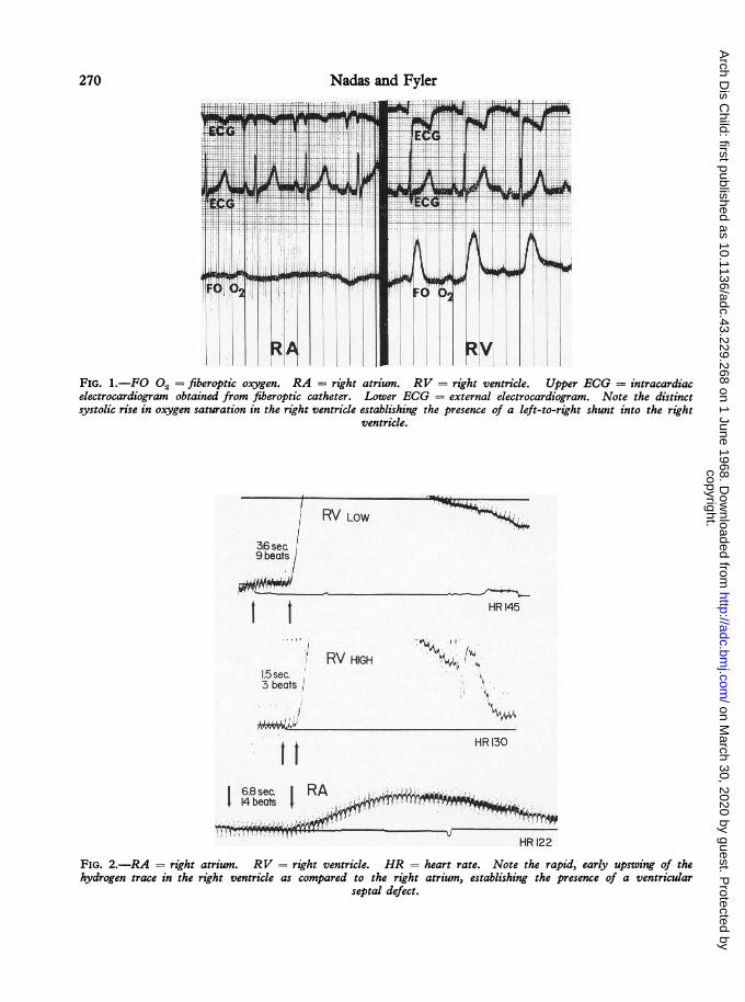
choice in establishing, or excluding, the diagnosisof ventricular septal defect. The most widelyaccepted oximetric definition of a left-to-rightshunt through the ventricular septum is a minimumof 10% increase in oxygen saturation from rightatrium to ventricle in one set of samples, or aminimum of 5% increase in each of two sets ofsamples. A more sensitive, in vivo, oximetricmethod is afforded by the fiberoptic catheterresponding instantaneously to phasic changes inoxygen saturation within the cardiac cycle; the jet ofhighly oxygenated blood may be detected by thisinstrument if skilfully placed in the vicinity of thedefect (Fig. 1). Other indicators of high sensitivityfor detection of left-to-right shunts include hydrogenion (the gas inhaled through a mask and detected bya platinum electrode in the ventricle) (Fig. 2) andCardiogreen injected through a catheter in the leftventricle, with sampling in the right ventricle.Radiopaque material injected through a catheterinto the left ventricle and traced by radiographs orcinefluorograms is another excellent and highlysensitive method of detection of a left-to-rightshunt through a ventricular septal defect (Fig. 3).A phonocatheter registering a pansystolic murmurin the right ventricle with little or no murmur inthe right atrium or the main pulmonary artery(Fig. 4) may serve as an excellent, indirect, qualita-tive tool for demonstrating a ventricular septaldefect.The localization of right-to-left shunts may be
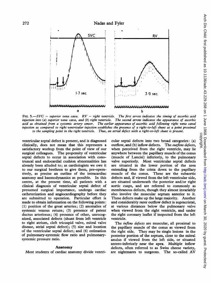
accomplished by injection of an indicator (ascorbicacid or Cardiogreen) in the right ventricle (Fig. 5)and sampling from the left ventricle, or by selectiveangiocardiography from the right ventricle.Of course, the clearest, and many times the
simplest, way of demonstrating a ventricular defectat cardiac catheterization is by passage of thecatheter from right ventricle to left ventricle oraorta.
In our opinion, the hydrogen ion method, fiberop-tic oximetry, and good quality left ventricularangiograms are probably the most sensitive detectorsof ventricular defect. Shunts as small as 5% ofthe cardiac output may be detected by thesetechniques. Preference among the three willdepend on the experience of the individual catheteri-zation laboratories; used optimally their sensitivitiesare about the same. Although in vitro oximetry isnot as sensitive as the three methods cited above,one can state with assurance that this method, withmultiple samplings, will probably uncover all thesurgically significant defects.
Surgical exploration of the right ventricle, in ouropinion, is an inappropriate method for the diagnosis
269
copyright. on M
arch 30, 2020 by guest. Protected by
http://adc.bmj.com
/A
rch Dis C
hild: first published as 10.1136/adc.43.229.268 on 1 June 1968. Dow
nloaded from
Nadas and Fyler
FIG. 1.-FO 0.2 = fiberoptic oxygen. RA = right atrium. RV = right ventricle. Upper ECG = intracardiacelectrocardiogram obtained from fiberoptic catheter. Lower ECG = external electrocardiogram. Note the distinctsystolic rise in oxygen saturation in the right ventricle establishing the presence of a left-to-right shunt into the right
ventricle.
* 36#etCSod
-, ;tMa
It
* ;.v
RV\ LOW
I
3- beats I
.. .. .i.a
ii
.
... ..H A
tl ..
:iI; *\
RVHGH
HR 130
T" :J.%,;tr 7. :,"fI*x ? b R A < 7 <r,j1'¼I'7...- ;.
Hf122
FIG. 2.-RA = right atrium. RV = right ventricle. HR = heart rate. Note the rapid, early upswing of thehydrogen trace in the right ventricle as compared to the right atrium, establishing the presence of a ventricular
septal defect.
.
m . ;=_WAAL
270
II .1 -,.. - .-P -I ip I N."I IT III -
copyright. on M
arch 30, 2020 by guest. Protected by
http://adc.bmj.com
/A
rch Dis C
hild: first published as 10.1136/adc.43.229.268 on 1 June 1968. Dow
nloaded from

Ventricular S
FIG. 3.-A lateral left ventricular angiocardiogram withcontrast injection via the retrograde route. The arrowindicates a jet of contrast material from the left ventricleinto the right ventricle, establishing the presence of a small
ventricular septal defect.
oeptal Defect 271of ventricular defect. Cardiotomy, with its inevi-table emotional tensions, poor visibility, limitedtime, and associated morbidity is not a diagnosticprocedure. Open-heart surgery ought not to beundertaken in an institution not equipped with acompetent, diagnostic team.The final, remaining, question is how accurate
is the clinical diagnosis of ventricular septal defect.The clinical profile is certainly characteristicenough to suggest the diagnosis in all cases worthconsidering for corrective surgery. Difficultiesmay arise with defects so small that the murmurlacks its usual characteristics, and in patients withpulmonary vascular obstructive disease or conges-tive heart failure, where the flow dynamics areseverely altered. In case of the mini-defect, withnormal heart sounds, x-rays, and electrocardio-grams, the precise diagnosis is only of academicinterest. In the patient with pulmonary vascularobstructive disease-Eisenmenger's syndrome-cardiac surgery is contraindicated; thus the accurateanatomical diagnosis is again only ofmarginal interestto the patient though it contains great fascinationfor the cardiologist. Finally, infants and childrenwith left-to-right shunts and congestive heartfailure have to be catheterized anyhow, to excludethe presence of a patent ductus arteriosus. Thus,one may say that the clinical diagnosis of ventricularseptal defect may be made quite accurately, in over90% of the cases where it is of practical importanceto the patient. At the same time, the fact that the
FIG. 4.-RA = right atrium. RV = right ventricle.BA = brachial artery tracing. SM = systolic murmur.The uppermost tracing from the phonocatheter shows thepresence of a murmur in the ventricle which is not seen in
the atrium.
I
copyright. on M
arch 30, 2020 by guest. Protected by
http://adc.bmj.com
/A
rch Dis C
hild: first published as 10.1136/adc.43.229.268 on 1 June 1968. Dow
nloaded from
Nadas and Fyler
RV
.W4W.W.iWiA
a bFIG. 5.-SVC = superior vena cava. RV = right ventricle. The first arrow indicates the timing of ascorbic acidinjection into (a) superior vena cava, and (b) right ventricle. The second arrow indicates the appearance of ascorbicacid as obtained from a systemic artery sensor. The earlier appearance of ascorbic acid following right vena cavalinjection as compared to right ventricular injection establishes the presence of a right-to-left shunt at a point proximal
to the sampling point in the right ventricle. Thus, an atrial defect with a right-to-left shunt is present.
ventricular septal defect is present, and is diagnosedclinically, does not mean that this represents asatisfactory workup from the point of view of oursurgical colleagues. The propensity of ventricularseptal defects to occur in association with cono-truncal and endocardial cushion abnormalities hasalready been alluded to; as cardiologists we owe itto our surgical brethren to give them, pre-opera-tively, as precise an outline of the intracardiacanatomy and haemodynamics as possible. In thiscentre, at the present time, all patients with aclinical diagnosis of ventricular septal defect ofpresumed surgical importance, undergo cardiaccatheterization and angiocardiography before theyare submitted to operation. Particular effort ismade to obtain information on the following points:(1) position of the great arteries; (2) anomalies ofsystemic venous return; (3) presence of patentductus arteriosus; (4) presence of other, unrecog-nized, associated defects (shunt from left ventricleto right atrium, cleft tricuspid valve, mitral valvedisease, atrial septal defect); (5) size and locationof the ventricular septal defect; and (6) estimationof pulmonary-systemic flow ratio and pulmonary-systemic pressure ratio.
AnatomyMost students of cardiac anatomy divide ventri-
cular septal defects into two broad categories: (a)outflow, and (b) inflow defects. The outflow defects,when perceived from the right ventricle, may lieanywhere between the papillary muscle of the conus(muscle of Lancisi) inferiorly, to the pulmonaryvalve superiorly. Most ventricular septal defectsare situated in the lower portion of the areaextending from the crista down to the papillarymuscle of the conus. These are the subaorticdefects and, if viewed from the left ventricular side,are situated underneath the posterior and/or rightaortic cusps, and are referred to commonly asmembranous defects, though they almost invariablyalso involve the muscular septum anterior to it.These defects make up the large majority. Anotherand considerably rarer outflow defect is supracristal,at various distances below the pulmonary valvewhen viewed from the right ventricle, and underthe right coronary leaflet if inspected from the leftventricle.The inflow defects are muscular, all proximal to
the papillary muscle of the conus as viewed fromthe right side. They may be single lesions in theposterior portion of the septum, close to the mitralanulus if viewed from the left side, or situatedantero-inferiorly near the apex. Multiple inflowdefects, often referred to as Swiss cheese variety,are nightmares to surgeons. The so-called AV
P l EN-
272
IijI.
3.9 sec.
IIA-A A I-AAA-Im .1 JA A .1 .11 -1-11AA J d AAA A WAAA
copyright. on M
arch 30, 2020 by guest. Protected by
http://adc.bmj.com
/A
rch Dis C
hild: first published as 10.1136/adc.43.229.268 on 1 June 1968. Dow
nloaded from

Ventricular Septal Defectcommune defects are large posterior openings tothe right of the muscle of Lancisi beneath theseptal leaflet of the tricuspid valve.
It is perhaps easiest to remember, and is not toofar from the truth, that the membranous outflowdefects adjoining the right and/or posterior aorticcusps make up 70% of the defects, and the highoutflow lesions, the inflow lesions, and the AVcanal defects contribute probably about 10% eachto the total.The size of the defects may vary from pinpoint
size to virtual absence of the septum. Defects withdiameters of at least one-half the aortic diameter arereferred to as 'large' (Selzer, 1949).
PhysiologyThe direction and magnitude of flow across the
ventricular septal defect depends on the size of thedefect and the pressure gradient between the twoventricles. Savard et al. (1960) found this relationaccurate enough to suggest that the pressuregradient between the two ventricles, in mm. Hgdivided by the left-to-right shunt in 1./min. perM.2 body weight, the so-called resistance index, couldpredict with reasonable accuracy the size of thedefect at surgery, up to 1 cm./m.2 Any defectgreater than this would still give the same index.This then could be used as a definition of a 'large'defect, in a physiological sense, and is not too farremoved from the anatomical definitions cited above,i.e. half the aortic diameter.The timing of the left-to-right shunt, within the
cardiac cycle, has recently been studied in ourlaboratory with the fiberoptic catheter (Gambleet al., 1965) and by Levin et al. (1967) usingbiplane cine-angiography. It seems that the flowoccurs in early systole in small defects, throughoutsystole in large defects without pulmonary vascularobstructive disease, and in systole as well as diastole,with a double peak, in patients with ventricularseptal defect and pulmonary vascular obstructivedisease (Fig. 1).That the physiology of ventricular septal defect
depends on the status of the pulmonary arterioles aswell as the size of the defects, has been amplydiscussed by Rudolph and Nadas (1962) in thepast and will not be repeated here. Suffice it tosay that the large defects (over 1 cm./m.2 in diameterphysiologically, or over half the aortic diameterpathologically) result in pressure equalizationbetween the systemic and pulmonary circuits.Applying Ohm's law, P = F x R, it seems clearthat the systemic, or near systemic, pressure levelsin the pulmonary circuit, associated with largeseptal defects, may be high pulmonary flow or
high pulmonary resistance situations or, mostlikely, a combination of both.The changes in the muscular pulmonary arteries
and pulmonary arterioles from newborn period toadulthood and the parallel changes occurring inpulmonary haemodynamics have also been amplydescribed within the past decade (Dammann andFerencz, 1956). Briefly, the normal involution ofthe media of the muscular arteries becomes evidentwithin the first couple of months of life but is notnormaUy completed till sometime within the firstyear. The precapillary arterioles normally have nomuscular media, except at their branching off fromthe muscular arteries. In patients with largeventricular septal defects and pulmonary arteryhypertension, the normal involution does not usuallytake place, anatomically, according to the normaltime-table. Medial hypertrophy is present ininfancy and, with the progressive intimal changesdeveloping in early adulthood, pulmonary vascularobstruction results (Naeye, 1966).
In the normal fetus, right and left ventricularpressures are equal due to the high resistance offeredby the unexpanded lung and the muscular arteries.With the first few breaths the lungs expand, resis-tance drops dramatically, and a pressure gradientdevelops between the two circuits, now separatedby the closure of the ductus arteriosus and theforamen ovale. Further drop in pulmonary resis-tance occurs through the ensuing months with thenormal involution of the muscular arteries. Thecourse of events in the patients with small ventricularseptal defects, though not well documented, issurely not too different. In babies with largeventricular septal defects, however, the drop inresistance does not occur with the same speed as inthe normal infants or in those with small defects.The drop in resistance due to expansion of the lungdoes occur indeed, but the further drop due to medialinvolution occurs probably only transiently if thesize of the defect stays the same. Taking theseanatomical and physiological changes into considera-tion, one may easily explain the following clinicalfacts: (1) the murmur of the ventricular septal defectis usually not heard well within the first days oflife, because resistances in the systemic and pul-monary circuits are similar enough that no significantpressure gradient between the two circuits exists;thus no significant left-to-right shunt, to give riseto a murmur, occurs; (2) in almost all cases asignificant murmur appears within the first monthof life because, when a defect is present, enoughinvolution of the pulmonary vasculature occurswithin this period to give rise to a sizeable shunt;(3) congestive heart failure in babies with large
273
copyright. on M
arch 30, 2020 by guest. Protected by
http://adc.bmj.com
/A
rch Dis C
hild: first published as 10.1136/adc.43.229.268 on 1 June 1968. Dow
nloaded from

274
ventricular septal defects almosiwithin the first 6 months, andfirst 3 months, of life, but almosweek, because the ratio of Rp/Rpermit a large left-to-right shunof life, but will certainly, withconducive to congestive heartfirst few months; (4) the appeararchest x-ray of pulmonary vasccannot be the result of increasecflow. Rather, other factors si
venous congestion or pulmoxdisease are more likely causes ofance; the reason is that the ratisystemic resistance is such that ashunt is not possible in the first
Clinical PictuiThe clinical profile of ventric
of course, depends principallydefect and the status of the pulrrWe should also stress that the depresented here, and as discuss4various publications and in textl1967; Nadas, 1963), pertains oseptal defect with left-to-right shright-to-left shunts present with
CAR
a-
2LIS
4LI S
IlTFIG. 6.- This child was proved to haiseptal defect. Note the atypical,systolic murmur recorded at the low(4LIS) on phonocardiography. 2LJcostal space. CAR = external cc
second tracing is the reference elo
Nadas and Fylert invariably occurs of the Eisenmenger syndrome, or pulmonarymostly within the vascular obstructive disease, and are indistinguish-st never in the first able, for practical purposes, from pulmonarydoes not usually vascular obstruction associated with atrial defect or
it in the first week aorto-pulmonary communications (Cutler et al.,a large defect, be 1954).failure within the The group characteristics of congenital heartice on the newborn disease with a left-to-right shunt are present incular engorgement more or lesser degrees in all patients with a ventri-d pulmonary blood cular defect as defined above. There is left chestach as pulmonary prominence, a hyperkinetic impulse, diastolic flownary parenchymal rumble, and active pulmonary plethora x-ray film.this x-ray appear- The following additional clinical findings are
io of pulmonary to specific for ventricular defect and may help ina large left-to-right assessing the severity of the disease. The cardiacdays of life. impulse is biventricular; the larger the pulmonary
systemic flow ratio, the more prominent is there apical impulse; the higher the pulmonary resistance,.ular septal defect, the more the xiphoid heave dominates. Theon the size of the pansystolic, loud murmur maximal at the xiphoidionary vasculature. process and transmitting along the left sternalscription as briefly border (but in contrast to mitral regurgitationed many times in murmur not too far to the posterior axilla) is)ooks (Keith et al., characteristic of ventricular defect with a sizeableinly to ventricular left-to-right shunt; as the shunt diminishes in size,unts. Those with due either to the development of vascular disease or,the clinical picture more favourably, to diminishing size of the defect,
the systolic murmur becomes fainter and shorter,losing its pansystolic character (Fig. 6). The mid-diastolic rumble usually follows the third soundand thus is separated appreciably from the pul-monary closure; though, by and large, the louderthe rumble, the greater the left-to-right shunt, thisrelation is far from linear (Fyler et al., 1958). Itshould also be mentioned that mid-diastolicrumbles due to increased mitral valve flow alone,
V ~ seldom, if ever, assume a presystolic crescendoconfiguration (Fig. 7). The intensity of the
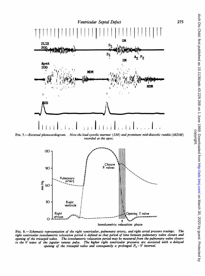
2 pulmonary valve closure is roughly related topulmonary arterial pressure, but in young childrenwith thin chests this may prove to be an unreliableguide. Better assessment of pulmonary arterypressure may be furnished at auscultation by theinverse relation of A2-P2 distance, and phono-cardiographically by the P2-v interval (Gamboaet al., 1965) (Fig. 8); the closer the second sound issplit, the nearer pulmonary artery pressure is tosystemic artery pressure.The over-all heart size and the left atrial size in
ve a small ventricular the x-ray are excellent measures of the size of the
early, decrescendo, left-to-right shunt and prove to be more dependableer left sternal border in our hands in this regard than the estimation ofIS = 2nd left inter- degrees of pulmonary plethora. The relative sizezrotid tracing. The of the left and right ventricles may give some impres-Cectrocardiogram. sion as to whether the ventricular defect is princi-
copyright. on M
arch 30, 2020 by guest. Protected by
http://adc.bmj.com
/A
rch Dis C
hild: first published as 10.1136/adc.43.229.268 on 1 June 1968. Dow
nloaded from
Ventricular Septal Defect
i'-.J1'''11 It' I I I HI 1'1sM
2L1S. .. illm
~~~11Ef'MdI!YinV - ~~~~~~~~~~~- sa AR"re&wrEN701i'iup q1 llu0,j'1' '1'' - -SI IIIIFI rlllw -- qw 1
SM£2 r2
Apex
200
, ! ~1'! t! H ,j'I , , i*&*,,~~~~~~~~~~'~ DI*M,mm~~~~~~1
I
I I I iIi.ji ji . I ,i. I I jII: I i i ...I .
FIG. 7.-External phonocardiogram.
120 -
90-
E: 601 -
E
Note the loud systolic murmur (SM) and prominent mid-diastolic rumble (MDM)recorded at the apex.
Isovolumetric relaxation phase
FIG. 8.-Schematic representation of the right ventricular, pulmonary artery, and right atrial pressure tracings. Theright ventricular isovolumetric relaxation period is defined as that period of time between pulmonary valve closure andopening of the tricuspid valve. The isovolumetric relaxation period may be measuredfrom the pulmonary valve closureto the V wave of the jugular venous pulse. The higher right ventricular pressures are asociated with a delayed
opening of the tricuspid valve and consequently a prolonged P2-V interval.
275
'qMNWAk%ft
Sl-, I-A&
D_
v
copyright. on M
arch 30, 2020 by guest. Protected by
http://adc.bmj.com
/A
rch Dis C
hild: first published as 10.1136/adc.43.229.268 on 1 June 1968. Dow
nloaded from

Nadas and Fylerpally a high-flow or a high-resistance type. Aprominent main pulmonary artery, in a patientwithout pulmonary stenosis, suggests pulmonaryartery hypertension but does not differentiate byany means the hyperkinetic from the obstructivetype.The electrocardiogram is an excellent tool for the
assessment of right ventricular, and thus indirectly,pulmonary arterial hypertension as well as thecontribution of high pulmonary blood flow to thepulmonary arterial pressure. DuShane et al. (1960)and Vince and Keith (1961) have repeatedly stressedthe importance of the electrocardiogram in thepre-operative assessment of patients with ventricularseptal defect. It may be said with considerableassurance that ventricular defect patients withoutright ventricular hypertrophy in the cardiogramwill not have appreciable pulmonary hypertension,and also that those without adequate left ventricularpotential in the ECG probably will not have asignificantly increased pulmonary blood flow. Ofcourse these criteria, and they may be obtained fromthe scalar as well as the vectorcardiographicrepresentations of the electrocardiogram, shouldnot be relied upon independently but rather inconjunction with other clinical findings. It maybe stated, however, that all patients with largeventricular defects will show some degree of rightventricular hypertrophy and that most patients whowill benefit from closure of ventricular defects willshow some left ventricular hypertrophy in theelectrocardiogram.
Natural HistoryWe have alluded already, in the section devoted
to the discussion on incidence, to the 'case of thedisappearing ventricular septal defect'. To paedia-tricians and paediatric cardiologists, ventricularseptal defect is probably the commonest congenitalcardiac malformation (2/1000 live births); incontrast, in the school surveys, incidences of only0-3-0-6/1000 are cited. Looking at the problemin another way, the figures for newborns suggestthat at this age about 30% of the patients withcongenital heart disease have ventricular septaldefect, data obtained from cardiac clinics dominatedby the paediatric age-group suggest a 20°% inci-dence, whereas from units frequented by adults an8% figure is reported. These figures, difficult toobtain and harder to interpret, gain validity fromthe common-sense observation that in paediatriccardiac clinics ventricular defects are present inlarge numbers, whereas they are as rare as hens'teeth in adult clinics and offices.What happens then to the patients with ventri-
cular septal defects ? Our more surgically-orientedcolleagues suggest, if not in writing but in formaland informal meetings, that they obviously die onaccount of the lack of surgical correction. Therecord does not seem to bear out this contention.In our own material (Fyler et al., 1958), less than5% of ventricular defects died and most of themwith bronchopneumonia, prematurity, and associ-ated anomalies. An 18% 'medical death rate' wasfound and contrasted with 25% 'surgical deathrate' by Ritter et al. (1965), but in only half of the18% was death attributable to congestive failure;in the others pneumonia, prematurity, and arrhyth-mias were the underlying causes of death. It isalso interesting to note that in this carefully analysedseries only one of the 85 patients who belonged towell-defined severity groups died without operation;19 out of 20 deaths belonged to a category designatedas 'remainder', by which the authors mean thatthe diagnosis was made at necropsy or on the basisof non-classifiable incomplete data. The vastmajority of these deaths, then, were in patients whoprobably did not receive optimal care for any lengthof time. Walker et al. (1965) cite a mortality of7/415 (less than 2%); but one of these occurred inan unexplained fashion in a baby with a defect ofless than 1 cm./m.2 and right ventricular pressureof 45 mm. Hg, and another one had congenitalheart block.
It is clear then that death can account for only asmall percentage of the disappearing defects. It isalso uniformly accepted that the vast majority ofthe few deaths occurred within the first year, andprobably within the first six months of life(Zacharioudakis, Terplan, and Lambert, 1957).Bloomfield (1964) could find only 25 ventriculardefects in patients of over 20 among 67,000 necrop-sies, and 16 of these died from causes unrelated totheir defects. Combing several series, this authorfound a 3*7/10,000 incidence among 142,000necropsies of patients over 15 to 20 years, and deathin most of these was probably not attributable tothe ventricular defect itself.The final consideration of the discussion of deaths
in patients with ventricular septal defect shouldrelate to the type of defect and to the nature oftheir demise. There seems to be no evidencewhatsoever that small defects, less than 1 cm./m.2,are likely to prove fatal. Bloomfield (1964) statesthat, 'pulmonary hypertension has never beenreported to occur or develop in small defects'. Ina statement based on the analysis of 115 reportednecropsy cases, Lucas et al. (1961) come to the sameconclusion on the basis of a smaller number ofclinical observations. Ritter et al. (1965) found no
276
copyright. on M
arch 30, 2020 by guest. Protected by
http://adc.bmj.com
/A
rch Dis C
hild: first published as 10.1136/adc.43.229.268 on 1 June 1968. Dow
nloaded from

Ventricular Septal Defectprogressive pulmonary vascular obstructive diseaseamong any of the patients with clinically smalldefects (pulmonary-systemic flow ratio of less than2 5). Kaplan et al. (1963) state that, 'we did notrecognize any instance of progression of smallventricular defects to severe pulmonary hyper-tension in adult life'. So that pulmonary vascularobstruction, one of the common causes of death,does not seem to occur with small defects. Conges-tive heart failure in Kaplan's group never occurredwith pulmonary arterial pressure of less than 30mm. Hg and was rare under 40 mm. Hg. Hoffmanand Rudolph (1965) did not find evidence of severefailure in anybody with pulmonary-systemic pressureratios of less than 50%. One can say then that onlylarge defects develop the two commonest fatalcomplications: pulmonary vascular obstruction andcongestive failure. Bacterial endocarditis, anotherpotentially fatal complication of ventricular defect,may indeed affect small as well as larger defects;the assumption that small defects are more likelyto be associated with bacterial endocarditis isprobably not true (Blumenthal, Griffiths, andMorgan, 1960). Finally, one should mentionanother possible mode of death which may strikepatients with small as well as large defects in thefifth or sixth decade, i.e. cardiomyopathy. Bloom-field (1964) speculates on this but the proof as yetis not completely convincing. One can say thenwith assurance that the disappearance of ventriculardefect, and particularly the small defect, in adultlife, is not due to premature death.The next explanation of the mystery might be
spontaneous, complete, or virtually complete,closure of the defect. The disappearance of aloud systolic murmur has suggested this course ofevents to clinicians for a long time (Weber, 1918),but it was not clearly proven by cardiac catheteri-zation till this decade, and its frequent occurrencewas documented only quite recently (Hoffman andRudolph, 1965). As many as 60% of newbornventricular defects were reported closed, or almostclosed, within the neonatal period. If one adds tothis Bloomfield's statement that 'at least 25% ofsmall defects among patients surviving infancy willclose in a lifetime' this will become a sizeablefigure indeed. In our clinic we have 3 patientsamong 73 recatheterizations in whom the defectcompletely closed. In addition, we have 50 inwhom the defect either closed completely, or becameappreciably smaller, by clinical criteria. The modeof spontaneous closure of ventricular defects has notbeen proven yet, but it seems likely that severalmechanisms could be responsible (Hoffman andRudolph, 1965; Bloomfield, 1964; Evans, Rowe,
and Keith, 1960). There are no data available topredict with accuracy how many and which type ofdefect will close spontaneously, but certain in-ferences can be made. First, the commonest timeof closure is in the neonatal period: but we haveseen it occur in the pre-school period, documentedonce between 6 and 9 years of age and another at14 years of age, and have no doubt that it may occurin adults as well. Small defects are more likelyto close than larger ones; Evans et al. suggest, andwe agree, that as many as 30% ofclinically diagnosedventricular defects with small shunts, normalelectrocardiograms, and short, regurgitant murmurs,will close spontaneously. It is important to em-phasize, however, that not too infrequently largedefects with significant pulmonary arterial hyper-tension will also close or at least become appreciablysmaller (Hoffman and Rudolph, 1965; Bloomfield,1964; Kaplan et al., 1963; Nadas et al., 1961). Itseems perfectly feasible, furthermore, that thesoft short murmur of a small ventricular defect,quite obvious in childhood, will become practicallyinaudible in an adult with a large chest.The considerations presented so far indicate
that spontaneous closure or diminution ofventriculardefect may explain the rarity of the defect inadults much more readily than premature death.In considering the matter further, one will have toraise the possibility that the defect does not disappearbut changes its clinical appearance by developingcomplications. This way the same patient mayhave been classified under the diagnosis of ventri-cular defect in early life and is classified as some-thing else by the time he, or she, starts attending anadult clinic. This consideration incidentally sup-ports the argument of the same clinic looking afterpatients with congenital heart disease from infancyup to adulthood.Among the most dreaded complications of ven-
tricular defect is the development of pulmonaryvascular obstructive disease. With the advent ofmedial and intimal changes in the arterioles, thepatient, who, in infancy, was non-cyanotic andsuffered from congestive failure due to a largepulmonary flow, becomes, as an adult, severelycyanotic, polycythaemic, and dyspnoeic. Thereare no overt evidences of failure, the systolic murmurbecomes faint or even disappears, pulmonaryincompetence murmur becomes evident, and theelectrocardiogram shows pure right ventricularhypertrophy. This, then, is the clinical picture ofEisenmenger's disease, which is totally different fromthe profile of ventricular defect as seen in childhood.It was mentioned earlier that ventricular defects ofEisenmenger variety are almost invariably large
277
copyright. on M
arch 30, 2020 by guest. Protected by
http://adc.bmj.com
/A
rch Dis C
hild: first published as 10.1136/adc.43.229.268 on 1 June 1968. Dow
nloaded from

Nadas and Fylerdefects. The high incidence of pulmonary vascularobstructive disease among adults, as contrasted toinfants and children, has been well documentedclinically; Kaplan et al. found 46% in 26 patientsover 20 years; the average age of patients withEisenmenger's complex in Bloomfield's series was
22 years. In our series of paediatric patients, 25%of isolated ventricular septal defects catheterizedhave pulmonary vascular obstructive disease.Earlier figures from our department indicate thatalmost one-half of our adolescents studied at cardiaccatheterization had pulmonary hypertension atsystemic levels. Histological confirmation of thisprevalence of pulmonary vascular obstructivechanges in patients beyond the teens was providedby the work of Edwards (1957) as well as byDammann and Ferencz (1956). Clearly, one mayfind some grade 4 and 5 changes in the lungs ofyoung children, but this is rare. Over the age of20, it becomes relatively common. So that one
may say with assurance that large defects inadults are likely to be associated with pulmonaryvascular disease, and thus a certain percentage ofchildren with ventricular defect may be classified,when they become adults, as Eisenmenger's disease.The true incidence of this complication is hard toassess; among Ritter's material (Ritter et al., 1965),covering all ages, 10% had Eisenmenger's disease.Figures much higher than this may have to bequoted for adults with large defects; these are thepatients who as infants and children had highpulmonary blood flow and pressure, with pulmonaryto systemic pressure ratio of over 75%.The emergence of subvalvar pulmonary stenosis
in patients with ventricular septal defect has beenwell documented for many years (Gasul et al., 1957).Most of these patients present in infancy with theclinical picture of large ventricular defect. Themurmur of infundibular stenosis, if present at all,is usually masked by the pansystolic murmur ofventricular defect. Findings that may suggest thepresence of additional pulmonary stenosis are: (1)a degree of right ventricular hypertrophy in theelectrocardiogram out of proportion to the evidenceof increased pulmonary flow (pulmonary plethora,heart size, and rumble), and (2) the murmur maytransmit better than usual to the supraclaviculararea and the neck. With the passage of time, inthese patients, evidences of a left-to-right shuntdiminish and those of a right-to-left shunt begin todominate, so that by the second or third decade,the clinical profile is indistinguishable from that ofFallot's tetralogy. This then is another way thatthe ventricular defect may disappear; it turns intothe tetralogy of Fallot. The incidence of the
development of this complication is also unknown;our own guess is that it is about 5% of patientswith large ventricular defects. One other pointshould be stressed, namely, that though most of thepatients who in later life present as tetralogy ofFallot did have some degree of pulmonary stenosisat the time of the first catheterization in childhood,this is not invariably the case. We have seen,rarely, infants with ventricular defect withoutappreciable gradient across the R-V outflow tractdevelop, de novo, pulmonary stenosis by the timethey are catheterized for a second time in their teens.A third complication of patients with ventricular
defect, which may alter the clinical profile over theyears, is development of aortic regurgitation.Anatomically, this consists of a prolapse of the rightcoronary, and occasionally the non-coronary, cuspof the aortic valve into the left ventricle or evenacross the defect into the right ventricle or thepulmonary artery. The defect is always of theoutflow type and may be unusually high, rightbelow the pulmonary valve. The development ofaortic regurgitation, due to the prolapsed cusp, maybe paralleled by the diminution, or even disappear-ance, of the ventricular left-to-right shunt as thedefect becomes plugged by the pendulous aorticleaflet. It is well documented now that clinicalaortic regurgitation is acquired invariably beyondthe first birthday, commonly at the end of the firstdecade. At first a faint, high frequency, proto-diastolic murmur appears in addition to the murmurof ventricular defect; as time goes on evidences ofaortic incompetence increase and signs of ventri-cular septal defect diminish or disappear. Notonly is the accurate incidence of this complicationnot known, though it surely is less common thanthe previously discussed two, and in no case is itany higher than 1-2%, but the rate of progress ofaortic regurgitation is not known either. Someobservers maintain that all patients with prolapsedaortic cusps deteriorate within a few months or ayear; we certainly have observed this course ofevents (Fig. 9) but we are also following patients atpresent whom we have observed for over 5 yearswithout accentuation of the signs of aortic regurgi-tation. On the whole, this is a malignant compli-cation leading to congestive failure and predisposingto bacterial endocarditis in a high percentage ofcases. Ventricular septal defect and aortic regurgi-tation is a much more life-threatening combinationthan ventricular defect with pulmonary stenosis,and though potentially surgically correctable, mayrun a more rapid downhill course than the equallyfatal pulmonary vascular obstructive disease.
Bacterial endocarditis is a dreaded but, fortunately
278
copyright. on M
arch 30, 2020 by guest. Protected by
http://adc.bmj.com
/A
rch Dis C
hild: first published as 10.1136/adc.43.229.268 on 1 June 1968. Dow
nloaded from

Ventricular Septal Defect
FIG. 9.-Two chest x-rays in a child with ventricular septal defect and aortic insufficiency. Note the increase inheart size due to increasing aortic insufficiency over a seven-year period.
nowadays, very rare complication of ventricularseptal defect. It is difficult to express accuratelythe risks of bacterial endocarditis in any patient at agiven age. Available figures vary from 1/2000 to1/1000 patient years (Keith et al., 1967). Thereseems to be a strong suggestion that neither thevery young infants nor patients beyond 30 arelikely to acquire this disease. Anatomically,vegetations may be found on the right ventricularside of the defect itself, or on the wall of the rightventricle opposite the defect, or even on the septalleaflet of the tricuspid valve. Among 15 patientswith ventricular defect and bacterial endocarditistreated in our own institution out of a total of 93children with endocarditis, 10 had complicatedlesions; 4 of these with aortic regurgitation (A. G.Zaver and A. S. Nadas, unpublished data).Very rarely, a child will be encountered with a
ventricular septal defect and a mitral valve deformitywhich is not an endocardial cushion anomaly. Moreoften mitral valve obstruction rather than insuffi-ciency has been recognized. In clinical terms, adiastolic rumble in the presence of a minimallyincreased pulmonary flow suggests mitral obstruc-tion; while in physiological terms, a high pulmonarycapillary wedge pressure in association with a smallleft-to-right shunt is also suggestive. The clinical andphysiological diagnosis is, of necessity, somewhat
uncertain, but, in our hands, clinical suspicions havebeen confirmed at necropsy. The overlap of theclinical observations between this combination ofdefects and isolated ventricular septal defect is suchthat only the most wary clinician will suspect thatmore than an isolated ventricular defect is present.
In summary then, one may say that the naturalhistory of ventricular defect is a complex one.There seems to be general agreement by mostworkers in the field that small defects are relativelyinnocuous, are likely to close spontaneously, andmay cause trouble only through the developmentof aortic regurgitation and/or bacterial endocarditis.Although large defects may also improve spon-taneously, they may prove to be fatal or severelydebilitating in infancy on account of congestivefailure, and in the third or fourth decade because ofpulmonary vascular obstruction and/or failure.Patients with large defects may also be plagued bybacterial endocarditis and aortic regurgitation.
Operative IndicationsCertain tentative conclusions, in regard to
operative indications, may be drawn from theforegoing remarks on the natural history of ventri-cular septal defects. It should be stressed that thethoughts expressed are the policies of our unit atthe present time (late 1967), and may very well have
279
copyright. on M
arch 30, 2020 by guest. Protected by
http://adc.bmj.com
/A
rch Dis C
hild: first published as 10.1136/adc.43.229.268 on 1 June 1968. Dow
nloaded from
Nadas and Fylerto be modified as new information from currentstudies on the natural history of the disease becomeavailable and the long-term results of surgery areassessed.
First, we find absolutely no justification, on thebasis of present knowledge, for the thesis proposedin the past to the effect that all ventricular defectsshould be closed. Only certain rather well-definedcategories of patients should be submitted to cardiacsurgery. The deciding factors are the pressuresand blood flow in the pulmonary artery.
Expressed in an oversimplified way one could saythat children with small shunts (pulmonary systemicflow ratio less than 1 * 5) should not be treatedsurgically; if they have normal pulmonary arterialpressures, the defect is not worth closing; if thepressure is significantly raised the risks becomeprohibitive and the benefits questionable.
Operations on patients with large shunts (pul-monary-systemic flow ratios of over 3) are man-datory. If a large shunt is associated with signifi-cant pulmonary arterial hypertension (pulmonaryartery mean pressure/systemic artery mean pressure/0 75) in a child, closure is recommended as soon aspossible. In asymptomatic young infants, in whomthe defect may become smaller, or the pulmonaryartery pressure may drop, we follow Hoffman's sug-gestion (Hoffman and Rudolph, 1965) of recatheter-izing within 6 months. If no improvement occurs,pulmonary artery banding is our procedure of choiceat the present time. We fully realize that withtechnical advances closure of the defect may verysoon be the preferred technique even in the veryyoung. Children with large shunts and smaller,if any, increases in pulmonary artery pressure, shouldbe operated upon electively at an age preferred bythe surgical and medical teams, which, in ourinstitution, is somewhere between 8 and 10 years.
Children with intermediate flow ratios (1 -5-3)present the most difficult decisions. If these flowratios are associated with significant pulmonaryartery hypertension, as defined above, surgery ismandatory. Since for any given pressure levelthese flow ratios represent a high degree of obstruc-tion, surgery, banding or closure, depending on thesurgical preference, is perhaps more imperative andmore urgent at any age. We do not at presentoperate on infants with intermediate flow ratios andpressure ratios of less than 0*5. In childhoodthese may be done electively depending on heartsize, the electrocardiogram, the symptomatology,parental attitude, and surgical skill.The surgical treatment of ventricular defect with
aortic regurgitation is a subject of great controversyat the present time; an intelligent decision is difficult
since we do not know the natural history of thiscombination of defects, and since the results ofsurgery are far from ideal. Enthusiastic surgeonsrecommend that ventricular defects, even withtrivial aortic regurgitation, should be treatedsurgically, irrespective of the size of the defect.This proposal is based on the optimistic assumptionthat closure of the defect will eliminate, or atleast arrest, aortic incompetence. Unfortunately,at the present time, we have no conclusive data tosupport this thesis, nor do we know what wouldhave happened to the aortic regurgitation had theventricular defect not been closed. The conserva-tive ones among us advocate that this lesion shouldnot be corrected surgically except as an emergencywhen the consequences of severe aortic regurgitationbecome manifest. In addition, patients with markedcardiac enlargement and severe aortic incompetenceare poor surgical risks. We believe that if a patient,when first seen, already has severe aortic regurgita-tion, closure of the defect with insertion of anaortic valve prosthesis or homograft is indicated.If aortic incompetence is trivial or even moderate,careful, at least six-monthly, follow-up examina-tions with x-ray, electrocardiogram, and possiblywith cardiac catheterization, with left ventricularvolume studies performedevery 2 years, are advisable,to demonstrate any significant progression. Only ifthese are manifest would we recommend operativeintervention on account of aortic regurgitation.Of course, the indications for surgical treatment ofthe ventricular septal defect are the same as men-tioned earlier.
Ventricular defect with pulmonary stenosis and aright-to-left shunt should be treated surgicallyaccording to the principles appropriate to the treat-ment of tetralogy of Fallot, i.e. shunt operations,Brock procedure, or complete repair according toprevalent surgical skills. Ventricular defect withpulmonary stenosis without a right-to-left shuntmay be treated conservatively for a long time; theseare benign lesions, and surgery is indicated forasymptomatic patients only if the right ventricularpressure is at systemic range.
Lest one be accused of abandoning clinicaljudgement in favour of a physiological numbersgame, we hasten to state that the above considera-tions are strongly modified all the time by the clinicalpicture. Babies in intractable congestive failureneed operation if they have a sizeable shunt almostirrespective of pulmonary arterial pressure. Poorgrowth and development, again in the presence of alarge shunt, would hasten surgical intervention inour institution, everything else being equal. Alarge heart by x-ray often modifies the significance
280
copyright. on M
arch 30, 2020 by guest. Protected by
http://adc.bmj.com
/A
rch Dis C
hild: first published as 10.1136/adc.43.229.268 on 1 June 1968. Dow
nloaded from

Ventricular Septal Defect 281
of flow ratios. Change in clinical parameters,symptomatology, electrocardiogram, and x-rayappearances, all should influence surgical decisions.Our psychiatrically oriented colleagues prefer,everything being equal, that cardiac surgery beperformed after 8 years of age; individual familysituations may justify postponement of operationeven longer. All these factors should not cancelout intelligent physiological evaluation but shouldsupplement it and result in better care for a largernumber of patients.
Operative ResultsOperative closure of a ventricular septal defect
in patients beyond infancy may be accomplishedwith an over-all mortality of 5-10% in most first-rate centres. Figures lower than this (1%) maybe cited for patients with low pulmonary vascularresistance, and a 25% risk may have to be acceptedfor those with high pulmonary resistance (morethan two-thirds systemic) and a net left-to-rightshunt. As mentioned earlier, the risks are prohibi-tive for those who have near systemic levels ofpulmonary resistance and a bidirectional shunt.What is accomplished by the surgical closure of
ventricular septal defects? Under ideal circum-stances, the murmur disappears, there is no residualshunt, the pulmonary arterial pressure is low, theheart size and the electrocardiogram return tonormal. Unfortunately, this perfect result doesnot occur in more than 50% of the survivors.Post-operatively, many of the children have someresiduum; the heart size may not be entirely normal,the electrocardiogram may show right bundle-branch block, a murmur may remain, and sensitiveindicators may demonstrate a small residual shuntat catheterization, and even the pulmonary arterialpressure may not return to normal. Any of theseresidual features may be quite disturbing to parentswho were promised, in return for the agony ofcardiac surgery, that the child's heart would be'normal'. The wise cardiologist will be careful topoint out before operation the likelihood of residualabnormalities and the desirability of post-operativerecatheterization in certain situations. The pre-sence of these, hopefully minor, abnormalities doesnot negate the helpfulness of the operation-theparents themselves often note the increase in growthrate, the quiet sleep, the steady slow heart rate, andthe increased exercise tolerance after operation.The heart just is not 'normal' and this fact cannotand should not be glossed over.What happens to the increased pulmonary
resistance in the survivors of operations for hyper-tensive ventricular septal defect ? In most instances
the pulmonary arterial pressure falls commensuratelywith the decrease in pulmonary flow, but thecalculated resistance, whatever that means, remainsunchanged in about 75% of the cases. In thelucky half of the remaining, the resistance dropssignificantly, but in the other unfortunate ones, theresistance continues to rise. At this time, theauthors are not aware of any diagnostic tool thatcould identify the latter group.
Infants with ventricular defect represent adifficult group from the operative point of view.Attempts at complete closure in infants under 6months of age are considered to be too hazardous,entailing a mortality of anywhere between 25 and50%. Consequently, in situations where surgeryis mandatory (intractable failure), almost allsurgeons in America prefer pulmonary arterybanding which carries a 10% risk in skilful hands.This approach is the preferred one in our institutionand in many other centres in the United States,for infants up to 2 years of age, but some especiallyskilful surgeons can accomplish complete repair inthe 6- to 24-month age range with mortality figuresof 10% or less (Cartmill et al., 1966). Obviously,this is preferable to banding if it can be accom-plished safely.The results of pulmonary artery banding in the
survivors is highly satisfactory. Congestive heartfailure disappears, the heart becomes smaller, andgrowth and development improves. We have seenpatients banded in infancy in whom the ventriculardefect, in the course of the first two years, decreasedin size and even closed. Others become cyanoticand present as tetralogy of Fallot patients 3 to 6years after banding. The majority, however,continue to do well, with a physiological picture ofventricular defect with moderate pulmonary stenosisand a small left-to-right shunt. Elective closure ofthe ventricular defect with removal of the band canbe accomplished at 5 to 10 years of age with a riskprobably no higher than 5-10%.
REFERENCES
Bloomfield, D. K. (1964). The natural history of ventricular septaldefect in patients surviving infancy. Circulation, 29, 914.
Blumenthal, S., Griffiths, S. P., and Morgan, B. C. (1960). Bacterialendocarditis in children with heart disease. A review based onthe literature and experience with 58 cases. Pediatrics, 26,993.
Cartmill, T. B., DuShane, J. W., McGoon, D. C., and Kirklin, J. W.(1966). Results of repair of ventricular septal defect. J.thorac. cardiovasc. Surg., 52, 486.
Cutler, J. G., Nadas, A. S., Goodale, W. T., Hickler, R. B., andRudolph, A. M. (1954). Pulmonary arterial hypertension withmarkedly increased pulmonary resistance: the pulmonaryvascular obstruction syndrome. Amer. J. Med., 17, 485.
Dammann, J. F., Jr., and Ferencz, C. (1956). Significance of thepulmonary vascular bed in congenital heart disease. III.Defects between the ventricles or great vessels in which both
copyright. on M
arch 30, 2020 by guest. Protected by
http://adc.bmj.com
/A
rch Dis C
hild: first published as 10.1136/adc.43.229.268 on 1 June 1968. Dow
nloaded from

282 Nadas and Fylerincreased pressure and blood flow may act upon the lungs inwhich there is a common ejectile force. Amer. Heart J3., 52,210.
DuShane, J. W., Weidman, W. H., Brandenburg, R. O., and Kirklin,J. W. (1960). The electrocardiogram in children with ventri-cular septal defect and severe pulmonary hypertension. Cor-relation with response of pulmonary arterial pressure to surgicalrepair. Circulation, 22, 49.
Edwards, J. E. (1957). Functional pathology of the pulmonaryvascular tree in congenital cardiac disease. The Lewis A.Conner Memorial Lecture. ibid., 15, 164.
Evans, J. R., Rowe, R. D., and Keith, J. D. (1960). Spontaneousclosure of ventricular septal defects. ibid., 22, 1044.
Fyler, D. C., Rudolph. A. M., Wittenborg, M. H., and Nadas, A. S.(1958). Ventricular septal defect in infants and children.ibid., 18, 833.
Gamble, W. J., Hugenholtz, P. G., Monroe, R. G., Polanyi, M.,and Nadas, A. S. (1965). The use of fiberoptics in clinicalcatheterization. I. Intracardiac oximetry. ibid., 31, 328.
Gamboa, R., Gersony, W. M., Hugenholtz, P. G., and Nadas, A. S.(1965). External measurement of the isovolumic relaxationphase as an indicator of pulmonary artery pressure in ventricularseptal defects. Amer. J. Cardiol., 16, 665.
Gasul, B. M., Dillon, R. F., Vrla, V., and Hait, G. (1957). Ventri-cular septal defects: their natural transformation into those withinfundibular stenosis or into the cyanotic or noncyanotic typeof tetralogy of Fallot. J. Amer. med. Ass., 164, 847.
Harris, L. E., and Steinberg, A. G. (1954). Abnormalities observedduring the first six days of life in 8716 live-born infants.Pediatrics, 14, 314.
Hoffman, J. I. E., and Rudolph, A. M. (1965). The natural historyof ventricular septal defects in infancy. Amer. J. Cardiol., 16,634.
Kaplan, S., Daoud, G. I., Benzing, G., III, Devine, F. J., Glass, I. H.,and McGuire, J. (1963). Natural history of ventricular septaldefect. Amer. J. Dis. Child., 105, 581.
Keith, J. D., Rowe, R. D., and Vlad, P. (1967). Heart Disease inInfancy and Childhood, 2nd ed. Macmillan, New York.
Levin, A. R., Spach, M. S., Canent, R. V., Jr., Boineau, J. P., Capp,M. P., Jain, V., and Barr, R. C. (1967). Intracardiac pressure-flow dynamics in isolated ventricular septal defects. Circulation,35, 430.
Lucas, R. V., Jr., Adams, P., Jr., Anderson, R. C., Meyne, N. G.,Lillehei, C. W., and Varco, R. L. (1961). The natural historyof isolated ventricular septal defect. A serial physiologic study.ibid., 24, 1372.
McIntosh, R., Merritt, K. K., Richards, M. R., Samuels, M. H.,and Bellows, M. T. (1954). The incidence of congenitalmalformations: a study of 5,964 pregnancies. Pediatrics, 14,505.
MacMahon, B., McKeown, T., and Record, R. G. (1953). Theincidence and life expectation of children with congenital heartdisease. Brit. Heart J., 15, 121.
Nadas, A. S. (1963). Pediatric Cardiology, 2nd ed. W. B. Saunders,Philadelphia.
-, Scott, L. P., Hauck, A. J., and Rudolph, A. M. (1961).Spontaneous functioning closing of ventricular septal defects.New Engl. J. Med., 264, 309.
Naeye, R. L. (1966). The pulmonary arterial bed in ventricularseptal defect: anatomic features in childhood. Circulation, 34,962.
Ritter, D. G., Feldt, R. H., Weidman, W. H., and DuShane, J. W.(1965). Ventricular septal defect. ibid., 32, Suppl. 3, 42.
Rose, V., and Keith, J. D. (1966). The prevalence of ventricularseptal defect in elementary school children in the city ofToronto. Canad. med. Ass. J., 95, 1132.
Rudolph, A. M., and Nadas, A. S. (1962). The pulmonary circula-tion and congenital heart disease: considerations of the role ofthe pulmonary circulation in certain systemic-pulmonarycommunications. New Engl. J. Med., 267, 968 and 1022.
Savard, M., Swan, H. J. C., Kirklin, J. W., and Wood, E. H. (1960).Haemodynamic alteration's associated with ventricular septaldefect. In Symposium on Congenital Heart Disease, p. 141.Ed. by A. D. Bass and F. K. Moe. American Association for theAdvancement of Science, Washington, D.C.
Selzer, A. (1949). Defect of the ventricular septum. Arch. intern.Med., 84, 798.
Van Praagh, R., Ongley, P. A., and Swan, H. J. C. (1964). Anatomictypes of single or common ventricle in man: morphologic andgeometric aspects of 60 necropsied cases. Amer. J. Cardiol.,13, 367.
Vince, D. J., and Keith, J. D. (1961). The electrocardiogram inventricular septal defect. Circulation, 23, 225.
Walker, W. J., Garcia-Gonzalez, E., Hall, R. J., Czarnecki, S. W.,Franklin, R. B., Das, S. K., and Cheitlin, M. D. (1965).Interventricular septal defect: analysis of 415 catheterized cases,ninety with serial haemodynamic studies. ibid., 31, 54.
Weber, F. P. (1918). Can the clinical manifestations of congenitalheart disease disappear with the general growth and develop-ment of the patient ? Brit. J. Dis. Child., 15, 113.
Zacharioudakis, S. C., Terplan, K., and Lambert, E. C. (1957).Ventricular septal defects in the infant age group. Circulation,16, 374.
copyright. on M
arch 30, 2020 by guest. Protected by
http://adc.bmj.com
/A
rch Dis C
hild: first published as 10.1136/adc.43.229.268 on 1 June 1968. Dow
nloaded from