VENTANA PD-L1 (SP142) Assay Guiding...
8
VENTANA PD-L1 (SP142) Assay Guiding immunotherapy Hiker’s path: VENTANA PD-L1 (SP142) Assay on urothelial carcinoma tissue Location: Point Conception, CA
Transcript of VENTANA PD-L1 (SP142) Assay Guiding...

VENTANA PD-L1 (SP142) AssayGuiding immunotherapy
Hiker’s path: VENTANA PD-L1 (SP142) Assay on urothelial carcinoma tissueLocation: Point Conception, CA
VENTANA PD-L1 (SP142) AssayIdentify patients most likely to benefit from TECENTRIQ™
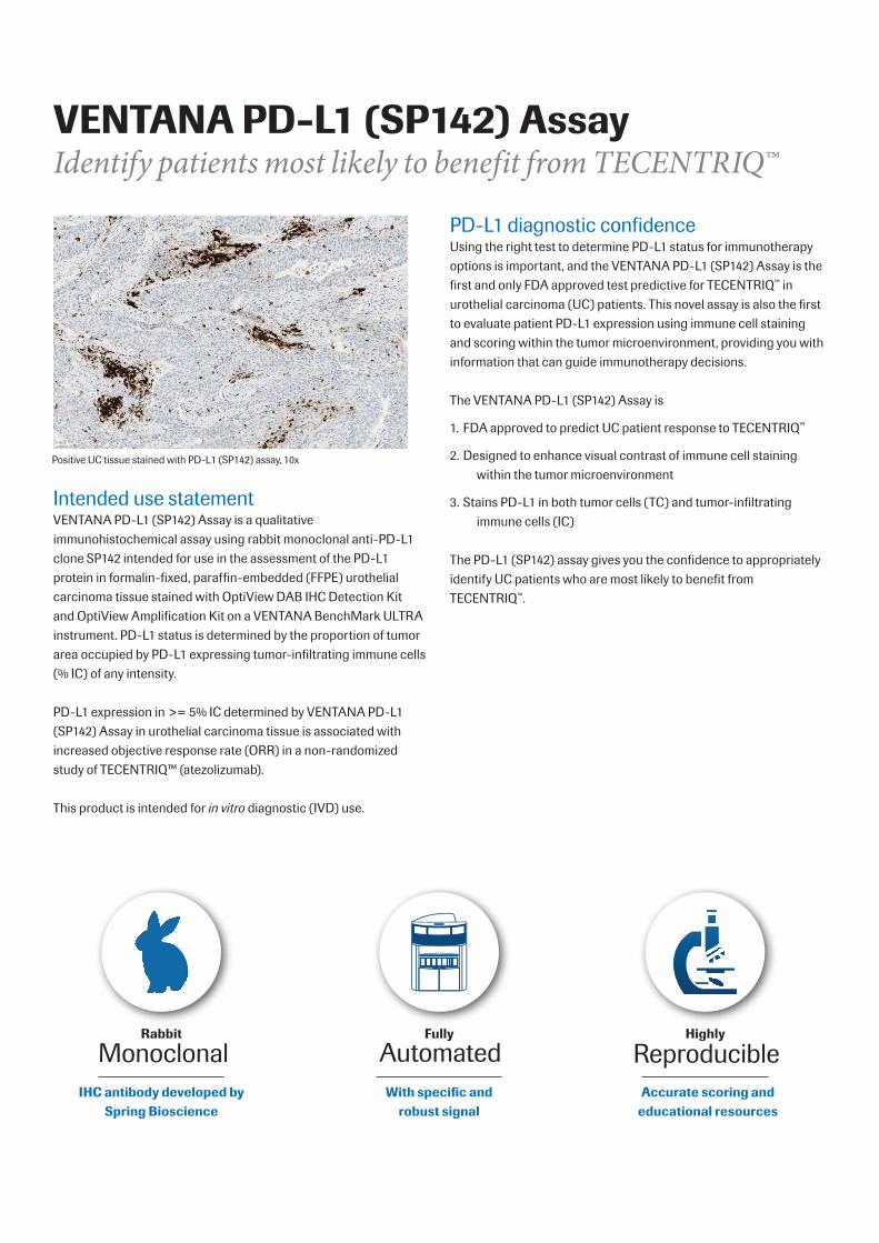
Positive UC tissue stained with PD-L1 (SP142) assay, 10x
Intended use statementVENTANA PD-L1 (SP142) Assay is a qualitative immunohistochemical assay using rabbit monoclonal anti-PD-L1 clone SP142 intended for use in the assessment of the PD-L1 protein in formalin-fixed, paraffin-embedded (FFPE) urothelial carcinoma tissue stained with OptiView DAB IHC Detection Kit and OptiView Amplification Kit on a VENTANA BenchMark ULTRA instrument. PD-L1 status is determined by the proportion of tumor area occupied by PD-L1 expressing tumor-infiltrating immune cells (% IC) of any intensity.
PD-L1 expression in >= 5% IC determined by VENTANA PD-L1 (SP142) Assay in urothelial carcinoma tissue is associated with increased objective response rate (ORR) in a non-randomized study of TECENTRIQ™ (atezolizumab).
This product is intended for in vitro diagnostic (IVD) use.
PD-L1 diagnostic confidence Using the right test to determine PD-L1 status for immunotherapy options is important, and the VENTANA PD-L1 (SP142) Assay is the first and only FDA approved test predictive for TECENTRIQ™ in urothelial carcinoma (UC) patients. This novel assay is also the first to evaluate patient PD-L1 expression using immune cell staining and scoring within the tumor microenvironment, providing you with information that can guide immunotherapy decisions.
The VENTANA PD-L1 (SP142) Assay is
1. FDA approved to predict UC patient response to TECENTRIQ™
2. Designed to enhance visual contrast of immune cell staining within the tumor microenvironment
3. Stains PD-L1 in both tumor cells (TC) and tumor-infiltrating immune cells (IC)
The PD-L1 (SP142) assay gives you the confidence to appropriately identify UC patients who are most likely to benefit from TECENTRIQ™.
With specific and robust signal
AutomatedFully
IHC antibody developed by Spring Bioscience
MonoclonalRabbit
Accurate scoring and educational resources
ReproducibleHighly
PD-L1 in urothelial carcinomaUrothelial carcinoma (also known as urothelial cell carcinoma, transitional cell carcinoma of the urinary tract, or urothelial bladder cancer) is the most common cancer of the urinary system worldwide. The majority of urothelial tumors arise in the bladder with the remainder originating in the renal pelvis, urethra or ureter. Transitional cell carcinoma (TCC) is the most common histologic subtype associated with bladder cancer and accounts for greater than 90% of all urothelial carcinoma cases in the industrialized world. Non-urothelial subtypes (e.g. squamous cell, adenocarcinoma, small cell carcinoma) are more frequent in other areas of the world.5
Elevated PD-L1 expression on tumor cells has been associated with a poor prognosis in patients with urothelial carcinoma.6 PD-L1 is widely expressed in tumor cells and tumor-infiltrating mononuclear cells (TIMCs) and PD-L1 expression in TIMCs appears to be associated with longer survival in patients who developed metastases.7 The association between PD-L1 expression in tumor cells or tumor-infiltrating immune cells and clinical benefit with PD-L1/PD-1 pathway inhibitors has been reported in clinical trials.8,4,9,11 Furthermore, targeting the PD-L1 pathway, based on IC expression, has demonstrated activity in patients with advanced urothelial carcinoma who have failed or refused standard-of-care therapies.10
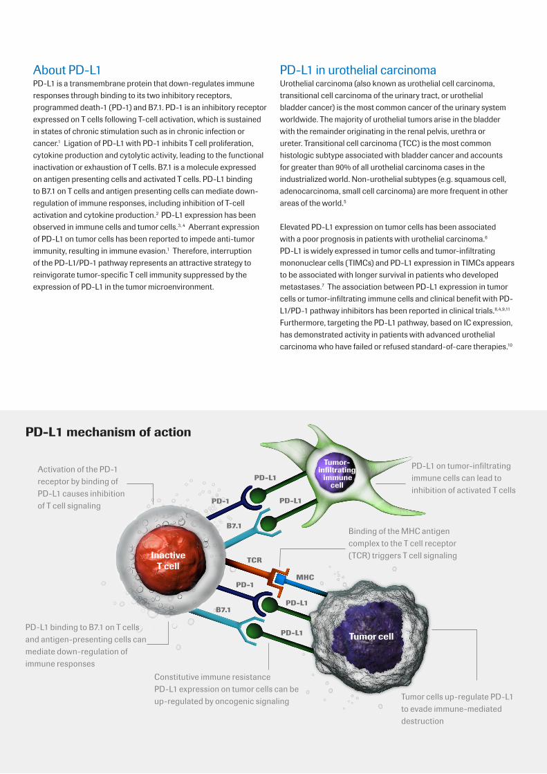
PD-L1 mechanism of action
About PD-L1PD-L1 is a transmembrane protein that down-regulates immune responses through binding to its two inhibitory receptors, programmed death-1 (PD-1) and B7.1. PD-1 is an inhibitory receptor expressed on T cells following T-cell activation, which is sustained in states of chronic stimulation such as in chronic infection or cancer.1 Ligation of PD-L1 with PD-1 inhibits T cell proliferation, cytokine production and cytolytic activity, leading to the functional inactivation or exhaustion of T cells. B7.1 is a molecule expressed on antigen presenting cells and activated T cells. PD-L1 binding to B7.1 on T cells and antigen presenting cells can mediate down-regulation of immune responses, including inhibition of T-cell activation and cytokine production.2 PD-L1 expression has been observed in immune cells and tumor cells.3, 4 Aberrant expression of PD-L1 on tumor cells has been reported to impede anti-tumor immunity, resulting in immune evasion.1 Therefore, interruption of the PD-L1/PD-1 pathway represents an attractive strategy to reinvigorate tumor-specific T cell immunity suppressed by the expression of PD-L1 in the tumor microenvironment.
MHC
TCR
B7.1
PD-L1
Tumor cells up-regulate PD-L1 to evade immune-mediated destruction
Constitutive immune resistance PD-L1 expression on tumor cells can be up-regulated by oncogenic signaling
Binding of the MHC antigen complex to the T cell receptor (TCR) triggers T cell signaling
PD-L1 on tumor-infiltrating immune cells can lead to inhibition of activated T cells
Activation of the PD-1 receptor by binding of PD-L1 causes inhibition of T cell signaling
Inactive T cell
Tumor cell
Tumor-infiltrating immune
cellPD-L1
PD-1
B7.1
PD-L1
PD-1
PD-L1
PD-L1 binding to B7.1 on T cells and antigen-presenting cells can mediate down-regulation of immune responses

VENTANA PD-L1 (SP142) Assay staining in immune cells
Examples of PD-L1 IC staining and score
The PD-L1 (SP142) Assay has been designed to stain and visualize PD-L1 protein on tumor-infiltrating immune cells. The assay is robust and specific across a range of PD-L1 expression levels and provides a strong staining signal through amplification.
High expression in urothelial carcinoma tissue, 10xHigh expression in urothelial carcinoma tissue, 10x
Low expression in urothelial carcinoma tissue, 10xLow expression in urothelial carcinoma tissue, 10x
% IC Staining is < 1% % IC Staining is >= 1% and < 5%
% IC Staining is >= 5% and < 10% % IC Staining is >= 10%

Discover your patients’ immunotherapy path with the VENTANA PD-L1 (SP142) Assay
Tumor-infiltrating immune cell staining assessment PD-L1 expression
Absence of any discernible PD-L1 staining-or- Presence of discernible PD-L1 staining of any intensity in tumor-infiltrating immune cells covering < 5% of tumor area occupied by tumor cells, associated intratumoral, and contiguous peritumoral stroma
<5%
Presence of discernible PD-L1 staining of any intensity in tumor-infiltrating immune cells covering >= 5% of tumor area occupied by tumor cells, associated intratumoral and contiguous peritumoral stroma
>= 5%
PD-L1 clinical relevance in urothelial carcinoma
Response rates
PD-L1 expression ORR*
<5% 9.5%
>= 5% 26.0%
Prevalence
PD-L1 expression Population
<5% 68%
>= 5% 32%
PD-L1 scoring algorithm for urothelial carcinoma
Outcomes and prevelance data based on IMvigor 210 (n = 311)• PD-L1 expression was determined using the VENTANA PD-L1 (SP142) Assay• PD-L1 expression in >= 5% of IC was associated with higher response rates, however levels of PD-L1 expression in <5% of IC did not
preclude response• Trial was an open-label, multicenter, single-arm phase II study that evaluated the safety and efficacy of TECENTRIQ™ in people with
locally advanced or mUC, regardless of PD-L1 expression• ClinicalTrials.gov number NCT02108652
* ORR - Objective response rate defined as a proportion of patients with reduction in tumor burden of a pre-defined amount
PD-L1 expression in the tumor microenvironment
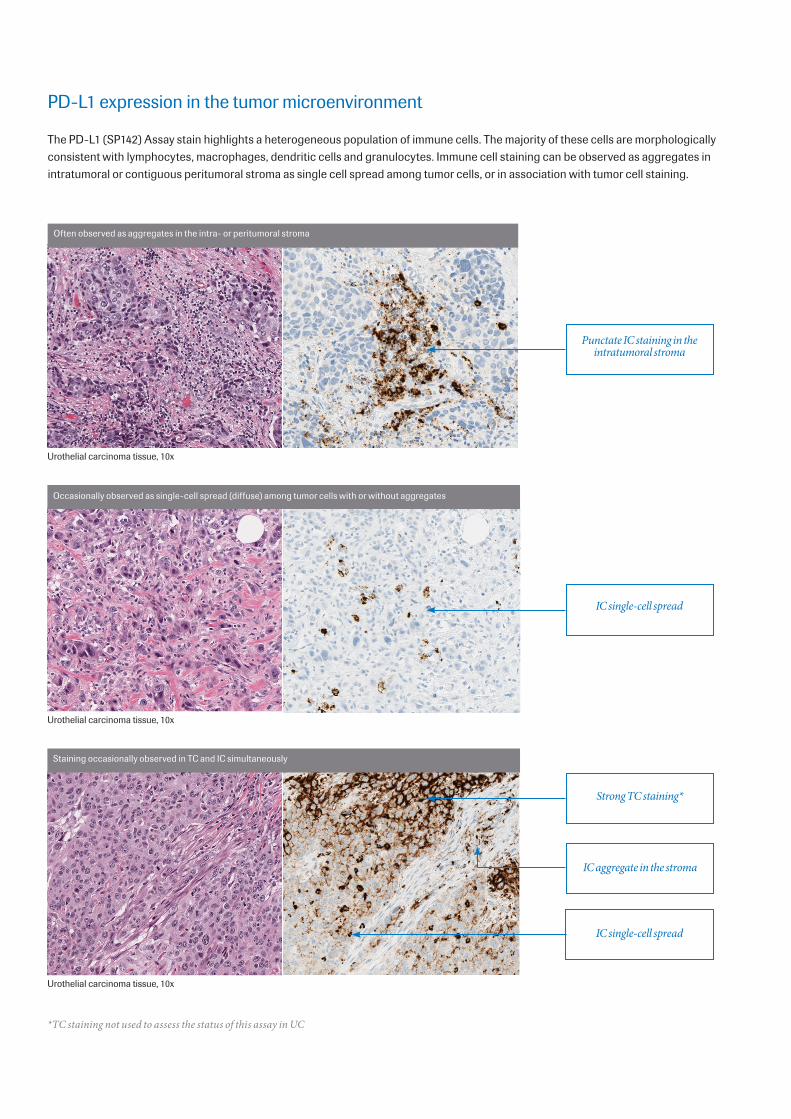
The PD-L1 (SP142) Assay stain highlights a heterogeneous population of immune cells. The majority of these cells are morphologically consistent with lymphocytes, macrophages, dendritic cells and granulocytes. Immune cell staining can be observed as aggregates in intratumoral or contiguous peritumoral stroma as single cell spread among tumor cells, or in association with tumor cell staining.
Urothelial carcinoma tissue, 10x
Urothelial carcinoma tissue, 10x
Urothelial carcinoma tissue, 10x
Punctate IC staining in the intratumoral stroma
IC single-cell spread
Strong TC staining*
IC aggregate in the stroma
IC single-cell spread
*TC staining not used to assess the status of this assay in UC
Often observed as aggregates in the intra- or peritumoral stroma
Occasionally observed as single-cell spread (diffuse) among tumor cells with or without aggregates
Staining occasionally observed in TC and IC simultaneously

PD-L1 assay performance
Inter-laboratory reproducibilityPositive agreement % (95% CI)*
Negative agreement % (95% CI)
Overall agreement % (95% CI)
Overall average agreement (across sites, days and readers)
98.3% (96.6-99.2%)
87.4% (83.8-90.2%)
92.8% (90.9-94.4%)
Between-reader agreement (average of all sites, two readers/site)
89.3% (78.1-96.0%)
86.6% (75.1-94.6%)
88.1% (84.6-90.8%)
Reader precision Positive agreement % (95% CI)
Negative agreement % (95% CI)
Overall agreement % (95% CI)
Inter-reader precision (average of all three readers’ comparisons)
92.6% (84.7-96.6%)
87.4% (83.8-90.2%)
92.8% (90.9-94.4%)
Intra-reader precision (average of all three readers’ agreement rates between first and second reads)
89.3% (77.6-95.3%)
86.6% (75.1-94.6%)
88.1% (84.6-90.8%)
* CI- confidence interval
VENTANA PD-L1 (SP142) Assay OptiView DAB IHC Detection Kit OptiView Amplification KitCatalog number 740-4859 07709374001 760-700 06396500001 860-099 06718663001Quantity 50 tests 250 tests 250 testsPositive control TonsilSpecies RabbitLocalization Membranous and/or Cytoplasmic

Ventana Medical Systems, Inc.1910 E. Innovation Park DriveTucson, AZ 85755USA1 520 877 21551 800 227 2155
www.roche.com www.ventana.com
© 2016 Ventana Medical Systems, Inc.
VENTANA, BENCHMARK and OPTIVIEW are trademarks of Roche. All other trademarks are the property of their respective owners. 6066A-6 0516
1. Blank, C and Mackensen, A, Contribution of the PD-L1/PD-1 pathway to T-cell exhaustion: an update on implications for chronic infections and tumor evasion. Cancer Immunol Immunother, 2007. 56(5): p. 739-745.
2. Butte MJ, Keir ME, Phamduy TB, et al. Programmed death-1 ligand 1 interacts specifically with the B7-1 costimulatory molecule to inhibit T cell responses. Immunity. 2007;27(1):111-122.
3. Dong H, Zhu G, Tamada K, Chen L. B7-H1, a third member of the B7 family, co-stimulates T-cell proliferation and interleukin-10 secretion. Nat Med. 1999;5(12):1365-1369.
4. Herbst RS, Soria JC, Kowanetz M, et al. Predictive correlates of response to the anti-PD-L1 antibody MPDL3280A in cancer patients. Nature. 2014;515(7528):563-567.
5. Chalasani V, Chin JL, Izawa JI. Histologic variants of urothelial bladder cancer and nonurothelial histology in bladder cancer. Can Urol Assoc J. 2009;3(6 Suppl 4):S193-198.
6. Nakanishi J, Wada Y, Matsumoto K, et al. Overexpression of B7-H1 (PD-L1) significantly associates with tumor grade and postoperative prognosis in human urothelial cancers. Cancer Immunol Immunother. 2007;56(8):1173-1182.
7. Bellmunt, J, Mullane, SA, Werner, L, et al., Association of PD-L1 expression on tumor-infiltrating mononuclear cells and overall survival in patients with urothelial carcinoma. Annals of oncology : official journal of the European Society for Medical Oncology / ESMO, 2015. 26(4): p. 812-817.
1. Brahmer JR, Drake CG, Wollner I, et al. Phase I study of single-agent anti-programmed death-1 (MDX-1106) in refractory solid tumors: safety, clinical activity, pharmacodynamics, and immunologic correlates. J Clin Oncol. 2010;28(19):3167-3175.
2. Topalian SL, Hodi FS, Brahmer JR, et al. Safety, activity, and immune correlates of anti-PD-1 antibody in cancer. N Engl J Med. 2012;366(26):2443-2454.
3. Powles T, Eder JP, Fine GD, et al. MPDL3280A (anti-PD-L1) treatment leads to clinical activity in metastatic bladder cancer. Nature. 2014;515(7528):558-562.
4. Fehrenbacher L, Spira A, Ballinger M, et al. Atezolizumab versus docetaxel for patients with previously treated non-small-cell lung cancer (POPLAR): a multicentre, open-label, phase 2 randomised controlled trial. The Lancet. 2016. Web.
References