VENEZUELAN EQUINE ENCEPHALOMYELITIS STANDARD OPERATING ... · VENEZUELAN EQUINE ENCEPHALOMYELITIS...
17
VENEZUELAN EQUINE ENCEPHALOMYELITIS STANDARD OPERATING PROCEDURES: 1. OVERVIEW OF ETIOLOGY AND ECOLOGY DRAFT AUGUST 2013
Transcript of VENEZUELAN EQUINE ENCEPHALOMYELITIS STANDARD OPERATING ... · VENEZUELAN EQUINE ENCEPHALOMYELITIS...
VENEZUELAN EQUINE ENCEPHALOMYELITIS STANDARD OPERATING PROCEDURES:1. OVERVIEW OF ETIOLOGY AND ECOLOGY
DRAFT AUGUST 2013
File name: VEE_FAD_PReP_E&E_SOP SOP number: SOP001
Lead section: Preparedness and Incident Coordination Version number: 1.0
Effective date: August 2013 Review date: August 2015
SOP Manual ii VEE Etiology and Ecology
The Foreign Animal Disease Preparedness and Response Plan (FAD PReP) Standard Operating
Procedures (SOPs) provide operational guidance for responding to an animal health emergency
in the United States.
These draft SOPs are under ongoing review. This document was last updated in August 2013.
Please send questions or comments to:
Preparedness and Incident Coordination
Veterinary Services
Animal and Plant Health Inspection Service
U.S. Department of Agriculture
4700 River Road, Unit 41
Riverdale, Maryland 20737-1231
Telephone: (301) 851-3595 Fax: (301) 734-7817
E-mail: [email protected]
While best efforts have been used in developing and preparing the FAD PReP SOPs, the U.S.
Government, U.S. Department of Agriculture (USDA), and the Animal and Plant Health
Inspection Service and other parties, such as employees and contractors contributing to this
document, neither warrant nor assume any legal liability or responsibility for the accuracy,
completeness, or usefulness of any information or procedure disclosed. The primary purpose of
these FAD PReP SOPs is to provide operational guidance to those government officials
responding to a foreign animal disease outbreak. It is only posted for public access as a reference.
The FAD PReP SOPs may refer to links to various other Federal and State agencies and private
organizations. These links are maintained solely for the user's information and convenience. If
you link to such site, please be aware that you are then subject to the policies of that site. In
addition, please note that USDA does not control and cannot guarantee the relevance, timeliness,
or accuracy of these outside materials. Further, the inclusion of links or pointers to particular
items in hypertext is not intended to reflect their importance, nor is it intended to constitute
approval or endorsement of any views expressed, or products or services offered, on these
outside websites, or the organizations sponsoring the websites.
Trade names are used solely for the purpose of providing specific information. Mention of a
trade name does not constitute a guarantee or warranty of the product by USDA or an
endorsement over other products not mentioned.
USDA prohibits discrimination in all its programs and activities on the basis of race, color,
national origin, sex, religion, age, disability, political beliefs, sexual orientation, or marital or
family status. (Not all prohibited bases apply to all programs.) Persons with disabilities who
require alternative means for communication of program information (Braille, large print,
audiotape, etc.) should contact USDA’s TARGET Center at (202) 720-2600 (voice and
telecommunications device for the deaf [TDD]).
To file a complaint of discrimination, write USDA, Director, Office of Civil Rights, Room
326-W, Whitten Building, 1400 Independence Avenue SW, Washington, DC 20250-9410 or call
(202) 720-5964 (voice and TDD). USDA is an equal opportunity provider and employer.
SOP Manual iii VEE Etiology and Ecology
Contents
1.1 Introduction ................................................................................................................. 1-2
1.1.2 Further Information ............................................................................................. 1-2
1.2 Purpose........................................................................................................................ 1-3
1.3 Etiology ....................................................................................................................... 1-3
1.3.1 Name…….. ........................................................................................................ 1-3 1.3.2 Virus Characteristics ........................................................................................... 1-3
1.3.3 Morphology ........................................................................................................ 1-3 1.3.4 VEEV Serotypes and Strains ............................................................................... 1-3
1.3.5 Differential Diagnoses ........................................................................................ 1-5 1.4 Ecology ....................................................................................................................... 1-5
1.4.1 General Overview ............................................................................................... 1-5 1.4.2 Susceptible Species ............................................................................................. 1-7
1.4.3 Introduction and Transmission of VEE ............................................................... 1-8 1.4.3.1 Vector Transmission ........................................................................................ 1-8
1.4.3.2 Air/Windborne Transmission ........................................................................... 1-9 1.4.4 Incubation Period ................................................................................................ 1-9
1.4.5 Morbidity and Mortality ...................................................................................... 1-9 1.4.5.1 Clinical Signs ................................................................................................. 1-10
1.5 Environmental Persistence of VEEV ......................................................................... 1-11
1.6 Risk of Introduction into the United States................................................................. 1-11
Attachment 1.A References and Selected Resources .............................................................. 1-12
Attachment 1.B Abbreviations .............................................................................................. 1-14
SOP Manual 1-1 VEE Etiology and Ecology
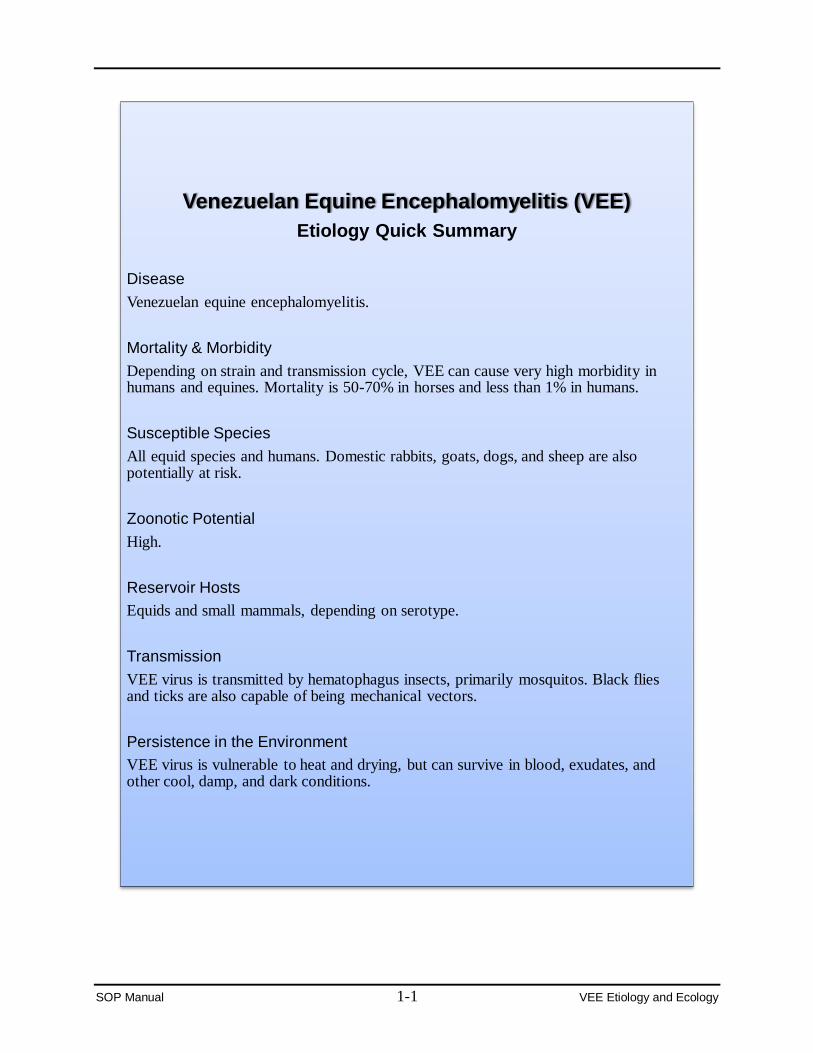
Venezuelan Equine Encephalomyelitis (VEE)
Etiology Quick Summary
Disease
Venezuelan equine encephalomyelitis.
Mortality & Morbidity
Depending on strain and transmission cycle, VEE can cause very high morbidity in humans and equines. Mortality is 50-70% in horses and less than 1% in humans.
Susceptible Species
All equid species and humans. Domestic rabbits, goats, dogs, and sheep are also potentially at risk.
Zoonotic Potential
High.
Reservoir Hosts
Equids and small mammals, depending on serotype.
Transmission
VEE virus is transmitted by hematophagus insects, primarily mosquitos. Black flies and ticks are also capable of being mechanical vectors.
Persistence in the Environment
VEE virus is vulnerable to heat and drying, but can survive in blood, exudates, and other cool, damp, and dark conditions.
SOP Manual 1-2 VEE Etiology and Ecology
1.1 Introduction
Venezuelan equine encephalomyelitis (VEE) is caused by the VEE virus (VEEV). The vector-
borne virus, predominantly transmitted by mosquitoes, primarily affects both domestic and wild
equid species and humans; the life cycle of VEEV is complex and affects many different
mammalian and mosquito species. The disease is characterized in horses by fever, loss of
appetite, and disorders of the central nervous system, such as muscle impairment, blindness, and
convulsions.
The spread of VEE varies in speed and intensity according to the viral subtype and densities of
mosquito populations, as transmission occurs by the vector biting an infected animal and then
feeding on a new host. Epizootic subtypes of VEEV are amplified in equids, may spread rapidly,
and can be highly pathogenic in horses, donkeys, mules, and other equids. In humans, the disease
results in flu-like symptoms for healthy individuals but can cause severe illness and death in
immune compromised, young, or older people.1
Outbreaks of VEE can be severe, causing high morbidity in humans and equines. VEE has the
potential to cause high mortality in equids. There has not been an epizootic outbreak of VEE in
the United States since 1971.2
1.1.1 Goals
As a preparedness goal, the Animal and Plant Health Inspection Service (APHIS) will provide
etiology and ecology summaries for VEE, and update these summaries at regular intervals.
As a response goal, the Unified Command and stakeholders will have a common set of etiology
and ecology definitions and descriptions, to ensure proper understanding of VEE when
establishing or revising goals, objectives, strategies, and procedures.
1.1.2 Further Information
This document is intended to be an overview, focusing on the threat of VEE to domestic equine
and human populations. Additional resources on VEE, as well as the articles cited in this
standard operating procedure (SOP), are listed in Attachment 1.A. Case definitions and
laboratory criteria are provided in the VEE Draft Case Definition from the APHIS Centers for
Epidemiology and Animal Health, National Surveillance Unit. Case definitions are under
ongoing review. This document does not comprehensively discuss vaccination, or its effects on
immunity.
Publicly available documents are available here:
http://www.aphis.usda.gov/animal_health/emergency_management/materials_ref.shtml, and
they are also on the APHIS Intranet (http://inside.aphis.usda.gov/vs/em/fadprep.shtml) for
APHIS employees.
1 Estrada-Franco JG, Navarro-Lopez R, Freier JE, Cordova D, Clements T, Moncayo A, Kang W, Gomez-
Hernandez C, Rodriguez-Dominguez G, Ludwig GV, Weaver SC. 2004. “Venezuelan Equine Encephalitis Virus,
Southern Mexico.” Emerging Infectious Diseases. 10(12): 2113-2121. 2 USDA APHIS. 2013. Draft Case Definition of Venezuelan Equine Encephalomyelitis.
SOP Manual 1-3 VEE Etiology and Ecology
1.2 Purpose
This document provides responders and stakeholders with a common understanding of the
disease agent.
1.3 Etiology
1.3.1 Name
This disease is called Venezuelan equine encephalomyelitis, and it is also often called
Venezuelan equine encephalitis. Some of the enzootic viral species within the VEE family are
known as Mosso das Pedras virus, Everglades virus, Mucambo virus, Tonate virus, Pixuna virus,
Cabassou virus, and Rio Negro virus.3
1.3.2 Virus Characteristics
According to the International Committee on Taxonomy of Viruses, this disease has the
following characteristics:
Family: Togaviridae
Genera: Alphavirus
Baltimore Classification: Group IV (+) ssRNA. 4
1.3.3 Morphology
The VEEV is a single-stranded ribonucleic acid (RNA) virus, approximately 70 nm in diameter,
with icosahedral symmetry. The 5’ end of the genome encodes four nonstructural proteins, nsP1,
nsP2, nsP3, and nsP4, and the 3’ end is responsible for three structural proteins, the capsid and
the E1 and E2 envelope proteins. The nonstructural proteins are involved in replicating the viral
genome and functions within the host’s cytoplasm.5
1.3.4 VEEV Serotypes and Strains
There are 6 subtypes (I-IV) within the VEEV complex. The epizootic strains are distributed
within subtype I, serotypes I-AB and I-C, and are generally thought to be responsible for major
epizootics and epidemics in equid species and humans; serotypes I-AB and I-C are believed to
have descended from enzootic strains. Subtypes ID and IE are comprised of enzootic, equine-
avirulent strains, but some are known to cause disease and death in humans. Recently, a subtype
IE strain in Mexico also caused neurologic signs in equine hosts but did not produce high-titer
viremia.6
3 Weaver SC, Barrett ADT. 2004. “Transmission cycles, host range, evolution and emergence of arboviral disease.” Nature Reviews Microbiology. 2: 789-801. 4 OIE. 2009. Technical Disease Card for Venezuelan equine encephalitis.
5 Weaver SC, Ferro C, Barrera R, Boshell J, Navarro J-C. 2004. “Venezuelan Equine Encephalitis.” Annual Review
of Entomology. 49: 141-174. 6 Weaver et al., 2004.

SOP Manual 1-4 VEE Etiology and Ecology
Table 1-1. The VEE Complex of Viruses7
Subtype Species Serotype Transmission Pattern
I
VEE virus AB Epizootic
VEE virus C
VEE virus D
Enzootic
VEE virus E
Mosso das Pedras virus F
II Everglades virus
III
Mucambo virus A
Tonate virus* B
Mucambo virus C
Mucambo virus D
IV Pixuna
V Cabassou virus
VI Rio Negro virus
Source: Weaver et al., 2004
*Bijou bridge virus is a well-known strain of Tonate virus that has been isolated in Colorado swallow bugs.
Serotype I-D viruses can alter via mutations in the E2 envelope glycoprotein to serotype I-AB or
I-C viruses that are able to amplify in equid hosts and cause epizootic outbreaks.5 For example,
the enzootic I-D strains differs from the epizootic I-C strain by only a few nucleotides, thus
epizootic, equine virulent disease can arise where enzootic disease is endemic.8 Changes in the
E2 protein are responsible for the higher equine virulence and host specificity that enable
epizootic outbreaks of VEE.9
7 Weaver et al., 2004. 8 Rico-Hesse R, Weaver SC, de Siger J, Medina G, Salas RA. 1995. “Emergence of a new epidemic/epizootic
Venezuelan equine encephalitis virus in South America.” PNAS. 92: 5278-5281. 9 Weaver et al, 2004.
SOP Manual 1-5 VEE Etiology and Ecology
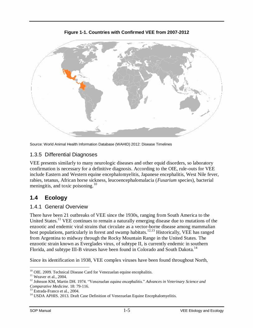
Figure 1-1. Countries with Confirmed VEE from 2007-2012
Source: World Animal Health Information Database (WAHID) 2012: Disease Timelines
1.3.5 Differential Diagnoses
VEE presents similarly to many neurologic diseases and other equid disorders, so laboratory
confirmation is necessary for a definitive diagnosis. According to the OIE, rule-outs for VEE
include Eastern and Western equine encephalomyelitis, Japanese encephalitis, West Nile fever,
rabies, tetanus, African horse sickness, leucoencephalomalacia (Fusarium species), bacterial
meningitis, and toxic poisoning.10
1.4 Ecology
1.4.1 General Overview
There have been 21 outbreaks of VEE since the 1930s, ranging from South America to the
United States.11
VEE continues to remain a naturally emerging disease due to mutations of the
enzootic and endemic viral strains that circulate as a vector-borne disease among mammalian
host populations, particularly in forest and swamp habitats.12,13
Historically, VEE has ranged
from Argentina to midway through the Rocky Mountain Range in the United States. The
enzootic strain known as Everglades virus, of subtype II, is currently endemic in southern
Florida, and subtype III-B viruses have been found in Colorado and South Dakota.14
Since its identification in 1938, VEE complex viruses have been found throughout North,
10 OIE. 2009. Technical Disease Card for Venezuelan equine encephalitis. 11 Weaver et al., 2004. 12 Johnson KM, Martin DH. 1974. “Venezuelan equine encephalitis.” Advances in Veterinary Science and
Comparative Medicine. 18: 79-116. 13 Estrada-Franco et al., 2004. 14 USDA APHIS. 2013. Draft Case Definition of Venezuelan Equine Encephalomyelitis.
SOP Manual 1-6 VEE Etiology and Ecology
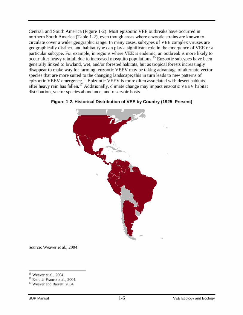
Central, and South America (Figure 1-2). Most epizootic VEE outbreaks have occurred in
northern South America (Table 1-2), even though areas where enzootic strains are known to
circulate cover a wider geographic range. In many cases, subtypes of VEE complex viruses are
geographically distinct, and habitat type can play a significant role in the emergence of VEE or a
particular subtype. For example, in regions where VEE is endemic, an outbreak is more likely to
occur after heavy rainfall due to increased mosquito populations.15
Enzootic subtypes have been
generally linked to lowland, wet, and/or forested habitats, but as tropical forests increasingly
disappear to make way for farming, enzootic VEEV may be taking advantage of alternate vector
species that are more suited to the changing landscape; this in turn leads to new patterns of
epizootic VEEV emergence.16
Epizootic VEEV is more often associated with desert habitats
after heavy rain has fallen.17
Additionally, climate change may impact enzootic VEEV habitat
distribution, vector species abundance, and reservoir hosts.
Figure 1-2. Historical Distribution of VEE by Country (1925–Present)
Source: Weaver et al., 2004
15 Weaver et al., 2004. 16 Estrada-Franco et al., 2004. 17 Weaver and Barrett, 2004.
SOP Manual 1-7 VEE Etiology and Ecology
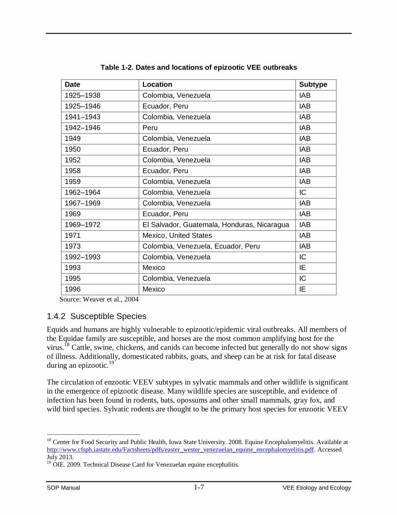
Table 1-2. Dates and locations of epizootic VEE outbreaks
Date Location Subtype
1925–1938 Colombia, Venezuela IAB
1925–1946 Ecuador, Peru IAB
1941–1943 Colombia, Venezuela IAB
1942–1946 Peru IAB
1949 Colombia, Venezuela IAB
1950 Ecuador, Peru IAB
1952 Colombia, Venezuela IAB
1958 Ecuador, Peru IAB
1959 Colombia, Venezuela IAB
1962–1964 Colombia, Venezuela IC
1967–1969 Colombia, Venezuela IAB
1969 Ecuador, Peru IAB
1969–1972 El Salvador, Guatemala, Honduras, Nicaragua IAB
1971 Mexico, United States IAB
1973 Colombia, Venezuela, Ecuador, Peru IAB
1992–1993 Colombia, Venezuela IC
1993 Mexico IE
1995 Colombia, Venezuela IC
1996 Mexico IE
Source: Weaver et al., 2004
1.4.2 Susceptible Species
Equids and humans are highly vulnerable to epizootic/epidemic viral outbreaks. All members of
the Equidae family are susceptible, and horses are the most common amplifying host for the
virus.18
Cattle, swine, chickens, and canids can become infected but generally do not show signs
of illness. Additionally, domesticated rabbits, goats, and sheep can be at risk for fatal disease
during an epizootic.19
The circulation of enzootic VEEV subtypes in sylvatic mammals and other wildlife is significant
in the emergence of epizootic disease. Many wildlife species are susceptible, and evidence of
infection has been found in rodents, bats, opossums and other small mammals, gray fox, and
wild bird species. Sylvatic rodents are thought to be the primary host species for enzootic VEEV
18 Center for Food Security and Public Health, Iowa State University. 2008. Equine Encephalomyelitis. Available at
http://www.cfsph.iastate.edu/Factsheets/pdfs/easter_wester_venezuelan_equine_encephalomyelitis.pdf. Accessed
July 2013. 19 OIE. 2009. Technical Disease Card for Venezuelan equine encephalitis.
SOP Manual 1-8 VEE Etiology and Ecology
due to their continually high rates of infection, their immunity, and their moderately high levels
of viremia.20
1.4.3 Introduction and Transmission of VEE
VEE is primarily transmitted by mosquito vectors, but the virus has been identified in black flies
and ticks as well. Humans are typically infected by mosquitos that have contracted an epizootic
virus from an equid source. Although some human viraemias may reach levels high enough to
infect mosquitos, people do not play a significant role in the transmission of VEEV. The high
viral titers produced in horses and other equids are essential to the transmission of VEEV in an
outbreak, because it leads to the infection of less competent mosquito hosts. Generally, arbovirus
hosts are small in size, such as the mammalian reservoirs of enzootic virus. The comparatively
large size of equine hosts enables epizootic VEE viruses to spread widely and quickly.
Additionally, outbreaks can be wider still if vector populations are simultaneously high due to
seasonal patterns or wet weather events.21
1.4.3.1 Vector Transmission
As a group of arboviruses, the VEE complex exploits multiple mosquito vectors, with different
species and genera of mosquito transmitting disease depending on the viral subtype and infected
species. The mosquito genera Ochlerotatus, Anopheles, Culex, Deinocerites, Mansonia, and
Psorophora have all been found to be infected with epizootic strains of VEEV. Enzootic VEE
has been found to be transmitted by mosquito subgenus Culex, but epizootic outbreaks are more
commonly associated with floodwater species, such as those of the Ochlerotatus and Psorophora
genera.22
In the one epidemic in the United States, in Texas (1971), Psorophora confinnis was
implicated as the most important VEE vector species.23
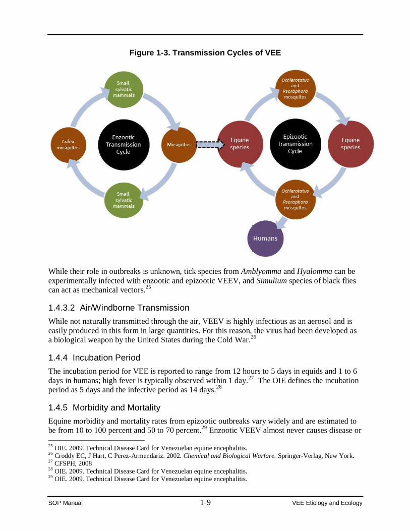
The transmission cycles of VEEV between enzootic and epizootic hosts and vectors are complex.
Figure 1-3 (adapted from Weaver et al., 2004) depicts the relationship between enzootic and
epizootic VEE hosts, vectors, and susceptible species; the dotted arrow reflects the mutation of
enzootic VEEV subtypes to epizootic, equine-virulent subtypes.24
20 Weaver et al., 2004. 21 Weaver et al., 2004. 22 Weaver and Barrett, 2004. 23 Bowen GS, Calisher CH. 1976. “Virological and serological studies of Venezuelan equine encephalomyelitis in
humans.” Journal of Clinical Microbiology. 4(1): 22-27. 24 Weaver et al., 2004.
SOP Manual 1-9 VEE Etiology and Ecology
Figure 1-3. Transmission Cycles of VEE
While their role in outbreaks is unknown, tick species from Amblyomma and Hyalomma can be
experimentally infected with enzootic and epizootic VEEV, and Simulium species of black flies
can act as mechanical vectors.25
1.4.3.2 Air/Windborne Transmission
While not naturally transmitted through the air, VEEV is highly infectious as an aerosol and is
easily produced in this form in large quantities. For this reason, the virus had been developed as
a biological weapon by the United States during the Cold War.26
1.4.4 Incubation Period
The incubation period for VEE is reported to range from 12 hours to 5 days in equids and 1 to 6
days in humans; high fever is typically observed within 1 day.27
The OIE defines the incubation
period as 5 days and the infective period as 14 days.28
1.4.5 Morbidity and Mortality
Equine morbidity and mortality rates from epizootic outbreaks vary widely and are estimated to
be from 10 to 100 percent and 50 to 70 percent.29
Enzootic VEEV almost never causes disease or
25 OIE. 2009. Technical Disease Card for Venezuelan equine encephalitis. 26 Croddy EC, J Hart, C Perez-Armendariz. 2002. Chemical and Biological Warfare. Springer-Verlag, New York. 27 CFSPH, 2008 28 OIE. 2009. Technical Disease Card for Venezuelan equine encephalitis. 29 OIE. 2009. Technical Disease Card for Venezuelan equine encephalitis.
SOP Manual 1-10 VEE Etiology and Ecology
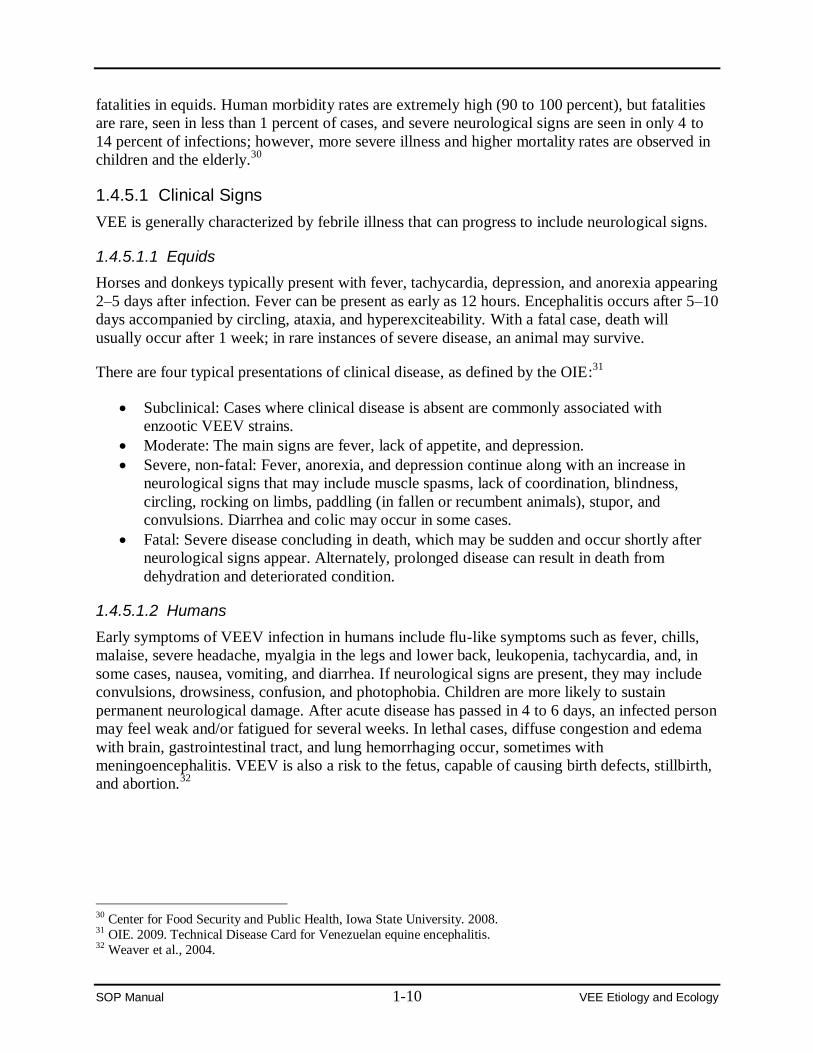
fatalities in equids. Human morbidity rates are extremely high (90 to 100 percent), but fatalities
are rare, seen in less than 1 percent of cases, and severe neurological signs are seen in only 4 to
14 percent of infections; however, more severe illness and higher mortality rates are observed in
children and the elderly.30
1.4.5.1 Clinical Signs
VEE is generally characterized by febrile illness that can progress to include neurological signs.
1.4.5.1.1 Equids
Horses and donkeys typically present with fever, tachycardia, depression, and anorexia appearing
2–5 days after infection. Fever can be present as early as 12 hours. Encephalitis occurs after 5–10
days accompanied by circling, ataxia, and hyperexciteability. With a fatal case, death will
usually occur after 1 week; in rare instances of severe disease, an animal may survive.
There are four typical presentations of clinical disease, as defined by the OIE:31
Subclinical: Cases where clinical disease is absent are commonly associated with
enzootic VEEV strains.
Moderate: The main signs are fever, lack of appetite, and depression.
Severe, non-fatal: Fever, anorexia, and depression continue along with an increase in
neurological signs that may include muscle spasms, lack of coordination, blindness,
circling, rocking on limbs, paddling (in fallen or recumbent animals), stupor, and
convulsions. Diarrhea and colic may occur in some cases.
Fatal: Severe disease concluding in death, which may be sudden and occur shortly after
neurological signs appear. Alternately, prolonged disease can result in death from
dehydration and deteriorated condition.
1.4.5.1.2 Humans
Early symptoms of VEEV infection in humans include flu-like symptoms such as fever, chills,
malaise, severe headache, myalgia in the legs and lower back, leukopenia, tachycardia, and, in
some cases, nausea, vomiting, and diarrhea. If neurological signs are present, they may include
convulsions, drowsiness, confusion, and photophobia. Children are more likely to sustain
permanent neurological damage. After acute disease has passed in 4 to 6 days, an infected person
may feel weak and/or fatigued for several weeks. In lethal cases, diffuse congestion and edema
with brain, gastrointestinal tract, and lung hemorrhaging occur, sometimes with
meningoencephalitis. VEEV is also a risk to the fetus, capable of causing birth defects, stillbirth,
and abortion.32
30 Center for Food Security and Public Health, Iowa State University. 2008. 31 OIE. 2009. Technical Disease Card for Venezuelan equine encephalitis. 32 Weaver et al., 2004.
SOP Manual 1-11 VEE Etiology and Ecology
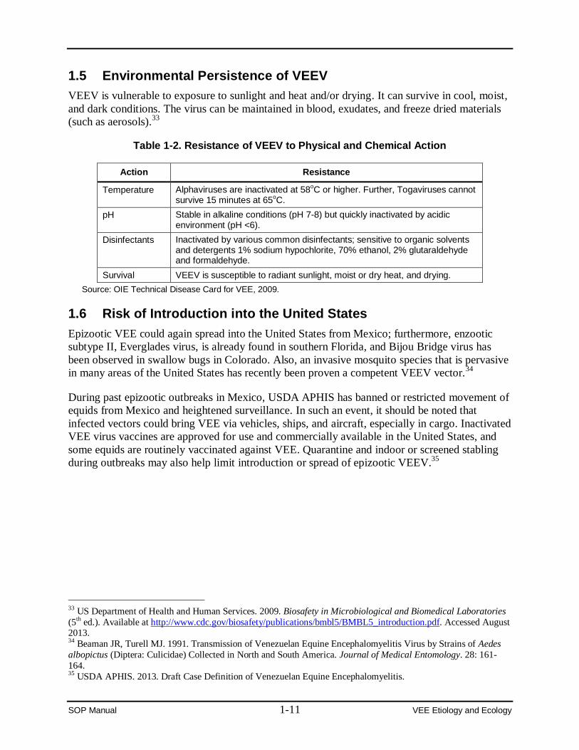
1.5 Environmental Persistence of VEEV
VEEV is vulnerable to exposure to sunlight and heat and/or drying. It can survive in cool, moist,
and dark conditions. The virus can be maintained in blood, exudates, and freeze dried materials
(such as aerosols).33
Table 1-2. Resistance of VEEV to Physical and Chemical Action
Action Resistance
Temperature Alphaviruses are inactivated at 58oC or higher. Further, Togaviruses cannot
survive 15 minutes at 65oC.
pH Stable in alkaline conditions (pH 7-8) but quickly inactivated by acidic environment (pH <6).
Disinfectants Inactivated by various common disinfectants; sensitive to organic solvents and detergents 1% sodium hypochlorite, 70% ethanol, 2% glutaraldehyde and formaldehyde.
Survival VEEV is susceptible to radiant sunlight, moist or dry heat, and drying.
Source: OIE Technical Disease Card for VEE, 2009.
1.6 Risk of Introduction into the United States
Epizootic VEE could again spread into the United States from Mexico; furthermore, enzootic
subtype II, Everglades virus, is already found in southern Florida, and Bijou Bridge virus has
been observed in swallow bugs in Colorado. Also, an invasive mosquito species that is pervasive
in many areas of the United States has recently been proven a competent VEEV vector.34
During past epizootic outbreaks in Mexico, USDA APHIS has banned or restricted movement of
equids from Mexico and heightened surveillance. In such an event, it should be noted that
infected vectors could bring VEE via vehicles, ships, and aircraft, especially in cargo. Inactivated
VEE virus vaccines are approved for use and commercially available in the United States, and
some equids are routinely vaccinated against VEE. Quarantine and indoor or screened stabling
during outbreaks may also help limit introduction or spread of epizootic VEEV.35
33 US Department of Health and Human Services. 2009. Biosafety in Microbiological and Biomedical Laboratories
(5th ed.). Available at http://www.cdc.gov/biosafety/publications/bmbl5/BMBL5_introduction.pdf. Accessed August 2013. 34 Beaman JR, Turell MJ. 1991. Transmission of Venezuelan Equine Encephalomyelitis Virus by Strains of Aedes
albopictus (Diptera: Culicidae) Collected in North and South America. Journal of Medical Entomology. 28: 161-
164. 35 USDA APHIS. 2013. Draft Case Definition of Venezuelan Equine Encephalomyelitis.
SOP Manual 1-12 VEE Etiology and Ecology
Attachment 1.A References and Selected Resources
Beaman JR, Turell MJ. 1991. Transmission of Venezuelan Equine Encephalomyelitis Virus by
Strains of Aedes albopictus (Diptera: Culicidae) Collected in North and South America. Journal
of Medical Entomology. 28: 161-164.
Bowen GS, Calisher CH. 1976. “Virological and serological studies of Venezuelan equine
encephalomyelitis in humans.” Journal of Clinical Microbiology. 4: 22-27.
Center for Food Security and Public Health, Iowa State University. 2008. Equine
Encephalomyelitis. Available at
http://www.cfsph.iastate.edu/Factsheets/pdfs/easter_wester_venezuelan_equine_encephalomyelit
is.pdf. Accessed July 2013.
Croddy EC, J Hart, C Perez-Armendariz. 2002. Chemical and Biological Warfare. Springer-
Verlag, New York.
Estrada-Franco JG, Navarro-Lopez R, Freier JE, Cordova D, Clements T, Moncayo A, Kang W,
Gomez-Hernandez C, Rodriguez-Dominguez G, Ludwig GV, Weaver SC. 2004. “Venezuelan
Equine Encephalitis Virus, Southern Mexico.” Emerging Infectious Diseases. 10: 2113-2121.
Johnson KM, Martin DH. 1974. “Venezuelan equine encephalitis.” Advances in Veterinary
Science and Comparative Medicine. 18: 79-116.
OIE. 2009. Technical Disease Card for Venezuelan equine encephalitis.
Rico-Hesse R, Weaver SC, de Siger J, Medina G, Salas RA. 1995. “Emergence of a new
epidemic/epizootic Venezuelan equine encephalitis virus in South America.” PNAS. 92: 5278-
5281.
USDA APHIS. 2013. Draft Case Definition Venezuelan Equine Encephalomyelitis.
US Department of Health and Human Services. 2009. Biosafety in Microbiological and
Biomedical Laboratories (5th ed.). Available at
http://www.cdc.gov/biosafety/publications/bmbl5/BMBL5_introduction.pdf. Accessed August
2013.
Weaver SC, Barrett ADT. 2004. “Transmission cycles, host range, evolution and emergence of
arboviral disease.” Nature Reviews Microbiology. 2: 789-801.
Weaver SC, Ferro C, Barrera R, Boshell J, Navarro J-C. 2004. “Venezuelan Equine
Encephalitis.” Annual Review of Entomology. 49: 141-174.
SOP Manual 1-13 VEE Etiology and Ecology
Weaver SC, Salas R, Rico-Hesse R, Ludwig GV, Oberste MS, Boshell J, Tesh RB. 1996. “Re-
emergence of epidemic Venezuelan equine encephalomyelitis in South America.” Lancet. 348:
336-340.

SOP Manual 1-14 VEE Etiology and Ecology
Attachment 1.B Abbreviations
APHIS Animal and Plant Health Inspection Service
FAD PReP Foreign Animal Disease Preparedness and Response Plan
VEE Venezuelan equine encephalomyelitis
VEEV Venezuelan equine encephalomyelitis virus
OIE World Organization for Animal Health
NAHEMS National Animal Health Emergency Management System
RNA ribonucleic acid
SOP standard operating procedure
TDD telecommunications device for the deaf
USDA United States Department of Agriculture