Vascular dementia - SciELO“Vascular Dementia: diagnostic criteria and supplemen-tary exams” was...
13
View & Review Dement Neuropsychol 2011 December;5(4):251-263 Engelhardt E, et al. Vascular dementia: diagnostic criteria and supplementary exams 251 Vascular dementia Diagnostic criteria and supplementary exams Recommendations of the Scientific Department of Cognitive Neurology and Aging of the Brazilian Academy of Neurology. Part I. Eliasz Engelhardt 1 , Carla Tocquer 2 , Charles André 3 , Denise Madeira Moreira 4 , Ivan Hideyo Okamoto 5 , José Luiz de Sá Cavalcanti 6 and Working Group on Alzheimer’s Disease and Vascular Dementia of the Brazilian Academy of Neurology Abstract – Vascular dementia (VaD) is the most prevalent form of secondary dementia and the second most common of all dementias. The present paper aims to define guidelines on the basic principles for treating patients with suspected VaD (and vascular cognitive impairment – no dementia) using an evidence-based, systematized approach. The knowledge used to define these guidelines was retrieved from searches of several databases (Medline, Scielo, Lilacs) containing scientific articles, systematic reviews, meta-analyses, largely published within the last 15 years or earlier when pertinent. Information retrieved and selected for relevance was used to analyze diagnostic criteria and to propose a diagnostic system encompassing diagnostic criteria, anamnesis, as well as supplementary and clinical exams (neuroimaging and laboratory). Wherever possible, instruments were selected that had versions previously adapted and validated for use in Brazil that take into account both schooling and age. This task led to proposed protocols for supplementary exams based on degree of priority, for application in clinical practice and research settings. Key words: recommendations, vascular dementia, criteria, neuroimaging, laboratory exams. Demência vascular: avaliação cognitiva, funcional e comportamental. Recomendações do Departamento Científico de Neurologia Cognitiva e do Envelhecimento da Academia Brasileira de Neurologia. Parte I. Resumo – A demência vascular (DV) é a forma mais prevalente de demência secundária e asegunda forma mais comum. O presente artigo visa definir recomendações dos princípios básicos para tratamento dos pacientes com suspeita de DV (e comprometimento cognitivo leve sem demência) usando uma abordagem sistematizada baseada em evidências. O conhecimento usado para definir estas recomendações foi recuperado de pesquisa de várias bases de dados (Medline, Scielo, Lilacs) contendo artigos científicos, revisões sistemáticas, meta- análises, publicados nos últimos 15 anos, ou antes, se pertinente. As informações recuperadas e selecionadas pela relevância foram usadas para analisar os critérios diagnósticos e propor um sistema diagnóstico incluindo critérios diagnósticos, anamnese, bem como exames complementares (neuroimagem e laboratório). Sempre que possível, os instrumentos foram selecionados com versões previamente adaptadas e validadas para uso no Brasil, segundo escolaridade e idade. Os protocolos propostos para exames complementares basearam-se no grau de prioridade, para aplicação na prática clínica ou em pesquisa. Palavras-chave: recomendações, demência vascular, critérios, neuroimagem, exames laboratoriais. 1 Full Professor (retired) – UFRJ, Coordinator of the Cognitive Neurology and Behavior Sector, INDC, CDA/IPUB, UFRJ, Rio de Janeiro RJ, Brazil; 2 Neurologist, Masters and PhD in Neuropsychology, Claude Bernard University, France; 3 Associate Professor of Neurology, Faculty of Medicine, UFRJ. Medical Director of SINAPSE Rehabilitation and Neurophysiology, Rio de Janeiro RJ, Brazil; 4 Adjunct Professor of Radiology, School of Medicine, UFRJ. Head of Radiology Sector, INDC, UFRJ, Rio de Janeiro RJ, Brazil; 5 Department of Neurology Neurosurgery, UNIFESP, Institute of Memory, UNIFESP, São Paulo SP, Brazil; 6 Adjunct Professor of Neurology, INDC, UFRJ. Cognitive Neurology and Behavior Sector, INDC, UFRJ, Rio de Janeiro RJ, Brazil. Eliasz Engelhardt – Av. N.S. de Copacabana, 749/708 - 22050-002 Rio de Janeiro RJ - Brazil. E-mail: [email protected] Disclosure: The authors report no conflits of interest. Received September 3, 2011. Accepted in final form November 2, 2011.
Transcript of Vascular dementia - SciELO“Vascular Dementia: diagnostic criteria and supplemen-tary exams” was...
View & ReviewDement Neuropsychol 2011 December;5(4):251-263
Engelhardt E, et al. Vascular dementia: diagnostic criteria and supplementary exams 251
Vascular dementiaDiagnostic criteria and supplementary exams
Recommendations of the Scientific Department of Cognitive Neurology and Aging of the Brazilian Academy of Neurology. Part I.
Eliasz Engelhardt1, Carla Tocquer2, Charles André3, Denise Madeira Moreira4, Ivan Hideyo Okamoto5, José Luiz de Sá Cavalcanti6 and Working Group on
Alzheimer’s Disease and Vascular Dementia of the Brazilian Academy of Neurology
Abstract – Vascular dementia (VaD) is the most prevalent form of secondary dementia and the second most
common of all dementias. The present paper aims to define guidelines on the basic principles for treating patients
with suspected VaD (and vascular cognitive impairment – no dementia) using an evidence-based, systematized
approach. The knowledge used to define these guidelines was retrieved from searches of several databases
(Medline, Scielo, Lilacs) containing scientific articles, systematic reviews, meta-analyses, largely published within
the last 15 years or earlier when pertinent. Information retrieved and selected for relevance was used to analyze
diagnostic criteria and to propose a diagnostic system encompassing diagnostic criteria, anamnesis, as well as
supplementary and clinical exams (neuroimaging and laboratory). Wherever possible, instruments were selected
that had versions previously adapted and validated for use in Brazil that take into account both schooling and
age. This task led to proposed protocols for supplementary exams based on degree of priority, for application in
clinical practice and research settings.
Key words: recommendations, vascular dementia, criteria, neuroimaging, laboratory exams.
Demência vascular: avaliação cognitiva, funcional e comportamental. Recomendações do Departamento
Científico de Neurologia Cognitiva e do Envelhecimento da Academia Brasileira de Neurologia. Parte I.
Resumo – A demência vascular (DV) é a forma mais prevalente de demência secundária e asegunda forma
mais comum. O presente artigo visa definir recomendações dos princípios básicos para tratamento dos pacientes
com suspeita de DV (e comprometimento cognitivo leve sem demência) usando uma abordagem sistematizada
baseada em evidências. O conhecimento usado para definir estas recomendações foi recuperado de pesquisa
de várias bases de dados (Medline, Scielo, Lilacs) contendo artigos científicos, revisões sistemáticas, meta-
análises, publicados nos últimos 15 anos, ou antes, se pertinente. As informações recuperadas e selecionadas
pela relevância foram usadas para analisar os critérios diagnósticos e propor um sistema diagnóstico incluindo
critérios diagnósticos, anamnese, bem como exames complementares (neuroimagem e laboratório). Sempre que
possível, os instrumentos foram selecionados com versões previamente adaptadas e validadas para uso no Brasil,
segundo escolaridade e idade. Os protocolos propostos para exames complementares basearam-se no grau de
prioridade, para aplicação na prática clínica ou em pesquisa.
Palavras-chave: recomendações, demência vascular, critérios, neuroimagem, exames laboratoriais.
1Full Professor (retired) – UFRJ, Coordinator of the Cognitive Neurology and Behavior Sector, INDC, CDA/IPUB, UFRJ, Rio de Janeiro RJ, Brazil; 2Neurologist, Masters and PhD in Neuropsychology, Claude Bernard University, France; 3Associate Professor of Neurology, Faculty of Medicine, UFRJ. Medical Director of SINAPSE Rehabilitation and Neurophysiology, Rio de Janeiro RJ, Brazil; 4Adjunct Professor of Radiology, School of Medicine, UFRJ. Head of Radiology Sector, INDC, UFRJ, Rio de Janeiro RJ, Brazil; 5Department of Neurology Neurosurgery, UNIFESP, Institute of Memory, UNIFESP, São Paulo SP, Brazil; 6Adjunct Professor of Neurology, INDC, UFRJ. Cognitive Neurology and Behavior Sector, INDC, UFRJ, Rio de Janeiro RJ, Brazil.
Eliasz Engelhardt – Av. N.S. de Copacabana, 749/708 - 22050-002 Rio de Janeiro RJ - Brazil. E-mail: [email protected]
Disclosure: The authors report no conflits of interest.
Received September 3, 2011. Accepted in final form November 2, 2011.
Dement Neuropsychol 2011 December;5(4):251-263
252 Vascular dementia: diagnostic criteria and supplementary exams Engelhardt E, et al.
IntroductionVascular dementia (VaD) is characterized by cogni-
tive impairment, functional decline, behavioral disorders and neurological symptoms secondary to cerebrovascular disease (CVD). Vascular cognitive impairment (VCI) in-cludes from very mild forms of impairment (VCI no de-mentia [CIND] and vascular mild cognitive impairment [VMCI]) to more severe forms, including VaD and its clini-cal stages,1-5 thus constituting a VCI/VaD spectrum. CVD can manifest associated with AD, constituting mixed forms such as AD+CVD and Mixed Dementia (MD).6-9 Pure forms of VCI/VaD associated with AD constitute vascular cognitive disorder (VCD),10 a concept later incorporated into VCI.11
VaD (and likewise CIND) is a clinically and anatomi-cally heterogeneous condition. The clinical characteristics of VaD differ to those of most neurodegenerative diseases, since the latter tend to typically present sequential and pre-dictable progression according to the underlying pathology (e.g. AD [amnestic form]). This heterogeneity stems from pathophysiologic aspects such as: (i) The presence of ischemic (or hemorrhagic) intracere-
bral lesions (vascular ictus, cerebral vascular accident [CVA], cerebrovascular attack) with varying neuropa-thologic and neuroimaging characteristics. Ischemic ic-tus can be found in sites of large caliber arteries (single, multiple, strategic infarcts) and of lesions in regions nourished by small caliber arteries and arterioles (mi-nor infarcts [which may be strategic], lesion in borde-ring areas, lacunes, white matter lesion). Hemorrhagic ictus can also cause similar pictures.12-15
(ii) The relevance of site, size and number of lesions. Le-sions must affect associative and/or limbic/paralimbic regions (“strategic areas”) 16, with initial symptoms and evolution varying accordingly. Thus, VaD can present a range of different neuropsychological patterns, with a predominance of executive dysfunction (mainly sub-cortical type), “scattered” impairments (due to multiple lesions) and impaired memory (hippocampal, median prosencephalic, and thalamic lesion) among others17. The size (volume) of infarct should be substantial (~100 cm3) in pure VaD and smaller (~50 cm3) in AD+CVD. The majority of patients with intermediate volume lesions (50 to 100 cm3) present with cognitive impairment.16,18-19 Infarcts smaller than the minimum stipulated volume can cause VaD, denoting the concept of “strategic site”, 19 later referred to as VaD due to “stra-tegic infarct”.20
Detailed descriptions of classifications and characteris-tics of the main subtypes of VaD can be found in a number of published sources.13,15,21
The goal of the working group involved in the module “Vascular Dementia: diagnostic criteria and supplemen-tary exams” was to put forward basic guidelines based on evidence for diagnosing VaD. This is the first task of its kind undertaken on VaD in our milieu, having led to a preliminary publication of a version of these guidelines22.
The previously published version was revised and split into two parts: (i) diagnostic criteria and supplementary exams (part I).(ii) cognitive, functional and behavioral assessment (part II).
This first part of the diagnostic module for VaD covers diagnostic criteria and anamnesis, in addition to clinical and supplementary exams.
MethodsThe guidelines (recommendations and suggestions)
were based on publications retrieved from electronic da-tabases (Medline, Scielo, Lilacs) and encompassed scientific articles, systematic reviews, meta-analyses, largely pub-lished within the last 15 years, or earlier when pertinent. Consensus and Studies on the theme or related subjects were also examined.11,23-31
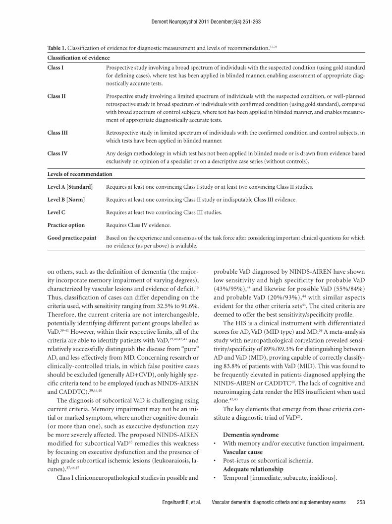
Classification of evidence and levels of recommendation The scientific evidence for diagnostic assessment was
evaluated according to pre-established levels of certain-ty (Classes I, II, III and IV) and recommendations were graded according to strength of evidence (Level A, B or C). Additionally, important clinical issues were addressed for which evidence is questionable (Practice Option) and, when no evidence was available, recommendations were made based on the experience and consensus of the task force under “Good Practice Point” 32,25 (Table 1).
These guidelines may not be applicable under some circumstances and decisions on whether to apply recom-mendations must be taken in light of the individual clinical presentation of the case and of the resources available.32
Diagnostic stepsThe diagnostic steps outlined below should be followed
systematically to determine the diagnosis of VaD as accu-rately as possible.
Diagnostic criteria The diagnostic criteria for VaD include the official sets
(CID-10-CDP33 and DSM-IV34) as well as those devised specifically for research (CADDTC,35 NINDS-AIREN,36 NINDS-AIREN modified).37 Ischemic scores are also rou-tinely used, the most common of which is the Hachinski (HIS).38
These criteria are similar on several aspects while differ
Dement Neuropsychol 2011 December;5(4):251-263
Engelhardt E, et al. Vascular dementia: diagnostic criteria and supplementary exams 253
on others, such as the definition of dementia (the major-ity incorporate memory impairment of varying degrees), characterized by vascular lesions and evidence of deficit.13 Thus, classification of cases can differ depending on the criteria used, with sensitivity ranging from 32.5% to 91.6%. Therefore, the current criteria are not interchangeable, potentially identifying different patient groups labelled as VaD.39-41 However, within their respective limits, all of the criteria are able to identify patients with VaD,39,40,42,43 and relatively successfully distinguish the disease from “pure” AD, and less effectively from MD. Concerning research or clinically-controlled trials, in which false positive cases should be excluded (generally AD+CVD), only highly spe-cific criteria tend to be employed (such as NINDS-AIREN and CADDTC).39,44,40
The diagnosis of subcortical VaD is challenging using current criteria. Memory impairment may not be an ini-tial or marked symptom, where another cognitive domain (or more than one), such as executive dysfunction may be more severely affected. The proposed NINDS-AIREN modified for subcortical VaD45 remedies this weakness by focusing on executive dysfunction and the presence of high grade subcortical ischemic lesions (leukoaraiosis, la-cunes).37,46,47
Class I cliniconeuropathological studies in possible and
probable VaD diagnosed by NINDS-AIREN have shown low sensitivity and high specificity for probable VaD (43%/95%),48 and likewise for possible VaD (55%/84%) and probable VaD (20%/93%),44 with similar aspects evident for the other criteria sets44. The cited criteria are deemed to offer the best sensitivity/specificity profile.
The HIS is a clinical instrument with differentiated scores for AD, VaD (MID type) and MD.38 A meta-analysis study with neuropathological correlation revealed sensi-tivity/specificity of 89%/89.3% for distinguishing between AD and VaD (MID), proving capable of correctly classify-ing 83.8% of patients with VaD (MID). This was found to be frequently elevated in patients diagnosed applying the NINDS-AIREN or CADDTC49. The lack of cognitive and neuroimaging data render the HIS insufficient when used alone.42,43
The key elements that emerge from these criteria con-stitute a diagnostic triad of VaD21.
Dementia syndrome • Withmemoryand/orexecutivefunctionimpairment.
Vascular cause • Post-ictus or subcortical ischemia.
Adequate relationship • Temporal [immediate, subacute, insidious].
Table 1. Classification of evidence for diagnostic measurement and levels of recommendation.32,25
Classification of evidence
Class I Prospective study involving a broad spectrum of individuals with the suspected condition (using gold standard
for defining cases), where test has been applied in blinded manner, enabling assessment of appropriate diag-
nostically accurate tests.
Class II Prospective study involving a limited spectrum of individuals with the suspected condition, or well-planned
retrospective study in broad spectrum of individuals with confirmed condition (using gold standard), compared
with broad spectrum of control subjects, where test has been applied in blinded manner, and enables measure-
ment of appropriate diagnostically accurate tests.
Class III Retrospective study in limited spectrum of individuals with the confirmed condition and control subjects, in
which tests have been applied in blinded manner.
Class IV Any design methodology in which test has not been applied in blinded mode or is drawn from evidence based
exclusively on opinion of a specialist or on a descriptive case series (without controls).
Levels of recommendation
Level A [Standard] Requires at least one convincing Class I study or at least two convincing Class II studies.
Level B [Norm] Requires at least one convincing Class II study or indisputable Class III evidence.
Level C Requires at least two convincing Class III studies.
Practice option Requires Class IV evidence.
Good practice point Based on the experience and consensus of the task force after considering important clinical questions for which
no evidence (as per above) is available.
Dement Neuropsychol 2011 December;5(4):251-263
254 Vascular dementia: diagnostic criteria and supplementary exams Engelhardt E, et al.
• Functional [lesion to structures of cognitive integration].Based on the diagnostic criteria, a general definition
of VaD can be derived and, considering the variations presented, its simplified unification can be formulated as shown below. The basic characteristic of VaD is cognitive impairment of multiple domains, with compromise often being non-uniform. For VaD, memory impairment is a requisite (in most criteria) together with one or more of the following signs and symptoms: aphasia, apraxia, agno-sia and executive dysfunction. Deficits of the disease must have a major impact on occupational or habitual activities and be marked by significant decline compared to pre-vious level of functioning. The presence of neurological signs and symptoms is also a requirement, in addition to laboratory evidence or neuroimaging findings indicative of CVD, deemed to be etiologically related to the condition. The condition should not manifest exclusively during the course of delirium or major psychiatric disorder21.
Recommendations – A diagnosis of VaD must be based on specific criteria, with NINDS-AIREN being the most frequently used in research settings (Level A). The HIS can be recommended although with some re-strictions, given a lack of cognitive and neuroimaging data (Level C). The diagnostic triad of VaD can repre-sent a brief option for diagnosis (Good Practice Point).
AnamnesisAnamnesis is fundamental and must include questions
on all aspects related to a dementia condition of vascular cause, such as mode of onset, pattern of progression, prior history (CVA, revascularization), comorbidities (SAH, DL, DM, anemia, sleep and psychiatric disorders), habits (eat-ing, life-style, tobacco and alcohol use), familial and edu-cational history. Specific questions on cognition, activities of daily living and behavior are necessary. It is important to obtain a full list of drugs used and prescribed, as well as alternative medications.21,24,50,51
Recommendations – Anamnesis is fundamental and data must be supplemented by a companion that is as well informed as possible (Level A).
Physical and neurological exams A general physical exam can disclose frequent comor-
bidities, particularly in elderly patients, that can rapidly worsen cognitive, functional and behavioral status. These comorbidities or complications can include depression, cardiovascular disease, infections, dehydration, collateral effects of medications, delirium, falls, incontinence, an-orexia and obesity. There is a strong correlation between
comorbidities and cognitive status in VaD (Class IV).52 Clinical neurovascular assessment (palpation, ausculta-tion) is also part of a thorough exam (see “Vascular neu-roimaging”). Neurological examination is necessary and the presence of neurological symptoms makes up part of the diagnostic criteria of VD51, 53 (Class II).
Recommendations – All patients presenting with, or suspected of having dementia must be submitted to a general physical exam aimed at detecting comorbidi-ties, in addition to a neurovascular exam (Good Practice Point), as well as a neurological exam (Level B).
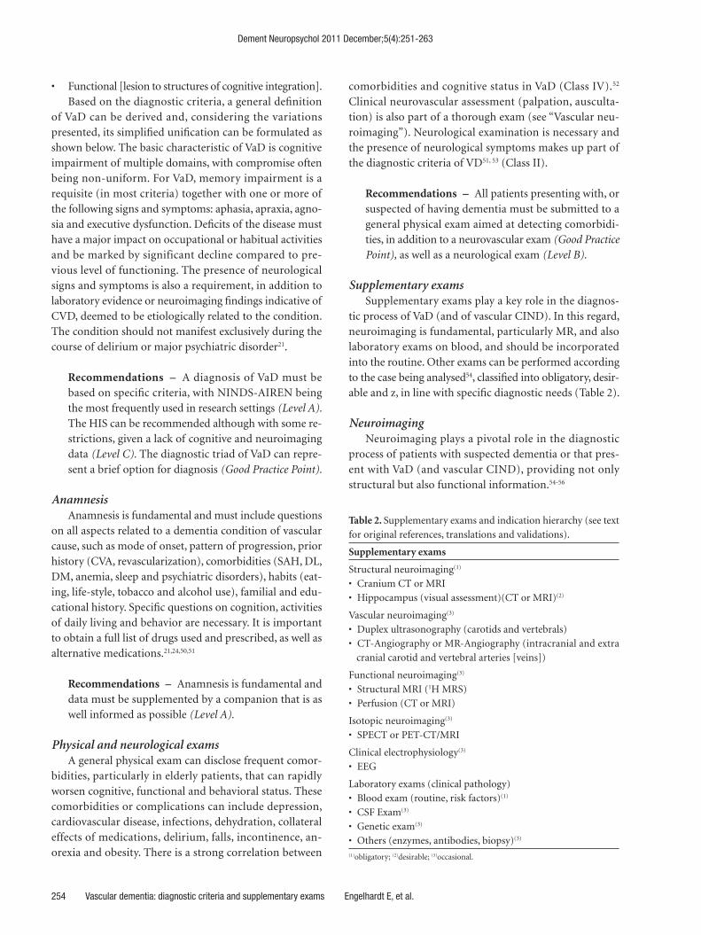
Supplementary examsSupplementary exams play a key role in the diagnos-
tic process of VaD (and of vascular CIND). In this regard, neuroimaging is fundamental, particularly MR, and also laboratory exams on blood, and should be incorporated into the routine. Other exams can be performed according to the case being analysed54, classified into obligatory, desir-able and z, in line with specific diagnostic needs (Table 2).
NeuroimagingNeuroimaging plays a pivotal role in the diagnostic
process of patients with suspected dementia or that pres-ent with VaD (and vascular CIND), providing not only structural but also functional information.54-56
Table 2. Supplementary exams and indication hierarchy (see text
for original references, translations and validations).
Supplementary exams
Structural neuroimaging(1)
• CraniumCTorMRI
• Hippocampus(visualassessment)(CTorMRI)(2)
Vascular neuroimaging(3)
• Duplexultrasonography(carotidsandvertebrals)
• CT-AngiographyorMR-Angiography(intracranialandextra
cranial carotid and vertebral arteries [veins])
Functional neuroimaging(3)
• StructuralMRI(1H MRS)
• Perfusion(CTorMRI)
Isotopic neuroimaging(3)
• SPECTorPET-CT/MRI
Clinical electrophysiology(3)
• EEG
Laboratory exams (clinical pathology)
• Bloodexam(routine,riskfactors)(1)
• CSFExam(3)
• Geneticexam(3)
• Others(enzymes,antibodies,biopsy)(3)
(1)obligatory; (2)desirable; (3)occasional.
Dement Neuropsychol 2011 December;5(4):251-263
Engelhardt E, et al. Vascular dementia: diagnostic criteria and supplementary exams 255
Structural neuroimagingThis can be obtained through computed tomography
scans (CT) or magnetic resonance imaging (MRI). Long considered merely for “excluding” brain lesions as the cause of dementia (e.g. tumors, hematomas, hydrocephaly), these techniques now exercise an important role of “including” diagnoses (e.g. neurodegenerative diseases, cerebrovascular diseases), through evidencing aspects considered character-istic of certain dementia types (e.g. hippocampal atrophy, as a marker of AD, or infarcts and lesions in white matter, as characteristics of VaD).57-59 It should be noted that de-spite confirmation of CVD on neuroimaging, the method cannot reliably diagnose VaD.60 However, the absence of CVD on neuroimaging offers strong evidence against de-mentia of vascular etiology.61
MRI is the method of choice for reaching a diagnosis of VaD (and vascular CIND), given its high sensitivity and spatial resolution, and ability to provide a greater amount of reliable data. The preferred magnetic field intensity is 1.5T or greater,62 where 0.5T may be acceptable.
The basic sequences needed are diffusion (DWI), 3D-T1,T2,T2-FLAIRandGE-T2(gradientecho).56,63-65 CT should be used when no MRI device is available and in special situations (pace-maker fitted, intracranial ferro-magnetic metal clips, psychological grounds etc.).22 Both techniques, within the scope of their characteristics, are able to obtain information on anatomy and presence of vascular lesions (infarcts, lacunes, changes in white mat-ter, hemorrhages) and other pathologies, providing infor-mation on quantitative (number and volume) and topo-graphic (localization) aspects. Ischemic changes in white matter (hypodensities – hyperintensities)(leukoaraiosis) can be assessed using visual and automated scales.15,64-70 The presence of hippocampal atrophy can also be assessed (see “Hippocampal atrophy”).
Structural neuroimaging must be performed as routine and a number of diagnostic criteria for VaD expressly re-quire neuroimaging as a core item71, 56, such as the NINDS-AIREN, in which the technique is essential for diagnosing probable VaD, where lack of the method leads to a default diagnosis of the “possible” category6. Moreover, the criteria specify which vascular territories are “relevant” for VaD. Use of NINDS-AIREN operating guidelines for classifying radiological aspects led to a significant increase in diagnos-tic reliability among professionals assessing images from 40% to 60% (Class II).
Hippocampal atrophy – The assessment of degree of atrophy can be carried out by CT or MRI (with the latter being more reliable) using the visual assessment volumet-ric measurements (manual or automated), with the lat-ter deemed more accurate.73-76 Comparative studies have
shown good correlation among these techniques.77,78 Visual scales can be employed in clinical practice as well as clinical research, particularly in cross-sectional studies.76,79-81 T2-MRI or FLAIR can be used to distinguish the nature of the hippocampal atrophy. High signal, although also seen in AD, is not characteristic of this condition, being more often found in hippocampal sclerosis. On the other hand, isch-emic changes in these regions can be better distinguished on FLAIR.82
MRI studies have shown hippocampal atrophy through visual (VaD) or volumetric assessment (subcortical VaD), to a lesser degree compared to AD but to a greater extent than in normal controls, in cases of dementia with similar severity.83-86 The findings on neuroimaging were confirmed by neuropathological studies showing that hippocampal volume was lower in VaD than in normal controls, but greater compared with atrophy found in AD.87-89
Diffusion tensor – Diffusion tensor imaging (DTI) is a technique for assessing the integrity of white matter fibers using quantitative fractionated anisotropy (DTI-FA) and tractography (DTI-TR). DTI-FA is an important technique in considering the large extension of white matter, and has been previously applied in clinical practice.90-95 DTI-TR can visualize the bundles interconnecting various regions whose interruption can cause a range of different discon-nection syndromes. The method is not routinely used in clinical practice.95,96
Vascular neuroimagingNeurovascular assessment entails, beyond clinical ex-
amination,duplexultrasonography(USG)oftheextra-cranial carotid and vertebral arteries and CT-angiography or MRI-angiography of intracranial and extracranial ca-rotid and vertebral arteries (from their point of origin).97,98 USGtendstobetheinitialexamandenablesvisualizationof the vascular wall, detecting atheromatous plaques and stenosis while also providing a measure of intima-media thickness of the carotids, an early marker for wall pathol-ogy and cardiovascular risk factor. The characteristics of blood flow can also be verified, constituting a functional aspect.99 MRI-angiography and CT-angiography visual-izes the entire cervical and intracranial tree (and venous system when necessary) and the vascular pathology pres-ent (atheromatous plaques, stenosis) which is related to flow and perfusion disorders with potential to cause brain lesions.97-102
Recommendations – Structural neuroimaging should be used in all patients with suspected dementia, prefer-ably using MRI (Level A). In the impossibility of using MRI, CT can serve as an alternative method (Level B).
Dement Neuropsychol 2011 December;5(4):251-263
256 Vascular dementia: diagnostic criteria and supplementary exams Engelhardt E, et al.
Hippocampal atrophy must be assessed in all patients with the aim of reaching a diagnosis of pure VaD or VaD associated to AD (Level B). Neurovascular assessment can be necessary for clinical clarification and determin-ing therapeutical interventions (Good Practice Point).
Functional neuroimaging Functional aspects of neuroimaging often provide use-
ful supplementary information to the diagnosis, such as proton MR spectroscopy (1H MRS), CT and MRI perfu-sion, besides isotopic techniques including Single Photon Emission Computed Tomography and Positron Emission Tomography (PET).
Proton spectroscopy – 1H MRS represents biochem-ical information via MRI, which can be obtained at the same time as structural acquisitions. Studies in AD have shown changes in mI/Cr in the posterior region of the cin-gulate (PC)103 and reduced Naa/Cr in hippocampi (HCs), with progressive decline according to stage of the disease.104 In addition, comparing alterations in HCs with those in PC for AD enables staging by spectroscopy.105
Studies of these structures in VaD (HCs, PC) are scarce. Investigations have been performed mainly for PC, com-paring findings in several different dementia types (AD, FTLD, DLB) and subcortical VaD in mild stages, involving a small number of patients, and have revealed reduced Naa/Cr, although within one standard deviation of the normal group.6 Other studies however, found no significant dif-ference in Naa/Cr ratio of PC between AD and VaD.107 A study focusing on HCs (and frontal and parietal lobes) in dementia without vascular lesions (AD) and with vascular lesions (lacunes)(subcortical VaD) showed a lower Naa/Cr ratio in HCs among AD cases, whereas this ratio was similar in dementia patients with lacunes and in normal controls.108 1H MRS can be used as a resource in diagnos-ing VaD, particularly as a differential item with AD and in suspected cases of AD+VaD (MD).6
Isotopic neuroimaging – SPECT and PET are used fairly frequently in diagnosing dementia. Some studies us-ing SPEC were designed to compare AD with other types of dementia and have shown sensitivity and specificity for AD vs. VaD of 71% and 75%, respectively.109 However, al-though results were variable, the techniques seems to be useful for distinguishing AD from VaD, but use for diag-nostic purposes without previous structural imaging is not advised.110 PET can differentiate AD from VaD by disclos-ing temporo-parietal pattern of hypometabolism in AD, or predominant frontal lobe damage in VaD.111 Different regional patterns of hypoperfusion as seen by SPECT, or hypometabolism seen on PET, can assist in differentiat-ing diverse neurodegenerative types and VaD. Images on
SPECT and PET in VaD show diverse patterns according to VaD subtype – such as multifocal pattern seen in dementia due to multiple infarcts, or with a more diffuse pattern as-sociated to extensive lesion of white matter and lacunes.57
Recommendations – The use of 1H MRS can be valu-able for certain cases of VaD and for differentiating with MD (Practice option). SPECT and PET can be used in cases where there is diagnostic doubt after clinical work-up and structural imaging, but should not be used as a standalone imaging assessment (Good Practice Point).
Blood examThe blood test is a necessary part of the assessment in
cases of cognitive disorder so as to: (i) identify comorbidi-ties and/or complications; (ii) reveal potential risk factors; (iii) explore causes of frequently associated confusional states, and (iv) less often, identify the primary cause of the dementia. Cognitive disorders can be associated to a broad array of metabolic, infectious, and toxic conditions which must be identified and treated.30
Besides routine items (full blood count, ESR, electrolytes, glucose, renal and hepatic function tests, and TSH), items should be ordered that represent vascular risk factors (VRFs) which merit separate consideration in VCI/VaD. VRFs are intrinsically linked to CVD (and transient vascular ictus or report of ictus symptoms in its absence).112 These are also associated to reports of ictus symptoms in the absence of diagnosed ictus or transient ischemic attack.113,114 VRFs are numerous and encompass metabolic, toxic, genetic, car-diovascular, and demographic factors, being divided into non-modifiable and modifiable (the majority) with this latter group being susceptible to preventive measures.12,31 A comprehensive study showed that among the numerous VRFs for cerebral vascular ictus, just 10 were associated to 90% of cases, namely: arterial hypertension, smoking, waist-hip ratio, high risk diet, moderate physical activity, diabetes mellitus, excessive alcohol intake, psychosocial stress and depression, cardiac causes and ratio of apolipoproteins B for A1 (Class I).115 It has been suggested that ≥3 VRFs among those cited place the brain at high risk of cognitive impairment.30 Cognitive decline associated to VRFs in the absence of clinical ictus of dementia has also been observed, with theory proposing that subclinical CVD proves an im-portant link among the main VRFs for ictus and cognitive function.117 Healthy elderly can present subclinical CVD,118 leading to the hypothesis that CVD, faster cerebral atrophy, abnormal cerebral white matter and clinically asymptomatic brain infarcts represent possible mechanism linking VRFs to risk of future ictus and cognitive dysfunction.119Geneticriskfactorsareexaminedseparately(see“Geneticexam”).
Dement Neuropsychol 2011 December;5(4):251-263
Engelhardt E, et al. Vascular dementia: diagnostic criteria and supplementary exams 257
Recommendations – Blood tests should be per-formed at first assessment, including routine items and those representing a potential cause of cognitive impairment or as comorbidity, including exams which represent VRFs (Level A). More in-depth tests can be necessary in selected cases (Good Practice Point).
Other examsThis category includes exams performed in special situ-
ations according to specific indications.Cerebrospinal fluid – The cerebrospinal fluid exam
(CSF) has an important place in diagnosing neurodegen-erative dementia (AD, FTD, DLB, CJD) through the study of markers based on β-amyloid peptide (βA) and total tau and phospho-tau protein.54 Levels of Aβ42 are reduced in AD (and in AD+CVD) and increased in VaD, enabling AD (and AD+CVD) to be discriminated from VaD. Aβ42 has proved an important resource for discriminating AD vs. VaD and possibly improving the diagnostic precision of cases classified as MD (presence of CVD on neuroimag-ing). Tau protein is high in AD (and in AD+CVD) and low in VaD.120-122 Levels of phospho-tau exhibit differentiated increase, being greater in AD, intermediate in AD+CVD and lower in VaD.122,123 Analysis correlating Aβ42 and total tau have shown high specificity in AD and low specificity in VaD 48% [29-67]), possibly owing to the presence of neurodegenerative lesion in this condition.120
Recommendations – The CSF exam is recommended in certain situations (inflammatory diseases, vasculi-tis, rapidly progressive dementia)(Good Practice Point). The markers based on tau protein and Aβ42 can be used as a complement in cases with diagnostic doubt (Level B).
Electroencephalograph – EEGincasesofAD,AD+CVD (with pathological confirmation) and controls dis-closes abnormalities in the majority of patients from path-ologicalgroups,wherebynormalEEGhasshownanegativepredictive value of 0.825 for AD diagnosis (Class II).124 Vi-sualEEGandquantitativeEEG(qEEG)canbeusedinthedifferential diagnosis between AD and subcortical VaD.125 EEGcanberequiredundersomecircumstances,suchasin cases of diagnostic doubt,126 but does not constitute a routine exam in dementia.127
Recommendations – TheEEGcanbeausefulcom-plement in the diagnostic process (Practice Option). It can be used in the differential diagnosis of tran-sient epileptic amnesia vs. transient ischemic attack (Level B).
Genetic exam – Vascular ictus of any etiology, whether familial or genetic, is a basic cause of VaD (and of vascular CIND).128-130 A positive family history seems to constitute a risk factor for ictus and genetic influence can vary accord-ing to ictus subtype.131,132 There is a strong relationship be-tween familiar conditions associated to ischemic and hem-orrhagic stroke, as well as those related to connective tissue disease and hematologic diseases, among others.128-130 Sin-gle gene causes of stroke are considered relatively rare with multiple genetic influences on VRFs more common, influ-encing pathogenesis and severity.129 The monogenic dis-orders associated to CVD include CADASIL (NOTCH 3), hereditary variant of cerebral amyloid angiopathy (CAA), the frequent sickle cell disease (HBB*S [homo and hetero-zygotes] and haplotypes βS),Fabry’sdisease(GLA),ho-mocystinuria (CBS and other genes), besides other rare conditions.26,64,133-136
Recommendations – The genetic exam for detec-tion of known pathogenic mutations can be carried out when available, mainly for genetic counselling and clinical research purposes. The exams should be done in a specialized center, with appropriate counselling of patient and family members (Good Practice Point).
Certain investigations can yield important information for diagnosis (such as concentrations of enzymes, amino-acids, antibodies among others). The biopsy of tissues such as the skin test in the CADASIL, as well as the brain, can be important in primary vasculitis.137,138
Recommendations – Specific exams and tissue biopsy can offer a specific diagnosis in some rarer conditions. The exam should be carried in specialized centers in carefully selected cases (Good Practice Point).
ConclusionThe assessment procedures for diagnosing VaD require
multi-disciplinary interaction toward reaching a diagnosis. This part of the proposal addressed the analysis of diag-nostic criteria, anamnesis, as well as clinical and supple-mentary exams (neuroimaging and laboratory) used for diagnosing VaD, classified according to proven evidence at various levels.
It should be highlighted that only around half of the population of patients with VCI/VaD present with demen-tia and the group envisages that the present study can be further refined to enable more precise diagnosis of this condition and that part of the spectrum of CIND and vas-cular MCI can be extended with the defining of suitable criteria and diagnostic process.
Dement Neuropsychol 2011 December;5(4):251-263
258 Vascular dementia: diagnostic criteria and supplementary exams Engelhardt E, et al.
Acknowledgements – We would like to extend our thankstoDr.GilbertoSousaAlvesandtoDr.FelipeKenjiSudo, members of the team of the CDA-IPUB-UFRJ for critical review of this paper and to Luzinete Alvarenga, li-brarian, for organizing the references.
References1. GauthierS,RockwoodK.DoesvascularMCIprogressata
different rate than does amnestic MCI? Int Psychogeriatr
2003;15(Suppl 1):257-259.
2. Hachinski V. Vascular dementia: a radical redefinition. De-
mentia 1994;5:130-132.
3. InglesJL,WentzelC,FiskJD,RockwoodK.Neuropsycho-
logical predictors of incident dementia in patients with
vascular cognitive impairment, without dementia. Stroke
2002;33:1999-2002.
4. Loeb C. Clinical criteria for the diagnosis of vascular demen-
tia. Eur Neurol 1988;28:87-92.
5. MeyerJS,XuG,ThornbyJ,ChowdhuryMH,QuachM.Is
mild cognitive impairment prodromal for vascular dementia
like Alzheimer’s disease? Stroke 2002;33:1981-1985.
6. Engelhardt E. Demência mista: do conceito ao tratamento.
Rev Bras Neurol 2004;40:33-54.
7. JellingerKA,AttemsJ.Istherepurevasculardementiainold
age? J Neurol Sci. 2010;299:150-154.
8. McKhannGM,KnopmanDS,ChertkowH,etal.The
diagnosis of dementia due to Alzheimer’s disease: Recom-
mendations from the National Institute on Aging and the
Alzheimer’s Association workgroup. The diagnosis of demen-
tia due to Alzheimer’s disease: Recommendations from the
National Institute on Aging and the Alzheimer’s Association
workgroup. Alzheimers Dement 2011 [Epub ahead of print]
9. ZekryD,HauwJJ,GoldG.Mixeddementia:epidemiology,
diagnosis,andtreatment.JAmGeriatrSoc2002;50:1431-
1438.
10. SachdevP.Vascularcognitivedisorder.IntJGeriatrPsychia-
try 1999;14:402-403.
11. RockwoodK,DavisH,MacKnightC,etal.Theconsortium
to investigate vascular impairment of cognition: methods
and first findings. Can J Neurol Sci 2003;30:237-243.
12. Engelhardt E, Laks J, Cavalcanti JLS, Moreira DM, Madalen
C. Demência vascular. Rev Bras Neurol 2004;40:5-25.
13. Engelhardt E. Demência vascular. In: Bottino CMC, Laks J,
Blay SL (Eds). Demência e transtornos cognitivos no idoso.
RiodeJaneiro:GuanabaraKoogan,2006:177-195.
14. ErkinjunttiT,RomanG,GauthierS,FeldmanH,Rockwood
K.Emergingtherapiesforvasculardementiaandvascular
cognitive impairment. Stroke 2004;35:1010-1017.
15. JellingerKA.Theenigmaofvascularcognitivedisorderand
vascular dementia. Acta Neuropathol 2007;113:349-388.
16. Zekry D, Duyckaerts C, Belmin J, et al. The vascular lesions
in vascular and mixed dementia: the weight of functional
neuroanatomy. Neurobiol Aging 2003;24:213-219.
17. Desmond DW. The neuropsychology of vascular cognitive
impairment: Is there a specific cognitive deficit? J Neurol Sci
2004;226:3-7.
18. MerinoJG,HachisnkiV.Demênciaeictus:loaimpor-
tância de la enfermedad cerebral coexistente. Rev Neurol
2003;36:61-63.
19. TomlinsonBE,BlessedG,RothM.Observationsonthe
brains of demented old people. J Neurol Sci 1970;11:205-242.
20. Brun A. Pathology and pathophysiology of cerebrovascular
dementia: pure subgoups of obstructive and hypoperfusive
etiology. Dementia 1994;5:145-147.
21. Engelhardt E. Demência mista: do conceito ao tratamento.
Rev Bras Neurol 2004;40:33-54.
22. Engelhardt E, Tocquer C, André C, Moreira DM, Okamoto
IH, Cavalcanti JLS. Demência vascular. Criterios diagnos-
ticos e exames complementares. Dement Neuropsychol
2011;5(Suppl 1):49-77.
23. GorelickPB,ScuteriA,BlackSE,etal.AmericanHeartAs-
sociation Stroke Council, Council on Epidemiology and
Prevention, Council on Cardiovascular Nursing, Council on
Cardiovascular Radiology and Intervention, and Council on
Cardiovascular Surgery and Anesthesia. Vascular contribu-
tions to cognitive impairment and dementia: a statement for
healthcare professionals from the american heart associa-
tion/american stroke association. Stroke 2011;42:2672-2713.
24. Hachinski V, Iadecola C, Petersen RC, et al. National Insti-
tute of Neurological Disorders and Stroke-Canadian Stroke
Network vascular cognitive impairment harmonization stan-
dards. Stroke 2006;37:2220-2241.
25. KnopmanDS,DeKoskyST,CummingsJL,etal.Practicepa-
rameter: Diagnosis of dementia (an evidence-based review).
Neurology 2001;56:1143-1Nitrini R, Caramelli P, Bottino
CMC, Damasceno BP, Brucki SMD, Anghinah R. Diagnós-
tico de doença de Alzheimer no Brasil. Critérios diagnósticos
e exames complementares recomendações do Departamen-
to Científico de Neurologia Cognitiva e do Envelhecimento
da Academia Brasileira de Neurologia Arq Neuropsiquiatr
2005;63:713-719.
26. Nitrini R, Caramelli P, Bottino CMC, Damasceno BP, Bru-
cki SMD, Anghinah R. Diagnóstico de doença de Alzheimer
no Brasil. Avaliação cognitiva e funcional. Recomendações
do Departamento Científico de Neurologia Cognitiva e do
Envelhecimento da Academia Brasileira de Neurologia. Arq
Neuropsiquiatr 2005;63:720-727.
27. RockwoodK,ParhadI,HachinskiV,etal.Diagnosisofvas-
cular dementia: Consortium of Canadian Centres for Clini-
cal Cognitive Research concensus statement. Can J Neurol
Sci 1994;21:358-364.
28. Madureira S, Verdelho A, Ferro J, et al. Development of a
neuropsychological battery for the Leukoaraiosis and Disa-
bility in the Elderly Study (LADIS): experience and baseline
data. Neuroepidemiology 2006;27:101-116.
29. WaldemarG,DuboisB,EmreM,etal.Recommendationsfor
the diagnosis and management of Alzheimer’s disease and
other disorders associated with dementia: EFNS guideline.
Eur J Neurol 2007;14:1-26.
Dement Neuropsychol 2011 December;5(4):251-263
Engelhardt E, et al. Vascular dementia: diagnostic criteria and supplementary exams 259
30. ZhaoQ,ZhouY,WangY,DongK,WangY.Anewdiagnostic
algorithm for vascular cognitive impairment: the proposed
criteria and evaluation of its reliability and validity. Chin
Med J 2010;123:311-319.
31. BraininM,BarnesM,BaronJC,etal.Guidanceforthepre-
paration of neurological management guidelines by EFNS
scientific task forces – revised recommendations 2004. Eur J
Neurol 2004;11:577-581.
32. Organização Mundial de Saúde (OMS) (Classificação Esta-
tística Internacional de Doenças e Problemas Relacionados
com a Saúde – Critérios Diagnósticos para Pesquisa) (CID-
10-CDP). 10ª edição. 1993
33. American Psychiatric Association Committee on nomen-
clature and statistics. Diagnostic and Statistical Manual of
Mental Disorders (DSM-IV), 4th ed. Washington, DC; 1994.
34. Chui HC, Victoroff JI, Margolin D, et al. Criteria for the diag-
nosis of ischemic vascular dementia proposed by the State
of California Alzheimer’s Disease Diagnostic and Treatment
Centers. Neurology 1992;42:473-480.
35. RománGC,TatemichiTK,ErkinjunttiT,etal.Vascularde-
mentia: diagnostic criteria for research studies: report of
theNINDS-AIRENInternationalWorkGroup.Neurology
1993;43:250-260.
36. Erkinjuntti T, Inzitari D, Pantoni L, et al. Research criteria
for subcortical vascular dementia in clinical trials. J Neural
Transm Suppl 2000;59:23-30.
37. Hachinski VC, Iliff LD, Zilhka E, et al. Cerebral blood flow
in dementia. Arch Neurol 1975;32:632-637.
38. Chui HC, Mack W, Jackson JE, et al. Clinical criteria for the
diagnosis of vascular dementia: a multicenter study of com-
parability and interrater reliability. Arch Neurol 2000;57:
191-196.
39. PohjasvaaraT,MäntlyäR,YlikoskiR,KasteM,ErkinjunttiT.
Comparison of different clinical criteria (DSM-III, ADDTC,
ICD-10, NINDS-AIREN, DSM-IV) for the diagnosis of vas-
cular dementia. National Institute of Neurological Disorders
and Stroke-Association Internationale pour la Recherche et
l’Enseignement en Neurosciences. Stroke 2000;31:2952-2957.
40. WetterlingT,KanitzRD,BorgisK-J.Comparisonofdifferent
diagnostic criteria for vascular dementia (ADDTC, DSM-IV,
ICD-10, NINDS-AIREN). Stroke 1996;27:30-36.
41. Wiederkehr S, Simard M, Fortin C, van Reekum R. Compa-
rability of the clinical diagnostic criteria for vascular demen-
tia: a critical review. Part I. J Neuropsychiatry Clin Neurosci
2008;20:150-161.
42. Wiederkehr S, Simard M, Fortin C, van Reekum R. Validi-
ty of the clinical diagnostic criteria for vascular dementia:
a critical review. Part II. J Neuropsychiatry Clin Neurosci
2008;20:162-177.
43. GoldG,BourasC,CanutoA,etal.Clinicopathologicalvali-
dation study of four sets of clinical criteria for vascular de-
mentia. Am J Psychiatry 2002;159:82-87.
44. Erkinjuntti T, Inzitari D, Pantoni L, et al. Research criteria
for subcortical vascular dementia in clinical trials. J Neural
Transm Suppl 2000;59:23-30.
45. Erkinjuntti T. Subcortical vascular dementia. Cerebrovasc
Dis 2002;13(Suppl 2):58-60.
46. RománGC,RoyallDR.Executivecontrolfunction:arational
basis for the diagnosis of vascular dementia. Alzheimer Dis
Assoc Disord 1999;13(Suppl 3):S69-S80.
47. Holmes C, Cairns N, Lantos P, Mann A. Validity of current cli-
nical criteria for Alzheimer’s disease, vascular dementia and
dementia with Lewy bodies. Br J Psychiatry 1999;174:45-50.
48. Moroney JT, Bagiella E, Desmond DW, et al. Meta-analysis
of the Hachinski Ischemic Score in pathologically verified
dementias. Neurology 1997;49:1096-1105.
49. McKhannGM,KnopmanDS,ChertkowH,etal.The
diagnosis of dementia due to Alzheimer’s disease: recom-
mendations from the National Institute on Aging and the
Alzheimer’s Association workgroup on diagnostic guide-
lines for Alzheimer’s disease. Alzheimers Dement 2011;7:
263-269.
50. Smid J, Nitrini R, Bahia VS, Caramelli P. Caracterização clí-
nica da demência vascular. Avaliação retrospectiva de uma
amostra de pacientes ambulatoriais. Arq Neuropsiquiatr
2001;59:390-393.
51. FuC,ChuteDJ,FaragES,GarakianJ,CummingsJL,Vinters
HV. Comorbidity in dementia: an autopsy study. Arch Pathol
Lab Med 2004;128:32-38.
52. Staekenborg SS, van der Flier WM, van Straaten ECW, Lane
R, Barkhof F, Scheltens P. Neurological signs in relation to
type of cerebrovascular disease in vascular dementia. Stroke
2008;39:317-322.
53. van der Flier WM, Scheltens P. Use of Laboratory and
Imaging Investigations in Dementia. J Neurol Neurosurg
Psychiatry 2005;76(Suppl V):v45-v52.
54. Jagust WJ. Neuroimaging in dementia. Neurol Clin 2000;18:
885-901.
55. Tartaglia MC, Rosen H, Miller L. Neuroimaging in Demen-
tia. Neurotherapeutics 2011;8:82-92.
56. O’Brien JT. Role of imaging techniques in the diagnosis of
dementia Br J Radiol 2007;80:S71-S77.
57. Scheltens P, Fox N, Barkhof F, De Carli C. Structural magne-
tic resonance imaging in the practical assessment of demen-
tia: beyond exclusion. Lancet Neurol 2002;1:13-21.
58. van Straaten EC, Scheltens P, Barkhof F. MRI and CT in the
diagnosis of vascular dementia. J Neurol Sci 2004;226:9-12.
59. BallardCG,BurtonEJ,BarberR,etal.NINDSAIRENneu-
roimaging criteria do not distinguish stroke patients with
and without dementia. Neurology 2004;63:983-988.
60. Erkinjuntti T, Haltia M, Palo J, Sulkava R, Paetau A. Accuracy
of the clinical diagnosis of vascular dementia: a prospective
clinical and post-mortem neuropathological study. J Neurol
Neurosurg Psychiatry 1988;51:1037-1044.
61. YueNC,ArnoldAM,LongstrethWTJr,etal.Sulcal,ventri-
cular, and white matter changes at MR imaging in the aging
brain: Data from the Cardiovascular Health Study. Radiology
1997;202:33-39.
62. Cavalieri M, Schmidt R. New development in diagnosis of
vascular cognitive impairment. J Neurol Sci 2010;299:11-14.
Dement Neuropsychol 2011 December;5(4):251-263
260 Vascular dementia: diagnostic criteria and supplementary exams Engelhardt E, et al.
63. BiffiA,GreenbergSM.Cerebralamyloidangiopathy:asys-
tematic review. J Clin Neurol. 2011;7:1-9.
64. Longstreth WT Jr, Dulberg C, Manolio TA, et al. Incidence,
manifestations, and predictors of brain infarcts defined by
serial cranial magnetic resonance imaging in the elderly: the
cardiovascular health study. Stroke 2002;33:2376-2382.
65. InzitariD,PracucciG,PoggesiA,etal.Changesinwhite
matter as determinant of global functional decline in older
independent outpatients: three year follow-up of LADIS
(leukoaraiosis and disability) study cohort. BMJ 2009;339:
b2477.
66. JellingerKA,AttemsJ.Prevalenceandimpactofcerebrovas-
cular pathology in Alzheimer’s disease and parkinsonism.
Acta Neurol Scand 2006:114:38-46.
67. Scheltens P, Erkinjunti T, Leys D, et al. White matter changes
on CT and MRI: an overview of visual rating scales. Euro-
pean task force on age-related white matter changes. Eur
Neurol 1998;39:80-89.
68. TiehuisAM,VinckenKL,MaliWP,etal.Automatedandvi-
sual scoring methods of cerebral white matter hyperinten-
sities: relation with age and cognitive function. Cerebrovasc
Dis 2008;25:59-66.
69. Wahlund LO, Barkhof F, Fazekas F, et al. A new rating scale
for ge-related white matter changes applicable to MRI and
CT. Stroke 2001;32:1318-1322.
70. KeyserlingH,MukundanJrS.TheroleofconventionalMR
and CT in the work-up of dementia patients. Neuroimag
Clin N Am 2005;15:789-802.
71. vanStraatenEC,ScheltensP,KnolDL,etal.Operational
definitions for the NINDS-AIREN criteria for vascular de-
mentia: an interobserver study. Stroke 2003;34:1907-1912.
72. deLeonMJ,GeorgeAE,StylopoulosLA,SmithG,Miller
D. In vivo studies of hippocampal atrophy in Alzheimer’s
disease. J Neural Transmiss (P-D Sect) 1989;1:34.
73. deLeonMJ,ConvitA,GeorgeAE,etal.Invivostructural
studies of the hippocampus in normal aging and in incipient
Alzheimer’sdisease.AnnNYAcadSci1996;777:1-13.
74. Ridha BH, Barnes J, van de Pol LA, et al. Application of au-
tomated medial temporal lobe atrophy scale to Alzheimer
disease. Arch Neurol 2007;64:849-854.
75. Scheltens P, Leys D, Barkhof F, et al. Atrophy of medial tem-
poral lobes on MRI in probable Alzheimer’s disease and
normal ageing: diagnostic value and neuropsychological
correlates. J Neurol Neurosurg Psychiatry 1992;55:967-972
76. HsuY-Y,SchuffN,DuA-T,etal.Comparisonofautoma-
ted and manual MRI volumetry of hippocampus in normal
aging and dementia. J Magnet Res Imag 2002;16:305-310.
77. Wahlund LO, Julin P, Johansson SE, Scheltens P. Visual rating
and volumetry of the medial temporal lobe on magnetic re-
sonance imaging in dementia: a comparative study. J Neurol
Neurosurg Psychiatry 2000;69:630-635.
78. deLeonMJ,GolombJ,GeorgeAE,etal.Theradiologic
prediction of Alzheimer disease: the atrophic hippocampal
formation. AJNR 1993;14:897-906.
79. deLeonMJ,GeorgeAE,GolombJ,etal.Frequencyofhippo-
campal formation atrophy in normal aging and Alzheimer’s
disease. Neurobiol Aging 1997;18:1-11.
80. LiY,LiJ,SegalS,etal.Hippocampalcerebrospinalfluidspa-
ces on MR imaging: Relationship to aging and Alzheimer
disease. Am J Neuroradiol 2006;27:912-918.
81. Jack Jr CR, Dickson DW, Parisi JE, et al. Antemortem MRI
findings correlate with hippocampal neuropathology in ty-
pical aging and dementia. Neurology 2002;58:750-757.
82. Bastos-Leite AJ, Scheltens P, Barkhof F. Pathological aging of
the brain: an overview. Top Magn Reson Imaging 2004;15:
369-389.
83. ChoH,KwonJ-H,SeoH-J.Medialtemporallobeatrophy
in vascular dementia: Visual temporal lobe rating scale. Arch
GerontGeriatr2009;48:415-418
84. Du AT, Schuff N, Laakso MP, et al. Effects of subcortical is-
chemic vascular dementia and AD on entorhinal cortex and
hippocampus. Neurology 2002;58:1635-1641.
85. FeinG,DiSclafaniV,TanabeJ,etal.Hippocampalandcorti-
cal atrophy predict dementia in subcortical ischemic vascular
disease. Neurology 2000;55:1626-1635.
86. KnopmanDS,ParisiJE,BoeveBF,etal.VascularDementia
in a Population-Based Autopsy Study. Arch Neurol 2003;60:
569-575.
87. KrilJJ,PatelS,HardingAJ,HallidayGM.Patientswithvas-
cular dementia due to microvascular pathology have sig-
nificant hippocampal neuronal loss, J Neurol Neurosurg
Psychiatry 2002;72:747-751.
88. ZarowC,VintersHV,EllisWG,etal.Correlatesofhippo-
campal neuron number in Alzheimer’s disease and ischemic
vascular dementia. Ann Neurol 2005;57:896-903.
89. Engelhardt E, Moreira DM, Laks J. The brain subcortical
white matter and aging. A quantitative fractional anisotropy
analysis. Dement Neuropsychol 2007;3:228-233.
90. Engelhardt E, Moreira DM. A substância branca cerebral.
Localização dos principais feixes com anisotropia fracionada
direcional. Rev Bras Neurol 2008a;44:19-34.
91. Engelhardt E, Moreira DM. A substância branca cerebral.
Dissecção virtual dos principais feixes: tratografia. Rev Bras
Neurol 2008;44:19-34.
92. EngelhardtE,MoreiraDM,AlvesGS,etal.Binswanger’s
disease and quantitative fractional anisotropy. Arq Neurop-
siquiatr 2009;67:179-184.
93. Engelhardt E, Moreira DM, Laks J. The brain subcortical
white matter and aging
94. A quantitative fractional anisotropy analysis. Dement Neu-
ropsychol 2009;3:228-233.
95. Jellison BJ, Field AS, Medow J, Lazar M, Salamat MS, Alexan-
der AL. Diffusion tensor imaging of cerebral white matter:
a pictorial review of physics, fiber tract anatomy, and tumor
imaging patterns. Am J Neuroradiol 2004;25:356-369.
96. EngelhardtE,MoreiraDM,AlvesGSEngelhardtE,Morei-
ra DM. A substancia branca cerebral: disseccao virtual dos
principais feixes: tratografia. Rev Bras Neurol 2008;44:19-34.
97. BrottTG,HalperinJL,AbbaraS,etal.2011ASA/ACCF/
AHA/ AANN/ AANS/ ACR/ ASNR/ CNS/ SAIP/ SCAI/ SIR/
Dement Neuropsychol 2011 December;5(4):251-263
Engelhardt E, et al. Vascular dementia: diagnostic criteria and supplementary exams 261
SNIS/SVM/SVS.Guidelineonthemanagementofpatients
with extracranial carotid and vertebral artery disease: execu-
tive summary. Stroke 2011a;42:e420-e463.
98. EnterlineDS,KapoorG.ApracticalapproachtoCTangio-
graphy of the neck and brain. Tech Vasc Interv Radiol 2006;9:
192-204.
99. SimonA,MegnienJL,ChironiG.Thevalueofcarotid
intima-media thickness for predicting cardiovascular risk.
Arterioscler Thromb Vasc Biol 2010;30:182-185.
100. GauvritJ,TrystramD,OppenheimC,LeclercX.Nouvelles
techniques en imagerie vasculaire cervico-encéphalique et
médullaire. J Radiol 2007;88:472-482.
101. vanLaarPJ,vanderGrondJ,MaliWP,HendrikseJ.Magne-
tic resonance evaluation of the cerebral circulation in obs-
tructive arterial disease. Cerebrovasc Dis 2006;21:297-306
102. VicenziniE,RicciardiMC,SirimarcoG,DiPieroV,LenziGL.
Extracranial and intracranial sonographic findings in verte-
bral artery diseases. J Ultrasound Med 2010;29:1811-1823.
103. KantarciK,JackCRJr,XuYC,etal.Regionalmetabolicpat-
terns in mild cognitive impairment and Alzheimer’s disease,
a 1H MRS study. Neurology 2000;55:210-217.
104. Engelhardt E, Moreira DM, Laks L, Marinho VM, Rozenthal
M, Oliveira Jr AC. Doença de Alzheimer e espectroscopia por
ressonância magnética do hipocampo. Arq Neuropsiquiatr
2001;59:865-870.
105. Engelhardt E, Moreira DM, Laks J, Cavalcanti JL. Alzheimer’s
disease and proton magnetic resonance spectroscopy of lim-
bic regions: a suggestion of a clinical-spectroscopic staging.
Arq Neuropsiquiatr 2005;63:195-200.
106. KantarciK,PetersenRC,BoeveBF,etal.1HMRspectros-
copy in common dementias. Neurology 2004;63:1393-1398.
107. Martínez-Bisba MC, Arana E, Martí-Bonmatí L, Mollá E,
Celda B. Cognitive impairment: classification by 1H mag-
netic resonance spectroscopy. Eur J Neurol 2004;11:187-193.
108. Capizzano AA, Schuff N, Amend DL, et al. Subcortical is-
chemic vascular dementia: assessment with quantitative
MR imaging and 1H MR spectroscopy. Am J Neuroradiol
2000;21:621-630.
109. DougalNJ,BrugginkS,EbmeierKP.Systematicreviewofthe
diagnostic accuracy of 99mTc-HMPAO-SPECT in dementia.
AmJGeriatrPsychiatry2004;12:554-570.
110. Talbot PR, Lloyd JJ, Snowden JS, Neary D, Testa HJ. A clini-
cal role for 99mTc-HMPAO SPECT in the investigation of
dementia? J Neurol Neurosurg Psychiatry 1998;64:306-313.
111. NagataK,MaruyaH,YuyaH,etal.CanPETdatadifferen-
tiateAlzheimer’sdiseasefromvasculardementia?AnnNY
Acad Sci 2000;903:252-261.
112. BrottTG,HalperinJL,AbbaraS,etal.2011ASA/ACCF/
AHA/ AANN/ AANS/ ACR/ ASNR/ CNS/ SAIP/ SCAI/SIR/
SNIS/SVM/SVSGuidelineontheManagementofPatients
With Extracranial Carotid and Vertebral Artery Disease.
Stroke. 2011b;42:e464-e540.
113. Howard VJ, McClure LA, Meschia JF, Pulley LV, Orr SC,
FridayGH.HighPrevalenceofstrokesymptomsamong
persons without a diagnosis of stroke or transient ischemic
attack in a general population the reasons for geographic and
racialdifferencesinstroke(REGARDS)study.ArchIntern
Med 2006;166:1952-1958.
114. WadleyVG,McClureLA,HowardVJ,etal.Cognitivestatus,
stroke symptom reports, and modifiable risk factors among
individuals with no diagnosis of stroke or transient ischemic
attack in the reasons for geographic and racial differences in
stroke(REGARDS)study.Stroke2007;8:1143-1147.
115. O’Donnell MJ, Xavier D, Liu L, et al.; on behalf of the IN-
TERSTROKEinvestigators.Riskfactorsforischaemic
and intracerebral haemorrhagic stroke in 22 countries
(theINTERSTROKEstudy):acase-controlstudy.Lancet
2010;376:112-123.
116. Wiederkehr S, Laurin D, Simard M, Verreault R, Lindsay J.
Vascular risk factors and cognitive functions in fondemen-
tedelderlyindividuals.JGeriatrPsychiatryNeurol2009;22:
196-206.
117. DesmondDW,ThomasK,TatemichiTK,PaikM,Stern
Y.Riskfactorsforcerebrovasculardiseaseascorrelatesof
cognitive function in a stroke-free cohort. Arch Neurol
1993;50:162-166.
118. RazN,Gunning-DixonFM,HeadD,DupuisJH,AkerJD.
Neuroanatomical correlates of cognitive aging: evidence
from structural magnetic imaging. Neuropsychology 1998;
12:95-114.
119. Elias MF, Sullivan LM, D’Agostino RB, et al. Framingham
stroke risk profile and lowered cognitive performance. Stroke
2004;35:404-409.
120. Andreasen N, Minthon L, Davidsson P, et al. Evaluation of
CSF-tau and CSF-Ab42 as diagnostic markers for Alzheimer
disease in clinical practice. Arch Neurol 2001;58:373-379.
121. ParnettiL,LanariA,SaggeseE,SpaccatiniC,GallaiV.Cere-
brospinal fluid biochemical markers in early detection and
in differential diagnosis of dementia disorders in routine
clinical practice. Neurol Sci 2003;24:199-200.
122. Stefani A, Bernardini S, Panella M, et al. AD with subcortical
white matter lesions and vascular dementia: CSF markers for
differential diagnosis. J Neurol Sci 2005;237:83-88.
123. NäggaK,GottfriesJ,BlennowK,MarcussonJ.Cerebrospi-
nal fluid phospho-tau, total tau and beta-amyloid(1-42) in
the differentiation between Alzheimer’s disease and vascular
dementia.DementiaGeriatrCogDisord2002;14:183-190.
124. Robinson DJ, Merskey H, Blume WT, Fry R, Williamson PC,
Hachinski VC. Electro-encephalography as an aid in the ex-
clusion of Alzheimer’s disease. Arch Neurol 1994;51:280-284.
125. GawelM,ZalewskaE,Szmidt-SałkowskaE,Kowalski
J.ThevalueofquantitativeEEGindifferentialdiagnosisof
Alzheimer’s disease and subcortical vascular dementia. J
Neurol Sci 2009;283:127-133.
126. Smith SJM. J Neurol Neurosurg Psychiatry 2005;76 Suppl
II:ii8-ii12.
127. FairbairnA,GouldN,KendallT,etal.Dementia-supporting
people with dementia and their carers in health and social
care(NICEGuideline42).London:NationalInstitutefor
Health and Clinical Excellence, and Social Care Institute for
Dement Neuropsychol 2011 December;5(4):251-263
262 Vascular dementia: diagnostic criteria and supplementary exams Engelhardt E, et al.
Excellence; 2006 (amended 2011). [periodic na internet].
2011 [acesso em 2011 abr]; 11:52. Disponível em: http://
www.nice.org.uk/nicemedia/pdf/CG42Dementiafinal.pdf
128. MeschiaJF,BrottTG,BrownJrRD.Geneticsofcerebrovas-
cular disorders. Mayo Clin Proc 2005;80:122-132.
129. Razvi SSM, Boné I. Single gene disorders causing ischaemic
stroke. J Neurol 2006;253:685-700.
130. WarlowC,vanGijnJ,DennisM,etal.(Eds).Stroke:practical
management. 3rd ed. Oxford: Blackwell Publishing, 2008.
131. Jerrard-DunneP,CloudG,HassanA,MarkusHS.Evaluating
the genetic component of ischemic stroke subtypes: a family
history study. Stroke 2003;34:1364-1369.
132. PolychronopoulosP,GioldasisG,EllulJ,etal.Familyhis-
tory of stroke in stroke types and subtypes. J Neurol Sci
2002;195:117-122.
133. Ashley-KochA,YangQ,OlneyRS.Sicklehemoglobin(HbS)
alleleandsicklecelldisease:aHuGEreview.AmJEpidemiol
2000;151:839-845.
134. Naoum PS. Sickle cell disease: from the beginning until it
was recognized as a public health disease. Rev Bras Hematol
Hemoter 2011;33:7-9.
135. Joutel A, Monet M, Domenga V, Riant F, Tournier-Lasserve
E. Pathogenic mu tations associated with cerebral autosomal
dominant arteriopathy with sub cortical infarcts and leuko-
encephalopathy differently affect Jagged1 bind ing and No-
tch3activityviatheRBP/JKsignalingPathway.AmJHum
Genet2004;74:338-347.
136. Ballabio E, Bersano A, Bresolin N, Candelise L. Monogenic
vessel diseases related to ischemic stroke: a clinical approach.
J Cerebr Blood Flow Metabol 2007;27:1649-1662.
137. Joutel A, Favrole P, Labauge P, et al. Skin biopsy immunos-
taining with a Notch3 monoclonal antibody for CADASIL
diagnosis. Lancet 2001;358:2049-2051.
138. SchultzA,SantoianniR,Hewan-LoweK.Vasculopathic
changes of CADASIL can be focal in skin biopsies. Ultras-
truct Pathol 1999;23:241-247.
Dement Neuropsychol 2011 December;5(4):251-263
Engelhardt E, et al. Vascular dementia: diagnostic criteria and supplementary exams 263
GROUP RECOMMENDATIONS IN ALZHEIMER’S DISEASE AND VASCULAR DEMENTIA OF THE BRAZILIAN ACADEMY OF NEUROLOGY
Amauri B. da Silva [UNINEURO, Recife (PE)]; Ana Cláudia Ferraz [Serviço de Neurologia do Hospital Santa Marcelina (SP)];
Analuiza Camozzato de Pádua [Universidade Federal de Ciên-
cias da Saúde de Porto Alegre (UFCSPA); Hospital de Clínicas de Porto
Alegre(UFRGS)(RS)];Antonio Lúcio Teixeira [Departamento
de Clínica Médica, Faculdade de Medicina da Universidade Federal de
MinasGerais,BeloHorizonte(MG)];Ayrton Roberto Massaro [Instituto de Reabilitação Lucy Montoro (SP)]; Benito Pereira Da-masceno [Departamento de Neurologia da Universidade Estadual de
Campinas (SP)]; Carlos Alberto Buchpiguel [Departamento de
Radiologia, Faculdade de Medicina da Universidade de São Paulo (SP)];
Cássio Machado C. Bottino [Programa Terceira Idade, Institu-
to de Psiquiatria do Hospital das Clínicas da Faculdade de Medicina da
Universidade de São Paulo (FMUSP) (SP)]; Cláudia C. Godinho [Serviço de Neurologia do Hospital de Clínicas de Porto Alegre, Univer-
sidadeFederaldoRioGrandedoSul(RS)];Cláudia Sellitto Porto [GrupodeNeurologiaCognitivaedoComportamentodaFaculdadede
Medicina da USP (SP)]; Delson José da Silva [Núcleo de Neuroci-
ênciasdoHospitaldasClínicasdaUniversidadeFederaldeGoiás(UFG);
InstitutoIntegradodeNeurociências(IINEURO),Goiânia(GO)];Elza Dias-Tosta [Presidente da Academia Brasileira de Neurologia, Hos-
pital de Base do Distrito Federal (DF)]; Emílio Herrera Junior [Departamento de Medicina Interna, Faculdade de Medicina de Catan-
duva (SP)]; Francisco de Assis Carvalho do Vale [Universidade
Federal de São Carlos (UFSCar), Departamento de Medicina (DMed)
(SP)]; Gabriel R. de Freitas [Instituto D’or de Pesquisa e Ensino;
Universidade Federal Fluminense (RJ)]; Hae Won Lee [Instituto de
Radiologia, Hospital das Clínicas da Faculdade de Medicina da Universi-
dade de São Paulo e Hospital Sírio-Libanês (SP)]; Jerusa Smid [Grupo
de Neurologia Cognitiva e do Comportamento do Hospital das Clínicas
da Faculdade de Medicina da Universidade de São Paulo (FMUSP) (SP)];
João Carlos Barbosa Machado [Aurus IEPE - Instituto de Ensino
e Pesquisa do Envelhecimento de Belo Horizonte; Faculdade de Ciências
MédicasdeMinasGerais(FCMMG),ServiçodeMedicinaGeriátricado
HospitalMaterDei(MG)];José Antonio Livramento [Laboratório
de Investigação Médica (LIM) 15, Faculdade de Medicina da Universidade
de São Paulo (SP)]; Letícia Lessa Mansur [GrupodeNeurologia
Cognitiva e do Comportamento do Departamento de Neurologia da
FMUSP; Departamento de Fisioterapia, Fonoaudiologia e Terapia Ocu-
pacional da Faculdade de Medicina da USP (SP)]; Márcia Lorena Fagundes Chaves [Serviço de Neurologia do Hospital de Clínicas de
PortoAlegre,UniversidadeFederaldoRioGrandedoSul(RS)];Márcia Radanovic [Laboratório de Neurociências - LIM27, Departamento e
Instituto de Psiquiatria da Faculdade de Medicina da Universidade de
São Paulo (FMUSP) (SP)]; Márcio Luiz Figueredo Balthazar [Universidade Estadual de Campinas (UNICAMP), Faculdade de Ciências
Médicas (FCM), Departamento de Neurologia (SP)]; Maria Teresa Carthery-Goulart [GrupodeNeurologiaCognitivaedoCompor-
tamento do Departamento de Neurologia da Faculdade de Medicina da
USP; Centro de Matemática, Computação e Cognição, Universidade Fede-
ral do ABC (SP)]; Mônica S. Yassuda [GrupodeNeurologiaCogni-
tiva e do Comportamento do Departamento de Neurologia da Faculdade
deMedicinadaUSP;DepartamentodeGerontologia,EscoladeArtes,
Ciências e Humanidades da USP (EACH/USP Leste) (SP)]; Nasser Allam [Universidade de Brasília (UnB), Laboratório de Neurociências e
Comportamento, Brasília (DF)]; Norberto Anizio Ferreira Frota [Universidade de Fortaleza (UNIFOR), Serviço de Neurologia do Hospital
GeraldeFortaleza(HGF)(CE)];Orestes Forlenza [Laboratório de
Neurociências - LIM27, Departamento e Instituto de Psiquiatria da Facul-
dade de Medicina da Universidade de São Paulo (FMUSP) (SP)]; Paulo Caramelli [Departamento de Clínica Médica, Faculdade de Medicina
daUniversidadeFederaldeMinasGerais,BeloHorizonte(MG)];Paulo Henrique Ferreira Bertolucci [Universidade Federal de São Paulo
(UNIFESP), Setor de Neurologia do Comportamento - Escola Paulista de
Medicina, São Paulo (SP)]; Regina Miksian Magaldi [Serviço de
GeriatriadoHospitaldasClínicasdaFMUSP,CentrodeReferenciaem
Distúrbios Cognitivos (CEREDIC) da FMUSP (SP)]; Renata Areza-Fegyveres [GrupodeNeurologiaCognitivaedoComportamentodo
Hospital das Clínicas da Faculdade de Medicina da Universidade de São
Paulo (FMUSP) (SP)]; Renato Anghinah [GrupodeNeurologia
Cognitiva e do Comportamento do Hospital das Clínicas da Faculdade
de Medicina da Universidade de São Paulo (FMUSP); Centro de Referên-
cia em Distúrbios Cognitivos (CEREDIC) da FMUSP (SP)]; Ricardo Nitrini [GrupodeNeurologiaCognitivaedoComportamentodoHos-
pital das Clínicas da Faculdade de Medicina da Universidade de São Paulo
(FMUSP); Centro de Referência em Distúrbios Cognitivos (CEREDIC)
da FMUSP (SP)]; Rodrigo Rizek Schultz [Setor de Neurologia do
Comportamento do Departamento de Neurologia e Neurocirurgia da
Universidade Federal de São Paulo, Núcleo de Envelhecimento Cerebral
(NUDEC) - Instituto da Memória (UNIFESP) (SP)]; Rogério Beato [GrupodePesquisaemNeurologiaCognitivaedoComportamento,De-
partamentodeMedicinaInterna,FaculdadedeMedicina,UFMG(MG)];
Sonia Maria Dozzi Brucki [GrupodeNeurologiaCognitivaedo
Comportamento da Faculdade de Medicina da Universidade de São Paulo;
Centro de Referência em Distúrbios Cognitivos (CEREDIC) da FMUSP;
Hospital Santa Marcelina (SP)]; Tânia Novaretti [Faculdade de Fi-
losofia e Ciências, Campus de Marília, da Universidade Estadual Paulista
(UNESP) (SP)]; Valéria Santoro Bahia [GrupodeNeurologiaCog-
nitiva e do Comportamento do Hospital das Clínicas da Faculdade de Me-
dicina da Universidade de São Paulo (FMUSP) (SP)]; Ylmar Corrêa Neto [Universidade Federal de Santa Catarina (UFSC), Departamento
de Clínica Médica, Florianópolis (SC)].