Variations on a Theme Living organisms are distinguished by their ability to reproduce their own...
104
Variations on a Theme • Living organisms are distinguished by their ability to reproduce their own kind • Genetics is the scientific study of heredity and variation • Heredity is the transmission of traits from one generation to the next • Variation is demonstrated by the differences in appearance that offspring show from parents and siblings
-
Upload
lora-wilkerson -
Category
Documents
-
view
221 -
download
6
Transcript of Variations on a Theme Living organisms are distinguished by their ability to reproduce their own...

Variations on a Theme
• Living organisms are distinguished by their ability to reproduce their own kind
• Genetics is the scientific study of heredity and variation
• Heredity is the transmission of traits from one generation to the next
• Variation is demonstrated by the differences in appearance that offspring show from parents and siblings


Inheritance of Genes
• Genes are the units of heredity, and are made up of segments of DNA
• Genes are passed to the next generation through reproductive cells called gametes (sperm and eggs)
• Each gene has a specific location called a locus on a certain chromosome
• Most DNA is packaged into chromosomes
• One set of chromosomes is inherited from each parent

Comparison of Asexual and Sexual Reproduction
• In asexual reproduction, one parent produces genetically identical offspring by mitosis
• A clone is a group of genetically identical individuals from the same parent
• In sexual reproduction, two parents give rise to offspring that have unique combinations of genes inherited from the two parents

(a) Hydra (b) Redwoods
Parent
Bud
0.5 mm

Fertilization and meiosis alternate in sexual life cycles
• A life cycle is the generation-to-generation sequence of stages in the reproductive history of an organism

Sets of Chromosomes in Human Cells
• Human somatic cells (any cell other than a gamete) have 23 pairs of chromosomes
• A karyotype is an ordered display of the pairs of chromosomes from a cell
• The two chromosomes in each pair are called homologous chromosomes, or homologs
• Chromosomes in a homologous pair are the same length and carry genes controlling the same inherited characters

APPLICATION

TECHNIQUE
Pair of homologousreplicated chromosomes
Centromere
Sisterchromatids
Metaphasechromosome
5 µm

• The sex chromosomes are called X and Y
• Human females have a homologous pair of X chromosomes (XX)
• Human males have one X and one Y chromosome
• The 22 pairs of chromosomes that do not determine sex are called autosomes

• Each pair of homologous chromosomes includes one chromosome from each parent
• The 46 chromosomes in a human somatic cell are two sets of 23: one from the mother and one from the father
• A diploid cell (2n) has two sets of chromosomes
• For humans, the diploid number is 46 (2n = 46)

Key
Maternal set ofchromosomes (n = 3)
Paternal set ofchromosomes (n = 3)
2n = 6
Centromere
Two sister chromatidsof one replicatedchromosome
Two nonsisterchromatids ina homologous pair
Pair of homologouschromosomes(one from each set)

• A gamete (sperm or egg) contains a single set of chromosomes, and is haploid (n)
• For humans, the haploid number is 23 (n = 23)
• Each set of 23 consists of 22 autosomes and a single sex chromosome
• In an unfertilized egg (ovum), the sex chromosome is X
• In a sperm cell, the sex chromosome may be either X or Y

• Fertilization is the union of gametes (the sperm and the egg)
• The fertilized egg is called a zygote and has one set of chromosomes from each parent
• The zygote produces somatic cells by mitosis and develops into an adult
Behavior of Chromosome Sets in the Human Life Cycle

• At sexual maturity, the ovaries and testes produce haploid gametes
• Gametes are the only types of human cells produced by meiosis, rather than mitosis
• Meiosis results in one set of chromosomes in each gamete
• Fertilization and meiosis alternate in sexual life cycles to maintain chromosome number

KeyHaploid (n)Diploid (2n)
Haploid gametes (n = 23)
Egg (n)
Sperm (n)
MEIOSIS FERTILIZATION
Ovary Testis
Diploidzygote(2n = 46)
Mitosis anddevelopment
Multicellular diploidadults (2n = 46)

The Variety of Sexual Life Cycles
• The alternation of meiosis and fertilization is common to all organisms that reproduce sexually
• The three main types of sexual life cycles differ in the timing of meiosis and fertilization

• In animals, meiosis produces gametes, which undergo no further cell division before fertilization
• Gametes are the only haploid cells in animals
• Gametes fuse to form a diploid zygote that divides by mitosis to develop into a multicellular organism

Key
Haploid (n)
Diploid (2n)
n nGametes
nn n
Mitosis
MEIOSIS FERTILIZATION
MEIOSIS
2n 2nZygote2n
Mitosis
Diploidmulticellularorganism
(a) Animals
Spores
Diploidmulticellularorganism(sporophyte)
(b) Plants and some algae
2n
Mitosis
Gametes
Mitosisn
nn
Zygote
FERTILIZATION
n
n
nMitosis
Zygote
(c) Most fungi and some protists
MEIOSIS FERTILIZATION
2n
Gametes
n
n
Mitosis
Haploid multi-cellular organism(gametophyte)
Haploid unicellular ormulticellular organism

Key
Haploid (n)
Diploid (2n)
Gametesn
n
n
2n 2nZygote
MEIOSIS FERTILIZATION
Mitosis
Diploidmulticellularorganism
(a) Animals

• Plants and some algae exhibit an alternation of generations
• This life cycle includes both a diploid and haploid multicellular stage
• The diploid organism, called the sporophyte, makes haploid spores by meiosis

• Each spore grows by mitosis into a haploid organism called a gametophyte
• A gametophyte makes haploid gametes by mitosis
• Fertilization of gametes results in a diploid sporophyte

Key
Haploid (n)
Diploid (2n)
n n
n
nn
2n2n
Mitosis
Mitosis
Mitosis
Zygote
SporesGametes
MEIOSIS FERTILIZATION
Diploidmulticellularorganism(sporophyte)
Haploid multi-cellular organism(gametophyte)
(b) Plants and some algae

• In most fungi and some protists, the only diploid stage is the single-celled zygote; there is no multicellular diploid stage
• The zygote produces haploid cells by meiosis
• Each haploid cell grows by mitosis into a haploid multicellular organism
• The haploid adult produces gametes by mitosis

Key
Haploid (n)
Diploid (2n)
Mitosis Mitosis
Gametes
Zygote
Haploid unicellular ormulticellular organism
MEIOSIS FERTILIZATION
n
nn n
n
2n
(c) Most fungi and some protists

• Depending on the type of life cycle, either haploid or diploid cells can divide by mitosis
• However, only diploid cells can undergo meiosis
• In all three life cycles, the halving and doubling of chromosomes contributes to genetic variation in offspring

Meiosis reduces the number of chromosome sets from diploid to haploid
• Like mitosis, meiosis is preceded by the replication of chromosomes
• Meiosis takes place in two sets of cell divisions, called meiosis I and meiosis II
• The two cell divisions result in four daughter cells, rather than the two daughter cells in mitosis
• Each daughter cell has only half as many chromosomes as the parent cell

The Stages of Meiosis
• In the first cell division (meiosis I), homologous chromosomes separate
• Meiosis I results in two haploid daughter cells with replicated chromosomes; it is called the reductional division
• In the second cell division (meiosis II), sister chromatids separate
• Meiosis II results in four haploid daughter cells with unreplicated chromosomes; it is called the equational division

Interphase
Homologous pair of chromosomesin diploid parent cell
Chromosomesreplicate
Homologous pair of replicated chromosomes
Sisterchromatids Diploid cell with
replicated chromosomes
Meiosis I
Homologouschromosomesseparate
1
Haploid cells withreplicated chromosomes
Meiosis II
2 Sister chromatidsseparate
Haploid cells with unreplicated chromosomes

• Meiosis I is preceded by interphase, in which chromosomes are replicated to form sister chromatids
• The sister chromatids are genetically identical and joined at the centromere
• The single centrosome replicates, forming two centrosomes

Metaphase IProphase I Anaphase I Telophase I andCytokinesis
Centrosome(with centriole pair)
Sisterchromatids Chiasmata
Spindle
Homologouschromosomes
Fragmentsof nuclearenvelope
Centromere(with kinetochore)
Metaphaseplate
Microtubuleattached tokinetochore
Sister chromatidsremain attached
Homologouschromosomesseparate
Cleavagefurrow

Prophase I
• Prophase I typically occupies more than 90% of the time required for meiosis
• Chromosomes begin to condense
• In synapsis, homologous chromosomes loosely pair up, aligned gene by gene

• In crossing over, nonsister chromatids exchange DNA segments
• Each pair of chromosomes forms a tetrad, a group of four chromatids
• Each tetrad usually has one or more chiasmata, X-shaped regions where crossing over occurred

Metaphase I
• In metaphase I, tetrads line up at the metaphase plate, with one chromosome facing each pole
• Microtubules from one pole are attached to the kinetochore of one chromosome of each tetrad
• Microtubules from the other pole are attached to the kinetochore of the other chromosome

Prophase I Metaphase I
Centrosome(with centriole pair)
Sisterchromatids Chiasmata
Spindle
Centromere(with kinetochore)
Metaphaseplate
Homologouschromosomes
Fragmentsof nuclearenvelope
Microtubuleattached tokinetochore

Anaphase I
• In anaphase I, pairs of homologous chromosomes separate
• One chromosome moves toward each pole, guided by the spindle apparatus
• Sister chromatids remain attached at the centromere and move as one unit toward the pole

Telophase I and Cytokinesis
• In the beginning of telophase I, each half of the cell has a haploid set of chromosomes; each chromosome still consists of two sister chromatids
• Cytokinesis usually occurs simultaneously, forming two haploid daughter cells

• In animal cells, a cleavage furrow forms; in plant cells, a cell plate forms
• NO chromosome replication occurs between the end of meiosis I and the beginning of meiosis II because the chromosomes are already replicated

Anaphase ITelophase I and
Cytokinesis
Sister chromatidsremain attached
Homologouschromosomesseparate
Cleavagefurrow

• Division in meiosis II also occurs in four phases:
– Prophase II
– Metaphase II
– Anaphase II
– Telophase II and cytokinesis
• Meiosis II is very similar to mitosis

Prophase II Metaphase II Anaphase II Telophase II andCytokinesis
Sister chromatidsseparate Haploid daughter cells
forming

Metaphase II
• In metaphase II, the sister chromatids are arranged at the metaphase plate
• Because of crossing over in meiosis I, the two sister chromatids of each chromosome are no longer genetically identical
• The kinetochores of sister chromatids attach to microtubules extending from opposite poles

Prophase II Metaphase II

Anaphase II
• In anaphase II, the sister chromatids separate
• The sister chromatids of each chromosome now move as two newly individual chromosomes toward opposite poles

Telophase II and Cytokinesis
• In telophase II, the chromosomes arrive at opposite poles
• Nuclei form, and the chromosomes begin decondensing
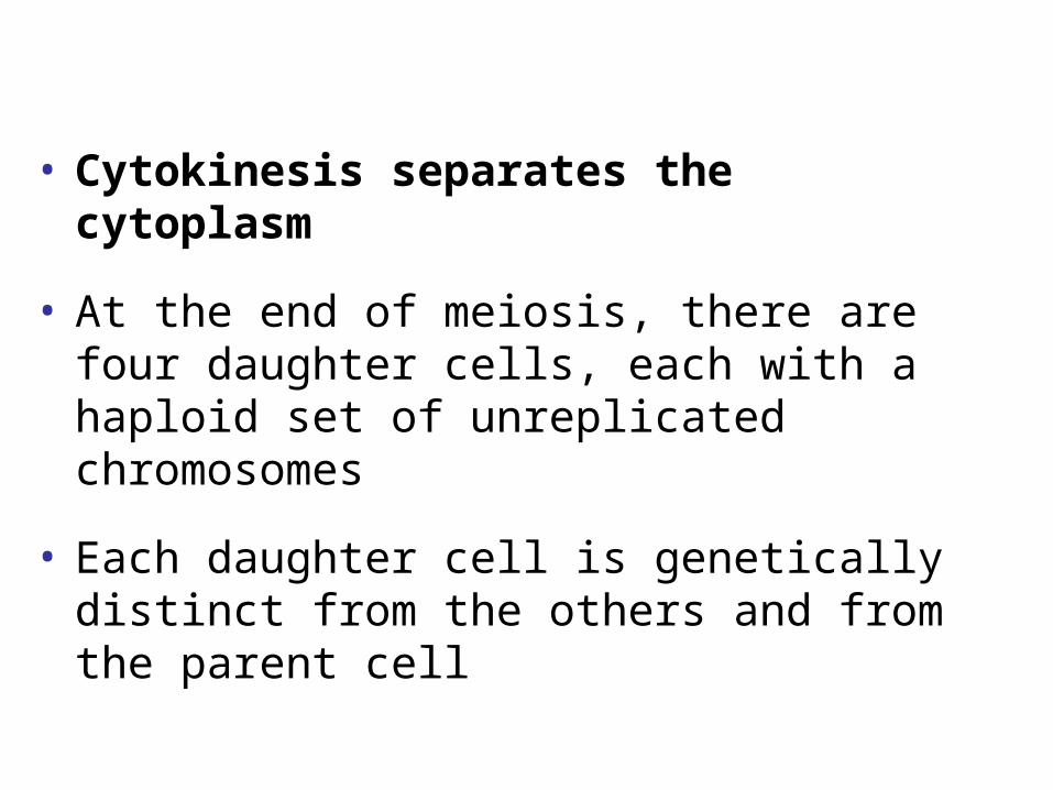
• Cytokinesis separates the cytoplasm
• At the end of meiosis, there are four daughter cells, each with a haploid set of unreplicated chromosomes
• Each daughter cell is genetically distinct from the others and from the parent cell

Anaphase IITelephase II and
Cytokinesis
Sister chromatidsseparate Haploid daughter cells
forming

A Comparison of Mitosis and Meiosis
• Mitosis conserves the number of chromosome sets, producing cells that are genetically identical to the parent cell
• Meiosis reduces the number of chromosomes sets from two (diploid) to one (haploid), producing cells that differ genetically from each other and from the parent cell
• The mechanism for separating sister chromatids is virtually identical in meiosis II and mitosis

MITOSIS MEIOSIS
MEIOSIS I
Prophase I
Chiasma
Chromosomereplication
Homologouschromosomepair
Chromosomereplication
2n = 6
Parent cell
Prophase
Replicated chromosome
Metaphase Metaphase I
Anaphase ITelophase I
Haploid n = 3
Daughter cells ofmeiosis I
MEIOSIS II
Daughter cells of meiosis II
nnnn
2n2n
Daughter cellsof mitosis
AnaphaseTelophase

SUMMARY
MeiosisMitosisProperty
DNAreplication
Number ofdivisions
Occurs during interphase beforemitosis begins
One, including prophase, metaphase,anaphase, and telophase
Synapsis ofhomologouschromosomes
Does not occur
Number ofdaughter cellsand geneticcomposition
Two, each diploid (2n) and geneticallyidentical to the parent cell
Role in theanimal body
Enables multicellular adult to arise fromzygote; produces cells for growth, repair,and, in some species, asexual reproduction
Occurs during interphase before meiosis I begins
Two, each including prophase, metaphase, anaphase, andtelophase
Occurs during prophase I along with crossing overbetween nonsister chromatids; resulting chiasmatahold pairs together due to sister chromatid cohesion
Four, each haploid (n), containing half as many chromosomesas the parent cell; genetically different from the parentcell and from each other
Produces gametes; reduces number of chromosomes by halfand introduces genetic variability among the gametes

• Three events are unique to meiosis, and all three occur in meiosis l:
– Synapsis and crossing over in prophase I: Homologous chromosomes physically connect and exchange genetic information
– At the metaphase plate, there are paired homologous chromosomes (tetrads), instead of individual replicated chromosomes
– At anaphase I, it is homologous chromosomes, instead of sister chromatids, that separate

• Sister chromatid cohesion allows sister chromatids of a single chromosome to stay together through meiosis I
• Protein complexes called cohesins are responsible for this cohesion
• In mitosis, cohesins are cleaved at the end of metaphase
• In meiosis, cohesins are cleaved along the chromosome arms in anaphase I (separation of homologs) and at the centromeres in anaphase II (separation of sister chromatids)

Genetic variation produced in sexual life cycles contributes to evolution
• Mutations (changes in an organism’s DNA) are the original source of genetic diversity
• Mutations create different versions of genes called alleles
• Reshuffling of alleles during sexual reproduction produces genetic variation

Origins of Genetic Variation Among Offspring
• The behavior of chromosomes during meiosis and fertilization is responsible for most of the variation that arises in each generation
• Three mechanisms contribute to genetic variation:
– Independent assortment of chromosomes
– Crossing over
– Random fertilization

Independent Assortment of Chromosomes
• Homologous pairs of chromosomes orient randomly at metaphase I of meiosis
• In independent assortment, each pair of chromosomes sorts maternal and paternal homologues into daughter cells independently of the other pairs

• The number of combinations possible when chromosomes assort independently into gametes is 2n, where n is the haploid number
• For humans (n = 23), there are more than 8 million (223) possible combinations of chromosomes

Possibility 1 Possibility 2
Two equally probablearrangements ofchromosomes at
metaphase I
Metaphase II
Daughtercells
Combination 1 Combination 2 Combination 3 Combination 4

Crossing Over
• Crossing over produces recombinant chromosomes, which combine genes inherited from each parent
• Crossing over begins very early in prophase I, as homologous chromosomes pair up gene by gene

• In crossing over, homologous portions of two nonsister chromatids trade places
• Crossing over contributes to genetic variation by combining DNA from two parents into a single chromosome

Prophase Iof meiosis
Pair ofhomologs
Nonsisterchromatidsheld togetherduring synapsis
Chiasma
Centromere
Anaphase I
Anaphase II
Daughtercells
Recombinant chromosomes
TEM

Random Fertilization
• Random fertilization adds to genetic variation because any sperm can fuse with any ovum (unfertilized egg)
• The fusion of two gametes (each with 8.4 million possible chromosome combinations from independent assortment) produces a zygote with any of about 70 trillion diploid combinations

The Evolutionary Significance of Genetic Variation Within Populations
• Natural selection results in the accumulation of genetic variations favored by the environment
• Sexual reproduction contributes to the genetic variation in a population, which originates from mutations
• Crossing over adds even more variation
• Each zygote has a unique genetic identity


Animations and Videos
• Meiosis Tutorial
• Meiosis - 1
• Meiosis - 2
• Stages of Meiosis
• Anaphase Separase
• Spermatogenesis
• Production of Male and Female Gametes
• Polar Body Formation

Animations and Videos
• Random Orientation of Chromosomes During Meiosis
• Independent Assortment
• Independent Assortment and Gamete Diversity
• Alleles That Do Not Assort Independently
• Comparison of Mitosis and Meiosis
• Bozeman - Diploid vs. Haploid Cells
• Activity- Mitosis and Meiosis
• Bozeman - Mitosis and Meiosis Simulation

Animations and Videos
• Meiosis - Nondisjunction – 1
• Meiosis - Nondisjunction – 2
• Crossing Over
• Changes in Chromosome Structure
• The Consequence of Inversion
• Bozeman - Cell Cycle (Mitosis and Meiosis)
• X Inactivation
• Bozeman - X Inactivation

Animations and Videos
• Density-Dependent Inhibition
• Cytotoxic T Lymphocytes Attack a Melanoma Cell
• Metastasis of a Cancer
• Amniocentesis Animation
• Chorionic Villus Sampling
• Karyotype Animation
• Human Melanoma Cells Dividing
• Chapter Quiz Questions – 1
• Chapter Quiz Questions – 2

Which of the following transmits genes from both parents to child, or from one generation of a family to another?
• DNA
• gametes
• somatic cells
• mitosis
• nucleotides

Which of the following transmits genes from both parents to child, or from one generation of a family to another?
• DNA
• gametes
• somatic cells
• mitosis
• nucleotides

Fertilization is to zygote as meiosis is to which of the following?
• mitosis
• diploid
• chromosome
• replication
• gamete

Fertilization is to zygote as meiosis is to which of the following?
• mitosis
• diploid
• chromosome
• replication
• gamete

Privet shrubs and humans each have a diploid number of 46 chromosomes per cell. Why are the two species so dissimilar?
• Privet chromosomes undergo only mitosis.
• Privet chromosomes are shaped differently.
• Human chromosomes have genes grouped together differently.
• The two species have appreciably different genes.
• Privets do not have sex chromosomes.

Why is it more practical to prepare karyotypes by viewing somatic diploid cells rather than haploid gametes?
• Somatic diploid cells do not contain organelles to interfere with karyotyping.
• Both sets of chromosomes, which are present in somatic diploid cells, need to be examined.
• DNA in haploid gametes will not stain.
• The chromosomes are larger in a somatic diploid cell.
• Haploid gametes do not have sex chromosomes.

Diploid cells may undergo either mitosis or meiosis. Haploid cells may undergo mitosis (for certain species) but not meiosis because
• the sister chromatids cannot separate.
• the synaptonemal complex is too strong.
• crossing over has occurred.
• cohesins are no longer present.
• homologous chromosomes cannot pair.

How and at what stage do chromosomes undergo independent assortment?
• meiosis I pairing of homologs
• anaphase I separation of homologs
• meiosis II separation of homologs
• meiosis I metaphase alignment
• meiosis I telophase separation

How and at what stage do chromosomes undergo independent assortment?
• meiosis I pairing of homologs
• anaphase I separation of homologs
• meiosis II separation of homologs
• meiosis I metaphase alignment
• meiosis I telophase separation

What allows sister chromatids to separate in which phase of meiosis?
• release of cohesin along sister chromatid arms in anaphase I
• crossing over of chromatids in prophase I
• release of cohesin at centromeres in anaphase I
• release of cohesin at centromeres in anaphase II
• crossing over of homologs in prophase I

Gametes produced from one meiotic event
• are genetically identical to each other.
• each have the same chromosome number.
• are genetically identical to the cells produced from meiosis I.
• are genetically identical to the parent cell.
• each have the same mutations.

Gametes produced from one meiotic event
• are genetically identical to each other.
• each have the same chromosome number.
• are genetically identical to the cells produced from meiosis I.
• are genetically identical to the parent cell.
• each have the same mutations.
Crossing over begins to occur during
• anaphase I
• anaphase II
• prophase I
• metaphase II
• telophase II

Crossing over begins to occur during
• anaphase I
• anaphase II
• prophase I
• metaphase II
• telophase II

In this cell, what phase is represented?
• mitotic metaphase
• meiosis I anaphase
• meiosis I metaphase
• meiosis II anaphase
• meiosis II metaphase

In this cell, what phase is represented?
• mitotic metaphase
• meiosis I anaphase
• meiosis I metaphase
• meiosis II anaphase
• meiosis II metaphase

When nutrients are low, cells of the budding yeast (Saccharomyces cerevisiae) exit the mitotic cell cycle and enter meiosis. In this exercise, you will track the DNA content of a population of yeast cells as they progress through meiosis.
Researchers grew a culture of yeast cells in a nutrient-rich medium and then transferred them to a nutrient-poor medium to induce meiosis. At different times after induction, the DNA content per cell was measured in a sample of the cells, and the average DNA content per cell was recorded in femtograms (fg; 1 femtogram 1 × 10–15 gram). Their data are shown in the table to the right.
Scientific Skills Exercise

Most of the yeast cells in the culture were in G1 of the cell cycle before being moved to the nutrient-poor medium to induce meiosis. How many femtograms of DNA are there in a yeast cell in G1? Estimate this value from the data in the table.
• 12 fg
• 24 fg
• 40 fg
• 48 fg

Most of the yeast cells in the culture were in G1 of the cell cycle before being moved to the nutrient-poor medium to induce meiosis. How many femtograms of DNA are there in a yeast cell in G1? Estimate this value from the data in the table.
• 12 fg
• 24 fg
• 40 fg
• 48 fg

How many femtograms of DNA are present in a cell in G2?
• 12 fg
• 24 fg
• 40 fg
• 48 fg

How many femtograms of DNA are present in a cell in G2?
• 12 fg
• 24 fg
• 40 fg
• 48 fg

How many femtograms of DNA are present in a cell at the end of meiosis I?
• 12 fg
• 24 fg
• 40 fg
• 48 fg

How many femtograms of DNA are present in a cell at the end of meiosis I?
• 12 fg
• 24 fg
• 40 fg
• 48 fg

How many femtograms of DNA are present in a cell at the end of meiosis II?
• 12 fg
• 24 fg
• 40 fg
• 48 fg

How many femtograms of DNA are present in a cell at the end of meiosis II?
• 12 fg
• 24 fg
• 40 fg
• 48 fg

A graph with labels indicating the different phases of the meiotic cell cycle (MI meiosis I; MII meiosis II) is shown to the right, based on the data from the table. Think carefully about the point on the graph where the line at the highest value begins to slope downward, indicated by the red arrow.
What specific point of meiosis does this “corner” represent?
• metaphase I
• prophase II
• cytokinesis
• anaphase I

Based on this data, how much DNA is present in a gamete of Saccharomyces cerevisiae?
• 12 fg
• 24 fg
• 48 fg

Based on this data, how much DNA is present in a gamete of Saccharomyces cerevisiae?
• 12 fg
• 24 fg
• 48 fg

Given the fact that 1 fg of DNA 9.78 105 base pairs (on average), you can convert the amount of DNA per cell to the length of DNA in numbers of base pairs. Millions of base pairs (Mb) is the standard unit for expressing genome size.
Calculate the approximate number of base pairs of DNA in the haploid yeast genome.
• 0.08 Mb (8.0 104 base pairs)
• 1.2 Mb (1.2 106 base pairs)
• 12 Mb (12 106 base pairs)
• 23 Mb (23 106 base pairs)

Given the fact that 1 fg of DNA 9.78 105 base pairs (on average), you can estimate the rate of DNA synthesis in Saccharomyces cerevisiae.
Approximately how many base pairs per minute were synthesized during the S phase of these yeast cells?
• 0.19 (1.9 10–1) base pair per minute
• 100,000 (1.0 105) base pairs per minute
• 200,000 (2.0 105) base pairs per minute
• 11,000,000 (11.0 106) base pairs per minute