NTNUfolk.ntnu.no/htorp/DoktorAvhandlinger_Ultralyd/2010_HåvardNordgaard.pdf · NTNU Norges...
111
Doktoravhandlinger ved NTNU, 2010:249 Håvard Bersås Nordgaard TRANSIT-TIME FLOWMETRY AND WALL SHEAR STRESS ANALYSIS OF CORONARY ARTERY BYPASS GRAFTS – A clinical and experimental study ISBN 978-82-471-2496-3 (trykt utg.) ISBN 978-82-471-2497-0 (elektr. utg.) ISSN 1503-8181 NTNU Norges teknisk-naturvitenskapelige universitet Avhandling for graden Det medisinske fakultet Institutt for sirkulasjon og bildediagnostikk Håvard Bersås Nordgaard Doktoravhandlinger ved NTNU, 2010:249
Transcript of NTNUfolk.ntnu.no/htorp/DoktorAvhandlinger_Ultralyd/2010_HåvardNordgaard.pdf · NTNU Norges...
Doktoravhandlinger ved NTNU, 2010:249
Håvard Bersås NordgaardTRANSIT-TIME FLOWMETRY ANDWALL SHEAR STRESS ANALYSIS OFCORONARY ARTERY BYPASS GRAFTS– A clinical and experimental study
ISBN 978-82-471-2496-3 (trykt utg.)ISBN 978-82-471-2497-0 (elektr. utg.)
ISSN 1503-8181
NTN
UN
orge
s te
knis
k-na
turv
itens
kape
lige
univ
ersi
tet
Avha
ndlin
g fo
r gr
aden
Det
med
isin
ske
faku
ltet
Inst
itutt
for
sirk
ulas
jon
og b
ilded
iagn
ostik
k
Håvard B
ersås Nordgaard
Doktoravhandlinger ved N
TNU
, 2010:249

philosophiae doctor philosophiae
philosophiae doctor philosophiae doctor

NTNUNorges teknisk-naturvitenskapelige universitet
Avhandling for graden philosophiae doctor
Det medisinske fakultet
Institutt for sirkulasjon og bildediagnostikk
©Håvard Bersås Nordgaard
ISBN 978-82-471-2496-3 (trykt utg.)
ISBN 978-82-471-2497-0 (elektr utg.)
ISSN 1503-8181
Doktoravhandlinger ved NTNU, 2010:249
Trykt av Tapir Uttrykk
TRANSIT-TIME FLOWMETRY AND WALL SHEAR STRESS
ANALYSIS OF CORONARY ARTERY BYPASS GRAFTS
– A clinical and experimental study
Håvard Bersås Nordgaard, MD
Department of Circulation and Medical Imaging
The Faculty of Medicine
Norwegian University of Science and Technology
Trondheim, Norway
TRANSIT-TIME FLOWMETRY AND WALL SHEAR STRESS
ANALYSIS OF CORONARY ARTERY BYPASS GRAFTS
– A clinical and experimental study
Håvard Bersås Nordgaard, MD
Department of Circulation and Medical Imaging
The Faculty of Medicine
Norwegian University of Science and Technology
Trondheim, Norway
TRANSIT-TIME FLOWMETRY AND WALL SHEAR STRESS
ANALYSIS OF CORONARY ARTERY BYPASS GRAFTS
– A clinical and experimental study
Håvard Bersås Nordgaard, MD
Department of Circulation and Medical Imaging
The Faculty of Medicine
Norwegian University of Science and Technology
Trondheim, Norway
TRANSIT-TIME FLOWMETRY AND WALL SHEAR STRESS
ANALYSIS OF CORONARY ARTERY BYPASS GRAFTS
– A clinical and experimental study
Håvard Bersås Nordgaard, MD
Department of Circulation and Medical Imaging
The Faculty of Medicine
Norwegian University of Science and Technology
Trondheim, Norway

Transitt-tid blodstrømsmåling og analyse av skjærekrefter i bypass-graft ved koronar hjertekirurgi. En eksperimentell og klinisk studie.
Bakgrunn Avhandlingen ”Transit-time flowmetry and wall shear stress analysis of coronary artery bypass grafts – A clinical and experimental study” er en studie av funksjonen i nye bypass-årer i ulike situasjoner, samt av skjærekrefter i disse under ulike eksperimentelle intervensjoner. I Norge utføres årlig ca. 3000 bypass-operasjoner pga. koronar hjertesykdom (angina pectoris). Ved åpen hjertekirurgi sys da erstatningsårer forbi de trange partier på hjertets egne kransarterier. Korrekt vurdering av forsnevringen i kransarteriene og intraoperativ kvalitetskontroll av åreforbindelsen (anastomosen) er viktig for at bypass-årer holder seg åpne. Doktoravhandlingen er basert på fire arbeider som alle er publisert i anerkjente internasjonale tidsskrifter. Arbeidet er en blanding av studier på hjertekirurgiske pasienter og dyreeksperimentelle studier. Resultater I den første studien ble et stort pasientmateriale med 1390 blodstrømsmålinger analysert. Ulike typer bypass-graft til de forskjellige kransarteriene på hjertet ble sammenlignet. Hovedfunnet var økende blodstrøm ved økende antall anastomoser (opptil tre) fra den samme erstatnings-åre og når denne er vene. ”Pulsatility index”, som til en viss grad indikerer motstand i åresystemet, var uavhengig av antall anastomoser, men hadde høyere verdi på høyre side av hjertet sammenlignet med venstre side.
I en annen klinisk studie ble de to mest anvendte transitt-tid blodstrømsmålere sammenlignet på samme bypass-åre. Undersøkelsen viste at MediStim flowmeter systematisk ga høyere pulsatility index-verdier enn Transonic flowmeter, selv om mengde blodstrøm per tidsenhet var lik. Årsaken var ulik filtrering av blodstrømssignalene i flowmetrene. Det bør derfor etableres retningslinjer for fortolkning og rapportering ved anvendelse av de respektive blodstrømsmålere. Det er kjent at konkurrerende blodstrøm fører til at bypass-graft hyppigere går tett eller får mindre kaliber. Dette forekommer når innsnevringen av kransarterien er mindre uttalt enn forventet og ikke begrenser blodstrømmen i hvile. I en grisemodell ble transitt-tid blodstrømsmåling ved forskjellig grad av konkurrerende blodstrøm studert, med og uten innsnevring i anatomosen. Resultatet var at konkurrerende blodstrøm har en stor innvirkning på målingene, mens en innsnevring i anastomosen på inntil 75 % ikke ga noen begrensning av blodstrømmen i bypass-åren. I den fjerde studien ble varierende grad av konkurrerende blodstrøm studert med avansert datasimulering, såkalt ”computational fluid dynamics”, på bakgrunn av en ny grisemodell. Her ble spesielt skjærekreftene (”shear stress”) mellom blodstrøm og årevegg beregnet. Resultatet var at full konkurrerende blodstrøm induserer ugunstige skjærekrefter i bypass-åren på grunn av redusert og retningsskiftende blodstrøm. Ved mindre grad eller fravær av konkurrerende blodstrøm var skjærekreftene gunstigere på grunn av høyere og mer retningsstabil blodstrøm. Ugunstige skjærekrefter kan føre til endotel-dysfunksjon, som er en viktig årsak til intima-hyperplasi og åreforkalkning. Disse funn indikerer at konkurrerende blodstrøm med ledsagende endotel-dysfunksjon er en negativ faktor i forhold til langtids holdbarhet av bypass-årer ved koronar hjertekirurgi. Navn kandidat: Håvard Bersås Nordgaard Institutt: Institutt for sirkulasjon og bildediagnostikk (ISB) Hovedveileder: Professor Rune Haaverstad, ISB/NTNU og Universitetet i Bergen Biveiledere: Overlege PhD Dag Ole Nordhaug, ISB/NTNU og St. Olavs Hospital
Overlege PhD Idar Kirkeby-Garstad, St. Olavs Hospital Post. doc. Lasse Løvstakken, ISB/NTNU
Ovennevnte avhandling er funnet verdig til å forsvares offentlig for graden philosophiae doctor (PhD).
Disputas finner sted i Auditoriet (ØHA11), Øya Helsehus. Fredag 17. desember 2010 kl.12.15


CONTENTS Page
1. ACKNOWLEDGEMENTS................................................................... 3
2. LIST OF PAPERS.................................................................................. 5
3. ABBREVIATIONS................................................................................ 6
4. INTRODUCTION................................................................................. 7 4.1 CORONAR ARTERY BYPASS GRAFTING.......................................... 7 4.2 CORONARY HAEMODYNAMICS.......................................................... 10
Coronary blood flow, autoregulation and stenosis.................................... 10 Wall shear stress and endothelial dysfunction........................................... 12
5. STUDY OBJECTIVES………………………..................................... 17
6. METHODOLOGICAL CONSIDERATIONS................................... 19 6.1 TRANSIT-TIME FLOW MEASUREMENTS......................................... 19
Procedure................................................................................................... 20 Interpretation............................................................................................. 22 Limitations................................................................................................. 24
6.2 COMPUTATIONAL FLUID DYNAMICS.............................................. 25 Limitations................................................................................................. 27
6.3 SURGICAL PROCEDURES AND EXPERIMENTAL SET-UP............ 27 Clinical setting…………………………………………………….…….. 27 Animal model……………………………………………………………. 28 Epicardial ultrasound imaging................................................................... 31
6.4 STATISTICAL ANALYSES.................................................................... 34
7. SUMMARY OF RESULTS................................................................. 35 7.1 Paper I........................................................................................................ 35 7.2 Paper II....................................................................................................... 36 7.3 Paper III..................................................................................................... 36 7.4 Paper IV..................................................................................................... 37
8. DISCUSSION........................................................................................ 39
9. MAIN CONCLUSIONS....................................................................... 46
10. PROSPECTS OF FUTURE RESEARCH......................................... 47
11. REFERENCES...................................................................................... 48
12. APPENDIX............................................................................................ 58
12.1 Letter to the Editor of the EJCTS in reference to Paper III……………… 59 12.2 Reply to Letter, EJCTS…………………………………………………... 60


3
1. ACKNOWLEDGEMENTS
This thesis was completed between May 2006 and December 2009 while I was
working as a research fellow at the Department of Circulation and Imaging,
Norwegian University of Science and Technology (NTNU). My office has been at St.
Elisabeth Department of Cardiothoracic Surgery, where I have been active as a
research fellow as well as a junior surgeon. To all the people who have contributed to
this thesis, I am sincerely thankful.
My main supervisor has been Professor Rune Haaverstad. I want to thank him for
initiating the research project, his support, ideas, encouragement, criticism and fast e-
mail responses! I have benefited from his long-term interest and scientific work
regarding coronary graft assessment, as well as his established experimental animal
research program. Further, he has introduced me to his great network of collaborators
at NTNU and St. Olavs Hospital, making my project a real team work. I have enjoyed
our cooperative relationship and friendship.
The Head of Department of Cardiothoracic Surgery, Asbjørn Karevold, has provided
me with continuous support and introduced me to the field of cardiothoracic surgery,
for which I am enormously grateful. It has been a privilege working as a junior
surgeon in the friendly atmosphere at St. Elisabeth.
I would like to thank my associate Dag Ole Nordhaug for inspirational and strategic
talks and for helping me in the animal lab with his vast experience and excellent
surgical skills.
It has also been a privilege to work with Nicola Vitale, and I appreciate his unlimited
contribution in terms of ideas and manuscript revisions.
I must thank Idar Kirkeby-Garstad for his inspiring talks, superb anaesthesia and other
contribution to the animal research.
I also want to acknowledge Lasse Løvstakken for his excellent ultrasound technology
skills. I hope our successful collaboration, which brought clinical and technical
research together, will continue in the future!

4
I am thankful for the fruitful cooperation with Abigail Swillens and Patrick Segers in
Ghent, particularly for their excellent knowledge in the field of biomechanics and
computational fluid dynamics.
I want to thank Oddveig Lyng at the Section for Comparative medicine for helping
me with the experiments. I also appreciate the important advice and technical support
given by Arne Grip, Arne Nilsen and Erik Swendsen at Medi-Stim ASA.
I would also like to thank the excellent surgeons at St. Elisabeth: Rafael Astudillo,
Hans Henrik Dedichen, Alexander Wahba and Anders Winnerkvist, as well as
surgical assistant Maryann Stenvik and all the nurses at the operating unit for their
great enthusiasm and surgical training. Thanks also to Sigurd Gunnes for his friendly
support during the writing of this thesis and to Olav Sellevold and Petter Aadahl for
contributions to the early revisions of the thesis.
The research presented in this thesis has been financially supported by the Blix family
fund, Sigrid Wolmars legat, Øyvind Mikkelsens minnefond, Fondstiftelse St. Olavs
Hospital, Odd Fellow Ordenen, Operating Room of the Future, and the Medical
Imaging Lab.
Above all, I want to thank my wonderful Marit and our beautiful children, Magnus,
Synnøve and Henrik, for their love and support. I will always be there for you.

5
2. LIST OF PAPERS
This thesis is based on the following original papers, which will be referred to by their
Roman numerals:
I. Transit-time blood flow measurements in sequential saphenous coronary
artery bypass grafts. Nordgaard H, Vitale N, Haaverstad R. Ann Thorac Surg
2009 May;87(5):1409-15
II. Different graft flow patterns due to competitive flow or stenosis in the
coronary anastomosis assessed by transit-time flowmetry in a porcine model.
Nordgaard H, Nordhaug D, Kirkeby-Garstad I, Lovstakken L, Vitale N,
Haaverstad R. Eur J Cardiothorac Surg 2009 Jul;36(1):137-42
III. Pulsatility index variations using two different transit-time flowmeters in
coronary artery bypass surgery. Nordgaard H, Vitale N, AstudilloR, Renzulli
A, Romundstad P, Haaverstad R. Eur J Cardiothorac Surg 2010
May;37(5):1063-7.
IV. Impact of competitive flow on wall shear stress in coronary surgery:
Computational fluid dynamics of a LIMA-LAD model. Nordgaard H,
Swillens A, Nordhaug D, Kirkeby-Garstad I, Van Loo D, Vitale N, Segers P,
Haaverstad R, Løvstakken L. Cardiovascular Research June 2010; In press

6
3. ABBREVIATIONS
BFI blood flow imaging
CFD computational fluid dynamics
CABG coronary artery bypass grafting
CO cardiac output
CX circumflex artery
ECG electrocardiogram
FFR fractional flow reserve
IVUS intravascular ultrasound
LAD left anterior descending artery
LIMA left internal mammary artery
LMS left main stem
MI myocardial infarction
MVR mitral valve replacement
MVr mitral valve repair
OPCAB off-pump coronary artery bypass
OM obtuse marginal
OSI oscillatory shear index
PCI percutaneous coronary intervention
PDA posterior descending artery
PI pulsatility index
PL posterolaterale branch
RCA right coronary artery
SD standard deviation
SSVG sequential saphenous vein graft
SVG saphenous vein graft
TTFM transit-time flow measurement
WSS wall shear stress
7
4. INTRODUCTION
4.1 Coronary artery bypass grafting (CABG)
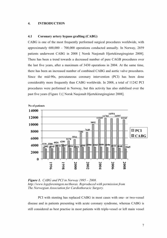
CABG is one of the most frequently performed surgical procedures worldwide, with
approximately 600,000 – 700,000 operations conducted annually. In Norway, 2859
patients underwent CABG in 2008 [ Norsk Nasjonalt Hjertekirurgiregister 2008].
There has been a trend towards a decreased number of pure CAGB procedures over
the last five years, after a maximum of 3430 operations in 2004. At the same time,
there has been an increased number of combined CABG and aortic valve procedures.
Since the mid-90s, percutaneous coronary intervention (PCI) has been done
considerably more frequently than CABG worldwide. In 2008, a total of 11242 PCI
procedures were performed in Norway, but this activity has also stabilised over the
past five years (Figure 1) [ Norsk Nasjonalt Hjertekirurgiregister 2008].
Figure 1. CABG and PCI in Norway 1995 – 2008. http://www.legeforeningen.no/thorax. Reproduced with permission from The Norwegian Association for Cardiothoracic Surgery.
PCI with stenting has replaced CABG in most cases with one- or two-vessel
disease and in patients presenting with acute coronary syndrome, whereas CABG is
still considered as best practise in most patients with triple-vessel or left main vessel
7
4. INTRODUCTION
4.1 Coronary artery bypass grafting (CABG)
CABG is one of the most frequently performed surgical procedures worldwide, with
approximately 600,000 – 700,000 operations conducted annually. In Norway, 2859
patients underwent CABG in 2008 [ Norsk Nasjonalt Hjertekirurgiregister 2008].
There has been a trend towards a decreased number of pure CAGB procedures over
the last five years, after a maximum of 3430 operations in 2004. At the same time,
there has been an increased number of combined CABG and aortic valve procedures.
Since the mid-90s, percutaneous coronary intervention (PCI) has been done
considerably more frequently than CABG worldwide. In 2008, a total of 11242 PCI
procedures were performed in Norway, but this activity has also stabilised over the
past five years (Figure 1) [ Norsk Nasjonalt Hjertekirurgiregister 2008].
Figure 1. CABG and PCI in Norway 1995 – 2008. http://www.legeforeningen.no/thorax. Reproduced with permission from The Norwegian Association for Cardiothoracic Surgery.
PCI with stenting has replaced CABG in most cases with one- or two-vessel
disease and in patients presenting with acute coronary syndrome, whereas CABG is
still considered as best practise in most patients with triple-vessel or left main vessel

8
disease and with impaired left ventricular function [Eagle et al 2004; Serruys et al
2009; Patel et al 2009].
Patients with complex coronary anatomy and calcified lesions are more often
treated by CABG, as PCI in such cases are technically less feasible [Haaverstad
2009]. Moreover, patients undergoing CABG nowadays often have a higher operative
risk due to old age and comorbidities. On-pump CABG is still the technique most
widely performed when surgical revascularisation is considered. In Norway, off-pump
CABG is usually reserved for patients with a heavily calcified wall of the ascending
aorta. The cannulation and cross-clamping may increase the risk of calcium emboli. In
2007, only 20 off-pump coronary artery bypass (OPCAB) operations were performed
in Norway [Haaverstad 2009].
The use of the left internal mammary artery (LIMA) and the great saphenous
vein are still the most common procedures of surgical myocardial revascularisation.
The LIMA-to-LAD has a superior long-term patency versus other conduits and
improves long-term survival and freedom from reinterventions [Cameron et al 1996;
Goldman et al 2004; Sabik et al 2005].
Currently, vein grafting is often carried out by the sequential grafting
technique, in which one or more side-to-side anastomoses are performed in addition
to the distal end-to-side anastomosis. The obvious advantages of sequential vein
grafting are shorter limb incisions and fewer proximal anastomoses with better
utilisation of the vein graft segment. However, sequential vein grafts may also carry
more blood flow to the myocardium compared to single vein grafts, although solid
evidence for this is still warranted [Christenson et al 1998; Dion et al 2001; Vural et al
2001; Kandemir et al 2007]. Furthermore, some data suggest that the long-term
patency of side-to-side anastomoses are better than the patency of end-to-side
anastomoses [Kieser et al 1986]. Sequential grafting may also optimise
haemodynamic conditions like flow pattern and wall shear stress [Frauenfelder et al
2007].
Graft failure is a concern after CABG. Improvement of the early as well as the
long-term graft patency is frequently addressed. Nowadays it is common to perform
intraoperative quality assessment of the bypass grafts. Several technologies are
available, but transit-time flowmetry is the most widespread method. The aim of
intraoperative graft assessment is to detect and thereby to revise technical of grafts
and anastomoses, thus improve clinical outcome and avoid re-interventions.

9
Table 1. Possible causes of early graft failure after CABG.
Reported findings References
Failure of prox. or dist. anastomosis [D'Ancona et al 2000] Overstretched graft [D'Ancona et al 2000; Tokuda et al 2007] Graft dissection [Walpoth et al 1998; Tokuda et al 2007] Obstructing intimal flap [Walpoth et al 1998; D'Ancona et al 2000; Tokuda et al 2007] Intramural haematoma [Walpoth et al 1998; D'Ancona et al 2000] Kinked or twisted graft [D'Ancona et al 2000; Tokuda et al 2007]
In CABG, the flow within bypass grafts may be influenced by the function of
native coronary arteries. Competitive flow may be seen when grafting is performed on
coronary vessels with low-grade stenosis. Clinically, competitive flow is assumed to
occur frequently in CABG, as coronary arteries with a luminal area stenosis of 50-70
% are often grafted. Most studies on graft function have shown better long-term
patency when they are directed distally to severe stenoses rather than beyond non-
significant lesions [Villareal et al 2000; Hirotani et al 2001; Sabik et al 2003; Bezon
et al 2003; Berger et al 2004; Nakajima et al 2007; Botman et al 2007; Sabik et al
2008; Kawamura et al 2008].
The “string phenomenon” of the LIMA graft, which displays a threadlike
structure on the angiogram, is assumed to be caused by competitive flow. However,
there is limited knowledge of the underlining patho-physiological and biomechanical
mechanisms of competitive flow in CABG. By addressing why grafts often fail during
competitive flow conditions, essential information with great consequences may be
achieved. This may subsequently facilitate the development of surgical,
pharmacological and genetic interventions for improvement of coronary grafts
patency.

10
4.2 Coronary haemodynamics
Because this thesis deals with blood flow and wall shear stress in coronary bypass
grafts, some basic fundamental anatomical and physiological concepts of the coronary
circulation will be presented.
Figure 2. The coronary artery tree. LMS = left main stem; CX = circumflex artery; LAD = left anterior descending artery; OM = obtuse marginal; LIMA = left internal mammary artery; RCA = right coronary artery; PDA = posterior descending artery; PL = posterolateral artery. Modified figure from: http://www.meddean.luc.edu/lumen/meded/Radio/curriculum/Vascular/Coronary_artery.jpg
Coronary blood flow, autoregulation and stenosis
At rest, the total coronary flow is 180-250 ml/min, i.e. 5-10 % of the total
cardiac output [Naidu et al 2001]. The primary regulatory mechanism of coronary
blood flow is determined by the local myocardial metabolism by release of adenosine
and other vasodilator mediators. At rest, the myocardium already has an extremely
high oxygen extraction. The higher oxygen demand during increased cardiac work
load or ischaemia is normally met by increased coronary blood flow. Thus, there is a
nearly linear relationship between the myocardial metabolic demand and the coronary
blood flow. Healthy individuals can increase coronary flow up to four-to-six fold
[Naidu et al 2001; Spaan et al 2006]. Other important control mechanisms are the

11
neurogenic and hormonal systems, which are generally more vasoconstrictive than
vasodilatory.
The vascular endothelium acts as control system by determining the vascular
tonus. The blood flow induced wall shear stress (WSS) increases the release of
several mediators from the intact endothelium, particularly the vasodilatory molecule
nitric oxide (NO). However, in damaged endothelium such as in patients with
atherosclerosis, the release of NO is reduced and the release of the vasoconstrictive
endothelin-1 is increased [Opie et al 2004].
Coronary blood flow is inversely related to the peripheral vascular resistance.
The vascular resistance is very low in the epicardial coronary arteries due to these
large branches. Thus, only a minimal pressure drop exists between the ascending aorta
and the distal end of healthy major coronary arteries. The smaller coronary branches
are arterioles embedded deeply into the myocardium and are also called resistance
arteries. They can adjust coronary blood flow according to the metabolic demands of
the myocardium through the metabolic, neurogenic and vascular control systems
(autoregulation).
The coronary arteries provide stable myocardial perfusion when the systolic
pressure ranges between 50 - 180 mmHg, allowing a relatively constant blood flow. If
the systolic perfusion pressure drops below 50 mmHg, the vasodilatory effect
controlled by the coronary autoregulation is gradually lost [Gould et al 1974; Opie et
al 2004]. An estimated 70 % diameter reduction, equivalent to a 90–95 % decrease in
luminal area, is required to decrease the basal, resting coronary blood flow [Gould et
al 1974].
Coronary flow varies considerably from rest to exercise. During exercise, the
coronary flow is already impaired when the internal diameter is reduced beyond 30 %
[Gould et al 1974]. The coronary flow is further influenced by the collateral blood
flow, the vascular spasm and a multiple or extensive stenosis, as well as the degree of
turbulence across the stenosis.
The impact of a stenosis on blood flow cannot directly be explained in the
context of Poiseuille's law due to the coronary autoregulation, the vessel tortuosity
and the pulsatile flow that is present in-vivo. Moreover, the law was based upon a
Newtonian fluid, i.e. an homogenous fluid with constant viscosity like water and not
blood. The law relates the flow Q to the length of the internal radius r of the vessel,
according to the following formula:

12
L8PrQ
4
, with viscosity , length L and pressure difference P [Opie et al 2004].
The equation states that the dominant factor on the flow Q is the internal radius r of
the vessel.
The coronary flow pattern is pulsatile due to the myocardial contraction and
relaxation. During systole, the subendocardial coronary branches are much more
compressed than the epicardial branches [Yada et al 1993]. Hence, most of the
coronary flow occurs in the diastolic phase of relaxation, most pronounced for the left
coronary artery. The right coronary artery has a more equal volume flow both in
systole and diastole. The heart rate also affects coronary blood flow as bradycardia
extends the diastole, thus increases the coronary flow. Tachycardia prolongs the
systolic phase of the cardiac cycle, thus reduces the coronary flow [Opie et al 2004].
Wall shear stress and endothelial dysfunction
The blood flow through arteries is mostly laminar with a parabolic profile: the highest
blood velocity in the centre and the lowest velocities close to the vessel wall. The
laminar blood flow may be considered as a series of layers that move with different
velocities, but in the same direction. The friction between these layers generates the
shear stress of blood. The WSS is the friction of blood flow against the vessel wall
(Figures 3-5).
For a Newtonian fluid, the WSS is proportional to the wall shear rate (yv ,
the change in flow velocity across the
yv
. The wall shear stress is defined as the shear stress at y = 0, i.e., at the
vessel wall (Figure 3).
The WSS may also be calculated in terms of volume flow Q and vessel radius
R by applying Poiseuille’s law: 3RQ4 . Thus, small changes in radius R greatly
flow and dimensions can be measured non-invasively, for instance by ultrasound
imaging. However, as for Poiseuille’s law, the formula has several major

13
assumptions: a) stiff, straight and smooth vessels; b) Newtonian fluid; c) laminar,
steady, non-pulsatile flow and d) zero velocity at the vessel wall.
In healthy arteries at rest, the time-average WSS over a cardiac cycle typically
varies within an approximate range of 1-7 Pa. A time-average WSS < 1-1.5 Pa is
considered as low. Non-physiological elevated WSS is reported to be > 7 Pa and is
usually seen in a narrowed lumen (Figure 4) [Malek et al 1999; Chatzizisis et al
2007].
Figure 3. The wall shear stress is defined as the shear stress at y = 0. Y is the distance within the flow profile in relation to the vessel wall.
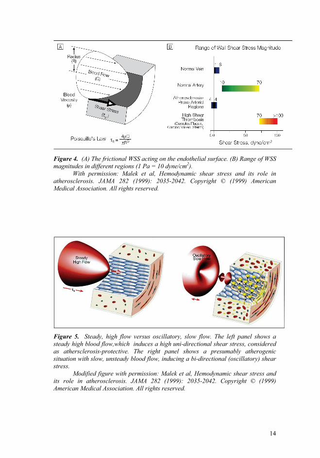
14
Figure 4. (A) The frictional WSS acting on the endothelial surface. (B) Range of WSS magnitudes in different regions (1 Pa = 10 dyne/cm2).
With permission: Malek et al, Hemodynamic shear stress and its role in atherosclerosis. JAMA 282 (1999): 2035-2042. Copyright © (1999) American Medical Association. All rights reserved.
Figure 5. Steady, high flow versus oscillatory, slow flow. The left panel shows a steady high blood flow,which induces a high uni-directional shear stress, considered as athersclerosis-protective. The right panel shows a presumably atherogenic situation with slow, unsteady blood flow, inducing a bi-directional (oscillatory) shear stress.
Modified figure with permission: Malek et al, Hemodynamic shear stress and its role in atherosclerosis. JAMA 282 (1999): 2035-2042. Copyright © (1999) American Medical Association. All rights reserved.

15
An established index for assessing the changes in direction and magnitude of
WSS is the oscillatory shear index (OSI). It is given by the following equation:
T
O
T
0
dt
dt1
21OSI ,
where T is the time of an entire cardiac cycle, and is the WSS vector [Ku et al 1985].
The numerator and the denominator display the total WSS (the sum of all positive and
negative WSS vectors) and the sum of all absolute values of the same WSS vectors
over a cardiac cycle, respectively. The OSI varies between 0 and 0.5, where 0
indicates a completely unidirectional shear force, and 0.5 indicates a purely unsteady,
oscillatory shear force with a net amount of zero WSS. Regions of high OSI are prone
to endothelial dysfunction and atherogenesis [Ku et al 1985; Kassab et al 2006;
Davies 2009].
Next to WSS, the other basic mechanical force acting within vessels is the
transmural pressure. This acts perpendicular to the vessel wall and stretches the wall.
Although there is a wide discrepancy between the extremely small force generated by
the normal WSS (1-7 Pa) and the transmural arterial blood pressure (100 mmHg =
13332 Pa), WSS has a major and well-documented influence on endothelial function
[Malek et al 1999; Davignon et al 2004; Slager et al 2005; Chatzizisis et al 2007;
Stone et al 2007; Katritsis et al 2007; Davies 2009; Koskinas et al 2009]. All
endothelial cells throughout the cardiovascular system are constantly exposed to WSS
generated by the blood flow. Further, WSS varies in magnitude and direction,
depending upon the geometry of the vessel, blood viscosity and flow pattern. Healthy
and intact endothelium is crucial for efficient control of vascular resistance and
function [Opie et al 2004; Davignon et al 2004; Chatzizisis et al 2007]. WSS exerts its
effects through the conversion of mechanical stress to biochemical response, also
described as a mechano-transduction [Davies 1995]. Thus, the endothelium triggers
the metabolic reactions by synthesising vasoactive mediators in response to WSS
changes.
The endothelium attempts to adapt to local flow conditions [Kassab et al 2006].
Vasoactive mediators regulate the adaptation to sudden changes in WSS, whereas
sustained and prolonged changes of WSS induce a slow, structural wall remodelling
that is mediated by the activation of several genes [Slager et al 2005; Kassab et al
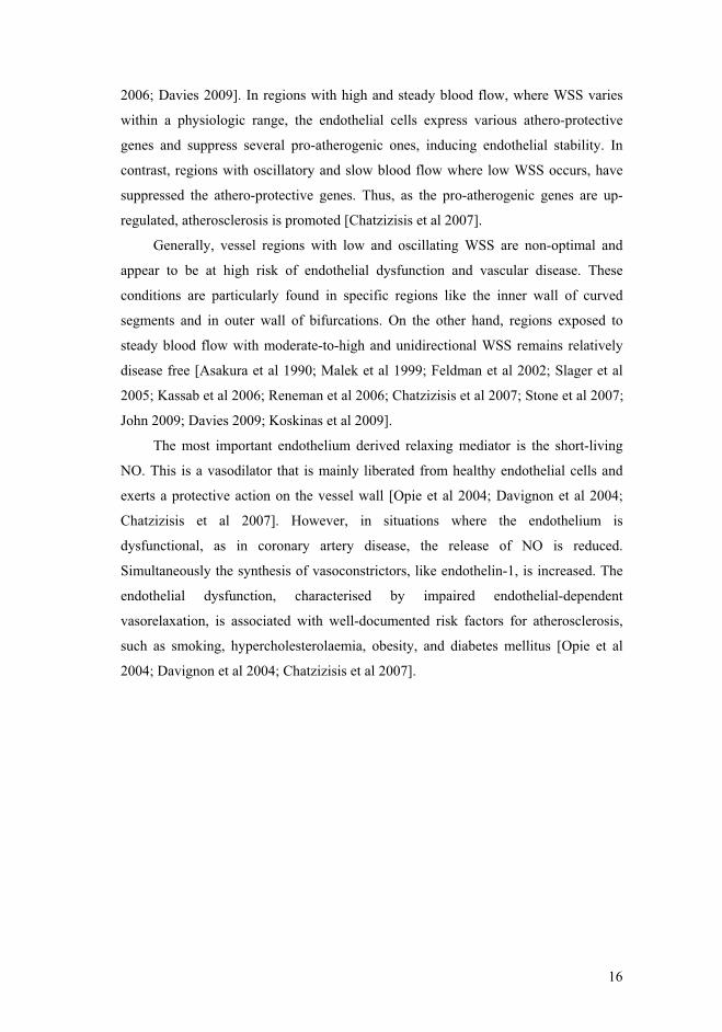
16
2006; Davies 2009]. In regions with high and steady blood flow, where WSS varies
within a physiologic range, the endothelial cells express various athero-protective
genes and suppress several pro-atherogenic ones, inducing endothelial stability. In
contrast, regions with oscillatory and slow blood flow where low WSS occurs, have
suppressed the athero-protective genes. Thus, as the pro-atherogenic genes are up-
regulated, atherosclerosis is promoted [Chatzizisis et al 2007].
Generally, vessel regions with low and oscillating WSS are non-optimal and
appear to be at high risk of endothelial dysfunction and vascular disease. These
conditions are particularly found in specific regions like the inner wall of curved
segments and in outer wall of bifurcations. On the other hand, regions exposed to
steady blood flow with moderate-to-high and unidirectional WSS remains relatively
disease free [Asakura et al 1990; Malek et al 1999; Feldman et al 2002; Slager et al
2005; Kassab et al 2006; Reneman et al 2006; Chatzizisis et al 2007; Stone et al 2007;
John 2009; Davies 2009; Koskinas et al 2009].
The most important endothelium derived relaxing mediator is the short-living
NO. This is a vasodilator that is mainly liberated from healthy endothelial cells and
exerts a protective action on the vessel wall [Opie et al 2004; Davignon et al 2004;
Chatzizisis et al 2007]. However, in situations where the endothelium is
dysfunctional, as in coronary artery disease, the release of NO is reduced.
Simultaneously the synthesis of vasoconstrictors, like endothelin-1, is increased. The
endothelial dysfunction, characterised by impaired endothelial-dependent
vasorelaxation, is associated with well-documented risk factors for atherosclerosis,
such as smoking, hypercholesterolaemia, obesity, and diabetes mellitus [Opie et al
2004; Davignon et al 2004; Chatzizisis et al 2007].

17
5. STUDY OBJECTIVES
Paper I
Routine TTFMs were performed in a large group of patients by the same surgeon. The
grafts considered for analysis were LIMA-LAD, single saphenous vein grafts (SVGs),
double sequential SVGs, and triple sequential SVGs. The aim was to study TTFMs of
different sequential SVGs in relation to single grafts, and to investigate how graft
flow is related to patient characteristics and haemodynamics.
Paper II
Nine pigs underwent off-pump coronary artery bypass surgery with the LIMA grafted
to LAD. TTFMs of the LIMA graft were recorded under the following four flow
conditions (Figure 6):
A. No competitive flow; the proximal LAD totally occluded (the baseline condition),
mimicking an ideal situation for placing the graft distal to a totally occluded
coronary artery.
B. Partial competitive flow; partial occlusion of the LAD to half its full flow,
mimicking a situation in which the anastomotic site was placed distal to a
significant stenosis, providing about half the volume flow compared to the full
flow.
C. Fully competitive flow; no occlusion of the proximal LAD, mimicking a situation
in which a graft was placed distal to a moderate (or less than critical) stenosis.
D. Stenotic anastomosis with no competitive flow; mimicking a typical site of surgical
error (the toe of the anastomosis).
Figure 6. A schematic layout of the four investigated flow conditions on each pig.
The aim was to assess the influence on graft flows due to competitive flow and
stenotic anastomosis in LIMA-LAD anastomoses. Sub-analysis of TTFMs was of
particular interest in this project.

18
Paper III
The objective was to compare the flow signals produced by the two most commonly
used transit-time flowmeters, manufactured by MediStim ASA and Transonic Inc.,
both having the same guidelines regarding the level of Pulsatility index (PI).
In the first part of the study a MediStim (VeriQ) or a Transonic (HT331)
flowmeter were directly compared by applying two flow probes simultaneously on
coronary bypass grafts. In the second part we aimed to investigate whether the PI is
influenced by different technical characteristics with regards to variation of filter
settings within the flowmeter.
Paper IV
In an experimental porcine model a LIMA-LAD anastomosis was performed to
investigate wall shear stress (WSS) during competitive flow conditions. By applying
computational fluid dynamics, WSS in a 3-D LIMA-LAD model were investigated
during three different flow conditions (Figure 6):
A. No competitive flow; simulation of an ideal situation
B. Partial competitive flow; simulation of a significant LAD stenosis
C. Fully competitive flow; simulation of a no-significant LAD stenosis
The aim of the study was to explore the influence of competitive flow on WSS
within the coronary grafts and vessels, and to analyse how different flow conditions
may trigger endothelial dysfunction and subsequent graft failure.

19
6. METHODOLOGICAL CONSIDERATIONS
6.1 Transit-time flow measurements
Almost every cardiac intervention, including valve repair and PCI, are routinely
followed by a reliable assessment to ensure adequate technical results. However, in
CABG, this kind of evaluation is not always a routine procedure, although technical
failures is quite common even if the operation is performed by an experienced
surgeons [D'Ancona et al 2000; Hol et al 2004].
The intention of intraoperative graft assessment is to reduce the risk of early
graft failure and thereby achieve optimal surgical results. Ideally, an intraoperative
quality assessment has the following characteristics:
Simple and quick application
Easy interpretation
Surgeon or technician independent
Provision of both anatomical and physiologic information
Inexpensive
Applicable for all relevant vessels
Highly sensitive
Highly reliable and specific
TTFM of bypass grafts is the most commonly used method for
intraoperative quality assessment in CABG. The theoretical basis of the transit-time
method has been known for more than 40 years, but it has only been applied in
clinical practice since the early 1990s. Initially, this delay was due to several technical
difficulties, like the extremely short “transit-time” of a few nanoseconds and the
complexity and stability of the electronic network. The progress in electronic
technology solved these problems. The first commercial transit-time flowmeter was
introduced by Transonic Inc. in 1983 (Personal communication with Arne Grip,
former CEO, MediStim ASA, Oslo). Later, TTFMs were proven to be an accurate
method for intraoperative verification of bypass grafts [Matre et al 1994; Beldi et al
2000]. A common misunderstanding is that the TTFM method is based on the
Doppler principle, as erroneously asserted by some authors [Wolf et al 2003]. TTFMs
are based on the ultrasonic transit-time principle according to the following formula:
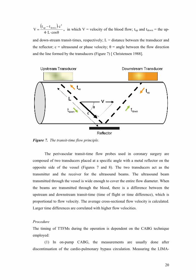
20
cosL4ctt
V2
downup , in which V = velocity of the blood flow; tup and tdown = the up-
and down-stream transit-times, respectively; L = distance between the transducer and
and the line formed by the transducers (Figure 7) [ Christensen 1988].
Figure 7. The transit-time flow principle.
The perivascular transit-time flow probes used in coronary surgery are
composed of two transducers placed at a specific angle with a metal reflector on the
opposite side of the vessel (Figures 7 and 8). The two transducers act as the
transmitter and the receiver for the ultrasound beams. The ultrasound beam
transmitted through the vessel is wide enough to cover the entire flow diameter. When
the beams are transmitted through the blood, there is a difference between the
upstream and downstream transit-time (time of flight or time difference), which is
proportional to flow velocity. The average cross-sectional flow velocity is calculated.
Larger time differences are correlated with higher flow velocities.
Procedure
The timing of TTFMs during the operation is dependent on the CABG technique
employed:
(1) In on-pump CABG, the measurements are usually done after
discontinuation of the cardio-pulmonary bypass circulation. Measuring the LIMA-

21
LAD flow is also recommended before removal of the aortic clamp because this time
point has no collateral flow. Some cardiac surgery units register graft flow both before
and after application of diluted papaverine, particularly within vein grafts. Some
surgeons recommend repeated flow measurements as well as when the sternal edges
are approximated during chest closing in order to assure correct graft position and
absence of graft kinking.
(2) In OPCAB, the arterial grafts are usually assessed both immediately after
they have been constructed and before chest closure. For vein grafts, the assessment
may be performed as with on-pump surgery.
The final measurements should be carried out under stable haemodynamic
conditions. The hook of the flow probe connected to the flowmeter is placed around
the bypass graft. The graft should fit within the flow probe because the ultrasound
beam must illuminate the entire graft.
When assessing a pedicled LIMA graft, a short segment of the graft should be
skeletonised to fit the probe hook. By measuring the graft both before and after
application of papaverine into the vein graft, the flow reserve for the individual graft
is also measured. For the LIMA graft, papaverine is usually applied only externally,
which is not as efficient as when it is given intraluminally.
In order to obtain a correct interpretation of the blood flow patterns, an ECG
recording should be combined with flowmetry to differentiate systole from diastole.

22
Figure 8. A transit-time flow probe is placed around a vein graft after weaning off the cardio-pulmonary bypass circulation.
Interpretation
There is a poor correlation between mean volume flow and the quality of the
anastomosis [Jaber et al 1998; D'Ancona et al 2001; Nordgaard et al 2009]. At rest,
the graft flow is not reduced until the luminal stenosis is severe.
The flow pattern in a bypass graft should mainly be diastolic as in a coronary
artery. Different indicators have been introduced for the quality assessment in CABG.
The most important are:
a) Diastolic filling % (DF%), which is calculated as
%100VV
V%DF
abs,Dabs,S
D ,
where VD and VS is diastolic- and systolic volume flows, respectively. The DF% is
only calculated when an ECG signal is connected to the flowmeter with sufficient
quality for defining systole and diastole. The DF% is perhaps the most important
indicator for TTFM interpretation [Morota et al 2002]. A DF % < 50 is suspicious of
disturbed graft flow because it represents a systolic graft flow pattern [Becit et al
2007].

23
b) Insufficiency % is the percentage retrograde flow:
c) Pulsatility index (PI) is calculated according to the following formula:
flowvolumemeanflowvolumeminflowvolumemaxPI
The PI has been described as an indicator of peripheral vascular resistance
[Louagie et al 1994; D'Ancona et al 2000; Becit et al 2007], although the sensitivity
and specificity are unknown. Peripheral resistance is governed by many factors, and
PI is not sufficiently specific to distinguish between stenoses of different severity.
Only very severe stenoses may produce significantly higher PIs [Evans et al 1980].
Several TTFM cut-off values have been suggested as indicators of adequate
graft flow [D'Ancona et al 2001; Kim et al 2005; Di Giammarco et al 2006]. The
largest prospective study on TTFMs was performed by Tokuda et al., who measured
blood flow by the MediStim flowmeter in 261 bypass grafts, and correlated the results
obtained with 3-month postoperative coronary angiography. They found that mean
flow <15 ml/min, PI > 5 and insufficiency % > 4 % in grafts to the left coronary
system had a higher incidence of graft failure. The failure of grafts to the right
coronary artery was predicted with mean flow < 20 ml/min, PI > 4.7, and
insufficiency % > 4.6 % [Tokuda et al 2007].
The most commonly used transit-time flowmeters, manufactured by MediStim
ASA (Oslo, Norway) and Transonic Systems Inc. (Ithaca, NY, USA), use a default
low-pass filter at 20 Hz and 10 Hz, respectively. They filter the signals differently,
which yields different wave forms (Figure 9) [Proakis et al 1995]. The different
maximum and minimum peak flows that are registered may influence the PI value.
Although several scientific papers have compared the available flowmeters
and concluded that they are accurate and precise with regard to volume flow, the issue
of different flow patterns has not previously been addressed [Laustsen et al 1996;
Beldi et al 2000; Cikirikcioglu et al 2006]. However, the latter is important because
smoother flow pattern and low PI are indications of good flow, in contrast to spiky
flow pattern and higher PI, which are associated with poor flow [Jaber et al 1998;
D'Ancona et al 2001; Kim et al 2005].
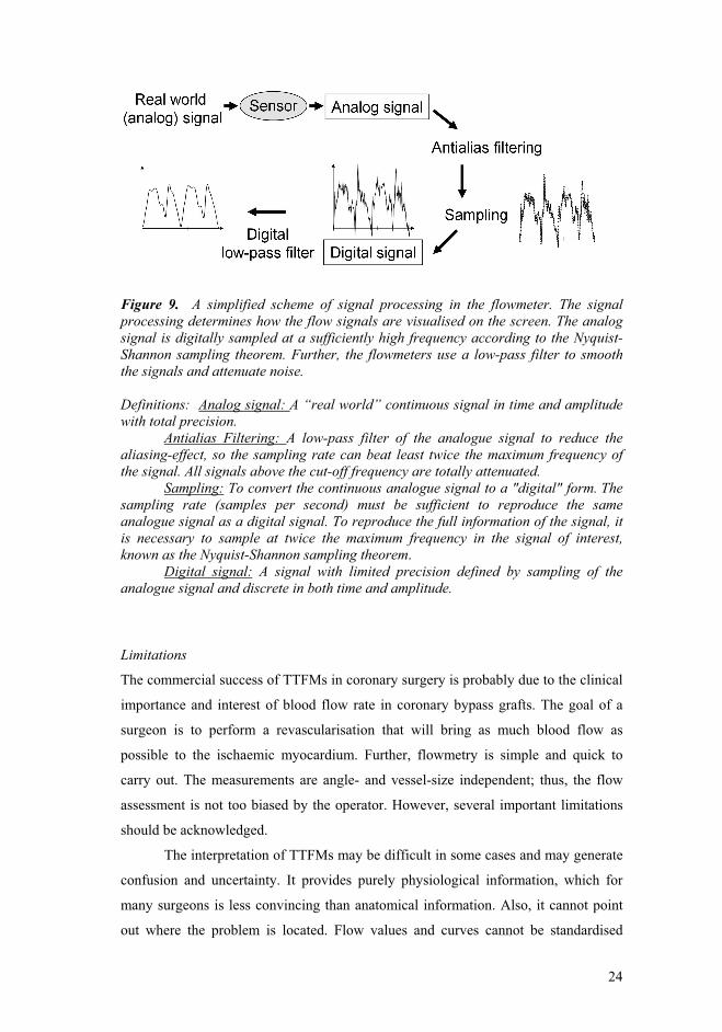
24
Figure 9. A simplified scheme of signal processing in the flowmeter. The signal processing determines how the flow signals are visualised on the screen. The analog signal is digitally sampled at a sufficiently high frequency according to the Nyquist-Shannon sampling theorem. Further, the flowmeters use a low-pass filter to smooth the signals and attenuate noise. Definitions: Analog signal: A “real world” continuous signal in time and amplitude with total precision.
Antialias Filtering: A low-pass filter of the analogue signal to reduce the aliasing-effect, so the sampling rate can beat least twice the maximum frequency of the signal. All signals above the cut-off frequency are totally attenuated.
Sampling: To convert the continuous analogue signal to a "digital" form. The sampling rate (samples per second) must be sufficient to reproduce the same analogue signal as a digital signal. To reproduce the full information of the signal, it is necessary to sample at twice the maximum frequency in the signal of interest, known as the Nyquist-Shannon sampling theorem.
Digital signal:
A signal with limited precision defined by sampling of the analogue signal and discrete in both time and amplitude.
Limitations
The commercial success of TTFMs in coronary surgery is probably due to the clinical
importance and interest of blood flow rate in coronary bypass grafts. The goal of a
surgeon is to perform a revascularisation that will bring as much blood flow as
possible to the ischaemic myocardium. Further, flowmetry is simple and quick to
carry out. The measurements are angle- and vessel-size independent; thus, the flow
assessment is not too biased by the operator. However, several important limitations
should be acknowledged.
The interpretation of TTFMs may be difficult in some cases and may generate
confusion and uncertainty. It provides purely physiological information, which for
many surgeons is less convincing than anatomical information. Also, it cannot point
out where the problem is located. Flow values and curves cannot be standardised

25
because of the great variability among patients (sex, races, etc.). Thus, the
interpretation of graft quality is to a large extent surgeon dependent.
6.2 Computational fluid dynamics (CFD)
A CFD model may be applied to investigate the WSS distribution in arteries [Krams
et al 1997; Lei et al 2001; Boutsianis et al 2004; Frauenfelder et al 2007; Stone et al
2007]. CFD is a subfield within fluid mechanics in which numerical methods and
algorithms are used to analyse and solve problems that involve fluid flows; in
particular, the Navier-Stokes equations govern how the motion of fluids is treated.
The numerical tools can be used to obtain a better understanding of local
haemodynamic and mechanical forces in arteries, including native coronary arteries
and bypass grafts [Krams et al 1997; Lei et al 2001; Boutsianis et al 2004;
Frauenfelder et al 2007].
The CFD technique allows parameters, such as vessel wall conditions and inlet
flow velocity, to be changed easily, so that they can simulate theoretical and realistic
conditions. Additionally, the CFD technique may be use to estimate shear stress and
other important haemodynamic factors. These characteristics make CFD to an
appropriate tool for research of vessel disease aetiologies, for instance intimal
hyperplasia, atherosclerosis and subsequent failure of coronary bypass grafts.
Our study group developed 3-D CFD simulations of the LIMA-LAD model
based on a geometric model and TTFMs (Figure 10). Details are presented in Paper
IV [Nordgaard et al 2010].
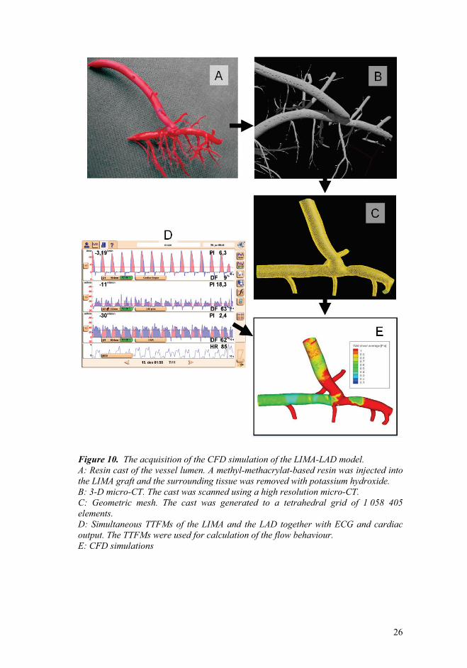
26
Figure 10. The acquisition of the CFD simulation of the LIMA-LAD model. A: Resin cast of the vessel lumen. A methyl-methacrylat-based resin was injected into the LIMA graft and the surrounding tissue was removed with potassium hydroxide. B: 3-D micro-CT. The cast was scanned using a high resolution micro-CT. C: Geometric mesh. The cast was generated to a tetrahedral grid of 1 058 405 elements. D: Simultaneous TTFMs of the LIMA and the LAD together with ECG and cardiac output. The TTFMs were used for calculation of the flow behaviour. E: CFD simulations

27
Limitations of CFD simulations
There are several potential sources of errors that may cause simulations to
differ from true conditions. The most common sources of error are as follows
[Versteeg et al 2007]:
Input data error: If the geometry and the flow data that come from in vivo
measurements are inaccurate.
Boundary conditions error: The model is simplified. All flow properties are
generally not exactly known because they are estimated. For instance, flow data
are needed at locations where the fluid enters and leaves the geometric model.
Discretisation error: Errors that occur when the entire numerical flow data are
transferred into small discrete domain of space and time, known as grid or mesh,
as algebraic expressions. These errors are intrinsic and of most concern to the
CFD model. They are mainly dependent on the quality and distribution of the
grids used in the simulation.
Modelling error: The real flow may involve complicated flow patterns that are not
perfectly described by current scientific theories, for instance in turbulence
modeling.
In paper IV, we compared the flow pattern in the CFD model with the one seen
by epicardial ultrasound imaging as a qualitative validation.
6.3 Surgical procedures and experimental set-up
CABG and experimental set-up in the clinical setting (papers I, III)
A median sternotomy was performed in all cases and the LIMA was harvested with its
pedicle. The greater saphenous vein was placed in a heparin solution with 0.05 %
papaverine after harvesting. Cardio-pulmonary bypass circulation was instituted with
moderate hypothermia (34 °C). The heart was arrested by infusion of antegrade
crystalloid or cold blood cardioplegia. In OPCAB operations (paper I: n = 61, 11 % of
all patients) the target vessel was snared with 4-0 Prolene (Ethicon, NJ, USA)
proximal to the coronary arteriotomy. After 3-5 minutes of ischaemic preconditioning,
the snare was released, and an epicardial stabiliser was applied to pull the heart and
28
immobilise the vessel site chosen for grafting. After arteriotomy, an intracoronary
shunt (CardioThoracic System, CA, USA) was positioned into the vessel lumen.
Grafting was attempted on all vessels measuring >1 mm and with >50 %
stenosis on the angiogram. The surgical plan was to carry out sequential grafting any
time it was considered feasible and the LIMA was always grafted end-to-side to the
LAD. The single SVG anastomosis was constructed end-to-side. In the sequential
SVG, the distal end-to-side anastomosis was constructed before the side-by-side
anastomoses. All LIMA and vein anastomoses were sutured with 7-0 Prolene. The
proximal anastomoses were generally performed after release of the aortic cross-
clamp and by applying a side-biting clamp on the ascending aorta.
The final TTFMs were done at a stable haemodynamic condition 10-15
minutes after weaning off CPB. In off-pump patients, the measurements were done
after completion of all distal anastomoses and removal of the cardiac stabilizer.
Three- or four mm flow probes connected to a transit-time flowmeter (Medi-Stim
ASA, Oslo, Norway) were used. Both for single and sequential SVG, the flow probe
was placed 2-4 cm below the proximal anastomosis. The LIMA flow was assessed by
applying the probe around a skeletonised area. In all grafts both mean graft flow and
PI were registered.
CABG and experimental set-up in the animal model (papers II, IV)
A total of 11 Noroc pigs (hybrid of ¼ Duroc, ¼ Yorkshire and ½ Norwegian
landrace) were operated upon. Animals of 65-70 kg weight were chosen because at
that size the dimensions of the coronary vessels are similar to humans, and the
myocardium has a relatively good tolerance to ischaemia. One pig died due to
ventricular fibrillation before completion of the investigation protocol. A second pig
was excluded from the study because it was haemodynamically unstable, making
measurements unreliable for statistical analysis.
Details of anaesthesia and the animal instrumentation are presented in Paper II
and 4 [Nordgaard et al 2009; Nordgaard et al 2010].
Pigs are quite intolerant to myocardial ischaemia and prone to ventricular
fibrillation. Ventricular fibrillation happened also during our experiments, especially
when making the anastomosis. The heart was promptly defibrillated, and in most of
the cases, the heart converted into sinus rhythm with a haemodynamically stable
29
condition. These events likely do not influence results because the pigs returned to
haemodynamic stability long before and during all measurements.
Amiodarone 150 mg and hexamethonium chloride 20 mg/kg
(Hexamethonium, Sigma-Aldrich, MO, USA) were given to avoid arrhythmias.
Hexamethonium chloride is a ganglionic blocker, which paralyses the autonomic
nervous system (sympatholytic and parasympatholytic), thus contributing to a more
stable pig model. This ganglion blocker provides a stable heart rate throughout the
experiment and presumably avoids ventricular fibrillations [Nordhaug 2003].
The pig was chosen as an animal model because the anatomy and physiology
of its cardiovascular system are very similar to humans. Nonetheless, the pig model
has normal coronary arteries with good run-off and no collateral flow, a very different
condition from patients with severe coronary disease. When planning the
experimental studies, we concluded that non-animal research models were unsuitable
to the purposes of our investigations. The minimum number of animals required for
meaningful results was utilised, and we aimed to reduce any pain to the animals
during the experiments. All experimental protocols were approved by the Norwegian
Animal Experiments Authority. All animals received care in compliance with the
European Convention on Animal Care, and the investigation conformed to the Guide
for Care and Use of Laboratory Animals published by the US National Institutes of
Health (NIH Publication No. 85-23, revised 1996).
All pigs underwent OPCAB surgery with a LIMA-to-LAD anastomosis. After
sternotomy, the LIMA was harvested with its pedicle. The LAD was identified and
investigated visually and sometimes with ultrasound images to identify diagonal
arteries. An epicardial stabiliser (Axius Vacuum Stabilizer, Guidant, CA, USA) was
applied to immobilise the LAD. Proximal to the anastomotic site, approximately 15-
20 mm (dependent on diagonals/ anatomy), the LAD was dissected from the
epicardial tissue in order to apply a 2-3 mm flow probe around the vessel. Adjacent to
this flow probe, another short area was dissected in order to fit an adjustable vascular
occluder (In Vivo Metric, CA, USA). A 16-mm flow probe was placed around the
pulmonary trunk to monitor cardiac output. The flow through the pulmonary trunk
was also used to define systole and diastole because the flowmeter was not always
able to read and interpret the ECG from the pig (Figure 11).
Prior to coronary arteriotomy, ischaemic preconditioning was performed.
Then, the LAD was snared distally and proximally with 4-0 Prolene (Ethicon, NJ,
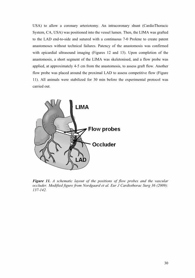
30
USA) to allow a coronary arteriotomy. An intracoronary shunt (CardioThoracic
System, CA, USA) was positioned into the vessel lumen. Then, the LIMA was grafted
to the LAD end-to-side and sutured with a continuous 7-0 Prolene to create patent
anastomoses without technical failures. Patency of the anastomosis was confirmed
with epicardial ultrasound imaging (Figures 12 and 13). Upon completion of the
anastomosis, a short segment of the LIMA was skeletonised, and a flow probe was
applied, at approximately 4-5 cm from the anastomosis, to assess graft flow. Another
flow probe was placed around the proximal LAD to assess competitive flow (Figure
11). All animals were stabilized for 30 min before the experimental protocol was
carried out.
Figure 11. A schematic layout of the positions of flow probes and the vascular occluder. Modified figure from Nordgaard et al. Eur J Cardiothorac Surg 36 (2009): 137-142.

31
Epicardial ultrasound imaging
A GE Vivid 7 scanner (GE Vingmed, Norway) with an i13L linear array mini-
transducer (GE Healthcare, WI, USA) was used. This scanner is capable of operating
at frequencies 7-14 MHz. We applied ultrasound imaging extensively for visualisation
of the anastomosis and the blood flow (Figure 12-13). In paper II, ultrasound imaging
was used to calculate the stenosis at the toe of the anastomosis (Figure 14).
We used a new imaging modality named “blood flow imaging” (BFI). It is
based on conventional colour Doppler imaging, but, in addition, shows the speckle
pattern of the blood flow with a very high frame rate. It visualizes the blood flow in
any direction of the two-dimensional image plane without limitations imposed by
aliases (the velocity aliasing or the angle-dependency). Using slow motion display of
subsequent speckle images, the movement of speckle pattern can be tracked by the
human eye. This new BFI provides a more intuitive display of the flow conditions,
making interpretation easier than conventional colour Doppler imaging [Lovstakken
et al 2008].
In addition to the assessment of coronary graft anastomosis, ultrasound
imaging may be useful to locate intra-myocardial coronary vessels, for evaluation of
severity of coronary stenoses, identification of the arteriotomy site (to avoid plaques
or stented segments), as well as assessment of the ascending aorta [Haaverstad et al
2002; Dessing et al 2004; Budde et al 2006; Lovstakken et al 2008; Ibrahim et al
2008; Budde et al 2009].

32
Figure 12. Intraoperative photo of the pig heart with flow probes and occluder. Epicardial ultrasound imaging of the LIMA-LAD anastomosis is being performed with the stabiliser placed on the epicardial tissue. The geometry and blood flow were assessed before and after constructing a stenotic anastomosis, as well as with and without competitive flow.
Figure 13. Simultaneous TTFM and epicardial ultrasound imaging on a patent LIMA-LAD anastomosis.

33
Figure 14. The luminal dimensions for estimating the extent of the stenoses were measured by epicardial ultrasound imaging. D1 = the vessel diameter of the stenotic toe; DA = diameter of the anastomotic inlet. A circular lumen was presumed. The formula for calculating the luminal stenosis:
100
2DA21D
1100DAArea
1DArea1%Stenosis 2
2

34
6.5 Statistical analysis
Data are presented as either the arithmetic mean ± one standard deviation for
normally distributed data or the median with range for data that were not normally
distributed. In paper III, the values are expressed as geometric mean and 95 %
confidence interval after logarithmic transformation. P-values less than 0.05 were
considered significant. Statistical analysis in Papers I-III was performed using SPSS
for Windows version 15 (SPSS Inc., Chicago, IL, USA). In paper III, STATA for
Windows version 10 (StataCorp, College Station, Texas, USA) and Excel were also
used.
In paper I, the flow values were compared utilising a linear mixed regression
model. The pair-wise post-hoc comparisons were adjusted for multiple comparisons
using the Bonferroni technique. Categorical variables were analysed using a chi-
square test, and continuous variables were analysed using a two-tailed Student's t-test
with equal or unequal variances based on the findings of an initial F-test for equality
of variance.
In paper II, Friedman’s tests for non-normally distributed variables were
applied. If the Friedman’s test revealed significant changes, post hoc analyses by
Wilcoxon signed-ranks were performed without correction for multiple comparisons.
Instead of using the Bonferroni correction for multiple comparisons, we chose to
report uncorrected P-values for the Wilcoxon signed-ranks, so the readers may more
easily assess the relevance of the statistical differences described [Perneger 1998].
In paper III, a logarithmic transformation of the collected flow data was
performed to achieve normal distribution. Two-sample t-tests were used on data with
no repeated measurements, while a random effects model was used to account for
repeated measurements on same individuals. The grafts were nested into LIMA-LAD,
single SVGs and double sequential SVGs to the left and right coronary arteries. The
comparisons between grafts from two different cardiac centres were performed using
two sample t-tests except for the single SVGs to the left coronary artery, where a
random effects model was used to account for repeated measurements. Wilcoxon
signed ranks tests were used to compare the simultaneous flow assessments on the
same grafts.

35
7. SUMMARY OF RESULTS
7.1 Paper I
Within the single vein graft group there were no differences between flows within
grafts to different target vessels except for diagonals, which had the lowest flow. The
blood flow increased substantially from single to double and further to triple
sequential vein grafts. Graft flow was higher in males versus females. Lower age and
higher left ventricular ejection fraction were associated with higher mean graft flows.
The aortic pressure did not influence on graft flow. The mean PI of vein grafts to the
right coronary system was significantly higher than the mean PI of vein grafts to the
left coronary system (PI ± SEM: 2.40 ± 0.06 and 2.00 ± 0.05, respectively). Similar PI
values were found between gender and groups of vein grafts within each coronary
system (Figure 15).
Figure 15. Left: Box-plot of the blood flows in different coronary bypass grafts. The bottom and top of the boxes show the 25th and 75th percentile, and bands near the middle of the box are the 50th percentile (the median). The ends of the vertical lines indicate the minimum and maximum values. Points outside the ends of the lines represent suspected outliers.
Right: Mean PI ± 2 SEM in single SVGs, double and triple sequential SVGs, divided by right and left coronary system. Modified figure from Nordgaard et al. Ann Thorac Surg 2009; 87: 1409-1415

36
7.2 Paper II
Competitive flow reduced the mean LIMA graft flow more than by a stenotic
anastomosis, which was assessed to be 75 % ± 11 % luminal stenosis as measured by
ultrasound. Reduction of the graft flow due to competition was particularly evident
during diastole. The systolic flow remained unchanged. Competitive flow decreased
both diastolic and systolic maximum peak flows, making the flow curve look
smoother. Moreover, competitive flow increased retrograde flow. The PI and rate of
insufficiency % (retrograde flow as a fraction of total flow) were more increased by
competitive flow than by stenosis. Diastolic filling % was substantially reduced
during competitive flow compared with both stenosis and baseline.
7.3 Paper III
The Transonic flowmeter calculated substantially lower PI values than the MediStim
flowmeter. A systematic and constant increase of PIs occurred when the filter setting
was raised from 5 Hz to 100 Hz. Under various filter settings (5 Hz, 10 Hz, 20 Hz, 30
Hz, 50 Hz and 100 Hz) the proportion of the changes depended on the level of the PI
values. Major changes were seen between the filter levels when the PI was high,
typically seen when the flow curves were spiky with high and low peaks. Minor
changes were observed when the PI was low, typically seen when the flow curves
were smoother (Figure 16).
Nineteen coronary bypass grafts were assessed simultaneously by the
MediStim (VeriQ) and Transonic (HT331) flowmeters. The differences in calculation
of PI between the two flowmeter increased as the flow pattern became more spiky.
Thus, the differences between the flowmeters are more pronounced when the flow
curve is spiky, typically seen in situations with poor graft flow and high resistance,
situations where flow analysis is critical.

37
Figure 16. Simultaneous graft flow measurements with the MediStim and Transonic flowmeters [Nordgaard et al 2009].
The upper panel shows a LIMA-to-LAD with flow pattern with sharp peaks. The PI values are 5.5 and 3.2 as calculated by the MediStim and Transonic flowmeters, respectively.
The lower panel shows a single SVG to PDA with smoother flow pattern, thus similar PIs in both flowmeters; PI 1.3 and 1.2 in the MediStim and Transonic flowmeter, respectively.
7.4 Paper IV
High competitive flow resulted in substantial lower WSS and more oscillatory shear
stress in the LIMA graft compared to the two other flow conditions. Partial
competitive flow resulted in WSS and OSI values that were similar to the no-
competitive flow condition (Table 2; Figure 17 and 18).
Table 2. WSS and OSI in the LIMA graft found during one cardiac cycle. Competitive flow WSS [Pa] OSI
High 0.3-0.6 0.15-0.35 Partial 0.6-3.0 <0.05 None 0.9-3.0 <0.05

38
Figure 17. The time-average distribution of the WSS in the LIMA–LAD model during one cardiac cycle. The upper and lower panels show the WSS in two different scales: 0.1 – >1 and 1 – >19 Pa, respectively. (A) No competitive flow shows WSS > 1 Pa in the LIMA graft. In comparison, lower WSS is seen during partial reduced competitive flow (B). Substantial lower WSS in the LIMA graft is seen during high competitive flow (C). Higher WSS is shown at the toe of the anastomosis where a moderate narrowing of the vessel lumen had formed.
Figure 18. The time-average oscillatory shear index (OSI) distribution during one cardiac cycle. Higher OSI values are found in LIMA graft and within the anastomosis during high, unimpaired competitive flow (C), while reduced (B) and no competitive flow (A) lead to very low OSI values.
39
8. DISCUSSION
Transit-time flowmetry as intraoperative quality assessment
Coronary bypass graft failure is a troubling complication that may cause various
problems, including refractory angina pectoris, myocardial infarction, arrhythmia,
heart failure, and premature death. Early graft failure is related to technical problems
involving the bypass grafts and anastomoses, which are difficult to detect during
surgery by visual assessment, palpation or conventional cardiac monitoring. Thus, to
avoid early graft failure, intraoperative graft assessment is required. Late graft failure
may be caused by both technical failures and other factors. We know that long-term
patency is dependent on competitive flow, pathology of the target vessel, type of graft
(venous or arterial) and time since CABG [Sabik et al 2005]. However, the exact
pathophysiological reason for graft failure has been difficult to define.
The first study of this thesis included flow measurements of 1390 coronary
bypass grafts that were operated on and assessed by the same senior surgeon. Thus,
operator-related biases were minimised due to all procedures being conducted by the
same surgeon and the same protocol of flow measurements being applied to all
patients. This continuity was essential for the statistical analyses.
A detailed statistical analysis of the patient characteristics and all flow values
gave important insights in the haemodynamic behaviour of different bypass grafts.
Sequential vein graft flow was higher as compared to single bypass grafts, with flow
substantially increasing from single versus double and triple sequential vein grafts.
However, the differences were not two or three-fold higher, as one would expect if the
resistance to flow were only determined by the peripheral coronary resistance. Other
factors, such as the length of the grafts and the flow pattern in the anastomoses
themselves, must contribute significantly to the total resistance.
In terms of the Poiseuille's law, the flow resistance is proportional to the length
of the conduit. Sequential bypass grafts are longer conduits, hence they produce more
resistance. Another possible explanation may be the interaction between the two distal
anastomoses. Flow disturbance caused by myocardial contractions or competitive
flow close an anastomosis may impair the flow into the other anastomosis.
The lowest flow was found in vein grafts to diagonals, which usually are small
vessels with a limited peripheral vascular bed compared to other coronary branches.

40
Mean graft flows were higher in men vs. women, probably due to men having
larger coronary arteries and larger ventricular mass [Levy et al 1987]. Higher graft
flow was generally seen in younger patients and in patients with good left ventricular
ejection fraction. A possible explanation may be that younger patients have larger and
more dynamic coronary vessels than older patients, which may allow a better run-off.
Reduced ejection fraction indicates congestive heart failure, which may reduce
coronary flow due to the reduction of the coronary vascular bed caused by myocardial
infarction, fibrosis and cardiomyopathy.
The aortic pressure did not influence on graft flow. However, no patients had
hypotension at the time of measurement. The mean blood pressure was around 90
mmHg during the flow measurements suggesting that the coronary auto regulatory
mechanism was intact.
The LIMA-LAD grafts showed lower flow compared to vein grafts directed to
the LAD. Lower flow is probably because of smaller dimensions and some degree of
spasm in the LIMA grafts, since no vasodilators (e.g. papaverine) were used routinely.
In study I similar PI values for single and sequential vein grafts were found.
This observation was unexpected because the PI is thought to reflect peripheral
resistance; hence, one would hypothesise that sequential grafts provide lower PIs
compared to single grafts. Thus, flow pattern and thereby PI are determined by more
factors than the run-off.
Higher PIs were found in vein grafts directed to the right coronary system,
which usually has higher systolic peaks and a more equal systolic and diastolic flow
pattern as compared to the left coronary system. Although the difference in PI values
reached statistical significance, the numeric difference was small. Our findings are in
contrast to Kim et al who suggested cut-off values for insufficient graft flow of PI > 3
to the left and > 5 to the right coronary artery [Kim et al 2005].
Graft flow alterations induced by competitive flow and stenotic anastomoses
Firstly, the comparisons of the different levels of competitive flows revealed that
competitive flow reduced the LIMA graft flow, mainly because of decreased diastolic
flow, whereas the systolic flow remained unchanged. Secondly, partial competitive
flow induced the same effect as fully competitive flow on both mean and diastolic
flow, although not to the same extent. Thirdly, retrograde flow and PI increased only
under fully competitive flow. We also found that competitive flow caused a smoother

41
flow pattern with less sharp peaks compared with the flow pattern at no competitive
flow.
The LIMA graft flows were not significantly altered by a 75 % ± 11 % stenosis
of the lumen of the anastomosis. Hence, our observations are in line with previous
flow studies concluded that substantial hemodynamical effects at rest are seen only in
a critical stenotic anastomosis [Jaber et al 1998; Jaber et al 1998].
The question how to distinguish competitive flow from a severe stenotic
anastomosis is an important issue because both conditions may alter the graft flow. In
addition, numerous other causes may alter the graft flow, such as graft dissection,
kinking, torsion and spasm, as well as high coronary resistance due to small target
vessels [Walpoth et al 1998; D'Ancona et al 2000; Wolfe 2001; Tokuda et al 2007].
The consequences of even tighter stenotic anastomoses were not investigated.
Naturally, the graft flow will abruptly decrease due to a tighter stenosis. In these
situations, other studies have reported a systolic dominant flow pattern, similar to our
findings with competitive flow conditions [Morota et al 2002].
TTFM of grafts flows and pattern may be difficult to interpret, especially in
situations when graft flow is lower than expected. When competitive flow is
suspected, one manoeuvre is to block the native flow while measuring the graft flow.
Such block may be conducted by snaring the target coronary artery proximal to the
anastomosis. If a significant competitive flow is present, the temporary occlusion of
the native flow will induce an increase of graft flow [D'Ancona et al 2001]. However,
there is concern that the temporary occlusion of a coronary artery proximal to an
anastomosis may cause injury of the vessel [Hangler et al 2001]. Temporary clamping
of grafts directed to collateral coronaries may also aid in the interpretation of graft
flow. Another simple and practical technique to assess competitive flow in the LIMA
graft is to measure the flow before and after removal of the aortic cross-clamp. If the
LIMA flow decreases substantially following removal of the aortic clamp,
competitive flow may be present.
Alternatively, epicardial ultrasound may be used [Haaverstad et al 2002]. In
our study, the Blood flow imaging (BFI) modality was applied to evaluate the patency
of the anastomosis by looking at its geometry and function as well as the degree of
stenosis [Lovstakken et al 2008]. We believe that this technique has a great potential
for intraoperative assessment during CABG because it is non-invasive and can easily
detect stenoses and competitive flow.

42
Comparison of the Transonic and the MediStim flowmeters
The majority of published data regarding clinical TTFM have been obtained with the
MediStim flowmeter [Goel et al 2003; Kim et al 2005; Hassanein et al 2005;
Gwozdziewicz et al 2006; Nordgaard et al 2009].
In our clinical series, we found a trend towards lower PI values provided by
the Transonic flowmeter versus the MediStim flowmeter. When we used both
flowmeters simultaneously on the same grafts, the Transonic flowmeter gave
systematically lower PI values. Furthermore, the difference was highly dependent
upon the flow pattern: sharp peaks led to remarkable differences between the two
flowmeters, while smoother flow pattern revealed similar PI values. In other words,
the difference between the flowmeters became greater as PI increased (Figure 16).
The graft flow assessment at different filter settings revealed a systematic and
constant increase of PIs when the filter in the flowmeter was gradually raised from 5
Hz to 100 Hz. Progressively higher filter settings led to a flow pattern with sharper
peaks, which caused higher PIs. On the contrary, lower filter settings produced
smoother flow pattern and lower PIs. The change of PI became greater when the flow
curves had sharp peaks, and these peaks were filtered away at lower filter settings.
Coronary bypass graft patency and failure
Several studies with early postoperative coronary angiography demonstrate graft
failure in the range of 4-14 % [Sanz et al 1990; Goldman et al 1991; Alderman et al
1998; D'Ancona et al 2000; Desai et al 2006; Tokuda et al 2007]. Early graft failure is
associated with low and disturbed graft flow, which is commonly related to technical
problems with the graft or anastomosis, both correctable if recognised
intraoperatively [Nwasokwa 1995]. A post-mortem study of 517 coronary bypass
grafts in 223 patients with fatal outcome within 30 days of CABG, discovered that 55
% of the patients had technical failures in the grafts or anastomoses that were not
recognised intraoperatively [Weman et al 1999].
In a 10-year prospective study on graft patency in 1254 patients who were
followed-up by repeated angiograms the most important predictors of long-term graft
patency were the early patency as well as the size of the peripheral vascular bed
[Goldman et al 2004]. Therefore, intraoperative evaluation of graft patency should be
mandatory.

43
Ultimately, the majority of SVGs occlude or become substantially narrowed
after 10-15 years [Fitzgibbon et al 1996; Motwani et al 1998]. Unlike LIMA grafts,
SVGs are subject to intimal hyperplasia, atherosclerosis, progressive stenosis and
occlusion [Motwani et al 1998]. During the first year 12-20 % of the SVGs occlude.
Intimal hyperplasia occurs mainly one month to five years after surgery [Nwasokwa
1995]. Atherosclerotic lesions that are also haemodynamically significant appear after
about 3-5 years [Nwasokwa 1995; Goldman et al 2004]. At approximately 10 years,
50 % of all SVGs are occluded, and half of the remaining grafts are expected to be
severely narrowed due to atherosclerosis [Nwasokwa 1995; Fitzgibbon et al 1996;
Motwani et al 1998]. Vein graft failures are believed to be the most common reason
for recurrent coronary symptoms following CABG [Nwasokwa 1995; Fabricius et al
2001; Goldman et al 2004].
Numerous studies have shown that about 10 % of all LIMA grafts are
occluded within 10-15 years. When LIMA occlusions occurs the mechanism seems to
be different from vein graft occlusions [Nwasokwa 1995; Shimizu et al 2000;
Goldman et al 2004; Berger et al 2004; Desai et al 2004; Sabik et al 2008]. A
characteristic lesion observed in the LIMA is the “string phenomenon”, a longitudinal
narrowing of the LIMA lumen to less than 1 mm over a length of 3-5 cm. The rate of
the string phenomenon has not been ascertained yet, but it has been estimated to
happen in 2 % of CABG patients [Villareal et al 2000]. Competitive flow in the
LIMA is believed to cause the string phenomenon, which is considered an early sign
of graft failure [Siebenmann et al 1993; Nasu et al 1995; Kawasuji et al 1996;
Hashimoto et al 1996; Pagni et al 1997; Pagni et al 1997; Hirotani et al 2001;
Gaudino et al 2002; Sabik et al 2003; Bezon et al 2003; Nakajima et al 2004; Berger
et al 2004; Sabik et al 2005; Botman et al 2007; Sabik et al 2008].
Biological properties of the LIMA graft are crucial determinants of its
resistance to atherosclerosis. The LIMA is an elastic artery and has an enhanced
endothelial function compared to vein grafts, as shown by greater production of
vasodilating mediators like NO [Nishioka et al 1998]. The LIMA characteristic of
higher basal release of vasodilating mediators is likely to play a vital role in
maintaining the superior vascular patency. In that respect, a promising step in
improving vein graft patency has been introduced by the so-called “no-touch” method
during vein harvesting. Early reports on this technique have shown better early- and
long-term graft patency compared to conventional vein harvesting [Souza et al 2002].

44
During conventional vein harvesting, the tissue around the saphenous vein is
removed and often exposed to high-pressure distension to overcome spasm and
leakages. The no-touch method involves harvesting the vein with minimal handling
with the intention of conserving the perivascular adipose tissue. Conserving the
perivascular tissue may protect the vein by reducing the circumferential stress
[Kassab et al 2006] as well as maintain long-term sufficient endothelial function
[Dashwood et al 2005]. Regarding the acute effects, conventional vein harvesting
induces considerable endothelial injury and reduced NO production compared to the
no-touch method [Dashwood et al 2005].
As mentioned in chapter 4.2, the shear stress induced by blood flow on the
endothelium triggers its biochemical responds; thus, it is a major contributor to
endothelial function. An unfavourable WSS induces an endothelial dysfunction,
which induces vascular disease such as intimal hyperplasia and atherosclerosis. In
paper IV, we studied the WSS under competitive flow conditions. At high competitive
flow, such as when a graft is placed distal to the non-significant coronary stenosis,
very low and oscillatory WSS was found. This unfavourable WSS may explain the
LIMA string phenomenon and reduced graft patency by competitive flow.
The WSS pattern observed under partial competitive flow, simulating the
clinical condition of a conduit grafted distal to the significant coronary stenosis, was
comparable to the no competitive flow condition (Figure 17 and 18). We found very
low and oscillatory WSS during high competitive flow, which simulates a non-
significant coronary stenosis. The WSS pattern during partial competitive flow,
simulating significant coronary stenosis, was comparable to the condition with no
competitive flow. Thus, the WSS pattern changed greatly upon the degree of
competitive flow. Our findings indicate that bypass grafts may tolerate a certain
degree of competitive flow, which is probably the most frequent situation. In contrast,
the unfavourable WSS caused by high competitive flow probably has an adverse
impact on graft patency. Thus, our findings are in line with other studies reporting the
relationship of graft patency and competitive flow [Siebenmann et al 1993; Nasu et al
1995; Kawasuji et al 1996; Hashimoto et al 1996; Pagni et al 1997; Pagni et al 1997;
Hirotani et al 2001; Gaudino et al 2002; Sabik et al 2003; Bezon et al 2003; Nakajima
et al 2004; Berger et al 2004; Sabik et al 2005; Botman et al 2007; Sabik et al 2008].
The consequences of low and oscillatory WSS may very likely be the LIMA string
phenomenon and subsequent LIMA graft failure [Kassab et al 2006; Reneman et al

45
2006; Chatzizisis et al 2007; Stone et al 2007; John 2009; Davies 2009]. Moreover,
another additional explanation for the changes seen at competitive flow conditions is
based on the fact that WSS influences on homeostasis and vessel lumen dimensions
[Gusic et al 2005; Kassab et al 2006]. The physiological responds may explain, in
part, the aetiology of the string phenomenon of the LIMA graft: The endothelium
maintains a sufficient WSS by affecting the vessel radius ( 3RQ4 , radius R greatly
competitive flow, the vessel narrows in order to increase the WSS, and hence, the
endothelial function.
46
9. MAIN CONCLUSIONS
- The statistical analyses of the 1390 flow measurements concluded that the graft
flow in vein grafts increase gradually from single to double and up to triple sequential
bypasses. The PIs of vein grafts to the right coronary system were significantly higher
than in grafts to the left coronary system. However, no PI differences exist between
single, double and triple sequential vein grafts. In addition, higher graft flow should
be expected in males compared to female patients.
- TTFMs cannot detect minor or moderate technical failures in the anastomoses
because they are not haemodynamically significant. However, competitive flow
influences the TTFMs early and substantially. During transit-time flowmetry the
surgeon should particularly be aware of the severity of the coronary stenosis, the size
of the target coronary vessel and the influence of competitive flow. These
considerations are essential before final interpretations of TTFMs are made.
- Although the transit-time flowmeters from MediStim ASA and Transonic Inc. have
presented the same guidelines regarding the PI values, they are not directly
comparable due to their different default filter settings. The level of filter settings in
the flowmeter determines the shapes in the flow curves, which results in different PI
values. Thus, it is important to refer to the type of flowmeter used when presenting
transit-time flow values.
- Competitive flow, which is known to influence negatively on graft patency, induces
low and oscillatory wall shear stress (WSS). The levels of WSS found in our
computational fluid dynamics model are supposed to have damaging effects on the
endothelium, inducing endothelial dysfunction. Thus, the low and oscillatory WSS
seen during high competitive flow may explain the reduced long-term patency of
bypass grafts and the LIMA “string phenomenon”. A lower level of competitive flow
may be better tolerated due to a higher and more unidirectional shear stress, similar to
the ideal situation with no competitive flow.

47
10. PROSPECTS OF FUTURE RESEARCH
Further developments within intraoperative quality assessment in CABG
There is a need for dedicated instruments designed for intraoperative quality
assessment, for instance scanning areas prior to incisions. The widespread use of new
technology will largely depend on the cost, ease and sensitivity of the method.
Transit-time flowmetry may remain the cornerstone for intraoperative quality
assessment of CABG, but using it in combination with ultrasound imaging may
provide better and more reliable information in the future. This new approach with
epicardial ultrasound and TTFMs needs to be validated in clinical studies.
Graft patency
Improvement of the long-term patency of grafts, especially vein grafts, must remain
an important aim for future research. Preservation of endothelial function is probably
essential for long-term patency. Thus, better knowledge of endothelial function,
especially in vein grafts, is mandatory. Does the conventional way to harvest a vein
by removing the surrounding tissue reduce the endothelial function and hence the
long-term patency? A better understanding of WSS and endothelial function,
atherosclerosis and intimal hyperplasia may lead us to improved operative strategies,
for instance keeping the surrounding tissue around the vein grafts. Better knowledge
may also guide us to a biological or pharmacological approach, such as gene therapy.
There is a need for clinical studies on the impact of WSS and other biomechanical
forces in different flow conditions and different bypass grafts. Changes in surgical
practice will only occur if favourable results can be demonstrated in humans.

48
Reference List
1. Alderman EL, Levy JH, Rich J, Nili M, Vidne B et al. Analyses of coronary graft patency after aprotinin use: Results from the international multicenter aprotinin graft patency experience (IMAGE) trial, , , . The Journal of Thoracic and Cardiovascular Surgery 1998 Nov;116(5):716-30.
2. Asakura T, Karino T. Flow patterns and spatial distribution of atherosclerotic lesions in human coronary arteries. Circ Res 1990 Apr;66(4):1045-66.
3. Becit N, Erkut B, Ceviz M, Unlu Y, Colak A, Kocak H. The impact of intraoperative transit time flow measurement on the results of on-pump coronary surgery. European Journal of Cardio-Thoracic Surgery 2007 Aug;32(2):313-8.
4. Beldi G, Bosshard A, Hess OM, Althaus U, Walpoth BH. Transit time flow measurement: experimental validation and comparison of three different systems. Ann Thorac Surg 2000 Jul;70(1):212-7.
5. Berger A, MacCarthy PA, Siebert U, Carlier S, Wijns W et al. Long-Term Patency of Internal Mammary Artery Bypass Grafts: Relationship With Preoperative Severity of the Native Coronary Artery Stenosis. Circulation 2004 Sep;110(11_suppl_1):II-36.
6. Bezon E, Choplain JN, Maguid YA, Aziz A, Barra JA. Failure of internal thoracic artery grafts: conclusions from coronary angiography mid-term follow-up. The Annals of Thoracic Surgery 2003 Sep;76(3):754-9.
7. Botman CJ, Schonberger J, Koolen S, Penn O, Botman H, Dib N, Eeckhout E, Pijls N. Does Stenosis Severity of Native Vessels Influence Bypass Graft Patency? A Prospective Fractional Flow Reserve-Guided Study. The Annals of Thoracic Surgery 2007 Jun;83(6):2093-7.
8. Boutsianis E, Dave H, Frauenfelder T, Poulikakos D, Wildermuth S, Turina M, Ventikos Y, Zund G. Computational simulation of intracoronary flow based on real coronary geometry. Eur J Cardiothorac Surg 2004 Aug;26(2):248-56.
9. Budde RP, Bakker PF, Grundeman PF, Borst C. High-frequency epicardial ultrasound: review of a multipurpose intraoperative tool for coronary surgery. Surg Endosc 2009 Mar;23(3):467-76.
10. Budde RP, Bakker PF, Meijer R, Borst C, Grundeman PF. Ultrasound mini-transducer with malleable handle for coronary artery surgery. Ann Thorac Surg 2006 Jan;81(1):322-6.

49
11. Cameron A, Davis K, Green G, Schaff H. Coronary Bypass Surgery with Internal-Thoracic-Artery Grafts -- Effects on Survival over a 15-Year Period. N Engl J Med 1996 Jan;334(4):216-20.
12. Chatzizisis YS, Coskun AU, Jonas M, Edelman ER, Feldman CL, Stone PH. Role of Endothelial Shear Stress in the Natural History of Coronary Atherosclerosis and Vascular Remodeling: Molecular, Cellular, and Vascular Behavior. Journal of the American College of Cardiology 2007 Jun;49(25):2379-93.
13. Christensen DA. Doppler and Other Ultrasonic Flowmeters. Ultrasonic Bioinstrumentation. John Wiley & Sons, 1988:203-206.
14. Christenson JT, Simonet F, Schmuziger M. Sequential vein bypass grafting: tactics and long-term results. Cardiovasc Surg 1998 Aug;6(4):389-97.
15. Cikirikcioglu M, Cikirikcioglu YB, Khabiri E, Djebaili M, Kalangos A, Walpoth BH. Pre-clinical validation of a new intra-operative "dual beam Doppler" blood flowmeter in an artificial circuit. Heart Surg Forum 2006;9(1):E499-E505.
16. D'Ancona G, Karamanoukian HL, Bergsland J. Is intraoperative measurement of coronary blood flow a good predictor of graft patency? Eur J Cardiothorac Surg 2001 Nov;20(5):1075-7.
17. D'Ancona G, Karamanoukian HL, Ricci M, Schmid S, Bergsland J, Salerno TA. Graft revision after transit time flow measurement in off-pump coronary artery bypass grafting. Eur J Cardiothorac Surg 2000 Mar;17(3):287-93.
18. Dashwood MR, Savage K, Dooley A, Shi-Wen X, Abraham DJ, Souza DSR. Effect of Vein Graft Harvesting on Endothelial Nitric Oxide Synthase and Nitric Oxide Production. The Annals of Thoracic Surgery 2005 Sep;80(3):939-44.
19. Davies PF. Flow-mediated endothelial mechanotransduction. Physiol Rev 1995 Jul;75(3):519-60.
20. Davies PF. Hemodynamic shear stress and the endothelium in cardiovascular pathophysiology. Nat Clin Pract Cardiovasc Med 2009 Jan;6(1):16-26.
21. Davignon J, Ganz P. Role of Endothelial Dysfunction in Atherosclerosis. Circulation 2004 Jun;109(23_suppl_1):III-27.
22. Desai ND, Cohen EA, Naylor CD, Fremes SE. A randomized comparison of radial-artery and saphenous-vein coronary bypass grafts. N Engl J Med 2004 Nov;351(22):2302-9.
23. Desai ND, Miwa S, Kodama D, Koyama T, Cohen G et al. A randomized comparison of intraoperative indocyanine green angiography and transit-time flow measurement to detect technical errors in coronary bypass grafts. J Thorac Cardiovasc Surg 2006 Sep;132(3):585-94.
50
24. Dessing TC, Budde RP, Meijer R, Bakker PF, Borst C, Grundeman PF. Geometry assessment of coronary artery anastomoses with construction errors by epicardial ultrasound. Eur J Cardiothorac Surg 2004 Aug;26(2):257-61.
25. Di Giammarco G, Pano M, Cirmeni S, Pelini P, Vitolla G, Di Mauro M. Predictive value of intraoperative transit-time flow measurement for short-term graft patency in coronary surgery. The Journal of Thoracic and Cardiovascular Surgery 2006 Sep;132(3):468-74.
26. Dion R, Glineur D, Derouck D, Verhelst R, Noirhomme P, El Khoury G, Degrave E, Hanet C. Complementary saphenous grafting: long-term follow-up. J Thorac Cardiovasc Surg 2001 Aug;122(2):296-304.
27. Eagle KA, Guyton RA, Davidoff R, Edwards FH, Ewy GA et al. ACC/AHA 2004 Guideline Update for Coronary Artery Bypass Graft Surgery: Summary Article: A Report of the American College of Cardiology/American Heart Association Task Force on Practice Guidelines (Committee to Update the 1999 Guidelines for Coronary Artery Bypass Graft Surgery). Circulation 2004 Aug;110(9):1168-76.
28. Evans DH, Barrie WW, Asher MJ, Bentley S, Bell PR. The relationship between ultrasonic pulsatility index and proximal arterial stenosis in a canine model. Circ Res 1980 Apr;46(4):470-5.
29. Fabricius AM, Gerber W, Hanke M, Garbade J, Autschbach R, Mohr FW. Early angiographic control of perioperative ischemia after coronary artery bypass grafting. European Journal of Cardio-Thoracic Surgery 2001 Jun;19(6):853-8.
30. Feldman CL, Ilegbusi OJ, Hu Z, Nesto R, Waxman S, Stone PH. Determination of in vivo velocity and endothelial shear stress patterns with phasic flow in human coronary arteries: A methodology to predict progression of coronary atherosclerosis. American Heart Journal 2002 Jun;143(6):931-9.
31. Fitzgibbon GM, Kafka HP, Leach AJ, Keon WJ, Hooper GD, Burton JR. Coronary bypass graft fate and patient outcome: Angiographic follow-up of 5,065 grafts related to survival and reoperation in 1,388 patients during 25 years. Journal of the American College of Cardiology 1996 Sep;28(3):616-26.
32. Frauenfelder T, Boutsianis E, Schertler T, Husmann L, Leschka S, Poulikakos D, Marincek B, Alkadhi H. Flow and wall shear stress in end-to-side and side-to-side anastomosis of venous coronary artery bypass grafts. Biomed Eng Online 2007;6:35.
33. Gaudino M, Alessandrini F, Nasso G, Bruno P, Manzoli A, Possati G. Severity of coronary artery stenosis at preoperative angiography and midterm mammary graft status. The Annals of Thoracic Surgery 2002 Jul;74(1):119-21.
34. Goel P, Dubey S, Makker A, Kohli VM. Evaluation of coronary artery bypass grafts by intraoperative transit time flow measurement. Indian Journal of Thoracic and Cardiovascular Surgery 2003 Apr;19(2):108-12.

51
35. Goldman S, Copeland J, Moritz T, Henderson W, Zadina K et al. Starting aspirin therapy after operation. Effects on early graft patency. Department of Veterans Affairs Cooperative Study Group. Circulation 1991 Aug;84(2):520-6.
36. Goldman S, Zadina K, Moritz T, Ovitt T, Sethi G et al. Long-term patency of saphenous vein and left internal mammary artery grafts after coronary artery bypass surgery: Results from a Department of Veterans Affairs Cooperative Study. Journal of the American College of Cardiology 2004 Dec;44(11):2149-56.
37. Gould KL, Lipscomb K, Hamilton GW. Physiologic basis for assessing critical coronary stenosis. Instantaneous flow response and regional distribution during coronary hyperemia as measures of coronary flow reserve. Am J Cardiol 1974 Jan;33(1):87-94.
38. Gusic RJ, Myung R, Petko M, Gaynor JW, Gooch KJ. Shear stress and pressure modulate saphenous vein remodeling ex vivo. J Biomech 2005 Sep;38(9):1760-9.
39. Gwozdziewicz M, Nemec P, Simek M, Hajek R, Troubil M. Sequential bypass grafting on the beating heart: blood flow characteristics. Ann Thorac Surg 2006 Aug;82(2):620-3.
40. Haaverstad R. Koronarkirurgi i Norge 2009. Hjerteforum 2009;3(22):107-10.
41. Haaverstad R, Vitale N, Tjomsland O, Tromsdal A, Torp H, Samstad SO. Intraoperative color Doppler ultrasound assessment of LIMA-to-LAD anastomoses in off-pump coronary artery bypass grafting. Ann Thorac Surg 2002 Oct;74(4):S1390-S1394.
42. Hangler HB, Pfaller K, Antretter H, Dapunt OE, Bonatti JO. Coronary endothelial injury after local occlusion on the human beating heart. The Annals of Thoracic Surgery 2001 Jan;71(1):122-7.
43. Hashimoto H, Isshiki T, Ikari Y, Hara K, Saeki F, Tamura T, Yamaguchi T, Suma H. Effects of competitive blood flow on arterial graft patency and diameter. Medium-term postoperative follow-up. J Thorac Cardiovasc Surg 1996 Feb;111(2):399-407.
44. Hassanein W, Albert AA, Arnrich B, Walter J, Ennker IC, Rosendahl U, Bauer S, Ennker J. Intraoperative transit time flow measurement: off-pump versus on-pump coronary artery bypass. Ann Thorac Surg 2005 Dec;80(6):2155-61.
45. Hirotani T, Kameda T, Shirota S, Nakao Y. An evaluation of the intraoperative transit time measurements of coronary bypass flow. Eur J Cardiothorac Surg 2001 Jun;19(6):848-52.
46. Hol PK, Lingaas PS, Lundblad R, Rein KA, Vatne K, Smith HJr, Nitter-Hauge S, Fosse E. Intraoperative angiography leads to graft revision in

52
coronary artery bypass surgery. The Annals of Thoracic Surgery 2004 Aug;78(2):502-5.
47. Ibrahim K, Vitale N, Tromsdal A, Kirkeby-Garstad I, Fraser AG, Haaverstad R. Enhanced intra-operative grading of ascending aorta atheroma by epiaortic ultrasound vs echocardiography. Int J Cardiol 2008 Aug;128(2):218-23.
48. Jaber SF, Koenig SC, BhaskerRao B, VanHimbergen DJ, Cerrito P, Ewert D, Gray L, Spence PA. Role of graft flow measurement technique in anastomotic quality assessment in minimally invasive CABG. Ann Thorac Surg 1998 Sep;66(3):1087-92.
49. Jaber SF, Koenig SC, BhaskerRao B, VanHimbergen DJ, Spence PA. Can visual assessment of flow waveform morphology detect anastomotic error in off-pump coronary artery bypass grafting? Eur J Cardiothorac Surg 1998 Nov;14(5):476-9.
50. John LCH. Biomechanics of Coronary Artery and Bypass Graft Disease: Potential New Approaches. The Annals of Thoracic Surgery 2009 Jan;87(1):331-8.
51. Kandemir O, Tokmakoglu H, Tezcaner T, Yorgancioglu C, Suzer K, Zorlutuna Y. Right coronary system grafts: alone or together with left system grafts--angiographic results. Ann Thorac Cardiovasc Surg 2007 Feb;13(1):27-31.
52. Kassab GS, Navia JA. Biomechanical considerations in the design of graft: the homeostasis hypothesis. Annu Rev Biomed Eng 2006;8:499-535.
53. Katritsis D, Kaiktsis L, Chaniotis A, Pantos J, Efstathopoulos EP, Marmarelis V. Wall shear stress: theoretical considerations and methods of measurement. Prog Cardiovasc Dis 2007 Mar;49(5):307-29.
54. Kawamura M, Nakajima H, Kobayashi J, Funatsu T, Otsuka Y, Yagihara T, Kitamura S. Patency rate of the internal thoracic artery to the left anterior descending artery bypass is reduced by competitive flow from the concomitant saphenous vein graft in the left coronary artery. European Journal of Cardio-Thoracic Surgery 2008 Oct;34(4):833-8.
55. Kawasuji M, Sakakibara N, Takemura H, Tedoriya T, Ushijima T, Watanabe Y. Is internal thoracic artery grafting suitable for a moderately stenotic coronary artery? J Thorac Cardiovasc Surg 1996 Aug;112(2):253-9.
56. Kieser TM, FitzGibbon GM, Keon WJ. Sequential coronary bypass grafts. Long-term follow-up. J Thorac Cardiovasc Surg 1986 May;91(5):767-72.
57. Kim KB, Kang CH, Lim C. Prediction of graft flow impairment by intraoperative transit time flow measurement in off-pump coronary artery bypass using arterial grafts. Ann Thorac Surg 2005 Aug;80(2):594-8.
58. Koskinas KC, Chatzizisis YS, Baker AB, Edelman ER, Stone PH, Feldman CL. The role of low endothelial shear stress in the conversion of

53
atherosclerotic lesions from stable to unstable plaque. Curr Opin Cardiol 2009 Oct.
59. Krams R, Wentzel JJ, Oomen JAF, Vinke R, Schuurbiers JCH, de Feyter PJ, Serruys PW, Slager CJ. Evaluation of Endothelial Shear Stress and 3D Geometry as Factors Determining the Development of Atherosclerosis and Remodeling in Human Coronary Arteries in Vivo : Combining 3D Reconstruction from Angiography and IVUS (ANGUS) with Computational Fluid Dynamics. Arterioscler Thromb Vasc Biol 1997 Oct;17(10):2061-5.
60. Ku DN, Giddens DP, Zarins CK, Glagov S. Pulsatile flow and atherosclerosis in the human carotid bifurcation. Positive correlation between plaque location and low oscillating shear stress. Arteriosclerosis 1985 May;5(3):293-302.
61. Laustsen J, Pedersen EM, Terp K, Steinbruchel D, Kure HH, Paulsen PK, Jorgensen H, Paaske WP. Validation of a new transit time ultrasound flowmeter in man. Eur J Vasc Endovasc Surg 1996 Jul;12(1):91-6.
62. Lei M, Giddens DP, Jones SA, Loth F, Bassiouny H. Pulsatile flow in an end-to-side vascular graft model: comparison of computations with experimental data. J Biomech Eng 2001 Feb;123(1):80-7.
63. Levy D, Savage DD, Garrison RJ, Anderson KM, Kannel WB, Castelli WP. Echocardiographic criteria for left ventricular hypertrophy: The Framingham heart study. The American Journal of Cardiology 1987 Apr;59(9):956-60.
64. Louagie YA, Haxhe JP, Jamart J, Buche M, Schoevaerdts JC. Intraoperative assessment of coronary artery bypass grafts using a pulsed Doppler flowmeter. Ann Thorac Surg 1994 Sep;58(3):742-9.
65. Lovstakken L, Ibrahim KS, Vitale N, Henriksen ST, Kirkeby-Garstad I, Torp H, Haaverstad R. Blood flow imaging: a new two-dimensional ultrasound modality for enhanced intraoperative visualization of blood flow patterns in coronary anastomoses. J Am Soc Echocardiogr 2008 Aug;21(8):969-75.
66. Malek AM, Alper S, Izumo S. Hemodynamic shear stress and its role in atherosclerosis. JAMA 1999 Dec;282(21):2035-42.
67. Matre K, Birkeland S, Hessevik I, Segadal L. Comparison of transit-time and Doppler ultrasound methods for measurement of flow in aortocoronary bypass grafts during cardiac surgery. Thorac Cardiovasc Surg 1994 Jun;42(3):170-4.
68. Morota T, Duhaylongsod FG, Burfeind WR, Huang CT. Intraoperative evaluation of coronary anastomosis by transit-time ultrasonic flow measurement. Ann Thorac Surg 2002 May;73(5):1446-50.
69. Motwani JG, Topol EJ. Aortocoronary Saphenous Vein Graft Disease : Pathogenesis, Predisposition, and Prevention. Circulation 1998 Mar;97(9):916-31.
70. Naidu RB, Bailey SR. Physiologic assessment of coronary artery stenoses. Curr Probl Cardiol 2001 Jul;26(7):423-92.
54
71. Nakajima H, Kobayashi J, Funatsu T, Shimahara Y, Kawamura M, Kawamura A, Yagihara T, Kitamura S. Predictive factors for the intermediate-term patency of arterial grafts in aorta no-touch off-pump coronary revascularization. European Journal of Cardio-Thoracic Surgery 2007 Nov;32(5):711-7.
72. Nakajima H, Kobayashi J, Tagusari O, Bando K, Niwaya K, Kitamura S. Competitive flow in arterial composite grafts and effect of graft arrangement in Off-Pump coronary revascularization. The Annals of Thoracic Surgery 2004 Aug;78(2):481-6.
73. Nasu M, Akasaka T, Okazaki T, Shinkai M, Fujiwara H et al. Postoperative flow characteristics of left internal thoracic artery grafts. The Annals of Thoracic Surgery 1995 Jan;59(1):154-62.
74. Nishioka H, Kitamura S, Kameda Y, Taniguchi S, Kawata T, Mizuguchi K. Difference in acetylcholine-induced nitric oxide release of arterial and venous grafts in patients after coronary bypass operations, . The Journal of Thoracic and Cardiovascular Surgery 1998 Sep;116(3):454-9.
75. Nordgaard H, Swillens A, Nordhaug D, Kirkeby-Garstad I, Van LD et al. Impact of competitive flow on wall shear stress in coronary surgery: Computational fluid dynamics of a LIMA-LAD model. Cardiovasc Res 2010 Jun.
76. Nordgaard HB, Nordhaug D, Kirkeby-Garstad I, Lovstakken L, Vitale N, Haaverstad R. Different graft flow patterns due to competitive flow or stenosis in the coronary anastomosis assessed by transit-time flowmetry in a porcine model. Eur J Cardiothorac Surg 2009 Jul;36(1):137-42.
77. Nordgaard, Vitale, Astudillo, Renzulli, Romundstad, Haaverstad. Pulsatility index variations using two different transit-time flowmeters in coronary artery bypass surgery. European Journal of Cardio-Thoracic Surgery. In press 2009.
78. Nordgaard HB, Vitale N, Haaverstad R. Transit-time blood flow measurements in sequential saphenous coronary artery bypass grafts. Ann Thorac Surg 2009 May;87(5):1409-15.
79. Nordhaug D. Thesis: Experimental treatment of acute ischemic heart failure. Ventriculo-arterial matching and cardiac efficiency. University Hospital North Norway Institute of Clinical Medicine, University of Tromsø 2003.
80. Norsk Nasjonalt Hjertekirurgiregister
81. Nwasokwa ON. Coronary Artery Bypass Graft Disease. Annals of Internal Medicine 1995 Oct;123(7):528-33.
www.legeforeningen.no/thorax
82. Opie LH, Heusch G. Oxygen Supply: Coronary Flow. In: Opie LH, ., eds. Heart Physiology: From Cell to Circulation. 4. ed. Lippincott Williams & Wilkins, 2004:279-305.

55
83. Pagni S, Storey J, Ballen J, Montgomery W, Chiang BY, Etoch S, Spence P. ITA versus SVG: a comparison of instantaneous pressure and flow dynamics during competitive flow. European Journal of Cardio-Thoracic Surgery 1997 Jun;11(6):1086-92.
84. Pagni S, Storey J, Ballen J, Montgomery W, Qaqish NK, Etoch S, Spence PA. Factors Affecting Internal Mammary Artery Graft Survival: How Is Competitive Flow from a Patent Native Coronary Vessel a Risk Factor?. Journal of Surgical Research 1997 Aug;71(2):172-8.
85. Patel MR, Dehmer GJ, Hirshfeld JW, Smith PK, Spertus JA. ACCF/SCAI/STS/AATS/AHA/ASNC 2009 Appropriateness Criteria for Coronary Revascularization: A Report by the American College of Cardiology Foundation Appropriateness Criteria Task Force, Society for Cardiovascular Angiography and Interventions, Society of Thoracic Surgeons, American Association for Thoracic Surgery, American Heart Association, and the American Society of Nuclear Cardiology Endorsed by the American Society of Echocardiography, the Heart Failure Society of America, and the Society of Cardiovascular Computed Tomography. Journal of the American College of Cardiology 2009 Feb;53(6):530-53.
86. Perneger TV. What's wrong with Bonferroni adjustments. BMJ 1998 Apr;316(7139):1236-8.
87. Proakis JG, Manolakis DK. Digital Signal Processing: Principles, Algorithms and Applications. Prentice Hall, 1995
88. Reneman RS, Arts T, Hoeks APG. Wall Shear Stress -- an Important Determinant of Endothelial Cell Function and Structure -- in the Arterial System in vivo. Journal of Vascular Research 2006;43(3).
89. Sabik JF, Blackstone EH. Coronary artery bypass graft patency and competitive flow. J Am Coll Cardiol 2008 Jan;51(2):126-8.
90. Sabik JF, Lytle BW, Blackstone EH, Houghtaling PL, Cosgrove DM. Comparison of saphenous vein and internal thoracic artery graft patency by coronary system. Ann Thorac Surg 2005 Feb;79(2):544-51.
91. Sabik JF, Lytle BW, Blackstone EH, Khan M, Houghtaling PL, Cosgrove DM. Does competitive flow reduce internal thoracic artery graft patency? Ann Thorac Surg 2003 Nov;76(5):1490-6.
92. Sanz G, Pajaron A, Alegria E, Coello I, Cardona M et al. Prevention of early aortocoronary bypass occlusion by low-dose aspirin and dipyridamole. Grupo Espanol para el Seguimiento del Injerto Coronario (GESIC). Circulation 1990 Sep;82(3):765-73.
93. Serruys PW, Morice MC, Kappetein AP, Colombo A, Holmes DR et al. Percutaneous coronary intervention versus coronary-artery bypass grafting for severe coronary artery disease. N Engl J Med 2009 Mar;360(10):961-72.

56
94. Shimizu T, Hirayama T, Suesada H, Ikeda K, Ito S, Ishimaru S. Effect of flow competition on internal thoracic artery graft: Postoperative velocimetric and angiographic study. Journal of Thoracic and Cardiovascular Surgery 2000 Sep;120(3):459-65.
95. Siebenmann R, Egloff L, Hirzel H, Rothlin M, Studer M, Tartini R. The internal mammary artery 'string phenomenon'. Analysis of 10 cases. European Journal of Cardio-Thoracic Surgery 1993 Jul;7(5):235-8.
96. Slager CJ, Wentzel JJ, Gijsen FJ, Schuurbiers JC, van der Wal AC, van der Steen AF, Serruys PW. The role of shear stress in the generation of rupture-prone vulnerable plaques. Nat Clin Pract Cardiovasc Med 2005 Aug;2(8):401-7.
97. Souza DS, Dashwood MR, Tsui JC, Filbey D, Bodin L, Johansson B, Borowiec J. Improved patency in vein grafts harvested with surrounding tissue: results of a randomized study using three harvesting techniques. Ann Thorac Surg 2002 Apr;73(4):1189-95.
98. Spaan J, Piek J, Hoffman J, Siebes M. Physiological Basis of Clinically Used Coronary Hemodynamic Indices. Circulation 2006 Jan;113(3):446-55.
99. Stone PH, Coskun AU, Kinlay S, Popma JJ, Sonka M et al. Regions of low endothelial shear stress are the sites where coronary plaque progresses and vascular remodelling occurs in humans: an in vivo serial study. Eur Heart J 2007 Mar;28(6):705-10.
100. Tokuda Y, Song MH, Ueda Y, Usui A, Akita T. Predicting Early Coronary Artery Bypass Graft Failure by Intraoperative Transit Time Flow Measurement. The Annals of Thoracic Surgery 2007 Dec;84(6):1928-33.
101. Versteeg H, Malalasekera W. An Introduction to Computational Fluid Dynamics: The Finite Volume Method. 2 ed. Person Education Limited, 2007
102. Villareal RP, Mathur VS. The string phenomenon: an important cause of internal mammary artery graft failure. Tex Heart Inst J 2000;27(4):346-9.
103. Vural KM, Sener E, Tasdemir O. Long-term patency of sequential and individual saphenous vein coronary bypass grafts. Eur J Cardiothorac Surg 2001 Feb;19(2):140-4.
104. Walpoth BH, Bosshard A, Genyk I, Kipfer B, Berdat PA, Hess OM, Althaus U, Carrel TP. Transit-time flow measurement for detection of early graft failure during myocardial revascularization. Ann Thorac Surg 1998 Sep;66(3):1097-100.
105. Weman SM, Salminen US, Penttila A, Mannikko A, Karhunen PJ. Post-mortem cast angiography in the diagnostics of graft complications in patients with fatal outcome following coronary artery bypass grafting (CABG). Int J Legal Med 1999;112(2):107-14.

57
106. Wolf RK, Falk V. Intraoperative assessment of coronary artery bypass grafts. J Thorac Cardiovasc Surg 2003 Sep;126(3):634-7.
107. Wolfe JA. The coronary artery bypass conduit: II. Assessment of the quality of the distal anastomosis. Ann Thorac Surg 2001 Dec;72(6):S2253-S2258.
108. Yada T, Hiramatsu O, Kimura A, Goto M, Ogasawara Y et al. In vivo observation of subendocardial microvessels of the beating porcine heart using a needle-probe videomicroscope with a CCD camera. Circ Res 1993 May;72(5):939-46.


58
Appendix
58
Appendix
58
Appendix
58
Appendix

Not included due to copyright Article in Press: Trachiotis GD. Value of diastolic flow with transit-time flow meters in coronary artery bypass surgery. Eur J Cardiothorac Surg (2010), doi:10.1016/j.ejcts.2010.07.005 Article in Press: Nordgaard HB, et al. Reply to Trachiotis. Eur J Cardiothorac Surg (2010), doi:10.1016/j.ejcts.2010.07.006

Paper I Paper I
Paper I Paper I

Transit-Time Blood Flow Measurements inSequential Saphenous Coronary ArteryBypass GraftsHåvard Nordgaard, MD, Nicola Vitale, MD, PhD, and Rune Haaverstad, MD, PhDDepartment of Circulation and Medical Imaging, The Norwegian University of Science and Technology, and Department ofCardiothoracic Surgery, St. Olav University Hospital, Trondheim, and Institute of Surgical Sciences, The Medical Faculty,University of Bergen, Norway
Background. Little information is available on transit-time flow measurements of sequential saphenous veingrafts. The aim of the study was evaluation of meanblood flow and pulsatility index of sequential saphenousvein grafts in a large population of patients operated onwith coronary artery bypass grafting.
Methods. In 581 patients 1,390 grafts were nested intoleft internal mammary artery to left anterior descendingartery, single vein grafts, or double and triple sequentialvein grafts, and analyzed.
Results. Within the single vein graft group there wereno differences between flow of grafts to different targetvessels except for diagonals (diagonal versus obtusemarginal, p < 0.001; versus posterior descending artery,p � 0.035; versus right coronary artery, p � 0.003). Flowsmeasured in single vein grafts were significantly lowerthan in double (p < 0.001) and triple sequential veingrafts (p < 0.001). Flows were lower in double versus
triple sequential vein grafts (p � 0.017) and higher inmen versus women (p < 0.001). Mean pulsatility index ofvein grafts were lower in the left versus the right coro-nary system, 2.0 � 0.01 and 2.4 � 0.06, respectively (p <0.001). Between sex and groups of vein grafts withineach coronary system, mean pulsatility index had sim-ilar values.
Conclusions. Blood flow increases from single to dou-ble and up to triple sequential grafts. Single graftsdirected to diagonals have the lowest flow. Graft bloodflows are higher in male versus female patients. Single,double, and triple saphenous vein grafts have similarpulsatility indexes. Pulsatility index of grafts to the rightcoronary system is significantly higher than that of graftsto the left coronary system.
(Ann Thorac Surg 2009;87:1409–15)© 2009 by The Society of Thoracic Surgeons
Coronary artery bypass graft (CABG) surgery is anestablished treatment for angina pectoris of isch-
emic origin and can increase life quality and expectancyas well as reduce ischemic complications [1, 2]. Long-term results depend on the patency of grafts [3]. Veingrafts show poor long-term patency and limit the long-term success of CABG. Approximately 60% to 70% ofsaphenous vein grafts (SVGs) will be narrowed or closeddown within a decade; in contrast, 90% of internal mam-mary artery grafts remain patent at 10 years [4–6].
Angiography is still the gold standard for intraopera-tive graft assessment, but it is rarely carried out routinelybecause of logistics and lack of time. Nowadays, transit-time flow measurement is the most common method forintraoperative assessment of CABG. Several studies havereported blood flows in arterial and saphenous vein graft(SVG) by transit-time flowmetry [7–12]. Nonetheless,little information is available regarding sequential SVGs,which together with left internal mammary artery(LIMA) to left anterior descending artery (LAD), are
still the routine conduits for surgical myocardialrevascularization.
To assess the performance of sequential SVGs, a com-parisons of mean blood flows and pulsatility index (PI) ofsingle versus sequential SVGs was carried out by theretrospective analysis of results obtained in a large pop-ulation of patients operated on with CABG by a singlesurgeon.
Material and Methods
From 2000 to 2005, 595 consecutive patients were oper-ated on by the same senior surgeon (R.H.). Routinetransit-time flow measurements of all grafts were carriedout at the end of the procedure as a quality control.Patients’ clinical characteristics are presented in Table 1.The grafts considered for analysis were LIMA to LAD,single SVG, and double and triple sequential SVG. Alto-gether, 347 (60%) patients received a sequential SVG.
Of the 595 patients, 14 patients (2%) were excluded
Accepted for publication Feb 9, 2009.
Address correspondence to Dr Haaverstad, Department of CardiothoracicSurgery, St. Olav University Hospital, Trondheim, N-7018, Norway;e-mail: [email protected].
Drs Nordgaard and Haaverstad disclose a financialrelationship with MediStim ASA.
© 2009 by The Society of Thoracic Surgeons 0003-4975/09/$36.00Published by Elsevier Inc doi:10.1016/j.athoracsur.2009.02.018
AD
UL
TC
AR
DIA
C

from the analysis because of missing data. In the remain-ing 581 patients, a total of 1,552 grafts were constructed.Of these, 162 grafts (10%) were excluded from furtheranalyses: 15 grafts with a mean flow less than 15 mL/minand PI greater than 5 as a result of a poor peripheralvascular bed (2 sequential SVGs, 3 LIMA-LAD, 10 singleSVGs) [13], 10 grafts made of different (right internalmammary artery, radial) or composite (Y graft, quadru-
ple sequential SVG) conduits, and 137 grafts with incom-plete flow data. Thus, the analysis was carried out on1,390 grafts.
All data were entered into a single clinical databaseaccording to the regulation of the Norwegian Social ScienceData Services. The local ethical committee granted ap-proval to this study on the basis of a quality control;therefore no informed consent was required.
Table 2. Mean Blood Flow and Pulsatility Index Values
Graft Target Vessel
Male Female
N (grafts) Flow (mL/min) PI N (grafts) Flow (mL/min) PI
Single LIMA LAD 386 34 � 22 2.4 � 0.9 109 27 � 15 2.2 � 0.8Single SVG Diagonal 60 36 � 18 1.9 � 0.8 15 32 � 17 1.6 � 0.3
PDA 200 47 � 26 2.5 � 1.5 59 42 � 28 2.2 � 1.0OM 78 54 � 29 2.1 � 1.0 24 48 � 26 2.0 � 0.7RCA 37 56 � 32 2.3 � 1.6 19 49 � 22 2.1 � 1.1Posterior-lateral 12 54 � 33 2.8 � 1.7 0 . . . . . .LAD 20 56 � 25 2.1 � 0.8 9 51 � 23 1.9 � 0.9
Double SSVG PDA-posterior 33 62 � 32 2.3 � 1.0 8 50 � 26 3.0 � 1.5OM-OM 57 62 � 33 2.1 � 0.9 13 44 � 20 1.9 � 0.5Diagonal-diagonal 9 74 � 42 1.9 � 0.9 0 . . . . . .OM-diagonal 153 73 � 33 1.9 � 0.9 43 56 � 28 2.1 � 1.2
Triple SSVG Left coronaryartery system
37 84 � 44 2.1 � 0.7 9 75 � 19 2.0 � 0.7
Mean values are � 1 standard deviation.
LAD � left anterior descending artery; LIMA � left internal mammary artery; OM � obtuse marginal; PDA � posterior descendingartery; PI � pulsatility index; RCA � right coronary artery; SSVG � sequential saphenous vein graft; SVG � saphenous vein graft.
Table 1. Patients’ Clinical Characteristicsa
Variable Total Male Female p Value
Patients (N) 581 445 136 �0.001b
Age (y) 66 � 10 65 � 10 70 � 9 �0.001b
Coronary vessel disease �0.084b
1 VD 66 42 (9%) 24 (18%)2 VD 97 73 (16%) 24 (18%)3 VD 238 181 (42%) 57 (41%)LMS only 8 7 (2%) 1 (1%)1 VD � LMS 13 10 (2%) 3 (2%)2 VD � LMS 42 33 (7%) 9 (7%)3 VD � LMS 117 99 (22%) 18 (13%)
Unstable angina 230 (41%) 181 (40%) 49 (36%) 0.3c
Distal anastomoses 3.4 � 1.2 3.5 � 1.2 3.1 � 1.3 0.001c
Euroscore 5 � 3 4 � 3 6 � 3 �0.001b
BMI (kg/m2) 27 � 4 27 � 4 26 � 5 0.6b
Left ventricular EF 0.63 � 0.15 0.62 � 0.14 0.65 � 0.16 0.049b
Redo 29 (5%) 23 (5%) 6 (4%) 0.7c
On-pump CABG 436 (75%) 353 (79%) 83 (61%) �0.001c
Off-pump CABG 61 (11%) 42 (9%) 19 (14%) 0.1c
CABG � AVR 73 (13%) 41 (9%) 32 (24%) �0.001c
CABG � MVR/ MVr 11 (2%) 8 (2%) 3 (2%) 0.8c
a Mean values are � 1 standard deviation. Probability value refers to male versus female. b Student’s t test for equality of mean. c �2 test.
AVR � aortic valve replacement; BMI � body mass index; CABG � coronary artery bypass grafting; EF � ejection fraction; LMS � leftmain stem stenosis; MVR � mitral valve replacement; MVr � mitral valve repair; VD � vessel disease.
1410 NORDGAARD ET AL Ann Thorac SurgBLOOD FLOW IN SEQUENTIAL VEIN GRAFTS 2009;87:1409–15
AD
UL
TC
AR
DIA
C

Operative TechniqueThe approach to the heart was through a median ster-notomy in all cases. A pedicled LIMA was harvested bydiathermia. The greater saphenous vein was stored inheparin solution with 0.05% papaverine after harvesting.Cardiopulmonary bypass was performed with moderatehypothermia (34°C). The heart was arrested by infusionof antegrade and retrograde crystalloid or cold bloodcardioplegia. In off-pump operations the target vesselwas snared with 4-0 pledgeted polypropylene suture(Prolene; Ethicon, Somerville, NJ) proximal to the coro-nary arteriotomy. After 3 to 5 minutes of ischemic pre-conditioning, the snare was released and an epicardialstabilizer applied to pull the heart and immobilize thevessel site chosen for grafting. After arteriotomy anintracoronary shunt (CardioThoracic System, Cupertino,CA) was positioned into the vessel lumen.
Grafting was attempted on all vessels measuring 1 mmor more in diameter with a 50% or greater stenosis. Thesurgical policy was to carry out sequential grafting anytime it was considered feasible. The LIMA was alwaysgrafted to the LAD end-to-side. The single SVG anasto-mosis was also constructed end-to-side, whereas in thesequential SVG the distal end-to-side anastomosis wasconstructed before the side-by-side anastomosis. AllLIMA and vein anastomoses were sutured with 7-0Prolene. The proximal anastomoses were made afterrelease of the aortic cross-clamp and by applying aside-biting clamp on the ascending aorta.
Transit-Time Flow MeasurementsGrafts were assessed in stable hemodynamic conditions10 to 15 minutes after cardiopulmonary bypass discon-tinuation. In off-pump patients, the flow measurementswere recorded after completion of all distal anastomoses.Mean systemic arterial blood pressure during measure-ments was 68 � 8 mm Hg.
Measurements were carried out by 3- or 4-mm flowprobes connected to a transit-time flowmeter (Medi-StimASA, Oslo, Norway). Both for single and sequential SVGsthe flow probe was placed about 2 cm below the proximal
anastomosis. The LIMA flow was assessed by applyingthe probe around a skeletonized distal site.
In all grafts the flow measurements included meanflow and the PI. The PI is a dimensionless positivenumber that is considered as an indicator of peripheralvascular resistance. This is automatically calculated bythe flowmeter according to the following formula: PI �[maximum volumetric peak flow � minimum volumetricpeak flow]/mean volumetric flow.
Based on work by Belboul and coworkers [14], meanflows and PIs were divided according to patients’ sex,target vessel, and left and right coronary circulation. Theintermediate coronary branch was considered as diago-nal. Because the posterior descending and posterior-lateral branches originated from the right coronary arteryin most cases, they were considered part of the rightcoronary system.
Table 3. Estimated Mean Flowsa
Males Females
Graft N Mean Flow (mL/min)
95% CI
N Mean Flow (mL/min)
95% CI
Low High Low High
LIMA to LAD 386 35 32 38 109 26 22 31SVG toDiagonal
60 42 36 49 15 34 26 41
Single SVG 347 50 47 53 111 42 37 46Double SSVG 252 69 65 72 64 60 55 65Triple SSVG 37 81 74 89 9 73 64 81
a Covariates appearing in the model are evaluated at the following values: age � 66 years; left ventricular ejection fraction � 0.63; mean arterialpressure � 68 mm Hg.
CI � confidence interval; LAD � left anterior descending artery; LIMA � left internal mammary artery; SSVG � sequential saphenous veingraft; SVG � saphenous vein graft.
Table 4. Factors Influencing Blood Flow in Coronary ArteryBypass Graftsa
FactorsFlow
Estimate SEM t Ratiop
Value
Male 8.7 2.4 3.7 �0.001EF �0.23 0.06 �3.56 �0.001Age 0.21 0.10 2.12 0.034LIMA to LAD �46 4 �11.9 �0.001Single SVG �39 5 �7.9 �0.001SVG to diagonal �31 4 �7.9 �0.001Double SSVG �13 4 �3.1 0.002Triple SSVG 0Intercept 61.3 11.9 5.2 �0.001
a The table shows the fixed factors influencing the flow in all coronarybypass grafts according to the linear mixed model. Triple SSVG is thereference level (N � 1,390).
EF � ejection fraction; LAD � left anterior descending artery;LIMA � left internal mammary artery; SEM � standard error of themean; SSVG � sequential saphenous vein graft; SVG � saphe-nous vein graft.
1411Ann Thorac Surg NORDGAARD ET AL2009;87:1409–15 BLOOD FLOW IN SEQUENTIAL VEIN GRAFTS
AD
UL
TC
AR
DIA
C
Operative TechniqueThe approach to the heart was through a median ster-notomy in all cases. A pedicled LIMA was harvested bydiathermia. The greater saphenous vein was stored inheparin solution with 0.05% papaverine after harvesting.Cardiopulmonary bypass was performed with moderatehypothermia (34°C). The heart was arrested by infusionof antegrade and retrograde crystalloid or cold bloodcardioplegia. In off-pump operations the target vesselwas snared with 4-0 pledgeted polypropylene suture(Prolene; Ethicon, Somerville, NJ) proximal to the coro-nary arteriotomy. After 3 to 5 minutes of ischemic pre-conditioning, the snare was released and an epicardialstabilizer applied to pull the heart and immobilize thevessel site chosen for grafting. After arteriotomy anintracoronary shunt (CardioThoracic System, Cupertino,CA) was positioned into the vessel lumen.
Grafting was attempted on all vessels measuring 1 mmor more in diameter with a 50% or greater stenosis. Thesurgical policy was to carry out sequential grafting anytime it was considered feasible. The LIMA was alwaysgrafted to the LAD end-to-side. The single SVG anasto-mosis was also constructed end-to-side, whereas in thesequential SVG the distal end-to-side anastomosis wasconstructed before the side-by-side anastomosis. AllLIMA and vein anastomoses were sutured with 7-0Prolene. The proximal anastomoses were made afterrelease of the aortic cross-clamp and by applying aside-biting clamp on the ascending aorta.
Transit-Time Flow MeasurementsGrafts were assessed in stable hemodynamic conditions10 to 15 minutes after cardiopulmonary bypass discon-tinuation. In off-pump patients, the flow measurementswere recorded after completion of all distal anastomoses.Mean systemic arterial blood pressure during measure-ments was 68 � 8 mm Hg.
Measurements were carried out by 3- or 4-mm flowprobes connected to a transit-time flowmeter (Medi-StimASA, Oslo, Norway). Both for single and sequential SVGsthe flow probe was placed about 2 cm below the proximal
anastomosis. The LIMA flow was assessed by applyingthe probe around a skeletonized distal site.
In all grafts the flow measurements included meanflow and the PI. The PI is a dimensionless positivenumber that is considered as an indicator of peripheralvascular resistance. This is automatically calculated bythe flowmeter according to the following formula: PI �[maximum volumetric peak flow � minimum volumetricpeak flow]/mean volumetric flow.
Based on work by Belboul and coworkers [14], meanflows and PIs were divided according to patients’ sex,target vessel, and left and right coronary circulation. Theintermediate coronary branch was considered as diago-nal. Because the posterior descending and posterior-lateral branches originated from the right coronary arteryin most cases, they were considered part of the rightcoronary system.
Table 3. Estimated Mean Flowsa
Males Females
Graft N Mean Flow (mL/min)
95% CI
N Mean Flow (mL/min)
95% CI
Low High Low High
LIMA to LAD 386 35 32 38 109 26 22 31SVG toDiagonal
60 42 36 49 15 34 26 41
Single SVG 347 50 47 53 111 42 37 46Double SSVG 252 69 65 72 64 60 55 65Triple SSVG 37 81 74 89 9 73 64 81
a Covariates appearing in the model are evaluated at the following values: age � 66 years; left ventricular ejection fraction � 0.63; mean arterialpressure � 68 mm Hg.
CI � confidence interval; LAD � left anterior descending artery; LIMA � left internal mammary artery; SSVG � sequential saphenous veingraft; SVG � saphenous vein graft.
Table 4. Factors Influencing Blood Flow in Coronary ArteryBypass Graftsa
FactorsFlow
Estimate SEM t Ratiop
Value
Male 8.7 2.4 3.7 �0.001EF �0.23 0.06 �3.56 �0.001Age 0.21 0.10 2.12 0.034LIMA to LAD �46 4 �11.9 �0.001Single SVG �39 5 �7.9 �0.001SVG to diagonal �31 4 �7.9 �0.001Double SSVG �13 4 �3.1 0.002Triple SSVG 0Intercept 61.3 11.9 5.2 �0.001
a The table shows the fixed factors influencing the flow in all coronarybypass grafts according to the linear mixed model. Triple SSVG is thereference level (N � 1,390).
EF � ejection fraction; LAD � left anterior descending artery;LIMA � left internal mammary artery; SEM � standard error of themean; SSVG � sequential saphenous vein graft; SVG � saphe-nous vein graft.
1411Ann Thorac Surg NORDGAARD ET AL2009;87:1409–15 BLOOD FLOW IN SEQUENTIAL VEIN GRAFTS
AD
UL
TC
AR
DIA
COperative TechniqueThe approach to the heart was through a median ster-notomy in all cases. A pedicled LIMA was harvested bydiathermia. The greater saphenous vein was stored inheparin solution with 0.05% papaverine after harvesting.Cardiopulmonary bypass was performed with moderatehypothermia (34°C). The heart was arrested by infusionof antegrade and retrograde crystalloid or cold bloodcardioplegia. In off-pump operations the target vesselwas snared with 4-0 pledgeted polypropylene suture(Prolene; Ethicon, Somerville, NJ) proximal to the coro-nary arteriotomy. After 3 to 5 minutes of ischemic pre-conditioning, the snare was released and an epicardialstabilizer applied to pull the heart and immobilize thevessel site chosen for grafting. After arteriotomy anintracoronary shunt (CardioThoracic System, Cupertino,CA) was positioned into the vessel lumen.
Grafting was attempted on all vessels measuring 1 mmor more in diameter with a 50% or greater stenosis. Thesurgical policy was to carry out sequential grafting anytime it was considered feasible. The LIMA was alwaysgrafted to the LAD end-to-side. The single SVG anasto-mosis was also constructed end-to-side, whereas in thesequential SVG the distal end-to-side anastomosis wasconstructed before the side-by-side anastomosis. AllLIMA and vein anastomoses were sutured with 7-0Prolene. The proximal anastomoses were made afterrelease of the aortic cross-clamp and by applying aside-biting clamp on the ascending aorta.
Transit-Time Flow MeasurementsGrafts were assessed in stable hemodynamic conditions10 to 15 minutes after cardiopulmonary bypass discon-tinuation. In off-pump patients, the flow measurementswere recorded after completion of all distal anastomoses.Mean systemic arterial blood pressure during measure-ments was 68 � 8 mm Hg.
Measurements were carried out by 3- or 4-mm flowprobes connected to a transit-time flowmeter (Medi-StimASA, Oslo, Norway). Both for single and sequential SVGsthe flow probe was placed about 2 cm below the proximal
anastomosis. The LIMA flow was assessed by applyingthe probe around a skeletonized distal site.
In all grafts the flow measurements included meanflow and the PI. The PI is a dimensionless positivenumber that is considered as an indicator of peripheralvascular resistance. This is automatically calculated bythe flowmeter according to the following formula: PI �[maximum volumetric peak flow � minimum volumetricpeak flow]/mean volumetric flow.
Based on work by Belboul and coworkers [14], meanflows and PIs were divided according to patients’ sex,target vessel, and left and right coronary circulation. Theintermediate coronary branch was considered as diago-nal. Because the posterior descending and posterior-lateral branches originated from the right coronary arteryin most cases, they were considered part of the rightcoronary system.
Table 3. Estimated Mean Flowsa
Males Females
Graft N Mean Flow (mL/min)
95% CI
N Mean Flow (mL/min)
95% CI
Low High Low High
LIMA to LAD 386 35 32 38 109 26 22 31SVG toDiagonal
60 42 36 49 15 34 26 41
Single SVG 347 50 47 53 111 42 37 46Double SSVG 252 69 65 72 64 60 55 65Triple SSVG 37 81 74 89 9 73 64 81
a Covariates appearing in the model are evaluated at the following values: age � 66 years; left ventricular ejection fraction � 0.63; mean arterialpressure � 68 mm Hg.
CI � confidence interval; LAD � left anterior descending artery; LIMA � left internal mammary artery; SSVG � sequential saphenous veingraft; SVG � saphenous vein graft.
Table 4. Factors Influencing Blood Flow in Coronary ArteryBypass Graftsa
FactorsFlow
Estimate SEM t Ratiop
Value
Male 8.7 2.4 3.7 �0.001EF �0.23 0.06 �3.56 �0.001Age 0.21 0.10 2.12 0.034LIMA to LAD �46 4 �11.9 �0.001Single SVG �39 5 �7.9 �0.001SVG to diagonal �31 4 �7.9 �0.001Double SSVG �13 4 �3.1 0.002Triple SSVG 0Intercept 61.3 11.9 5.2 �0.001
a The table shows the fixed factors influencing the flow in all coronarybypass grafts according to the linear mixed model. Triple SSVG is thereference level (N � 1,390).
EF � ejection fraction; LAD � left anterior descending artery;LIMA � left internal mammary artery; SEM � standard error of themean; SSVG � sequential saphenous vein graft; SVG � saphe-nous vein graft.
1411Ann Thorac Surg NORDGAARD ET AL2009;87:1409–15 BLOOD FLOW IN SEQUENTIAL VEIN GRAFTS
AD
UL
TC
AR
DIA
C
Operative TechniqueThe approach to the heart was through a median ster-notomy in all cases. A pedicled LIMA was harvested bydiathermia. The greater saphenous vein was stored inheparin solution with 0.05% papaverine after harvesting.Cardiopulmonary bypass was performed with moderatehypothermia (34°C). The heart was arrested by infusionof antegrade and retrograde crystalloid or cold bloodcardioplegia. In off-pump operations the target vesselwas snared with 4-0 pledgeted polypropylene suture(Prolene; Ethicon, Somerville, NJ) proximal to the coro-nary arteriotomy. After 3 to 5 minutes of ischemic pre-conditioning, the snare was released and an epicardialstabilizer applied to pull the heart and immobilize thevessel site chosen for grafting. After arteriotomy anintracoronary shunt (CardioThoracic System, Cupertino,CA) was positioned into the vessel lumen.
Grafting was attempted on all vessels measuring 1 mmor more in diameter with a 50% or greater stenosis. Thesurgical policy was to carry out sequential grafting anytime it was considered feasible. The LIMA was alwaysgrafted to the LAD end-to-side. The single SVG anasto-mosis was also constructed end-to-side, whereas in thesequential SVG the distal end-to-side anastomosis wasconstructed before the side-by-side anastomosis. AllLIMA and vein anastomoses were sutured with 7-0Prolene. The proximal anastomoses were made afterrelease of the aortic cross-clamp and by applying aside-biting clamp on the ascending aorta.
Transit-Time Flow MeasurementsGrafts were assessed in stable hemodynamic conditions10 to 15 minutes after cardiopulmonary bypass discon-tinuation. In off-pump patients, the flow measurementswere recorded after completion of all distal anastomoses.Mean systemic arterial blood pressure during measure-ments was 68 � 8 mm Hg.
Measurements were carried out by 3- or 4-mm flowprobes connected to a transit-time flowmeter (Medi-StimASA, Oslo, Norway). Both for single and sequential SVGsthe flow probe was placed about 2 cm below the proximal
anastomosis. The LIMA flow was assessed by applyingthe probe around a skeletonized distal site.
In all grafts the flow measurements included meanflow and the PI. The PI is a dimensionless positivenumber that is considered as an indicator of peripheralvascular resistance. This is automatically calculated bythe flowmeter according to the following formula: PI �[maximum volumetric peak flow � minimum volumetricpeak flow]/mean volumetric flow.
Based on work by Belboul and coworkers [14], meanflows and PIs were divided according to patients’ sex,target vessel, and left and right coronary circulation. Theintermediate coronary branch was considered as diago-nal. Because the posterior descending and posterior-lateral branches originated from the right coronary arteryin most cases, they were considered part of the rightcoronary system.
Table 3. Estimated Mean Flowsa
Males Females
Graft N Mean Flow (mL/min)
95% CI
N Mean Flow (mL/min)
95% CI
Low High Low High
LIMA to LAD 386 35 32 38 109 26 22 31SVG toDiagonal
60 42 36 49 15 34 26 41
Single SVG 347 50 47 53 111 42 37 46Double SSVG 252 69 65 72 64 60 55 65Triple SSVG 37 81 74 89 9 73 64 81
a Covariates appearing in the model are evaluated at the following values: age � 66 years; left ventricular ejection fraction � 0.63; mean arterialpressure � 68 mm Hg.
CI � confidence interval; LAD � left anterior descending artery; LIMA � left internal mammary artery; SSVG � sequential saphenous veingraft; SVG � saphenous vein graft.
Table 4. Factors Influencing Blood Flow in Coronary ArteryBypass Graftsa
FactorsFlow
Estimate SEM t Ratiop
Value
Male 8.7 2.4 3.7 �0.001EF �0.23 0.06 �3.56 �0.001Age 0.21 0.10 2.12 0.034LIMA to LAD �46 4 �11.9 �0.001Single SVG �39 5 �7.9 �0.001SVG to diagonal �31 4 �7.9 �0.001Double SSVG �13 4 �3.1 0.002Triple SSVG 0Intercept 61.3 11.9 5.2 �0.001
a The table shows the fixed factors influencing the flow in all coronarybypass grafts according to the linear mixed model. Triple SSVG is thereference level (N � 1,390).
EF � ejection fraction; LAD � left anterior descending artery;LIMA � left internal mammary artery; SEM � standard error of themean; SSVG � sequential saphenous vein graft; SVG � saphe-nous vein graft.
1411Ann Thorac Surg NORDGAARD ET AL2009;87:1409–15 BLOOD FLOW IN SEQUENTIAL VEIN GRAFTS
AD
UL
TC
AR
DIA
C

Statistical AnalysisData are presented as the mean � 1 standard deviation.All data were reviewed retrospectively. The grafts werenaturally nested into four groups: LIMA-LAD, and sin-gle, double, and triple sequential SVGs. Comparison offlow values within these groups and overall comparisonamong the groups were done using a linear mixed model.Sex, age, blood pressure, and ejection fraction wereinitially included in the model, and statistically nonsig-nificant terms were sequentially removed. The mixedmodel also incorporated correlation of grafts within eachpatient [15]. Pairwise post-hoc comparisons were ad-justed for multiple comparisons using the Bonferronitechnique. Categorical variables were analyzed using a �2
test, and continuous variables using a two-tailed Stu-dent’s t test with equal or unequal variances based on thefindings of an initial F test for equality of variance. Aprobability value of less than 0.05 was considered signif-icant. Statistical analysis was performed by SPSS forWindows 15 (SPSS Inc, Chicago, IL).
Results
Of the 1,390 grafts constituting the study, 1,385 werefound satisfactory at the transit-time flow measurementscontrol, whereas 5 grafts (0.4%; 2 LIMA-to-LADs, 3 singleSVGs to obtuse marginal, posterior descending, andLAD) required revision of the anastomosis because ofunexpected low flows and high PIs attributable to tech-nical failure or incorrect placement of the anastomosis.After revision both flows and PI were within satisfactoryranges.
Graft FlowMean flow values of grafts directed to different targetvessels are shown in Table 2. In Figure 1, the box plots
depict flow values when categorized in LIMA-to-LAD,single SVG, and double and triple sequential SVGs,whereas Table 3 shows the estimated mean values fromthe statistical model. Within the single SVG group therewere no significant differences between flows in graftsdirected to different target vessels except for the diagonal(diagonal versus obtuse marginal, p � 0.001; diagonal ver-sus posterior descending artery, p � 0.035; diagonal versusright coronary artery, p � 0.003). This behavior was takeninto consideration for further analysis by grouping to-gether all single SVGs, whereas those directed to thediagonal were treated as an additional group. Within thedouble sequential group the target vessels of the graftsmade no significant difference (p � 0.093).
Table 4 shows the fixed factors influencing the flow inall CABGs according to the linear mixed model. Overall,graft flows were 9 mL/min higher for men than forwomen (p � 0.001). Age and left ventricular ejectionfraction also made significant contributions to the model.Flow was not significantly influenced by the perfusionpressure (p � 0.115).
The pairwise post-hoc comparison showed flows insingle SVGs were significantly lower when comparedwith double (p � 0.001) and triple sequential SVGs (p �0.001; Fig 1, Table 3). Flows in single SVG to diagonalwere significantly lower than flows in double and triplesequential SVG (p � 0.001, respectively). Flows in doublesequential SVGs were significantly lower than those intriple sequential SVGs (p � 0.017).
Pulsatility IndexMean PI values of grafts directed to different targetvessels are shown in Table 2. Figure 2 shows PI values insingle and sequential SVG to the left and right coronarysystem. Overall, the mean PI � standard error of themean of SVG to the left or right coronary system were2.0 � 0.05 and 2.4 � 0.06, respectively, with the difference
Fig 1. Transit-time blood flows in different coronary bypass grafts(581 patients; 1,390 grafts) in a box plot diagram. All triple sequen-tial saphenous vein grafts (SSVGs) were directed to the left coronaryartery system. (LAD � left anterior descending artery; LIMA � leftinternal mammary artery; SVG � saphenous vein graft.)
Fig 2. Pulsatility index (PI) in single saphenous vein grafts (SVGs)and double and triple sequential saphenous vein grafts (SSVGs),divided by right and left coronary system presented in a box plotdiagram. (SEM � standard error of the mean.)
1412 NORDGAARD ET AL Ann Thorac SurgBLOOD FLOW IN SEQUENTIAL VEIN GRAFTS 2009;87:1409–15
AD
UL
TC
AR
DIA
C

being significant (p � 0.001). There were no significantdifferences between PI values for any type of graftswithin the single and double SVG group to the left orright coronary system (p � 0.001). Moreover, PI values ofsingle versus double versus triple SVG group within theleft coronary system were not significantly different (p �0.244). Similarly, PI values of single versus double SVGsto the right coronary system were also not significantlydifferent (p � 0.126). Pulsatility index values were similarfor men and women (p � 0.534).
Comment
Although several clinical series have demonstrated thatthe sequential bypass grafting technique has excellentlong-term results [16–18], this technique remains a con-troversial one. This is particularly true considering thatthe majority of studies on sequential technique go backat least a decade, when transit-time flowmetry wasunavailable.
Our study analyzed routine recordings of intraopera-tive transit-time flow measurements of sequential SVGsin a large population of patients operated on by onesurgeon. Thus, the same surgical technique of coronaryartery grafting and standard of measurements were ap-plied in all patients, avoiding operator-related biases atthe time of statistical analysis of the outcome.
The first finding was an increased blood flow in se-quential SVG compared with single SVG. This observa-tion was consistent for both male and female patients,with male patients exhibiting the highest flows. Specifi-cally, male patients were predicted to have a graft bloodflow 8.7 mL/min higher than women of the same age andleft ventricular ejection fraction. The blood flow in-creased according to the number of distal anastomoses:from a lower flow in single SVGs to a higher flow indouble sequentials and up to the highest flow in triplesequentials. The trend was confirmed by our linearmixed model predicting that in patients of the same age,sex, and left ventricular ejection fraction, a triple sequen-tial graft has 13 and 39 mL/min flow higher than doublesequential and single SVG, respectively. This behavior isrecognized to be caused by decreased total resistance,better run-off, and minimized impedance difference.
The variables generally used for evaluation of the flowrate are graft quality and diameter, and the resistanceposed by the native coronary vessel. If one considers thatthe diameters of SVGs are relatively constant for a givenpatient, and the resistance posed by an SVG is negligiblewhen compared with that of its coronary counterpart, theresistance of the native coronary vessels remains theprincipal determinant of the flow rate [17]. Therefore, ifindividual resistances of the grafted coronaries are as-sumed to be equivalent, a double sequential graft shouldpose only half of the resistance of an individual graft.Thus, the single SVGs are more resistant than sequentialconduits, and are expected to have a lower flow com-pared with the sequential ones. Although all the differ-ences among the three flow values reached statisticalsignificance, flows in double and triple sequential SVGs
were not twofold or threefold higher, respectively, com-pared with flows in single SVGs. This observation isconsistent with the conclusion by Gwozdziewicz andcoworkers [19] that blood flow through an individualbypass is comparable with that through the distal seg-ment (end-to-side anastomosis) of a double sequentialbypass. A likely explanation may be the relation betweenthe capacity of the graft and the blood flow velocity in thegraft. Although a sequential SVG would be able toprovide a blood supply two or three times larger than asingle SVG, this will not occur because blood flow veloc-ity should be similarly high. The native coronary bloodflow velocity is relatively fixed because it is dependent onthe cross-sectional area of the coronary lumen and re-gional left ventricular mass [20].
Within the single SVG group, grafts directed to diag-onal branches exhibited significantly lower flows thanthose directed to other target vessels, and this held trueboth for men and women. The explanation is that diag-onals may be vessels of a smaller diameter and with lessperipheral flow reserve, thus posing a higher coronaryresistance. Significantly lower flows of single grafts todiagonals were also reported by Hassenein and col-leagues [21]. Overall, flows were significantly higher inmen versus women because the former are assumed tohave coronary arteries of larger diameter than the latteras well as larger ventricular mass [22].
Surprisingly, and in contrast to comparable studies[9–12], we measured a lower flow in the LIMA-LADgrafts compared with vein grafts directed to the LAD. Webelieve this may be caused by some degree of spasm ofthe LIMA graft, as no vasodilators (eg, papaverine) wereused routinely.
With regard to the PI, our results show that single,double, and triple SVGs all had similar PI values withoutsignificant differences among the three groups. The onlydifference was the higher PI in the right coronary systemcompared with the left, and this was true both for maleand female patients. These findings are opposite to whatone could expect as sequential grafts provide higherflows owing to a reduced peripheral vascular resistance.Thus, sequential grafts should theoretically providelower PIs compared with single SVGs. Nonetheless, ourdata indicate that the increased blood flow from single tosequential SVGs is not associated with a decrease of PI.The likely explanation is that the shape of the flow curvesof the single and sequential grafts are similar, resulting insimilar PI values. Thus PI, a dimensionless numberderived from an equation considering the maximum andminimum peaks and mean flow measurements, willchange concomitantly with marked changes of the flowcurve. This is typically seen as a gross increase of resis-tance in the bypass grafts (ie, technical failure of theanastomosis, graft kinking or torsion) or coronary arteries(ie, small diameter, poor runoff). This is in line with thereports by Morota and coworkers [23], who showed alarge increase of PI as a consequence of a progressivetightening of the LIMA-LAD graft in the pig causing areduced mean flow. On the other hand, the right coro-nary system had significantly higher PI values com-
1413Ann Thorac Surg NORDGAARD ET AL2009;87:1409–15 BLOOD FLOW IN SEQUENTIAL VEIN GRAFTS
AD
UL
TC
AR
DIA
C

pared with the left across all the graft types because ofa more spiky and systolic flow pattern, indicatinghigher vascular resistance in the right system. How-ever, it is also known that blood flow to the rightcoronary artery takes place during systole as a result ofminor compression of the epicardial vessels duringright ventricular contraction [13].
The limitations of our study are, first, that total flowmeasurements were taken in sequential grafts by apply-ing the probe about 2 cm distal to the aortic anastomosis;therefore the assessment of flow characteristics such asthe systolic and diastolic patterns of each distal anasto-mosis could not be carried out. On the other hand,Gwozdziewicz and coworkers [19] reported that graftinga sequential graft proximal to the larger artery in se-quence did not appear to have a significant effect on theblood flow in the distal segment of a sequential graft.Second, coronary flow reserve was not investigated asinjection of papaverine in the bypass grafts was notcarried out routinely. Third, exclusion of the few graftswith PI greater than 5 and mean flow less than 15 mL/minwas done because of poor runoff as assessed by thecombined information from the angiogram and the lim-ited peripheral vascular bed seen in the surgical field.However, this issue was not confirmed by postoperativeangiography as this was not performed routinely. Afollow-up angiography would have been needed to provethe long-term outcome of sequential grafts.
On clinical grounds the reason to carry out transit-timeflow measurements is as first-line quality control of CABG,with the aim to rule out all the likely causes of early graftfailures. Ideally flow measurements should be followedby an angiogram later in the patient’s follow-up. Unfor-tunately an angiogram performed on a routine basis in apatient without recurrent angina is just wishful thinking,because it is costly, difficult, and impractical owing to theheavy caseloads of the catheterization laboratories. Weare aware that clear cutoff transit-time flow values topredict the patency for different types of grafts to differ-ent target vessels have not been defined, owing to a widevariability among patients and among different grafts.Nonetheless flow measurements remain the most ap-plied specific investigation readily available on graftperformance, and may be considered proof of a satisfac-tory surgical job. In our unit transit-time flow measure-ments of each graft are part of the operation report.
In conclusion, at retrospective analysis, sequentialSVGs had a significantly higher blood flow comparedwith single SVGs, with flow increasing from single SVGto double and triple sequential SVG. Single SVGs di-rected to diagonals had the lowest flow. Blood flows insingle and sequential grafts were higher in men versuswomen. Single, double, and triple SVGs had similar PIs.The PI of grafts directed to the right coronary system wassignificantly higher compared with grafts to the leftcoronary system.
We thank Siren Torsvik Oernes, MS, for statistical analysis ofdata.
References
1. Long-term results of prospective randomised study of coro-nary artery bypass surgery in stable angina pectoris. Euro-pean Coronary Surgery Study Group. Lancet 1982;2:1173–80.
2. Coronary Artery Surgery Study (CASS): a randomized trialof coronary artery bypass surgery. Survival data. Circulation1983;68:939–50.
3. Weman SM, Salminen US, Penttila A, Mannikko A, Kar-hunen PJ. Post-mortem cast angiography in the diagnosticsof graft complications in patients with fatal outcome follow-ing coronary artery bypass grafting (CABG). Int J Legal Med1999;112:107–14.
4. Campeau L, Enjalbert M, Lesperance J, Vaislic C, GrondinCM, Bourassa MG. Atherosclerosis and late closure of aor-tocoronary saphenous vein grafts: sequential angiographicstudies at 2 weeks, 1 year, 5 to 7 years, and 10 to 12 years aftersurgery. Circulation 1983;68(Suppl):II-1–7.
5. Cooper GJ, Underwood MJ, Deverall PB. Arterial and ve-nous conduits for coronary artery bypass. A current review.Eur J Cardiothorac Surg 1996;10:129–40.
6. Goldman S, Zadina K, Moritz T, et al. Long-term patency ofsaphenous vein and left internal mammary artery graftsafter coronary artery bypass surgery: results from a Depart-ment of Veterans Affairs Cooperative Study. J Am CollCardiol 2004;44:2149–56.
7. Beldi G, Bosshard A, Hess OM, Althaus U, Walpoth BH.Transit time flow measurement: experimental validation andcomparison of three different systems. Ann Thorac Surg2000;70:212–7.
8. Laustsen J, Pedersen EM, Terp K, et al. Validation of a newtransit time ultrasound flowmeter in man. Eur J Vasc Endo-vasc Surg 1996;12:91–6.
9. Hirotani T, Kameda T, Shirota S, Nakao Y. An evaluation ofthe intraoperative transit time measurements of coronarybypass flow. Eur J Cardiothorac Surg 2001;19:848–52.
10. Kjaergard HK, Irmukhamedov A, Christensen JB, SchmidtTA. Flow in coronary bypass conduits on-pump and off-pump. Ann Thorac Surg 2004;78:2054–6.
11. Onorati F, Pezzo F, Esposito A, et al. Single versus sequentialsaphenous vein grafting of the circumflex system: A flow-metric study. Scand Cardiovasc J 2007;41:265–71.
12. Schmitz C, Ashraf O, Schiller W, et al. Transit time flowmeasurement in on-pump and off-pump coronary arterysurgery. J Thorac Cardiovasc Surg 2003;126:645–50.
13. D’Ancona G, Karamanoukian HL, Bergsland J. Is intraoper-ative measurement of coronary blood flow a good predictorof graft patency? Eur J Cardiothorac Surg 2001;20:1075–7.
14. Belboul A, Radberg G, Roberts D, Dernevik L. Intraoperativeassessment of coronary flow and coronary vascular resis-tance during coronary bypass surgery. Scand Cardiovasc J1999;33:23–8.
15. Henderson WG, Moritz T, Goldman S, et al. The statisticalanalysis of graft patency data in a clinical trial of antiplateletagents following coronary artery bypass grafting. ControlClin Trials 1988;9:189–205.
16. Dion R, Glineur D, Derouck D, et al. Complementary saphe-nous grafting: long-term follow-up. J Thorac CardiovascSurg 2001;122:296–304.
17. Vural KM, Sener E, Tasdemir O. Long-term patency ofsequential and individual saphenous vein coronary bypassgrafts. Eur J Cardiothorac Surg 2001;19:140–4.
18. Kandemir O, Tokmakoglu H, Tezcaner T, Yorgancioglu C,Suzer K, Zorlutuna Y. Right coronary system grafts: alone ortogether with left system grafts—angiographic results. AnnThorac Cardiovasc Surg 2007;13:27–31.
19. Gwozdziewicz M, Nemec P, Simek M, Hajek R, Troubil M.Sequential bypass grafting on the beating heart: blood flowcharacteristics. Ann Thorac Surg 2006;82:620–3.
20. Vernon Anderson H, Stokes M, Leon H, et al. Coronaryartery flow velocity is related to lumen area and regional leftventricular mass. Circulation 2000;102:48–54.
1414 NORDGAARD ET AL Ann Thorac SurgBLOOD FLOW IN SEQUENTIAL VEIN GRAFTS 2009;87:1409–15
AD
UL
TC
AR
DIA
C
21. Hassanein W, Albert A, Florath I, et al. Concomitant aorticvalve replacement and coronary bypass: the effect of valvetype on the blood flow in bypass grafts. Eur J CardiothoracSurg 2007;31:391–6.
22. Levy D, Savage DD, Garrison RJ, et al. Echocardiographic
criteria for left ventricular hypertrophy: the FraminghamHeart Study. Am J Cardiol 1987;59:956–60.
23. Morota T, Duhaylongsod FG, Burfeind WR, Huang CT. Intraop-erative evaluation of coronary anastomosis by transit-time ultra-sonic flow measurement. Ann Thorac Surg 2002;73:1446–50.
INVITED COMMENTARY
Sequential grafting techniques during coronary arterybypass grafting (CABG) have been in use for decades andhave been adopted by different surgeons to varying extents.Purported advantages have included efficient use of avail-able conduits, potentially improved graft patency ratesattributable to presumed higher flow rates, and the need forfewer proximal anastomoses for sequential aortocoronarygrafts. Intraoperative transit time graft flow measurementand calculation of the pulsatility index have likewise beenavailable for many years and have been adopted by somesurgeons as a quality control measure.
Nordgaard and coauthors [1] have provided a retro-spective analysis of intraoperative transit time flow mea-surements from a consecutive cohort of CABG patientsoperated on at a single center by a single surgeon usinga standardized technique during a 5-year time period.The stated objective of this report was to compare themean flow rates and the pulsatility indices in single graftswith those of double and triple sequential grafts. Sequen-tial grafting techniques were preferred and were used forsaphenous vein grafts (SVG) whenever feasible. Routinetransit time flow measurements using a standardizedtechnique were used for quality control. Results weretabulated for left internal mammary artery-left anteriordescending artery and for SVG-diagonal, other singleSVG grafts, and double and triple sequential grafts.
There were 581 evaluable patients with a total of 1552grafts, of which 1390 were evaluated. Some patients andsome grafts were excluded from analysis, most oftenbecause of inadequate data. Nordgaard and coauthorsbased their analysis on previous authors’ findings, divid-ing the data according to gender, target vessel, and left orright coronary systems.
Among the pertinent findings were that measuredmean graft flows increased with the number of recipientvessels. Single grafts had the lowest mean flows, followedby double sequential grafts, whereas triple sequentialgrafts had the highest mean flows. These findings per-sisted after statistically controlling for gender, recipientvessel, and left or right coronary system grafting. Inter-estingly, the magnitude of increased flow compared with
single grafts was less than double for double grafts ortriple for triple sequential grafts. The authors offer sometheoretical geometric and physiologic explanations forthis finding.
The authors have provided a thorough analysis of aconsiderable amount of mean flow and pulsatility indexdata from a consecutive cohort of patients in whichsurgeon and technique biases were minimized. The find-ings suggest that sequential grafts do result in higherproximal vein flows and that triple sequential grafts carrythe highest proximal flows.
The authors also correctly point out several limitationsto their study: Some patients and some grafts were notincluded, and the technique used only measured totalgraft flow, so nothing could be said about the relativeproportions of flow into each recipient vessel. Flowreserve was not evaluated. Follow-up angiographic dataand patient outcome data that would indicate the ade-quacy or durability of revascularization are not provided.The authors do suggest that from their experience, transittime mean flow and pulsatility index measurement mightbe considered as useful intraoperative graft quality con-trol measures.
James Hart, MD
EthiconRoute 22 WSomerville, NJ 08876e-mail: [email protected]
Reference
1. Nordgaard H, Vitale N, Haaverstad R. Transit-time bloodflow measurements in sequential saphenous coronary arterybypass grafts. Ann Thorac Surg 2009;87:1409–15.
Dr Hart discloses a financial relationship with EthiconInc and Johnson & Johnson.
1415Ann Thorac Surg NORDGAARD ET AL2009;87:1409–15 BLOOD FLOW IN SEQUENTIAL VEIN GRAFTS
© 2009 by The Society of Thoracic Surgeons 0003-4975/09/$36.00Published by Elsevier Inc doi:10.1016/j.athoracsur.2009.03.038
AD
UL
TC
AR
DIA
C


Paper II Paper II
Paper II Paper II


Different graft flow patterns due to competitive flow or stenosisin the coronary anastomosis assessed by transit-time flowmetry
in a porcine model§,§§
Havard Nordgaard a,*, Dag Nordhaug a,b, Idar Kirkeby-Garstad b,Lasse Løvstakken a, Nicola Vitale a, Rune Haaverstad a,c
aDepartment of Circulation and Medical Imaging, Norwegian University of Science and Technology, N-7489 Trondheim, NorwaybDepartment of Cardiothoracic Surgery, St. Olavs University Hospital, Trondheim, Norwayc Institute of Surgical Sciences, The Medical Faculty, University of Bergen, Bergen, Norway
Received 31 August 2008; received in revised form 17 February 2009; accepted 23 February 2009; Available online 18 April 2009
Abstract
Objective: To assess whether coronary graft flow patterns are affected differently by native coronary competitive flow or by stenosis of thecoronary anastomosis. Methods: Nine pigs (65—70 kg) underwent off-pump grafting of the left internal mammary artery to the left anteriordescending artery (LAD). Transit-time flow patterns in the mammary grafts were recorded under four different conditions: (1) baseline flow(proximal LAD occluded), (2) full competitive flow, (3) partial competitive flow and (4) after creation of a stenosis in the anastomosis. Competitiveflow was achieved by an adjustable occluder on the left anterior descending artery. The mean luminal stenosis of the anastomosis was 75 � 11%,calculated by epicardial ultrasound. Mean flow, systolic and diastolic antegrade and retrograde flow during different flow conditions werecalculated as ratios of baseline flow and compared. Different derived flow indexes were calculated and compared in the samemanner. Friedman’stest and post hoc analyses by Wilcoxon signed-ranks were performed without correction for multiple comparisons. Results: Mean graft flow wasmore reduced by competitive flow than by a stenotic anastomosis of 75 � 11%. Competitive flow significantly decreased diastolic antegrade flowand both diastolic and systolic maximum peak flows, but increased retrograde flow, compared with baseline and stenosis. Furthermore,competitive flow and stenosis could be distinguished by analysis of several derived indexes. Pulsatility index (maximum � minimum flow/meanflow) and insufficiency percent (retrograde flow as fraction of total flow) was increased significantly more by competitive flow than by stenosis.Diastolic filling percent was significantly reduced at competitive flow comparedwith stenosis and baseline. Conclusions: Themammary graft flowwas significantly reduced by native coronary competitive flow, but marginally decreased by a stenotic anastomosis of 75% mean luminal stenosis.Reduction of graft flow due to competition was particularly evident in diastole. A detailed flow pattern analysis may differentiate betweencompetitive flow and stenosis of the anastomosis.# 2009 European Association for Cardio-Thoracic Surgery. Published by Elsevier B.V. All rights reserved.
Keywords: Heart surgery; CABG; Anastomosis; Coronary graft assessment; Transit-time flow measurement; Epicardial ultrasound
1. Introduction
Transit-timebloodflowmeasurement has gainedpopularityfor assessing patency of coronary bypass grafts. It is easy andconvenient for intra-operative use and provides accurate graftflow measurements with few sources of technical error [1].However, clear cut-off values to predict the patency for
different types of grafts to different target vessels have notbeen reported because of a wide variability among patientsand grafts. Generally, a mean graft flow of under 10—15 ml/min, pulsatility index>5 and a poor diastolic flowpattern havebeen accepted as indicating poor graft performance, necessi-tating further investigation of the graft and the anastomosis[2]. When interpreting transit-time flow measurements, it isimportant to take thehemodynamic conditionsof the coronarysystem into consideration. Thus low flow within bypass graftsmight havenumerous causes, such as high coronary resistance,small target vessels, kinking, dissection or spasm in the graft,competitive flow and anastomotic failure. Hence the use ofgraft flow for assessing the anastomosis may give unreliableresults and lead to unnecessary revisions.
This study aimed to assess the impairment of graft flowsdue to competitive flow from the coronary artery or stenosis
www.elsevier.com/locate/ejctsEuropean Journal of Cardio-thoracic Surgery 36 (2009) 137—142
§ Presented at the 22nd Annual Meeting of the European Association forCardio-thoracic Surgery, Lisbon, Portugal, September 14—17, 2008.§§ Disclosure: GE Vingmed Ultrasound and Medi-Stim ASA, Norway, providedfinancial support for equipment and disposables. Conflict of interest state-ment: The Department of Circulation and Medical Imaging, NorwegianUniversity of Science and Technology, has a corporate partnership with GEVingmed Ultrasound and Medi-Stim ASA.* Corresponding author. Tel.: +47 91717994; fax: +47 73867029.E-mail address: [email protected] (H. Nordgaard).
1010-7940/$ — see front matter # 2009 European Association for Cardio-Thoracic Surgery. Published by Elsevier B.V. All rights reserved.doi:10.1016/j.ejcts.2009.02.036

of the coronary anastomosis, as well as the extent of theirimpact. Transit-time flow patterns were measured at fourdifferent flow conditions in a left internal mammary artery(LIMA) to left anterior descending coronary artery (LAD) in aporcine model.
2. Materials and methods
Ten Noroc pigs (hybrid of 1/4 Duroc, 1/4 Yorkshire and 1/2Norwegian landrace, weight 65—70 kg) underwent off-pumpcoronary artery bypass grafting (OPCAB) with LIMA-to-LADanastomosis through a median sternotomy. One pig wasexcluded from the study because of hemodynamic instabilitythroughout the study so that nine animals were included inthe final analysis. The animals received good care inaccordance with the European convention on animal careand Norwegian national regulations. Approval was given bythe Norwegian ethics committee on animal research. Allanimals were treated by the same anesthesiologist andsurgical team. At completion of the experiments, the animalswere killed with an intravenous injection of high dosepentobarbital and potassium chloride.
2.1. Anesthesia and surgical technique
All pigs received premedication with intramuscularazaperone 4 mg/kg and ketamine 0.20 mg/kg. Anesthesiawas induced with intravenous atropine 1 mg, fentanyl0.01 mg/kg and pentobarbital 10 mg/kg. General anesthesiawas maintained by infusions of fentanyl 0.02 mg/kg/h andmidazolam 0.3 mg/kg/h. Amiodarone 150 mg and hexam-ethonium chloride 20 mg/kg were given to avoid arrhyth-mias. Full heparinization was achieved with heparin 10—20 000 IE intravenously. The pigs were ventilated with roomair through a tracheotomy tube, and ventilator settings wereadjusted according to blood gas measurements. Centralvenous catheters were introduced into both internal jugularveins for infusions and measurements of central venouspressure. A catheter was placed in the descending thoracicaorta for continuous measurement of arterial pressure. Thebladder was drained through a cystotomy. After mediansternotomy, the LIMAwas harvested with its pedicle. A 16 mmflow transit-time flow probe (VeriQ flowmeter, Medi-StimASA, Oslo, Norway) was placed around the pulmonary trunkto measure cardiac output. ECG leads were connected to theflowmeter. An Axius vacuum stabilizer (Guidant, Santa Clara,CA) was applied to immobilize the LAD at the site chosen forgrafting. A 2 or 3 mm flow probe was placed immediatelyproximal to the coronary arteriotomy to assess nativecoronary flow. Adjacent to this flow probe, an adjustablevascular occluder (In Vivo Metric, US) was applied to createeither occlusion of the LAD or partial competitive flow(Figs. 1 and 3). Full competitive flow was achieved bycomplete pressure release of the vascular occluder. Ischemicpreconditioning was performed by five repeated cycles of30 s of LAD occlusion with 30 s of reperfusion in between.After arteriotomy of the LAD, an intracoronary shunt wasplaced in the vessel lumen. The LIMA—LAD anastomosis wasthen sutured with a continuous 7-0 Prolene to create patentanastomoses without technical failures. Patency of the
anastomosis was confirmed with epicardial ultrasoundimaging. At completion of the anastomosis, a 3 mm flowprobe was also placed on the LIMA graft to assess graft flow(Figs. 1 and 3).
2.2. Epicardial ultrasound imaging
The status of the anastomosis was assessed by epicardialultrasound imaging before and after constructing a stenosisof the anastomosis, and with or without competitive flow.
H. Nordgaard et al. / European Journal of Cardio-thoracic Surgery 36 (2009) 137—142138
Fig. 1. Layout of the four flow conditions with typical corresponding flowcurves. (A) Baseline flow with proximal LAD totally occluded. (B) Full compe-titive flow with no snaring of the proximal LAD. (C) Partial competitive flowwith partial occlusion of LAD (approximately 50% of full competitive flow). (D)Stenotic anastomosis with no competitive flow.
Fig. 2. B-mode ultrasound images of the same LIMA—LAD anastomosis. (A)Fully patent anastomosis. (B) Stenotic anastomosis after placement of a deepstitch at the toe.

The ultrasound imaging was performed with a GE Vivid 7scanner (GE Vingmed, Norway) equipped with an i13L lineararray probe (GE Healthcare, Waukesha, WI), capable ofoperating at frequencies from 7 to 14 MHz. Ultrasound gelwas applied in the field of the anastomosis to get a properstand-off, suitable for better images. The anastomosis wasvisualized in transverse and longitudinal scan planes using B-mode tissue imaging and a new ultrasound flow imagingmodality termed ‘blood flow imaging’ (BFI). The BFI modalityis based on conventional color Doppler imaging and canvisualize blood flow in any direction of the two-dimensionalimage plane without limitations imposed by aliases [3](Videos 1 and 2). The luminal stenosis was estimated with theuse of MATLAB v7.0 (Mathworks Inc., USA).
2.3. Experimental protocol
Transit-time flow patterns in the LIMA grafts wererecorded under four different conditions: (A) baseline flow:the proximal LAD was totally occluded; (B) full competitiveflow: no occlusion of the proximal LAD; (C) partialcompetitive flow: partial occlusion of the LAD to roughlyhalf its competitive flow; (D) stenotic anastomosis (Fig. 1).After a fully patent anastomosis was constructed and
controlled by epicardial ultrasound, a stenosis of theanastomosis was made by placing a deep stitch at theanastomotic toe with the aim to reduce its cross sectionalarea (Fig. 2). The calculated mean degree of stenosis was75 � 11% as measured by epicardial ultrasound [3].
All flow and ultrasound measurements were made after30 min stabilization and at similar and stable hemodynamicconditions (heart rate, cardiac output and blood pressure)after removal of the stabilizer from the epicardial surface.All recordings of flow values in the LAD, LIMA and pulmonarytrunkwere obtained simultaneously and stored in a database.The flow curves were analyzed off-line using the Medi-Stim2.0 VeriQ software. The LAD flow represented the compe-titive flow (Figs. 1 and 3) (Videos 1 and 2).
For each flow condition five consecutive stable waveletsfrom each recording probe were stored for further analysis.The flow in the pulmonary trunk defined the systolic anddiastolic flow phase. The following flow values were
calculated in the LIMA graft under all four conditions:diastolic antegrade flow, systolic antegrade flow, totalretrograde flow, diastolic maximum peak flow, and systolicmaximum peak flow. The following values were used tocalculate the following derived indexes:
2.4. Statistics
Data were analyzed using SPSS, version 15.0 (SPSS Inc.,Chicago, IL) and expressed as mean � 1 standard deviationfor normally distributed data and as median with range fordata not normally distributed. Friedman’s tests for non-normally distributed variables were applied. If the Fried-man’s test revealed significant changes, post hoc analyses byWilcoxon signed-ranks were performed without correctionfor multiple comparisons. To compare the flow measure-ments between individual pigs, ratio values to baseline flowwere calculated. A p value <0.05 was considered significant.
3. Results
The overall blood pressure, heart rate and cardiac outputwere 94 � 14 mmHg, 102 � 19/min and 7.0 � 1.6 l/min,
H. Nordgaard et al. / European Journal of Cardio-thoracic Surgery 36 (2009) 137—142 139
Table 1True values of mean flow and derived indexes. Median and range (n = 9).
Name Baseline Full competitive flow Partial competitive flow Stenosis of anastomosis p
Mean flow 48 (35—50) 29 (3—38)*,y 38 (18—52) * 41 (19—60) 0.001Pulsatility index 3.3 (1.6—10.1) 5.8 (2.2—12.8) * 4.0 (2.0—9.7) 3.9 (1.5—10.1) 0.006Diastolic filling% 67 (56—85) 46 (31—58)*,y 56 (40—71) * 61 (46—75) <0.001Insufficiency% 2.3 (0—13.1) 9.4 (0.21—37.0)*,y 5.1 (0—14.5) 2.0 (0—14.6) 0.002
p refers to Friedman’s test.* p < 0.05 refers to comparison with baseline condition analyzed by post hoc test.y p < 0.05 refers to comparison with a luminal LIMA—LAD stenosis of 75 � 11%, analyzed by post hoc test.
Fig. 3. Layout showing the position of the three flow probes during experi-ments and their corresponding flow curves by the Medi-Stim VeriQ flowmeter.(A) Pulmonary trunk flow. (B) LAD flow. (C) LIMA flow. The vascular occluderwas applied around the proximal LAD to adjust competitive flow. These flowcurves were recorded at full competitive flow; light gray represents diastolicflow, dark gray is the systolic flow. Antegrade and retrograde flow is the flowabove and below the zero line, respectively.
Mean flow ¼ antegrade� retrograde flowInsufficiency ð%Þ ¼ ðretrograde flow=antegrade flowÞ � 100Diastolic filling ð%Þ ¼ ðdiastolic flow=ðsystolicþ diastolic flowÞ � 100Pulsatility index ðPIÞ ¼ ðmaximum peak flow�minimum peak flowÞ=mean flow:
This is automatically calculated by the flowmeter:
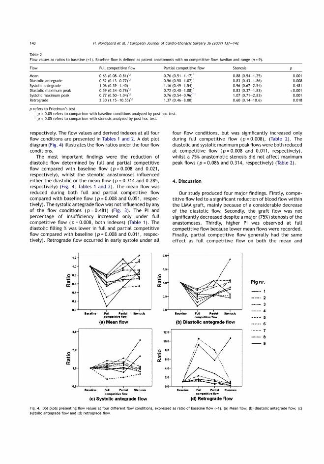
respectively. The flow values and derived indexes at all fourflow conditions are presented in Tables 1 and 2. A dot plotdiagram (Fig. 4) illustrates the flow ratios under the four flowconditions.
The most important findings were the reduction ofdiastolic flow determined by full and partial competitiveflow compared with baseline flow ( p = 0.008 and 0.021,respectively), whilst the stenotic anastomoses influencedeither the diastolic or the mean flow ( p = 0.314 and 0.285,respectively) (Fig. 4; Tables 1 and 2). The mean flow wasreduced during both full and partial competitive flowcompared with baseline flow ( p = 0.008 and 0.051, respec-tively). The systolic antegrade flowwas not influenced by anyof the flow conditions ( p = 0.481) (Fig. 3). The PI andpercentage of insufficiency increased only under fullcompetitive flow ( p = 0.008, both indexes) (Table 1). Thediastolic filling % was lower in full and partial competitiveflow compared with baseline ( p = 0.008 and 0.011, respec-tively). Retrograde flow occurred in early systole under all
four flow conditions, but was significantly increased onlyduring full competitive flow ( p = 0.008), (Table 2). Thediastolic and systolic maximum peak flows were both reducedat competitive flow ( p = 0.008 and 0.011, respectively),whilst a 75% anastomotic stenosis did not affect maximumpeak flows ( p = 0.086 and 0.314, respectively) (Table 2).
4. Discussion
Our study produced four major findings. Firstly, compe-titive flow led to a significant reduction of blood flow withinthe LIMA graft, mainly because of a considerable decreaseof the diastolic flow. Secondly, the graft flow was notsignificantly decreased despite a major (75%) stenosis of theanastomoses. Thirdly, higher PI was observed at fullcompetitive flow because lower mean flows were recorded.Finally, partial competitive flow generally had the sameeffect as full competitive flow on both the mean and
H. Nordgaard et al. / European Journal of Cardio-thoracic Surgery 36 (2009) 137—142140
Fig. 4. Dot plots presenting flow values at four different flow conditions, expressed as ratio of baseline flow (=1). (a) Mean flow, (b) diastolic antegrade flow, (c)systolic antegrade flow and (d) retrograde flow.
Table 2Flow values as ratios to baseline (=1). Baseline flow is defined as patent anastomosis with no competitive flow. Median and range (n = 9).
Flow Full competitive flow Partial competitive flow Stenosis p
Mean 0.63 (0.08—0.81)*,y 0.76 (0.51—1.17) * 0.88 (0.54—1.25) 0.001Diastolic antegrade 0.52 (0.13—0.77)*,y 0.56 (0.50—1.07) * 0.83 (0.43—1.86) 0.008Systolic antegrade 1.06 (0.39—1.40) 1.16 (0.49—1.54) 0.96 (0.67—2.54) 0.481Diastolic maximum peak 0.59 (0.34—0.78)*,y 0.72 (0.40—1.08) * 0.83 (0.37—1.83) <0.001Systolic maximum peak 0.77 (0.50—1.04)*,y 0.76 (0.54—0.96)*,y 1.07 (0.71—2.83) 0.001Retrograde 2.30 (1.15—10.55)*,y 1.37 (0.46—8.00) 0.60 (0.14—10.6) 0.018
p refers to Friedman’s test.* p < 0.05 refers to comparison with baseline conditions analyzed by post hoc test.y p < 0.05 refers to comparison with stenosis analyzed by post hoc test.

diastolic flow, whereas retrograde flow, PI and insufficiencywere significantly influenced only under full competitiveflow.
Our various experimental settings evoked three differentflow conditions often encountered by the surgeon in practice.Full competitive flow mimics the situation of a graft placeddistal to a less than critical stenosis of the coronary vessel.This may occur when coronary vessels with less than 50—70%stenosis are grafted, although such stenosis has no hemody-namic consequences at rest. Partial competitive flow occurswhen a coronary artery is grafted distal to a significantstenosis providing about half the volume flow compared withthe full value. The third condition was no-flow, an idealsituation for placing the graft distally on a totally occludedcoronary.
This study showed that the LIMA graft was muchinfluenced by competitive flow, as both the full and partialcompetitive flow conditions significantly altered the flowpatterns. Despite the superior long-term patency ofmammary grafts, the effect of competitive flow on thepatency of LIMA grafts is still controversial [4—15]. Thenarrowing phenomenon (string sign) of the distal mammarygraft is believed to be associated with competitive flow[4,7,16], because the blood flow within the mammaryartery is so low that it cannot meet the metabolicrequirements of its cell walls, causing the graft to narrowand eventually fail.
Clinical studies of competitive flow do not measure flowsbecause they are retrospective and based on angiographicdata. The occurrence of competitive flow has been inferredfrom the concomitant presence of a non-significant stenosisof the grafted vessel [7,9,17]. Furthermore, other studiessuggest that arterial grafts such as LIMA are more influencedby competitive flow than are saphenous vein grafts [4,7,10—16]. The non-muscular saphenous vein grafts cannot adjusttheir lumens in response to metabolic requirements as muchas arterial grafts. Thus, the response of vein grafts to low flowis limited.
To consider the effects of stenotic anastomoses we foundthat a 75% luminal stenosis of the anastomosis did not reduceblood flow through the grafts. This supports the findings ofMorota et al. [18] and Jaber et al. [19], who also showed thatblood flowmeasurements remained stable when anastomoticstenoses varied from mild (<25%) to moderately severe(<75%).
Our results are in line with the patho-physiologicalprinciples underlining blood flow in stenotic vessels describedby Poiseuille’s law. Nonetheless, this effect of a stenosis onblood flow is reduced in the LAD because of the autoregula-tion within the peripheral vascular bed. The LAD ischaracterized by the largest lumen and most extensiveperipheral vascular bed of all coronary arteries. Therefore toreduce the LAD perfusion flow by stenosis, the vessel lumenmust be considerably restricted (e.g. from 80 to 90%). Withsuch narrowing autoregulation will soon be exhausted, andany subsequent small reduction will have major hemody-namic consequences [20]. Clinically, a stenosis of the LAD<75% will reduce maximal flow capacity, causing ischemicinduced chest pain during exertion. During rest a stenosis>75% may significantly reduce blood flow, which can lead tochronic myocardial hypoxia.
A practical question raised by our findings is how todistinguish between competitive flows from a stenoticanastomosis, whenever low flows are recorded after CABG,since both conditions may decrease blood flow.We found thatcompetitive flow altered mainly the diastolic phase of graftflow, whereas the systolic flow remained unchanged.Additionally, a smoother flow pattern might be present,because the maximum peaks in systole and diastole decreaseduring competitive flow in LIMA—LAD anastomoses. Thus, acareful analysis of flow patterns and their timing shouldalways be carried out in situations with low graft flow. If theflow pattern is still difficult to interpret then snaring thecoronary artery proximal to the anastomotic site might be offurther help. Thus D’Ancona et al. strongly recommendedthat graft flow should bemeasured with andwithout proximalsnaring of the coronary artery [2]. With competitive flow, aswe found, occlusion of the coronary artery will increase graftflow; conversely, with a flow decrease an obstruction distal tothe graft may be suspected (i.e. stenotic anastomosis,coronary stenosis distal to the anastomotic site or poor run-off). Temporary clamping of grafts directed to collateralcoronaries may also aid in the interpretation of blood flow.
Hangler et al. strongly warned against the use of occludersduring CABG, as they found evidence of coronary endothelialinjury on electron microscopy after different occludingtechniques [21]. To avoid vessel snaring, many surgeonsmeasure the flow in the LIMA graft before removing the aorticcross clamp. Alternatively, as we have reported, epicardialultrasound is fast and efficient for assessing coronary graftanastomoses [22]. In the present study the new blood flowimaging modality was used to evaluate the patency of theanastomosis by looking at its morphology and function, andparticularly the degree of stenosis [3]. We believe that thistechnique has a great part to play in the intra-operativeassessment of CABG as it is non-invasive and can easily detectstenoses and competitive flow [22].
The main limitations of this study were that the pigs hadnormal coronary arteries with good run-off, and that theanimal model may not be directly comparable to patientswith advanced coronary disease. The measured flow valuesmay also be different in different species. Thus, the true flowvalues and derived indexes may not be directly applicable tohumans, although the 75% degree of anastomotic stenosis inour model still represented a failed anastomosis likely tocause stable angina. The consequences of an even tighterstenosis on graft flow were not investigated. Finally, thestatistical limitations were the multiple comparisons and alimited number of measurements. The widely used Bonfer-roni correction for multiple comparisons is designed for usewith independent multiple measurements and does not fullyapply to this situation. We instead report uncorrected pvalues for the Wilcoxon signed-ranks, which the readers maythemselves more easily assess the relevance of the statisticaldifferences described.
In conclusion, in a porcine model, the LIMA flow wassignificantly reduced by native coronary artery competitiveflow but marginally decreased by an anastomotic meanluminal stenosis of 75%. Reduction of graft flow due tocompetitive flow was particularly evident in diastole. Thus acareful analysis of flow patterns may help to discriminatebetween competitive flow and an anastomotic stenosis. A
H. Nordgaard et al. / European Journal of Cardio-thoracic Surgery 36 (2009) 137—142 141

detailed knowledge of different flow patterns by the surgeonis mandatory for a reliable interpretation of transit-timeflowmetry. In a near future epicardial tissue and Dopplerimaging assessment may be performed together with transit-time flowmetry because the combined information suppliedby each technique might potentially improve intra-operativeevaluation of coronary grafts.
Acknowledgements
The authors gratefully acknowledge the advice from EirikSkogvoll, Oddveig Lyng, Solveig Kolseth, Hans HenrikDedichen, Jørn Ove Sæternes and Ivar Nordrum.
References
[1] Walpoth BH, Bosshard A, Genyk I, Kipfer B, Berdat PA, Hess OM, Althaus U,Carrel TP. Transit-time flow measurement for detection of early graftfailure during myocardial revascularization. Ann Thorac Surg1998;66:1097—100.
[2] D’Ancona G, Karamanoukian HL, Bergsland J. Is intraoperative measure-ment of coronary blood flow a good predictor of graft patency? Eur JCardiothorac Surg 2001;20:1075—7.
[3] Lovstakken L, Ibrahim KS, Vitale N, Henriksen ST, Kirkeby-Garstad I, TorpH, Haaverstad R. Blood flow imaging: a new two-dimensional ultrasoundmodality for enhanced intraoperative visualization of blood flow patternsin coronary anastomoses. J Am Soc Echocardiogr 2008;21:969—75.
[4] Bezon E, Choplain JN, Maguid YA, Aziz AA, Barra JA. Failure of internalthoracic artery grafts: conclusions from coronary angiography mid-termfollow-up. Ann Thorac Surg 2003;76:754—9.
[5] Botman CJ, Schonberger J, Koolen S, Penn O, Botman H, Dib N, EeckhoutE, Pijls N. Does stenosis severity of native vessels influence bypass graftpatency? A prospective fractional flow reserve-guided study. Ann ThoracSurg 2007;83:2093—7.
[6] Gaudino M, Alessandrini F, Nasso G, Bruno P, Manzoli A, Possati G. Severityof coronary artery stenosis at preoperative angiography and midtermmammary graft status. Ann Thorac Surg 2002;74:119—21.
[7] Hashimoto H, Isshiki T, Ikari Y, Hara K, Saeki F, Tamura T, Yamaguchi T,Suma H. Effects of competitive blood flow on arterial graft patency anddiameter. J Thorac Cardiovasc Surg 1996;111:399—407.
[8] Hirotani T, Kameda T, Shirota S, Nakao Y. An evaluation of the intrao-perative transit time measurements of coronary bypass flow. Eur JCardiothorac Surg 2001;19:848—52.
[9] Kawasuji M, Sakakibara N, Takemura H, Tedoriya T, Ushijima T, WatanabeY. Is internal thoracic artery grafting suitable for a moderately stenoticcoronary artery? J Thorac Cardiovasc Surg 1996;112:253—9.
[10] Nakajima H, Kobayashi J, Tagusari O, Bando K, Niwaya K, Kitamura S.Competitive flow in arterial composite grafts and effect of graft arrange-ment in off-pump coronary revascularization. Ann Thorac Surg2004;78:481—6.
[11] Pagni S, Storey J, Ballen J, Montgomery W, Chiang BY, Etoch S, Spence PA.ITAversus SVG: a comparison of instantaneous pressure and flow dynamicsduring competitive flow. Eur J Cardiothorac Surg 1997;11:1086—92.
[12] Pagni S, Storey J, Ballen J, Montgomery W, Qaqish NK, Etoch S, Spence PA.Factors affecting internal mammary artery graft survival: how is compe-titive flow from a patent native coronary vessel a risk factor? J Surg Res1997;71:172—8.
[13] Sabik III JF, Lytle BW, Blackstone EH, Khan M, Houghtaling PL, CosgroveDM. Does competitive flow reduce internal thoracic artery graft patency?Ann Thorac Surg 2003;76:1490—6.
[14] Sabik III JF, Lytle BW, Blackstone EH, Houghtaling PL, Cosgrove DM.Comparison of saphenous vein and internal thoracic artery graft patencyby coronary system. Ann Thorac Surg 2005;79:544—51.
[15] Sabik III JF, Blackstone EH. Coronary artery bypass graft patency andcompetitive flow. J Am Coll Cardiol 2008;51:126—8.
[16] Villareal RP, Mathur VS. The string phenomenon: an important cause ofinternal mammary artery graft failure. Tex Heart Inst J 2000;27:346—9.
[17] Kim KB, Kang CH, Lim C. Prediction of graft flow impairment by intrao-perative transit time flow measurement in off-pump coronary arterybypass using arterial grafts. Ann Thorac Surg 2005;80:594—8.
[18] Morota T, Duhaylongsod FG, Burfeind WR, Huang CT. Intraoperativeevaluation of coronary anastomosis by transit-time ultrasonic flow mea-surement. Ann Thorac Surg 2002;73:1446—50.
[19] Jaber SF, Koenig SC, BhaskerRao B, VanHimbergen DJ, Spence PA. Canvisual assessment of flowwaveformmorphology detect anastomotic errorin off-pump coronary artery bypass grafting? Eur J Cardiothorac Surg1998;14:476—9.
[20] Spaan JAE, Piek JJ, Siebes M. Coronary circulation and hemodynamics.In: Kurachi Y, Sperelakis N, Terzic A, editors. Heart physiology andpathophysiology. 4th ed., Academic Press; 2001. p. 19—43.
[21] Hangler HB, Pfaller K, Antretter H, Dapunt OE, Bonatti JO. Coronaryendothelial injury after local occlusion on the human beating heart. AnnThorac Surg 2001;71:122—7.
[22] Haaverstad R, Vitale N, Tjomsland O, Tromsdal A, Torp H, Samstad SO.Intraoperative color Doppler ultrasound assessment of LIMA-to-LAD ana-stomoses in off-pump coronary artery bypass grafting. Ann Thorac Surg2002;74:1390—4.
Appendix A. Conference discussion
Dr B. Walpoth (Geneva, Switzerland): Thank you for presenting aninteresting experimental assessment of this new device from MediStim.Basically there are some problems with what you showed. First of all, it hasbeen shown before, secondly, it is well known to all cardiac surgeons that youwill never have full competitive flow. You need a relevant stenosis in order tomake a bypass. And as you have shown and which was known that if you have astenosis of the native vessels which is not in excess of 75 to 80%, you will nothave major changes in your flow measurement.
However, I fully agree with you that the addition of imaging technique is animportant feature, because sometimes you measure low-flow situations ingrafts without any obvious reasons, but if imaging is available it can help on-site in the operating room to make a decision.
My question is the following. You said with 75% stenosis at the toe of youranastomosis you didn’t see major flow changes. You saw some changes but notas much as you would have expected. How did you assess the 75% stenosis? Toobtain a correct degree of stenosis you need a biplanar angiographic evaluationand 75% might not be sufficient.
Dr Nordgaard: We used our ultrasound images of the anastomoses. Theimages were afterwards analyzed using the MatLab software. Severaldiameters of each LIMA—LAD anastomosis were measured for calculation ofthe degree of luminal stenosis. In the operation field we assessed theanastomosis using ultrasound imaging since our aim was creating a stenosis of asignificant degree at the toe of the anastomosis.
I do not agree with your comment that full competitive flow doesn’t existin coronary surgery. Often surgeons do graft vessels with about 50% luminalstenosis. Then the hemodynamic still acts as full competitive flow because theblood flow is not influenced at this degree of stenosis.
Dr Walpoth: This is correct, yes.
Dr A. Pavie (Paris, France): On your presentation I would like to make asmall comment, and I am a little provocative. You suggest that it is moreimportant to have good indication to avoid competitive flow, but it is still veryimportant to do good surgery. I am not sure that we can follow you. It iscertainly important to do a good indication, but if we do good surgery, it isprobably better.
Appendix B. Supplementary data
Supplementary data associated with this article can be found, in the onlineversion, at doi:10.1016/j.ejcts.2009.02.036.
H. Nordgaard et al. / European Journal of Cardio-thoracic Surgery 36 (2009) 137—142142

Paper III Paper III
Paper III Paper III

Pulsatility index variations using two different transit-timeflowmeters in coronary artery bypass surgery§
Havard B. Nordgaard a,*, Nicola Vitale b, Rafael Astudillo b, Attilio Renzulli d,Pal Romundstad c, Rune Haaverstad a,e
aDepartment of Circulation and Medical Imaging, The Norwegian University of Science and Technology, N-7489 Trondheim, NorwaybDepartment of Cardiothoracic Surgery, St. Olavs University Hospital, Trondheim, Norway
cDepartment of Public Health, Norwegian University of Science and Technology, Trondheim, NorwaydDepartment of Cardiac Surgery, University of Magna Grecia, Catanzaro, Italy
eDepartment of Surgical Sciences, University of Bergen, Bergen, Norway
Received 17 August 2009; received in revised form 11 November 2009; accepted 17 November 2009; Available online 23 December 2009
Abstract
Objective: Transit-time flow measurement is widely accepted as an intra-operative assessment in coronary artery bypass grafting (CABG).However, the two most commonly applied flowmeters, manufactured by MediStim ASA and Transonic Inc., have different default filter settings of20 and 10 Hz, respectively. This may cause different flow measurements, which will influence the reported results. The aim was to comparepulsatility index (PI) values recorded by the MediStim and Transonic flowmeters in two different clinical settings: (1) analysis of the flow patternsrecorded simultaneously by both flowmeters in the same CABGs; and (2) evaluation of flow patterns under different levels of filter settings in thesame grafts.Methods:Graft flow and PI weremeasured using the two different flowmeters simultaneously in 19 bypass grafts. Finally, eight graftswere assessed under different digital filter settings at 5, 10, 20, 30, 50 and 100 Hz. Results: The Transonic flowmeter provided substantially lowerPI as compared with the MediStim flowmeter. By increasing the filter setting in the flowmeter, PI increased considerably. Conclusions: TheMediStimflowmeter displayed a lower PI than the Transonic, due to a lower filter setting. In the Transonic, flow signals are filtered at a lower level,rendering a ‘smoother’ pattern of flow curves. Because different filter settings determine different PIs, caution must be taken when flow valuesand flowmeters are compared. The type of flowmeter should be indicated whenever graft flow measurements and derived indexes are provided.# 2009 European Association for Cardio-Thoracic Surgery. Published by Elsevier B.V. All rights reserved.
Keywords: CABG; Coronary graft assessment; Flowmetry; Pulsatile index; PI; Transit-time
1. Introduction
Intra-operative assessment of graft patency is an impor-tant aspect of coronary artery bypass grafting (CABG).Transit-time flow measurement (TTFM) has been in clinicaluse for more than a decade and is the most applied techniquefor intra-operative coronary graft assessment. Transit-timetechnology has proven to have a high reliability and accuracywith few sources of technical errors and provides volumetricflow patterns and derived indexes, such as the pulsatilityindex (PI) [1—4].
The PI is considered a non-invasive method for theassessment of peripheral vascular resistance of coronaryarteries [5—9], as well as in arteries of other organs (i.e.brain, kidney, placenta, eye, and so on) [5]. It was firstintroduced by Gosling and King [6], and was originally defined
as the peak-to-peak height of the waveform divided by themean height during a single cardiac cycle, as assessed byDoppler ultrasound. With the introduction of TTFM, thedefinition of PI was replaced and simplified, and at presentthe following formula is applied: PI = [maximum volumetricpeak flow � minimum volumetric peak flow]/mean volu-metric flow.
The two most commonly used transit-time flowmetersare manufactured by MediStim ASA (Oslo, Norway) andTransonic Systems Inc. (Ithaca, NY, USA) and have differentdefault filter settings of 20 and 10 Hz, respectively.Since the level of filter settings in a flowmeter mayinfluence the wave form, different maximum and minimumpeak flows may be registered and influence the PI values[10].
The aim of this study was to assess the potentialvariability of the PI as calculated by the MediStim andTransonic flowmeters, and to investigate whether increasein filtering of the flowmeter signals influences flow curvesand PI values.
www.elsevier.com/locate/ejctsEuropean Journal of Cardio-thoracic Surgery 37 (2010) 1063—1067
§ The Medical Faculty, Norwegian University of Science and Technology, has acorporate partnership with MediStim ASA.* Corresponding author. Tel.: +47 91717994; fax: +47 73867029.E-mail address: [email protected] (H.B. Nordgaard).
1010-7940/$ — see front matter # 2009 European Association for Cardio-Thoracic Surgery. Published by Elsevier B.V. All rights reserved.doi:10.1016/j.ejcts.2009.11.030

2. Material and methods
The idea to carry out this study came to us whilestudying two clinical series of patients operated upon withCABG, each of them assessed by TTFM of coronary grafts bythe MediStim or the Transonic flowmeter, respectively (seeTable 1). The first series was operated on by RH at theUniversity Hospital of Trondheim, Norway, whereas thesecond series came from the University Hospital ofCatanzaro, Italy, being operated upon by AR. All singleand sequential saphenous vein grafts (SVGs), and leftinternal mammary artery (LIMA) grafts were included. Allflow measurements were carried out after weaning ofcardiopulmonary bypass and after administration ofprotamine. Factors such subclavian artery stenosis orspasm of the LIMA graft were not taken into consideration.We observed that the PI calculated by the Transonic wasgenerally lower than the PI by MediStim. This differencewas either due to a real difference between the twoflowmeters (i.e. different levels of filter setting), or it wasdetermined by the intrinsic biases associated with acomparison between different patient populations andsurgeons. Moreover, we decided to search the literature forthe largest clinical series reporting PI data of coronarygrafts calculated by the MediStim or Transonic flowmeters,to find out whether the trend of different PI valuescalculated by the two machines were consistent in otherseries as well (see Table 2). Most of the relevant dataavailable were obtained by the MediStim flowmeter [11—16], whereas just two series presented by the sameinvestigators were found reporting PI values by Transonic[17,18]. Despite differences in the amount of data, wenoted a trend towards lower PI values provided by theTransonic flowmeter versus the MediStim flowmeter. Thus,we decided to carry out the following study to ascertainwhether this observation held true based on the technicalcharacteristics of transit-time flowmeters.
2.1. Digital signal processing technology in transit-timeflowmeters
The transit-time flowmeters measure the time differ-ence between an upstream and downstream ultrasonicpulse when transmitted and received by two spatiallyseparated transducers in the flow probe. This time-delaydifference can be converted to volume flow, which is
displayed on-screen as a digitised signal. The flow isrecorded as analogue signals that are initially digitallysampled at a sufficiently high frequency according to theNyquist—Shannon sampling theorem [19]. Further, theflowmeter typically uses a low-pass filter to smooth thesignal and attenuate noise. MediStim and Transonicflowmeters use default low-pass filters at 20 and 10 Hz,respectively. In other words, the flowmeters filter thesignals differently, which may determine different flowcurve patterns.
H.B. Nordgaard et al. / European Journal of Cardio-thoracic Surgery 37 (2010) 1063—10671064
Table 1Mean PI of two patient populations assessed by MediStim and Transonic flowmeter, respectively.
Graft Group A Group B
N Mean 95% CI N Mean 95% CI p
Lower Upper Lower Upper
LIMA-LAD 482 2.1 2.1 2.2 100 1.7 1.6 1.8 <0.001Single SVG to LCA 198 1.8 1.7 1.9 36 1.7 1.5 2.0 0.52Double SSVG to LCA 264 1.8 1.7 1.9 19 1.4 1.0 1.8 0.03Single SVG to RCA 317 2.1 1.9 2.2 25 1.6 1.3 2.0 0.03Double SSVG to RCA 41 2.2 1.9 2.5 61 1.7 1.5 1.9 0.001
Group A: MediStim flowmeter; Group B: Transonic flowmeter; CI: confidence interval; LCA: left coronary artery; RCA: right coronary artery; SSVG: sequentialsaphenous vein graft; and SVG: saphenous vein graft.
Table 2Mean PI from different published studies.
Flowmeter Author Type of graft (N) PI � SD
MediStim Becit [11] LIMA-LAD (102) 2.32 � 0.88SVG to Diagonal (48) 2.26 � 0.81SVG to OM (80) 2.51 � 0.94SVG to RCA (73) 2.23 � 0.94
Goel* [12] LIMA-LAD (162) 2.71 � 0.95SVG overall (224) 2.48 � 1.04
Gwozdziewicz [13] SSVG (50) 1.8 � 0.4
Kim*** [14] To LCA (67) 2.4 � 1.3To RCA (36) 3.1 � 1.6
Hassanein [15] LIMA-LAD (421) 1.98 � 1.08SVG to Diagonal (69) 1.72 � 0.58SVG to OM (71) 1.88 � 0.86SVG to RCA (107) 1.80 � 1.82
Nordgaard** [16] LIMA-LAD (495) 2.4 � 0.9SVG to LCA (527)y 2.0 (0.05 SEM)SVG to RCA (368)y 2.4 (0.06 SEM)
Transonic Onorati [17] LIMA-LAD (199) 1.53 � 0.62SVG to RCA (102) 1.67 � 1.20SVG to OM (61) 2.06 � 0.89SVG to Diag (25) 2.25 � 1.25
Onorati [18] LIMA-LAD (45)a 1.2 � 0.4LIMA-LAD (45)b 1.0 � 0.7SVG to OMa 1.46 � 0.9SSVG to OMb 0.71 � 0.4
a Single SVG group.b Sequential SVG group.* OPCAB in 36% of cases.** OPCAB in 11% of cases.*** Only arterial grafts were used: mammary (n = 78), gastroepiploic (n = 38)
and radial artery (n = 1).y Includes values from single and sequential SVG as no PI difference was
found between the groups [16].
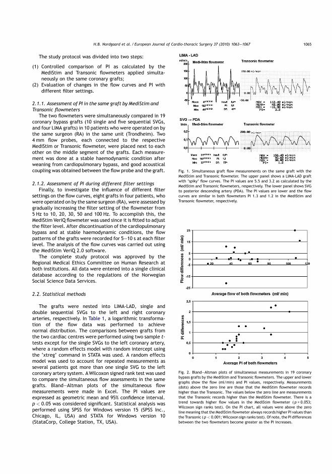
The study protocol was divided into two steps:
(1) Controlled comparison of PI as calculated by theMediStim and Transonic flowmeters applied simulta-neously on the same coronary grafts;
(2) Evaluation of changes in the flow curves and PI withdifferent filter settings.
2.1.1. Assessment of PI in the same graft by MediStim andTransonic flowmeters
The two flowmeters were simultaneously compared in 19coronary bypass grafts (10 single and five sequential SVGs,and four LIMA grafts) in 10 patients who were operated on bythe same surgeon (RA) in the same unit (Trondheim). Two4 mm flow probes, each connected to the respectiveMediStim or Transonic flowmeter, were placed next to eachother on the middle segment of the grafts. Each measure-ment was done at a stable haemodynamic condition afterweaning from cardiopulmonary bypass, and good acousticalcoupling was obtained between the flow probe and the graft.
2.1.2. Assessment of PI during different filter settingsFinally, to investigate the influence of different filter
settings on the flow curves, eight grafts in four patients, whowere operated on by the same surgeon (RA), were assessed bygradually increasing the filter setting of the flowmeter from5 Hz to 10, 20, 30, 50 and 100 Hz. To accomplish this, theMediStim VeriQ flowmeter was used since it is fitted to adjustthe filter level. After discontinuation of the cardiopulmonarybypass and at stable haemodynamic conditions, the flowpatterns of the grafts were recorded for 5—10 s at each filterlevel. The analysis of the flow curves was carried out usingthe MediStim VeriQ 2.0 software.
The complete study protocol was approved by theRegional Medical Ethics Committee on Human Research atboth institutions. All data were entered into a single clinicaldatabase according to the regulations of the NorwegianSocial Science Data Services.
2.2. Statistical methods
The grafts were nested into LIMA-LAD, single anddouble sequential SVGs to the left and right coronaryarteries, respectively. In Table 1, a logarithmic transforma-tion of the flow data was performed to achievenormal distribution. The comparisons between grafts fromthe two cardiac centres were performed using two sample t-tests except for the single SVGs to the left coronary artery,where a random effects model with random intercept usingthe ‘xtreg’ command in STATA was used. A random effectsmodel was used to account for repeated measurements asseveral patients got more than one single SVG to the leftcoronary artery system. AWilcoxon signed rank test was usedto compare the simultaneous flow assessments in the samegrafts. Bland—Altman plots of the simultaneous flowmeasurements were made in Excel. The PI values areexpressed as geometric mean and 95% confidence interval.p < 0.05 was considered significant. Statistical analysis wasperformed using SPSS for Windows version 15 (SPSS Inc.,Chicago, IL, USA) and STATA for Windows version 10(StataCorp, College Station, TX, USA).
H.B. Nordgaard et al. / European Journal of Cardio-thoracic Surgery 37 (2010) 1063—1067 1065
Fig. 1. Simultaneous graft flow measurements on the same graft with theMediStim and Transonic flowmeter. The upper panel shows a LIMA-LAD graftwith ‘spiky’ flow curves. The PI values are 5.5 and 3.2 as calculated by theMediStim and Transonic flowmeters, respectively. The lower panel shows SVGto posterior descending artery (PDA). The PI values are lower and the flowcurves are similar in both flowmeters PI 1.3 and 1.2 in the MediStim andTransonic flowmeter, respectively.
Fig. 2. Bland—Altman plots of simultaneous measurements in 19 coronarybypass grafts by the MediStim and Transonic flowmeters. The upper and lowergraphs show the flow (ml/min) and PI values, respectively. Measurements(dots) above the zero line are those that the MediStim flowmeter recordshigher than the Transonic. The values below the zero line are measurementsthat the Transonic records higher than the MediStim flowmeter. There is atrend towards higher flow values in the MediStim flowmeter (p = 0.053;Wilcoxon sign ranks test). On the PI chart, all values were above the zeroline meaning that the MediStim flowmeter always records higher PI values thanthe Transonic (p < 0.001; Wilcoxon sign ranks test). Of note, the PI differencesbetween the two flowmeters become greater as the PI increases.

3. Results
3.1. Assessment of PI by MediStim versus Transonicflowmeter in the same grafts
The MediStim and Transonic flowmeter provided meanPI and standard deviations of 2.7 � 1.2 and 1.8 � 0.6,respectively ( p < 0.001; Wilcoxon signed ranks test). Fig. 1clearly demonstrates the difference between the twoflowmeters. The Transonic flowmeter achieved system-atically lower PI values. The degree of differencedepended on the flow pattern. In a spiky flow curvepattern the resultant PI was high, with remarkabledifferences between the two flowmeters. However, witha smoother flow pattern the PI was low, and the twoflowmeters produced similar values, demonstrating thatthe difference between the flowmeters becomes greater asPI increases (Fig. 2).
3.2. Assessment of PI under different filter settings
Typical changes in the flow pattern occurred underdifferent filter settings. At low-filter setting, a smootherflow pattern appeared with no sharp flow spikes. Byincreasing the filter setting, a ‘noisier’ flow pattern appearedwhich produced higher PIs. Fig. 3 visualises the different flowcurves at different filter settings with the MediStim VeriQflowmeter, and in this case as well, the difference betweenfilter settings becomes greater as PI increases.
4. Discussion
This study identified important differences between thetwo most frequently used TTFMs. Because of a lower filtersetting, the Transonic flowmeter produced smoother flowcurves than the MediStim, which influenced the calculationof the PIs. Specifically, PIs calculated by Transonic are lowerthan those calculated by MediStim.
Direct comparisons of flow and PI results of the same graftsby the two flowmeters provided evidence that the Transonicproduced a lower PI than the MediStim, which was observedconsistently during the testing of 19 grafts. Furthermore, PIdifferences between the two flowmeters seemed to be morepronounced at higher PI values, as shown in Fig. 1.
The next step in our analysis was to find out whether alower PI was achieved by applying a lower filter setting,which produced smoother flow curves. We discovered that aconstant increase in PI value occurred in concomitance withan increase in the filter setting, and that higher filter settingsare characterised by a pronounced spiky pattern thatdetermined very high maximum and low minimum peakson the flow charts, resulting in increased PI values.
The PI is calculated automatically by the flowmeters. TheMediStim flowmeter and the Transonic flowmeter measure thehigher and lower peaks during a blood flow recording time of 2and 8 s, respectively. Therefore, one would expect higher andlower peaks, resulting in higher PIs, to most likely be recordedby the Transonic flowmeter, rather than the MediStim becauseof its longer recording time. Interestingly, but still of unknown
H.B. Nordgaard et al. / European Journal of Cardio-thoracic Surgery 37 (2010) 1063—10671066
Fig. 3. The upper panel shows PI values of eight different grafts measured under various filter settings (5, 10, 20, 30, 50 and 100 Hz, respectively). The proportion ofthe changes depends on the level of the PI values. There are major changes between the filter levels when PI is high, minor changes when PI is low. The lower panelsshow two corresponding flow curves under increasing filter settings. The upper flow curves of a single saphenous vein graft (SVG) to the posterior-lateral branch (PL)present a greater increase in PI values due to higher filter settings. The lower flow curves of a sequential saphenous vein graft (SSVG) to obtuse marginal (OM) anddiagonal (D) show a more moderate change in PI values due to lower filter settings. Increased filter settings lead to a spikier flow pattern, causing higher PIs. Lowerfilter setting results in a smoother flow pattern, and consequently a lower PI. PDA: posterior descending artery. RCA: right coronary artery.

reason, this was not the case since the Transonic producessystematically lower PIs due to its lower filter setting.
Similar outcomes, which document the differencesbetween these two flowmeters, have only previouslybeen published in an abstract.1 Fifty-five grafts from 23patients were assessed intra-operatively by both devices. Onthe one hand, a significantly higher PI of 2.7 was shown withthe MediStim as compared with a PI of 2.2 by the Transonic.On the other hand, the differences in mean flow were alsosignificant: it was 45.6 ml min�1 using the MediStim ascompared with 62.1 ml min�1 with the Transonic. Unfortu-nately, the authors did not comment on these findings.
In vitro and in vivo comparisons of TTFMs have both showna high accuracy and precision in regard to volumetric flow[1,3,20]. To the best of our knowledge, this variation in flowpattern as seen in different flowmeters has not beenpreviously described, although it is highly relevant. Thesimultaneous comparison of the two flowmeters confirmedthe differences seen clinically as presented in Table 1.
On clinical grounds, the difference in waveforms due todifferent filter settings may have important repercussions.The shape of the flow curve is an element which comes intoplay when assessing grafts that are difficult to interpret (lowflows and high PI). In cases involving a failed anastomosis or agraft directed to coronary arteries with high peripheralvascular resistance, the waveform will present multiplespikes that will be higher than expected. On the one hand, adevice equipped with a high filter setting will produce veryspiky flow curves, making it difficult to evaluate the graftflow. On the other hand, a device with a low filter setting willsmooth out most of the spikes, making the graft assessmentappear to be better than it actually is.
Unfortunately, there are no clear cut-off values of meanflow and PI that allow surgeons to adequately establish thequality of a coronary graft. There has been much debate overthe identification of these cut-off values, but so far, no generalagreement has been reached due to the many variablesinvolved in graft assessment [14,21,22]. The important issueofhaving separate cut-off values for different flowmeters has notbeen discussed. Therefore, based on the results of ourinvestigation, it is important to refer to the type of flowmeterused when presenting flow values.
In conclusion, MediStim and Transonic TTFMs arenot directly comparable because of their filter settings at20 and 10 Hz, respectively. Different levels of filter settings inthe flowmeters determine different shapes in the flowcurves, which results in different PI values. In particular,more pronounced differences in PIs were noted when the PIwas around 3. Thus, the type of flowmeter should always bereported together with the graft flows and PI.
Acknowledgements
The authors gratefully acknowledge the assistance byTransonic Inc. and MediStim ASA for providing equipment anddisposables.
References
[1] Beldi G, Bosshard A, Hess OM, Althaus U, Walpoth BH. Transit time flowmeasurement: experimental validation and comparison of three differentsystems. Ann Thorac Surg 2000;70:212—7.
[2] Walpoth BH, Bosshard A, Genyk I, Kipfer B, Berdat PA, Hess OM, Althaus U,Carrel TP. Transit-time flow measurement for detection of early graftfailure during myocardial revascularization. Ann Thorac Surg 1998;66:1097—100.
[3] Laustsen J, Pedersen EM, Terp K, Steinbruchel D, Kure HH, Paulsen PK,Jorgensen H, Paaske WP. Validation of a new transit time ultrasoundflowmeter in man. Eur J Vasc Endovasc Surg 1996;12:91—6.
[4] Groom R, Tryzelaar J, Forest R, Niimi K, Cecere G, Donegan D, Katz S,Weldner P, Quinn R, Braxton J, Blank S, Kramer R, Morton J. Intra-operative quality assessment of coronary artery bypass grafts. Perfusion2001;16: 511—8.
[5] Aleksic M, Heckenkamp J, Gawenda M, Brunkwall J. Pulsatility indexdetermination by flowmeter measurement: a new indicator for vascularresistance? Eur Surg Res 2004;36:345—9.
[6] Gosling RG, King DH. Arterial assessment by Doppler-shift ultrasound.Proc R Soc Med 1974;67:447—9.
[7] D’Ancona G, Karamanoukian HL, Ricci M, Schmid S, Bergsland J, SalernoTA. Graft revision after transit time flow measurement in off-pumpcoronary artery bypass grafting. Eur J Cardiothorac Surg 2000;17:287—93.
[8] Evans DH, Barrie WW, Asher MJ, Bentley S, Bell PR. The relationshipbetween ultrasonic pulsatility index and proximal arterial stenosis in acanine model. Circ Res 1980;46:470—5.
[9] Morota T, Duhaylongsod FG, Burfeind WR, Huang CT. Intra-operativeevaluation of coronary anastomosis by transit-time ultrasonic flow mea-surement. Ann Thorac Surg 2002;73:1446—50.
[10] Proakis JG, Manolakis DK. Design of digital filters. In: Digital SignalProcessing: Principles, Algorithms and Applications. Prentice Hall;1995 . p. 614—737 [chapter 8].
[11] Becit N, Erkut B, Ceviz M, Unlu Y, Colak A, Kocak H. The impact of intra-operative transit time flow measurement on the results of on-pumpcoronary surgery. Eur J Cardiothorac Surg 2007;32:313—8.
[12] Goel P, Dubey S, Makker A, Kohli VM. Evaluation of coronary artery bypassgrafts by intra-operative transit time flow measurement. Ind J ThoracCardiovasc Surg 2003;19:108—12.
[13] Gwozdziewicz M, Nemec P, Simek M, Hajek R, Troubil M. Sequentialbypass grafting on the beating heart: blood flow characteristics. AnnThorac Surg 2006;82:620—3.
[14] Kim KB, Kang CH, Lim C. Prediction of graft flow impairment by intra-operative transit time flow measurement in off-pump coronary arterybypass using arterial grafts. Ann Thorac Surg 2005;80:594—8.
[15] Hassanein W, Albert AA, Arnrich B, Walter J, Ennker IC, Rosendahl U,Bauer S, Ennker J. Intra-operative transit time flow measurement: off-pump versus on-pump coronary artery bypass. Ann Thorac Surg2005;80:2155—61.
[16] Nordgaard H, Vitale N, Haaverstad R. Transit-time blood flow measure-ments in sequential saphenous coronary artery bypass grafts. Ann ThoracSurg 2009;87:1409—15.
[17] Onorati F, Olivito S, Mastroroberto P, Virgilio A, Esposito A, Perrotti A,Renzulli A. Perioperative patency of coronary artery bypass grafting is notinfluenced by off-pump technique. Ann Thorac Surg 2005;80:2132—40.
[18] Onorati F, Pezzo F, Esposito A, Impiombato B, Comi MC, Polistina M,Renzulli A. Single versus sequential saphenous vein grafting of thecircumflex system: a flowmetric study. Scand Cardiovasc J 2007;41:265—71.
[19] Pharr M, Humphreys G. Sampling and reconstruction. In: Physically basedrendering. From theory to implementation1st ed., Elsevier Science &Technology; 2004. p. 279—94 [chapter 7].
[20] Cikirikcioglu M, Cikirikcioglu YB, Khabiri E, Djebaili MK, Kalangos A,Walpoth BH. Pre-clinical validation of a new intra-operative ‘‘dual beamDoppler’’ blood flowmeter in an artificial circuit. Heart Surg Forum2006;9:499—505.
[21] Di Giammarco G, Pano M, Cirmeni S, Pelini P, Vitolla G, Di Mauro M.Predictive value of intra-operative transit-time flow measurement forshort-term graft patency in coronary surgery. J Thorac Cardiovasc Surg2006;132:468—74.
[22] D’Ancona G, Karamanoukian HL, Bergsland J. Is intra-operative measure-ment of coronary blood flow a good predictor of graft patency? Eur JCardiothorac Surg 2001;20:1075—7.
H.B. Nordgaard et al. / European Journal of Cardio-thoracic Surgery 37 (2010) 1063—1067 1067
1 Abstract: Wakwe W, Poston R. Which flowmeter predicts postoperativegraft patency more consistently and effectively after OPCABG; 2004. http://medschool.umaryland.edu/OSR/docs/ForumBooklet/2004.pdf.

EJCTS-7757; No. of Pages 1
Please cite this article in press as: Nordgaard HB, et al. Corrigendum to ‘‘Pulsatility index variations using two different transit-time
flowmeters in coronary artery bypass surgery’’ [Eur. J. Cardiothorac. Surg. 37 (5) (2010) 1063—1067]. Eur J Cardiothorac Surg (2010),
doi:10.1016/j.ejcts.2010.06.001
Corrigendum
Corrigendum to ‘‘Pulsatility index variations using two differenttransit-time flowmeters in coronary artery bypass surgery’’
[Eur. J. Cardiothorac. Surg. 37 (5) (2010) 1063—1067]
Havard B. Nordgaard a,*, Nicola Vitale b, Rafael Astudillo b, Attilio Renzulli d,Pal Romundstad c, Rune Haaverstad a,e
aDepartment of Circulation and Medical Imaging, The Norwegian University of Science and Technology, N-7489 Trondheim, NorwaybDepartment of Cardiothoracic Surgery, St. Olavs University Hospital, Trondheim, Norway
cDepartment of Public Health, Norwegian University of Science and Technology, Trondheim, NorwaydDepartment of Cardiac Surgery, University of Magna Grecia, Catanzaro, Italy
eDepartment of Surgical Sciences, University of Bergen, Bergen, Norway
The author regrets that an error appeared in the original version of this paper.The Conclusions section of the abstract should read as follows:Conclusions: The Transonic flowmeter displayed a lower PI than the MediStim, due to a lower filter setting. In the Transonic,
flow signals are filtered at a lower level, rendering a ‘smoother’ pattern of flow curves. Because different filter settingsdetermine different PIs, caution must be taken when flow values and flowmeters are compared. The type of flowmeter should beindicated whenever graft flow measurements and derived indexes are provided.
www.elsevier.com/locate/ejctsEuropean Journal of Cardio-thoracic Surgery xxx (2010) xxx—xxx
DOI of original article: 10.1016/j.ejcts.2009.11.030.* Corresponding author. Tel.: +47 91717994; fax: +47 73867029.E-mail address: [email protected] (H.B. Nordgaard).
1010-7940/$ — see front matter # 2010 European Association for Cardio-Thoracic Surgery. Published by Elsevier B.V. All rights reserved.doi:10.1016/j.ejcts.2010.06.001 DOCTOPIC:

Paper IV Paper IV
Paper IV Paper IV

Is not included due to copyright

Dissertations at the Faculty of Medicine, NTNU 1977
1. Knut Joachim Berg: EFFECT OF ACETYLSALICYLIC ACID ON RENAL FUNCTION 2. Karl Erik Viken and Arne Ødegaard: STUDIES ON HUMAN MONOCYTES CULTURED IN
VITRO 1978
3. Karel Bjørn Cyvin: CONGENITAL DISLOCATION OF THE HIP JOINT. 4. Alf O. Brubakk: METHODS FOR STUDYING FLOW DYNAMICS IN THE LEFT
VENTRICLE AND THE AORTA IN MAN. 1979
5. Geirmund Unsgaard: CYTOSTATIC AND IMMUNOREGULATORY ABILITIES OF HUMAN BLOOD MONOCYTES CULTURED IN VITRO
1980 6. Størker Jørstad: URAEMIC TOXINS 7. Arne Olav Jenssen: SOME RHEOLOGICAL, CHEMICAL AND STRUCTURAL
PROPERTIES OF MUCOID SPUTUM FROM PATIENTS WITH CHRONIC OBSTRUCTIVE BRONCHITIS
1981 8. Jens Hammerstrøm: CYTOSTATIC AND CYTOLYTIC ACTIVITY OF HUMAN
MONOCYTES AND EFFUSION MACROPHAGES AGAINST TUMOR CELLS IN VITRO 1983
9. Tore Syversen: EFFECTS OF METHYLMERCURY ON RAT BRAIN PROTEIN. 10. Torbjørn Iversen: SQUAMOUS CELL CARCINOMA OF THE VULVA.
1984 11. Tor-Erik Widerøe: ASPECTS OF CONTINUOUS AMBULATORY PERITONEAL
DIALYSIS. 12. Anton Hole: ALTERATIONS OF MONOCYTE AND LYMPHOCYTE FUNCTIONS IN
REALTION TO SURGERY UNDER EPIDURAL OR GENERAL ANAESTHESIA. 13. Terje Terjesen: FRACTURE HEALING AND STRESS-PROTECTION AFTER METAL
PLATE FIXATION AND EXTERNAL FIXATION. 14. Carsten Saunte: CLUSTER HEADACHE SYNDROME. 15. Inggard Lereim: TRAFFIC ACCIDENTS AND THEIR CONSEQUENCES. 16. Bjørn Magne Eggen: STUDIES IN CYTOTOXICITY IN HUMAN ADHERENT
MONONUCLEAR BLOOD CELLS. 17. Trond Haug: FACTORS REGULATING BEHAVIORAL EFFECTS OG DRUGS.
1985 18. Sven Erik Gisvold: RESUSCITATION AFTER COMPLETE GLOBAL BRAIN ISCHEMIA. 19. Terje Espevik: THE CYTOSKELETON OF HUMAN MONOCYTES. 20. Lars Bevanger: STUDIES OF THE Ibc (c) PROTEIN ANTIGENS OF GROUP B
STREPTOCOCCI. 21. Ole-Jan Iversen: RETROVIRUS-LIKE PARTICLES IN THE PATHOGENESIS OF
PSORIASIS. 22. Lasse Eriksen: EVALUATION AND TREATMENT OF ALCOHOL DEPENDENT
BEHAVIOUR. 23. Per I. Lundmo: ANDROGEN METABOLISM IN THE PROSTATE.
1986 24. Dagfinn Berntzen: ANALYSIS AND MANAGEMENT OF EXPERIMENTAL AND
CLINICAL PAIN. 25. Odd Arnold Kildahl-Andersen: PRODUCTION AND CHARACTERIZATION OF
MONOCYTE-DERIVED CYTOTOXIN AND ITS ROLE IN MONOCYTE-MEDIATED CYTOTOXICITY.
26. Ola Dale: VOLATILE ANAESTHETICS. 1987
27. Per Martin Kleveland: STUDIES ON GASTRIN. 28. Audun N. Øksendal: THE CALCIUM PARADOX AND THE HEART. 29. Vilhjalmur R. Finsen: HIP FRACTURES
1988 30. Rigmor Austgulen: TUMOR NECROSIS FACTOR: A MONOCYTE-DERIVED
REGULATOR OF CELLULAR GROWTH.

31. Tom-Harald Edna: HEAD INJURIES ADMITTED TO HOSPITAL. 32. Joseph D. Borsi: NEW ASPECTS OF THE CLINICAL PHARMACOKINETICS OF
METHOTREXATE. 33. Olav F. M. Sellevold: GLUCOCORTICOIDS IN MYOCARDIAL PROTECTION. 34. Terje Skjærpe: NONINVASIVE QUANTITATION OF GLOBAL PARAMETERS ON LEFT
VENTRICULAR FUNCTION: THE SYSTOLIC PULMONARY ARTERY PRESSURE AND CARDIAC OUTPUT.
35. Eyvind Rødahl: STUDIES OF IMMUNE COMPLEXES AND RETROVIRUS-LIKE ANTIGENS IN PATIENTS WITH ANKYLOSING SPONDYLITIS.
36. Ketil Thorstensen: STUDIES ON THE MECHANISMS OF CELLULAR UPTAKE OF IRON FROM TRANSFERRIN.
37. Anna Midelfart: STUDIES OF THE MECHANISMS OF ION AND FLUID TRANSPORT IN THE BOVINE CORNEA.
38. Eirik Helseth: GROWTH AND PLASMINOGEN ACTIVATOR ACTIVITY OF HUMAN GLIOMAS AND BRAIN METASTASES - WITH SPECIAL REFERENCE TO TRANSFORMING GROWTH FACTOR BETA AND THE EPIDERMAL GROWTH FACTOR RECEPTOR.
39. Petter C. Borchgrevink: MAGNESIUM AND THE ISCHEMIC HEART. 40. Kjell-Arne Rein: THE EFFECT OF EXTRACORPOREAL CIRCULATION ON
SUBCUTANEOUS TRANSCAPILLARY FLUID BALANCE. 41. Arne Kristian Sandvik: RAT GASTRIC HISTAMINE. 42. Carl Bredo Dahl: ANIMAL MODELS IN PSYCHIATRY.
1989 43. Torbjørn A. Fredriksen: CERVICOGENIC HEADACHE. 44. Rolf A. Walstad: CEFTAZIDIME. 45. Rolf Salvesen: THE PUPIL IN CLUSTER HEADACHE. 46. Nils Petter Jørgensen: DRUG EXPOSURE IN EARLY PREGNANCY. 47. Johan C. Ræder: PREMEDICATION AND GENERAL ANAESTHESIA IN OUTPATIENT
GYNECOLOGICAL SURGERY. 48. M. R. Shalaby: IMMUNOREGULATORY PROPERTIES OF TNF- AND THE RELATED
CYTOKINES. 49. Anders Waage: THE COMPLEX PATTERN OF CYTOKINES IN SEPTIC SHOCK. 50. Bjarne Christian Eriksen: ELECTROSTIMULATION OF THE PELVIC FLOOR IN FEMALE
URINARY INCONTINENCE. 51. Tore B. Halvorsen: PROGNOSTIC FACTORS IN COLORECTAL CANCER.
1990 52. Asbjørn Nordby: CELLULAR TOXICITY OF ROENTGEN CONTRAST MEDIA. 53. Kåre E. Tvedt: X-RAY MICROANALYSIS OF BIOLOGICAL MATERIAL. 54. Tore C. Stiles: COGNITIVE VULNERABILITY FACTORS IN THE DEVELOPMENT AND
MAINTENANCE OF DEPRESSION. 55. Eva Hofsli: TUMOR NECROSIS FACTOR AND MULTIDRUG RESISTANCE. 56. Helge S. Haarstad: TROPHIC EFFECTS OF CHOLECYSTOKININ AND SECRETIN ON
THE RAT PANCREAS. 57. Lars Engebretsen: TREATMENT OF ACUTE ANTERIOR CRUCIATE LIGAMENT
INJURIES. 58. Tarjei Rygnestad: DELIBERATE SELF-POISONING IN TRONDHEIM. 59. Arne Z. Henriksen: STUDIES ON CONSERVED ANTIGENIC DOMAINS ON MAJOR
OUTER MEMBRANE PROTEINS FROM ENTEROBACTERIA. 60. Steinar Westin: UNEMPLOYMENT AND HEALTH: Medical and social consequences of a
factory closure in a ten-year controlled follow-up study. 61. Ylva Sahlin: INJURY REGISTRATION, a tool for accident preventive work. 62. Helge Bjørnstad Pettersen: BIOSYNTHESIS OF COMPLEMENT BY HUMAN ALVEOLAR
MACROPHAGES WITH SPECIAL REFERENCE TO SARCOIDOSIS. 63. Berit Schei: TRAPPED IN PAINFUL LOVE. 64. Lars J. Vatten: PROSPECTIVE STUDIES OF THE RISK OF BREAST CANCER IN A
COHORT OF NORWEGIAN WOMAN. 1991
65. Kåre Bergh: APPLICATIONS OF ANTI-C5a SPECIFIC MONOCLONAL ANTIBODIES FOR THE ASSESSMENT OF COMPLEMENT ACTIVATION.

66. Svein Svenningsen: THE CLINICAL SIGNIFICANCE OF INCREASED FEMORAL ANTEVERSION.
67. Olbjørn Klepp: NONSEMINOMATOUS GERM CELL TESTIS CANCER: THERAPEUTIC OUTCOME AND PROGNOSTIC FACTORS.
68. Trond Sand: THE EFFECTS OF CLICK POLARITY ON BRAINSTEM AUDITORY EVOKED POTENTIALS AMPLITUDE, DISPERSION, AND LATENCY VARIABLES.
69. Kjetil B. Åsbakk: STUDIES OF A PROTEIN FROM PSORIATIC SCALE, PSO P27, WITH RESPECT TO ITS POTENTIAL ROLE IN IMMUNE REACTIONS IN PSORIASIS.
70. Arnulf Hestnes: STUDIES ON DOWN´S SYNDROME. 71. Randi Nygaard: LONG-TERM SURVIVAL IN CHILDHOOD LEUKEMIA. 72. Bjørn Hagen: THIO-TEPA. 73. Svein Anda: EVALUATION OF THE HIP JOINT BY COMPUTED TOMOGRAMPHY AND
ULTRASONOGRAPHY. 1992
74. Martin Svartberg: AN INVESTIGATION OF PROCESS AND OUTCOME OF SHORT-TERM PSYCHODYNAMIC PSYCHOTHERAPY.
75. Stig Arild Slørdahl: AORTIC REGURGITATION. 76. Harold C Sexton: STUDIES RELATING TO THE TREATMENT OF SYMPTOMATIC NON-
PSYCHOTIC PATIENTS. 77. Maurice B. Vincent: VASOACTIVE PEPTIDES IN THE OCULAR/FOREHEAD AREA. 78. Terje Johannessen: CONTROLLED TRIALS IN SINGLE SUBJECTS. 79. Turid Nilsen: PYROPHOSPHATE IN HEPATOCYTE IRON METABOLISM. 80. Olav Haraldseth: NMR SPECTROSCOPY OF CEREBRAL ISCHEMIA AND REPERFUSION
IN RAT. 81. Eiliv Brenna: REGULATION OF FUNCTION AND GROWTH OF THE OXYNTIC
MUCOSA. 1993
82. Gunnar Bovim: CERVICOGENIC HEADACHE. 83. Jarl Arne Kahn: ASSISTED PROCREATION. 84. Bjørn Naume: IMMUNOREGULATORY EFFECTS OF CYTOKINES ON NK CELLS. 85. Rune Wiseth: AORTIC VALVE REPLACEMENT. 86. Jie Ming Shen: BLOOD FLOW VELOCITY AND RESPIRATORY STUDIES. 87. Piotr Kruszewski: SUNCT SYNDROME WITH SPECIAL REFERENCE TO THE
AUTONOMIC NERVOUS SYSTEM. 88. Mette Haase Moen: ENDOMETRIOSIS. 89. Anne Vik: VASCULAR GAS EMBOLISM DURING AIR INFUSION AND AFTER
DECOMPRESSION IN PIGS. 90. Lars Jacob Stovner: THE CHIARI TYPE I MALFORMATION. 91. Kjell Å. Salvesen: ROUTINE ULTRASONOGRAPHY IN UTERO AND DEVELOPMENT IN
CHILDHOOD. 1994
92. Nina-Beate Liabakk: DEVELOPMENT OF IMMUNOASSAYS FOR TNF AND ITS SOLUBLE RECEPTORS.
93. Sverre Helge Torp: erbB ONCOGENES IN HUMAN GLIOMAS AND MENINGIOMAS. 94. Olav M. Linaker: MENTAL RETARDATION AND PSYCHIATRY. Past and present. 95. Per Oscar Feet: INCREASED ANTIDEPRESSANT AND ANTIPANIC EFFECT IN
COMBINED TREATMENT WITH DIXYRAZINE AND TRICYCLIC ANTIDEPRESSANTS. 96. Stein Olav Samstad: CROSS SECTIONAL FLOW VELOCITY PROFILES FROM TWO-
DIMENSIONAL DOPPLER ULTRASOUND: Studies on early mitral blood flow. 97. Bjørn Backe: STUDIES IN ANTENATAL CARE. 98. Gerd Inger Ringdal: QUALITY OF LIFE IN CANCER PATIENTS. 99. Torvid Kiserud: THE DUCTUS VENOSUS IN THE HUMAN FETUS. 100. Hans E. Fjøsne: HORMONAL REGULATION OF PROSTATIC METABOLISM. 101. Eylert Brodtkorb: CLINICAL ASPECTS OF EPILEPSY IN THE MENTALLY RETARDED. 102. Roar Juul: PEPTIDERGIC MECHANISMS IN HUMAN SUBARACHNOID HEMORRHAGE. 103. Unni Syversen: CHROMOGRANIN A. Phsysiological and Clinical Role.
1995
104. Odd Gunnar Brakstad: THERMOSTABLE NUCLEASE AND THE nuc GENE IN THE DIAGNOSIS OF Staphylococcus aureus INFECTIONS.

105. Terje Engan: NUCLEAR MAGNETIC RESONANCE (NMR) SPECTROSCOPY OF PLASMA IN MALIGNANT DISEASE.
106. Kirsten Rasmussen: VIOLENCE IN THE MENTALLY DISORDERED. 107. Finn Egil Skjeldestad: INDUCED ABORTION: Timetrends and Determinants. 108. Roar Stenseth: THORACIC EPIDURAL ANALGESIA IN AORTOCORONARY BYPASS
SURGERY. 109. Arild Faxvaag: STUDIES OF IMMUNE CELL FUNCTION in mice infected with MURINE
RETROVIRUS. 1996
110. Svend Aakhus: NONINVASIVE COMPUTERIZED ASSESSMENT OF LEFT VENTRICULAR FUNCTION AND SYSTEMIC ARTERIAL PROPERTIES. Methodology and some clinical applications.
111. Klaus-Dieter Bolz: INTRAVASCULAR ULTRASONOGRAPHY. 112. Petter Aadahl: CARDIOVASCULAR EFFECTS OF THORACIC AORTIC CROSS-
CLAMPING. 113. Sigurd Steinshamn: CYTOKINE MEDIATORS DURING GRANULOCYTOPENIC
INFECTIONS. 114. Hans Stifoss-Hanssen: SEEKING MEANING OR HAPPINESS? 115. Anne Kvikstad: LIFE CHANGE EVENTS AND MARITAL STATUS IN RELATION TO
RISK AND PROGNOSIS OF CANCER. 116. Torbjørn Grøntvedt: TREATMENT OF ACUTE AND CHRONIC ANTERIOR CRUCIATE
LIGAMENT INJURIES. A clinical and biomechanical study. 117. Sigrid Hørven Wigers: CLINICAL STUDIES OF FIBROMYALGIA WITH FOCUS ON
ETIOLOGY, TREATMENT AND OUTCOME. 118. Jan Schjøtt: MYOCARDIAL PROTECTION: Functional and Metabolic Characteristics of Two
Endogenous Protective Principles. 119. Marit Martinussen: STUDIES OF INTESTINAL BLOOD FLOW AND ITS RELATION TO
TRANSITIONAL CIRCULATORY ADAPATION IN NEWBORN INFANTS. 120. Tomm B. Müller: MAGNETIC RESONANCE IMAGING IN FOCAL CEREBRAL
ISCHEMIA. 121. Rune Haaverstad: OEDEMA FORMATION OF THE LOWER EXTREMITIES. 122. Magne Børset: THE ROLE OF CYTOKINES IN MULTIPLE MYELOMA, WITH SPECIAL
REFERENCE TO HEPATOCYTE GROWTH FACTOR. 123. Geir Smedslund: A THEORETICAL AND EMPIRICAL INVESTIGATION OF SMOKING,
STRESS AND DISEASE: RESULTS FROM A POPULATION SURVEY. 1997
124. Torstein Vik: GROWTH, MORBIDITY, AND PSYCHOMOTOR DEVELOPMENT IN INFANTS WHO WERE GROWTH RETARDED IN UTERO.
125. Siri Forsmo: ASPECTS AND CONSEQUENCES OF OPPORTUNISTIC SCREENING FOR CERVICAL CANCER. Results based on data from three Norwegian counties.
126. Jon S. Skranes: CEREBRAL MRI AND NEURODEVELOPMENTAL OUTCOME IN VERY LOW BIRTH WEIGHT (VLBW) CHILDREN. A follow-up study of a geographically based year cohort of VLBW children at ages one and six years.
127. Knut Bjørnstad: COMPUTERIZED ECHOCARDIOGRAPHY FOR EVALUTION OF CORONARY ARTERY DISEASE.
128. Grethe Elisabeth Borchgrevink: DIAGNOSIS AND TREATMENT OF WHIPLASH/NECK SPRAIN INJURIES CAUSED BY CAR ACCIDENTS.
129. Tor Elsås: NEUROPEPTIDES AND NITRIC OXIDE SYNTHASE IN OCULAR AUTONOMIC AND SENSORY NERVES.
130. Rolf W. Gråwe: EPIDEMIOLOGICAL AND NEUROPSYCHOLOGICAL PERSPECTIVES ON SCHIZOPHRENIA.
131. Tonje Strømholm: CEREBRAL HAEMODYNAMICS DURING THORACIC AORTIC CROSSCLAMPING. An experimental study in pigs.
1998 132. Martinus Bråten: STUDIES ON SOME PROBLEMS REALTED TO INTRAMEDULLARY
NAILING OF FEMORAL FRACTURES. 133. Ståle Nordgård: PROLIFERATIVE ACTIVITY AND DNA CONTENT AS PROGNOSTIC
INDICATORS IN ADENOID CYSTIC CARCINOMA OF THE HEAD AND NECK. 134. Egil Lien: SOLUBLE RECEPTORS FOR TNF AND LPS: RELEASE PATTERN AND
POSSIBLE SIGNIFICANCE IN DISEASE.

135. Marit Bjørgaas: HYPOGLYCAEMIA IN CHILDREN WITH DIABETES MELLITUS 136. Frank Skorpen: GENETIC AND FUNCTIONAL ANALYSES OF DNA REPAIR IN HUMAN
CELLS. 137. Juan A. Pareja: SUNCT SYNDROME. ON THE CLINICAL PICTURE. ITS DISTINCTION
FROM OTHER, SIMILAR HEADACHES. 138. Anders Angelsen: NEUROENDOCRINE CELLS IN HUMAN PROSTATIC CARCINOMAS
AND THE PROSTATIC COMPLEX OF RAT, GUINEA PIG, CAT AND DOG. 139. Fabio Antonaci: CHRONIC PAROXYSMAL HEMICRANIA AND HEMICRANIA
CONTINUA: TWO DIFFERENT ENTITIES? 140. Sven M. Carlsen: ENDOCRINE AND METABOLIC EFFECTS OF METFORMIN WITH
SPECIAL EMPHASIS ON CARDIOVASCULAR RISK FACTORES. 1999
141. Terje A. Murberg: DEPRESSIVE SYMPTOMS AND COPING AMONG PATIENTS WITH CONGESTIVE HEART FAILURE.
142. Harm-Gerd Karl Blaas: THE EMBRYONIC EXAMINATION. Ultrasound studies on the development of the human embryo.
143. Noèmi Becser Andersen:THE CEPHALIC SENSORY NERVES IN UNILATERAL HEADACHES. Anatomical background and neurophysiological evaluation.
144. Eli-Janne Fiskerstrand: LASER TREATMENT OF PORT WINE STAINS. A study of the efficacy and limitations of the pulsed dye laser. Clinical and morfological analyses aimed at improving the therapeutic outcome.
145. Bård Kulseng: A STUDY OF ALGINATE CAPSULE PROPERTIES AND CYTOKINES IN RELATION TO INSULIN DEPENDENT DIABETES MELLITUS.
146. Terje Haug: STRUCTURE AND REGULATION OF THE HUMAN UNG GENE ENCODING URACIL-DNA GLYCOSYLASE.
147. Heidi Brurok: MANGANESE AND THE HEART. A Magic Metal with Diagnostic and Therapeutic Possibilites.
148. Agnes Kathrine Lie: DIAGNOSIS AND PREVALENCE OF HUMAN PAPILLOMAVIRUS INFECTION IN CERVICAL INTRAEPITELIAL NEOPLASIA. Relationship to Cell Cycle Regulatory Proteins and HLA DQBI Genes.
149. Ronald Mårvik: PHARMACOLOGICAL, PHYSIOLOGICAL AND PATHOPHYSIOLOGICAL STUDIES ON ISOLATED STOMACS.
150. Ketil Jarl Holen: THE ROLE OF ULTRASONOGRAPHY IN THE DIAGNOSIS AND TREATMENT OF HIP DYSPLASIA IN NEWBORNS.
151. Irene Hetlevik: THE ROLE OF CLINICAL GUIDELINES IN CARDIOVASCULAR RISK INTERVENTION IN GENERAL PRACTICE.
152. Katarina Tunòn: ULTRASOUND AND PREDICTION OF GESTATIONAL AGE. 153. Johannes Soma: INTERACTION BETWEEN THE LEFT VENTRICLE AND THE SYSTEMIC
ARTERIES. 154. Arild Aamodt: DEVELOPMENT AND PRE-CLINICAL EVALUATION OF A CUSTOM-
MADE FEMORAL STEM. 155. Agnar Tegnander: DIAGNOSIS AND FOLLOW-UP OF CHILDREN WITH SUSPECTED OR
KNOWN HIP DYSPLASIA. 156. Bent Indredavik: STROKE UNIT TREATMENT: SHORT AND LONG-TERM EFFECTS 157. Jolanta Vanagaite Vingen: PHOTOPHOBIA AND PHONOPHOBIA IN PRIMARY
HEADACHES 2000
158. Ola Dalsegg Sæther: PATHOPHYSIOLOGY DURING PROXIMAL AORTIC CROSS-CLAMPING CLINICAL AND EXPERIMENTAL STUDIES
159. xxxxxxxxx (blind number) 160. Christina Vogt Isaksen: PRENATAL ULTRASOUND AND POSTMORTEM FINDINGS – A
TEN YEAR CORRELATIVE STUDY OF FETUSES AND INFANTS WITH DEVELOPMENTAL ANOMALIES.
161. Holger Seidel: HIGH-DOSE METHOTREXATE THERAPY IN CHILDREN WITH ACUTE LYMPHOCYTIC LEUKEMIA: DOSE, CONCENTRATION, AND EFFECT CONSIDERATIONS.
162. Stein Hallan: IMPLEMENTATION OF MODERN MEDICAL DECISION ANALYSIS INTO CLINICAL DIAGNOSIS AND TREATMENT.
163. Malcolm Sue-Chu: INVASIVE AND NON-INVASIVE STUDIES IN CROSS-COUNTRY SKIERS WITH ASTHMA-LIKE SYMPTOMS.

164. Ole-Lars Brekke: EFFECTS OF ANTIOXIDANTS AND FATTY ACIDS ON TUMOR NECROSIS FACTOR-INDUCED CYTOTOXICITY.
165. Jan Lundbom: AORTOCORONARY BYPASS SURGERY: CLINICAL ASPECTS, COST CONSIDERATIONS AND WORKING ABILITY.
166. John-Anker Zwart: LUMBAR NERVE ROOT COMPRESSION, BIOCHEMICAL AND NEUROPHYSIOLOGICAL ASPECTS.
167. Geir Falck: HYPEROSMOLALITY AND THE HEART. 168. Eirik Skogvoll: CARDIAC ARREST Incidence, Intervention and Outcome. 169. Dalius Bansevicius: SHOULDER-NECK REGION IN CERTAIN HEADACHES AND
CHRONIC PAIN SYNDROMES. 170. Bettina Kinge: REFRACTIVE ERRORS AND BIOMETRIC CHANGES AMONG
UNIVERSITY STUDENTS IN NORWAY. 171. Gunnar Qvigstad: CONSEQUENCES OF HYPERGASTRINEMIA IN MAN 172. Hanne Ellekjær: EPIDEMIOLOGICAL STUDIES OF STROKE IN A NORWEGIAN
POPULATION. INCIDENCE, RISK FACTORS AND PROGNOSIS 173. Hilde Grimstad: VIOLENCE AGAINST WOMEN AND PREGNANCY OUTCOME. 174. Astrid Hjelde: SURFACE TENSION AND COMPLEMENT ACTIVATION: Factors
influencing bubble formation and bubble effects after decompression. 175. Kjell A. Kvistad: MR IN BREAST CANCER – A CLINICAL STUDY. 176. Ivar Rossvoll: ELECTIVE ORTHOPAEDIC SURGERY IN A DEFINED POPULATION.
Studies on demand, waiting time for treatment and incapacity for work. 177. Carina Seidel: PROGNOSTIC VALUE AND BIOLOGICAL EFFECTS OF HEPATOCYTE
GROWTH FACTOR AND SYNDECAN-1 IN MULTIPLE MYELOMA. 2001
178. Alexander Wahba: THE INFLUENCE OF CARDIOPULMONARY BYPASS ON PLATELET FUNCTION AND BLOOD COAGULATION – DETERMINANTS AND CLINICAL CONSEQUENSES
179. Marcus Schmitt-Egenolf: THE RELEVANCE OF THE MAJOR hISTOCOMPATIBILITY COMPLEX FOR THE GENETICS OF PSORIASIS
180. Odrun Arna Gederaas: BIOLOGICAL MECHANISMS INVOLVED IN 5-AMINOLEVULINIC ACID BASED PHOTODYNAMIC THERAPY
181. Pål Richard Romundstad: CANCER INCIDENCE AMONG NORWEGIAN ALUMINIUM WORKERS
182. Henrik Hjorth-Hansen: NOVEL CYTOKINES IN GROWTH CONTROL AND BONE DISEASE OF MULTIPLE MYELOMA
183. Gunnar Morken: SEASONAL VARIATION OF HUMAN MOOD AND BEHAVIOUR 184. Bjørn Olav Haugen: MEASUREMENT OF CARDIAC OUTPUT AND STUDIES OF
VELOCITY PROFILES IN AORTIC AND MITRAL FLOW USING TWO- AND THREE-DIMENSIONAL COLOUR FLOW IMAGING
185. Geir Bråthen: THE CLASSIFICATION AND CLINICAL DIAGNOSIS OF ALCOHOL-RELATED SEIZURES
186. Knut Ivar Aasarød: RENAL INVOLVEMENT IN INFLAMMATORY RHEUMATIC DISEASE. A Study of Renal Disease in Wegener’s Granulomatosis and in Primary Sjögren’s Syndrome
187. Trude Helen Flo: RESEPTORS INVOLVED IN CELL ACTIVATION BY DEFINED URONIC ACID POLYMERS AND BACTERIAL COMPONENTS
188. Bodil Kavli: HUMAN URACIL-DNA GLYCOSYLASES FROM THE UNG GENE: STRUCTRUAL BASIS FOR SUBSTRATE SPECIFICITY AND REPAIR
189. Liv Thommesen: MOLECULAR MECHANISMS INVOLVED IN TNF- AND GASTRIN-MEDIATED GENE REGULATION
190. Turid Lingaas Holmen: SMOKING AND HEALTH IN ADOLESCENCE; THE NORD-TRØNDELAG HEALTH STUDY, 1995-97
191. Øyvind Hjertner: MULTIPLE MYELOMA: INTERACTIONS BETWEEN MALIGNANT PLASMA CELLS AND THE BONE MICROENVIRONMENT
192. Asbjørn Støylen: STRAIN RATE IMAGING OF THE LEFT VENTRICLE BY ULTRASOUND. FEASIBILITY, CLINICAL VALIDATION AND PHYSIOLOGICAL ASPECTS
193. Kristian Midthjell: DIABETES IN ADULTS IN NORD-TRØNDELAG. PUBLIC HEALTH ASPECTS OF DIABETES MELLITUS IN A LARGE, NON-SELECTED NORWEGIAN POPULATION.
194. Guanglin Cui: FUNCTIONAL ASPECTS OF THE ECL CELL IN RODENTS 195. Ulrik Wisløff: CARDIAC EFFECTS OF AEROBIC ENDURANCE TRAINING:
HYPERTROPHY, CONTRACTILITY AND CALCUIM HANDLING IN NORMAL AND FAILING HEART
196. Øyvind Halaas: MECHANISMS OF IMMUNOMODULATION AND CELL-MEDIATED CYTOTOXICITY INDUCED BY BACTERIAL PRODUCTS
197. Tore Amundsen: PERFUSION MR IMAGING IN THE DIAGNOSIS OF PULMONARY EMBOLISM
198. Nanna Kurtze: THE SIGNIFICANCE OF ANXIETY AND DEPRESSION IN FATIQUE AND PATTERNS OF PAIN AMONG INDIVIDUALS DIAGNOSED WITH FIBROMYALGIA: RELATIONS WITH QUALITY OF LIFE, FUNCTIONAL DISABILITY, LIFESTYLE, EMPLOYMENT STATUS, CO-MORBIDITY AND GENDER
199. Tom Ivar Lund Nilsen: PROSPECTIVE STUDIES OF CANCER RISK IN NORD-TRØNDELAG: THE HUNT STUDY. Associations with anthropometric, socioeconomic, and lifestyle risk factors
200. Asta Kristine Håberg: A NEW APPROACH TO THE STUDY OF MIDDLE CEREBRAL ARTERY OCCLUSION IN THE RAT USING MAGNETIC RESONANCE TECHNIQUES
2002 201. Knut Jørgen Arntzen: PREGNANCY AND CYTOKINES 202. Henrik Døllner: INFLAMMATORY MEDIATORS IN PERINATAL INFECTIONS 203. Asta Bye: LOW FAT, LOW LACTOSE DIET USED AS PROPHYLACTIC TREATMENT OF
ACUTE INTESTINAL REACTIONS DURING PELVIC RADIOTHERAPY. A PROSPECTIVE RANDOMISED STUDY.
204. Sylvester Moyo: STUDIES ON STREPTOCOCCUS AGALACTIAE (GROUP B STREPTOCOCCUS) SURFACE-ANCHORED MARKERS WITH EMPHASIS ON STRAINS AND HUMAN SERA FROM ZIMBABWE.
205. Knut Hagen: HEAD-HUNT: THE EPIDEMIOLOGY OF HEADACHE IN NORD-TRØNDELAG
206. Li Lixin: ON THE REGULATION AND ROLE OF UNCOUPLING PROTEIN-2 IN INSULIN PRODUCING ß-CELLS
207. Anne Hildur Henriksen: SYMPTOMS OF ALLERGY AND ASTHMA VERSUS MARKERS OF LOWER AIRWAY INFLAMMATION AMONG ADOLESCENTS
208. Egil Andreas Fors: NON-MALIGNANT PAIN IN RELATION TO PSYCHOLOGICAL AND ENVIRONTENTAL FACTORS. EXPERIENTAL AND CLINICAL STUDES OF PAIN WITH FOCUS ON FIBROMYALGIA
209. Pål Klepstad: MORPHINE FOR CANCER PAIN 210. Ingunn Bakke: MECHANISMS AND CONSEQUENCES OF PEROXISOME
PROLIFERATOR-INDUCED HYPERFUNCTION OF THE RAT GASTRIN PRODUCING CELL
211. Ingrid Susann Gribbestad: MAGNETIC RESONANCE IMAGING AND SPECTROSCOPY OF BREAST CANCER
212. Rønnaug Astri Ødegård: PREECLAMPSIA – MATERNAL RISK FACTORS AND FETAL GROWTH
213. Johan Haux: STUDIES ON CYTOTOXICITY INDUCED BY HUMAN NATURAL KILLER CELLS AND DIGITOXIN
214. Turid Suzanne Berg-Nielsen: PARENTING PRACTICES AND MENTALLY DISORDERED ADOLESCENTS
215. Astrid Rydning: BLOOD FLOW AS A PROTECTIVE FACTOR FOR THE STOMACH MUCOSA. AN EXPERIMENTAL STUDY ON THE ROLE OF MAST CELLS AND SENSORY AFFERENT NEURONS
2003 216. Jan Pål Loennechen: HEART FAILURE AFTER MYOCARDIAL INFARCTION. Regional
Differences, Myocyte Function, Gene Expression, and Response to Cariporide, Losartan, and Exercise Training.
217. Elisabeth Qvigstad: EFFECTS OF FATTY ACIDS AND OVER-STIMULATION ON INSULIN SECRETION IN MAN
218. Arne Åsberg: EPIDEMIOLOGICAL STUDIES IN HEREDITARY HEMOCHROMATOSIS: PREVALENCE, MORBIDITY AND BENEFIT OF SCREENING.

219. Johan Fredrik Skomsvoll: REPRODUCTIVE OUTCOME IN WOMEN WITH RHEUMATIC DISEASE. A population registry based study of the effects of inflammatory rheumatic disease and connective tissue disease on reproductive outcome in Norwegian women in 1967-1995.
220. Siv Mørkved: URINARY INCONTINENCE DURING PREGNANCY AND AFTER DELIVERY: EFFECT OF PELVIC FLOOR MUSCLE TRAINING IN PREVENTION AND TREATMENT
221. Marit S. Jordhøy: THE IMPACT OF COMPREHENSIVE PALLIATIVE CARE 222. Tom Christian Martinsen: HYPERGASTRINEMIA AND HYPOACIDITY IN RODENTS –
CAUSES AND CONSEQUENCES 223. Solveig Tingulstad: CENTRALIZATION OF PRIMARY SURGERY FOR OVARAIN
CANCER. FEASIBILITY AND IMPACT ON SURVIVAL 224. Haytham Eloqayli: METABOLIC CHANGES IN THE BRAIN CAUSED BY EPILEPTIC
SEIZURES 225. Torunn Bruland: STUDIES OF EARLY RETROVIRUS-HOST INTERACTIONS – VIRAL
DETERMINANTS FOR PATHOGENESIS AND THE INFLUENCE OF SEX ON THE SUSCEPTIBILITY TO FRIEND MURINE LEUKAEMIA VIRUS INFECTION
226. Torstein Hole: DOPPLER ECHOCARDIOGRAPHIC EVALUATION OF LEFT VENTRICULAR FUNCTION IN PATIENTS WITH ACUTE MYOCARDIAL INFARCTION
227. Vibeke Nossum: THE EFFECT OF VASCULAR BUBBLES ON ENDOTHELIAL FUNCTION 228. Sigurd Fasting: ROUTINE BASED RECORDING OF ADVERSE EVENTS DURING
ANAESTHESIA – APPLICATION IN QUALITY IMPROVEMENT AND SAFETY 229. Solfrid Romundstad: EPIDEMIOLOGICAL STUDIES OF MICROALBUMINURIA. THE
NORD-TRØNDELAG HEALTH STUDY 1995-97 (HUNT 2) 230. Geir Torheim: PROCESSING OF DYNAMIC DATA SETS IN MAGNETIC RESONANCE
IMAGING 231. Catrine Ahlén: SKIN INFECTIONS IN OCCUPATIONAL SATURATION DIVERS IN THE
NORTH SEA AND THE IMPACT OF THE ENVIRONMENT 232. Arnulf Langhammer: RESPIRATORY SYMPTOMS, LUNG FUNCTION AND BONE
MINERAL DENSITY IN A COMPREHENSIVE POPULATION SURVEY. THE NORD-TRØNDELAG HEALTH STUDY 1995-97. THE BRONCHIAL OBSTRUCTION IN NORD-TRØNDELAG STUDY
233. Einar Kjelsås: EATING DISORDERS AND PHYSICAL ACTIVITY IN NON-CLINICAL SAMPLES
234. Arne Wibe: RECTAL CANCER TREATMENT IN NORWAY – STANDARDISATION OF SURGERY AND QUALITY ASSURANCE
2004 235. Eivind Witsø: BONE GRAFT AS AN ANTIBIOTIC CARRIER 236. Anne Mari Sund: DEVELOPMENT OF DEPRESSIVE SYMPTOMS IN EARLY
ADOLESCENCE 237. Hallvard Lærum: EVALUATION OF ELECTRONIC MEDICAL RECORDS – A CLINICAL
TASK PERSPECTIVE 238. Gustav Mikkelsen: ACCESSIBILITY OF INFORMATION IN ELECTRONIC PATIENT
RECORDS; AN EVALUATION OF THE ROLE OF DATA QUALITY 239. Steinar Krokstad: SOCIOECONOMIC INEQUALITIES IN HEALTH AND DISABILITY.
SOCIAL EPIDEMIOLOGY IN THE NORD-TRØNDELAG HEALTH STUDY (HUNT), NORWAY
240. Arne Kristian Myhre: NORMAL VARIATION IN ANOGENITAL ANATOMY AND MICROBIOLOGY IN NON-ABUSED PRESCHOOL CHILDREN
241. Ingunn Dybedal: NEGATIVE REGULATORS OF HEMATOPOIETEC STEM AND PROGENITOR CELLS
242. Beate Sitter: TISSUE CHARACTERIZATION BY HIGH RESOLUTION MAGIC ANGLE SPINNING MR SPECTROSCOPY
243. Per Arne Aas: MACROMOLECULAR MAINTENANCE IN HUMAN CELLS – REPAIR OF URACIL IN DNA AND METHYLATIONS IN DNA AND RNA
244. Anna Bofin: FINE NEEDLE ASPIRATION CYTOLOGY IN THE PRIMARY INVESTIGATION OF BREAST TUMOURS AND IN THE DETERMINATION OF TREATMENT STRATEGIES
245. Jim Aage Nøttestad: DEINSTITUTIONALIZATION AND MENTAL HEALTH CHANGES AMONG PEOPLE WITH MENTAL RETARDATION
246. Reidar Fossmark: GASTRIC CANCER IN JAPANESE COTTON RATS

247. Wibeke Nordhøy: MANGANESE AND THE HEART, INTRACELLULAR MR RELAXATION AND WATER EXCHANGE ACROSS THE CARDIAC CELL MEMBRANE
2005 248. Sturla Molden: QUANTITATIVE ANALYSES OF SINGLE UNITS RECORDED FROM THE
HIPPOCAMPUS AND ENTORHINAL CORTEX OF BEHAVING RATS 249. Wenche Brenne Drøyvold: EPIDEMIOLOGICAL STUDIES ON WEIGHT CHANGE AND
HEALTH IN A LARGE POPULATION. THE NORD-TRØNDELAG HEALTH STUDY (HUNT)
250. Ragnhild Støen: ENDOTHELIUM-DEPENDENT VASODILATION IN THE FEMORAL ARTERY OF DEVELOPING PIGLETS
251. Aslak Steinsbekk: HOMEOPATHY IN THE PREVENTION OF UPPER RESPIRATORY TRACT INFECTIONS IN CHILDREN
252. Hill-Aina Steffenach: MEMORY IN HIPPOCAMPAL AND CORTICO-HIPPOCAMPAL CIRCUITS
253. Eystein Stordal: ASPECTS OF THE EPIDEMIOLOGY OF DEPRESSIONS BASED ON SELF-RATING IN A LARGE GENERAL HEALTH STUDY (THE HUNT-2 STUDY)
254. Viggo Pettersen: FROM MUSCLES TO SINGING: THE ACTIVITY OF ACCESSORY BREATHING MUSCLES AND THORAX MOVEMENT IN CLASSICAL SINGING
255. Marianne Fyhn: SPATIAL MAPS IN THE HIPPOCAMPUS AND ENTORHINAL CORTEX 256. Robert Valderhaug: OBSESSIVE-COMPULSIVE DISORDER AMONG CHILDREN AND
ADOLESCENTS: CHARACTERISTICS AND PSYCHOLOGICAL MANAGEMENT OF PATIENTS IN OUTPATIENT PSYCHIATRIC CLINICS
257. Erik Skaaheim Haug: INFRARENAL ABDOMINAL AORTIC ANEURYSMS – COMORBIDITY AND RESULTS FOLLOWING OPEN SURGERY
258. Daniel Kondziella: GLIAL-NEURONAL INTERACTIONS IN EXPERIMENTAL BRAIN DISORDERS
259. Vegard Heimly Brun: ROUTES TO SPATIAL MEMORY IN HIPPOCAMPAL PLACE CELLS
260. Kenneth McMillan: PHYSIOLOGICAL ASSESSMENT AND TRAINING OF ENDURANCE AND STRENGTH IN PROFESSIONAL YOUTH SOCCER PLAYERS
261. Marit Sæbø Indredavik: MENTAL HEALTH AND CEREBRAL MAGNETIC RESONANCE IMAGING IN ADOLESCENTS WITH LOW BIRTH WEIGHT
262. Ole Johan Kemi: ON THE CELLULAR BASIS OF AEROBIC FITNESS, INTENSITY-DEPENDENCE AND TIME-COURSE OF CARDIOMYOCYTE AND ENDOTHELIAL ADAPTATIONS TO EXERCISE TRAINING
263. Eszter Vanky: POLYCYSTIC OVARY SYNDROME – METFORMIN TREATMENT IN PREGNANCY
264. Hild Fjærtoft: EXTENDED STROKE UNIT SERVICE AND EARLY SUPPORTED DISCHARGE. SHORT AND LONG-TERM EFFECTS
265. Grete Dyb: POSTTRAUMATIC STRESS REACTIONS IN CHILDREN AND ADOLESCENTS
266. Vidar Fykse: SOMATOSTATIN AND THE STOMACH 267. Kirsti Berg: OXIDATIVE STRESS AND THE ISCHEMIC HEART: A STUDY IN PATIENTS
UNDERGOING CORONARY REVASCULARIZATION 268. Björn Inge Gustafsson: THE SEROTONIN PRODUCING ENTEROCHROMAFFIN CELL,
AND EFFECTS OF HYPERSEROTONINEMIA ON HEART AND BONE 2006
269. Torstein Baade Rø: EFFECTS OF BONE MORPHOGENETIC PROTEINS, HEPATOCYTE GROWTH FACTOR AND INTERLEUKIN-21 IN MULTIPLE MYELOMA
270. May-Britt Tessem: METABOLIC EFFECTS OF ULTRAVIOLET RADIATION ON THE ANTERIOR PART OF THE EYE
271. Anne-Sofie Helvik: COPING AND EVERYDAY LIFE IN A POPULATION OF ADULTS WITH HEARING IMPAIRMENT
272. Therese Standal: MULTIPLE MYELOMA: THE INTERPLAY BETWEEN MALIGNANT PLASMA CELLS AND THE BONE MARROW MICROENVIRONMENT
273. Ingvild Saltvedt: TREATMENT OF ACUTELY SICK, FRAIL ELDERLY PATIENTS IN A GERIATRIC EVALUATION AND MANAGEMENT UNIT – RESULTS FROM A PROSPECTIVE RANDOMISED TRIAL
274. Birger Henning Endreseth: STRATEGIES IN RECTAL CANCER TREATMENT – FOCUS ON EARLY RECTAL CANCER AND THE INFLUENCE OF AGE ON PROGNOSIS

275. Anne Mari Aukan Rokstad: ALGINATE CAPSULES AS BIOREACTORS FOR CELL THERAPY
276. Mansour Akbari: HUMAN BASE EXCISION REPAIR FOR PRESERVATION OF GENOMIC STABILITY
277. Stein Sundstrøm: IMPROVING TREATMENT IN PATIENTS WITH LUNG CANCER – RESULTS FROM TWO MULITCENTRE RANDOMISED STUDIES
278. Hilde Pleym: BLEEDING AFTER CORONARY ARTERY BYPASS SURGERY - STUDIES ON HEMOSTATIC MECHANISMS, PROPHYLACTIC DRUG TREATMENT AND EFFECTS OF AUTOTRANSFUSION
279. Line Merethe Oldervoll: PHYSICAL ACTIVITY AND EXERCISE INTERVENTIONS IN CANCER PATIENTS
280. Boye Welde: THE SIGNIFICANCE OF ENDURANCE TRAINING, RESISTANCE TRAINING AND MOTIVATIONAL STYLES IN ATHLETIC PERFORMANCE AMONG ELITE JUNIOR CROSS-COUNTRY SKIERS
281. Per Olav Vandvik: IRRITABLE BOWEL SYNDROME IN NORWAY, STUDIES OF PREVALENCE, DIAGNOSIS AND CHARACTERISTICS IN GENERAL PRACTICE AND IN THE POPULATION
282. Idar Kirkeby-Garstad: CLINICAL PHYSIOLOGY OF EARLY MOBILIZATION AFTER CARDIAC SURGERY
283. Linn Getz: SUSTAINABLE AND RESPONSIBLE PREVENTIVE MEDICINE. CONCEPTUALISING ETHICAL DILEMMAS ARISING FROM CLINICAL IMPLEMENTATION OF ADVANCING MEDICAL TECHNOLOGY
284. Eva Tegnander: DETECTION OF CONGENITAL HEART DEFECTS IN A NON-SELECTED POPULATION OF 42,381 FETUSES
285. Kristin Gabestad Nørsett: GENE EXPRESSION STUDIES IN GASTROINTESTINAL PATHOPHYSIOLOGY AND NEOPLASIA
286. Per Magnus Haram: GENETIC VS. AQUIRED FITNESS: METABOLIC, VASCULAR AND CARDIOMYOCYTE ADAPTATIONS
287. Agneta Johansson: GENERAL RISK FACTORS FOR GAMBLING PROBLEMS AND THE PREVALENCE OF PATHOLOGICAL GAMBLING IN NORWAY
288. Svein Artur Jensen: THE PREVALENCE OF SYMPTOMATIC ARTERIAL DISEASE OF THE LOWER LIMB
289. Charlotte Björk Ingul: QUANITIFICATION OF REGIONAL MYOCARDIAL FUNCTION BY STRAIN RATE AND STRAIN FOR EVALUATION OF CORONARY ARTERY DISEASE. AUTOMATED VERSUS MANUAL ANALYSIS DURING ACUTE MYOCARDIAL INFARCTION AND DOBUTAMINE STRESS ECHOCARDIOGRAPHY
290. Jakob Nakling: RESULTS AND CONSEQUENCES OF ROUTINE ULTRASOUND SCREENING IN PREGNANCY – A GEOGRAPHIC BASED POPULATION STUDY
291. Anne Engum: DEPRESSION AND ANXIETY – THEIR RELATIONS TO THYROID DYSFUNCTION AND DIABETES IN A LARGE EPIDEMIOLOGICAL STUDY
292. Ottar Bjerkeset: ANXIETY AND DEPRESSION IN THE GENERAL POPULATION: RISK FACTORS, INTERVENTION AND OUTCOME – THE NORD-TRØNDELAG HEALTH STUDY (HUNT)
293. Jon Olav Drogset: RESULTS AFTER SURGICAL TREATMENT OF ANTERIOR CRUCIATE LIGAMENT INJURIES – A CLINICAL STUDY
294. Lars Fosse: MECHANICAL BEHAVIOUR OF COMPACTED MORSELLISED BONE – AN EXPERIMENTAL IN VITRO STUDY
295. Gunilla Klensmeden Fosse: MENTAL HEALTH OF PSYCHIATRIC OUTPATIENTS BULLIED IN CHILDHOOD
296. Paul Jarle Mork: MUSCLE ACTIVITY IN WORK AND LEISURE AND ITS ASSOCIATION TO MUSCULOSKELETAL PAIN
297. Björn Stenström: LESSONS FROM RODENTS: I: MECHANISMS OF OBESITY SURGERY – ROLE OF STOMACH. II: CARCINOGENIC EFFECTS OF HELICOBACTER PYLORI AND SNUS IN THE STOMACH
2007 298. Haakon R. Skogseth: INVASIVE PROPERTIES OF CANCER – A TREATMENT TARGET ?
IN VITRO STUDIES IN HUMAN PROSTATE CANCER CELL LINES 299. Janniche Hammer: GLUTAMATE METABOLISM AND CYCLING IN MESIAL
TEMPORAL LOBE EPILEPSY

300. May Britt Drugli: YOUNG CHILDREN TREATED BECAUSE OF ODD/CD: CONDUCT PROBLEMS AND SOCIAL COMPETENCIES IN DAY-CARE AND SCHOOL SETTINGS
301. Arne Skjold: MAGNETIC RESONANCE KINETICS OF MANGANESE DIPYRIDOXYL DIPHOSPHATE (MnDPDP) IN HUMAN MYOCARDIUM. STUDIES IN HEALTHY VOLUNTEERS AND IN PATIENTS WITH RECENT MYOCARDIAL INFARCTION
302. Siri Malm: LEFT VENTRICULAR SYSTOLIC FUNCTION AND MYOCARDIAL PERFUSION ASSESSED BY CONTRAST ECHOCARDIOGRAPHY
303. Valentina Maria do Rosario Cabral Iversen: MENTAL HEALTH AND PSYCHOLOGICAL ADAPTATION OF CLINICAL AND NON-CLINICAL MIGRANT GROUPS
304. Lasse Løvstakken: SIGNAL PROCESSING IN DIAGNOSTIC ULTRASOUND: ALGORITHMS FOR REAL-TIME ESTIMATION AND VISUALIZATION OF BLOOD FLOW VELOCITY
305. Elisabeth Olstad: GLUTAMATE AND GABA: MAJOR PLAYERS IN NEURONAL METABOLISM
306. Lilian Leistad: THE ROLE OF CYTOKINES AND PHOSPHOLIPASE A2s IN ARTICULAR CARTILAGE CHONDROCYTES IN RHEUMATOID ARTHRITIS AND OSTEOARTHRITIS
307. Arne Vaaler: EFFECTS OF PSYCHIATRIC INTENSIVE CARE UNIT IN AN ACUTE PSYCIATHRIC WARD
308. Mathias Toft: GENETIC STUDIES OF LRRK2 AND PINK1 IN PARKINSON’S DISEASE 309. Ingrid Løvold Mostad: IMPACT OF DIETARY FAT QUANTITY AND QUALITY IN TYPE
2 DIABETES WITH EMPHASIS ON MARINE N-3 FATTY ACIDS 310. Torill Eidhammer Sjøbakk: MR DETERMINED BRAIN METABOLIC PATTERN IN
PATIENTS WITH BRAIN METASTASES AND ADOLESCENTS WITH LOW BIRTH WEIGHT
311. Vidar Beisvåg: PHYSIOLOGICAL GENOMICS OF HEART FAILURE: FROM TECHNOLOGY TO PHYSIOLOGY
312. Olav Magnus Søndenå Fredheim: HEALTH RELATED QUALITY OF LIFE ASSESSMENT AND ASPECTS OF THE CLINICAL PHARMACOLOGY OF METHADONE IN PATIENTS WITH CHRONIC NON-MALIGNANT PAIN
313. Anne Brantberg: FETAL AND PERINATAL IMPLICATIONS OF ANOMALIES IN THE GASTROINTESTINAL TRACT AND THE ABDOMINAL WALL
314. Erik Solligård: GUT LUMINAL MICRODIALYSIS 315. Elin Tollefsen: RESPIRATORY SYMPTOMS IN A COMPREHENSIVE POPULATION
BASED STUDY AMONG ADOLESCENTS 13-19 YEARS. YOUNG-HUNT 1995-97 AND 2000-01; THE NORD-TRØNDELAG HEALTH STUDIES (HUNT)
316. Anne-Tove Brenne: GROWTH REGULATION OF MYELOMA CELLS 317. Heidi Knobel: FATIGUE IN CANCER TREATMENT – ASSESSMENT, COURSE AND
ETIOLOGY 318. Torbjørn Dahl: CAROTID ARTERY STENOSIS. DIAGNOSTIC AND THERAPEUTIC
ASPECTS 319. Inge-Andre Rasmussen jr.: FUNCTIONAL AND DIFFUSION TENSOR MAGNETIC
RESONANCE IMAGING IN NEUROSURGICAL PATIENTS 320. Grete Helen Bratberg: PUBERTAL TIMING – ANTECEDENT TO RISK OR RESILIENCE ?
EPIDEMIOLOGICAL STUDIES ON GROWTH, MATURATION AND HEALTH RISK BEHAVIOURS; THE YOUNG HUNT STUDY, NORD-TRØNDELAG, NORWAY
321. Sveinung Sørhaug: THE PULMONARY NEUROENDOCRINE SYSTEM. PHYSIOLOGICAL, PATHOLOGICAL AND TUMOURIGENIC ASPECTS
322. Olav Sande Eftedal: ULTRASONIC DETECTION OF DECOMPRESSION INDUCED VASCULAR MICROBUBBLES
323. Rune Bang Leistad: PAIN, AUTONOMIC ACTIVATION AND MUSCULAR ACTIVITY RELATED TO EXPERIMENTALLY-INDUCED COGNITIVE STRESS IN HEADACHE PATIENTS
324. Svein Brekke: TECHNIQUES FOR ENHANCEMENT OF TEMPORAL RESOLUTION IN THREE-DIMENSIONAL ECHOCARDIOGRAPHY
325. Kristian Bernhard Nilsen: AUTONOMIC ACTIVATION AND MUSCLE ACTIVITY IN RELATION TO MUSCULOSKELETAL PAIN
326. Anne Irene Hagen: HEREDITARY BREAST CANCER IN NORWAY. DETECTION AND PROGNOSIS OF BREAST CANCER IN FAMILIES WITH BRCA1GENE MUTATION

327. Ingebjørg S. Juel : INTESTINAL INJURY AND RECOVERY AFTER ISCHEMIA. AN EXPERIMENTAL STUDY ON RESTITUTION OF THE SURFACE EPITHELIUM, INTESTINAL PERMEABILITY, AND RELEASE OF BIOMARKERS FROM THE MUCOSA
328. Runa Heimstad: POST-TERM PREGNANCY 329. Jan Egil Afset: ROLE OF ENTEROPATHOGENIC ESCHERICHIA COLI IN CHILDHOOD
DIARRHOEA IN NORWAY 330. Bent Håvard Hellum: IN VITRO INTERACTIONS BETWEEN MEDICINAL DRUGS AND
HERBS ON CYTOCHROME P-450 METABOLISM AND P-GLYCOPROTEIN TRANSPORT 331. Morten André Høydal: CARDIAC DYSFUNCTION AND MAXIMAL OXYGEN UPTAKE
MYOCARDIAL ADAPTATION TO ENDURANCE TRAINING 2008
332. Andreas Møllerløkken: REDUCTION OF VASCULAR BUBBLES: METHODS TO PREVENT THE ADVERSE EFFECTS OF DECOMPRESSION
333. Anne Hege Aamodt: COMORBIDITY OF HEADACHE AND MIGRAINE IN THE NORD-TRØNDELAG HEALTH STUDY 1995-97
334. Brage Høyem Amundsen: MYOCARDIAL FUNCTION QUANTIFIED BY SPECKLE TRACKING AND TISSUE DOPPLER ECHOCARDIOGRAPHY – VALIDATION AND APPLICATION IN EXERCISE TESTING AND TRAINING
335. Inger Anne Næss: INCIDENCE, MORTALITY AND RISK FACTORS OF FIRST VENOUS THROMBOSIS IN A GENERAL POPULATION. RESULTS FROM THE SECOND NORD-TRØNDELAG HEALTH STUDY (HUNT2)
336. Vegard Bugten: EFFECTS OF POSTOPERATIVE MEASURES AFTER FUNCTIONAL ENDOSCOPIC SINUS SURGERY
337. Morten Bruvold: MANGANESE AND WATER IN CARDIAC MAGNETIC RESONANCE IMAGING
338. Miroslav Fris: THE EFFECT OF SINGLE AND REPEATED ULTRAVIOLET RADIATION ON THE ANTERIOR SEGMENT OF THE RABBIT EYE
339. Svein Arne Aase: METHODS FOR IMPROVING QUALITY AND EFFICIENCY IN QUANTITATIVE ECHOCARDIOGRAPHY – ASPECTS OF USING HIGH FRAME RATE
340. Roger Almvik: ASSESSING THE RISK OF VIOLENCE: DEVELOPMENT AND VALIDATION OF THE BRØSET VIOLENCE CHECKLIST
341. Ottar Sundheim: STRUCTURE-FUNCTION ANALYSIS OF HUMAN ENZYMES INITIATING NUCLEOBASE REPAIR IN DNA AND RNA
342. Anne Mari Undheim: SHORT AND LONG-TERM OUTCOME OF EMOTIONAL AND BEHAVIOURAL PROBLEMS IN YOUNG ADOLESCENTS WITH AND WITHOUT READING DIFFICULTIES
343. Helge Garåsen: THE TRONDHEIM MODEL. IMPROVING THE PROFESSIONAL COMMUNICATION BETWEEN THE VARIOUS LEVELS OF HEALTH CARE SERVICES AND IMPLEMENTATION OF INTERMEDIATE CARE AT A COMMUNITY HOSPITAL COULD PROVIDE BETTER CARE FOR OLDER PATIENTS. SHORT AND LONG TERM EFFECTS
344. Olav A. Foss: “THE ROTATION RATIOS METHOD”. A METHOD TO DESCRIBE ALTERED SPATIAL ORIENTATION IN SEQUENTIAL RADIOGRAPHS FROM ONE PELVIS
345. Bjørn Olav Åsvold: THYROID FUNCTION AND CARDIOVASCULAR HEALTH 346. Torun Margareta Melø: NEURONAL GLIAL INTERACTIONS IN EPILEPSY 347. Irina Poliakova Eide: FETAL GROWTH RESTRICTION AND PRE-ECLAMPSIA: SOME
CHARACTERISTICS OF FETO-MATERNAL INTERACTIONS IN DECIDUA BASALIS 348. Torunn Askim: RECOVERY AFTER STROKE. ASSESSMENT AND TREATMENT; WITH
FOCUS ON MOTOR FUNCTION 349. Ann Elisabeth Åsberg: NEUTROPHIL ACTIVATION IN A ROLLER PUMP MODEL OF
CARDIOPULMONARY BYPASS. INFLUENCE ON BIOMATERIAL, PLATELETS AND COMPLEMENT
350. Lars Hagen: REGULATION OF DNA BASE EXCISION REPAIR BY PROTEIN INTERACTIONS AND POST TRANSLATIONAL MODIFICATIONS
351. Sigrun Beate Kjøtrød: POLYCYSTIC OVARY SYNDROME – METFORMIN TREATMENT IN ASSISTED REPRODUCTION
352. Steven Keita Nishiyama: PERSPECTIVES ON LIMB-VASCULAR HETEROGENEITY: IMPLICATIONS FOR HUMAN AGING, SEX, AND EXERCISE
353. Sven Peter Näsholm: ULTRASOUND BEAMS FOR ENHANCED IMAGE QUALITY

354. Jon Ståle Ritland: PRIMARY OPEN-ANGLE GLAUCOMA & EXFOLIATIVE GLAUCOMA. SURVIVAL, COMORBIDITY AND GENETICS
355. Sigrid Botne Sando: ALZHEIMER’S DISEASE IN CENTRAL NORWAY. GENETIC AND EDUCATIONAL ASPECTS
356. Parvinder Kaur: CELLULAR AND MOLECULAR MECHANISMS BEHIND METHYLMERCURY-INDUCED NEUROTOXICITY
357. Ismail Cüneyt Güzey: DOPAMINE AND SEROTONIN RECEPTOR AND TRANSPORTER GENE POLYMORPHISMS AND EXTRAPYRAMIDAL SYMPTOMS. STUDIES IN PARKINSON’S DISEASE AND IN PATIENTS TREATED WITH ANTIPSYCHOTIC OR ANTIDEPRESSANT DRUGS
358. Brit Dybdahl: EXTRA-CELLULAR INDUCIBLE HEAT-SHOCK PROTEIN 70 (Hsp70) – A ROLE IN THE INFLAMMATORY RESPONSE ?
359. Kristoffer Haugarvoll: IDENTIFYING GENETIC CAUSES OF PARKINSON’S DISEASE IN NORWAY
360. Nadra Nilsen: TOLL-LIKE RECEPTOR 2 –EXPRESSION, REGULATION AND SIGNALING 361. Johan Håkon Bjørngaard: PATIENT SATISFACTION WITH OUTPATIENT MENTAL
HEALTH SERVICES – THE INFLUENCE OF ORGANIZATIONAL FACTORS. 362. Kjetil Høydal : EFFECTS OF HIGH INTENSITY AEROBIC TRAINING IN HEALTHY
SUBJECTS AND CORONARY ARTERY DISEASE PATIENTS; THE IMPORTANCE OF INTENSITY,, DURATION AND FREQUENCY OF TRAINING.
363. Trine Karlsen: TRAINING IS MEDICINE: ENDURANCE AND STRENGTH TRAINING IN CORONARY ARTERY DISEASE AND HEALTH.
364. Marte Thuen: MANGANASE-ENHANCED AND DIFFUSION TENSOR MR IMAGING OF THE NORMAL, INJURED AND REGENERATING RAT VISUAL PATHWAY
365. Cathrine Broberg Vågbø: DIRECT REPAIR OF ALKYLATION DAMAGE IN DNA AND RNA BY 2-OXOGLUTARATE- AND IRON-DEPENDENT DIOXYGENASES
366. Arnt Erik Tjønna: AEROBIC EXERCISE AND CARDIOVASCULAR RISK FACTORS IN OVERWEIGHT AND OBESE ADOLESCENTS AND ADULTS
367. Marianne W. Furnes: FEEDING BEHAVIOR AND BODY WEIGHT DEVELOPMENT: LESSONS FROM RATS
368. Lene N. Johannessen: FUNGAL PRODUCTS AND INFLAMMATORY RESPONSES IN HUMAN MONOCYTES AND EPITHELIAL CELLS
369. Anja Bye: GENE EXPRESSION PROFILING OF INHERITED AND ACQUIRED MAXIMAL OXYGEN UPTAKE – RELATIONS TO THE METABOLIC SYNDROME.
370. Oluf Dimitri Røe: MALIGNANT MESOTHELIOMA: VIRUS, BIOMARKERS AND GENES. A TRANSLATIONAL APPROACH
371. Ane Cecilie Dale: DIABETES MELLITUS AND FATAL ISCHEMIC HEART DISEASE. ANALYSES FROM THE HUNT1 AND 2 STUDIES
372. Jacob Christian Hølen: PAIN ASSESSMENT IN PALLIATIVE CARE: VALIDATION OF METHODS FOR SELF-REPORT AND BEHAVIOURAL ASSESSMENT
373. Erming Tian: THE GENETIC IMPACTS IN THE ONCOGENESIS OF MULTIPLE MYELOMA
374. Ole Bosnes: KLINISK UTPRØVING AV NORSKE VERSJONER AV NOEN SENTRALE TESTER PÅ KOGNITIV FUNKSJON
375. Ola M. Rygh: 3D ULTRASOUND BASED NEURONAVIGATION IN NEUROSURGERY. A CLINICAL EVALUATION
376. Astrid Kamilla Stunes: ADIPOKINES, PEROXISOME PROFILERATOR ACTIVATED RECEPTOR (PPAR) AGONISTS AND SEROTONIN. COMMON REGULATORS OF BONE AND FAT METABOLISM
377. Silje Engdal: HERBAL REMEDIES USED BY NORWEGIAN CANCER PATIENTS AND THEIR ROLE IN HERB-DRUG INTERACTIONS
378. Kristin Offerdal: IMPROVED ULTRASOUND IMAGING OF THE FETUS AND ITS CONSEQUENCES FOR SEVERE AND LESS SEVERE ANOMALIES
379. Øivind Rognmo: HIGH-INTENSITY AEROBIC EXERCISE AND CARDIOVASCULAR HEALTH
380. Jo-Åsmund Lund: RADIOTHERAPY IN ANAL CARCINOMA AND PROSTATE CANCER 2009 381. Tore Grüner Bjåstad: HIGH FRAME RATE ULTRASOUND IMAGING USING PARALLEL
BEAMFORMING 382. Erik Søndenaa: INTELLECTUAL DISABILITIES IN THE CRIMINAL JUSTICE SYSTEM
383. Berit Rostad: SOCIAL INEQUALITIES IN WOMEN’S HEALTH, HUNT 1984-86 AND 1995-97, THE NORD-TRØNDELAG HEALTH STUDY (HUNT)
384. Jonas Crosby: ULTRASOUND-BASED QUANTIFICATION OF MYOCARDIAL DEFORMATION AND ROTATION
385. Erling Tronvik: MIGRAINE, BLOOD PRESSURE AND THE RENIN-ANGIOTENSIN SYSTEM
386. Tom Christensen: BRINGING THE GP TO THE FOREFRONT OF EPR DEVELOPMENT 387. Håkon Bergseng: ASPECTS OF GROUP B STREPTOCOCCUS (GBS) DISEASE IN THE
NEWBORN. EPIDEMIOLOGY, CHARACTERISATION OF INVASIVE STRAINS AND EVALUATION OF INTRAPARTUM SCREENING
388. Ronny Myhre: GENETIC STUDIES OF CANDIDATE TENE3S IN PARKINSON’S DISEASE
389. Torbjørn Moe Eggebø: ULTRASOUND AND LABOUR 390. Eivind Wang: TRAINING IS MEDICINE FOR PATIENTS WITH PERIPHERAL ARTERIAL
DISEASE 391. Thea Kristin Våtsveen: GENETIC ABERRATIONS IN MYELOMA CELLS 392. Thomas Jozefiak: QUALITY OF LIFE AND MENTAL HEALTH IN CHILDREN AND
ADOLESCENTS: CHILD AND PARENT PERSPECTIVES 393. Jens Erik Slagsvold: N-3 POLYUNSATURATED FATTY ACIDS IN HEALTH AND
DISEASE – CLINICAL AND MOLECULAR ASPECTS 394. Kristine Misund: A STUDY OF THE TRANSCRIPTIONAL REPRESSOR ICER.
REGULATORY NETWORKS IN GASTRIN-INDUCED GENE EXPRESSION 395. Franco M. Impellizzeri: HIGH-INTENSITY TRAINING IN FOOTBALL PLAYERS.
EFFECTS ON PHYSICAL AND TECHNICAL PERFORMANCE 396. Kari Hanne Gjeilo: HEALTH-RELATED QUALITY OF LIFE AND CHRONIC PAIN IN
PATIENTS UNDERGOING CARDIAC SURGERY 397. Øyvind Hauso: NEUROENDOCRINE ASPECTS OF PHYSIOLOGY AND DISEASE 398. Ingvild Bjellmo Johnsen: INTRACELLULAR SIGNALING MECHANISMS IN THE INNATE
IMMUNE RESPONSE TO VIRAL INFECTIONS 399. Linda Tømmerdal Roten: GENETIC PREDISPOSITION FOR DEVELOPMENT OF
PREEMCLAMPSIA – CANDIDATE GENE STUDIES IN THE HUNT (NORD-TRØNDELAG HEALTH STUDY) POPULATION
400. Trude Teoline Nausthaug Rakvåg: PHARMACOGENETICS OF MORPHINE IN CANCER PAIN
401. Hanne Lehn: MEMORY FUNCTIONS OF THE HUMAN MEDIAL TEMPORAL LOBE STUDIED WITH fMRI
402. Randi Utne Holt: ADHESION AND MIGRATION OF MYELOMA CELLS – IN VITRO STUDIES –
403. Trygve Solstad: NEURAL REPRESENTATIONS OF EUCLIDEAN SPACE 404. Unn-Merete Fagerli: MULTIPLE MYELOMA CELLS AND CYTOKINES FROM THE
BONE MARROW ENVIRONMENT; ASPECTS OF GROWTH REGULATION AND MIGRATION
405. Sigrid Bjørnelv: EATING– AND WEIGHT PROBLEMS IN ADOLESCENTS, THE YOUNG HUNT-STUDY
406. Mari Hoff: CORTICAL HAND BONE LOSS IN RHEUMATOID ARTHRITIS. EVALUATING DIGITAL X-RAY RADIOGRAMMETRY AS OUTCOME MEASURE OF DISEASE ACTIVITY, RESPONSE VARIABLE TO TREATMENT AND PREDICTOR OF BONE DAMAGE
407. Siri Bjørgen: AEROBIC HIGH INTENSITY INTERVAL TRAINING IS AN EFFECTIVE TREATMENT FOR PATIENTS WITH CHRONIC OBSTRUCTIVE PULMONARY DISEASE
408. Susanne Lindqvist: VISION AND BRAIN IN ADOLESCENTS WITH LOW BIRTH WEIGHT 409. Torbjørn Hergum: 3D ULTRASOUND FOR QUANTITATIVE ECHOCARDIOGRAPHY 410. Jørgen Urnes: PATIENT EDUCATION IN GASTRO-OESOPHAGEAL REFLUX DISEASE.
VALIDATION OF A DIGESTIVE SYMPTOMS AND IMPACT QUESTIONNAIRE AND A RANDOMISED CONTROLLED TRIAL OF PATIENT EDUCATION
411. Elvar Eyjolfsson: 13C NMRS OF ANIMAL MODELS OF SCHIZOPHRENIA 412. Marius Steiro Fimland: CHRONIC AND ACUTE NEURAL ADAPTATIONS TO STRENGTH
TRAINING 413. Øyvind Støren: RUNNING AND CYCLING ECONOMY IN ATHLETES; DETERMINING
FACTORS, TRAINING INTERVENTIONS AND TESTING

414. Håkon Hov: HEPATOCYTE GROWTH FACTOR AND ITS RECEPTOR C-MET. AUTOCRINE GROWTH AND SIGNALING IN MULTIPLE MYELOMA CELLS
415. Maria Radtke: ROLE OF AUTOIMMUNITY AND OVERSTIMULATION FOR BETA-CELL DEFICIENCY. EPIDEMIOLOGICAL AND THERAPEUTIC PERSPECTIVES
416. Liv Bente Romundstad: ASSISTED FERTILIZATION IN NORWAY: SAFETY OF THE REPRODUCTIVE TECHNOLOGY
417. Erik Magnus Berntsen: PREOPERATIV PLANNING AND FUNCTIONAL NEURONAVIGATION – WITH FUNCTIONAL MRI AND DIFFUSION TENSOR TRACTOGRAPHY IN PATIENTS WITH BRAIN LESIONS
418. Tonje Strømmen Steigedal: MOLECULAR MECHANISMS OF THE PROLIFERATIVE RESPONSE TO THE HORMONE GASTRIN
419. Vidar Rao: EXTRACORPOREAL PHOTOCHEMOTHERAPY IN PATIENTS WITH CUTANEOUS T CELL LYMPHOMA OR GRAFT-vs-HOST DISEASE
420. Torkild Visnes: DNA EXCISION REPAIR OF URACIL AND 5-FLUOROURACIL IN HUMAN CANCER CELL LINES
2010 421. John Munkhaugen: BLOOD PRESSURE, BODY WEIGHT, AND KIDNEY FUNCTION IN
THE NEAR-NORMAL RANGE: NORMALITY, RISK FACTOR OR MORBIDITY ? 422. Ingrid Castberg: PHARMACOKINETICS, DRUG INTERACTIONS AND ADHERENCE TO
TREATMENT WITH ANTIPSYCHOTICS: STUDIES IN A NATURALISTIC SETTING 423. Jian Xu: BLOOD-OXYGEN-LEVEL-DEPENDENT-FUNCTIONAL MAGNETIC
RESONANCE IMAGING AND DIFFUSION TENSOR IMAGING IN TRAUMATIC BRAIN INJURY RESEARCH
424. Sigmund Simonsen: ACCEPTABLE RISK AND THE REQUIREMENT OF PROPORTIONALITY IN EUROPEAN BIOMEDICAL RESEARCH LAW. WHAT DOES THE REQUIREMENT THAT BIOMEDICAL RESEARCH SHALL NOT INVOLVE RISKS AND BURDENS DISPROPORTIONATE TO ITS POTENTIAL BENEFITS MEAN?
425. Astrid Woodhouse: MOTOR CONTROL IN WHIPLASH AND CHRONIC NON-TRAUMATIC NECK PAIN
426. Line Rørstad Jensen: EVALUATION OF TREATMENT EFFECTS IN CANCER BY MR IMAGING AND SPECTROSCOPY
427. Trine Moholdt: AEROBIC EXERCISE IN CORONARY HEART DISEASE 428. Øystein Olsen: ANALYSIS OF MANGANESE ENHANCED MRI OF THE NORMAL AND
INJURED RAT CENTRAL NERVOUS SYSTEM 429. Bjørn H. Grønberg: PEMETREXED IN THE TREATMENT OF ADVANCED LUNG
CANCER 430. Vigdis Schnell Husby: REHABILITATION OF PATIENTS UNDERGOING TOTAL HIP
ARTHROPLASTY WITH FOCUS ON MUSCLE STRENGTH, WALKING AND AEROBIC ENDURANCE PERFORMANCE
431. Torbjørn Øien: CHALLENGES IN PRIMARY PREVENTION OF ALLERGY. THE PREVENTION OF ALLERGY AMONG CHILDREN IN TRONDHEIM (PACT) STUDY.
432. Kari Anne Indredavik Evensen: BORN TOO SOON OR TOO SMALL: MOTOR PROBLEMS IN ADOLESCENCE
433. Lars Adde: PREDICTION OF CEREBRAL PALSY IN YOUNG INFANTS. COMPUTER BASED ASSESSMENT OF GENERAL MOVEMENTS
434. Magnus Fasting: PRE- AND POSTNATAL RISK FACTORS FOR CHILDHOOD ADIPOSITY
435. Vivi Talstad Monsen: MECHANISMS OF ALKYLATION DAMAGE REPAIR BY HUMAN AlkB HOMOLOGUES
436. Toril Skandsen: MODERATE AND SEVERE TRAUMATIC BRAIN INJURY. MAGNETIC RESONANCE IMAGING FINDINGS, COGNITION AND RISK FACTORS FOR DISABILITY
437. Ingeborg Smidesang: ALLERGY RELATED DISORDERS AMONG 2-YEAR OLDS AND ADOLESCENTS IN MID-NORWAY – PREVALENCE, SEVERITY AND IMPACT. THE PACT STUDY 2005, THE YOUNG HUNT STUDY 1995-97
438. Vidar Halsteinli: MEASURING EFFICIENCY IN MENTAL HEALTH SERVICE DELIVERY: A STUDY OF OUTPATIENT UNITS IN NORWAY
439. Karen Lehrmann Ægidius: THE PREVALENCE OF HEADACHE AND MIGRAINE IN RELATION TO SEX HORMONE STATUS IN WOMEN. THE HUNT 2 STUDY
440. Madelene Ericsson: EXERCISE TRAINING IN GENETIC MODELS OF HEART FAILURE

441. Marianne Klokk: THE ASSOCIATION BETWEEN SELF-REPORTED ECZEMA AND COMMON MENTAL DISORDERS IN THE GENERAL POPULATION. THE HORDALAND HEALTH STUDY (HUSK)
442. Tomas Ottemo Stølen: IMPAIRED CALCIUM HANDLING IN ANIMAL AND HUMAN CARDIOMYOCYTES REDUCE CONTRACTILITY AND INCREASE ARRHYTHMIA POTENTIAL – EFFECTS OF AEROBIC EXERCISE TRAINING
443. Bjarne Hansen: ENHANCING TREATMENT OUTCOME IN COGNITIVE BEHAVIOURAL THERAPY FOR OBSESSIVE COMPULSIVE DISORDER: THE IMPORTANCE OF COGNITIVE FACTORS
444. Mona Løvlien: WHEN EVERY MINUTE COUNTS. FROM SYMPTOMS TO ADMISSION FOR ACUTE MYOCARDIAL INFARCTION WITH SPECIAL EMPHASIS ON GENDER DIFFERECES
445. Karin Margaretha Gilljam: DNA REPAIR PROTEIN COMPLEXES, FUNCTIONALITY AND SIGNIFICANCE FOR REPAIR EFFICIENCY AND CELL SURVIVAL
446. Anne Byriel Walls: NEURONAL GLIAL INTERACTIONS IN CEREBRAL ENERGY – AND AMINO ACID HOMEOSTASIS – IMPLICATIONS OF GLUTAMATE AND GABA
447. Cathrine Fallang Knetter: MECHANISMS OF TOLL-LIKE RECEPTOR 9 ACTIVATION 448. Marit Følsvik Svindseth: A STUDY OF HUMILIATION, NARCISSISM AND TREATMENT
OUTCOME IN PATIENTS ADMITTED TO PSYCHIATRIC EMERGENCY UNITS 449. Karin Elvenes Bakkelund: GASTRIC NEUROENDOCRINE CELLS – ROLE IN GASTRIC
NEOPLASIA IN MAN AND RODENTS 450. Kirsten Brun Kjelstrup: DORSOVENTRAL DIFFERENCES IN THE SPATIAL
REPRESENTATION AREAS OF THE RAT BRAIN 451. Roar Johansen: MR EVALUATION OF BREAST CANCER PATIENTS WITH POOR
PROGNOSIS 452. Rigmor Myran: POST TRAUMATIC NECK PAIN. EPIDEMIOLOGICAL,
NEURORADIOLOGICAL AND CLINICAL ASPECTS 453. Krisztina Kunszt Johansen: GENEALOGICAL, CLINICAL AND BIOCHEMICAL STUDIES
IN LRRK2 – ASSOCIATED PARKINSON’S DISEASE 454. Pål Gjerden: THE USE OF ANTICHOLINERGIC ANTIPARKINSON AGENTS IN
NORWAY. EPIDEMIOLOGY, TOXICOLOGY AND CLINICAL IMPLICATIONS 455. Else Marie Huuse: ASSESSMENT OF TUMOR MICROENVIRONMENT AND
TREATMENT EFFECTS IN HUMAN BREAST CANCER XENOGRAFTS USING MR IMAGING AND SPECTROSCOPY
456. Khalid S. Ibrahim: INTRAOPERATIVE ULTRASOUND ASSESSMENT IN CORONARY ARTERY BYPASS SURGERY – WITH SPECIAL REFERENCE TO CORONARY ANASTOMOSES AND THE ASCENDING AORTA
457. Bjørn Øglænd: ANTHROPOMETRY, BLOOD PRESSURE AND REPRODUCTIVE DEVELOPMENT IN ADOLESCENCE OF OFFSPRING OF MOTHERS WHO HAD PREECLAMPSIA IN PREGNANCY
458. John Olav Roaldset: RISK ASSESSMENT OF VIOLENT, SUICIDAL AND SELF-INJURIOUS BEHAVIOUR IN ACUTE PSYCHIATRY – A BIO-PSYCHO-SOCIAL APPROACH
459. Håvard Dalen: ECHOCARDIOGRAPHIC INDICES OF CARDIAC FUNCTION – NORMAL VALUES AND ASSOCIATIONS WITH CARDIAC RISK FACTORS IN A POPULATION FREE FROM CARDIOVASCULAR DISEASE, HYPERTENSION AND DIABETES: THE HUNT 3 STUDY
460. Beate André: CHANGE CAN BE CHALLENGING. INTRODUCTION TO CHANGES AND IMPLEMENTATION OF COMPUTERIZED TECHNOLOGY IN HEALTH CARE
461. Latha Nrugham: ASSOCIATES AND PREDICTORS OF ATTEMPTED SUICIDE AMONG DEPRESSED ADOLESCENTS – A 6-YEAR PROSPECTIVE STUDY
462. Håvard Bersås Nordgaard: TRANSIT-TIME FLOWMETRY AND WALL SHEAR STRESS ANALYSIS OF CORONARY ARTERY BYPASS GRAFTS – A CLINICAL AND EXPERIMENTAL STUDY