Mr Civel – ABM1 / Microbiologie Le laboratoire de Microbiologie.
Upload
vuongduongCategory
view
244download
0
UvA-DARE is a service provided by the library of the University of Amsterdam (http://dare.uva.nl)
UvA-DARE (Digital Academic Repository)
The nuclear protein Sge1 of Fusarium oxysporum is required for parasitic growth
Michielse, C.B.; van Wijk, R.; Reijnen, L.; Manders, E.M.M.; Boas, S.; Olivain, C.;Alabouvette, C.; Rep, M.Published in:PLoS Pathogens
DOI:10.1371/journal.ppat.1000637
Link to publication
Citation for published version (APA):Michielse, C. B., van Wijk, R., Reijnen, L., Manders, E. M. M., Boas, S., Olivain, C., ... Rep, M. (2009). Thenuclear protein Sge1 of Fusarium oxysporum is required for parasitic growth. PLoS Pathogens, 5(10),e1000637. DOI: 10.1371/journal.ppat.1000637
General rightsIt is not permitted to download or to forward/distribute the text or part of it without the consent of the author(s) and/or copyright holder(s),other than for strictly personal, individual use, unless the work is under an open content license (like Creative Commons).
Disclaimer/Complaints regulationsIf you believe that digital publication of certain material infringes any of your rights or (privacy) interests, please let the Library know, statingyour reasons. In case of a legitimate complaint, the Library will make the material inaccessible and/or remove it from the website. Please Askthe Library: http://uba.uva.nl/en/contact, or a letter to: Library of the University of Amsterdam, Secretariat, Singel 425, 1012 WP Amsterdam,The Netherlands. You will be contacted as soon as possible.
Download date: 14 Sep 2018

The Nuclear Protein Sge1 of Fusarium oxysporum IsRequired for Parasitic GrowthCaroline B. Michielse1.¤*, Ringo van Wijk1., Linda Reijnen1, Erik M. M. Manders1, Sonja Boas1, Chantal
Olivain2, Claude Alabouvette2, Martijn Rep1
1 Plant Pathology, Swammerdam Institute for Life Sciences, University of Amsterdam, Amsterdam, The Netherlands, 2 UMR 1229 INRA Universite de Bourgogne
Microbiologie du Sol et de l’Environnement, Dijon, France
Abstract
Dimorphism or morphogenic conversion is exploited by several pathogenic fungi and is required for tissue invasion and/orsurvival in the host. We have identified a homolog of a master regulator of this morphological switch in the plantpathogenic fungus Fusarium oxysporum f. sp. lycopersici. This non-dimorphic fungus causes vascular wilt disease in tomatoby penetrating the plant roots and colonizing the vascular tissue. Gene knock-out and complementation studies establishedthat the gene for this putative regulator, SGE1 (SIX Gene Expression 1), is essential for pathogenicity. In addition, microscopicanalysis using fluorescent proteins revealed that Sge1 is localized in the nucleus, is not required for root colonization andpenetration, but is required for parasitic growth. Furthermore, Sge1 is required for expression of genes encoding effectorsthat are secreted during infection. We propose that Sge1 is required in F. oxysporum and other non-dimorphic (plant)pathogenic fungi for parasitic growth.
Citation: Michielse CB, van Wijk R, Reijnen L, Manders EMM, Boas S, et al. (2009) The Nuclear Protein Sge1 of Fusarium oxysporum Is Required for ParasiticGrowth. PLoS Pathog 5(10): e1000637. doi:10.1371/journal.ppat.1000637
Editor: Barbara Jane Howlett, University of Melbourne, Australia
Received March 31, 2009; Accepted September 25, 2009; Published October 23, 2009
Copyright: � 2009 Michielse et al. This is an open-access article distributed under the terms of the Creative Commons Attribution License, which permitsunrestricted use, distribution, and reproduction in any medium, provided the original author and source are credited.
Funding: Most of this work was funded by the Utopa Foundation. The funders had no role in study design, data collection and analysis, decision to publish, orpreparation of the manuscript.
Competing Interests: The authors have declared that no competing interests exist.
* E-mail: [email protected]
¤ Current address: Institute of Botany, Westfalische Wilhelms-University, Munster, Germany
. These authors contributed equally to this work.
Introduction
The fungus Fusarium oxysporum is found in both agricultural and
non-cultivated soils throughout the world. The species consists of
non-pathogenic and pathogenic isolates, both known as efficient
colonizers of the root rhizosphere. The pathogenic isolates,
grouped into formae specialis depending on their host range [1,2],
cause wilt or rot disease in important agricultural and ornamental
plant species, such as tomato, banana, cotton and tulip bulbs,
thereby causing serious problems in commercial crop production
[3,4]. Recently, F. oxysporum has also been reported as an emerging
human pathogen, causing opportunistic mycoses [5–7].
In the absence of plant roots F. oxysporum survives in the soil
either as dormant propagules (chlamydospores) or by growing
saprophytically on organic matter [1,8]. When growing on roots of
a suitable host F. oxysporum appears to switch from a saprophyte
into a pathogen. As a pathogen F. oxysporum needs to overcome
host defence responses and sustain growth within the host in order
to establish disease. To do so, F. oxysporum likely undergoes
reprogramming of gene expression. In the last decade, genes have
been identified that do not seem to be required for saprophytic
growth, but are involved in or required for pathogenicity and/or
are specifically expressed during in planta growth. Examples are
SIX1, encoding a small secreted protein, and FOW2 and FTF1,
both encoding Zn(II)2Cys6-type transcriptional regulators [9–12].
In an insertional mutagenesis screen aimed at identification of
pathogenicity factors of Fusarium oxysporum f. sp. lycopercisi (Fol), a
gene now called SGE1 (SIX Gene Expression 1) was identified that
shows homology to the transcriptional regulators Candida albicans
WOR1 and Histoplasma capsulatum RYP1 [9]. These transcription
factors have been identified as major regulators of morphological
switching in these human pathogens: from a filamentous to a yeast
form in H. capsulatum and from a white to opaque cell type in C.
albicans [10–13]. In both fungi, these morphological transitions are
correlated with the ability to cause disease. Targeted deletion of
RYP1 in H. capsulatum or WOR1 in C. albicans locks the fungus in its
filamentous form or white cell type, respectively.
In this work we characterize SGE1 and show that it shares many
characteristics with WOR1 and RYP1. In addition, we show that
expression of effector genes is lost in the SGE1 deletion mutant.
We conclude that Sge1 plays a major role during parasitic growth,
defined as extensive in planta growth leading to wilt symptoms, in F.
oxysporum f. sp. lycopersici.
Results
Isolation and characterization of SGE1 and FoPAC2In an insertional mutagenesis screen aimed at identifying genes
involved in pathogenicity a non-pathogenic mutant (5G2) and one
severely reduced in pathogenicity (101E1) were identified that
both carried a single T-DNA insertion into the ORF of
FOXG_10510 [9], hereafter called SGE1 (SIX Gene Expression
1). The SGE1 ORF contains no introns and encodes a protein
of 330 amino acids (http://www.broad.mit.edu/annotation/
PLoS Pathogens | www.plospathogens.org 1 October 2009 | Volume 5 | Issue 10 | e1000637

genome/fusarium_group/MultiHome.html). Sequence analysis
revealed that the N-terminus (amino acids 1–120) contains a
TOS9 (COG5037) and a Gti1_Pac2 family domain (Pfam09729)
and is conserved in the fungal kingdom; all fungi of which the
genome sequence was examined, including ascomycetes, basidio-
mycetes and zygomycetes, contain related genes that divide in two
groups based on sequence similarity of the predicted proteins.
Most ascomycetes have one member in each group, except
Neurospora crassa which lacks a member of the SGE1 group
(Figure 1A). The basidiomycete Coprinus cinereus and the zygomy-
cete Rhizopus oryzae contain more than two members, still with at
least one member in each group (data not shown). The branching
order within the two groups does not always follow species
phylogeny, making orthology questionable. Examples are the
placement of FoSge1 and FGSG_12164 basal to the Magnaporthe
grisea homolog (MGG_00850), the placement of CAWG_04607 of
C. albicans basal to Pac2 of Schizosaccharomyces pombe (and closer to
basidiomycete homologs) and of NCU06864 of N. crassa basal to
homologs of other filamentous fungi (pezizomycotina) (Figure 1A).
Sge1 is in the same group as Histoplasma capsulatum Ryp1 and
Candida albicans Wor1, both identified as regulators for morpho-
logical switching [10–13], and Schizosaccharomyces pombe Gti1, which
plays a role in gluconate uptake upon glucose starvation [14]. A
potential protein kinase A phosphorylation site (KRWTDS/G) is
conserved between these proteins (Figure 1B). In addition, a
nuclear localization motif is present in Sge1 (+93 to +100) that is
shared with Ryp1 (Figure 1B). The F. oxysporum protein related to
Sge1, encoded by FOXG_12728, shows high similarity to S. pombe
Pac2, a protein controlling the onset of sexual development [15].
Interestingly, in the same insertional mutagenesis screen men-
tioned above, a Fol mutant with reduced pathogenicity (30C11)
was identified in which one of two T-DNA insertions resides in the
FOXG_12728 ORF [9], hereafter called FoPAC2.
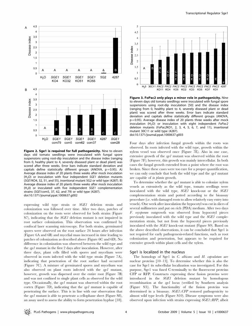
SGE1 is strictly required for pathogenicityTo assess the involvement of SGE1 and FoPAC2 in pathogenic-
ity, gene knock-out mutants were generated by homologous
recombination. Four independent SGE1 and eight independent
FoPAC2 knock-out mutants were obtained, with the deletions
confirmed by PCR and Southern analysis (Figure S1, S3 and S4).
The SGE1 deletion mutants were non-pathogenic on tomato in a
root dip bioassay and corroborated the severely reduced to non-
pathogenic phenotype of the insertional mutagenesis mutants
(Figure 2A). Re-introduction of the SGE1 gene in locus by
homologous recombination in a knock-out mutant (Figure S2
and S3) restored pathogenicity (Figure 2B), confirming that the loss
of pathogenicity was due to loss of SGE1. Deletion of FoPAC2 only
had a minor effect on pathogenicity. All knock-out mutants were
significantly different from the wild type in disease causing ability,
but seven out of the eight mutants were also significantly different
from the original insertion mutant (30C11) (Figure 3). This
indicates that FoPAC2 plays at most a minor role during infection
and that most probably additional defects in the 30C11 mutant
added to the reduced pathogenicity phenotype. Since the loss of
pathogenicity was complete upon deletion of SGE1, we focussed
on this gene for further analysis.
SGE1 is not required for vegetative growth, butquantitatively affects conidiation
Previously, we reported that vegetative growth of the insertional
mutants 5G2 and 101E1 are indistinguishable from that of the
wild type on various carbon sources [9]. To more fully analyze
potential metabolic defects of the sge1 mutant, we made use of
BIOLOG FF MicroPlates, in which each well contains a different
carbon source [16]. Also in this assay no reproducible differences
were observed between growth of the wild type and the SGE1
deletion mutant on 95 different carbon sources (Figure S5).
Microconidia and macroconidia generated in minimal or CMC
liquid medium were phenotypically indistinguishable from wild
type (Figure S6). However, the SGE1 deletion mutants produced
about 6-fold less microconidia compared to the wild type in both
media, and this phenotype was only partially restored in the SGE1
complementation mutants (Figure 4A). The conidial germination
rates were comparable to wild type (Figure 4B), indicating that,
although less microconidia are formed, they are fully viable. These
observations indicate that SGE1 is quantitatively involved in
conidiogenesis, but is not required for conidial fitness, overall
(colony) morphology, vegetative growth or carbon source
utilization.
SGE1 expression is upregulated during infection oftomato roots
C. albicans WOR1 and H. capsulatum RYP1 are 45-fold and 4-fold
upregulated upon transition from white to opaque cells in C.
albicans and from filamentous growth to yeast cells in H. capsulatum,
respectively [11,17]. To determine the relative expression levels of
SGE1 during saprophytic and parasitic growth quantitative PCR
was performed. Expression levels of SGE1 were determined both
in axenic culture and during tomato root infection at different time
points after inoculation and compared to the level of the
constitutively expressed elongation factor 1 alpha gene (EF-1a).
We found that SGE1 expression is upregulated 2- to 5-fold during
infection with maximal expression eight days after inoculation
(Figure 5).
SGE1 is not essential for colonization or penetration ofthe root surface
To determine at which stage during infection the SGE1 deletion
mutant is halted, tomato root colonization by the mutant was
visualized using fluorescent binocular and confocal laser scanning
microscopy. Tomato seedlings were infected with a GFP
Author Summary
Plant pathogenic fungi have evolved many ways to infecttheir hosts and can have devastating effects on commer-cial crop production. Dissecting their infection strategiesand understanding the molecular pathways involved inpathogenesis have been and continue to be the subject ofintensive research. New insights gained may help todevelop disease controlling strategies. Fusarium oxy-sporum has become a model for root invading, pathogenicfungi. This fungus attacks a wide range of plant speciesworldwide and the only effective disease control strategiesin the field are crop rotation and usage of resistant plantvarieties, if at all available. In the last decade, many geneshave been identified that play a role during pathogenesis,many of which are linked to general strain fitness. Only fewgenes have been identified that are required for patho-genesis but do not affect vegetative growth. This paperdescribes the characterization of such a gene. Its proteinproduct, Sge1, is conserved in the fungal kingdom andrepresents a new class of transcriptional regulatorsinvolved in morphological switching in dimorphic fungalpathogens. In F. oxysporum, the Sge1 protein is requiredfor parasitic growth and is associated with expression ofparasitic phase-specific genes. We suggest that thefunction of Sge1 is conserved in (plant) pathogenic fungi.
Transcriptional Regulator Sge1
PLoS Pathogens | www.plospathogens.org 2 October 2009 | Volume 5 | Issue 10 | e1000637

Figure 1. Representation of Sge1 homologs in fungi. A) Phylogenetic (neighbour joining, mid-point rooted) tree of Sge1 and FoPac2 withhomologs from C. albicans (Wor1 and CAWG_04607), S. pombe (SpGti1 and SpPac2), H. capsulatum (HcRyp1 and HCAG_05432), Magnaporthe grisea(MGG_08850 and MGG_06564), Aspergillus fumigatus (Afu6g04490 and Afu3g09640), Fusarium graminearum (FGSG_12164 and FGSG_10796), Botrytiscinerea (BC1G_11680 and BC1G_14615), Sclerotinia sclerotiorum (SS1G_00031 and SS1G_11157), Cryptococcus neoformans (CNAG_01983 andCNAG_05835), Ustilago maydis (UM05853 and UM06496) and Neurospora crassa (NCU06864), constructed using MacVector software. Only fullyaligned parts of the multiple sequence alignment were used (manual curation). Bootstrap percentages are provided only for branches receiving 60%or more support (1000 replications). Branch length reflects the extent of sequence divergence. B) Protein sequence alignment of the Sge1 N-terminalregion with H. capsulatum Ryp1, C. albicans Wor1 and S. pombe Gti1. Conserved residues are shaded black, similar residues are shaded grey. Thearrow head indicates the conserved threonine residue within the potential protein kinase A phosphorylation site and the asterisk indicates themutated residue in Sge1R66S. Predicted nucleur localization signals are boxed. The protein sequence alignment were created using VectorNTIsoftware.doi:10.1371/journal.ppat.1000637.g001
Transcriptional Regulator Sge1
PLoS Pathogens | www.plospathogens.org 3 October 2009 | Volume 5 | Issue 10 | e1000637
expressing wild type strain or SGE1 deletion strain and
colonization was followed over time. After two days, patches of
colonization on the roots were observed for both strains (Figure
S7), indicating that the SGE1 deletion mutant is not impaired in
root surface colonization. This observation was confirmed by
confocal laser scanning microscopy. For both strains, germinated
spores were observed on the root surface 24 hours after infection
(Figure 6A and 6B) and mycelial mass increased in time leading to
patches of colonization as described above (Figure 6C and 6D). No
difference in colonization was observed between the wild type and
the sge1 mutant in the first 2 days after inoculation. However, after
three days, plant cells filled with spores and mycelium were
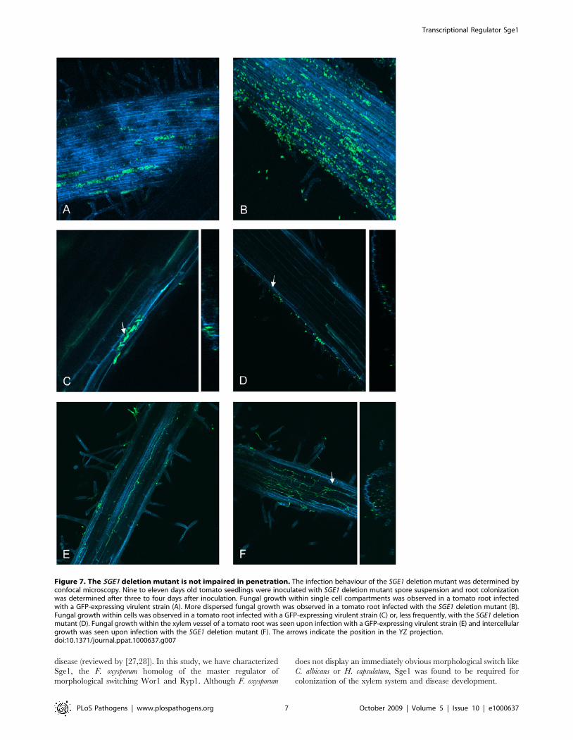
observed in roots infected with the wild type strain (Figure 7A),
indicating that penetration of the root surface had occurred
(Figure 7C). A mixture of germinated spores and mycelium was
also observed on plant roots infected with the sge1 mutant,
however, growth was dispersed over the entire root (Figure 7B)
and was not confined to single plant cells as observed for the wild
type. Occasionally, the sge1 mutant was observed within the root
cortex (Figure 7D), indicating that the sge1 mutant is capable of
penetrating the surface. This is in line with our observation that
the sge1 mutant is able to penetrate a cellophane sheet (Figure S8),
an assay used to assess the ability to form penetration hyphae [18].
Four days after infection fungal growth within the roots was
observed. In roots infected with the wild type, growth within the
xylem vessel was observed once (Figure 7E). Also in one case,
extensive growth of the sge1 mutant was observed within the root
(Figure 7F); however, this growth was mainly intercellular. In both
cases the fungal growth extended from a point where the root was
broken. Since these cases were too rare for a proper quantification,
we can only conclude that both the wild type and the sge1 mutant
are capable of in planta growth.
To determine whether the sge1 mutant is able to colonize xylem
vessels as extensively as the wild type, tomato seedlings were
inoculated with the wild type, SGE1 knock-out or the SGE1
complementation strain and potted according to the bioassay
procedure (i.e. with damaged roots to allow relatively easy entry into
vessels). One week after inoculation the hypocotyl was cut in slices of
several millimeters and put on rich (PDA) medium. After two days
F. oxysporum outgrowth was observed from hypocotyl pieces
previously inoculated with the wild type and the SGE1 comple-
mentation strain, but not from the hypocotyl pieces previously
inoculated with the SGE1 knock-out mutant (Figure S9). Based on
the above described observations, it can be concluded that Sge1 is
not required for early pathogenesis-related functions, such as root
colonization and penetration, but appears to be required for
extensive growth within plant cells and the xylem.
Sge1 is localized in the nucleusThe homologs of Sge1 in C. albicans and H. capsulatum are
nuclear proteins [10–13]. To determine whether this is also the
case for Sge1 its subcellular localization was investigated. For this
purpose, Sge1 was fused C-terminally to the fluorescent proteins
CFP or RFP. Constructs expressing these fusion proteins were
introduced in the SGE1 deletion mutant by homologous
recombination at the sge1 locus (verified by Southern analysis
(Figure S3)). The functionality of the fusion proteins was
determined in a bioassay. SGE1::CFP restored pathogenicity to
almost wild type levels (Figure S10). Disease symptoms were also
observed upon infection with strains expressing SGE1::RFP, albeit
Figure 2. Sge1 is required for full pathogenicity. Nine to elevendays old tomato seedlings were inoculated with fungal sporesuspensions using root-dip inoculation and the disease index (rangingfrom 0, healthy plant to 4, severely diseased plant or dead plant) wasscored after three weeks. Error bars indicate standard deviation andcapitals define statistically different groups (ANOVA, p = 0.95). A)Average disease index of 20 plants three weeks after mock inoculation(H2O) or inoculation with four independent SGE1 deletion mutants(SGE1KO4, 32, 51, and 55), insertional mutant 5G2 or wild type (4287). B)Average disease index of 20 plants three weeks after mock inoculation(H20) or inoculated with five independent SGE1 complementationstrains (SGE1com5, 37, 62, and 79) or wild type (4287).doi:10.1371/journal.ppat.1000637.g002
Figure 3. FoPac2 only plays a minor role in pathogenicity. Nineto eleven days old tomato seedlings were inoculated with fungal sporesuspensions using root-dip inoculation [50] and the disease index(ranging from 0, healthy plant to 4, severely diseased plant or deadplant) was scored after three weeks. Error bars indicate standarddeviation and capitals define statistically different groups (ANOVA,p = 0.95). Average disease index of 20 plants three weeks after mockinoculation (H2O) or inoculation with eight independent FoPac2deletion mutants (FoPac2KO1, 2, 3, 4, 5, 6, 7, and 11), insertionalmutant 30C11 or wild type (4287).doi:10.1371/journal.ppat.1000637.g003
Transcriptional Regulator Sge1
PLoS Pathogens | www.plospathogens.org 4 October 2009 | Volume 5 | Issue 10 | e1000637

severely reduced compared to the wild type infection (Figure S10).
Subcellular localization for both fusion proteins was similar in that
they are localized in the nucleus in both spores and hyphae
(Figure 8). Nuclear localization was verified by introduction of a
construct encoding histone H2B::GFP [19] into the SGE1::RFP
strain. Both fusion proteins localized to the same compartment
(Figure 8). These observations support the potential role of Sge1 as
a transcriptional regulator in F. oxysporum.
The putative Pka phosphorylation site is required forSge1 function
As described above, the Sge1 protein contains a potential Pka
phosphorylation site and this site is conserved in all Sge1 homologs
(Figure 1B). Replacement of the conserved threonine residue by an
alanine impaired S. pombe Gti1 function [14]. In an attempt to
identify Sge1 interacting partners using a yeast two-hybrid screen,
the SGE1 gene, fused to the portion of GAL4 encoding the Gal4
DNA binding domain, was introduced in Saccharamyces cerevisiae. To
our surprise, after numerous transformation attempts only one
colony was obtained. After re-isolation of the plasmid from this
colony followed by sequencing it turned out that a point mutation
in the SGE1 sequence (C196A) had occurred resulting in an amino
acid change from an arginine into a serine at position 66, altering
the Pka phosphorylation site (Figure 1). In contrast to wild type
SGE1, this mutant form of SGE1 could be easily re-transformed to
yeast. We concluded that SGE1 is normally toxic in S. cerevisiae and
that the R.S mutation leads to tolerance in this yeast. To
determine the effect of this mutation on the activity of Sge1 in Fol,
a construct expressing Sge1R66S was introduced in the SGE1
deletion mutant. Correct gene replacement was again confirmed
by PCR (Figure S11). All strains encoding the Sge1R66S protein
were non-pathogenic (Figure 9), suggesting that an intact Pka
phosphorylation site is required for Sge1 to function properly.
SGE1 regulates expression of effector genesThe transcriptional regulators Ryp1 and Wor1 regulate
expression of phase specific genes [11,20]. Since Sge1 shares
many features with Ryp1 and Wor1, we hypothesized that
expression of F. oxysporum genes specifically expressed during
infection could be altered. Examples of such genes are those
encoding effectors, small in planta secreted proteins, called Six
(Secreted in xylem) proteins in Fol. Recently, it was shown that F.
oxysporum secretes numerous Six proteins during infection [21]
(Houterman and Rep, unpublished). SIX1, encoding Avr3, is
strongly induced upon penetration of the root cortex and plays a
role during pathogenicity [22,23]. In addition, Six3 (Avr2) and
Six4 (Avr1) have been shown to play a role in virulence as well as
resistance protein recognition [24,25]. Since the SGE1 deletion
mutant does not show extensive in planta growth, precluding
assessment of SIX gene expression during root infection, we
incubated the mutant together with tomato cells in culture, a
situation known to induce SIX1 expression [22]. MSK8 tomato
cells were inoculated with a conidial suspension of the wild type or
Figure 4. Sge1 is involved in conidiogenesis. Microconidiaproduction was determined after five days of growth in minimalmedium and counted in a Burker-Turk haemocytometer. A) The averagenumber of microconidia of five independent experiments each with fivereplicates. B) The average germination rate of the isolated microconidiaon PDA medium after six hours of incubation at 25uC. Error barsindicate standard deviation and capitals define statistically differentgroups (ANOVA, p = 0.95).doi:10.1371/journal.ppat.1000637.g004
Figure 5. SGE1 is upregulated during infection of tomato roots.Nine to eleven days old tomato seedlings were inoculated with eitherwater or wild type Fol using root-dip inoculation. Roots were harvested4, 8, 12, and 16 days after inoculation followed by RNA isolation andcDNA synthesis. Quantitative PCR was used to determine the expressionlevels of the constitutively expressed gene EF-1a and SGE1 in axenicculture (minimal medium) and during infection. Relative expressionlevels were subsequently calculated using the comparative C(t) method.*, significantly different from the SGE1 expression level in axenic cultureat a 95% confidence interval.doi:10.1371/journal.ppat.1000637.g005
Transcriptional Regulator Sge1
PLoS Pathogens | www.plospathogens.org 5 October 2009 | Volume 5 | Issue 10 | e1000637

the SGE1 deletion mutant. After 24 h the cultures were harvested
and the presence of SIX gene transcripts was determined by RT-
PCR and compared to transcript levels in axenic cultures. As
expected, SIX1 gene expression was very low when the wild type
strain was grown in axenic culture and expression was strongly
upregulated upon incubation with MSK8 cells. A similar
expression pattern was observed for SIX2 (Figure 10). In contrast,
the SIX3 and SIX5 genes were expressed in the wild type strain in
axenic culture as well as in the presence of MSK8 cells (Figure 10).
Interestingly, in the sge1 mutant expression of all four SIX genes
was lost both in axenic culture and in the presence of MSK8 cells.
Expression was restored in the complemented strain (Figure 10).
These results show that expression of at least four SIX genes tested
is dependent on the presence of SGE1.
The SGE1 deletion mutant exhibits biocontrol activityBiocontrol activity has been reported to be dependent on
colonization of superficial cell layers, a capacity shared between
pathogenic and protective strains and has been reported to be
independent of competition for putative penetration sites or
nutrients [26]. Fusarium oxysporum f. sp. lycopersici exerts biocontrol
activity on flax when added in a 100-fold excess relative to a F.
oxysporum strain pathogenic towards flax. To determine whether
the sge1 mutant is still able to protect flax against wilt disease like its
parental strain, flax cv. viking was treated with either a pathogenic
isolate of F. oxysporum f. sp. lini (Foln3) alone or in a 1:100 ratio with
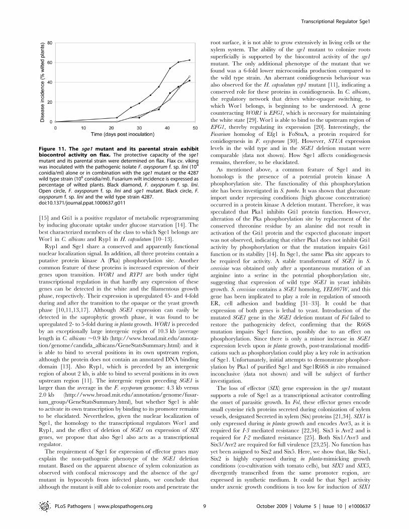
either the sge1 mutant or the wild type strain 4287. The first wilt
symptoms were observed 22 days after inoculation in the
treatment with Foln3 (Figure 11). In the Foln3/sge1 mutant and
Foln3/wild type combination treatments the first disease symp-
toms were delayed by 3 days. Disease symptoms were always
reduced in the Foln3/sge1 mutant and Foln3/wild type treatments
compared to the Foln3 treatment: 48 days post inoculation disease
symptoms were reduced by 27 and 33%, by the sge1 mutant and
the wild type, respectively (Figure 11). The ANOVA performed on
AUDPCs indicated that this difference was significant at the
probability of 91%. We conclude that the sge1 mutant is able to
protect flax against Fusarium wilt as well as the wild type strain,
suggesting that it can colonize roots efficiently.
Discussion
Fungi have various ways to adapt their morphology to the
environment, one example being dimorphism. Dimorphism is a
strategy frequently employed by fungal pathogens where this
developmental transition is correlated with the ability to cause
Figure 6. The SGE1 deletion mutant is not impaired in root colonization. The infection behaviour of the SGE1 deletion mutant wasdetermined by confocal microscopy. Nine to eleven days old tomato seedlings were inoculated with SGE1 deletion mutant spore suspension and rootcolonization was determined after one to two days after inoculation. Germinated spores were observed on a tomato root infected with a GFP-expressing virulent strain (A) or with the SGE1 deletion mutant (B). Patches of colonization were found on a tomato root infected with a GFP-expressing virulent strain (C) or with the SGE1 deletion mutant (D).doi:10.1371/journal.ppat.1000637.g006
Transcriptional Regulator Sge1
PLoS Pathogens | www.plospathogens.org 6 October 2009 | Volume 5 | Issue 10 | e1000637
disease (reviewed by [27,28]). In this study, we have characterized
Sge1, the F. oxysporum homolog of the master regulator of
morphological switching Wor1 and Ryp1. Although F. oxysporum
does not display an immediately obvious morphological switch like
C. albicans or H. capsulatum, Sge1 was found to be required for
colonization of the xylem system and disease development.
Figure 7. The SGE1 deletion mutant is not impaired in penetration. The infection behaviour of the SGE1 deletion mutant was determined byconfocal microscopy. Nine to eleven days old tomato seedlings were inoculated with SGE1 deletion mutant spore suspension and root colonizationwas determined after three to four days after inoculation. Fungal growth within single cell compartments was observed in a tomato root infectedwith a GFP-expressing virulent strain (A). More dispersed fungal growth was observed in a tomato root infected with the SGE1 deletion mutant (B).Fungal growth within cells was observed in a tomato root infected with a GFP-expressing virulent strain (C) or, less frequently, with the SGE1 deletionmutant (D). Fungal growth within the xylem vessel of a tomato root was seen upon infection with a GFP-expressing virulent strain (E) and intercellulargrowth was seen upon infection with the SGE1 deletion mutant (F). The arrows indicate the position in the YZ projection.doi:10.1371/journal.ppat.1000637.g007
Transcriptional Regulator Sge1
PLoS Pathogens | www.plospathogens.org 7 October 2009 | Volume 5 | Issue 10 | e1000637

Sge1, together with its homolog FoPac2, described in this work,
belong to a class of conserved fungal proteins. Both proteins have
apparent orthologs across the fungal kingdom and the N-terminal
domain of these proteins is always more conserved than the C-
terminus, which is generally rich in glutamine residues. Despite
their similarity, the SGE1 and FoPAC2 deletion mutants generated
in this study display different phenotypes, indicating that the
functions of these proteins are not redundant. The FoPac2 and
Sge1 homologs in S. pombe, Pac2 and Gti1, respectively, also have a
different function although both proteins are involved in
regulation of transition processes. Pac2 is a negative regulator of
sexual development, which is induced under nitrogen starvation
Figure 8. Sge1 is localized in the nucleus. The subcellular localization of the fluorescent proteins Sge1::RFP and Sge1::CFP was determined afterfive days of growth in minimal medium using a fluorescence microscope. A) Subcellular localization of Sge1::RFP. B) Subcellular localization ofH2B::GFP expressed in the Sge1::RFP background. C) Subcellular localization of Sge1::CFP.doi:10.1371/journal.ppat.1000637.g008
Figure 9. An intact Pka phosphorylation site in Sge1 isessential for pathogenicity. Nine to eleven days old tomatoseedlings were inoculated with fungal spore suspensions using root-dip inoculation and the disease index (ranging from 0, healthy plant to4, severely diseased plant or dead plant) was scored after three weeks.Error bars indicate standard deviation and capitals define statisticallydifferent groups (ANOVA, p = 0.95). Average disease index of 20 plantsthree weeks after mock inoculation (H2O) or inoculation with fiveindependent Sge1R66S complementation mutants (SGEPM6, 11, 19, 36and 54), an SGE1 complementation mutant (SGE1com28) or wild type(4287).doi:10.1371/journal.ppat.1000637.g009
Figure 10. SGE1 regulates expression of effector genes.Expression levels of SIX1, SIX2, SIX3, SIX5, SGE1 and the constitutivelyexpressed gene FEM1 were determined in the SGE1 deletion (#4 and#32) and complementation mutants (#5 and #28) and in the wild type(WT) grown in vitro and under in planta-mimicking conditions. TotalRNA was isolated from mycelium harvested after five days of growth inminimal medium (in vitro) and from mycelium grown for 24 h in BY-medium in the presence of one week old MSK8 cells (in planta-mimicking) followed by RT-PCR. 2, negative control. +, positive(genomic DNA) control.doi:10.1371/journal.ppat.1000637.g010
Transcriptional Regulator Sge1
PLoS Pathogens | www.plospathogens.org 8 October 2009 | Volume 5 | Issue 10 | e1000637
[15] and Gti1 is a positive regulator of metabolic reprogramming
by inducing gluconate uptake under glucose starvation [14]. The
best characterized members of the class to which Sge1 belongs are
Wor1 in C. albicans and Ryp1 in H. capsulatum [10–13].
Ryp1 and Sge1 share a conserved and apparently functional
nuclear localization signal. In addition, all three proteins contain a
putative protein kinase A (Pka) phosphorylation site. Another
common feature of these proteins is increased expression of their
genes upon transition. WOR1 and RYP1 are both under tight
transcriptional regulation in that hardly any expression of these
genes can be detected in the white and the filamentous growth
phase, respectively. Their expression is upregulated 45- and 4-fold
during and after the transition to the opaque or the yeast growth
phase [10,11,13,17]. Although SGE1 expression can easily be
detected in the saprophytic growth phase, it was found to be
upregulated 2- to 5-fold during in planta growth. WOR1 is preceded
by an exceptionally large intergenic region of 10.3 kb (average
length in C. albicans ,0.9 kb (http://www.broad.mit.edu/annota-
tion/genome/candida_albicans/GeneStatsSummary.html) and it
is able to bind to several positions in its own upstream region,
although the protein does not contain an annotated DNA binding
domain [13]. Also Ryp1, which is preceded by an intergenic
region of about 2 kb, is able to bind to several positions in its own
upstream region [11]. The intergenic region preceding SGE1 is
larger than the average in the F. oxysporum genome: 4.3 kb versus
2.0 kb (http://www.broad.mit.edu/annotation/genome/fusar-
ium_group/GeneStatsSummary.html), but whether Sge1 is able
to activate its own transcription by binding to its promoter remains
to be elucidated. Nevertheless, given the nuclear localization of
Sge1, the homology to the transcriptional regulators Wor1 and
Ryp1, and the effect of deletion of SGE1 on expression of SIX
genes, we propose that also Sge1 also acts as a transcriptional
regulator.
The requirement of Sge1 for expression of effector genes may
explain the non-pathogenic phenotype of the SGE1 deletion
mutant. Based on the apparent absence of xylem colonization as
observed with confocal microscopy and the absence of the sge1
mutant in hypocotyls from infected plants, we conclude that
although the mutant is still able to colonize roots and penetrate the
root surface, it is not able to grow extensively in living cells or the
xylem system. The ability of the sge1 mutant to colonize roots
superficially is supported by the biocontrol activity of the sge1
mutant. The only additional phenotype of the mutant that we
found was a 6-fold lower microconidia production compared to
the wild type strain. An aberrant conidiogenesis behaviour was
also observed for the H. capsulatum ryp1 mutant [11], indicating a
conserved role for these proteins in conidiogenesis. In C. albicans,
the regulatory network that drives white-opaque switching, to
which Wor1 belongs, is beginning to be understood. A gene
counteracting WOR1 is EFG1, which is necessary for maintaining
the white state [29]. Wor1 is able to bind to the upstream region of
EFG1, thereby regulating its expression [20]. Interestingly, the
Fusarium homolog of Efg1 is FoStuA, a protein required for
conidiogenesis in F. oxysporum [30]. However, STUA expression
levels in the wild type and in the SGE1 deletion mutant were
comparable (data not shown). How Sge1 affects conidiogenesis
remains, therefore, to be elucidated.
As mentioned above, a common feature of Sge1 and its
homologs is the presence of a potential protein kinase A
phosphorylation site. The functionality of this phosphorylation
site has been investigated in S. pombe. It was shown that gluconate
import under repressing conditions (high glucose concentration)
occurred in a protein kinase A deletion mutant. Therefore, it was
speculated that Pka1 inhibits Gti1 protein function. However,
alteration of the Pka phosphorylation site by replacement of the
conserved threonine residue by an alanine did not result in
activation of the Gti1 protein and the expected gluconate import
was not observed, indicating that either Pka1 does not inhibit Gti1
activity by phosphorylation or that the mutation impairs Gti1
function or its stability [14]. In Sge1, the same Pka site appears to
be required for activity. A stable transformant of SGE1 in S.
cerevisiae was obtained only after a spontaneous mutation of an
arginine into a serine in the potential phosphorylation site,
suggesting that expression of wild type SGE1 in yeast inhibits
growth. S. cerevisiae contains a SGE1 homolog, YEL007W, and this
gene has been implicated to play a role in regulation of smooth
ER, cell adhesion and budding [31–33]. It could be that
expression of both genes is lethal to yeast. Introduction of the
mutated SGE1 gene in the SGE1 deletion mutant of Fol failed to
restore the pathogenicity defect, confirming that the R66S
mutation impairs Sge1 function, possibly due to an effect on
phosphorylation. Since there is only a minor increase in SGE1
expression levels upon in planta growth, post-translational modifi-
cations such as phosphorylation could play a key role in activation
of Sge1. Unfortunately, initial attempts to demonstrate phosphor-
ylation by Pka1 of purified Sge1 and Sge1R66S in vitro remained
inconclusive (data not shown) and will be subject of further
investigation.
The loss of effector (SIX) gene expression in the sge1 mutant
supports a role of Sge1 as a transcriptional activator controlling
the onset of parasitic growth. In Fol, these effector genes encode
small cysteine rich proteins secreted during colonization of xylem
vessels, designated Secreted in xylem (Six) proteins [21,34]. SIX1 is
only expressed during in planta growth and encodes Avr3, as it is
required for I-3 mediated resistance [22,34]. Six3 is Avr2 and is
required for I-2 mediated resistance [25]. Both Six1/Avr3 and
Six3/Avr2 are required for full virulence [23,25]. No function has
yet been assigned to Six2 and Six5. Here, we show that, like Six1,
Six2 is highly expressed during in planta-mimicking growth
conditions (co-cultivation with tomato cells), but SIX3 and SIX5,
divergently transcribed from the same promoter region, are
expressed in synthetic medium. It could be that Sge1 activity
under axenic growth conditions is too low for induction of SIX1
Figure 11. The sge1 mutant and its parental strain exhibitbiocontrol activity on flax. The protective capacity of the sge1mutant and its parental strain were determined on flax. Flax cv. vikingwas inoculated with the pathogenic isolate F. oxysporum f. sp. lini (104
conidia/ml) alone or in combination with the sge1 mutant or the 4287wild type strain (106 conidia/ml). Fusarium wilt incidence is expressed aspercentage of wilted plants. Black diamond, F. oxysporum f. sp. lini.Open circle, F. oxysporum f. sp. lini and sge1 mutant. Black circle, F.oxysporum f. sp. lini and the wild type strain 4287.doi:10.1371/journal.ppat.1000637.g011
Transcriptional Regulator Sge1
PLoS Pathogens | www.plospathogens.org 9 October 2009 | Volume 5 | Issue 10 | e1000637

and SIX2, but high enough to induce SIX3 and SIX5 expression.
The increased SGE1 expression levels during in planta growth may
then lead to expression of all SIX genes. Currently, a SGE1 over-
expression mutant is being generated in order to determine
whether increased SGE1 expression alone can lead to expression of
all SIX genes under axenic growth conditions. At the moment, it is
not known whether Sge1 influences the expression of the SIX
genes directly or indirectly, for instance through involvement of
other (transcription) factors, nor how SGE1 expression itself is
regulated. Preliminary promoter analysis of the SIX genes revealed
a potential common motif (unpublished data). The role of this
motif in SIX gene expression and the question whether Sge1 is able
to bind DNA and the SIX gene promoter regions in particular
remain to be elucidated.
All the genes tested in the SGE1 deletion mutant (SIX1, SIX2,
SIX3, and SIX5) were found to be dependent on Sge1 for their
expression, even during growth in synthetic medium. Although
deletion of individual SIX genes (SIX1 or SIX3) only leads to a
minor reduction in pathogenicity, the loss of expression of all SIX
genes in the sge1 mutant may be the primary cause of the complete
non-pathogenic phenotype, due to the inability to suppress host
defence responses. Still, loss of pathogenicity upon deletion of
SGE1 may not be due to loss of production of effector proteins
only. The majority of pathogenicity genes found in F. oxysporum
have pleiotropic functions [35,36]. It is therefore unlikely that
expression of these genes is altered in the sge1 mutant, since this
mutant did not display growth defects other than a minor effect in
conidiogenesis. One described F. oxysporum mutant that is only
disturbed in pathogenicity is the fow2 mutant [37]. Expression of
FOW2 nor that of two other pathogenicity genes, the protein
kinase gene SNF1 [38] and velvet homolog gene FOXG_00016
[39], is altered in the SGE1 deletion mutant (unpublished data).
However, preliminary results suggest that the expression of the
FTF1 transcription factor gene, implicated to be involved in
pathogenicity [40], seems to be altered in the sge1 mutant
(unpublished data). In addition, secondary metabolite profiling
revealed that the sge1 mutant is reduced in the production of
fusaric acid, beauvericin and a number of unknown metabolites
compared to the wild type (U. Thrane and C.B. Michielse,
unpublished data). High concentrations of fusaric acid may
contribute to pathogenicity by reducing host resistance [41,42].
Thus, processes implicated in pathogenesis other than SIX gene
expression are also altered in the sge1 mutant, supporting the
hypothesis that Sge1 plays a central role during parasitic growth.
Deletion of the Sge1 homolog in Botrytis cinerea, BC1G_11680, also
leads to a severely reduced pathogenicity phenotype (C.B.
Michielse and P. Tudzynski, unpublished data), indicating that
the role of Sge1-like proteins in (plant) pathogenic fungi might be
conserved.
The regulation of expression of phase specific genes (SIX1,
SIX2), and genes involved in virulence (SIX1, SIX3) by Sge1
resembles the functions of Wor1 in C. albicans and Ryp1 in H.
capsulatum. Wor1 has been shown to bind directly to the upstream
region of target genes [20]. Interestingly, these target genes all
have a large upstream region, indicating that large upstream
regions might be a prerequisite for Wor1 binding. Whether this is
a common feature shared between Wor1 and Sge1 is still
unknown. Future efforts include the identification of additional
genes regulated by Sge1 and, eventually, to unravel the network of
transcriptional regulators involved in activating the pathogenicity
program in F. oxysporum. This will also reveal whether Sge1 indeed
plays a master role in this network, like its homologs in dimorphic
fungi. Given the striking conservation of Sge1 across fungi, we
expect that many features of this network will turn out to be
similar in pathogens of both plants and animals. Obviously, since
Sge1 homologs are also present in non-pathogenic fungi, this
regulatory network must serve a fundamental morphological or
physiological switch function and pathogens would have adopted
this network for adapting to the host environment.
Materials and Methods
Strains, plant material and culture conditionsFusarium oxysporum f. sp. lycopersici strain 4287 (race 2;
FGSC9935) was used as the parent strain for fungal transforma-
tion. It was stored as a monoconidial culture at 280uC and
revitalized on potato dextrose agar (PDA, Difco) at 25uC.
Agrobacterium tumefaciens EHA105 [43] was used for Agrobacterium-
mediated transformation of F. oxysporum and was grown in 2YT
medium [44] containing 20 mg/ml rifampicin at 28uC. Introduc-
tion of the plasmids into the Agrobacterium strain was performed as
described by Mattanovich et al [45]. Escherichia coli DH5 alpha
(Invitrogen) was used for construction, propagation, and amplifi-
cation of the plasmids and was grown in LB medium at 37uCcontaining either 100 mg/ml ampicillin or 50 mg/ml kanamycin
depending on the resistance marker of the plasmid used. Plant line
Moneymaker ss590 (Gebr. Eveleens b.v., The Netherlands) was
used to assess pathogenicity of F. oxysporum strains and transfor-
mants. Biocontrol assays were performed on flax (Linum usitatissi-
mum) cv. viking using F. oxysporum f. sp. lini (Foln3, MYA-1201)
isolated from a diseased flax plant in French Britany as pathogenic
strain.
Construction of gene replacement and complementationconstructs
To generate the SGE1 disruption construct, pSGE1KO, PCR
was performed using a BAC clone containing the SGE1 gene as a
template with primer combination FP878–FP879 and FP880–
FP881 in order to amplify a 901 bp HindIII fragment correspond-
ing to the upstream region and a 1032 bp KnpI fragment
corresponding to the downstream region, respectively (Table S1).
The fragments were sequentially cloned into pPK2hphgfp [9] and
proper orientation of the inserts was checked by PCR.
The SGE1 complementation construct, pSGE1com, was gener-
ated by cloning a 2234 bp HindIII fragment containing the SGE1
ORF including 901 bp upstream and 341 bp downstream
sequences into pUC19, resulting in pUC19SGE1. The HindIII
SGE1 fragment was subsequently transferred from pUC19SGE1
into pRW1p [24], resulting in pSGE1com. The SGE1 comple-
mentation construct carrying a point mutation in the ORF of
SGE1, pSGE1comPM, was generated by replacing a 465 bp SacII/
BglII SGE1 fragment in pSGE1com by a 465 bp SacII/BglII
fragment isolated from the yeast-two-hybrid bait vector pASS-
GE1PM. This vector was re-isolated from Saccharomyces cerevisiae
after previously being transformed with pASSGE1. The latter
vector was generated by cloning an NcoI-EcoRI 1011 bp PCR
product obtained with the primers FP1484 and FP1485 corre-
sponding to the SGE1 ORF in pAS2.1 (Clontech).
To generate the FoPAC2 gene disruption construct, a 1037 bp
upstream fragment and a 749 bp downstream fragment was
amplified from genomic DNA by PCR with the primer pairs
FP1796–FP1797 and FP1798–FP1799, respectively. The PCR
products were cloned into pGEM-T Easy (Promega) and,
subsequently, the KpnI/PacI upstream and the AscI/HindIII
downstream fragment were sequentially cloned in pRW2h [24].
The Sge1-fluorescent protein fusion constructs pSGE1::CFP
and pSGE1::RFP were generated in a multi-step approach. First,
pUC19SGE1 was amplified by PCR with the primers FP1120 and
Transcriptional Regulator Sge1
PLoS Pathogens | www.plospathogens.org 10 October 2009 | Volume 5 | Issue 10 | e1000637

FP1121 flanked at the 59 end by an ApaI and a SpeI restriction site,
respectively. The amplified product was digested with ApaI and
religated, resulting in pUC19SGE1as, containing the SGE1 ORF
with the ApaI and SpeI preceding the SGE1 stop codon. Secondly,
a CFP [46] and a mRFP [47] fragment were generated by PCR
using primer pair FP1122–FP1123 and FP1120–FP1121, respec-
tively. The CFP and RFP PCR products were digested with ApaI
and SpeI and directionally cloned in pUC19SGE1as, resulting in
pUCSGE1::CFP and pUCSGE1::RFP, respectively. Finally, a
HindIII fragment corresponding to each fusion cassette was
cloned into HindIII digested pRW1p and proper orientation
of the fragments was checked by PCR. The gpdAH2B::GFP
expression cassette was isolated as a XbaI/NheI fragment from
pH2B::GFP (kind gift from Dr. Ram, Leiden, The Netherlands)
and cloned into XbaI/NheI digested pRW2h [24] to generate
pRWH2B::GFP.
Fungal transformation and disease assayAgrobacterium-mediated transformation of F. oxysporum f. sp.
lycopersici was performed as described by Mullins and Chen [48]
with minor adjustments [49]. Depending on the selection marker
used, transformants were selected on Czapek Dox agar (CDA,
Oxoid) containing 100 mg/ml Hygromycin (Duchefa) or on CDA
containing 0.1 M TrisHCl pH 8 and 100 mg/ml Zeocin (Invivo-
Gen).
Plant infection was performed using 9 to 11 days old seedlings
(Moneymaker ss590), following the root-dip inoculation method
[50]. Disease index was scored and statistical analysis performed as
described earlier [9].
Analysis of conidiogenesis, conidia germination, carbonsource utilization and cellophane penetration
For quantization of microconidia production, five indepen-
dent experiments were performed, each with five replicates.
Microconidia were harvested after five days of growth in 50 ml
minimal medium (3% sucrose, 10 mM KNO3 and 0.17% yeast
nitrogen base without amino acids and ammonia) and 106
spores were used to inoculate 100 ml minimal medium. After
five days the microconidia produced were harvested and
counted in a Burker-Turk haemocytometer. The isolated
conidia were also used to determine germination rates. To this
end 600 spores were added into a 6-wells plate with each well
containing 250 ml PDA and incubated overnight at 4uC, then
transferred to 25uC and germinated conidia were counted after
six hours of incubation. Macroconidia development was
analyzed in liquid carboxymethyl cellulose medium as described
by Ohara and Tsuge [30]. Conidia were fixed in 0.4% p-
formaldehyde and stained with Hoechst 33342 (250 mg/ml) and
calcofluor white (25 mg/ml) to visualize nuclei and cell walls,
respectively. Stained cells were observed with a BX50
fluorescence microscope and a U-MWU filter (Olympus).
Carbon-source utilization was tested in a BIOLOG FF
MicroPlate (BIOLOG). A conidial suspension (104 conidia in
150 ml) of each strain was inoculated in each well of the plate and
incubated at 25uC. The absorbance of each well at 600 nm was
measured with a microtiter plate reader (Packerd Spectra Count)
after 1, 2, 3 and 4 days of incubation.
The cellophane penetration assay was performed as described
[18], with minor adjustments. CDA was used as basal medium in
the assay and was inoculated with 105 conidia. After incubation
of 5 days at 25uC, the cellophane was removed and after a
subsequent incubation of 2 days at 25uC fungal growth was
scored.
Southern analysisGenomic DNA was isolated as described by Kolar et al [51] with
minor adjustments [9]. For Southern analysis, 10 mg genomic
DNA of each transformant was digested with Acc65I or BglII for
SGE1 or FoPAC2 transformants, respectively, and incubated
overnight at 37uC. The samples were loaded on a 1% 0.56Tris-borate/EDTA gel and run for 18 hours at 45 V. The
digested DNA was transferred to Hybond-N+ (Amersham
Pharmacia) as described by Sambrook et al [44]. The probes used
for Southern analysis are: (1) a 432 bp SGE1 upstream fragment
obtained by PCR with primers FP842 and FP1174 (Table S1) and
(2) a 488 bp FoPAC2 downstream fragment obtained by PCR with
primers FP2198 and FP 2199. The DecaLabelTM DNA Labelling
Kit (Fermentas) was used to label probes with [a-32P]dATP.
Hybridization was done overnight at 65uC in 0.5 M sodium
phosphate buffer, pH 7.2, containing 7% SDS and 1 mM EDTA.
Blots were washed with 0.26 SSC, 0.1% SDS. Hybridization
signals were visualized by phosphorimaging (Molecular Dynam-
ics).
RT and quantitative PCRSamples for expression analysis were obtained by adding 0,5 ml
of 107 conidia/ml of wild type, SGE1 deletion or SGE1
complementation strain to 4,5 ml of a one week old MSK8 cell
culture grown in BY-medium [52]. After 24 hours of incubation at
22uC, the material was harvested, washed two times with sterile
water and freeze-dried. Total RNA was isolated using Trizol
(Invitrogen) and prior to cDNA synthesis the RNA was treated
with DnaseI (Fermentas). First strand cDNA was synthesized with
M-MuLV reverse transcriptase following the instruction of the
manufacturer (Fermentas). Supertaq (SphaeroQ), 1 ml of the
cDNA reaction and primers FP1999/FP2000, FP998/FP1001,
FP962/FP963, FP1993/FP1994, FP2131/FP2132 and FP157/
FP158 (Table S1) to detect SIX1 (CAE55879), SIX2 (CAE55868),
SIX3 (CAJ83999), SIX5 (ACN87967), SGE1 (FOXG_10510) and
FEM1 (AAL47843) transcripts, respectively, were used in the RT-
PCR.
Quantitative PCR (qPCR) was performed with Platinum SYBR
Green qPCR (Invitrogen) using a 7500 Realtime PCR System
(Applied Biosystems). To quantify mRNA levels of SGE1 and of
the constitutively expressed EF-1a gene, we used primers FP2131/
FP2132 and FP2029/FP2030, respectively (Table S1). EF-1a was
used to calculate the relative expression levels of SGE1 in axenic
and MSK8 cultures infected with F. oxysporum wild type, SGE1
deletion or SGE1 complementation mutants. Real time PCR
primer efficiencies were calculated using LinRegPCR [53] and
relative expression levels were calculated according to the
comparative C(t) method [54].
Microscopic analysisSge1::mRFP, Sge1::CFP and H2B::GFP fusion proteins were
visualized using a BX50 fluorescence microscope with the
appropriate excitation and emission filters for CFP, mRFP and
GFP (Olympus). For this purpose, 10 ml of five days old minimal
medium cultures was spotted onto glass slides.
A Nikon A1 microscope was used to monitor tomato root
infection by the wild type and SGE1 deletion mutant. Excitation
was provided for the GFP signal with an Argon (488 nm) laser
(emission: 405–455 nm) and for the root auto-fluorescence with an
UV diode (405 nm) laser (emission: 420–470 nm). Images were
line-sequential scanned (2 mm slices, 102461024 pixels) using
water objective plan fluor 206 Imm, 0.75 NA. Pictures were
analyzed with the Nikon NIS and ImageJ software. For
preparation of the slides, 9 days old tomato seedlings were
Transcriptional Regulator Sge1
PLoS Pathogens | www.plospathogens.org 11 October 2009 | Volume 5 | Issue 10 | e1000637

carefully removed from the potting soil, rinsed with water and
intact roots were inoculated with 20 ml tap water containing 107
conidia/ml and incubated for 4 days at room temperature in a
petridish. The roots were rinsed with water, cut from the
hypocotyl, placed in a drop of water on a glass slide and covered
with a cover glass. A bridge mounted on the glass slide prevented
squashing of the root material.
Inoculum preparation and biocontrol assayInocula were prepared as described [55], except that minimal
liquid medium [56] in which sucrose was replaced by glucose
(5 g/l) and sodium nitrate by ammonium tartrate (1 g/l) was used
instead of Malt medium. A heat treated (100uC for 1 h) silty-loam
soil from Dijon (35.1% clay, 47% loam, 15.1% sand, and 1.22%
organic C [pH = 6.9]) was added to module trays containing 96
wells each of 50 ml. To prevent contamination between
treatments, only every second row of wells was filled with soil.
The soil in each well was inoculated with 5 mL of conidia
suspension. The concentration of the conidia suspensions were
adjusted to obtain the following inoculum densities: 16104
conidia ml21 of soil for the pathogenic strain Foln3 and 16106
conidia ml21 of soil for the two others strains. The soil surface was
covered with a thin layer of calcinated clay granules (Oil Dri
Chem-Sorb, Brenntag Bourgogne, Montchanin, France) and
three seeds of flax, cv. viking, were sown in each pot. A thin layer
of Chem-Sorb was used to cover the seeds. Plants were grown in a
growth chamber in the first 2 weeks; the growing conditions were
8 h 15uC N/16 h 17uC D, with a light intensity of
33 mE m22 s21. The plants were thinned to one plant per pot,
and from week 3 the temperature was kept at 22uC N/25uC D.
There were three replicates of 16 individual plants per treatment,
randomly arranged, and the experiment was replicated. Plants
were watered every day, and once a week water was replaced by a
500-fold dilution of a commercial nutrient stock solution
(‘‘Hydrodrokani AO’’, Hydro Agri, Nanterre, France). Plants
showing characteristic symptoms of yellowing were recorded
twice a week and removed. To compare the ability of strains to
induce disease or, on the contrary, to protect the plant against
wilt, ANOVA was performed on Area under the Disease Progress
Curves followed by Newman and Keuls test at the probability of
95%.
Supporting Information
Table S1 Primers used in this study.
Found at: doi:10.1371/journal.ppat.1000637.s001 (0.01 MB PDF)
Figure S1 Analysis of transformants deleted for SGE1. A knock-
out construct containing a hygromycin-GFP expression cassette
flanked by 901 bp up- and 1032 bp downstream sequences of
SGE1 was introduced in the wild type strain by Agrobacterium-
mediated transformation. A) Schematic representation of the
knock-out strategy for SGE1 drawn to scale. The arrow heads
indicate the positions of the original T-DNA insertions. The small
arrows represent the primers used to check homologous
recombination (a and b) and the absence of the open reading
frame (c and d). B) Verification of homologous recombination by
PCR using primers a and b. C) Verification of the absence of the
SGE1 open reading frame by PCR using primers c and d. The
1 kb DNA ladder of Fermentas (www.fermentas.com) is used as a
marker. 2, negative control. +, positive control (genomic DNA).
The figures are composed from different parts of an ethidium
bromide gel, which results in minor colour differences.
Found at: doi:10.1371/journal.ppat.1000637.s002 (0.41 MB PDF)
Figure S2 Analysis of transformants complemented with SGE1.
A complementation construct containing a phleomycin expression
cassette and the SGE1 gene including 901 bp up- and 341 bp
downstream sequences was introduced in the SGE1 knock-out
mutant #32 by Agrobacterium-mediated transformation. A) Sche-
matic representation of the complementation strategy for SGE1
drawn to scale. The small arrows represent the primers used to
check the presence of the open reading frame (c and d). B)
Verification of the presence of the SGE1 ORF by PCR using
primers c and d. The 1 kb DNA ladder of Fermentas (www.
fermentas.com) is used as a marker. 2, negative control.
Found at: doi:10.1371/journal.ppat.1000637.s003 (0.09 MB PDF)
Figure S3 Southern analysis of the SGE1 deletion and
complementation mutants. Southern analysis was performed to
verify correct homologous recombination at the SGE1 locus in the
SGE1 disruptants and complemented strains. To this end,
chromosomal DNA of the various mutants was digested with
Acc65I, blotted and hybridized with a 432 bp probe corresponding
to the SGE1 promoter. The SGE1 locus of the wild type strain is
visible as a 1.6 kb fragment corresponding to the SGE1 upstream
region and to the 59 part of the SGE1 ORF. In the 5G2 insertional
mutagenesis mutant, the SGE1 ORF is disrupted due to a T-DNA
insertion (see Figure S1A). As a result the 1.6 kb fragment
observed in a wild type situation is replaced by a 1.8 kb fragment
corresponding to a part of the SGE1 promoter region and the
hygromycin expression cassette present on the T-DNA. In the
SGE1 disruption mutants introduction of the gene replacement
cassette by homologous recombination led to the expected
replacement of the 1.6 kb fragment with a fragment containing
part of the SGE1 upstream region and the gene disruption cassette
which should be larger than 4.9 kb. Introduction of the SGE1
complementation cassette, including the SGE1::FP fusion protein
complementation cassettes, by homologous recombination re-
stored the wild type SGE1 locus. WT, wild type. 5G2, insertional
mutagenesis mutant 5G2. KO, SGE1 knock-out mutants. com,
SGE1 complementation mutants. CC, SGE1::CFP complementa-
tion mutants. RC, SGE1::RFP complementation mutants.
Found at: doi:10.1371/journal.ppat.1000637.s004 (0.05 MB PDF)
Figure S4 Analysis of transformants deleted for FoPAC2. A
knock-out construct containing a hygromycin expression cassette
flanked by 1037 bp up- and 749 bp downstream sequences of
FoPAC2 was introduced in the wild type strain by Agrobacterium-
mediated transformation. A) Schematic representation of the
knock-out strategy for FoPAC2 drawn to scale. The arrow head
indicates the position of the original T-DNA insertion. The small
arrows represents the primers used to check homologous
recombination (a and b) and the absence of the open reading
frame (c and d). B) Verification of the absence of the FoPAC2 open
reading frame by PCR using primers c and d. C) Verification of
homologous recombination by PCR using primers a and b. D)
Southern analysis of the FoPAC2 deletion mutants. Chromosomal
DNA was digested with BglII, blotted and hybridized with a
488 bp probe corresponding to upstream region. The FoPAC2
locus of the wild type strain is visible as a 7.0 kb fragment. In the
30C11 insertional mutagenesis mutant, the FoPAC2 ORF is
disrupted due to a T-DNA insertion (see Figure S4A). As a result
the 7.0 kb fragment observed in a wild type situation is replaced by
a 12.3 kb fragment corresponding to a part of the FoPAC2
upstream region and the hygromycin expression cassette present
on the T-DNA. In the FoPAC2 disruption mutants introduction of
the gene replacement cassette by homologous recombination led
to the expected replacement of the 7.0 kb fragment with a 11.4 kb
fragment containing part of the FoPAC2 upstream region and the
Transcriptional Regulator Sge1
PLoS Pathogens | www.plospathogens.org 12 October 2009 | Volume 5 | Issue 10 | e1000637
gene disruption cassette. The 1 kb DNA ladder of Fermentas
(www.fermentas.com) is used as a marker. 2, negative control. +,
positive control (genomic DNA). Panels B and C are composed of
different parts of an ethidium bromide gel, which results in minor
colour differences.
Found at: doi:10.1371/journal.ppat.1000637.s005 (0.15 MB PDF)
Figure S5 The SGE1 deletion mutant is not impaired in carbon
source utilization. To analyze carbon utilization of the sge1
mutant, BIOLOG FF MicroPlates containing in each well a
different carbon source were used. A conidial suspension (104
conidia in 150 ml) of the wild type or the SGE1 deletion stain was
inoculated in each well and incubated at 25uC. The absorbance of
each well at 600 nm was measured with a microtiter plate reader
(Packerd Spectra Count) after 4 days of incubation. The ratios
were calculated by dividing the measured values of the mutant
strain by those of the wild type strain. Only values higher than 1.5
or lower than 0.5 were marked (boxed) as carbon sources on which
the sge1 mutant appeared to display a different growth rate than
the wild type strain in this experiment. An additional plate assay
containing these carbon sources showed that these differences
were not reproducible (data not shown).
Found at: doi:10.1371/journal.ppat.1000637.s006 (0.40 MB TIF)
Figure S6 Micro- and macroconidia of the SGE1 deletion
mutant are morphologically indistinguishable from wild type.
Micro- and macroconidia development was analyzed in liquid
carboxymethyl cellulose medium. Conidia were fixed in 0.4% p-
formaldehyde and stained with Hoechst 33342 (250 mg/ml) and
calcofluor white (25 mg/ml) to visualize nuclei and cell walls,
respectively. The left panel depicts a phase contrast recording and
the right panel depicts a UV-exposed recording of conidia of the
sge1 mutant.
Found at: doi:10.1371/journal.ppat.1000637.s007 (1.38 MB PDF)
Figure S7 SGE1 is not essential for superficial root colonization.
The root colonization behaviour of the SGE1 deletion mutant was
determined by binocular microscopy. Nine to eleven days old
tomato seedlings were inoculated with wild type or an SGE1
deletion mutant spore suspension and root colonization was
determined after two to five days after inoculation. Patches of
colonization were already visible after three days. The left panel
depicts phase contrast recordings of a GFP-expressing virulent
strain (38H3) and of the SGE1 deletion mutant. The right panel
depicts UV-exposed recordings.
Found at: doi:10.1371/journal.ppat.1000637.s008 (0.82 MB PDF)
Figure S8 The SGE1 mutant is not impaired in cellophane
penetration. The capacity of the SGE1 deletion strain to penetrate
cellophane was determined using a cellophane penetration assay.
CDA medium covered by a cellophane sheet was inoculated with a
drop containing 105 conidia. After incubation of 5 days at 25uC(left panel), the cellophane was removed and after a subsequent
incubation of 2 days at 25uC fungal growth of both the wild type
and the SGE1 deletion mutant was clearly observed (right panel).
Found at: doi:10.1371/journal.ppat.1000637.s009 (0.25 MB PDF)
Figure S9 The sge1 mutant is impaired in extensive in planta
growth. To determine whether the SGE1 deletion mutant is able to
growth within xylem vessels, tomato seedlings were inoculated
with the wild type, SGE1 knock-out or the SGE1 complementation
strain and potted into soil according to the bioassay procedure.
One week after inoculation the hypocotyl was cut in slices of
several millimeters which were placed on rich (PDA) medium. F.
oxysporum outgrowth was observed from the hypocotyl pieces
previously inoculated with the wild type and the SGE1
complementation strain, but not from the hypocotyl pieces
previously inoculated with the SGE1 knock-out mutant.
Found at: doi:10.1371/journal.ppat.1000637.s010 (0.26 MB PDF)
Figure S10 Pathogenicity is partially restored in SGE1::FP
complementation strains. Nine to eleven days old tomato seedlings
were inoculated with fungal spore suspensions following the root-
dip inoculation method and the disease index (ranging from 0
healthy plant to 4 severely diseased plant/dead plant) was scored
after three weeks. Average disease index of 20 plants three weeks
after mock inoculation (H2O) or inoculation with a SGE1 deletion
mutant (SGE1KO32), SGE1 (SGE1com28), SGE1::CFP (SGE1-
comCC4, 5, and 10) and SGE1::RFP (SGEcomRC2, 6, and 11)
complementation mutants or wild type (4287). Error bars indicate
standard deviation and capitals define statistically different groups
(ANOVA, p = 0.95).
Found at: doi:10.1371/journal.ppat.1000637.s011 (0.03 MB PDF)
Figure S11 Analysis of transformants complemented with
SGE1PM. A complementation construct containing a phleomycin
expression cassette and the SGE1PM gene, encoding Sge1R66S,
including 901 bp up- and 341 bp downstream sequences, was
introduced in the SGE1 knock-out mutant #32 by Agrobacterium-
mediated transformation. A) Verification of the presence of the
SGE1 ORF by PCR using primers c and d (see Figure S1A). B)
Verification of homologous recombination by PCR using primers
a and b (see Figure S2). The 1 kb DNA ladder of Fermentas (www.
fermentas.com) is used as a marker. 2, negative control. Deletion
mutant SGE1KO32 and complementation mutant SGE1com28
were used as a negative and positive control, respectively.
Found at: doi:10.1371/journal.ppat.1000637.s012 (0.34 MB PDF)
Acknowledgments
We thank Antonio Di Pietro for providing F. oxysporum strain 4287, Arthur
Ram for the vector pH2B::GFP, Wilfried Jonkers for technical assistance,
Ben Cornelissen for critically reading this manuscript and Ulf Thrane for
metabolic profile analysis. Furthermore, we thank Harold Lemereis, Thijs
Hendrix, and Ludek Tikovsky for managing the plant growth facilities and
their assistance with bioassays.
Author Contributions
Conceived and designed the experiments: CBM RvW CO CA. Performed
the experiments: CBM RvW LR EMMM SB CO. Analyzed the data:
CBM RvW LR CO. Wrote the paper: CBM. Originated research leading
up to this paper and provided guidance and review: MR.
References
1. Gordon TR, Martyn RD (1997) The evolutionary biology of Fusarium oxysporum.
Ann Rev Phytopathol 35: 111–128.
2. Armstrong GM, Armstrong JK (1981) Formae speciales and races of Fusarium
oxysporum and Alternaria alternata. In: Cook R, ed (1981) Fusarium: Diseases, Biology
and Taxonomy Penn State University Press. pp 391–399.
3. Brayford D (1996) Fusarium oxysporum f. sp. radicis-lycopersici, IMI descriptions of
fungi and bacteria no. 1270. Mycopathologia 133: 61–63.
4. Ploetz RC (1990) Popolation biology of Fusarium oxysporum f. sp. cubense. In:
Ploetz RC, ed (1990) Fusarium wilt of banana. St. Paul, Minnesota: APS Press.
pp 63–76.
5. Albisetti M, Lauener RP, Gungor T, Schar G, Niggli FK, et al. (2004)
Disseminated Fusarium oxysporum infection in hemophagocytic lymphohistiocy-
tosis. Infection 32: 364–366.
6. Anaissie EJ, Kuchar RT, Rex JH, Francesconi A, Kasai M, et al. (2001)
Fusariosis associated with pathogenic Fusarium species colonization of a hospital
water system: a new paradigm for the epidemiology of opportunistic mold
infections. Clin Infect Dis 33: 1871–1878.
7. Romano C, Miracco C, Difonzo EM (1998) Skin and nail infections due to
Fusarium oxysporum in Tuscany, Italy. Mycoses 41: 433–437.
8. Agrios GN (1997) Plant Pathology. San Diego, California: Academic Press.
Transcriptional Regulator Sge1
PLoS Pathogens | www.plospathogens.org 13 October 2009 | Volume 5 | Issue 10 | e1000637

9. Michielse CB, van Wijk R, Reijnen L, Cornelissen BJ, Rep M (2009) Insight into
the molecular requirements for pathogenicity of Fusarium oxysporum f. sp. lycopersici
through large-scale insertional mutagenesis. Genome Biol 10: R4.
10. Huang G, Wang H, Chou S, Nie X, Chen J, et al. (2006) Bistable expression of
WOR1, a master regulator of white-opaque switching in Candida albicans. Proc
Natl Acad Sci U S A 103: 12813–12818.
11. Nguyen VQ, Sil A (2008) Temperature-induced switch to the pathogenic yeast
form of Histoplasma capsulatum requires Ryp1, a conserved transcriptional
regulator. Proc Natl Acad Sci U S A 105: 4880–4885.
12. Srikantha T, Borneman AR, Daniels KJ, Pujol C, Wu W, et al. (2006) TOS9
regulates white-opaque switching in Candida albicans. Eukaryot Cell 5: 1674–
1687.
13. Zordan RE, Galgoczy DJ, Johnson AD (2006) Epigenetic properties of white-
opaque switching in Candida albicans are based on a self-sustaining transcriptional
feedback loop. Proc Natl Acad Sci U S A 103: 12807–12812.
14. Caspari T (1997) Onset of gluconate-H+ symport in Schizosaccharomyces pombe is
regulated by the kinases Wis1 and Pka1, and requires the gti1+ gene product.
J Cell Sci 110: 2599–2608.
15. Kunitomo H, Sugimoto A, Wilkinson CR, Yamamoto M (1995) Schizosacchar-
omyces pombe pac2+ controls the onset of sexual development via a pathway
independent of the cAMP cascade. Curr Genet 28: 32–38.
16. Preston-Mafham J, Boddy L, Randerson PF (2002) Analysis of microbial
community functional diversity using sole-carbon-source utilisation profiles - a
critique. FEMS Microbiol Ecol 42: 1–14.
17. Tsong AE, Miller MG, Raisner RM, Johnson AD (2003) Evolution of a
combinatorial transcriptional circuit: a case study in yeasts. Cell 115: 389–399.
18. Prados Rosales RC, Di Pietro A (2008) Vegetative hyphal fusion is not essential
for plant infection by Fusarium oxysporum. Eukaryot Cell 7: 162–171.
19. Maruyama J, Nakajima H, Kitamoto K (2001) Visualization of nuclei in
Aspergillus oryzae with EGFP and analysis of the number of nuclei in each
conidium by FACS. Biosci Biotechnol Biochem 65: 1504–1510.
20. Zordan RE, Miller MG, Galgoczy DJ, Tuch BB, Johnson AD (2007)
Interlocking transcriptional feedback loops control white-opaque switching in
Candida albicans. PLoS Biol 5: e256. doi:10.1371/journal.pbio.0050256.
21. Houterman PM, Speijer D, Dekker HL, De Koster CG, Cornelissen BJC, et al.
(2007) The mixed xylem sap proteome of Fusarium oxysporum-infected tomato
plants. Mol Plant Pathol 8: 215–221.
22. van der Does HC, Duyvesteijn RG, Goltstein PM, van Schie CC, Manders EM,
et al. (2008) Expression of effector gene SIX1 of Fusarium oxysporum requires living
plant cells. Fungal Genet Biol 45: 1257–1264.
23. Rep M, Meijer M, Houterman PM, van der Does HC, Cornelissen BJ (2005)
Fusarium oxysporum evades I-3-mediated resistance without altering the matching
avirulence gene. Mol Plant Microbe Interact 18: 15–23.
24. Houterman PM, Cornelissen BJ, Rep M (2008) Suppression of plant resistance
gene-based immunity by a fungal effector. PLoS Pathog 4: e1000061.
doi:10.1371/journal.ppat.1000061.
25. Houterman PM, Ma L, van Ooijen G, de Vroomen MJ, Cornelissen BJ, et al.
(2009) The effector protein Avr2 of the xylem colonizing fungus Fusarium
oxysporum activates the tomato resistance protein I-2 intracellularly. Plant J 58:
970–978.
26. Alabouvette C, Olivain C, Migheli Q, Steinberg C (2009) Microbiological
control of soil-borne diseases: capacities of non pathogenic strains of Fusarium
oxysporum to control Fusarium wilts. New Phytol, in press.
27. Klein BS, Tebbets B (2007) Dimorphism and virulence in fungi. Curr Opin
Microbiol 10: 314–319.
28. Nadal M, Garcia-Pedrajas MD, Gold SE (2008) Dimorphism in fungal plant
pathogens. FEMS Microbiol Lett 284: 127–134.
29. Sonneborn A, Tebarth B, Ernst JF (1999) Control of white-opaque phenotypic
switching in Candida albicans by the Efg1p morphogenetic regulator. Infect
Immun 67: 4655–4660.
30. Ohara T, Tsuge T (2004) FoSTUA, encoding a basic helix-loop-helix protein,
differentially regulates development of three kinds of asexual spores, macroco-
nidia, microconidia, and chlamydospores, in the fungal plant pathogen Fusarium
oxysporum. Eukaryot Cell 3: 1412–1422.
31. Li F, Palecek SP (2005) Identification of Candida albicans genes that induce
Saccharomyces cerevisiae cell adhesion and morphogenesis. Biotechnol Prog 21:
1601–1609.
32. Wright R, Parrish ML, Cadera E, Larson L, Matson CK, et al. (2003) Parallel
analysis of tagged deletion mutants efficiently identifies genes involved inendoplasmic reticulum biogenesis. Yeast 20: 881–892.
33. Zettel MF, Garza LR, Cass AM, Myhre RA, Haizlip LA, et al. (2003) The
budding index of Saccharomyces cerevisiae deletion strains identifies genesimportant for cell cycle progression. FEMS Microbiol Lett 223: 253–258.
34. Rep M, van der Does HC, Meijer M, van Wijk R, Houterman PM, et al. (2004)A small, cysteine-rich protein secreted by Fusarium oxysporum during colonization
of xylem vessels is required for I-3-mediated resistance in tomato. Mol Microbiol
53: 1373–1383.35. Di Pietro A, Madrid MP, Caracuel Z, Delgado-Jarana J, Roncero MI (2003)
Fusarium oxysporum: exploring the molecular arsenal of a vascular wilt fungus. MolPlant Pathol 4: 315–325.
36. Michielse CB, Rep M (2009) Pathogene profile update: Fusarium oxysporum. MolPlant Pathol 10: 311–324.
37. Imazaki I, Kurahashi M, Iida Y, Tsuge T (2007) Fow2, a Zn(II)2Cys6-type
transcription regulator, controls plant infection of the vascular wilt fungusFusarium oxysporum. Mol Microbiol 63: 737–753.
38. Ospina-Giraldo MD, Mullins E, Kang S (2003) Loss of function of the Fusarium
oxysporum SNF1 gene reduces virulence on cabbage and Arabidopsis. Curr Genet
44: 49–57.
39. Lopez-Berges MS, A DIP, Daboussi MJ, Wahab HA, Vasnier C, et al. (2009)Identification of virulence genes in Fusarium oxysporum f. sp. lycopersici by large-
scale transposon tagging. Mol Plant Pathol 10: 95–107.40. Ramos B, Alves-Santos FM, Garcia-Sanchez MA, Martin-Rodrigues N,
Eslava AP, et al. (2007) The gene coding for a new transcription factor (ftf1)of Fusarium oxysporum is only expressed during infection of common bean. Fungal
Genet Biol 44: 864–876.
41. Bouizgarne B, El-Maarouf-Bouteau H, Frankart C, Reboutier D, Madiona K, etal. (2006) Early physiological responses of Arabidopsis thaliana cells to fusaric acid:
toxic and signalling effects. New Phytol 169: 209–218.42. Gaumann E (1957) Fusaric acid as a wilt toxin. Phytopathol 47: 342–357.
43. Hood EE, Gelvin SB, Melchers S, Hoekema A (1993) New Agrobacterium helper
plasmids for gene transfer to plants (EHA105). Trans Res 2: 208–218.44. Sambrook J, Russel DW (2001) Molecular Cloning: A laboratory manual. New
York: Cold Spring Harbor laboratory press.45. Mattanovich D, Ruker F, Machado AC, Laimer M, Regner F, et al. (1989)
Efficient transformation of Agrobacterium spp. by electroporation. Nucleic AcidsRes 17: 6747.
46. Bolwerk A, Lagopodi AL, Lugtenberg BJ, Bloemberg GV (2005) Visualization of
interactions between a pathogenic and a beneficial Fusarium strain duringbiocontrol of tomato foot and root rot. Mol Plant Microbe Interact 18: 710–721.
47. Campbell RE, Tour O, Palmer AE, Steinbach PA, Baird GS, et al. (2002) Amonomeric red fluorescent protein. Proc Natl Acad Sci U S A 99: 7877–7882.
48. Mullins ED, Chen X, Romaine P, Raina r, Geiser DM, et al. (2001)
Agrobacterium-mediated transformation of Fusarium oxysporum: an efficient toolfor insertional mutagenesis and gene transfer. Phytopathol 91: 173–180.
49. Takken FL, Van Wijk R, Michielse CB, Houterman PM, Ram AF, et al. (2004)A one-step method to convert vectors into binary vectors suited for Agrobacterium-
mediated transformation. Curr Genet 45: 242–248.50. Wellman FL (1939) A technique for studying host resistance and pathogenicity in
tomato Fusarium wilt. Phytopathol 29: 945–956.
51. Kolar M, Punt PJ, van den Hondel CA, Schwab H (1988) Transformation ofPenicillium chrysogenum using dominant selection markers and expression of an
Escherichia coli lacZ fusion gene. Gene 62: 127–134.52. Felix G, Grosskopf DG, Regenass M, Basse CW, Boller T (1991) Elicitor-
Induced Ethylene Biosynthesis in Tomato Cells: Characterization and Use as a
Bioassay for Elicitor Action. Plant Physiol 97: 19–25.53. Ramakers C, Ruijter JM, Deprez RH, Moorman AF (2003) Assumption-free
analysis of quantitative real-time polymerase chain reaction (PCR) data.Neurosci Lett 339: 62–66.
54. Livak KJ (2001) ABI Prism 7700 Sequence Detection System. User bulletin 2,
Applied Biosystems.55. Trouvelot S, Olivain C, Recorbet G, Migheli Q, Alabouvette C (2002) Recovery
of Fusarium oxysporum Fo47 Mutants affected in their biocontrol activity aftertransposition of the Fot1 element. Phytopathol 92: 936–945.
56. Correll JC, Klittich CJR, Leslie JF (1987) Nitrate non-utilizing mutants ofFusarium oxysporum and their use in vegetative compatibility tests. Phytopathol 77:
1640–1646.
Transcriptional Regulator Sge1
PLoS Pathogens | www.plospathogens.org 14 October 2009 | Volume 5 | Issue 10 | e1000637