UvA-DARE (Digital Academic Repository) Pathophysiology of ... · insight in the pathophysiology of...
221
UvA-DARE is a service provided by the library of the University of Amsterdam (http://dare.uva.nl) UvA-DARE (Digital Academic Repository) Pathophysiology of pneumococcal meningitis Geldhoff, M. Link to publication Citation for published version (APA): Geldhoff, M. (2016). Pathophysiology of pneumococcal meningitis. General rights It is not permitted to download or to forward/distribute the text or part of it without the consent of the author(s) and/or copyright holder(s), other than for strictly personal, individual use, unless the work is under an open content license (like Creative Commons). Disclaimer/Complaints regulations If you believe that digital publication of certain material infringes any of your rights or (privacy) interests, please let the Library know, stating your reasons. In case of a legitimate complaint, the Library will make the material inaccessible and/or remove it from the website. Please Ask the Library: http://uba.uva.nl/en/contact, or a letter to: Library of the University of Amsterdam, Secretariat, Singel 425, 1012 WP Amsterdam, The Netherlands. You will be contacted as soon as possible. Download date: 28 May 2019
-
Upload
duongthien -
Category
Documents
-
view
214 -
download
0
Transcript of UvA-DARE (Digital Academic Repository) Pathophysiology of ... · insight in the pathophysiology of...

UvA-DARE is a service provided by the library of the University of Amsterdam (http://dare.uva.nl)
UvA-DARE (Digital Academic Repository)
Pathophysiology of pneumococcal meningitisGeldhoff, M.
Link to publication
Citation for published version (APA):Geldhoff, M. (2016). Pathophysiology of pneumococcal meningitis.
General rightsIt is not permitted to download or to forward/distribute the text or part of it without the consent of the author(s) and/or copyright holder(s),other than for strictly personal, individual use, unless the work is under an open content license (like Creative Commons).
Disclaimer/Complaints regulationsIf you believe that digital publication of certain material infringes any of your rights or (privacy) interests, please let the Library know, statingyour reasons. In case of a legitimate complaint, the Library will make the material inaccessible and/or remove it from the website. Please Askthe Library: http://uba.uva.nl/en/contact, or a letter to: Library of the University of Amsterdam, Secretariat, Singel 425, 1012 WP Amsterdam,The Netherlands. You will be contacted as soon as possible.
Download date: 28 May 2019

Pathophysiology of
pneumococcal meningitis
Madelijn Geldho�
Pathophysiology of pneumococcal m
eningitis M
adelijn Geldho� 2016
41937 Geldhoff, Madelijn Cover en Uitn Diner font.indd 1 16-08-16 11:40


Pathophysiology of pneumococcal meningitis
Madelijn Geldhoff
41973 Geldhoff_v3.indd 1 24-08-16 00:39

ColofonPrinting: GVO drukkers & vormgevers B.V., EdeDesign and lay-out: Ferdinand van Nispen tot PannerdenCitroenvlinder DTP & Vormgeving, my-thesis.nl Cover photo: Streptococcus pneumoniae, medical illustration Centers of Disease Control and Prevention.
ISBN: 978-94-6332-062-7
© 2016, Madelijn GeldhoffAll rights reserved. No parts of this thesis may be reproduced or transmitted in any form or by any means, without permission of the author.
Printing of this thesis was financially supported by:the Department of Neurology of the Academic Medical Center, University of Amsterdam, JeanGui B.V. Utrecht.
41973 Geldhoff_v3.indd 2 24-08-16 00:39

Pathophysiology of pneumococcal meningitis
ACADEMISCH PROEFSCHRIFT
ter verkrijging van de graad van doctor
aan de Universiteit van Amsterdam
op gezag van de Rector Magnificus
prof. dr. ir. K.I.J. Maex
ten overstaan van een door het College voor Promoties ingestelde commissie,
in het openbaar te verdedigen in de Agnietenkapel
op vrijdag 30 september 2016, te 12.00 uur
door Madelijn Geldhoff
geboren te Lienden
41973 Geldhoff_v3.indd 3 24-08-16 00:39

Promotiecommissie
Promotores: Prof. dr. D. van de Beek Universiteit van Amsterdam
Prof. dr. T. van der Poll Universiteit van Amsterdam
Copromotor: Dr. M.C. Brouwer Universiteit van Amsterdam
Overige leden: Prof. dr. W. Bitter Vrije Universiteit Amsterdam
Prof. dr. A.M. van Furth Vrije Universiteit Amsterdam
Prof. dr. M.D. de Jong Universiteit van Amsterdam
Prof. dr. P. Portegies Universiteit van Amsterdam
Prof. dr. J. Stam Universiteit van Amsterdam
Dr. W.J. Wiersinga Universiteit van Amsterdam
Faculteit der Geneeskunde
41973 Geldhoff_v3.indd 4 24-08-16 00:39

Voor Miep, Brecht en …
41973 Geldhoff_v3.indd 5 24-08-16 00:39

41973 Geldhoff_v3.indd 6 24-08-16 00:39

Content
Chapter 1 Introduction 9
Chapter 2 Pathogenesis and pathophysiology of pneumococcal meningitis 15
Chapter 3 Characterization of a pneumococcal meningitis mouse model 85
Chapter 4 Inflammasome activation mediates inflammation and outcome in humans and mice with pneumococcal meningitis
101
Chapter 5 Genetic variation in inflammasome genes is associated with outcome in bacterial meningitis
119
Chapter 6 Streptococcus pneumoniae arginine synthesis genes promote growth and virulence in pneumococcal meningitis
135
Chapter 7 Cerebrospinal fluid inflammatory mediators and outcome in patients with pneumococcal meningitis
157
Chapter 8 Summary and general discussion 177
Nederlandse samenvatting 189
List of abbreviatons 199
Dankwoord 203
Curriculum vitae 209
List of publications 211
Portfolio 214
41973 Geldhoff_v3.indd 7 24-08-16 00:39

41973 Geldhoff_v3.indd 8 24-08-16 00:39

1INTRODUCTION
41973 Geldhoff_v3.indd 9 24-08-16 00:39

CHAPTER 1
10
Bacterial meningitis is a serious infectious disease, involving the membranes surrounding the brain and spinal cord, and the subarachnoid space. In the Netherlands most common causative agents are Streptococcus pneumoniae (72%) and Neisseria meningitidis (11%).1 The incidence of pneumococcal meningitis in the Netherlands is 0.7 per 100.000, and has rapidly declined over the last decade as a result of herd immunity established by the introduction of a pneumococcal conjugate vaccine in the national childhood immunization programme.2 However, mortality and morbidity of pneumococcal meningitis remain high.3
S. pneumoniae was first isolated coincidentally by Pasteur and Sternberg in 1881 from saliva of a patient.4,5 In 1883 the pneumococcus was first associated with pneumonia, and in 1884 the Gram stain was developed which led to more specific identification of the pneumococcus and it’s association with other infectious diseases such as otitis and meningitis.6 In the beginning of the 20th century the chemical composition of the pneumococcus was revealed and different pneumococcal capsular polysaccharides were identified and their role in virulence was described.6 Studies showed that serum from infected individuals protected from disease and in 1911 the first pneumococcal vaccine was produced from killed pneumococci and tested in humans by Wright et al.6 By 1940 over 85 different pneumococcal serotypes were identified and the role of the capsular polysaccharides in recognition by the host immune system was established.6 It was shown that opsonization by complement and capsular type specific antibodies leads to phagocytosis and killing of the bacteria by submucosal macrophages.7
The pneumococcus is a human commensal bacterium that resides at the nasopharyngeal mucosa in up to 5-90% of the population and is transferred between people mainly via coughing and sneezing.8 Nasopharyngeal carriage usually results in protection against infection with the specific serotype.9 Although in the majority of cases S. pneumoniae colonization is asymptomatic, it may proceed to infection in some individuals.10 Most common pneumococcal infections are upper respiratory tract infections, otitis, sinusitis and pneumonia.11 Invasive disease such as sepsis and meningitis are less common but more serious diseases with high mortality and morbidity.1,12 The highest incidence of pneumococcal disease occurs in children under the age of two and the elderly above 65 years of age.8
Before the discovery of antibiotics in 1929 by Fleming pneumococcal meningitis was an invariably fatal disease.13 Since the general introduction of penicillin in the 1940’s the mortality of pneumococcal meningitis has declined to 30% and has remained steady since then for a long period despite advances in supportive medical care.1 The only adjuvant treatment that has proven to be effective in adult patients with pneumococcal meningitis to date is dexamethasone.14 The introduction of dexamethasone as an adjuvant treatment next to antibiotics, has decreased mortality of pneumococcal meningitis from 30 to 20% and unfavorable outcome from 50 to 39%.1 However, still up to 40% of the patients surviving pneumococcal meningitis suffer from neurologic sequelae, including hearing loss, aphasia, paresis, cranial nerve palsies and cognitive impairment.3,15 Pathologic findings on brain imaging occur in up to 56% and most commonly include sinusitis or otitis, ischemic
41973 Geldhoff_v3.indd 10 24-08-16 00:39
1
INTRODUCTION
11
stroke, cerebral edema, cerebritis and hydrocephalus.16 Several clinical risk factors for unfavorable outcome of pneumococcal meningitis have been identified, including age, immunocompromised state (e.g., patients on immunosuppressive drugs, asplenia, diabetes mellitus, alcoholism or HIV infection), systemic infection, and parameters indicating poorly controlled cerebrospinal fluid (CSF) infection (CSF white blood cell count <1000 /mm3, high CSF protein count, low CSF-blood glucose ratio).16 Genetic risk factors include deficiencies in the innate immune response, which lead to overwhelming pneumococcal infections in these patients and is associated with high morbidity and mortality.17 As opposed to immunodeficiency and uncontrolled infection, the host immune response itself may lead to host tissue damage and unfavorable outcome.18 Several components of the host immune response associated with brain damage and adverse disease outcome have been described in experimental animals. Previous studies showed that a genetic variation in complement component 5 and increased CSF levels of complement factors C5a and the terminal complement complex (C5b-9) are associated with unfavorable outcome in patients with bacterial meningitis.19 However, little is known about the host inflammatory response with respect to disease outcome in humans. The aim of this thesis is to gain more insight in the pathophysiology of pneumococcal meningitis and to identify new targets for potential adjuvant treatments in the future.
In chapter 2 the pathophysiology of pneumococcal meningitis is reviewed. We discuss the different routes of infection, the host innate and adaptive immune response, brain damage described in humans and different animal models and potential targets for adjunctive therapy. In chapter 3 we describe the development and validation of an experimental mouse model for pneumococcal meningitis. We describe the different features of the immune response and pathophysiology. This animal model is used to study the role of the inflammasome pathway in the pathogenesis of pneumococcal meningitis in chapter 4. In chapter 5 we describe the association of a single-nucleotide polymorphism in two genes that are involved in the inflammasome-signaling pathway with unfavorable outcome in patients with pneumococcal meningitis. In chapter 6 we show that pneumococcal virulence also plays a role in the outcome of pneumococcal meningitis in patients. The absence of a pneumococcal arginine synthetase system is associated with favorable outcome in patients. In chapter 7 we report an explorative study in which we measured a large set of inflammatory mediators in the CSF of patients with pneumococcal meningitis. In chapter 8 we discuss our results with regard to other studies, we address any methodological shortcomings and give directions for future research.
41973 Geldhoff_v3.indd 11 24-08-16 00:39
CHAPTER 1
12
References1. van de Beek D, de Gans J, Spanjaard L, Weisfelt M, Reitsma JB, Vermeulen M. Clinical features and
prognostic factors in adults with bacterial meningitis. N Engl J Med 2004;351:1849-59.2. Bijlsma MW, Brouwer MC, Kasanmoentalib ES, et al. Community-acquired bacterial meningitis in adults
in the Netherlands, 2006-14: a prospective cohort study. Lancet Infect Dis 2015.3. Brouwer MC, Heckenberg SG, de Gans J, Spanjaard L, Reitsma JB, van de Beek D. Nationwide
implementation of adjunctive dexamethasone therapy for pneumococcal meningitis. Neurology 2010.4. Sternberg. A fatal form ofsepticaemia in the rabbit, produced by the subcutaneous injection of human
saliva. Annual Reports of the National Board of Health 1881;3:87-1085. Pasteur L. Note sur la maladie nouvelle provoquee par la salive d’un enfant mort de la rage. Bulletin de
I’Academie de Medicine (Paris) 1881;10:94-103.6. Watson DA, Musher DM, Jacobson JW, Verhoef J. A brief history of the pneumococcus in biomedical
research: a panoply of scientific discovery. Clin Infect Dis 1993;17:913-24.7. Hanes FM. An Immunological Study of Pneumococcus Mucosus. J Exp Med 1914;19:38-51.8. Robinson KA, Baughman W, Rothrock G, et al. Epidemiology of invasive Streptococcus pneumoniae
infections in the United States, 1995-1998: Opportunities for prevention in the conjugate vaccine era. JAMA 2001;285:1729-35.
9. McCool TL, Cate TR, Moy G, Weiser JN. The immune response to pneumococcal proteins during experimental human carriage. J Exp Med 2002;195:359-65.
10. Musher DM, Groover JE, Reichler MR, et al. Emergence of antibody to capsular polysaccharides of Streptococcus pneumoniae during outbreaks of pneumonia: association with nasopharyngeal colonization. Clin Infect Dis 1997;24:441-6.
11. Pletz MW, Maus U, Krug N, Welte T, Lode H. Pneumococcal vaccines: mechanism of action, impact on epidemiology and adaption of the species. Int J Antimicrob Agents 2008;32:199-206.
12. Drijkoningen JJ, Rohde GG. Pneumococcal infection in adults: burden of disease. Clin Microbiol Infect 2014;20 Suppl 5:45-51.
13. Rosenberg DH, Arling PA. Landmark article Aug 12, 1944: Penicillin in the treatment of meningitis. By D.H. Rosenberg and P.A.Arling. JAMA 1984;251:1870-6.
14. Brouwer MC, McIntyre P, Prasad K, van de Beek D. Corticosteroids for acute bacterial meningitis. Cochrane Database Syst Rev 2015;9:CD004405.
15. de Gans J, van de Beek D. Dexamethasone in adults with bacterial meningitis. N Engl J Med 2002;347:1549-56.
16. Weisfelt M, van de Beek D, Spanjaard L, Reitsma JB, de Gans J. Clinical features, complications, and outcome in adults with pneumococcal meningitis: a prospective case series. Lancet Neurol 2006;5:123-9.
17. Picard C, von Bernuth H, Ghandil P, et al. Clinical features and outcome of patients with IRAK-4 and MyD88 deficiency. Medicine (Baltimore) 2010;89:403-25.
18. Mook-Kanamori BB, Geldhoff M, van der Poll T, van de Beek D. Pathogenesis and pathophysiology of pneumococcal meningitis. Clin Microbiol Rev 2011;24:557-91.
19. Woehrl B, Brouwer MC, Murr C, et al. Complement component 5 contributes to poor disease outcome in humans and mice with pneumococcal meningitis. J Clin Invest 2011;121:3943-53.
41973 Geldhoff_v3.indd 12 24-08-16 00:39

41973 Geldhoff_v3.indd 13 24-08-16 00:39

41973 Geldhoff_v3.indd 14 24-08-16 00:39

2
Pathogenesis and pathophysiology of
pneumococcal meningitis
Geldhoff M*, Mook-Kanamori BB*, van der Poll T, van de Beek D.
* authors contributed equally
Clinical Microbiology Reviews 2011; 24: 557-91.
41973 Geldhoff_v3.indd 15 24-08-16 00:39

CHAPTER 2
16
Abstract
Pneumococcal meningitis continues to be associated with high rates of mortality and long-term neurological sequelae. The most common route of infection starts by nasopharyngeal colonization by Streptococcus pneumoniae, which must avoid mucosal entrapment and evade the host immune system after local activation. During invasive disease, pneumococcal epithelial adhesion is followed by bloodstream invasion and activation of the complement and coagulation systems. The release of inflammatory mediators facilitates pneumococcal crossing of the blood-brain barrier into the brain, where the bacteria multiply freely and trigger activation of circulating antigen-presenting cells and resident microglial cells. The resulting massive inflammation leads to further neutrophil recruitment and inflammation, resulting in the well-known features of bacterial meningitis, including cerebrospinal fluid pleocytosis, cochlear damage, cerebral edema, hydrocephalus, and cerebrovascular complications. Experimental animal models continue to further our understanding of the pathophysiology of pneumococcal meningitis and provide the platform for the development of new adjuvant treatments and antimicrobial therapy. This review discusses the most recent views on the pathophysiology of pneumococcal meningitis, as well as potential targets for (adjunctive) therapy.
41973 Geldhoff_v3.indd 16 24-08-16 00:39

2
PATHOGENESIS AND PATHOPHYSIOLOGY OF PNEUMOCOCCAL MENINGITIS
17
Introduction
Community-acquired bacterial meningitis continues to exact a heavy toll, even in developed countries, despite the implementation of childhood vaccination programs and effective antimicrobial agents.1,2 The most common etiologic agents are Streptococcus pneumoniae and Neisseria meningitidis, with the first being responsible for two-thirds of cases in Europe and the United States.3-5 Today, despite advances in medical care, mortality from pneumococcal meningitis ranges from 16 to 37%, and neurological sequelae, including hearing loss, focal neurological deficits, and cognitive impairment, are estimated to occur in 30 to 52% of surviving patients.5-9
During past decades, experimental animal models have shown that the outcome of bacterial meningitis is related to the severity of inflammation in the subarachnoid space and that the outcome can be improved by modulation of the inflammatory response, e.g., with dexamethasone.10 Many randomized clinical trials of dexamethasone in bacterial meningitis have been performed, but the results remain ambiguous.4,11-15 An individual patient data meta-analysis of 5 large recent trials showed no effect of dexamethasone.16 However, a prospective cohort study showed a decrease in mortality from 30 to 20% in adults with pneumococcal meningitis after successful nationwide implementation of dexamethasone in The Netherlands.17 Nevertheless, new adjunctive therapies are needed to improve the prognosis of bacterial meningitis.
Previously, we reviewed the epidemiology, diagnosis, and antimicrobial treatment of acute bacterial meningitis.4 In the current review, we focus on current understandings of the pathophysiology and pathogenic mechanisms associated with pneumococcal meningitis. Finally, we discuss targets for future therapeutic strategies.
Colonization
Mucosal colonizationThe human nasopharynx is the main reservoir for S. pneumoniae, where it usually leads to asymptomatic colonization. Carriage rates of S. pneumoniae are highest among young children (37%) and may rise to up to 58% in crowded situations such as day care centers.18 In adults, crowding may also lead to increased carriage rates, specifically in hospitals, long-term care facilities, shelters, and prisons, where carriage rates of up to 40% have been reported, compared to 4% in the general adult population.19-21 The bacterium is transferred between people mainly by coughing and sneezing. During colonization, adherence, nutrition, and replication are the pneumococcus’ main priorities. To reach these objectives, the pneumococcus is confronted with the host’s natural barriers at the respiratory mucosa, the host’s immune system, and other pathogens colonizing the same niche.
41973 Geldhoff_v3.indd 17 24-08-16 00:39

CHAPTER 2
18
Natural barrier evasionTwo important natural barriers preventing pneumococci from binding to the respiratory mucosal surface are the respiratory mucus and lysozyme.22-24 The pneumococcus has evolved several strategies to overcome these barriers and reach the respiratory epithelial cell layer.
Mucus entrapment and subsequent clearing may be prevented by the pneumococcus by three ways. First, the capsule of the pneumococcus repulses the sialic acid residues of mucus by its negative charge, thereby decreasing the likelihood of entrapment.23 Second, the pneumococcus expresses several exoglycosidases, including neuraminidase A (NanA), beta-galactosidase A (BgaA), beta-N-acetylglucosaminidase (StrH), and neuraminidase B (NanB), which are capable of deglycosylating mucus glycoconjugates, thereby decreasing mucus viscosity and preventing mucus entrapment.25-27 Third, pneumolysin (Ply), a pore-forming toxin, decreases epithelial cell ciliary beating, thereby enabling the pneumococcus to bind to epithelial cells without being removed with the mucus (Figure 1A).28,29
Figure 1: (A) Mucus breakdown. S. pneumoniae colonization of the nasopharynx is facilitated by mucus degradation by the enzymes NanA, BgaA, StrH, and NanB. Ply decreases epithelial cell ciliary beating, enhancing bacterial adherence. (B) Evasion of proteolytic enzymes. Pneumococcal cell wall peptidoglycans may be destroyed by lysozyme. PdgA and Adr deacetylate pneumococcal cell surface peptidoglycan molecules, rendering them resistant to lysozyme. (C) Epithelial cell binding. S. pneumoniae binds host GalNac by using SpxB, Smi, MsrA, and PlpA. (D) Intracellular translocation. By binding the pIgR with PspC (or PAF receptor [PAFr] with ChoP), pneumococci can use the pIgR or PAF receptor recycling pathway to be transported through the epithelial cell layer. (E) Inter- and pericellular translocation. Plasminogen bound by Gly3Ph, CbpE, and enolase enhances epithelial cell binding and degrades interepithelial adherens junctions, allowing pericellular migration.
Lysozyme is a muramidase which cleaves peptidoglycan, a polymer of sugars and amino acids present in the cell wall of many pathogens, including S. pneumoniae.30 Acetylated peptidoglycan molecules of the pneumococcal cell wall (PCW) are specifically prone to lysozyme destruction. The pneumococcus expresses two enzymes, peptidoglycan
41973 Geldhoff_v3.indd 18 24-08-16 00:39

2
PATHOGENESIS AND PATHOPHYSIOLOGY OF PNEUMOCOCCAL MENINGITIS
19
N-acetylglucosamine-deacetylase A (PdgA) and an O-acetyltransferase (Adr), which are able to deacetylate peptidoglycan molecules on the pneumococcal surface, rendering the bacterium resistant to lysozyme (Fig. 1B).30-32 Both enzymes have been shown to be important during colonization, as PdgA or Adr knockout pneumococci are more prone to exogenous lysozyme and are outcompeted by wild-type (WT) pneumococci in an intranasal model of pneumococcal colonization.30
Host mucosal immune systemAt the nasopharyngeal mucosal site, the pneumococcus is targeted by components of the host innate immune system, such as secretory IgA (sIgA), lactoferrin, and components of the complement system.33-36
Soluble IgA interferes with binding of the pneumococcus to the nasopharyngeal mucosa and facilitates opsonization of bacteria, which enables phagocytosis by antigen-presenting cells (APCs) and neutrophils.37-39 Pneumococci have several methods to limit opsonization by sIgA. First, the capsule itself prevents binding of sIgA.40 Second, capsule-bound IgA is cleaved by a pneumococcal IgA1 protease. This protease cleaves sIgA at the hinge region, inhibiting IgA-mediated opsonization and promoting binding to the respiratory mucosa.41,42 The remaining Fab fragment of sIgA binds to the PCW, thereby exposing choline-binding proteins (Cbps) and decreasing the negative charge of the capsule, which also facilitates bacterial adhesion to the epithelial cell (Fig. 1B).42
Lactoferrin is an iron scavenger present in multiple human body fluids, including saliva and nasal secretions.43 Lactoferrin acts bacteriostatically by depleting iron necessary for bacterial metabolism. Unbound lactoferrin (apolactoferrin) also has direct bactericidal properties, independent of iron scavenging, toward various pathogens, including S. pneumoniae.35,44,45 The mechanism by which apolactoferrin destroys bacteria is not completely clear, but it appears to disrupt the bacterial cell, leading to cell lysis.46 Lactoferrin is also present in neutrophils and may enhance bacterial phagocytosis and killing.47 The pneumococcus prevents apolactoferrin-mediated killing by the expression of pneumococcal surface protein A (PspA), a choline-binding protein expressed on the outer surface of the pneumococcal cell. PspA binds human apolactoferrin at its active site, thereby inhibiting apolactoferrin-mediated bacterial killing.35
A third, important component of the mucosal innate immune system is the complement cascade. Activation of the complement pathway results in cleavage of several complement factors, leading to bacterial opsonization and phagocytosis, leukocyte recruitment, and the assembly of a membrane attack complex (MAC) which forms pores in the pathogen’s membrane, inducing cell lysis.48 Complement plays an important role in the immune response against S. pneumoniae, since mice as well as humans with complement deficiencies are more susceptible to the transition of pneumococcal colonization to invasive disease.33,34,49
C-reactive protein (CRP) serves as an important innate immune defense mechanism of the respiratory tract.50 CRP is a protein produced by the liver in the acute phase of
41973 Geldhoff_v3.indd 19 24-08-16 00:39

CHAPTER 2
20
an infection.48 CRP binds to phosphorylcholine on apoptotic cells and several bacteria, including the pneumococcus.51,52 Through binding on the bacterial cell surface, CRP can activate the classical complement pathway through complement factor 1q (C1q).53 Subsequent opsonophagocytosis by the complement system leads to more effective phagocytosis by macrophages. In addition, CRP can bind the Fcγ receptor (FcγR) on macrophages and dendritic cells, thereby enhancing phagocytosis and macrophage cytokine production.54-56
The complement cascade is activated in three ways: the classical complement pathway, the alternative complement pathway, and the lectin-induced complement pathway. The classical complement pathway is characteristically activated by antibody-antigen complexes. Natural IgM, a part of which is directed against pneumococcal C polysaccharides (teichoic acid), contributes to the activation of the classical pathway.57 However, the classical pathway may also be activated through other mechanisms, such as by the binding of acute-phase proteins such as CRP to the pneumococcal surface and subsequent binding of complement component C1q, direct binding of C1q to the bacterium, and binding of C1q to the C-type lectin SIGN-R1.48,58 When C1q was depleted from human serum, in vitro opsonophagocytosis of S. pneumoniae was severely affected.59 In addition, C1q-deficient mice showed a severely impaired immune response and worse outcomes in an experimental model of pneumococcal meningitis.60 Furthermore, mice deficient in the pattern recognition receptor SIGN-R1 had reduced activation of the classical complement pathway.58 In this study, C1q was directly activated upon activation of SIGN-R1 by pneumococcal polysaccharides in the spleen, leading to activation of the classical complement cascade and complement component C3 activation, with subsequent pneumococcal opsonization.58 SIGN-R1 is highly abundant on cells of the splenic red pulp and is an important factor in the spleen’s function to control invasive pneumococcal disease. Another study showed that splenic macrophages of SIGN-R1 knockout mice were unable to activate splenic B cells to produce pneumococcus-specific IgM.61 Therefore, splenic SIGN-R1-mediated activation of B cells may explain, at least partially, the susceptibility of splenectomized patients to invasive pneumococcal disease.
Activation of C1q by the classical or mannose-binding lectin (MBL) pathway leads to cleavage of complement component C2. In a Swedish cohort, 40 patients with a homozygous C2 deficiency due to a deletion in the C2 gene were described.62 Invasive infections, mainly pneumococcal infections, were found in 23 (58%) of these patients.62
The alternative pathway is also activated during infection with S. pneumoniae and occurs by the direct binding of complement component C3 to the pneumococcal surface.63 The importance of the alternative pathway in pneumococcal opsonization was shown in mice made deficient in factor D, a peptidase involved in activation of the alternative pathway.64 Opsonophagocytosis of S. pneumoniae was delayed in factor D-deficient mice compared to wild-type mice, indicating an important role for this complement pathway in the early phase of infection.64 In line with this, a recent study showed that mice deficient
41973 Geldhoff_v3.indd 20 24-08-16 00:39
2
PATHOGENESIS AND PATHOPHYSIOLOGY OF PNEUMOCOCCAL MENINGITIS
21
in complement factor B, another peptidase involved in activation of the alternative complement pathway were more susceptible to pneumococcal otitis media.65
The lectin-induced complement pathway appears to be less important in pneumococcal disease than the classical and alternative pathways. Polymorphisms in MBL, one of the most important activators of the lectin complement pathway, were not associated with increased risk of pneumococcal invasive disease in a genetic association study.66 A larger cohort showed a significant increase in risk for pneumococcal invasive disease, with three codon variants in the MBL locus.67 In a third study, 140 patients with invasive pneumococcal disease, defined by positive blood culture for S. pneumoniae, were assessed for three structural variant MBL alleles and one promoter allele.68 In this study, no association was found between susceptibility or outcome of invasive pneumococcal disease and any of the structural MBL variants or promoter alleles. In a subgroup analysis of the 22 patients in the cohort with pneumococcal invasive disease and meningitis, there was no association between susceptibility or outcome and the MBL genotype.68 However, a meta-analysis combining the results of the above three studies demonstrated an association between susceptibility to invasive pneumococcal disease and homozygosity for one of the three structural variants in the MBL gene, with an odds ratio (OR) of 2.57 (95% confidence interval [CI], 1.38 to 4.80).69 In a cohort of 57 HIV-positive patients, an increased risk for invasive pneumococcal disease was found to be associated neither with MBL polymorphisms nor with polymorphisms in the downstream molecule MBL-associated serine protease 2 (MASP-2).70 One genetic association study has been performed regarding outcome and MBL genotypes. This study included only 60 patients with community-acquired pneumococcal pneumonia and did not detect an association between MBL genotype and outcome.71 Experimental studies showed weak to no binding of MBL to S. pneumoniae compared to other bacteria.72,73 Another experimental study showed that although MBL bound to S. pneumoniae, it did not increase opsonophagocytosis, and that complement activation by the classical pathway was much more important.74
Another group of proteins that can activate the lectin-induced complement pathway are ficolins. Two ficolin variants, H-ficolin and L-ficolin, have been studied for the capability of binding to S. pneumoniae; only L-ficolin was found to bind some of the pneumococcal strains tested.73 However, no frequency differences were found for polymorphisms in L-ficolin among 290 patients with invasive pneumococcal disease compared to 720 controls from a similar population.75
The pneumococcus has evolved several strategies to limit complement-mediated opsonophagocytosis. The pneumococcal capsule plays a central role by limiting the amount of complement deposited on the pneumococcal surface and impeding the access to cell-bound complement.76 Furthermore, pneumolysin has been shown to decrease complement opsonization of the pneumococcal cell.77 This is thought to result from the consumption of complement factors by released pneumolysin. In addition, several other pneumococcal outer surface proteins have been shown to affect complement deposition on
41973 Geldhoff_v3.indd 21 24-08-16 00:39

CHAPTER 2
22
the pneumococcus, including pneumococcal surface protein C (PspC), PspA, PsaA, and PhpA.36,77-83
PspC, also referred to as CbpA or SpsA, a choline-binding protein attached to the cell wall, is able to bind complement component C3b, thereby preventing opsonization.36,78,79,81 Furthermore, PspC binds human factor H, a factor which inhibits activation of two complement components of the alternative and lectin pathways. By binding and activating factor H, the pneumococcus locally blocks the unfolding of these two complement pathways.81,84-86 In addition, PspC binds the complement inhibitor C4b-binding protein, which blocks activation of the classical complement pathway.87 PspA has been shown to interfere with the binding of complement component C3 on the bacterial surface, thereby inhibiting complement-mediated opsonization.77,80,82 PhpA is a pneumococcal surface protein with C3-degrading properties.83 Since activation of the complement cascade is crucial in the defense against pneumococcal invasive disease, pneumococcal complement binding proteins are important targets for vaccine development.88-91
Binding to epitheliumThe pneumococcal capsule is advantageous in circumventing the host barriers and reaching the respiratory mucosa but covers PCW binding sites for epithelial cell binding. The pneumococcus adjusts its binding properties to its environment through a process called phase variation.92-94 In this process, the amount of polysaccharide in the capsule varies from an opaque (thick capsule) to a transparent (thin capsule) phase, either covering or exposing binding sites on the pneumococcal surface.92 During colonization, the thick capsule prevents mucus entrapment as well as immunoglobulin and complement binding, thereby preventing opsonophagocytosis.23,95-97 Once the pneumococcus has reached the nasopharyngeal epithelium, the transparent phase becomes prominent, unveiling several adhesion molecules for binding to the host epithelium.92,94
At the host respiratory epithelium, the pneumococcus binds to glycoconjugates expressed on the epithelial cells of the respiratory mucosa (e.g., N-acetyl-D-galactosamine [GalNac]). Pneumococcal binding molecules interacting with the host glycoconjugates remain elusive. However, several bacterial genes involved in GalNac binding have been identified, including spxB, ami, msrA, and plpA (Fig. 1C).98-100 Their gene products are involved either directly in binding of glycoconjugates or indirectly by inducing upregulation of their binding molecules on the epithelial lining.98,101-104 Binding of the pneumococcus to GalNac is promoted by NanA, a pneumococcal glycosidase that separates sialic acid from mucin, glycolipids, glycoproteins, and oligosaccharides, thereby enhancing the expression of N-acetylglucosamine binding sites on host epithelial cells.27,105 Cleaved sialic acid residues serve as a carbohydrate source for bacterial metabolism.25,26
Pneumococcal binding is further enhanced by hydrophobic and electrostatic forces, binding of pneumococcal phosphorylcholine to the platelet activating factor (PAF) receptor, and binding of pneumococcal surface protein C (PspC) to the polymeric
41973 Geldhoff_v3.indd 22 24-08-16 00:39

2
PATHOGENESIS AND PATHOPHYSIOLOGY OF PNEUMOCOCCAL MENINGITIS
23
immunoglobulin (pIgR) receptor, all facilitating epithelial cell transcytosis (see Bloodstream Survival).37,106,107 Pneumococci also display pili on their surfaces, facilitating adherence to human buccal cells in the nasopharynx; however, which components of the respiratory mucosa interact with the pili are unknown.108-110
Co-colonizationThe nasopharynx may be colonized by up to 700 different microbial species, including residential flora, transient colonizing microbes, and pathogenic species.111,112 Microbial survival is therefore dependent on cooperative and competitive strategies, several of which were recently described in the context of pneumococcal infection.113,114 Pneumococcal intermicrobial interactions include secondary invasive disease following viral infection, prior innate immunity activation following exposure to another pathogen, and the sharing of virulence/survival factors between pneumococcal serotypes.115
Viral infection and subsequent bacterial infection have been investigated extensively.115-117 Prior exposure to influenza virus has been associated with secondary invasive pneumococcal disease.118,119 The importance of preexposure to influenza virus was recently underlined during the H1N1 pandemic, in which a third of fatal H1N1 cases exhibited evidence of concurrent bacterial pneumonia.120 The underlying pathogenesis of enhanced susceptibility to invasive pneumococcal disease after influenza virus infection remains unclear but might be related to an altered expression of adhesion molecules. Prior exposure to viral infection has been demonstrated to increase the expression of epithelial cell adhesion molecules both in vitro and in vivo.121 The exposure of adhesion molecules on the epithelial lining is further aided by influenza virus neuraminidase (NA), which cleaves terminal sialic acid residues, thereby facilitating pneumococcal binding after viral exposure.122 In mice, pneumococcal binding was reduced when NA was blocked pharmacologically or when either the pneumococcus or influenza virus was mutated to be NA deficient.123 Of particular interest has been the PAF receptor, which may be used by pneumococci for adherence to and transcytosis of the epithelium. Though the PAF receptor is upregulated following viral exposure, murine PAF receptor knockout studies yielded conflicting results regarding the contribution of PAF receptor to pneumococcal adherence and subsequent invasion.124-126 These conflicting results might be explained by variations in pneumococcal serotype, dosing, and timing of coinfection. There are alternative explanations to PAF receptor upregulation for the association of viral and bacterial infections, including mechanical lung epithelium damage, overall impaired pulmonary function, and an altered immune response to secondary infection following viral exposure.115 Ex vivo studies in which the tracheal epithelium was severely damaged following viral infection did not show increased binding of S. pneumoniae but showed a decreased mucociliary velocity leading to a higher local bacterial burden after secondary infection.127
Nasopharyngeal interactions between cocolonizing bacteria can lead to growth inhibition, synergism, and exchange of genetic material. Epidemiologic data suggested a
41973 Geldhoff_v3.indd 23 24-08-16 00:39

CHAPTER 2
24
negative association between nasopharyngeal colonization of Staphylococcus aureus and S. pneumoniae.128,129 In vitro studies suggested that S. aureus killing was the result of pneumococcal H2O2 production, but this effect has not been reproduced invariably in vivo.114,130 Bacteria may also compete or synergize in the nasopharynx by using the host response. Cocolonization of S. pneumoniae and Haemophilus influenzae led to rapid neutrophil-mediated clearance of S. pneumoniae.131 In vitro studies revealed that cell components of H. influenzae specifically stimulated the complement-dependent phagocytosis of S. pneumoniae; depletion of either complement or neutrophils abolished this competitive phenomenon.131
Finally, multiple pneumococcal strains may cocolonize the nasopharynx, usually leading to intraspecies competition and competitive outgrowth of a single strain.132,133 One proposed mechanism for this intraspecies competition involves the use of bacteriocins, so-called pneumocins in pneumococci, which are small peptides capable of killing bacteria of the same or closely related species.134 Additionally, S. pneumoniae is naturally able to integrate DNA from killed and closely related pathogens into its own genome, thus gaining a competitive advantage.132 In in vitro cocultures, pneumococci that were made bacteriocin deficient were rapidly outcompeted by parent strains or pneumococci of other serotypes.113
Invasive disease
Patients at riskInvasive pneumococcal disease may take place when two situations coincide: first, the host is colonized with a pneumococcal strain that it has not yet established immunity to, and second, an alteration of the natural barriers or host immune system has occurred.135,136 Invasive pneumococcal disease is seen during the extremes of age (less than 2 or more than 50 years of age); in patients with underlying conditions, such as splenectomy or asplenic states, sickle cell disease, multiple myeloma, hypogammaglobulinemia, alcoholism, chronic liver or kidney disease, malignancy, malnutrition, Wiskott-Aldrich syndrome, thalassemia major, diabetes mellitus, and basilar skull fracture with leakage of cerebrospinal fluid (CSF); and in children with cochlear implants.1,2,137-148 The use of immunosuppressive drugs, a history of splenectomy, or the presence of diabetes mellitus, alcoholism, or infection with HIV is found in 20% of adults with pneumococcal meningitis.144,147 Furthermore, damage to the naso- and oropharyngeal mucosae may be elicited by local pneumococcal infection, such as sinusitis or otitis, by viral respiratory infections (specifically by influenza virus [see “Cocolonization”), by smoking, or by allergy.9,149-151
Invading host endothelial and epithelial cellsPneumococci are relatively ineffective at invading host endothelial and epithelial cells. However, pressures of the host natural barriers, cocolonization of other microorganisms,
41973 Geldhoff_v3.indd 24 24-08-16 00:39

2
PATHOGENESIS AND PATHOPHYSIOLOGY OF PNEUMOCOCCAL MENINGITIS
25
and an activated innate immune response drive pathogens to develop new strategies. Epithelial endo- and transcytosis is an important strategy of invasion and also allows intraepithelial bacterial reservoirs and subsequent recolonization of the nasopharynx. Two mechanisms of epithelial transmigration by S. pneumoniae have been described (Fig. 1D). First, pneumococcal phosphorylcholine (ChoP) may bind to the PAF receptor on activated epithelial and endothelial cells.106 ChoP is a component of cell wall-associated acids and lipoteichoic acids (LTAs) on the surfaces of transparent pneumococci.152 By binding the PAF receptor, the pneumococcus may enter the PAF receptor recycling pathway, which transports the bacterium to the basal membrane of the host epithelial cell, which may lead to invasive disease.106,153 Intranasal challenge of mice deficient in the PAF receptor resulted in reduced rates of pneumococcal colonization, pneumonia, and invasive disease.125
A second mechanism involves the binding of the pneumococcal choline-binding protein PspC (also known as CbpA or SpsA) to the extracellular portion of epithelial pIgR, referred to as “secretory component”.37,107 Following attachment, the pneumococcus uses the pIgR recycling pathway, analogous to the PAF receptor pathway, to be transported between the apical and basal membranes of the epithelial cell.37,154 Pneumococcal expression of PspC has been shown to be an important factor for colonization and invasive disease, although its effect on virulence may vary between pneumococcal strains.79,154-157 The PspC binding of pIg receptor is observed only in humans, not in mice, rats, or rabbits.37 In addition, PspC also binds sialic acid and lacto-N-neotetraose on respiratory epithelial cells, further facilitating colonization.157 The level of pIg receptor directly correlates with the degree of pneumococcal attachment and epithelial invasion.154 pIg receptors are expressed in a decreasing gradient from the upper to the lower respiratory tract, while the opposite pattern is observed for the PAF receptor.154,158 Therefore, it has been suggested that where pIg receptor serves mainly as a pneumococcal receptor in the nasopharynx, the PAF receptor acts as a ligand for attachment and invasion of the pulmonary epithelium.154
Inter- or pericellular migration is another mechanism by which bacteria may cross epithelial or endothelial cell layers (Fig. 1E).159 Plasminogen, bound by the pneumococcal receptors enolase, Gly3Ph, and CbpE, plays a central role in this process and has been shown to serve two purposes.160-162 First, plasminogen increases adhesion of pneumococci to the epithelial surface.163 Second, bound plasmin is able to cleave proteins involved in the intercellular adherens junctions, which bind epithelial cells together to form a mechanical barrier to underlying tissues.163 This disruption is mediated by the degradation of cadherin, an essential component of interepithelial adherens junctions.163 Murine pneumococcal nasopharyngeal colonization studies demonstrated that epithelial barrier function was diminished through the downregulation of cadherins in a Toll-like receptor (TLR)-dependent manner.164 Third, epithelial permeability is also modulated by the innate immune system in a transforming growth factor beta (TGF-β)-dependent manner, possibly to allow for adequate migration of immune cells and inflammatory mediators
41973 Geldhoff_v3.indd 25 24-08-16 00:39

CHAPTER 2
26
into infected areas.165 Thus, the breakdown of the tight junctions, though necessary for an adequate immune response, may allow for pneumococcal access to the basal membrane and subsequent invasive disease.
Extracellular matrixAt the basal side of the epithelium or endothelium lies the basement membrane, which is comprised mainly of a network of collagen type I, laminin, and proteoglygans.166 Like many bacteria, pneumococci use hyaluronan lyase to degrade major components of the extracellular matrix (ECM), hyaluronan, and certain chondroitins, thereby facilitating invasive disease.167 The importance of hyaluronan lyase for the development of invasive pneumococcal disease was demonstrated in mice, as intranasally administered hyaluronidase adjuvant enhanced the development of invasive disease after an otherwise noninvasive intranasal inoculation of pneumococci.168 Moreover, pneumococci isolated from patients with pneumococcal meningitis expressed higher levels of hyaluronidase than pneumococci isolated from asymptomatic carriers.169
Fibronectin, a large multidomain ECM glycoprotein, is found in nearly every human tissue environment that the pneumococcus is likely to encounter and is bound by several pneumococcal adhesins, among which the most important are the pneumococcal adhesion and virulence A (PavA) and B (PavB) proteins.170,171 In murine infection models, PavA-deficient pneumococci had impaired adherence to murine epithelium and endothelial cells and were unable to sustain long-term nasopharyngeal colonization.172,173 Furthermore, although pneumococci lacking PavA showed similar growth to WT pneumococci in a sepsis model, PavA mutants were rapidly cleared from the central nervous system (CNS) after intracranial infections.172 Possibly, PavA not only serves to directly bind fibronectin but also plays a role in the effective adherence and virulence mediated by other, so far unknown determinants.173
Bloodstream survival
Complement systemOnce in the bloodstream, pneumococci are confronted with additional host defense mechanisms. Complement represents the first step of innate immunity against bacteremia. The classical complement pathway plays a dominant role in pneumococcal clearance, although the classical and alternative complement pathways are also activated by streptococcal species.174,175 Pneumococci have developed two ways to minimize complement-mediated opsonization and phagocytosis. First, pneumococci undergo a second phase variation and become encapsulated. The polysaccharide capsule serves as a nonspecific barrier, significantly reducing complement deposition on the bacterial surface and limiting subsequent interaction with phagocytes.152,176 In murine studies, systemically administered unencapsulated pneumococci were shown to be avirulent.134
41973 Geldhoff_v3.indd 26 24-08-16 00:39

2
PATHOGENESIS AND PATHOPHYSIOLOGY OF PNEUMOCOCCAL MENINGITIS
27
Second, pneumococcal surface proteins PspA, PspC, and pneumolysin target specific complement components, thereby reducing complement-mediated bacterial clearance. PspA, which is expressed ubiquitously among pneumococci, inhibits C1q and subsequent C3b deposition.174 PspC binds human factor H, thereby blocking the formation of C3 convertase (C3bBb), leading to lower C3b production and limiting opsonophagocytosis.177,178 Pneumococci can also attach to erythrocytes through a process called immune adherence, which is dependent on the binding of complement components C3b, C4b, C1q, and MBL to both the pneumococcus and erythrocyte receptor CR1.177,179,180 Immune complexes containing pneumococci, bound by complement to erythrocytes, are then transferred to macrophages, after which the erythrocytes are returned to the circulation.181 Recent in vitro studies showed that PspA and PspC work synergistically to limit complement-mediated adherence and transfer to phagocytes.177
Pneumolysin, released during pneumococcal autolysis, readily binds the Fc portion of IgG, thereby potently activating the classical complement pathway, increasing bacterial virulence by independently depleting complement factors away from the bacterium, and limiting opsonophagocytosis.182 Murine bacteremia studies showed that pneumolysin-deficient pneumococci are either cleared from the bloodstream or allowed to develop into chronic bacteremia.183 Furthermore, serum complement depletion may be particularly important in circumstances of overall limited complement availability, such as liver cirrhosis, and may further increase pneumococcal virulence at sites of limited complement presence, such as the nasopharynx.184,185
Lastly, the acute-phase CRP binds phosphorylcholine (Chop) on the PCW and subsequently interacts with C1q, leading to the activation of the classical complement pathway.186-189 In mice, CRP is not an acute-phase protein, and treatment with human CRP reduced mortality following pneumococcal infection.190,191 In vitro studies showed that CRP reduced pneumococcal binding to the epithelial cell PAF receptor.192
Recognition by the host immune systemPneumococci are recognized by APCs through the binding of pattern recognition receptors, which are specifically directed toward general motifs of molecules expressed by pathogens that are essential for pathogen survival. Pattern recognition receptors involved in sensing pneumococci include TLR2, TLR4, TLR9, and nucleotide oligomerization domain 1 (Nod1).193-204 Upon activation of these receptors, APCs release various cytokines, which induce a cascade of inflammatory reactions, including the recruitment of neutrophils.48 The most important cytokines released by phagocytic cells are tumor necrosis factor alpha (TNF-α), interleukin-1 (IL-1), and IL-6.205 IL-1β and TNF-α act on local vascular endothelial cells, increasing vascular permeability and vasodilatation and upregulating adhesion molecules such as E-selectin, P-selectin, and vascular cell adhesion molecule 1 (VCAM-1) to enable the influx of neutrophils and other lymphocytes from the blood to the site of infection (Fig. 2).206,207
41973 Geldhoff_v3.indd 27 24-08-16 00:39

CHAPTER 2
28
Initiation of coagulationMost patients with invasive pneumococcal disease show evidence of coagulation activation.208,209 Inflammation-induced thrombin generation is not dependent on direct interaction of bacteria and the coagulation cascade but rather on the exposure of blood to tissue factor (TF).210 TF is expressed primarily on cells outside the vasculature and is exposed to coagulation factors during vascular damage.211,212 Low levels of circulating TF have been detected in healthy individuals, in whom the role of TF in thrombin generation remains uncertain.213-216 The expression of TF in blood cells is limited to monocytes and can be elevated considerably during inflammation or sepsis.217 The upregulation of TF is largely IL-6 dependent, as studies have shown abrogation of TF-dependent thrombin generation when IL-6 is blocked.218
Figure 2. S. PNEUMONIAE adheres to endothelial cells by using PspC, which binds laminin and pIgR, enabling transcytosis across the endothelium. Once in the CSF, pneumococci multiply freely and release bacterial products such as LTA and Ply, which are recognized by TLR2 and TLR4 on circulating APCs. The subsequent release of proinflammatory cytokines and chemokines from macrophages and microglial cells results in upregulation of endothelial cell P- and E-selectin and ICAM (which binds MAC-1 on leukocytes), leading to increased neutrophil recruitment into the CSF.
Upon exposure to blood, TF forms a complex with factor VII and catalyzes the conversion of factor X into factor Xa. Factor Xa allows prothrombin conversion to thrombin, although this reaction occurs to a significant extent only after thrombin-induced feedback activation
41973 Geldhoff_v3.indd 28 24-08-16 00:39

2
PATHOGENESIS AND PATHOPHYSIOLOGY OF PNEUMOCOCCAL MENINGITIS
29
of factor VIII and factor V, nonenzymatic cofactors in the tenase and prothrombinase complexes, respectively.210,214 The prothrombinase and tenase complexes convert prothrombin (factor II) into thrombin (factor IIa), which then leads to the conversion of fibrinogen to the clot-forming fibrin protein.219 The activity of prothrombinase and tenase complexes is markedly enhanced by the presence of activated platelets, which become activated during inflammation but may also be activated directly by thrombin itself.220
Inflammation-mediated thrombin formation is regulated by three anticoagulant mechanisms: antithrombin (AT), the protein C system, and tissue factor pathway inhibitor (TFPI), all of which may be impaired during systemic infection.210 Antithrombin inhibits thrombin and factor Xa, though during severe infection antithrombin levels are markedly lower due to impaired synthesis, degradation, and consumption during thrombin generation.221 Circulating protein C, which upon conversion to activated protein C by the thrombin-thrombomodulin complex degrades the essential coagulation factors Va and VIIIa, is hampered during severe inflammation by enzymatic degradation by neutrophil-derived elastase and by impaired synthesis as well as decreased activation by depressed levels of thrombomodulin.222,223 Lastly, the importance of TFPI has been demonstrated in studies in healthy human volunteers injected with endotoxin, in whom administration of TFPI induced a marked inhibition of coagulation.224 Animal studies showed that rabbits deficient in TFPI were more susceptible to severe disseminated intravascular coagulation (DIC), and primates infused with TFPI were able to survive exposure to otherwise lethal amounts of Escherichia coli.225
The degradation of fibrin clots is mediated by plasmin, the active form of plasminogen, which is activated by tissue-type plasminogen activator (tPA) and urokinase-type plaminogen activator (uPA), both of which are stimulated by the inflammatory cytokines TNF-α and IL-1β.226 During severe infection, these cytokines subsequently induce plasminogen activator inhibitor type 1 (PAI-1), thereby limiting fibrinolysis and resulting in a net procoagulant state.226 Higher levels of PAI-1 in patients with meningococcal septicemia or disseminated intravascular coagulation have been shown to be associated with poor outcomes and mortality.227,228
At relatively high concentrations, thrombin forms a complex with thrombomodulin and activates thrombin-activatable fibrinolysis inhibitor (TAFI; also known as plasma carboxypeptidase B, carboxypeptidase U, and carboxypeptidase R).229,230 Activated TAFI inhibits fibrinolysis by limiting plasmin formation through the inhibition of plasminogen and tPA incorporation into fibrin clots.231 Furthermore, TAFI is able to inhibit several proinflammatory substrates, such as bradykinin and complement components C3 and C5a.232 The importance of TAFI and C5a was first demonstrated in a mouse model in which TAFI knockout mice showed a higher mortality when challenged with sublethal doses of lipopolysaccharide (LPS) and cobra venom factor.233
41973 Geldhoff_v3.indd 29 24-08-16 00:39

CHAPTER 2
30
Central nervous system invasion
Intracellular translocation across the blood-brain barrierCerebral vascular endothelial cells show marked differences from their systemic counterparts. They exhibit very tight junctions, low rates of pinocytosis, and relatively large numbers of mitochondria.234 In human brain microvascular endothelial cell cultures, the pneumococcus was able to adhere to the vascular endothelial PAF receptor, allowing transmigration through the endothelial cell to the basolateral site.235 This mechanism of transcytosis is similar to that seen at the pulmonary epithelium (see Invasive Disease) and is mediated by binding of pneumococcal phosphorylcholine to the PAF receptor.106,125 Pneumococci in the transparent phase are more efficient at invading the brain endothelial cell layer than opaque variants, which are dependent on the expression of phosphorylcholine.235 Concordantly, PAF receptor-deficient mice showed less translocation of pneumococci across the blood-brain barrier and, therefore, a decreased incidence of pneumococcal meningitis after intravenous challenge.153 Many of these studies have been performed with brain vascular endothelial cells. However, another important site of entry might be the choroid plexus epithelium, as shown for Streptococcus suis, which induces epithelial cell death and blood-brain barrier disruption in porcine choroid plexus epithelium but may also translocate intracellularly across the plexus epithelium.236,237
Nasopharyngeal colonization models demonstrated binding of pneumococcal PspC to pIgR on local epithelial cells, facilitating pneumococcal invasion.154 However, in a cell line of human brain microvascular endothelial cells, the pIgR was not expressed.154 In
vitro and animal experiments showed that pneumococcal PspC may bind the laminin receptor on brain microvascular endothelial cells.238 This receptor, by which endothelial cells are bound to the major component of basement membranes, laminin, was also shown to be a ligand for neurotropic viruses and prions.238-240 Laminin appears to be involved in binding of bacteria that may cause meningitis, such as S. pneumoniae, N. meningitidis, and H. influenzae, to brain microvascular endothelial cells.238 Pneumococcal PspC binds to laminin, and in a mouse model of pneumococcal sepsis, a pneumococcal PspC mutant caused a decreased frequency of pneumococcal meningitis.238 These results indicate that the interaction between laminin and pneumococcal PspC plays a role in intracellular translocation of pneumococci across the blood-brain barrier.
Intercellular Translocation across the Blood-Brain BarrierPneumococci may translocate into the CSF intercellularly, by disruption of the interepithelial tight junctions. In an animal model of pneumococcal meningitis, tight junctions between brain microvascular endothelial cells became disrupted in the course of the disease.234 This may be due to damage caused by the pneumococcus or by factors of the host immune response.241-243 Analogous to the nasopharyngeal setting, pneumolysin was capable of disrupting an endothelial cell layer in an in vitro endothelial cell culture, which may enhance blood-brain barrier disruption in vivo.243
41973 Geldhoff_v3.indd 30 24-08-16 00:39

2
PATHOGENESIS AND PATHOPHYSIOLOGY OF PNEUMOCOCCAL MENINGITIS
31
After crossing the dense vascular endothelial cell lining, pneumococci have several methods of disrupting and invading the basement membrane. The first involves binding of plasminogen to the bacterial surface, which may subsequently be activated by tPA.244 In patients with bacterial meningitis, levels of uPA correlated with breakdown of the blood-brain barrier and pleocytosis.245 In vitro models showed that pneumococcus-mediated activation of plasminogen resulted in damage of extracellular matrix components and the basement membrane, although conversely, an in vivo mouse model failed to demonstrate an effect of tPA or uPA receptor on pneumococcal transmigration across the blood-brain barrier.244 Finally, pneumococci may bind fibronectin, vitronectin, and collagen in the extracellular matrix, which may enhance blood-brain barrier disruption.246-248
Central nervous system immune response
Immune activationDuring multiplication, pneumococci concurrently undergo autolysis, which eventually leads to a stationary phase where multiplication and autolysis rates are similar.249 The released bacterial products are highly immunogenic and may lead to an increased inflammatory response in the host.250 Bactericidal antibiotics causing bacterial lysis may also induce a similar effect and lead to a temporarily increased host inflammatory response and increased disease severity.251-253
A variety of pneumococcal compounds are proinflammatory. The pathophysiological aspects of the different compounds may be reproduced by intracisternal inoculation of heat-killed unencapsulated pneumococci, purified PCW, cell wall lipoteichoic acid, or cell wall peptidoglycan.254 Heat-killed encapsulated pneumococci or purified pneumococcal capsular polysaccharides inoculated intracisternally into rabbits did not cause meningitis, indicating that the pneumococcal capsule is not immunogenic in the CSF.254 Inoculation with knockout pneumococcal strains is another way to study the immunogenicity of pneumococcal compounds. In a murine model of pneumococcal meningitis, intracisternal inoculation with pneumolysin-deficient pneumococci resulted in lower bacterial loads, better clinical scores, and longer survival of the host.255 However, histological inflammatory changes in this study were similar to those induced by wild-type pneumococci.255
Anatomical localization of blood-brain barrier invasion by leukocytesNeutrophils are thought to cross the blood-brain barrier mainly at the venous side of the penetrating cerebral blood vessels.256 Here they migrate to the perivascular space, which is continuous with the subarachnoid space. However, some neutrophils penetrate the brain parenchyma. Neutrophilic infiltrates in the brain have been seen primarily in spaces adjacent to CSF, such as the corpus callosum, periventricular space, and the meninges.257 Neutrophils mediate bacterial killing by phagocytosis of opsonized bacteria.48 Phagocytosis is initiated by recognition and binding of bacteria by a neutrophil
41973 Geldhoff_v3.indd 31 24-08-16 00:39

CHAPTER 2
32
and is facilitated by opsonization of the bacteria by complement and antibody. Following binding, the neutrophil engulfs the bacteria, after which the cell membrane closes around the pathogens and is cut off, forming a free membrane-covered entity within the cell called an endosome.48 In the activated neutrophil, the endosome containing the pathogens is fused with a lysosome present in the cell, which contains several bactericidal mediators, including nitric and oxygen species, but also activated lysozymes, and the bacteria are killed. In addition to intracellular killing, neutrophils also secrete nitric and oxygen species, establishing a bactericidal milieu around the cell.48 Adversely, these nitric and oxygen species may damage the surrounding tissue when they are present in large amounts and may be responsible, at least in part, for the neuronal damage seen in pneumococcal meningitis. This topic is discussed further in Neuronal Damage and Histopathology.
Table 1: Effects of pattern recognition receptor knockout or deficiency.
Model/setting* Outcome ReferenceTLR2 KO mice Higher cerebellar and blood bacterial titers, increased disease
severity, no difference in cytokine response264
Significantly increased disease severity, higher CSF bacterial loads, and earlier death
263
CD14 KO mice Significantly increased disease severity, higher CSF bacterial loads, and earlier death
263
TLR2/CD14 double-KO mice
Significantly increased disease severity, higher CSF bacterial loads, and earlier death
263
TLR4 KO mice No difference from WT mice 265TLR2/TLR4 double-KO mice
Decreased inflammatory response and increased disease severity in TLR2 and TLR4 double mutants
265
TLR2/TLR4/TLR9 triple-KO mice
No differences in immune response, bacterial load, or survival compared with TLR2/TLR4 double-knockout mice
265
Nod2-deficient microglial and astroglial cell line
Reduced levels of TNF-α and IL-6 production 266
Nod2 KO mice Decreased MIP-1α and TNF-α production and decreased cerebral demyelination and gliosis
266
SIGN-R1 on primary mouse and rat microglial cells
Involved in the uptake of pneumococcal capsular polysaccharides into the cell
289
Caspase-1 KO mice Less severe inflammation and improved survival in a mouse model of pneumococcal meningitis
282
IRAK-4 deficiency in children
Increased susceptibility to invasive pneumococcal infections, including meningitis
293
MyD88 deficiency in children
Increased susceptibility to invasive pneumococcal infections, including meningitis
294
NEMO deficiency in patients
Increased susceptibility to invasive pneumococcal infections, including meningitis
297, 298
MyD88 KO mice Increased mortality due to pneumococcal sepsis and meningitis, accompanied by decreased symptoms of infection and inflammatory parameters
269, 299
aKO, knockout.
41973 Geldhoff_v3.indd 32 24-08-16 00:39

2
PATHOGENESIS AND PATHOPHYSIOLOGY OF PNEUMOCOCCAL MENINGITIS
33
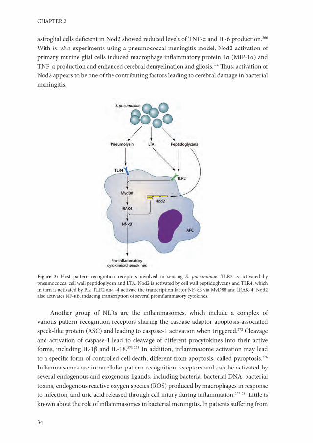
Pattern recognition receptorsImmune activation in the cerebrospinal fluid is initiated by the recognition of different bacterial pathogen-associated molecular patterns (PAMPs) by APCs (Table 1).258,259 These APCs are present at low levels in the CSF, or are situated in the meninges, choroid plexus, perivascular space, or brain parenchyma as astrocytes and microglial cells.260-262 Major pattern recognition receptors involved in initial sensing of pneumococci in the CNS are TLR2, TLR4, TLR9, and Nod-like receptors (NLRs) (Fig. 3).202,263-266
TLR2 recognizes PCW LTA.196,267 TLR2 signaling is enhanced by the TLR2 coreceptor, CD14, and by LPS binding protein (LBP).196,198 In a model of pneumococcal meningitis, TLR2-deficient mice showed increased disease severity with increased blood-brain barrier disruption and intracranial complications and increased bacterial loads.263,264 Cytokine production was similar in TLR2-deficient and wild-type mice with pneumococcal meningitis, except for that of TNF-α, which was significantly higher in TLR2-deficient mice.263,268 Since the phenotype of TLR2-deficient mice with pneumococcal meningitis was not as severe as that seen with mice lacking MyD88, an important general adaptor molecule for TLR signaling, it was proposed that other TLRs besides TLR2 may play a role in sensing pneumococci in the CNS.264,269 TLR4 recognizes pneumococcal pneumolysin.201
TLR4-deficient mice did not differ significantly from wild-type mice in their host immune response, cerebrovascular changes, or outcome during pneumococcal meningitis.265 However, in mice deficient in both TLR2 and TLR4, a marked reduction in inflammatory mediators, increased bacterial replication in the CNS, and reduced survival were seen compared to those for wild-type mice or mice with a single TLR deficiency.265 Thus, in meningitis, both TLR2 and TLR4 are important receptors in detecting the pneumococcus and initiating a robust inflammatory response to the pathogen, and one receptor may compensate for the absence of the other.265
TLR9 is an intracellular pattern recognition receptor and is activated by CpG repeats in bacterial DNA.270 In vitro, S. pneumoniae was able to activate alveolar and peripheral macrophages through TLR9 and induced IL-8 production in TLR9-transfected human embryonic kidney cells.195,202 In vivo, TLR9-deficient mice showed reduced resistance to S. pneumoniae after intranasal challenge.202 However, in a model of pneumococcal meningitis, triple mutant TLR2/TLR4/TLR9-deficient mice did not show significant differences in immune response, bacterial load, or survival compared with TLR2/TLR4-deficient mice.265 Therefore, TLR9 appears to play a minor role in pneumococcal meningitis, although this was assessed only in TLR triple-knockout mice.
NLRs are a second group of intracellular pattern recognition receptors involved in detecting pneumococci.203 NLRs belong to a family of receptors which, upon activation, induce activation of NF-κB or mitogen-activated protein kinase (MAPK) pathways and inflammatory caspases.203 In human embryonic kidney 293 cells, Nod2 was activated by internalized pneumococci through sensing of meso-diaminopimelic acid (meso-DAP) motifs of the bacterial peptidoglycan.203,271 In vitro experiments showed that microglial and astroglial cells are activated by S. pneumoniae through Nod2.266 Murine microglial and
41973 Geldhoff_v3.indd 33 24-08-16 00:39
CHAPTER 2
34
astroglial cells deficient in Nod2 showed reduced levels of TNF-α and IL-6 production.266 With in vivo experiments using a pneumococcal meningitis model, Nod2 activation of primary murine glial cells induced macrophage inflammatory protein 1α (MIP-1α) and TNF-α production and enhanced cerebral demyelination and gliosis.266 Thus, activation of Nod2 appears to be one of the contributing factors leading to cerebral damage in bacterial meningitis.
Figure 3: Host pattern recognition receptors involved in sensing S. pneumoniae. TLR2 is activated by pneumococcal cell wall peptidoglycan and LTA. Nod2 is activated by cell wall peptidoglycans and TLR4, which in turn is activated by Ply. TLR2 and -4 activate the transcription factor NF-κB via MyD88 and IRAK-4. Nod2 also activates NF-κB, inducing transcription of several proinflammatory cytokines.
Another group of NLRs are the inflammasomes, which include a complex of various pattern recognition receptors sharing the caspase adaptor apoptosis-associated speck-like protein (ASC) and leading to caspase-1 activation when triggered.272 Cleavage and activation of caspase-1 lead to cleavage of different procytokines into their active forms, including IL-1β and IL-18.273-275 In addition, inflammasome activation may lead to a specific form of controlled cell death, different from apoptosis, called pyroptosis.276 Inflammasomes are intracellular pattern recognition receptors and can be activated by several endogenous and exogenous ligands, including bacteria, bacterial DNA, bacterial toxins, endogenous reactive oxygen species (ROS) produced by macrophages in response to infection, and uric acid released through cell injury during inflammation.277-281 Little is known about the role of inflammasomes in bacterial meningitis. In patients suffering from
41973 Geldhoff_v3.indd 34 24-08-16 00:39

2
PATHOGENESIS AND PATHOPHYSIOLOGY OF PNEUMOCOCCAL MENINGITIS
35
bacterial meningitis, cerebrospinal fluid levels of caspase-1 were increased.282 In children with bacterial meningitis, as well as a rat model of pneumococcal meningitis, increased IL-1β levels were measured in the CSF.283,284 Koedel et al. showed that mice lacking caspase-1 displayed less severe inflammation and improved survival in a pneumococcal meningitis mouse model.282 Similar results were found in a pneumococcal meningitis model with IL-18 knockout mice, indicating a role for inflammasome activation in the pathophysiology of pneumococcal meningitis.285
A fourth group of pathogen recognition receptors involved in sensing S. pneumoniae are the C-type lectins, which are highly expressed on splenic dendritic cells and also on peritoneal macrophages.286 A member of this group, SIGN-R1, was shown to facilitate phagocytosis by recognition of the pneumococcal capsular polysaccharide.286,287 Mice lacking functional SIGN-R1 fail to effectively phagocytose S. pneumoniae, leading to an inability to clear the infection and resulting in increased inflammatory parameters and reduced survival in both a model of pneumococcal peritoneal sepsis and one of intranasally induced pneumonia.286,288 Furthermore, SIGN-R1 plays a role in the activation of the classical complement pathway by binding C1q.58 Park et al. showed the presence of SIGN-R1 on microglial cells in mouse and rat brains, which was functionally active in taking up pneumococcal capsular polysaccharides into the cell.289 Therefore, SIGN-R1 may be an important pathogen recognition receptor in the brain during pneumococcal meningitis.
Downstream signaling moleculesUpon stimulation of one of the above pattern recognition receptors, an intracellular cascade is activated and leads to the production of inflammatory molecules, usually cytokines or chemokines, which modulate the immune response by activating or attracting specialized immune cells. Deficiencies and polymorphisms in the pathogen recognition receptor downstream signaling cascade in humans have been associated with invasive pneumococcal disease, including meningitis.
The most extensively characterized TLR downstream signaling protein in pneumococcal invasive disease is IRAK-4 (Fig. 3).290 This adaptor protein is one of the links in TLR- and IL-1 receptor (IL-1R)-induced activation of MyD88 and NF-κB, which ultimately results in cytokine production.291,292 Specifically, children with IRAK-4 deficiency are susceptible to (recurrent) invasive pneumococcal infections, which are associated with high mortality.293 In a group of pediatric patients with normally expressed IRAK-4 but with recurrent invasive pneumococcal disease, deficiencies in the common adaptor molecule of TLR and IL-1R pathways, MyD88, were found.294 Deficiencies in IRAK-4 and MyD88 give indistinguishable phenotypes. Both patient groups are unresponsive to all TLR1, -2, -5, -6, -7, and -8 agonists, TLR9 agonists, and IL-1R agonists.294-296 In IRAK-4- or MyD88-deficient patients, the TLR3 signaling pathway is not affected, and the TLR4 pathway is affected only partially. Both TLR3 and -4 can still signal through the MyD88-independent TRIF pathway, leading to cytokine production.294
41973 Geldhoff_v3.indd 35 24-08-16 00:39
CHAPTER 2
36
Stimulation of whole blood of IRAK-4- or MyD88-deficient patients with several different TLR agonists showed impaired production of IL-1β, IL-6, IL-8, IL-10, IL-12, monocyte chemoattractant protein 1 (MCP-1), MIP-1α, and MIP-1β.294 Stimulation with a TLR3 or TLR4 agonist showed impaired production of IL-6, IL-10, and IL-12, as well as that of IL-8 in the case of TLR3 stimulation and IL-1β in the case of TLR4 stimulation.294 Among patients with an IRAK-4 or MyD88 deficiency, 68% suffer from invasive pneumococcal disease, and S. pneumoniae is responsible for 53% of all episodes of infectious episodes in these patients.290 Invasive bacterial disease in these patients consists of meningitis in 41% of IRAK-4-deficient patients and 52% of MyD88-deficient patients.290 IRAK-4 and MyD88 appear to be specifically important at a young age, as no fatal disease has been reported after the age of 8 years, with no invasive infections after the age of 14 years.290 Two patients have been described as having a homozygous mutation in the gene encoding NEMO, an adaptor molecule of the MyD88-dependent TLR, IL-1R, and TNF receptor (TNF-R) signaling pathways, and this mutation is associated with invasive pneumococcal disease.297,298
In mice, MyD88 deficiency resulted in increased susceptibility to systemic infection after colonization and increased mortality due to pneumococcal sepsis and meningitis.269,299 Pneumococcal infection in MyD88−/− mice was accompanied by decreased symptoms of infection and inflammatory parameters, similar to the phenotype seen in patients lacking functional MyD88 or IRAK-M.269,290,297 Deficiencies in the TLR and IL-1R signaling pathways have been associated with recurrent pneumococcal disease, illustrating the importance of these pathways in controlling pneumococcal infection.69
Proinflammatory cytokinesThe early response cytokines IL-1, TNF-α, and IL-6 are produced after pneumococcal recognition.300,301 Several cells have been found to be capable of sensing pneumococci and produce proinflammatory cytokines: perivascular and meningeal macrophages, vascular endothelial cells, astrocytes, and microglial cells.241,302-306 These early-phase cytokines induce upregulation of several adhesion factors on the vascular endothelium, mediating leukocyte influx (see above).206,207 The majority of leukocytes recruited to the CSF are polymorphonuclear neutrophils, and influx occurs largely in the first 6 h of infection.303
TNF-α is an important early proinflammatory response cytokine. Patients with bacterial meningitis have increased CSF TNF-α levels early in the course of disease.242,307-310 Intrathecal levels of TNF-α correlated with severity of blood-brain barrier disruption, disease severity, and neurologic sequelae in a study including 48 patients with bacterial meningitis.242 In this study, TNF-α levels decreased within 24 h after the onset of antibiotic treatment.242 In animal models of pneumococcal meningitis, TNF-α was produced mainly in the first 6 to 24 h of the immune response.311,312 One hour after intrathecal injection of recombinant TNF-α, CSF leukocyte recruitment was observed in a rabbit model.313 Intrathecal administration of anti-TNF-α antibody together with S. pneumoniae reduced CSF leukocytosis, protein content, and brain edema in these experiments.313 TNF-α
41973 Geldhoff_v3.indd 36 24-08-16 00:39

2
PATHOGENESIS AND PATHOPHYSIOLOGY OF PNEUMOCOCCAL MENINGITIS
37
administered intravenously also mediated blood-brain barrier opening, facilitating bacterial traversal into the CSF.314 However, TNF-α production is also essential for defense, as TNF-α-deficient mice showed decreased survival in a pneumococcal meningitis model.315 Thus, TNF-α has been shown to be a marker of the acute inflammatory response and is associated with inflammation-related complications of bacterial meningitis but is also essential for an adequate host response to the infection.
IL-1β is a proinflammatory cytokine produced by, e.g., perivascular and meningeal macrophages.303 CSF IL-1β levels are increased in the first 18 h of infection.316 Pro-IL-1β is cleaved into its active form by caspase-1, which is regulated by a group of different receptors called the inflammasome.275 Reported data on the role of IL-1β in bacterial meningitis are somewhat contradictory. Levels of IL-1β were not associated with the degree of blood-brain barrier disruption in patients with bacterial meningitis.242 However, a pneumococcal model using caspase-1 knockout mice showed decreased levels of IL-1β and decreased intracranial pressure (ICP), leukocyte recruitment, and brain edema compared to those in WT mice.282 IL-1β administered intrathecally did not lead to CSF pleocytosis or brain edema in a rabbit model of pneumococcal meningitis.313 However, antibodies against IL-1β decreased leukocyte influx induced by TNF-α.313 Mice deficient in the receptor for IL-1α and IL-1β (IL-1R) showed impaired survival and decreased cytokine responses without alterations in CSF pleocytosis.317 Thus, although IL-1β did not influence CSF pleocytosis in pneumococcal meningitis, other caspase-1-cleaved cytokines may be responsible for the reduced pleocytosis observed in caspase-1 knockout mice.
IL-6 is a proinflammatory as well as anti-inflammatory cytokine and has been shown to be upregulated in the acute phase of many infection models.318 In a mouse pneumococcal meningitis model, IL-6 knockout mice displayed increased CSF pleocytosis but decreased cerebral edema, blood-brain barrier disruption, and intracranial pressure.319 This was also described for a model of pneumococcal pneumonia where IL-6 was shown to downregulate multiple proinflammatory as well as anti-inflammatory cytokines.320 Thus, in pneumococcal meningitis, IL-6 attenuates CSF leukocyte recruitment but does not inhibit complications related to fluid shift.
Gamma interferon (IFN-γ) is one of the major cytokines of the T-helper 1 (Th1) pathway. IFN-γ was increased in the CSF of patients with pneumococcal meningitis.321,322 IFN-γ was also expressed in brain tissue of rats with pneumococcal meningitis.323 The exact role of IFN-γ in pneumococcal meningitis remains unclear. IL-12p70, an important stimulus for IFN-γ production, could be detected in patients with pneumococcal meningitis and in animal models of pneumococcal meningitis.322 Macrophage inflammatory factor (MIF) was found to be increased in the CSF of patients with pneumococcal meningitis and has also been associated with disease severity, suggesting a role for MIF in the pathophysiology of pneumococcal meningitis.324,325
41973 Geldhoff_v3.indd 37 24-08-16 00:39
CHAPTER 2
38
Anti-inflammatory cytokinesAnti-inflammatory cytokines include IL-10 and TGF-β.326-329 IL-6 may act partially as an anti-inflammatory cytokine and has been discussed earlier.320 IL-10 is an anti-inflammatory cytokine with multiple effects, including downregulation of proinflammatory cytokines and costimulatory molecules on macrophages and impairment of neutrophil phagocytosis and killing.326,329,330 IL-10 has been shown to downregulate TNF-α, IL-6, and keratinocyte-derived chemokine (KC), thereby reducing CSF pleocytosis in pneumococcal meningitis.331 Nonetheless, in experimental pneumococcal meningitis, IL-10 knockout mice did not have altered bacterial loads or survival.331 This anti-inflammatory cytokine has been described as an important repressor of sepsis-associated neuronal damage. Its pathophysiology is unclear, but it appears that inflammatory mediators as well as bacterial components cross the blood-brain barrier and induce a local inflammatory response.332-334 In mice overexpressing IL-10, the development of sepsis-associated neuronal damage as a result of pneumococcal sepsis has been shown to be decreased.332 In line with this, Koedel et al. showed that intravenously administered recombinant IL-10, as opposed to intracisternally administered IL-10, reduced the levels of CSF proinflammatory cytokines, CSF pleocytosis, cerebral edema, and intracranial pressure in a rat model of pneumococcal meningitis.335 Interestingly, intracisternally administered IL-10 had the opposite effect, as it increased CSF pleocytosis in rats with pneumococcal meningitis and induced an inflammatory response in uninfected rats.335 Thus, systemic IL-10 reduces cerebral inflammation and secondary complications in pneumococcal meningitis.
TGF-β is an anti-inflammatory cytokine with multiple functions, including differentiation and maintenance of regulatory T cells (Tregs), differentiation of Th17 T cells, and inhibition of Th1 and Th2 T-cell maturation and differentiation, but TGF-β also suppresses macrophage activation and production of several proinflammatory cytokines, such as IL-1β, IL-6, and TNF, by microglial cells.327,328,336 Activated Tregs produce TGF-β in an autocrine fashion and are thought to modulate the immune response in such a way that the host’s tissues are minimally damaged while the invading pathogen is effectively eliminated, by downregulating the acute inflammatory response. In a mouse model of pneumococcal meningitis, TGF-β was associated with cerebral vasculitis, a frequent complication in patients with meningitis.6,337 Mice with leukocytes deficient in TGF-β receptor II (TGF-βRII) showed increased neutrophil influx into the subarachnoid space, which was accompanied by increased bacterial clearance and survival of the host.337 In addition, TGF-βRII knockout mice showed decreased blood-brain barrier disruption, intracranial pressure, and cerebral vasculitis.337 However, when TGF-β2 or TGF-β1 was administered intraperitoneally in a rat model of sterile meningitis induced by a PCW lysate, cerebral edema, intracranial pressure, and cerebral blood flow (CBF) decreased.338 Thus, leukocyte TGF-βRII signaling has an unfavorable effect on the course of pneumococcal meningitis, although systemic TGF-β production appears to decrease the complications of meningitis.
41973 Geldhoff_v3.indd 38 24-08-16 00:39

2
PATHOGENESIS AND PATHOPHYSIOLOGY OF PNEUMOCOCCAL MENINGITIS
39
ChemokinesChemokines are a subgroup of cytokines with chemotactic activity recruiting effector immune cells to the site of infection.48 Multiple chemokines have been reported to be upregulated in the CSF of patients with pneumococcal meningitis, including MIP-1delta (CCL15), NAP-2 (CXCL7), MIF, MCP-2 (CCL8), PARC (CCL18), MIP-3α (CCL20), ENA-78 (CXCL5), GRO-α (CXCL-1), IL-8 (CXCL-8), MCP-1 (CCL2), MIP-1α (CCL3), and MIP-1β (CCL4).310,312,339-342 In animal models of pneumococcal meningitis, additional chemokines have been identified by protein arrays for brain tissue, including MIP-1γ (CCL9), MIP-2 (CXCL-2), lymphotactin (XCL-1), TCA-3 (CCL1), eotaxin (CCL11), MCP-5 (CCL12), eotaxin-2 (CCL24), TECK (CCL25), PF-4 (CXCL4), CRG-2 (CXCL10), SDF-1α (CXCL12), BLC (CXCL13), and CXCL16.343 The role in pneumococcal meningitis of many of these chemokines has not been elucidated yet.
IL-8 is one of the well-characterized chemokines involved in pneumococcal meningitis. IL-8 was found to be chemotactic for neutrophils in the CSF of patients with bacterial meningitis.340 Furthermore, CSF IL-8 levels increased as a result of blocking leukocyte recruitment in rabbits with pneumococcal meningitis, indicating local production of chemotactic cytokines.344 In patients with bacterial meningitis, no correlation was found between the CSF white blood cell (WBC) count and IL-8.340 Ostergaard et al. showed that not intracisternal but rather systemic IL-8 levels induced CSF pleocytosis in a rabbit model of pneumococcal meningitis.345 Thus, IL-8 appears to regulate CSF pleocytosis from the systemic compartment, comparable with the proinflammatory cytokine TNF-α and the anti-inflammatory cytokines IL-10 and TGF-β.
The CCL chemokines MCP-1, MIP-1α, and MIP-2 were produced in vitro by astrocytes and microglial cells in response to PCW structures.305,346 In vitro, antibodies against MCP-1, MIP-1α, and MIP-1β inhibited monocyte chemotactic properties of CSF from patients with pneumococcal meningitis.340 Furthermore, intracisternal inoculation of recombinant MIP-1 or MIP-2 induced blood-brain barrier disruption, CSF leukocytosis, and cerebral edema in a rabbit model of pneumococcal meningitis; blocking MIP-1 or MIP-2 delayed these inflammatory alterations by 2 h.313 Another experiment showed that blocking the receptor for MIP-1, i.e., CCR2, specifically reduced the influx of monocytes into the subarachnoid space in a mouse model of pneumococcal meningitis, while not changing bacterial clearing.347 Thus, both MIP-1 and MIP-2 are produced by immune cells resident in the brain and attract monocytes and neutrophils from the bloodstream into the CSF in the acute stage of infection. The role of MCP-1 in pneumococcal meningitis has not been studied extensively.
Of the CXCL chemokines, ENA-78 was found to be upregulated in patients with bacterial meningitis and exhibited specifically neutrophil chemotactic properties together with IL-8.341 GRO-α was also found at high levels in the CSF of patients with bacterial meningitis, as well as in a rat model of pneumococcal meningitis, but it did not exert any chemotactic activity.283,341 In summary, multiple chemokines have been shown to be upregulated in pneumococcal meningitis. Most of them have a role in attracting leukocytes
41973 Geldhoff_v3.indd 39 24-08-16 00:39

CHAPTER 2
40
to the CSF. However, the roles of many other chemokines have not been investigated extensively.
Leukocyte migration adhesion moleculesIn response to proinflammatory cytokines, selectins and integrins are upregulated on the blood endothelium and leukocytes are attracted from the bloodstream.256,348 Cerebral perivascular and meningeal macrophages play a key role in attracting leukocytes across the blood-brain barrier into the CSF.302 About 90% of the attracted leukocyte population consists of neutrophilic granulocytes, with the other 10% being predominantly monocytes.268,302,347 Rats depleted of perivascular and meningeal macrophages by use of clodronate showed decreased leukocyte recruitment into the CSF despite increased expression of MIP-2, IL-6, and VCAM-1.302 Furthermore, these depleted rats showed increased bacterial outgrowth in the CSF and poorer clinical scores than those for control rats with pneumococcal meningitis.302 Thus, leukocyte attraction to the subarachnoid space seems to be crucial for efficacious clearing of S. pneumoniae from the subarachnoid space and dependent on perivascular and meningeal macrophage activation but appears to be mediated by cytokines other than IL-6 and MIP-2.
Other cytokines and chemokines attracting leukocytes to the subarachnoid space are TNF-α, IL-8 (systemic), MIP-2, and ENA-78 (see above).313,341 Monocytes are also attracted from the bloodstream into the CSF but appear to play a minor role in the pathogenesis of pneumococcal meningitis.347
Leukocytes cross the blood-brain barrier by binding to selectins on the endothelium.256 Binding to P- and E-selectin promotes leukocyte rolling across the endothelium.256 Blocking L-selectin by fucoidin treatment reduced leukocytosis and disruption of the blood-brain barrier in rabbits challenged intrathecally with pneumococcal antigen.256 Integrins are also upregulated on the vascular endothelium, facilitating binding of leukocytes and subsequent blood-brain barrier migration.284 An important integrin involved in leukocyte recruitment in pneumococcal meningitis is ICAM-1, which is known to bind MAC-1 (CD11b/CD18) on the leukocyte surface.349,350 Rabbits treated intravenously with antibodies against CD18 showed decreased CSF leukocytosis, blood-brain barrier permeability, and brain edema and improved survival after intracisternal challenge with PCW or S. pneumoniae.349,350 Interestingly, antibodies directed against CD11b did not alter CSF leukocytosis in the same rabbit model of pneumococcal meningitis, which may implicate a role for CD11a/CD18 or CD11c/CD18. In line with this, CD11a/CD18-deficient mice showed increased rates of meningitis and otitis media following intraperitoneal infection with S. pneumoniae.350,351
The integrin ICAM-1 was shown to be expressed on brain vascular endothelial cells in response to PCW, through an autocrine loop involving TNF-α.241 In a rat model of meningitis induced by PCW, antibodies against ICAM-1 reduced the increase in CBF, increase in ICP, brain edema, and CSF leukocyte counts observed in the first hours after
41973 Geldhoff_v3.indd 40 24-08-16 00:39

2
PATHOGENESIS AND PATHOPHYSIOLOGY OF PNEUMOCOCCAL MENINGITIS
41
induction of meningitis.352 In an infant mouse model of pneumococcal peritonitis, ICAM-1 deficiency did not reduce the incidence of meningitis, and histopathologically there was no difference in the severity of inflammation (469).353 Thus, ICAM-1 is not solely responsible for leukocyte recruitment to the brain.
Other chemoattractantsPAF is a protein produced by neutrophils and endothelial cells in response to inflammatory stimuli, and it facilitates adhesion of leukocytes to the vascular endothelium.354-356 In rabbits, PAF administered intrathecally induced blood-brain barrier permeability and cerebral edema at doses much lower than those at which it induced leukocytosis.349 Antibodies against CD18 blocked these effects.349
In response to pneumococci, endothelial cells and neutrophils are stimulated to produce reactive nitrogen species (RNS), such as NO, by endothelial nitric oxide synthetase (eNOS) and inducible nitric oxide synthetase (iNOS), respectively.241,357 The cerebral vasculature appears to be the main location where ROS are active, and increased levels of ROS are associated with blood-brain barrier disruption.358,359 In patients with meningitis, positive correlations were found between CSF derivatives of NO production and CSF leukocyte counts and protein concentrations in a group of 27 children with bacterial meningitis; however, only 2 of these children had confirmed pneumococcal meningitis.360 Mice deficient for iNOS showed decreased blood-brain barrier disruption and decreased IL-1β, IL-6, TNF-α, MIP-1α, and MIP-2 mRNA levels in the brain.357 The opposite was true for eNOS-deficient mice, which showed more profound leukocyte infiltrates, increased cytokine levels, and decreased survival due to pneumococcal meningitis.361 A third form of NOS, neuronal NOS (nNOS), appears to play a minor role in fluid balance-related complications of pneumococcal meningitis.362 In addition to RNS, ROS such as O2
− are produced by the enzyme NADPH oxidase in neutrophils, macrophages, and endothelial cells in response to infection.363 In mice deficient for the subunit of NADPH oxidase in nonphagocytic cells, such as endothelial cells (p47), detrimental effects on blood-brain barrier permeability, subarachnoid space inflammation, and bacterial outgrowth were found.363 Mice deficient for the subunit of NADPH oxidase in phagocytic cells (gp91) did not show any inflammatory differences from WT mice in the course of pneumococcal meningitis.363 Thus, RNS/ROS produced specifically by cerebral endothelial cells, as opposed to granulocytes and macrophages, contribute to the blood-brain barrier damage and associated complications observed during pneumococcal meningitis.
The fibrinolysis factor uPA is also implicated in leukocyte recruitment to the brain in pneumococcal meningitis. In a group of 12 patients with bacterial meningitis (67% of cases were caused by S. pneumoniae), CSF uPA levels were associated with leukocyte recruitment and blood-brain barrier disruption.245 In this study, serum uPA levels correlated with unfavorable clinical outcomes for these patients with bacterial meningitis.245 Mice deficient in uPA showed reduced CSF leukocytosis, although blood-brain barrier permeability, ICP, expression of chemokines, bacterial killing, and clinical
41973 Geldhoff_v3.indd 41 24-08-16 00:39

CHAPTER 2
42
outcomes were not different from those for WT mice.364 Interestingly, deficiency in tPA did not have any implications in a mouse pneumococcal meningitis model.364
The complement systemA fourth chemoattractant factor for CSF leukocytes is the complement system. Low or undetectable CSF levels of C3, C4, and B were found in uninfected control subjects.365 In response to infection, the liver produces an array of acute-phase proteins which includes several complement components.366,367 Circulating monocytes, macrophages, and epithelial cells of the pulmonary and gastrointestinal tracts also produce substantial amounts of complement components.367-370 In addition, brain resident macrophages and monocytes recruited to the CSF during meningitis may also locally produce complement components. C3 was also found to be produced by astrocytes and neurons in response to HIV or proinflammatory cytokines.371-373 Cultured human brain pericytes from a patient with Alzheimer’s disease have been shown to produce C1q, and activated astroglial cells can produce C1q, which has been associated with increased blood-brain barrier damage in a rat model of neurotoxicity.374,375 Microglial cells have been shown to upregulate C1q, C3, C4, and C5a production in response to injury.376 Thus, levels of complement components are increased in the peripheral blood but may also be produced locally in the brain during infection or inflammation.
During infection or inflammation, the immune response in the brain compartment may do more harm than good. Under normal circumstances, the brain expresses multiple inhibitory factors for complement activation. One of these, factor H, was found to be expressed constitutively in neurons, brain endothelial cells, microglial cells, and astrocytes in mice.377 In a mouse model of antibody-mediated inflammation, expression of factor H in these cells was suppressed; when recombinant factor H was administered, complement opsonization, axonal injury, and leukocyte infiltration decreased.377 Thus, in addition to monocytes and macrophages, brain resident cells may contribute to the production of complement factors leading to leukocyte influx during inflammation.
In a rabbit model of pneumococcal meningitis where cobra venom (known to consume complement factor 3) was administered systemically, treated mice showed decreased survival accompanied by increased bacterial outgrowth in the CSF.49 CSF pleocytosis was similar between the treated and untreated groups, but neutrophils were severely impaired in phagocytosis and killing of bacteria.49 In line with these results, mice deficient in C1q or C3 also showed increased bacterial outgrowth in the CSF in a pneumococcal meningitis model, which was accompanied by decreased survival.49,60 C1q and C3 knockout mice displayed a tempered inflammatory response which was reflected by a decreased leukocyte count in the CSF, decreased brain cytokine and chemokine levels, and fewer meningitis-associated intracranial complications. However, survival was decreased in this model as a result of more fulminant sepsis accompanied by systemic complications.60 Similar results were found in mice deficient for the C3b receptor (CR3), which is also an integrin involved in binding of leukocytes to the endothelium.378
41973 Geldhoff_v3.indd 42 24-08-16 00:39

2
PATHOGENESIS AND PATHOPHYSIOLOGY OF PNEUMOCOCCAL MENINGITIS
43
CR3−/− mice showed increased bacterial outgrowth compared to WT mice, with decreased survival, as a result of decreased neutrophilic superoxidase production in the CR3−/− mice leading to ineffective bacterial killing, while CSF pleocytosis was not different between groups.378 In rabbits, intracisternally administered C5a resulted in rapid CSF pleocytosis and increased CSF protein levels which peaked 1 h after injection.379 Furthermore, the CSF of rabbits with pneumococcal meningitis lost its chemotactic activity to neutrophils after incubation with an antibody against C5a.380
MMPsMatrix metalloproteinases (MMPs) are Zn2+- and Ca2+-dependent endopeptidases capable of breaking down and remodeling extracellular matrix components such as fibronectin, laminin, proteoglycans, and type IV collagen.381,382 MMPs are produced mainly by activated neutrophils and, to a lesser extent, also by macrophages, monocytes, and possibly TNF-α-stimulated endothelial cells.383,384 Furthermore, constitutive MMP expression is found on microglial cells and astrocytes and may be modulated during neuroinflammation and meningitis.385-387
The action of MMPs was initially believed to be limited to the breakdown of ECM during leukocyte migration across the subendothelial layer.388 However, experimental bacterial meningitis models revealed that inhibition of MMPs did not result in a reduction of CSF pleocytosis, although blood-brain barrier permeability disruption was attenuated.389 More recent studies have revealed a wide range of MMP substrates, such as chemokines, growth factors, and adhesion molecules, as well as cytokines and cytokine receptors, allowing MMPs to influence the course of various inflammatory conditions.388,390
In patients with bacterial meningitis, CSF levels of MMP-8 and MMP-9 were elevated.309 Moreover, higher levels of MMP-9 were detected in children with meningitis who developed hearing impairment or secondary epilepsy than in those who recovered without neurological deficit.309
To avoid unwanted proteolytic activity, the activity of all MMPs is tightly regulated by binding to inhibitory proteins called tissue inhibitors of metalloproteinases (TIMPs). MMP-9, for instance, forms complexes with and is inactivated by TIMP-1, both of which are upregulated during pneumococcal meningitis.389 In a murine model of pneumococcal meningitis, the induction of TIMP-1 was delayed in relation to that of MMP-9, favoring increased collagen IV degradation and subsequent increased blood-brain barrier permeability.391 Thus, treatment options including drugs specifically targeting MMPs are being investigated.392 Interestingly, in a rat model of pneumococcal meningitis, adjuvant treatment of pneumococcal meningitis with dexamethasone resulted in lower MMP-9 mRNA expression, suggesting a possible mechanism of corticosteroids as an adjuvant treatment for bacterial meningitis.393
41973 Geldhoff_v3.indd 43 24-08-16 00:39

CHAPTER 2
44
Oxidative stressOne of the characterizing features of pneumococcal meningitis is the marked recruitment of leukocytes into the CSF.144,147 The subsequent release of large amounts of RNS and ROS has been documented for patient populations as well as in animal models and plays a central role in the development of intracranial complications and brain damage.394-396 In the past decade, ROS and RNS have been investigated extensively as potential targets for adjuvant treatment (reviewed by Klein et al.).397
During pneumococcal meningitis, RNS are produced by iNOS and eNOS.357 NOS inhibition studies with experimental pneumococcal meningitis models have yielded contradictory results, at least in part due to a lack of drug specificity for single isoforms of NOS.398 Moreover, iNOS and eNOS knockout studies show that the source of RNS is pivotal in determining its function during disease progression. Thus, NO derived from iNOS appears to contribute to blood-brain barrier disruption and to production of proinflammatory mediators (such as IL-1-β, TNF-α, and MIP-2), whereas eNOS-derived NO plays a largely protective role.357,361
Reactive oxygen species, such as superoxide anion (O2−), hydrogen peroxide (H2O2),
and hydroxyl radicals, are produced by brain resident immune cells as part of the host response to invasive bacterial infections.399 Pneumococci themselves are also an important source of H2O2, which not only is able to cause direct cytotoxic damage but also interacts with host NO to form the highly reactive species peroxynitrite (ONOO−).400-402 When present in large quantities, ROS overwhelm the resident antioxidant mechanisms (such as superoxide dismutase and glutathione), leading to tissue exposure to oxidative stress.399 Interventions aimed at scavenging ROS or enhancing antioxidant activity have generally resulted in reductions of intracranial complications such as elevated ICP, increased CBF, brain edema, and neuronal injury.397,398
Peroxynitrite, which is formed by the combination of superoxide radicals and NO, is a very reactive, short-lived molecule whose direct detection has proven difficult; the compound nitrotyrosine (which is formed by the reaction of NOO−with tyrosine) is widely used as a marker.403 In patients with bacterial meningitis, elevated CSF levels of nitrotyrosine were associated with an unfavorable outcome and with lower CSF concentrations of the antioxidant ascorbic acid, suggesting antioxidant depletion by the RNS.395 Furthermore, in autopsy studies, nitrotyrosine was detected in the leptomeninges, subarachnoid granulocytes, and penetrating cortical and leptomeningeal vasculature.
Peroxynitrite can damage neurons and glial cells in two ways. First, it causes damage by means of lipid peroxidation and cell membrane destabilization, which occurs by peroxynitrite attack on lipid peroxidation and is consistently seen in brain homogenates of rats with pneumococcal meningitis.397,404 Blocking of lipid peroxidation with aminosteroids limits neuronal damage.404 Alternatively, peroxynitrite can cause DNA fragmentation and subsequent poly(ADP-ribose) polymerase (PARP) activation, which leads to cell energy depletion and cell death.398 PARP knockout mice, as well as mice treated with a PARP
41973 Geldhoff_v3.indd 44 24-08-16 00:39

2
PATHOGENESIS AND PATHOPHYSIOLOGY OF PNEUMOCOCCAL MENINGITIS
45
inhibitor, demonstrated lower levels of inflammation and a better clinical course during pneumococcal meningitis.398,404
Klein et al. have reviewed the mechanisms of oxidative damage (activation of cytokines and chemokines, neutrophil activation, lipid peroxidation, DNA and mitochondrial damage, tyrosine nitration, MMP activation/TIMP inactivation, K+ channel activity alterations, and prostaglandin synthesis) as well as the resulting pathophysiologic alterations in pneumococcal meningitis.397
CoagulationThe importance of coagulation and fibrinolytic dysregulation during pneumococcal meningitis is illustrated by the large number of cerebrovascular complications, which occur in up to one-third of patients.147 Analysis of CSF in patients with bacterial meningitis revealed increased levels of both coagulation and fibrinolytic factors (Table 2).245,405 More recently, PAI-1 was shown to be associated with the occurrence of brain infarctions, though no causal relationship was determined.406 Furthermore, in a recent autopsy series of patients who died of pneumococcal meningitis, fibrin thrombi and cerebral infarctions were found in the absence of inflammatory vessel wall infiltrates, suggesting that disseminated cerebral intravascular coagulation might be an additional explanation for ischemic stroke in pneumococcal meningitis.407 The precise mechanism of cerebral
Table 2: Coagulation studies.Setting Material Factor Change in
concnRef
Human studies
Thirthy-eight patients with bacterial meningitis (GCSa of <9 vs GCS >9)
Serum PLT/dPLTPTrINRD-dimers
↑↓↑↑
467
Ninety-two patients with bacterial meningitis vs. controls and patients with viral meningitis.
CSF sTFTaTpT fragment F1+2 t-PAPAI-1D-dimer
↑↑↑↑↑↑
406
Twelve patients with bacterial meningitis vs. 10 patients with GBS and 10 controls.
Serum/CSF uPAuPARPAI-1PA-dependent platelet activation
↑/↑=/↑=/↑↑
245
Twelve patients with bacterial meningitis vs. 10 patients with GBS and 10 controls.
Serum/CSF tPA ↑↑/↑ 405
Murine studies
C57BL/6tPA-/-uPAR-/-
BrainFrozen samplesSection
tPA/uPAPAI-1/2uPARMIP-2KCAlbumin
↑/=↑/↑↑↑↑↑
364
aGCS, Glasgow coma score.
41973 Geldhoff_v3.indd 45 24-08-16 00:39
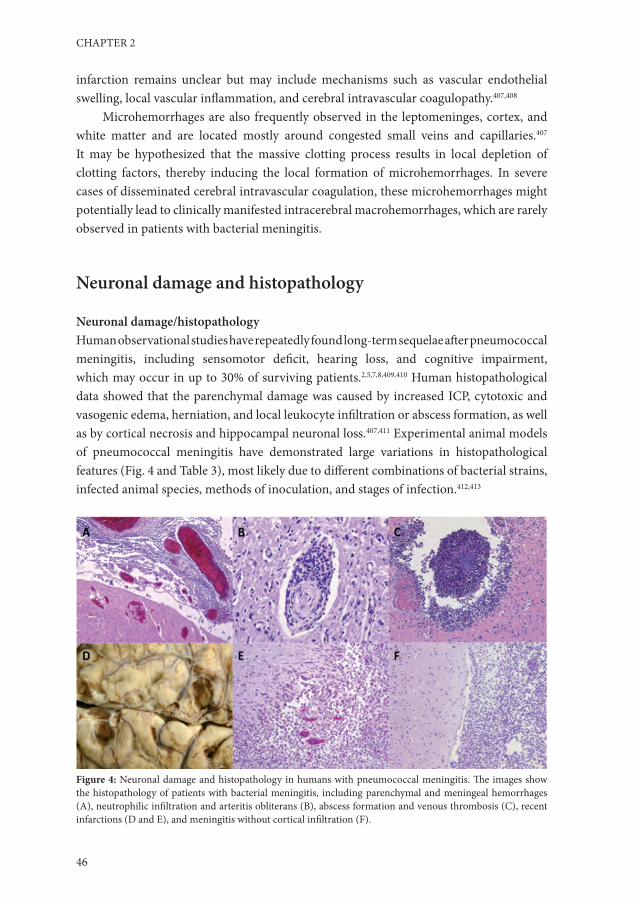
CHAPTER 2
46
infarction remains unclear but may include mechanisms such as vascular endothelial swelling, local vascular inflammation, and cerebral intravascular coagulopathy.407,408
Microhemorrhages are also frequently observed in the leptomeninges, cortex, and white matter and are located mostly around congested small veins and capillaries.407 It may be hypothesized that the massive clotting process results in local depletion of clotting factors, thereby inducing the local formation of microhemorrhages. In severe cases of disseminated cerebral intravascular coagulation, these microhemorrhages might potentially lead to clinically manifested intracerebral macrohemorrhages, which are rarely observed in patients with bacterial meningitis.
Neuronal damage and histopathology
Neuronal damage/histopathologyHuman observational studies have repeatedly found long-term sequelae after pneumococcal meningitis, including sensomotor deficit, hearing loss, and cognitive impairment, which may occur in up to 30% of surviving patients.2,5,7,8,409,410 Human histopathological data showed that the parenchymal damage was caused by increased ICP, cytotoxic and vasogenic edema, herniation, and local leukocyte infiltration or abscess formation, as well as by cortical necrosis and hippocampal neuronal loss.407,411 Experimental animal models of pneumococcal meningitis have demonstrated large variations in histopathological features (Fig. 4 and Table 3), most likely due to different combinations of bacterial strains, infected animal species, methods of inoculation, and stages of infection.412,413
Figure 4: Neuronal damage and histopathology in humans with pneumococcal meningitis. The images show the histopathology of patients with bacterial meningitis, including parenchymal and meningeal hemorrhages (A), neutrophilic infiltration and arteritis obliterans (B), abscess formation and venous thrombosis (C), recent infarctions (D and E), and meningitis without cortical infiltration (F).
41973 Geldhoff_v3.indd 46 24-08-16 00:40

2
PATHOGENESIS AND PATHOPHYSIOLOGY OF PNEUMOCOCCAL MENINGITIS
47
Microgial activationMicroglial cells, a specific subset of cells related to monocytes and dendritic cells, form the initial line of defense of brain parenchyma against damage, injury, or infection and play a pivotal role in tissue repair, removal of dead debris, and recruitment of other immune cells to the site of infection.414-416 Together with meningeal and perivascular macrophages, microglia express a wide palette of TLRs and become activated during bacterial infections.417 Once activated, microglia are capable of producing large amounts of proinflammatory cytokines as well as reactive oxygen and reactive nitrogen intermediates, thus potentially playing both neuroprotective and neurotoxic roles.418-420
Table 3: Neuronal damage.
Process or type of damage
Outcome References
Microglial activation Microglial activation is induced by pneumococcus and mediated by TLRs 2, 4, and 9
421, 444, 419, 266
Activin A may mediate microglial proliferation and activation 423, 424, 425
Neuronal apoptosis The early phase of hippocampal apoptosis is caspase dependent and mediated by H2O2, pneumolysin, and AIF
436, 435, 401, 437, 185, 432, 433
The late phase of hippocampal apoptosis is caspase independent and mediated by pneumococcal cell wall products, in a microglial TLR-2-dependent manner
400, 438, 439, 432
Sepsis-mediated neuronal damage
Neuronal damage may result in sepsis without concurrent bacterial growth in the CNS compartment
332, 445, 446, 447
Cochlear damage Cochlear damage is correlated with CSF inflammation and is mediated by TLR and MyD88 signaling pathways
459, 451, 452, 460
NO may contribute to cochlear damage; iNOS and eNOS are upregulated during pneumococcal meningitis, and damage can be attenuated using NO inhibitors/O2 scavengers
462, 457, 410, 461
In vitro studies have demonstrated that microglial cells express TLRs 2, 4, and 9, which upon activation by specific TLR agonists induce proinflammatory cytokine and NO production, as well as increased phagocytic activity.306,421 More specifically, the neurotoxic effects of CpG DNA, which is released into the subarachnoidal space in large amounts by pneumococci during bacterial growth and following antibiotic treatment, were shown to be mediated largely via stimulation of TLR9 on microglial cells.419 Furthermore, pneumolysin, which is a ligand for TLR4, was also found to be a potent activator of microglial cells, causing microglial cytotoxicity at high concentrations.421
Activin A, a member of the TGF-β superfamily, is a neuroprotective cytokine which has been shown to be expressed constitutively in CSF and elevated in patients during bacterial meningitis.422,423 Elevated activin A levels in CSF have also been detected in a rabbit meningitis model and were produced by cultured microglial cells following treatment with agonists of TLRs 2, 4, and 9.424,425 Furthermore, in vitro-cultured murine microglial cells stimulated with LPS showed that cotreatment with activin A increased
41973 Geldhoff_v3.indd 47 24-08-16 00:40

CHAPTER 2
48
microglial proliferation and negatively regulated production of NO, IL-1β, IL-6, and TNF-α.423
Although no studies have been performed regarding the effects of dexamethasone on microglial activation during pneumococcal meningitis, in vitro studies using LPS-stimulated glial cells demonstrated that microglial activation after bacterial exposure is limited by corticosteroid treatment, providing a possible explanation for the observed beneficial effects of dexamethasone treatment in vivo.11,12,426,427
A recently published hypothesis suggested that microglia may respond differently to a stimulus preceded by another stimulus, a phenomenon called “priming”.390 Aging itself may result in enhanced microglial activation following single or repeated stimulation.428,429 Thus, the usually tightly controlled microglial activation may become self-amplifying and even neurotoxic.334 Whether this dysregulation of microglial function plays a role in pneumococcal meningitis seems plausible but has not yet been investigated.
Neuronal apoptosisNeuronal damage is caused by the dual effects of an overwhelming inflammatory reaction and direct effects of bacterial toxins.430 Though the hippocampus is not exposed to pneumococci or infiltrating leukocytes directly, it is surrounded by interstitial fluid which is contiguous with the CSF, allowing secreted bacterial toxins and immune system mediators to diffuse into the parenchyma.401,431 Recent research with murine models has shown that pneumococcus-mediated hippocampal apoptosis occurs in at least two phases, separated by both time and mechanism.432,433
The early phase is initiated by the pneumococcal toxins H2O2 and pneumolysin and results in caspase-independent apoptosis-like pyknotic cell death of both mature and immature neurons throughout the dentate gyrus of the hippocampus.401,432 This is subsequently followed by caspase-dependent apoptosis of the immature neurons in the subgranular region of the dentate gyrus, triggered primarily by bacterial cell wall stimulation of leukocytes.413,432
Pneumolysin, which is also implicated in apoptosis of microglial and brain microvascular endothelial cells, was shown to colocalize to dying neurons in the dentate gyrus in a rabbit model of pneumococcal meningitis.185,401 Animals infected with either pneumolysin- or H2O2-deficient pneumococci showed only partial attenuation of early neuronal apoptosis.401 Additional blocking of H2O2 in animals infected with pneumolysin-deficient pneumococci led to a marked further reduction of cell death, suggesting that H2O2 and pneumolysin together are responsible for early hippocampal apoptosis. 401
Because pneumococci do not express catalase, they are capable of producing high levels of H2O2, which can diffuse freely through the cellular membranes of target cells to damage intracellular structures.434 H2O2 triggers the release of Ca2+ from the endoplasmic reticulum and its influx from the extracellular space. This results in a loss of mitochondrial membrane stability and in further increases of Ca2+ and ROS production. Pneumolysin possibly contributes directly to mitochondrial permeabilization through pore formation,
41973 Geldhoff_v3.indd 48 24-08-16 00:40

2
PATHOGENESIS AND PATHOPHYSIOLOGY OF PNEUMOCOCCAL MENINGITIS
49
after which apoptosis inducing factor (AIF) is cleaved from the mitochondrial membrane by calpein and cathespin is transported through the cytosol into the nucleus.435,436 There, AIF causes chromatin condensation and large-scale DNA fragmentation, leading to cell death.437 Paradoxically, AIF may also have antiapoptotic properties through the regulation of ROS through peroxide scavenging.437
The late, caspase-dependent phase of neuronal apoptosis is PCW and TLR2 dependent.400,432 The observation that in vitro exposure of isolated cultured neurons to PCW products does not lead to cell death led to the hypothesis that late-phase apoptosis may be dependent on the host inflammatory response.432 Neurons themselves do not express TLR2 or -4 and are not sensitive to exposure to the corresponding bacterial ligands. However, when cocultured with microglial cells, neurons revealed caspase-dependent, TLR-mediated late apoptosis when they were exposed to PCW or LPS.438,439 Therefore, the inflammatory response of microglial cells and invading neutrophils may underlie the caspase-dependent hippocampal apoptosis during pneumococcal meningitis.
Several experimental treatments aimed at reducing hippocampal apoptosis have been studied in animal models of pneumococcal meningitis. Much attention has gone to nonbacteriolytic antibiotic therapies, such as rifampin, daptomycin, and clindamycin (see Targets for Adjunctive Therapy). In experimental rabbit models, rifampin given either alone or as a pretreatment before ceftriaxone resulted in a reduction of hippocampal apoptosis, though no reduction in mortality was observed.440-442 Likewise, both daptomycin and clindamycin treatments yielded less hippocampal neuronal damage than that with ceftriaxone treatment in rabbit and rat models, respectively.443,444 Finally, inhibition of MMP and TNF-α converting enzyme (TACE) was also shown to reduce cortical damage and neuronal apoptosis, as well as preserving learning performance in rats with pneumococcal meningitis.392
Sepsis and hippocampal damageRecent findings with experimental animal models have suggested that neuronal damage may also be caused by pneumococcal growth outside the CNS compartment.445 Mice exposed intravascularly to purified PCW developed hippocampal apoptosis at 6 h postinoculation, an effect that was not reproduced in TLR2- or NOD2-deficient mice and was limited in mice overexpressing IL-10.332 These findings suggest a possible third, earlier, IL-10-repressible mechanism of neuronal damage, which may precede pneumococcal invasion of the CNS.332 Additionally, in the setting of experimental meningitis, bacteremia has been shown to contribute not only to increased hippocampal apoptosis but also to dysregulation of CBF autoregulation, reduced meningeal inflammation, and attenuated CSF pleocytosis.446,447 The mechanisms involved remain largely unclear.
Cochlear damage and hearing Hearing loss is a common long-term complication in survivors of bacterial meningitis. Up to 30% of survivors of pneumococcal meningitis experience uni- or bilateral
41973 Geldhoff_v3.indd 49 24-08-16 00:40

CHAPTER 2
50
hearing loss, which is often permanent and may be quite severe.147,448-450 The underlying pathophysiology has been studied in several animal models.451-456 Cochlear involvement may result from direct spread of pneumococcal infection from the meninges, CSF, and cochlear perilymphatic system.451,454,457 Alternatively, a hematogenous route may occur following bacteremia or sepsis.454,457
The resulting cochlear infiltration of pneumoccoci and neutrophils results in a severe granulocytic inflammation of the perilymphatic spaces and in the release of proinflammatory cytokines and cytotoxic mediators.458 Hearing loss was correlated with the level of CSF inflammation in a rabbit model of pneumococcal meningitis, and intrathecal administration of TNF-α alone was sufficient to induce cochlear injury similar to that observed with bacterial meningitis.451,459 Furthermore, TLR and MyD88 knockout mice demonstrated less hearing loss following pneumococcal meningitis.460 In a rat model, cochlear expression of iNOS and eNOS was upregulated following pneumococcal meningitis, and RNS-mediated cochlear damage could be attenuated both electrophysiologically and histopathologically by RNS scavengers.410,457,461 Earlier studies with guinea pigs showed that local perfusion of the scala tympani with NO donor compounds resulted in cochlear damage and could be attenuated by NO inhibitors or O2 scavengers.462
Cerebrovascular complicationsCerebrovascular complications are very common during pneumococcal meningitis.2,9 Arterial stroke occurs in up to 30% of patients, cerebral venous thrombosis in 9%, and intracerebral hemorrhage in up to 9%.6,147,463 Autopsy studies in the 1930s through 1960s showed inflammatory infiltrations of cerebral arteries and veins.464-466 Taking these together with angiographic descriptions of segmental arterial narrowing in patients with ischemic stroke complicating pneumococcal meningitis, the general assumption has been that infarctions during bacterial meningitis are caused by vasculitis.
In a recent human histopathological analysis of patients with pneumococcal meningitis, among whom half of patients had evidence of cerebral infarctions and 67% showed microhemorrhages, there was no evidence of large-vessel vasculitis.407 Moreover, the observed small-vessel vasculitis did not colocalize with areas of infarction, and in this series, no evidence of disseminated intravascular coagulation in the systemic compartment was observed.407 These results suggest the possibility of cerebral intravascular coagulation, independent of systemic coagulopathy or cerebral vasculitis, as the cause for both cerebral infarctions and hemorrhages.
The pathogenesis of cerebral infarction remains unclear and is the subject of ongoing research, which has focused largely on two areas: first, the dysregulation of the coagulation and fibrinolytic pathways, not only systemically but also locally, as exemplified by the upregulation of PAI-1 and elevated levels of prothrombin fragments F1 and -2 and soluble tissue factor in the CSF of patients with pneumococcal meningitis; and second, endothelial cell dysfunction, which may lead to localized swelling and release of procoagulant factors and proinflammatory cytokines. Also, endothelin, which is one of several potent vasoactive
41973 Geldhoff_v3.indd 50 24-08-16 00:40

2
PATHOGENESIS AND PATHOPHYSIOLOGY OF PNEUMOCOCCAL MENINGITIS
51
peptides, has been shown to be elevated in CSF during acute stages of infection.245,406,467-470 In a rat model, treatment with bosentan (an endothelin antagonist) normalized otherwise reduced CBF. Although endothelin inhibition lowered cortical necrosis, no effect on hippocampal damage was observed.470
Targets for adjunctive therapy
Inhibition of complement activationComplement activation is crucial in the early phases of host defense against pneumococcal disease. Generally, complement activation leads to formation of a membrane pore (the MAC) in the pathogen, leading to cell lysis. However, complement components C3a, C4a, and C5a are cleaved in the activation of the complement cascade and serve as anaphylatoxins.471 They recruit leukocytes to the site of infection, enhance neutrophil survival, and inhibit neutrophil oxidative burst.472-474 In a murine sepsis model, a C1 inhibitor improved survival through complement inhibition.475 In experimental pneumococcal meningitis, inhibition of C1 resulted in reduced meningeal inflammatory responses, decreased cytokine levels, decreased bacterial outgrowth, and improved survival in rats.476 Interference in the final common complement pathway may present a promising future target for adjunctive therapy (Table 4).
Inhibition of proinflammatory cytokinesTNF-α is essential for a robust inflammatory response but may also elicit inflammation-related complications.313,315 Thalidomide is a TNF-α inhibitor which is used in the treatment of multiple myeloma.480 In a rabbit model induced by intrathecally administered heat-killed pneumococci, intraperitoneally administered thalidomide was associated with decreased CSF TNF-α levels (but not IL-1β levels) and decreased CSF pleocytosis, but there was no effect on blood-brain barrier permeability.481 When TNF-α was blocked by a TACE inhibitor in an experimental mouse model of pneumococcal meningitis, survival was increased in WT and TLR2−/− mice.482 Thus, TACE inhibition may improve survival, even in a host with deficient TLR2 signaling.482
Blocking IL-6 intravenously in a rat model of pneumococcal meningitis reduced CSF pleocytosis and protein content.483 Similar results were found with antibodies against IL-10 administered intravenously, which also decreased CSF IL-6 levels.335 Administration of recombinant TGF-β2 intraperitoneally in the acute phase of pneumococcal meningitis in rats reduced the subarachnoid inflammatory response by inhibiting the increase in CBF and brain water content.338
41973 Geldhoff_v3.indd 51 24-08-16 00:40

CHAPTER 2
52
Table 4: Therapeutic/adjuvant treatments in experimental settings.
Method of treatment Target/treatment Outcome ReferenceInhibition of leukocytemigration
L-selectin/fucoidin Lowers CSF pleocytosis and protein content, CBF, ICP, and cytokine production (IL-1 and TNF-α)
485, 486, 487, 488, 256, 344, 350
ICAM-1/ABs Reduced CSF leukocyte count, CBF, ICP, and brain edema
352
CD-18/ABs Reduction of CSF pleocytosis, blood-brain barrier permeability, and brain edema and increased survival
350
G-CSF (Pre)treatment with G-CSF leads to lower levels of CSF pleocytosis and proinflammatory cytokines
494, 495, 496
P-selectin/pertussis toxin and E-selectin
Lowers CSF pleocytosis 500
Inhibition of patternrecognition receptors
ERK1/2/kinase inhibitor AG126
Decreased microglial production of proinflammatory cytokines and chemokines, pleocytosis, CBF, and brain edema
305
Inhibition of proinflammatory cytokines
TNF-α/thalidomide Decreased TNF-α levels (but not IL-1_ levels), decreased CSF pleocytosis
477
TNF-α/TACE Increased survival in murine model in both WT and TLR2 knockout mice
482
IL-6/ABs Reduced CSF pleocytosis and CSF protein content in rat model
483
IL-10/ABs Reduced CSF pleocytosis, CSF protein content, and IL-6 level
335
Use of nonbacteriolytic antibodies
Daptomycin Reduced levels of inflammatory cytokines and cortical damage
548, 549, 444
Rifampin Reduced bacterial protein synthesis, lowered mortality in murine model
551
Reduced release of PCW products, inflammation, and neuronal damage in combination with ceftriaxone
441, 552, 442
Moxifloxacin Reduced bacterial cell wall components LTA and TA in CSF in rabbits
551
No reduction in mortality over cephalosporin therapy
555
Radical scavenging iNOS inhibitors/NAC Reduction of proinflammatory cytokine production, cortical damage, CSF pleocytosis, and hearing loss
556, 557, 397,461, 558
Inhibition of caspases Caspase-3/BDNF Reduction of neuronal apoptosis 505, 506All caspases/BAF Reduction in cognitive decline 503
Inhibition of complement
C1 Reduction in meningeal inflammation response 476
Inhibition of MMPs BB-94 Reduction of blood-brain barrier permeability, lowers ICP
389
GM6001 Decreased TNF-α levels, reduction of neuronal apoptosis
478
MMP and TACE/TNF484
Reduction of cortical necrosis No reduction of hippocampal apoptosis
479
MMP and TACE/BB-1101
Reduction of both cortical necrosis and hippocampal apoptosis, preserved learning performance
392
41973 Geldhoff_v3.indd 52 24-08-16 00:40

2
PATHOGENESIS AND PATHOPHYSIOLOGY OF PNEUMOCOCCAL MENINGITIS
53
Inhibition of pattern recognition receptorsInhibiting TLR signaling with the aim of decreasing subsequent cytokine responses presents a promising strategy. The kinase inhibitor tyrphostin AG126 was shown to inhibit phosphorylation of the signaling molecule extracellular signal-regulated kinase 1/2 (ERK1/2) in microglial cells.305 ERK1/2 is activated in blood monocytes in response to LPS through activation of CD14 and TLR4.484 In microglial cells, treatment with this kinase inhibitor resulted in decreased production of proinflammatory cytokines and chemokines.305 When the kinase inhibitor was administered intraperitoneally in a mouse pneumococcal meningitis model, leukocyte recruitment to the CSF, CBF, brain edema, and TNF-α production were reduced.305 This provided evidence that blocking the TLR signaling pathway may reduce the severity of disease in pneumococcal meningitis.
Inhibition of leukocyte influx into the CNSOne of the strategies to prevent brain damage is to limit leukocyte recruitment to the CSF or to increase leukocyte apoptosis. Recruitment of leukocytes (mainly polymorphonuclear leukocytes [PMNs]) to the subarachnoid space results in the clearance of bacteria, which is accompanied by the production of several toxic mediators that may induce damage not only to the bacteria but also to the brain.
The first step in extravasation of PMNs involves binding of the leukocytes to selectins on the vessel endothelium. Fucoidin is a polysaccharide that blocks the leukocyte receptor L-selectin. Intravenous treatment with fucoidin in several animal models of pneumococcal meningitis reduced CSF pleocytosis, in association with reduced CSF protein content, modestly decreased CSF lactate, and decreased CBF and ICP, without influencing blood-brain barrier permeability, cerebral edema, and outcome.256,485,486 Furthermore, fucoidin prevented the increase in CSF TNF-α and IL-1 levels in response to intrathecally administered PCW in rabbits; however, when live bacteria were administered intrathecally and the rabbits were treated with ampicillin, fucoidin had no effect on cytokine production.487 A similar study without antibiotic treatment reported a reduction in IL-1 and an increase in CSF IL-8 in fucoidin-treated rabbits with pneumococcal meningitis.344 In contrast to these studies, in a rat model of pneumococcal meningitis, fucoidin treatment led to decreased survival.488 The leukocyte concentration in CSF was lower in fucoidin-treated rats, but systemic leukocytosis was increased, as was systemic bacterial outgrowth. However, rats in this study were pretreated with fucoidin, which may have led to the differences in the systemic immune response.488
After binding of leukocytes to selectins, firm adhesion to the vascular endothelium is mediated by ICAM-1. In a rat model of meningitis with PCW component-induced inflammation, antibodies against ICAM-1 reduced CSF leukocyte counts, CBF, ICP, and brain edema in the first 6 h.352 In experimental pneumococcal meningitis, an intravenously administered antibody against CD18, a subunit on leukocytes for binding ICAM-1, induced either by live bacteria or by PCW, reduced CSF leukocyte counts, blood-brain barrier permeability, and brain edema and increased survival.350 However, anti-CD18
41973 Geldhoff_v3.indd 53 24-08-16 00:40

CHAPTER 2
54
antibodies tended only to inhibit CSF leukocyte counts in a rabbit model of PCW-induced inflammation.489
Vascular endothelial growth factor (VEGF) is a peptide involved in angiogenesis, but it has also been described to function as a macrophage and granulocyte chemoattractant.490 Levels of VEGF were associated with CSF leukocyte counts in experimental meningitis induced by heat-killed pneumococci.491 However, blocking of VEGF in rabbits with pneumococcal meningitis did not reduce the extent of brain edema, leukocyte influx, or blood-brain barrier permeability.491
A different approach involves the induction of an increased systemic proliferation of leukocytes, which would lead to better control of systemic infection and, subsequently, to enhanced control of CNS infection. In patients with meningitis, granulocyte colony-stimulating factor (G-CSF) and macrophage colony-stimulating factor (M-CSF) have been found to be elevated in the CSF.492,493 In these studies, G-CSF and M-CSF levels correlated with CSF leukocytosis.493 In mice with pneumococcal meningitis, expression of granulocyte-macrophage colony-stimulating factor (GM-CSF) in the brain was increased.319 Rabbits pretreated with G-CSF intravenously 1 h before intrathecal inoculation with S. pneumoniae showed increased peripheral but not subarachnoid leukocytosis and increased CSF levels of TNF and IL-1 but no reduction of subarachnoid bacterial outgrowth or neuron-specific enolase, an indicator of neuronal cell damage.316 A similar experiment showed no influence on subarachnoid bacterial killing, but systemic pleocytosis and bacterial killing were increased.494 CSF leukocytosis and protein content and levels of IL-8, TNF-α, and IL-1β were decreased in G-CSF-pretreated animals, indicating a decreased subarachnoid inflammatory response.494 Similar results were found in a study with rats.495 However, late administration of G-CSF (28 h after infection) did not have any influence on disease parameters.495 In 22 patients with pneumococcal meningitis, adjunctive treatment with recombinant G-CSF was performed together with standard treatment with ceftriaxone and dexamethasone. G-CSF was continued for 4 days unless leukocyte counts exceeded 40 × 109 cells/liter. Lactate and glucose levels returned to normal more quickly in G-CSF-treated patients than in historical controls, and no adverse events were recorded. However, this was not a randomized controlled study.496
Another approach to limit neutrophil-mediated damage in pneumococcal meningitis is to induce apoptosis in neutrophils by use of roscovitine. Mice treated with a combination of antibiotics and roscovitine showed increased resolution of inflammation, decreased cerebral hemorrhages, and faster recoveries.497
In meningitis caused by Cryptococcus neoformans, the fungal capsular polysaccharide glucuronoxylomannan (GMX) inhibited leukocyte extravasation despite high IL-8 levels in the CSF.498 The mechanisms by which GMX inhibits leukocytosis are unknown; however, when GMX was administered intravenously in an experimental rabbit model of meningeal inflammation with heat-killed pneumococci, CSF TNF-α levels and leukocytosis decreased, in association with reduced brain edema and inflammation on brain histopathology.499
41973 Geldhoff_v3.indd 54 24-08-16 00:40

2
PATHOGENESIS AND PATHOPHYSIOLOGY OF PNEUMOCOCCAL MENINGITIS
55
A similar mechanism has been described for pertussis toxin, which interferes with the binding of PMNs to P-selectin and E selectin, the first steps of diapedesis. Intravenous treatment with pertussis toxin in a rabbit model of meningeal inflammation induced by heat-killed pneumococci altered CSF pleocytosis compared to that in untreated animals.500
Inhibition of CaspasesCaspase activation has been implicated in the activation of proinflammatory cytokines as well as in the mediation of programmed cell death of cerebral endothelial cells and neurons in the hippocampal dendate gyrus.282,400,432 Experimental models of pneumococcal meningitis using mice deficient in caspase-1 or after pharmacological blocking of caspase-1 demonstrated lower levels of IL-1β and subsequent diminished proinflammatory cytokine production, as well as fewer meningitis-induced complications.282 The protective effect of caspase-3 inhibition on the development of neuronal damage was demonstrated in a rat model of pneumococcal meningitis and was independent of cytokine modulation.501 Also, the broad-spectrum caspase inhibitor z-VAD-fmk was shown to reduce hippocampal neuronal apoptosis in a rabbit model of pneumococcal meningitis compared to that in untreated controls.502 Moreover, rats inoculated with group B streptococci demonstrated less cognitive decline following adjunctive treatment with the pan-caspase inhibitor bocaspartyl (OMe)-fluoromethylketone.503 An interesting observation is that exogenous administration of brain-derived neutrotrophic factor (BDNF), which has been shown to block caspase-3, reduced neuronal apoptosis in both rat and murine models.504-506 BDNF was found to be upregulated naturally during bacterial meningitis and after treatment with antibiotics with adjunctive dexamethasone yet lowered during standard antibiotic treatment, suggesting a possible mechanism of corticosteroid therapy.507 So far, no clinical trials have been performed with caspase inhibitors as adjunctive therapy.
Adjunctive Dexamethasone TherapyDexamethasone is a widely used anti-inflammatory drug. The mechanisms by which dexamethasone inhibits inflammation are not clear, but it decreases proinflammatory cytokine production in monocytes, dendritic cells, astroglial cells, and neutrophils, increases the production of anti-inflammatory cytokines such as IL-10, inhibits ROS production by leukocytes, and decreases leukocyte adherence.241,304,508-511 Dexamethasone acts on multiple molecules of the TLR downstream signaling cascade, including TAK-1, ERK1/2, MAPK, NF-κB, and STAT3.512,513 Astroglial cells stimulated with PCW components produced decreased amounts of NO and TNF-α when they were treated with dexamethasone.304,514,515 Brain microvascular endothelial cells also showed reduced levels of TNF-α and IL-1 and decreased expression of ICAM-1.241 On the molecular level, in peripheral blood mononuclear cells dexamethasone was shown to inhibit S. pneumoniae-induced IκBκ phosphorylation and degradation and binding of NF-κB to DNA, both of which are downstream effector mechanisms of TLR signaling.516 Dexamethasone induced increased levels of IκBκ mRNA, which may bind and inhibit the
41973 Geldhoff_v3.indd 55 24-08-16 00:40

CHAPTER 2
56
p65 subunit of NF-κB. These effects resulted in decreased IL-8 production by peripheral blood mononuclear cells.516
In experimental pneumococcal meningitis, adjunctive dexamethasone reduced TNF-α, lactate, and NO levels when it was administered together with antibiotics. Furthermore, dexamethasone decreased ICP, brain edema, and CSF pleocytosis in rats with PCW-induced meningeal inflammation.517-520 In a rabbit model, neuron-specific enolase, a marker of overall neuronal damage, was reduced in animals treated with ceftriaxone and dexamethasone compared to those treated with ceftriaxone alone, though an increase in hippocampal apoptosis was also observed.521 Moreover, in rats treated with adjunctive dexamethasone, the observed increase in hippocampal apoptosis was accompanied by impaired learning performance, and uninfected rats treated with dexamethasone also demonstrated increased hippocampal damage.522,523 In an analysis of several prospective multicenter trials in which patients with bacterial meningitis were treated with either adjunctive dexamethasone or placebo, the use of dexamethasone was not associated with cognitive impairment.8,409
Adjuvant treatment with dexamethasone resulted in a reduction of hearing loss in rabbit and gerbil models of pneumococcal meningitis but did not have a significant effect in infant rats.524-526 A recent meta-analysis of human trials evaluating adjuvant dexamethasone treatment suggested that dexamethasone may reduce hearing loss among survivors.16 Clearly, further trials are necessary to assess the effects of dexamethasone on cochlear injury and hearing loss.
In children with bacterial meningitis, CSF TNF-α and IL-1 levels were decreased if patients had been treated with adjunctive dexamethasone therapy.527 In a large randomized controlled trial in Vietnam investigating the efficacy of dexamethasone addition to conventional antibiotic regimens in adults with bacterial meningitis, CSF samples from a large group of patients were examined.528 For 195 of a total of 341 patients included in this study, CSF was available at baseline (when therapy was started) and at a follow-up puncture 1 to 4 days later. Of these 195 patients, 88 had received dexamethasone along with antibiotics starting at baseline, and 107 received placebo and antibiotics. For 24% of patients, S. pneumoniae was confirmed as the causative agent by CSF culture. Other causative agents included S. suis (43%) and N. meningitidis (8%). CSF samples from these patients were analyzed for IL-6, IL-8, IL-10, IL-12, IL-1β, and TNF-α. Cytokine levels in CSF from the first lumbar puncture at baseline were similar between both groups. Dexamethasone treatment reduced the levels of IL-6 and IL-8 and increased the levels of IL-10 (median, 37 pg/ml versus 33 pg/ml; P value = 0.01) in the CSF at follow-up compared to those with placebo.528 Levels of IL-12, IL-1β, and TNF-α were similar between both groups at follow-up. In addition to the differences in anti-inflammatory cytokine profiles, opening pressure of the follow-up lumbar puncture was reduced and the CSF glucose level and CSF/plasma glucose ratio were restored sooner in patients receiving dexamethasone. CSF lactate and protein levels as well as leukocyte counts were similar at follow-up in the dexamethasone and placebo groups.528
41973 Geldhoff_v3.indd 56 24-08-16 00:40

2
PATHOGENESIS AND PATHOPHYSIOLOGY OF PNEUMOCOCCAL MENINGITIS
57
Many randomized clinical trials of dexamethasone for treatment of bacterial meningitis have been performed, but the results have remained somewhat ambiguous.4,11,15,16,427 An individual patient data meta-analysis of 5 large recent trials showed no effect of dexamethasone.16 A prospective cohort study showed a decrease in mortality from 30 to 20% for adults with pneumococcal meningitis after nationwide implementation of dexamethasone therapy in the Netherlands.17 Dexamethasone treatment has been implemented as routine therapy for patients with suspected or proven pneumococcal meningitis in many countries.2,529
Adjunctive Glycerol TherapyGlycerol (glycerine 1-2-3-propanetriol), a hyperosmolar compound, is an essential component of cell membranes. It has been used in neurosurgery, neurology, and ophthalmology to decrease raised tissue pressures.530-535 Toxicological data show that it is safe and is associated with few, rare, mild, mostly gastrointestinal side effects.536 Furthermore, glycerol is inexpensive and readily available, facilitating widespread implementation if it is effective. The effects of glycerol in the neurological/neurosurgical setting have been hypothesized to lie in the resulting increase of plasma osmolality, which was shown to reduce the excretion of CSF by some 20 to 30%, leading to increased cerebral blood flow and improvement of brain oxygenation.537 For acute stroke, glycerol has been shown to confer only a short-term advantage, without demonstrating benefits in the long-term outcome.534
The first clinical trial evaluating glycerol as a potential adjuvant treatment for bacterial meningitis was conducted with 122 Finnish children with bacterial meningitis and suggested a reduction in hearing impairment as well as in long-term neurological sequelae.538 More recently, a large South American trial using adjuvant glycerol for the treatment of children with bacterial meningitis provided additional support for its efficacy in the reduction of neurological sequelae, although hearing loss and mortality were not diminished.539,540 However, reliable interpretation of the findings was compromised by several methodological problems.541 Nevertheless, the results fueled additional trials, most recently a study in Malawi where 256 adults with bacterial meningitis were randomized to receive either placebo or glycerol as adjuvant treatment.542 The trial was halted prematurely when an interim analysis after 100 deaths showed increased death in the glycerol group.542 The discrepancies in the various studies most likely lie in the variation in study populations, such as age, comorbidity, and causative pathogen, as well as in the variation in treatment regimens and methods of assessing outcome parameters.
Relatively few animal studies have been performed with glycerol, and they have not been able to demonstrate clinical or histological benefits of adjuvant glycerol therapy.543 Moreover, in a rabbit model, pneumococcal meningitis treated with glycerol alone was associated with an increased level of hippocampal neuronal apoptosis.544 A study in which healthy mice were treated with high doses of glycerol (much higher than trial dosages) showed that treatment was associated with the occurrence of seizures, which were also seen more in the glycerol-
41973 Geldhoff_v3.indd 57 24-08-16 00:40

CHAPTER 2
58
treated patients in the Malawi trial.542,545 This finding was not in concurrence with the South American trial and may be explained by either the shorter glycerol regimen (2 days instead of 4 days) or increased disease severity in the Malawi group.542
Though glycerol seemed attractive as a potential adjuvant treatment for bacterial meningitis, the lack of effectiveness in experimental models, combined with its harmful effects in the Malawi trial, question the value of additional studies on adjunctive glycerol for the treatment of adult meningitis.546
Nonbacteriolytic AntibioticsThe massive inflammatory response following pneumococcal meningitis has been shown to play a key role in the development of brain damage and subsequent poor outcomes.547 In part, the inflammatory response is determined by bacterial lysis products, as shown in experimental pneumococcal meningitis models where inoculation with PCW led to massive inflammation and neuronal damage.250,254 Although bacteriolytic antibiotic regimens may limit the overall amount of release of bacterial products, temporary increases in the release of bacterial components have been documented following treatment.251 These observations have fueled the study of nonbacteriolytic antimicrobials as future therapy options.
Specifically, recent research of nonbacteriolytic antimicrobials has focused on the potential use of daptomycin, rifampin, and moxifloxacin. Daptomycin, a lipopeptide antibiotic, has been shown to effectively clear S. pneumoniae in experimental rat, mouse, and rabbit meningitis models.444,548 Treatment with daptomycin led to lower levels of inflammatory cytokines and possibly to less cortical brain damage and neuronal apoptosis than those for treatment with ceftriaxone alone.444,549 Although adjunctive treatment with dexamethasone did not result in a reduction of overall pneumococcal clearance from the CSF, daptomycin penetration into the inflamed meninges was reduced in the presence of adjunctive corticosteroid therapy.550
Rifampin inhibits bacterial protein synthesis and was shown to lead to diminished levels of PCW product release in vitro.551 In a murine model of pneumococcal meningitis, a decrease in mortality was observed in mice treated with rifampin versus ceftriaxone, an effect which was most notable during the first hours following antibiotic therapy.552 Moreover, recent studies with a rabbit model demonstrated that short-term pretreatment with rifampin before ceftriaxone reduced the release of PCW products, inflammation, and neuronal damage compared to those with treatment with ceftriaxone alone.440-442
In a rabbit model of pneumococcal meningitis, treatment with moxifloxacin, a relatively novel quinolone, was shown to result in lower levels of the proinflammatory cell wall components LTA and TA in CSF than those obtained by treatment with ceftriaxone.551 Pneumococcal clearance from the CSF was comparable after treatment with either moxifloxacin or ceftriaxone, and drug levels were not reduced following adjunctive treatment with dexamethasone.553,554 However, in a murine model, moxifloxacin failed to reduce mortality compared to standard cephalosporin therapy.555
41973 Geldhoff_v3.indd 58 24-08-16 00:40

2
PATHOGENESIS AND PATHOPHYSIOLOGY OF PNEUMOCOCCAL MENINGITIS
59
Radical ScavengingInvestigations of potential adjunctive antioxidant therapies have been limited to animal studies and so far lack sufficient evidence for clinical testing. A recent comprehensive review by Klein et al. summarizes the efforts and findings in this area of potential treatment options.397 Among the most promising candidates for future clinical applications are the iNOS inhibitors, the peroxynitrite scavenger Mn(III)tetrakis(4-benzoic acid)-porphyrin (MnTBAP), uric acid, alpha-pheny-tert-butyl-nitrone (PBN), and N-acetyl-L-cysteine (NAC).397 In spite of some beneficial effects, such as reductions of proinflammatory cytokine pleocytosis, reductions in cortical damage, attenuated CSF pleocytosis, and a diminished incidence of hearing loss, adverse outcomes have been reported, such as impaired bacterial killing, increased neuronal apoptosis, and impaired learning function.461,556-558 Of all potential antioxidant agents, only NAC is currently used routinely in clinical practice (for the treatment of acetaminophen intoxications), and therefore it is a likely candidate for evaluation in a clinical setting.
Conclusions
Despite significant advances in treatment and vaccinations, pneumococcal meningitis remains one of the most important infectious diseases worldwide and continues to be associated with high mortality and morbidity. The growing emergence of drug resistance as well as shifts in serotype incidence is fueling further development of novel antibiotic and adjuvant treatment strategies. In addition to the widespread introduction of dexamethasone, other options for adjuvant drugs may lie in modulating ROS/RNS-mediated damage, in caspase inhibition, or in drugs targeting specific mediators in the inflammatory, complement, or coagulation cascades. Extensive research in this area is being performed using experimental animal meningitis models, though so far no clinical treatment trials with humans have been performed. Although the limitations of animal models of meningitis lie in the great variability between species, inoculation methods, and ages of infected animals, experimental medicine continues to provide the backbone for both intervention and pathophysiology studies and will hopefully pave the way to new knowledge and treatment of this deadly disease.
41973 Geldhoff_v3.indd 59 24-08-16 00:40

CHAPTER 2
60
References1. Brouwer MC, Tunkel AR, van de Beek D. Epidemiology, diagnosis, and antimicrobial treatment of acute
bacterial meningitis. Clin Microbiol Rev 2010;23:467-92.2. van de Beek D, de Gans J, Tunkel AR, Wijdicks EF. Community-acquired bacterial meningitis in adults. N
Engl J Med 2006;354:44-53.3. Arda B, Sipahi OR, Atalay S, Ulusoy S. Pooled analysis of 2,408 cases of acute adult purulent meningitis
from Turkey. Med Princ Pract 2008;17:76-9.4. Brouwer MC, McIntyre P, de Gans J, Prasad K, van de Beek D. Corticosteroids for acute bacterial meningitis.
Cochrane Database Syst Rev 2010;9:CD004405.5. van de Beek D, de Gans J, Spanjaard L, Weisfelt M, Reitsma JB, Vermeulen M. Clinical features and
prognostic factors in adults with bacterial meningitis. N Engl J Med 2004;351:1849-59.6. Kastenbauer S, Pfister H-W. Pneumococcal meningitis in adults: spectrum of complications and prognostic
factors in a series of 87 cases. Brain 2003;126:1015-25.7. van de Beek D, Schmand B, de Gans J, et al. Cognitive impairment in adults with good recovery after
bacterial meningitis. J INFECT DIS 2002;186:1047-52.8. Weisfelt M, Hoogman M, Van De Beek D, De Gans J, Dreschler WA, Schmand BA. Dexamethasone and
long-term outcome in adults with bacterial meningitis. Ann Neurol 2006;60:456-68.9. Weisfelt M, van de Beek D, Spanjaard L, Reitsma JB, de Gans J. Clinical features, complications, and
outcome in adults with pneumococcal meningitis: a prospective case series. Lancet Neurol 2006;5:123-9.10. Tauber MG, Khayam-Bashi H, Sande MA. Effects of ampicillin and corticosteroids on brain water
content, cerebrospinal fluid pressure, and cerebrospinal fluid lactate levels in experimental pneumococcal meningitis. J INFECT DIS 1985;151:528-34.
11. de Gans J, van de Beek D. Dexamethasone in adults with bacterial meningitis. N Engl J Med 2002;347:1549-56.
12. Fitch MT, van de Beek D. Drug Insight: steroids in CNS infectious diseases--new indications for an old therapy. Nat Clin Pract Neurol 2008;4:97-104.
13. McIntyre P. Adjunctive dexamethasone in meningitis: does value depend on clinical setting? Lancet Neurol 2010;9:229-31.
14. Schut ES, Brouwer MC, de Gans J, Florquin S, Troost D, van de Beek D. Delayed cerebral thrombosis after initial good recovery from pneumococcal meningitis. Neurology 2009;73:1988-95.
15. van de Beek D, de Gans J. Dexamethasone in adults with community-acquired bacterial meningitis. Drugs 2006;66:415-27.
16. van de Beek D, Farrar JJ, de Gans J, et al. Adjunctive dexamethasone in bacterial meningitis: a meta-analysis of individual patient data. Lancet Neurol 2010;9:254-63.
17. Brouwer MC, Heckenberg SG, de Gans J, Spanjaard L, Reitsma JB, van de Beek D. Nationwide implementation of adjunctive dexamethasone therapy for pneumococcal meningitis. Neurology 2010.
18. Bogaert D, Engelen MN, Timmers-Reker AJ, et al. Pneumococcal carriage in children in The Netherlands: a molecular epidemiological study. J Clin Microbiol 2001;39:3316-20.
19. Regev-Yochay G, Raz M, Dagan R, et al. Nasopharyngeal carriage of Streptococcus pneumoniae by adults and children in community and family settings. Clin Infect Dis 2004;38:632-9.
20. Hoge CW, Reichler MR, Dominguez EA, et al. An epidemic of pneumococcal disease in an overcrowded, inadequately ventilated jail. N Engl J Med 1994;331:643-8.
21. Ihekweazu C, Basarab M, Wilson D, et al. Outbreaks of serious pneumococcal disease in closed settings in the post-antibiotic era: A systematic review. The Journal of infection 2010.
22. Coonrod JD, Varble R, Yoneda K. Mechanism of killing of pneumococci by lysozyme. J INFECT DIS 1991;164:527-32.
23. Nelson AL, Roche AM, Gould JM, Chim K, Ratner AJ, Weiser JN. Capsule Enhances Pneumococcal Colonization by Limiting Mucus-Mediated Clearance. Infection and Immunity 2007;75:83-90.
24. Shimada J, Moon SK, Lee HY, et al. Lysozyme M deficiency leads to an increased susceptibility to Streptococcus pneumoniae-induced otitis media. BMC Infect Dis 2008;8:134.
25. Burnaugh AM, Frantz LJ, King SJ. Growth of Streptococcus pneumoniae on Human Glycoconjugates Is Dependent upon the Sequential Activity of Bacterial Exoglycosidases. Journal of Bacteriology 2008;190:221-30.
41973 Geldhoff_v3.indd 60 24-08-16 00:40

2
PATHOGENESIS AND PATHOPHYSIOLOGY OF PNEUMOCOCCAL MENINGITIS
61
26. King SJ, Hippe KR, Weiser JN. Deglycosylation of human glycoconjugates by the sequential activities of exoglycosidases expressed by Streptococcus pneumoniae. Mol Microbiol 2006;59:961-74.
27. Tong HH, James M, Grants I, Liu X, Shi G, DeMaria TF. Comparison of structural changes of cell surface carbohydrates in the eustachian tube epithelium of chinchillas infected with a Streptococcus pneumoniae neuraminidase-deficient mutant or its isogenic parent strain. Microb Pathog 2001;31:309-17.
28. Feldman C, Anderson R, Cockeran R, Mitchell T, Cole P, Wilson R. The effects of pneumolysin and hydrogen peroxide, alone and in combination, on human ciliated epithelium in vitro. Respir Med 2002;96:580-5.
29. Feldman C, Mitchell TJ, Andrew PW, et al. The effect of Streptococcus pneumoniae pneumolysin on human respiratory epithelium in vitro. Microb Pathog 1990;9:275-84.
30. Davis KM, Akinbi HT, Standish AJ, Weiser JN. Resistance to mucosal lysozyme compensates for the fitness deficit of peptidoglycan modifications by Streptococcus pneumoniae. PLoS Pathog 2008;4:e1000241.
31. Crisóstomo MI, Vollmer W, Kharat AS, et al. Attenuation of penicillin resistance in a peptidoglycan O-acetyl transferase mutant of Streptococcus pneumoniae. Mol Microbiol 2006;61:1497-509.
32. Vollmer W, Tomasz A. Peptidoglycan N-acetylglucosamine deacetylase, a putative virulence factor in Streptococcus pneumoniae. Infection and Immunity 2002;70:7176-8.
33. Bogaert D, Thompson CM, Trzcinski K, Malley R, Lipsitch M. The role of complement in innate and adaptive immunity to pneumococcal colonization and sepsis in a murine model. Vaccine 2010;28:681-5.
34. Picard C, Puel A, Bustamante J, Ku CL, Casanova JL. Primary immunodeficiencies associated with pneumococcal disease. Curr Opin Allergy Clin Immunol 2003;3:451-9.
35. Shaper M, Hollingshead SK, Benjamin WH, Briles DE. PspA protects Streptococcus pneumoniae from killing by apolactoferrin, and antibody to PspA enhances killing of pneumococci by apolactoferrin [corrected]. Infection and Immunity 2004;72:5031-40.
36. Jarva H, Janulczyk R, Hellwage J, Zipfel PF, Björck L, Meri S. Streptococcus pneumoniae evades complement attack and opsonophagocytosis by expressing the pspC locus-encoded Hic protein that binds to short consensus repeats 8-11 of factor H. J Immunol 2002;168:1886-94.
37. Kaetzel CS. Polymeric Ig receptor: defender of the fort or Trojan horse? Curr Biol 2001;11:R35-8.38. Kurono Y, Shimamura K, Shigemi H, Mogi G. Inhibition of bacterial adherence by nasopharyngeal
secretions. Ann Otol Rhinol Laryngol 1991;100:455-8.39. Janoff EN, Fasching C, Orenstein JM, Rubins JB, Opstad NL, Dalmasso AP. Killing of Streptococcus
pneumoniae by capsular polysaccharide-specific polymeric IgA, complement, and phagocytes. J Clin Invest 1999;104:1139-47.
40. Fasching CE, Grossman T, Corthesy B, Plaut AG, Weiser JN, Janoff EN. Impact of the Molecular Form of Immunoglobulin A on Functional Activity in Defense against Streptococcus pneumoniae. Infection and Immunity 2007;75:1801-10.
41. Sabharwal V, Ram S, Figueira M, Park IH, Pelton SI. Role of Complement in Host Defense against Pneumococcal Otitis Media. Infection and Immunity 2009;77:1121-7.
42. Weiser JN, Bae D, Fasching C, Scamurra RW, Ratner AJ, Janoff EN. Antibody-enhanced pneumococcal adherence requires IgA1 protease. Proc Natl Acad Sci USA 2003;100:4215-20.
43. Raphael GD, Jeney EV, Baraniuk JN, Kim I, Meredith SD, Kaliner MA. Pathophysiology of rhinitis. Lactoferrin and lysozyme in nasal secretions. J Clin Invest 1989;84:1528-35.
44. Arnold RR, Brewer M, Gauthier JJ. Bactericidal activity of human lactoferrin: sensitivity of a variety of microorganisms. Infect Immun 1980;28:893-8.
45. Arnold RR, Russell JE, Champion WJ, Brewer M, Gauthier JJ. Bactericidal activity of human lactoferrin: differentiation from the stasis of iron deprivation. Infect Immun 1982;35:792-9.
46. Senkovich O, Cook WJ, Mirza S, et al. Structure of a complex of human lactoferrin N-lobe with pneumococcal surface protein a provides insight into microbial defense mechanism. J Mol Biol 2007;370:701-13.
47. Esposito AL, Clark CA, Poirier WJ. An assessment of the factors contributing to the killing of type 3 Streptococcus pneumoniae by human polymorphonuclear leukocytes in vitro. APMIS 1990;98:111-21.
48. Janeway C. Immunobiology : the immune system in health and disease. 6th ed. New York: Garland Science; 2005.
49. Tuomanen E, Hengstler B, Zak O, Tomasz A. The role of complement in inflammation during experimental pneumococcal meningitis. Microb Pathog 1986;1:15-32.
50. Gould JM, Weiser JN. Expression of C-reactive protein in the human respiratory tract. Infect Immun 2001;69:1747-54.
41973 Geldhoff_v3.indd 61 24-08-16 00:40

CHAPTER 2
62
51. Kim SJ, Gershov D, Ma X, Brot N, Elkon KB. Opsonization of apoptotic cells and its effect on macrophage and T cell immune responses. Ann N Y Acad Sci 2003;987:68-78.
52. Winkelstein JA, Tomasz A. Activation of the alternative pathway by pneumococcal cell walls. J Immunol 1977;118:451-4.
53. Suresh MV, Singh SK, Ferguson DA, Agrawal A. Role of the property of C-reactive protein to activate the classical pathway of complement in protecting mice from pneumococcal infection. J Immunol 2006;176:4369-74.
54. Mortensen RF, Duszkiewicz JA. Mediation of CRP-dependent phagocytosis through mouse macrophage Fc-receptors. J Immunol 1977;119:1611-6.
55. Thomas-Rudolph D, Du Clos TW, Snapper CM, Mold C. C-reactive protein enhances immunity to Streptococcus pneumoniae by targeting uptake to Fc gamma R on dendritic cells. J Immunol 2007;178:7283-91.
56. Mold C, Du Clos TW. C-reactive protein increases cytokine responses to Streptococcus pneumoniae through interactions with Fc gamma receptors. J Immunol 2006;176:7598-604.
57. Mold C, Rodic-Polic B, Du Clos TW. Protection from Streptococcus pneumoniae infection by C-reactive protein and natural antibody requires complement but not Fc gamma receptors. J Immunol 2002;168:6375-81.
58. Kang YS, Do Y, Lee HK, et al. A dominant complement fixation pathway for pneumococcal polysaccharides initiated by SIGN-R1 interacting with C1q. Cell 2006;125:47-58.
59. Yuste J, Sen A, Truedsson L, et al. Impaired opsonization with C3b and phagocytosis of Streptococcus pneumoniae in sera from subjects with defects in the classical complement pathway. Infect Immun 2008;76:3761-70.
60. Rupprecht TA, Angele B, Klein M, et al. Complement C1q and C3 are critical for the innate immune response to Streptococcus pneumoniae in the central nervous system. J Immunol 2007;178:1861-9.
61. Koppel EA, Litjens M, van den Berg VC, van Kooyk Y, Geijtenbeek TB. Interaction of SIGNR1 expressed by marginal zone macrophages with marginal zone B cells is essential to early IgM responses against Streptococcus pneumoniae. Mol Immunol 2008;45:2881-7.
62. Jonsson G, Truedsson L, Sturfelt G, Oxelius VA, Braconier JH, Sjoholm AG. Hereditary C2 deficiency in Sweden: frequent occurrence of invasive infection, atherosclerosis, and rheumatic disease. Medicine (Baltimore) 2005;84:23-34.
63. Winkelstein JA, Abramovitz AS, Tomasz A. Activation of C3 via the alternative complement pathway results in fixation of C3b to the pneumococcal cell wall. J Immunol 1980;124:2502-6.
64. Xu Y, Ma M, Ippolito GC, Schroeder HW, Jr., Carroll MC, Volanakis JE. Complement activation in factor D-deficient mice. Proc Natl Acad Sci U S A 2001;98:14577-82.
65. Tong HH, Li YX, Stahl GL, Thurman JM. Enhanced susceptibility to acute pneumococcal otitis media in mice deficient in complement C1qa, factor B, and factor B/C2. Infect Immun 2010;78:976-83.
66. Moens L, Van Hoeyveld E, Peetermans WE, De Boeck C, Verhaegen J, Bossuyt X. Mannose-binding lectin genotype and invasive pneumococcal infection. Hum Immunol 2006;67:605-11.
67. Roy S, Knox K, Segal S, et al. MBL genotype and risk of invasive pneumococcal disease: a case-control study. Lancet 2002;359:1569-73.
68. Kronborg G, Weis N, Madsen HO, et al. Variant mannose-binding lectin alleles are not associated with susceptibility to or outcome of invasive pneumococcal infection in randomly included patients. J INFECT DIS 2002;185:1517-20.
69. Brouwer MC, de Gans J, Heckenberg SG, Zwinderman AH, van der Poll T, van de Beek D. Host genetic susceptibility to pneumococcal and meningococcal disease: a systematic review and meta-analysis. Lancet Infect Dis 2009;9:31-44.
70. Horcajada JP, Lozano F, Munoz A, et al. Polymorphic receptors of the innate immune system (MBL/MASP-2 and TLR2/4) and susceptibility to pneumococcal bacteremia in HIV-infected patients: a case-control study. Curr HIV Res 2009;7:218-23.
71. Endeman H, Herpers BL, de Jong BA, et al. Mannose-binding lectin genotypes in susceptibility to community-acquired pneumonia. Chest 2008;134:1135-40.
72. Neth O, Jack DL, Dodds AW, Holzel H, Klein NJ, Turner MW. Mannose-binding lectin binds to a range of clinically relevant microorganisms and promotes complement deposition. Infect Immun 2000;68:688-93.
73. Krarup A, Sorensen UB, Matsushita M, Jensenius JC, Thiel S. Effect of capsulation of opportunistic pathogenic bacteria on binding of the pattern recognition molecules mannan-binding lectin, L-ficolin, and H-ficolin. Infect Immun 2005;73:1052-60.
41973 Geldhoff_v3.indd 62 24-08-16 00:40

2
PATHOGENESIS AND PATHOPHYSIOLOGY OF PNEUMOCOCCAL MENINGITIS
63
74. Brouwer N, Dolman KM, van Houdt M, Sta M, Roos D, Kuijpers TW. Mannose-binding lectin (MBL) facilitates opsonophagocytosis of yeasts but not of bacteria despite MBL binding. J Immunol 2008;180:4124-32.
75. Chapman SJ, Vannberg FO, Khor CC, et al. Functional polymorphisms in the FCN2 gene are not associated with invasive pneumococcal disease. Mol Immunol 2007;44:3267-70.
76. Hyams C, Camberlein E, Cohen JM, Bax K, Brown JS. The Streptococcus pneumoniae capsule inhibits complement activity and neutrophil phagocytosis by multiple mechanisms. Infect Immun 2010;78:704-15.
77. Quin LR, Moore QC, 3rd, McDaniel LS. Pneumolysin, PspA, and PspC contribute to pneumococcal evasion of early innate immune responses during bacteremia in mice. Infect Immun 2007;75:2067-70.
78. Dave S, Carmicle S, Hammerschmidt S, Pangburn MK, McDaniel LS. Dual roles of PspC, a surface protein of Streptococcus pneumoniae, in binding human secretory IgA and factor H. J Immunol 2004;173:471-7.
79. Kerr AR, Paterson GK, Mccluskey J, et al. The Contribution of PspC to Pneumococcal Virulence Varies between Strains and Is Accomplished by Both Complement Evasion and Complement-Independent Mechanisms. Infection and Immunity 2006;74:5319-24.
80. Ochs MM, Bartlett W, Briles DE, et al. Vaccine-induced human antibodies to PspA augment complement C3 deposition on Streptococcus pneumoniae. Microb Pathog 2008;44:204-14.
81. Quin LR, Carmicle S, Dave S, Pangburn MK, Evenhuis JP, McDaniel LS. In vivo binding of complement regulator factor H by Streptococcus pneumoniae. J INFECT DIS 2005;192:1996-2003.
82. Ren B, McCrory MA, Pass C, et al. The virulence function of Streptococcus pneumoniae surface protein A involves inhibition of complement activation and impairment of complement receptor-mediated protection. J Immunol 2004;173:7506-12.
83. Zhang Y, Masi AW, Barniak V, Mountzouros K, Hostetter MK, Green BA. Recombinant PhpA protein, a unique histidine motif-containing protein from Streptococcus pneumoniae, protects mice against intranasal pneumococcal challenge. Infect Immun 2001;69:3827-36.
84. Dave S, Brooks-Walter A, Pangburn MK, McDaniel LS. PspC, a pneumococcal surface protein, binds human factor H. Infect Immun 2001;69:3435-7.
85. Neeleman C, Geelen SP, Aerts PC, et al. Resistance to both complement activation and phagocytosis in type 3 pneumococci is mediated by the binding of complement regulatory protein factor H. Infect Immun 1999;67:4517-24.
86. Yuste J, Khandavilli S, Ansari N, et al. The effects of PspC on complement-mediated immunity to Streptococcus pneumoniae vary with strain background and capsular serotype. Infect Immun 2010;78:283-92.
87. Dieudonne-Vatran A, Krentz S, Blom AM, et al. Clinical isolates of Streptococcus pneumoniae bind the complement inhibitor C4b-binding protein in a PspC allele-dependent fashion. J Immunol 2009;182:7865-77.
88. Briles DE, Hollingshead SK, Paton JC, et al. Immunizations with pneumococcal surface protein A and pneumolysin are protective against pneumonia in a murine model of pulmonary infection with Streptococcus pneumoniae. J INFECT DIS 2003;188:339-48.
89. Daniels CC, Coan P, King J, et al. The proline-rich region of pneumococcal surface proteins A and C contains surface-accessible epitopes common to all pneumococci and elicits antibody-mediated protection against sepsis. Infect Immun 2010;78:2163-72.
90. Ferreira DM, Darrieux M, Silva DA, et al. Characterization of protective mucosal and systemic immune responses elicited by pneumococcal surface protein PspA and PspC nasal vaccines against a respiratory pneumococcal challenge in mice. Clin Vaccine Immunol 2009;16:636-45.
91. Moreno AT, Oliveira ML, Ferreira DM, et al. Immunization of mice with single PspA fragments induces antibodies capable of mediating complement deposition on different pneumococcal strains and cross-protection. Clin Vaccine Immunol 2010;17:439-46.
92. Weiser JN, Austrian R, Sreenivasan PK, Masure HR. Phase variation in pneumococcal opacity: relationship between colonial morphology and nasopharyngeal colonization. Infection and Immunity 1994;62:2582-9.
93. Li-Korotky H-S, Banks JM, Lo C-Y, et al. Interaction of pneumococcal phase variation and middle ear pressure/gas composition: an in vitro model of simulated otitis media. Microbial Pathogenesis 2008;45:201-6.
94. Cundell DR, Weiser JN, Shen J, Young A, Tuomanen EI. Relationship between colonial morphology and adherence of Streptococcus pneumoniae. Infection and Immunity 1995;63:757-61.
95. Kim JO, Romero-Steiner S, Sorensen UB, et al. Relationship between cell surface carbohydrates and intrastrain variation on opsonophagocytosis of Streptococcus pneumoniae. Infect Immun 1999;67:2327-33.
41973 Geldhoff_v3.indd 63 24-08-16 00:40

CHAPTER 2
64
96. Hyams C, Yuste J, Bax K, Camberlein E, Weiser JN, Brown JS. Streptococcus pneumoniae resistance to complement-mediated immunity is dependent on the capsular serotype. Infect Immun 2010;78:716-25.
97. Goldenberg HB, McCool TL, Weiser JN. Cross-reactivity of human immunoglobulin G2 recognizing phosphorylcholine and evidence for protection against major bacterial pathogens of the human respiratory tract. J INFECT DIS 2004;190:1254-63.
98. Wizemann TM, Moskovitz J, Pearce BJ, et al. Peptide methionine sulfoxide reductase contributes to the maintenance of adhesins in three major pathogens. Proc Natl Acad Sci USA 1996;93:7985-90.
99. Spellerberg B, Cundell DR, Sandros J, et al. Pyruvate oxidase, as a determinant of virulence in Streptococcus pneumoniae. Mol Microbiol 1996;19:803-13.
100. Cundell DR, Pearce BJ, Sandros J, Naughton AM, Masure HR. Peptide permeases from Streptococcus pneumoniae affect adherence to eucaryotic cells. Infect Immun 1995;63:2493-8.
101. Andersson B, Dahmén J, Frejd T, et al. Identification of an active disaccharide unit of a glycoconjugate receptor for pneumococci attaching to human pharyngeal epithelial cells. J Exp Med 1983;158:559-70.
102. Cundell DR, Tuomanen EI. Receptor specificity of adherence of Streptococcus pneumoniae to human type-II pneumocytes and vascular endothelial cells in vitro. Microbial Pathogenesis 1994;17:361-74.
103. Idänpään-Heikkilä I, Simon PM, Zopf D, et al. Oligosaccharides interfere with the establishment and progression of experimental pneumococcal pneumonia. J INFECT DIS 1997;176:704-12.
104. Krivan HC, Roberts DD, Ginsburg V. Many pulmonary pathogenic bacteria bind specifically to the carbohydrate sequence GalNAc beta 1-4Gal found in some glycolipids. Proc Natl Acad Sci USA 1988;85:6157-61.
105. King SJ, Hippe KR, Gould JM, et al. Phase variable desialylation of host proteins that bind to Streptococcus pneumoniae in vivo and protect the airway. Mol Microbiol 2004;54:159-71.
106. Cundell DR, Gerard NP, Gerard C, Idanpaan-Heikkila I, Tuomanen EI. Streptococcus pneumoniae anchor to activated human cells by the receptor for platelet-activating factor. Nature 1995;377:435-8.
107. Elm C, Braathen R, Bergmann S, et al. Ectodomains 3 and 4 of human polymeric Immunoglobulin receptor (hpIgR) mediate invasion of Streptococcus pneumoniae into the epithelium. J Biol Chem 2004;279:6296-304.
108. Bagnoli F, Moschioni M, Donati C, et al. A Second Pilus Type in Streptococcus pneumoniae Is Prevalent in Emerging Serotypes and Mediates Adhesion to Host Cells. Journal of Bacteriology 2008;190:5480-92.
109. Nelson AL, Ries J, Bagnoli F, et al. RrgA is a pilus-associated adhesin in Streptococcus pneumoniae. Mol Microbiol 2007;66:329-40.
110. Song X-M, Connor W, Hokamp K, Babiuk LA, Potter AA. The growth phase-dependent regulation of the pilus locus genes by two-component system TCS08 in Streptococcus pneumoniae. Microbial Pathogenesis 2008;46:28-35.
111. Aas JA, Paster BJ, Stokes LN, Olsen I, Dewhirst FE. Defining the normal bacterial flora of the oral cavity. J Clin Microbiol 2005;43:5721-32.
112. Brugger SD, Hathaway LJ, Muhlemann K. Detection of Streptococcus pneumoniae strain cocolonization in the nasopharynx. J Clin Microbiol 2009;47:1750-6.
113. Dawid S, Roche AM, Weiser JN. The blp Bacteriocins of Streptococcus pneumoniae Mediate Intraspecies Competition both In Vitro and In Vivo. Infection and Immunity 2007;75:443-51.
114. Park B, Nizet V, Liu GY. Role of Staphylococcus aureus Catalase in Niche Competition against Streptococcus pneumoniae. Journal of Bacteriology 2008;190:2275-8.
115. Mccullers JA. Insights into the Interaction between Influenza Virus and Pneumococcus. Clinical Microbiology Reviews 2006;19:571-82.
116. Brundage JF. Interactions between influenza and bacterial respiratory pathogens: implications for pandemic preparedness. Lancet Infect Dis 2006;6:303-12.
117. Morens DM, Taubenberger JK, Fauci AS. Predominant role of bacterial pneumonia as a cause of death in pandemic influenza: implications for pandemic influenza preparedness. J INFECT DIS 2008;198:962-70.
118. Chien Y-W, Klugman KP, Morens DM. Bacterial pathogens and death during the 1918 influenza pandemic. N Engl J Med 2009;361:2582-3.
119. Geddes AM. Influenza and bacterial pneumonia. Int J Antimicrob Agents 2009;34:293-4.120. (CDC) CfDCaP. Bacterial coinfections in lung tissue specimens from fatal cases of 2009 pandemic influenza
A (H1N1) - United States, May-August 2009. MMWR Morb Mortal Wkly Rep 2009;58:1071-4.121. Avadhanula V, Rodriguez CA, Devincenzo JP, et al. Respiratory viruses augment the adhesion of
bacterial pathogens to respiratory epithelium in a viral species- and cell type-dependent manner. J Virol 2006;80:1629-36.
41973 Geldhoff_v3.indd 64 24-08-16 00:40

2
PATHOGENESIS AND PATHOPHYSIOLOGY OF PNEUMOCOCCAL MENINGITIS
65
122. Mccullers JA, Bartmess KC. Role of neuraminidase in lethal synergism between influenza virus and Streptococcus pneumoniae. J INFECT DIS 2003;187:1000-9.
123. Peltola VT, Mccullers JA. Respiratory viruses predisposing to bacterial infections: role of neuraminidase. The Pediatric Infectious Disease Journal 2004;23:S87-97.
124. Mccullers JA, Iverson AR, Mckeon R, Murray PJ. The platelet activating factor receptor is not required for exacerbation of bacterial pneumonia following influenza. Scand J Infect Dis 2008;40:11-7.
125. Rijneveld AW, Weijer S, Florquin S, et al. Improved host defense against pneumococcal pneumonia in platelet-activating factor receptor-deficient mice. J Infect Dis 2004;189:711-6.
126. van der Sluijs KF, van Elden LJR, Nijhuis M, et al. Involvement of the platelet-activating factor receptor in host defense against Streptococcus pneumoniae during postinfluenza pneumonia. Am J Physiol Lung Cell Mol Physiol 2006;290:L194-9.
127. Pittet LA, Hall-Stoodley L, Rutkowski MR, Harmsen AG. Influenza Virus Infection Decreases Tracheal Mucociliary Velocity and Clearance of Streptococcus pneumoniae. American Journal of Respiratory Cell and Molecular Biology 2010;42:450-60.
128. Bogaert D, van Belkum A, Sluijter M, et al. Colonisation by Streptococcus pneumoniae and Staphylococcus aureus in healthy children. Lancet 2004;363:1871-2.
129. Regev-Yochay G, Dagan R, Raz M, et al. Association between carriage of Streptococcus pneumoniae and Staphylococcus aureus in Children. JAMA 2004;292:716-20.
130. Margolis E. Hydrogen Peroxide-Mediated Interference Competition by Streptococcus pneumoniae Has No Significant Effect on Staphylococcus aureus Nasal Colonization of Neonatal Rats. Journal of Bacteriology 2009;191:571-5.
131. Lysenko ES, Ratner AJ, Nelson AL, Weiser JN. The role of innate immune responses in the outcome of interspecies competition for colonization of mucosal surfaces. PLoS Pathog 2005;1:e1.
132. Lux T, Nuhn M, Hakenbeck R, Reichmann P. Diversity of bacteriocins and activity spectrum in Streptococcus pneumoniae. J Bacteriol 2007;189:7741-51.
133. Nobbs AH, Lamont RJ, Jenkinson HF. Streptococcus adherence and colonization. Microbiol Mol Biol Rev 2009;73:407-50, Table of Contents.
134. Preston JA, Dockrell DH. Virulence factors in pneumococcal respiratory pathogenesis. Future microbiology 2008;3:205-21.
135. Bogaert D, De Groot R, Hermans PWM. Streptococcus pneumoniae colonisation: the key to pneumococcal disease. Lancet Infect Dis 2004;4:144-54.
136. Malley R, Lipsitch M, Stack A, et al. Intranasal immunization with killed unencapsulated whole cells prevents colonization and invasive disease by capsulated pneumococci. Infect Immun 2001;69:4870-3.
137. Adriani KS, van de Beek D, Brouwer MC, Spanjaard L, de Gans J. Community-acquired recurrent bacterial meningitis in adults. Clin Infect Dis 2007;45:e46-51.
138. Arditi M, Mason EO, Jr., Bradley JS, et al. Three-year multicenter surveillance of pneumococcal meningitis in children: clinical characteristics, and outcome related to penicillin susceptibility and dexamethasone use. Pediatrics 1998;102:1087-97.
139. Biernath KR, Reefhuis J, Whitney CG, et al. Bacterial meningitis among children with cochlear implants beyond 24 months after implantation. Pediatrics 2006;117:284-9.
140. Geiseler PJ, Nelson KE, Levin S, Reddi KT, Moses VK. Community-acquired purulent meningitis: a review of 1,316 cases during the antibiotic era, 1954-1976. Rev Infect Dis 1980;2:725-45.
141. Kragsbjerg P, Kallman J, Olcen P. Pneumococcal meningitis in adults. Scand J Infect Dis 1994;26:659-66.142. Miller ML, Gao G, Pestina T, Persons D, Tuomanen E. Hypersusceptibility to invasive pneumococcal
infection in experimental sickle cell disease involves platelet-activating factor receptor. J Infect Dis 2007;195:581-4.
143. Musher DM. Infections caused by Streptococcus pneumoniae: clinical spectrum, pathogenesis, immunity, and treatment. Clin Infect Dis 1992;14:801-7.
144. Østergaard C, Konradsen HB, Samuelsson S. Clinical presentation and prognostic factors of Streptococcus pneumoniae meningitis according to the focus of infection. BMC Infect Dis 2005;5:93.
145. Rosch JW, Boyd AR, Hinojosa E, et al. Statins protect against fulminant pneumococcal infection and cytolysin toxicity in a mouse model of sickle cell disease. J Clin Invest 2010;120:627-35.
146. van de Beek D, Drake JM, Tunkel AR. Nosocomial bacterial meningitis. N Engl J Med 2010;362:146-54.147. Weisfelt M, de Gans J, van der Poll T, van de Beek D. Pneumococcal meningitis in adults: new approaches
to management and prevention. Lancet Neurol 2006;5:332-42.
41973 Geldhoff_v3.indd 65 24-08-16 00:40

CHAPTER 2
66
148. Weisfelt M, van de Beek D, Spanjaard L, de Gans J. Nosocomial bacterial meningitis in adults: a prospective series of 50 cases. J Hosp Infect 2007;66:71-8.
149. Juhn YJ, Kita H, Yawn BP, et al. Increased risk of serious pneumococcal disease in patients with asthma. J Allergy Clin Immunol 2008;122:719-23.
150. Nuorti JP, Butler JC, Farley MM, et al. Cigarette smoking and invasive pneumococcal disease. Active Bacterial Core Surveillance Team. N Engl J Med 2000;342:681-9.
151. Watson M, Gilmour R, Menzies R, Ferson M, McIntyre P. The association of respiratory viruses, temperature, and other climatic parameters with the incidence of invasive pneumococcal disease in Sydney, Australia. Clin Infect Dis 2006;42:211-5.
152. Kadioglu A, Weiser JN, Paton JC, Andrew PW. The role of Streptococcus pneumoniae virulence factors in host respiratory colonization and disease. Nat Rev Microbiol 2008;6:288-301.
153. Radin JN, Orihuela CJ, Murti G, Guglielmo C, Murray PJ, Tuomanen EI. beta-Arrestin 1 participates in platelet-activating factor receptor-mediated endocytosis of Streptococcus pneumoniae. Infect Immun 2005;73:7827-35.
154. Zhang JR, Mostov KE, Lamm ME, et al. The polymeric immunoglobulin receptor translocates pneumococci across human nasopharyngeal epithelial cells. Cell 2000;102:827-37.
155. Brock SC, McGraw PA, Wright PF, Crowe JE, Jr. The human polymeric immunoglobulin receptor facilitates invasion of epithelial cells by Streptococcus pneumoniae in a strain-specific and cell type-specific manner. Infect Immun 2002;70:5091-5.
156. Hammerschmidt S, Tillig MP, Wolff S, Vaerman JP, Chhatwal GS. Species-specific binding of human secretory component to SpsA protein of Streptococcus pneumoniae via a hexapeptide motif. Mol Microbiol 2000;36:726-36.
157. Rosenow C, Ryan P, Weiser JN, et al. Contribution of novel choline-binding proteins to adherence, colonization and immunogenicity of Streptococcus pneumoniae. Mol Microbiol 1997;25:819-29.
158. Mestecky J. Mucosal immunology. 3rd ed. Amsterdam ; Boston: Elsevier Academic Press; 2005.159. Pancholi V, Fontan P, Jin H. Plasminogen-mediated group A streptococcal adherence to and pericellular
invasion of human pharyngeal cells. Microb Pathog 2003;35:293-303.160. Attali C, Frolet C, Durmort C, Offant J, Vernet T, Di Guilmi AM. Streptococcus pneumoniae choline-
binding protein E interaction with plasminogen/plasmin stimulates migration across the extracellular matrix. Infect Immun 2008;76:466-76.
161. Bergmann S, Rohde M, Chhatwal GS, Hammerschmidt S. alpha-Enolase of Streptococcus pneumoniae is a plasmin(ogen)-binding protein displayed on the bacterial cell surface. Mol Microbiol 2001;40:1273-87.
162. Bergmann S, Rohde M, Hammerschmidt S. Glyceraldehyde-3-phosphate dehydrogenase of Streptococcus pneumoniae is a surface-displayed plasminogen-binding protein. Infect Immun 2004;72:2416-9.
163. Attali C, Durmort C, Vernet T, Di Guilmi AM. The Interaction of Streptococcus pneumoniae with Plasmin Mediates Transmigration across Endothelial and Epithelial Monolayers by Intercellular Junction Cleavage. Infection and Immunity 2008;76:5350-6.
164. Beisswenger C, Lysenko ES, Weiser JN. Early Bacterial Colonization Induces Toll-Like Receptor-Dependent Transforming Growth Factor Signaling in the Epithelium. Infection and Immunity 2009;77:2212-20.
165. Beisswenger C, Coyne CB, Shchepetov M, Weiser JN. Role of p38 MAP kinase and transforming growth factor-beta signaling in transepithelial migration of invasive bacterial pathogens. J Biol Chem 2007;282:28700-8.
166. Alberts B. Essential cell biology. 3rd ed. New York: Garland Science; 2009.167. Jedrzejas MJ. Unveiling molecular mechanisms of bacterial surface proteins: Streptococcus pneumoniae as
a model organism for structural studies. Cell Mol Life Sci 2007;64:2799-822.168. Zwijnenburg PJ, Van Der Poll T, Florquin S, van Deventer SJ, Roord JJ, van Furth AM. Experimental
pneumococcal meningitis in mice: a model of intranasal infection. J INFECT DIS 2001;183:1143-6.169. Kostyukova NN, Volkova MO, Ivanova VV, Kvetnaya AS. A study of pathogenic factors of Streptococcus
pneumoniae strains causing meningitis. FEMS Immunol Med Microbiol 1995;10:133-7.170. Holmes AR, McNab R, Millsap KW, et al. The pavA gene of Streptococcus pneumoniae encodes a
fibronectin-binding protein that is essential for virulence. Mol Microbiol 2001;41:1395-408.171. Jensch I, Gamez G, Rothe M, et al. PavB is a surface-exposed adhesin of Streptococcus pneumoniae
contributing to nasopharyngeal colonization and airways infections. Mol Microbiol 2010;77:22-43.172. Kadioglu A, Brewin H, Härtel T, et al. Pneumococcal protein PavA is important for nasopharyngeal
carriage and development of sepsis. Mol Oral Microbiol 2010;25:50-60.
41973 Geldhoff_v3.indd 66 24-08-16 00:40

2
PATHOGENESIS AND PATHOPHYSIOLOGY OF PNEUMOCOCCAL MENINGITIS
67
173. Pracht D, Elm C, Gerber J, et al. PavA of Streptococcus pneumoniae modulates adherence, invasion, and meningeal inflammation. Infection and Immunity 2005;73:2680-9.
174. Jarva H, Jokiranta TS, Würzner R, Meri S. Complement resistance mechanisms of streptococci. Mol Immunol 2003;40:95-107.
175. Paterson GK, Mitchell TJ. Innate immunity and the pneumococcus. Microbiology (Reading, Engl) 2006;152:285-93.
176. Abeyta M, Hardy GG, Yother J. Genetic alteration of capsule type but not PspA type affects accessibility of surface-bound complement and surface antigens of Streptococcus pneumoniae. Infection and Immunity 2003;71:218-25.
177. Li J, Glover DT, Szalai AJ, Hollingshead SK, Briles DE. PspA and PspC Minimize Immune Adherence and Transfer of Pneumococci from Erythrocytes to Macrophages through Their Effects on Complement Activation. Infection and Immunity 2007;75:5877-85.
178. Zipfel PF, Skerka C, Hellwage J, et al. Factor H family proteins: on complement, microbes and human diseases. Biochem Soc Trans 2002;30:971-8.
179. Hament JM, van Dijk H, Fleer A, et al. Pneumococcal immune adherence to human erythrocytes. Eur J Clin Invest 2003;33:169-75.
180. Nelson RA, Jr. The immune-adherence phenomenon; an immunologically specific reaction between microorganisms and erythrocytes leading to enhanced phagocytosis. Science 1953;118:733-7.
181. Cornacoff JB, Hebert LA, Smead WL, VanAman ME, Birmingham DJ, Waxman FJ. Primate erythrocyte-immune complex-clearing mechanism. J Clin Invest 1983;71:236-47.
182. Alcantara RB, Preheim LC, Gentry-Nielsen MJ. Pneumolysin-induced complement depletion during experimental pneumococcal bacteremia. Infection and Immunity 2001;69:3569-75.
183. Orihuela CJ, Gao G, Francis KP, Yu J, Tuomanen EI. Tissue-specific contributions of pneumococcal virulence factors to pathogenesis. J INFECT DIS 2004;190:1661-9.
184. Alcantara RB, Preheim LC, Gentry MJ. Role of Pneumolysin’s complement-activating activity during pneumococcal bacteremia in cirrhotic rats. Infect Immun 1999;67:2862-6.
185. Marriott HM, Mitchell TJ, Dockrell DH. Pneumolysin: a double-edged sword during the host-pathogen interaction. Curr Mol Med 2008;8:497-509.
186. Agrawal A, Simpson MJ, Black S, Carey MP, Samols D. A C-reactive protein mutant that does not bind to phosphocholine and pneumococcal C-polysaccharide. J Immunol 2002;169:3217-22.
187. Volanakis JE, Kaplan MH. Specificity of C-reactive protein for choline phosphate residues of pneumococcal C-polysaccharide. Proc Soc Exp Biol Med 1971;136:612-4.
188. Claus DR, Siegel J, Petras K, Osmand AP, Gewurz H. Interactions of C-reactive protein with the first component of human complement. J Immunol 1977;119:187-92.
189. Siegel J, Rent R, Gewurz H. Interactions of C-reactive protein with the complement system. I. Protamine-induced consumption of complement in acute phase sera. J Exp Med 1974;140:631-47.
190. Szalai AJ, Briles DE, Volanakis JE. Human C-reactive protein is protective against fatal Streptococcus pneumoniae infection in transgenic mice. J Immunol 1995;155:2557-63.
191. Szalai AJ, Briles DE, Volanakis JE. Role of complement in C-reactive-protein-mediated protection of mice from Streptococcus pneumoniae. Infect Immun 1996;64:4850-3.
192. Gould JM, Weiser JN. The inhibitory effect of C-reactive protein on bacterial phosphorylcholine platelet-activating factor receptor-mediated adherence is blocked by surfactant. J INFECT DIS 2002;186:361-71.
193. Han F, Yu H, Tian C, et al. Role for Toll-Like Receptor 2 in the Immune Response to Streptococcus pneumoniae Infection in Mouse Otitis Media. Infection and Immunity 2009;77:3100-8.
194. Letiembre M, Echchannaoui H, Bachmann P, et al. Toll-like receptor 2 deficiency delays pneumococcal phagocytosis and impairs oxidative killing by granulocytes. Infection and Immunity 2005;73:8397-401.
195. Mogensen TH, Paludan SR, Kilian M, Ostergaard L. Live Streptococcus pneumoniae, Haemophilus influenzae, and Neisseria meningitidis activate the inflammatory response through Toll-like receptors 2, 4, and 9 in species-specific patterns. J Leukoc Biol 2006;80:267-77.
196. Schröder NWJ, Morath S, Alexander C, et al. Lipoteichoic acid (LTA) of Streptococcus pneumoniae and Staphylococcus aureus activates immune cells via Toll-like receptor (TLR)-2, lipopolysaccharide-binding protein (LBP), and CD14, whereas TLR-4 and MD-2 are not involved. J Biol Chem 2003;278:15587-94.
197. van Rossum AMC, Lysenko ES, Weiser JN. Host and bacterial factors contributing to the clearance of colonization by Streptococcus pneumoniae in a murine model. Infection and Immunity 2005;73:7718-26.
41973 Geldhoff_v3.indd 67 24-08-16 00:40

CHAPTER 2
68
198. Yoshimura A, Lien E, Ingalls RR, Tuomanen E, Dziarski R, Golenbock D. Cutting edge: recognition of Gram-positive bacterial cell wall components by the innate immune system occurs via Toll-like receptor 2. J Immunol 1999;163:1-5.
199. Branger J, Knapp S, Weijer S, et al. Role of Toll-like receptor 4 in gram-positive and gram-negative pneumonia in mice. Infection and Immunity 2004;72:788-94.
200. Lee KS, Scanga CA, Bachelder EM, Chen Q, Snapper CM. TLR2 synergizes with both TLR4 and TLR9 for induction of the MyD88-dependent splenic cytokine and chemokine response to Streptococcus pneumoniae. Cell Immunol 2007;245:103-10.
201. Malley R, Henneke P, Morse SC, et al. Recognition of pneumolysin by Toll-like receptor 4 confers resistance to pneumococcal infection. Proc Natl Acad Sci USA 2003;100:1966-71.
202. Albiger B, Dahlberg S, Sandgren A, et al. Toll-like receptor 9 acts at an early stage in host defence against pneumococcal infection. Cell Microbiol 2007;9:633-44.
203. Opitz B, Püschel A, Schmeck B, et al. Nucleotide-binding oligomerization domain proteins are innate immune receptors for internalized Streptococcus pneumoniae. J Biol Chem 2004;279:36426-32.
204. Zola TA, Lysenko ES, Weiser JN. Mucosal clearance of capsule-expressing bacteria requires both TLR and nucleotide-binding oligomerization domain 1 signaling. J Immunol 2008;181:7909-16.
205. Roche AM, King SJ, Weiser JN. Live Attenuated Streptococcus pneumoniae Strains Induce Serotype-Independent Mucosal and Systemic Protection in Mice. Infection and Immunity 2007;75:2469-75.
206. Fassbender K, Schminke U, Ries S, et al. Endothelial-derived adhesion molecules in bacterial meningitis: association to cytokine release and intrathecal leukocyte-recruitment. J Neuroimmunol 1997;74:130-4.
207. Tang T, Frenette PS, Hynes RO, Wagner DD, Mayadas TN. Cytokine-induced meningitis is dramatically attenuated in mice deficient in endothelial selectins. J Clin Invest 1996;97:2485-90.
208. Levi M, Opal SM. Coagulation abnormalities in critically ill patients. Crit Care 2006;10:222.209. Mammen EF. The haematological manifestations of sepsis. J Antimicrob Chemother 1998;41 Suppl A:17-
24.210. Levi M, Van Der Poll T. Inflammation and coagulation. Critical Care Medicine 2010;38:S26-S34.211. Drake TA, Morrissey JH, Edgington TS. Selective cellular expression of tissue factor in human tissues.
Implications for disorders of hemostasis and thrombosis. Am J Pathol 1989;134:1087-97.212. Fleck RA, Rao LV, Rapaport SI, Varki N. Localization of human tissue factor antigen by immunostaining
with monospecific, polyclonal anti-human tissue factor antibody. Thromb Res 1990;59:421-37.213. Giesen PL, Rauch U, Bohrmann B, et al. Blood-borne tissue factor: another view of thrombosis. Proc Natl
Acad Sci U S A 1999;96:2311-5.214. Butenas S, Orfeo T, Mann KG. Tissue factor in coagulation: Which? Where? When? Arterioscler Thromb
Vasc Biol 2009;29:1989-96.215. Hathcock JJ, Nemerson Y. Platelet deposition inhibits tissue factor activity: in vitro clots are impermeable
to factor Xa. Blood 2004;104:123-7.216. Rauch U, Nemerson Y. Tissue factor, the blood, and the arterial wall. Trends Cardiovasc Med 2000;10:139-
43.217. Osterud B. Tissue factor expression in blood cells. Thromb Res 2010;125 Suppl 1:S31-4.218. van der Poll T, Levi M, Hack CE, et al. Elimination of interleukin 6 attenuates coagulation activation in
experimental endotoxemia in chimpanzees. J Exp Med 1994;179:1253-9.219. Levi M, Ten Cate H. Disseminated intravascular coagulation. N Engl J Med 1999;341:586-92.220. Ruggeri ZM. Platelets in atherothrombosis. Nat Med 2002;8:1227-34.221. Levi M, van der Poll T, Buller HR. Bidirectional relation between inflammation and coagulation. Circulation
2004;109:2698-704.222. Eckle I, Seitz R, Egbring R, Kolb G, Havemann K. Protein C degradation in vitro by neutrophil elastase. Biol
Chem Hoppe Seyler 1991;372:1007-13.223. Faust SN, Levin M, Harrison OB, et al. Dysfunction of endothelial protein C activation in severe
meningococcal sepsis. N Engl J Med 2001;345:408-16.224. de Jonge E, Dekkers PE, Creasey AA, et al. Tissue factor pathway inhibitor dose-dependently inhibits
coagulation activation without influencing the fibrinolytic and cytokine response during human endotoxemia. Blood 2000;95:1124-9.
225. Randolph MM, White GL, Kosanke SD, et al. Attenuation of tissue thrombosis and hemorrhage by ala-TFPI does not account for its protection against E. coli--a comparative study of treated and untreated non-surviving baboons challenged with LD100 E. coli. Thromb Haemost 1998;79:1048-53.
41973 Geldhoff_v3.indd 68 24-08-16 00:40

2
PATHOGENESIS AND PATHOPHYSIOLOGY OF PNEUMOCOCCAL MENINGITIS
69
226. van der Poll T, Levi M, Buller HR, et al. Fibrinolytic response to tumor necrosis factor in healthy subjects. J Exp Med 1991;174:729-32.
227. Mesters RM, Florke N, Ostermann H, Kienast J. Increase of plasminogen activator inhibitor levels predicts outcome of leukocytopenic patients with sepsis. Thromb Haemost 1996;75:902-7.
228. Westendorp RG, Hottenga JJ, Slagboom PE. Variation in plasminogen-activator-inhibitor-1 gene and risk of meningococcal septic shock. Lancet 1999;354:561-3.
229. Boffa MB, Wang W, Bajzar L, Nesheim ME. Plasma and recombinant thrombin-activable fibrinolysis inhibitor (TAFI) and activated TAFI compared with respect to glycosylation, thrombin/thrombomodulin-dependent activation, thermal stability, and enzymatic properties. J Biol Chem 1998;273:2127-35.
230. Von dem Borne PA, Bajzar L, Meijers JC, Nesheim ME, Bouma BN. Thrombin-mediated activation of factor XI results in a thrombin-activatable fibrinolysis inhibitor-dependent inhibition of fibrinolysis. J Clin Invest 1997;99:2323-7.
231. Morser J, Gabazza EC, Myles T, Leung LL. What has been learnt from the thrombin-activatable fibrinolysis inhibitor-deficient mouse? J Thromb Haemost 2010;8:868-76.
232. Campbell WD, Lazoura E, Okada N, Okada H. Inactivation of C3a and C5a octapeptides by carboxypeptidase R and carboxypeptidase N. Microbiol Immunol 2002;46:131-4.
233. Asai S, Sato T, Tada T, et al. Absence of procarboxypeptidase R induces complement-mediated lethal inflammation in lipopolysaccharide-primed mice. J Immunol 2004;173:4669-74.
234. Quagliarello VJ, Long WJ, Scheld WM. Morphologic alterations of the blood-brain barrier with experimental meningitis in the rat. Temporal sequence and role of encapsulation. J Clin Invest 1986;77:1084-95.
235. Ring A, Weiser JN, Tuomanen EI. Pneumococcal trafficking across the blood-brain barrier. Molecular analysis of a novel bidirectional pathway. J Clin Invest 1998;102:347-60.
236. Tenenbaum T, Essmann F, Adam R, et al. Cell death, caspase activation, and HMGB1 release of porcine choroid plexus epithelial cells during Streptococcus suis infection in vitro. Brain Res 2006;1100:1-12.
237. Tenenbaum T, Papandreou T, Gellrich D, et al. Polar bacterial invasion and translocation of Streptococcus suis across the blood-cerebrospinal fluid barrier in vitro. Cell Microbiol 2009;11:323-36.
238. Orihuela CJ, Mahdavi J, Thornton J, et al. Laminin receptor initiates bacterial contact with the blood brain barrier in experimental meningitis models. J Clin Invest 2009;119:1638-46.
239. Akache B, Grimm D, Pandey K, Yant SR, Xu H, Kay MA. The 37/67-kilodalton laminin receptor is a receptor for adeno-associated virus serotypes 8, 2, 3, and 9. Journal of Virology 2006;80:9831-6.
240. Gauczynski S, Peyrin JM, Haik S, et al. The 37-kDa/67-kDa laminin receptor acts as the cell-surface receptor for the cellular prion protein. EMBO J 2001;20:5863-75.
241. Freyer D, Manz R, Ziegenhorn A, et al. Cerebral endothelial cells release TNF-alpha after stimulation with cell walls of Streptococcus pneumoniae and regulate inducible nitric oxide synthase and ICAM-1 expression via autocrine loops. J Immunol 1999;163:4308-14.
242. Sharief MK, Ciardi M, Thompson EJ. Blood-brain barrier damage in patients with bacterial meningitis: association with tumor necrosis factor-alpha but not interleukin-1 beta. J INFECT DIS 1992;166:350-8.
243. Zysk G, Schneider-Wald BK, Hwang JH, et al. Pneumolysin is the main inducer of cytotoxicity to brain microvascular endothelial cells caused by Streptococcus pneumoniae. Infection and Immunity 2001;69:845-52.
244. Eberhard T, Kronvall G, Ullberg M. Surface bound plasmin promotes migration of Streptococcus pneumoniae through reconstituted basement membranes. Microb Pathog 1999;26:175-81.
245. Winkler F, Kastenbauer S, Koedel U, Pfister HW. Role of the urokinase plasminogen activator system in patients with bacterial meningitis. Neurology 2002;59:1350-5.
246. van der Flier M, Chhun N, Wizemann TM, Min J, McCarthy JB, Tuomanen EI. Adherence of Streptococcus pneumoniae to immobilized fibronectin. Infection and Immunity 1995;63:4317-22.
247. Bergmann S, Lang A, Rohde M, et al. Integrin-linked kinase is required for vitronectin-mediated internalization of Streptococcus pneumoniae by host cells. Journal of Cell Science 2009;122:256-67.
248. Kostrzynska M, Wadstrom T. Binding of laminin, type IV collagen, and vitronectin by Streptococcus pneumoniae. Zentralbl Bakteriol 1992;277:80-3.
249. Tomasz A, Moreillon P, Pozzi G. Insertional inactivation of the major autolysin gene of Streptococcus pneumoniae. J Bacteriol 1988;170:5931-4.
250. Tuomanen E, Liu H, Hengstler B, Zak O, Tomasz A. The induction of meningeal inflammation by components of the pneumococcal cell wall. J INFECT DIS 1985;151:859-68.
41973 Geldhoff_v3.indd 69 24-08-16 00:40

CHAPTER 2
70
251. Nau R, Eiffert H. Minimizing the release of proinflammatory and toxic bacterial products within the host: a promising approach to improve outcome in life-threatening infections. FEMS Immunol Med Microbiol 2005;44:1-16.
252. Nau R, Eiffert H. Modulation of release of proinflammatory bacterial compounds by antibacterials: potential impact on course of inflammation and outcome in sepsis and meningitis. Clin Microbiol Rev 2002;15:95-110.
253. Trostdorf F, Reinert RR, Schmidt H, et al. Quinupristin/dalfopristin attenuates the inflammatory response and reduces the concentration of neuron-specific enolase in the cerebrospinal fluid of rabbits with experimental Streptococcus pneumoniae meningitis. J Antimicrob Chemother 1999;43:87-94.
254. Tuomanen E, Tomasz A, Hengstler B, Zak O. The relative role of bacterial cell wall and capsule in the induction of inflammation in pneumococcal meningitis. J INFECT DIS 1985;151:535-40.
255. Wellmer A, Zysk G, Gerber J, et al. Decreased virulence of a pneumolysin-deficient strain of Streptococcus pneumoniae in murine meningitis. Infection and Immunity 2002;70:6504-8.
256. Granert C, Raud J, Xie X, Lindquist L, Lindbom L. Inhibition of leukocyte rolling with polysaccharide fucoidin prevents pleocytosis in experimental meningitis in the rabbit. J Clin Invest 1994;93:929-36.
257. Trostdorf F, Bruck W, Schmitz-Salue M, et al. Reduction of meningeal macrophages does not decrease migration of granulocytes into the CSF and brain parenchyma in experimental pneumococcal meningitis. J Neuroimmunol 1999;99:205-10.
258. Akira S, Hoshino K, Kaisho T. The role of Toll-like receptors and MyD88 in innate immune responses. J Endotoxin Res 2000;6:383-7.
259. Schnare M, Holt AC, Takeda K, Akira S, Medzhitov R. Recognition of CpG DNA is mediated by signaling pathways dependent on the adaptor protein MyD88. Curr Biol 2000;10:1139-42.
260. de Graaf MT, Smitt PA, Luitwieler RL, et al. Central memory CD4(+) T cells dominate the normal cerebrospinal fluid. Cytometry B Clin Cytom 2010.
261. Chinnery HR, Ruitenberg MJ, McMenamin PG. Novel characterization of monocyte-derived cell populations in the meninges and choroid plexus and their rates of replenishment in bone marrow chimeric mice. J Neuropathol Exp Neurol 2010;69:896-909.
262. Gehrmann J, Matsumoto Y, Kreutzberg GW. Microglia: intrinsic immuneffector cell of the brain. Brain Res Brain Res Rev 1995;20:269-87.
263. Echchannaoui H, Frei K, Schnell C, Leib SL, Zimmerli W, Landmann R. Toll-like receptor 2-deficient mice are highly susceptible to Streptococcus pneumoniae meningitis because of reduced bacterial clearing and enhanced inflammation. J Infect Dis 2002;186:798-806.
264. Koedel U, Angele B, Rupprecht T, et al. Toll-like receptor 2 participates in mediation of immune response in experimental pneumococcal meningitis. J Immunol 2003;170:438-44.
265. Klein M, Obermaier B, Angele B, et al. Innate Immunity to Pneumococcal Infection of the Central Nervous System Depends on Toll‐Like Receptor (TLR) 2 and TLR4. J INFECT DIS 2008;198:1028-36.
266. Liu X, Chauhan VS, Young AB, Marriott I. NOD2 mediates inflammatory responses of primary murine glia to Streptococcus pneumoniae. Glia 2010;58:839-47.
267. Schwandner R, Dziarski R, Wesche H, Rothe M, Kirschning CJ. Peptidoglycan- and lipoteichoic acid-induced cell activation is mediated by toll-like receptor 2. J Biol Chem 1999;274:17406-9.
268. Letiembre M, Echchannaoui H, Ferracin F, Rivest S, Landmann R. Toll-like receptor-2 deficiency is associated with enhanced brain TNF gene expression during pneumococcal meningitis. J Neuroimmunol 2005;168:21-33.
269. Koedel U, Rupprecht T, Angele B, et al. MyD88 is required for mounting a robust host immune response to Streptococcus pneumoniae in the CNS. Brain 2004;127:1437-45.
270. Hemmi H, Takeuchi O, Kawai T, et al. A Toll-like receptor recognizes bacterial DNA. Nature 2000;408:740-5.
271. Franchi L, Warner N, Viani K, Nuñez G. Function of Nod-like receptors in microbial recognition and host defense. Immunol Rev 2009;227:106-28.
272. Franchi L, Eigenbrod T, Munoz-Planillo R, Nunez G. The inflammasome: a caspase-1-activation platform that regulates immune responses and disease pathogenesis. Nat Immunol 2009;10:241-7.
273. Akita K, Ohtsuki T, Nukada Y, et al. Involvement of caspase-1 and caspase-3 in the production and processing of mature human interleukin 18 in monocytic THP.1 cells. J Biol Chem 1997;272:26595-603.
274. Dinarello CA. An IL-1 family member requires caspase-1 processing and signals through the ST2 receptor. Immunity 2005;23:461-2.
41973 Geldhoff_v3.indd 70 24-08-16 00:40

2
PATHOGENESIS AND PATHOPHYSIOLOGY OF PNEUMOCOCCAL MENINGITIS
71
275. Ray CA, Black RA, Kronheim SR, et al. Viral inhibition of inflammation: cowpox virus encodes an inhibitor of the interleukin-1 beta converting enzyme. Cell 1992;69:597-604.
276. Fernandes-Alnemri T, Wu J, Yu JW, et al. The pyroptosome: a supramolecular assembly of ASC dimers mediating inflammatory cell death via caspase-1 activation. Cell Death Differ 2007;14:1590-604.
277. Tsuchiya K, Hara H, Kawamura I, et al. Involvement of absent in melanoma 2 in inflammasome activation in macrophages infected with Listeria monocytogenes. J Immunol 2010;185:1186-95.
278. Kim S, Bauernfeind F, Ablasser A, et al. Listeria monocytogenes is sensed by the NLRP3 and AIM2 inflammasome. Eur J Immunol 2010;40:1545-51.
279. Gurcel L, Abrami L, Girardin S, Tschopp J, van der Goot FG. Caspase-1 activation of lipid metabolic pathways in response to bacterial pore-forming toxins promotes cell survival. Cell 2006;126:1135-45.
280. Said-Sadier N, Padilla E, Langsley G, Ojcius DM. Aspergillus fumigatus stimulates the NLRP3 inflammasome through a pathway requiring ROS production and the Syk tyrosine kinase. PLoS One 2010;5:e10008.
281. Gasse P, Riteau N, Charron S, et al. Uric acid is a danger signal activating NALP3 inflammasome in lung injury inflammation and fibrosis. Am J Respir Crit Care Med 2009;179:903-13.
282. Koedel U, Winkler F, Angele B, Fontana A, Flavell RA, Pfister H-W. Role of Caspase-1 in experimental pneumococcal meningitis: Evidence from pharmacologic Caspase inhibition and Caspase-1-deficient mice. Ann Neurol 2002;51:319-29.
283. Barichello T, Santos Id, Savi GD, et al. TNF-α, IL-1β, IL-6, and cinc-1 levels in rat brain after meningitis induced by Streptococcus pneumoniae. Journal of Neuroimmunology 2010:1-4.
284. Carrol ED, Guiver M, Nkhoma S, et al. High pneumococcal DNA loads are associated with mortality in Malawian children with invasive pneumococcal disease. Pediatr Infect Dis J 2007;26:416-22.
285. Zwijnenburg PJG, Van Der Poll T, Florquin S, et al. Interleukin-18 gene-deficient mice show enhanced defense and reduced inflammation during pneumococcal meningitis. Journal of Neuroimmunology 2003;138:31-7.
286. Lanoue A, Clatworthy MR, Smith P, et al. SIGN-R1 contributes to protection against lethal pneumococcal infection in mice. J Exp Med 2004;200:1383-93.
287. Kang YS, Kim JY, Bruening SA, et al. The C-type lectin SIGN-R1 mediates uptake of the capsular polysaccharide of Streptococcus pneumoniae in the marginal zone of mouse spleen. Proc Natl Acad Sci U S A 2004;101:215-20.
288. Koppel EA, Wieland CW, van den Berg VC, et al. Specific ICAM-3 grabbing nonintegrin-related 1 (SIGNR1) expressed by marginal zone macrophages is essential for defense against pulmonary Streptococcus pneumoniae infection. Eur J Immunol 2005;35:2962-9.
289. Park JY, Choi HJ, Prabagar MG, et al. The C-type lectin CD209b is expressed on microglia and it mediates the uptake of capsular polysaccharides of Streptococcus pneumoniae. Neurosci Lett 2009;450:246-51.
290. Picard C, von Bernuth H, Ghandil P, et al. Clinical features and outcome of patients with IRAK-4 and MyD88 deficiency. Medicine (Baltimore) 2010;89:403-25.
291. Rock FL, Hardiman G, Timans JC, Kastelein RA, Bazan JF. A family of human receptors structurally related to Drosophila Toll. Proc Natl Acad Sci U S A 1998;95:588-93.
292. Zhang FX, Kirschning CJ, Mancinelli R, et al. Bacterial lipopolysaccharide activates nuclear factor-kappaB through interleukin-1 signaling mediators in cultured human dermal endothelial cells and mononuclear phagocytes. J Biol Chem 1999;274:7611-4.
293. Ku CL, von Bernuth H, Picard C, et al. Selective predisposition to bacterial infections in IRAK-4-deficient children: IRAK-4-dependent TLRs are otherwise redundant in protective immunity. J Exp Med 2007;204:2407-22.
294. von Bernuth H, Picard C, Jin Z, et al. Pyogenic bacterial infections in humans with MyD88 deficiency. Science 2008;321:691-6.
295. McDonald DR, Brown D, Bonilla FA, Geha RS. Interleukin receptor-associated kinase-4 deficiency impairs Toll-like receptor-dependent innate antiviral immune responses. J Allergy Clin Immunol 2006;118:1357-62.
296. Ku CL, Picard C, Erdos M, et al. IRAK4 and NEMO mutations in otherwise healthy children with recurrent invasive pneumococcal disease. J Med Genet 2007;44:16-23.
297. von Bernuth H, Puel A, Ku CL, et al. Septicemia without sepsis: inherited disorders of nuclear factor-kappa B-mediated inflammation. Clin Infect Dis 2005;41 Suppl 7:S436-9.
298. Ku CL, Dupuis-Girod S, Dittrich AM, et al. NEMO mutations in 2 unrelated boys with severe infections and conical teeth. Pediatrics 2005;115:e615-9.
41973 Geldhoff_v3.indd 71 24-08-16 00:40

CHAPTER 2
72
299. Albiger B, Sandgren A, Katsuragi H, et al. Myeloid differentiation factor 88-dependent signalling controls bacterial growth during colonization and systemic pneumococcal disease in mice. Cell Microbiol 2005;7:1603-15.
300. Täuber MG, Moser B. Cytokines and chemokines in meningeal inflammation: biology and clinical implications. Clin Infect Dis 1999;28:1-11; quiz 2.
301. van Furth AM, Roord JJ, van Furth R. Roles of proinflammatory and anti-inflammatory cytokines in pathophysiology of bacterial meningitis and effect of adjunctive therapy. Infection and Immunity 1996;64:4883-90.
302. Polfliet MM, Zwijnenburg PJ, van Furth AM, et al. Meningeal and perivascular macrophages of the central nervous system play a protective role during bacterial meningitis. J Immunol 2001;167:4644-50.
303. Zysk G, Bruck W, Huitinga I, et al. Elimination of blood-derived macrophages inhibits the release of interleukin-1 and the entry of leukocytes into the cerebrospinal fluid in experimental pneumococcal meningitis. J Neuroimmunol 1997;73:77-80.
304. Freyer D, Weih M, Weber JR, et al. Pneumococcal cell wall components induce nitric oxide synthase and TNF-alpha in astroglial-enriched cultures. Glia 1996;16:1-6.
305. Hanisch UK, Prinz M, Angstwurm K, et al. The protein tyrosine kinase inhibitor AG126 prevents the massive microglial cytokine induction by pneumococcal cell walls. Eur J Immunol 2001;31:2104-15.
306. Ribes S, Ebert S, Regen T, et al. Toll-Like Receptor Stimulation Enhances Phagocytosis and Intracellular Killing of Nonencapsulated and Encapsulated Streptococcus pneumoniae by Murine Microglia. Infection and Immunity 2010;78:865-71.
307. Brivet FG, Jacobs FM, Megarbane B. Cerebral output of cytokines in patients with pneumococcal meningitis. Crit Care Med 2005;33:2721-2; author reply 2-3.
308. Glimaker M, Kragsbjerg P, Forsgren M, Olcen P. Tumor necrosis factor-alpha (TNF alpha) in cerebrospinal fluid from patients with meningitis of different etiologies: high levels of TNF alpha indicate bacterial meningitis. J INFECT DIS 1993;167:882-9.
309. Leppert D, Leib SL, Grygar C, Miller KM, Schaad UB, Hollander GA. Matrix metalloproteinase (MMP)-8 and MMP-9 in cerebrospinal fluid during bacterial meningitis: association with blood-brain barrier damage and neurological sequelae. Clin Infect Dis 2000;31:80-4.
310. Ubenauf KM, Krueger M, Henneke P, Berner R. Lipopolysaccharide binding protein is a potential marker for invasive bacterial infections in children. Pediatr Infect Dis J 2007;26:159-62.
311. Barichello T, dos Santos I, Savi GD, et al. Tumor necrosis factor alpha (TNF-alpha) levels in the brain and cerebrospinal fluid after meningitis induced by Streptococcus pneumoniae. Neuroscience Letters 2009;467:217-9.
312. Ostergaard C, Brandt C, Konradsen HB, Samuelsson S. Differences in survival, brain damage, and cerebrospinal fluid cytokine kinetics due to meningitis caused by 3 different Streptococcus pneumoniae serotypes: evaluation in humans and in 2 experimental models. J INFECT DIS 2004;190:1212-20.
313. Saukkonen K, Sande S, Cioffe C, et al. The role of cytokines in the generation of inflammation and tissue damage in experimental gram-positive meningitis. J Exp Med 1990;171:439-48.
314. Tsao N, Chang WW, Liu CC, Lei HY. Development of hematogenous pneumococcal meningitis in adult mice: the role of TNF-alpha. FEMS Immunol Med Microbiol 2002;32:133-40.
315. Gerber J, Bottcher T, Hahn M, Siemer A, Bunkowski S, Nau R. Increased mortality and spatial memory deficits in TNF-alpha-deficient mice in ceftriaxone-treated experimental pneumococcal meningitis. Neurobiol Dis 2004;16:133-8.
316. Schmidt H, Stuertz K, Bruck W, et al. Intravenous granulocyte colony-stimulating factor increases the release of tumour necrosis factor and interleukin-1beta into the cerebrospinal fluid, but does not inhibit the growth of Streptococcus pneumoniae in experimental meningitis. Scand J Immunol 1999;49:481-6.
317. Zwijnenburg PJ, van der Poll T, Florquin S, Roord JJ, Van Furth AM. IL-1 receptor type 1 gene-deficient mice demonstrate an impaired host defense against pneumococcal meningitis. J Immunol 2003;170:4724-30.
318. Gadient RA, Patterson PH. Leukemia inhibitory factor, Interleukin 6, and other cytokines using the GP130 transducing receptor: roles in inflammation and injury. Stem Cells 1999;17:127-37.
319. Paul R, Koedel U, Winkler F, et al. Lack of IL-6 augments inflammatory response but decreases vascular permeability in bacterial meningitis. Brain 2003;126:1873-82.
320. van der Poll T, Keogh CV, Guirao X, Buurman WA, Kopf M, Lowry SF. Interleukin-6 gene-deficient mice show impaired defense against pneumococcal pneumonia. J INFECT DIS 1997;176:439-44.
41973 Geldhoff_v3.indd 72 24-08-16 00:40
2
PATHOGENESIS AND PATHOPHYSIOLOGY OF PNEUMOCOCCAL MENINGITIS
73
321. Glimaker M, Olcen P, Andersson B. Interferon-gamma in cerebrospinal fluid from patients with viral and bacterial meningitis. Scand J Infect Dis 1994;26:141-7.
322. Kornelisse RF, Hack CE, Savelkoul HF, et al. Intrathecal production of interleukin-12 and gamma interferon in patients with bacterial meningitis. Infection and Immunity 1997;65:877-81.
323. Diab A, Zhu J, Lindquist L, Wretlind B, Bakhiet M, Link H. Haemophilus influenzae and Streptococcus pneumoniae induce different intracerebral mRNA cytokine patterns during the course of experimental bacterial meningitis. Clin Exp Immunol 1997;109:233-41.
324. Ostergaard C, Benfield T. Macrophage migration inhibitory factor in cerebrospinal fluid from patients with central nervous system infection. Crit Care 2009;13:R101.
325. Geldhoff M, Mook-Kanamori BB, van de Beek D. Macrophage migration inhibitory factor, infection, the brain, and corticosteroids. Crit Care 2009;13:170.
326. de Waal Malefyt R, Abrams J, Bennett B, Figdor CG, de Vries JE. Interleukin 10(IL-10) inhibits cytokine synthesis by human monocytes: an autoregulatory role of IL-10 produced by monocytes. J Exp Med 1991;174:1209-20.
327. Ledeboer A, Breve JJ, Poole S, Tilders FJ, Van Dam AM. Interleukin-10, interleukin-4, and transforming growth factor-beta differentially regulate lipopolysaccharide-induced production of pro-inflammatory cytokines and nitric oxide in co-cultures of rat astroglial and microglial cells. Glia 2000;30:134-42.
328. Suzumura A, Sawada M, Yamamoto H, Marunouchi T. Transforming growth factor-beta suppresses activation and proliferation of microglia in vitro. J Immunol 1993;151:2150-8.
329. Thomssen H, Kahan M, Londei M. Differential effects of interleukin-10 on the expression of HLA class II and CD1 molecules induced by granulocyte/macrophage colony-stimulating factor/interleukin-4. Eur J Immunol 1995;25:2465-70.
330. Laichalk LL, Danforth JM, Standiford TJ. Interleukin-10 inhibits neutrophil phagocytic and bactericidal activity. FEMS Immunol Med Microbiol 1996;15:181-7.
331. Zwijnenburg PJG, Van Der Poll T, Florquin S, Roord JJ, van Furth AM. Interleukin-10 negatively regulates local cytokine and chemokine production but does not influence antibacterial host defense during murine pneumococcal meningitis. Infection and Immunity 2003;71:2276-9.
332. Orihuela CJ, Fillon S, Smith-Sielicki SH, et al. Cell Wall-Mediated Neuronal Damage in Early Sepsis. Infection and Immunity 2006;74:3783-9.
333. Turrin NP, Gayle D, Ilyin SE, et al. Pro-inflammatory and anti-inflammatory cytokine mRNA induction in the periphery and brain following intraperitoneal administration of bacterial lipopolysaccharide. Brain Res Bull 2001;54:443-53.
334. van Gool WA, van de Beek D, Eikelenboom P. Systemic infection and delirium: when cytokines and acetylcholine collide. Lancet 2010;375:773-5.
335. Koedel U, Bernatowicz A, Frei K, Fontana A, Pfister HW. Systemically (but not intrathecally) administered IL-10 attenuates pathophysiologic alterations in experimental pneumococcal meningitis. J Immunol 1996;157:5185-91.
336. Li MO, Flavell RA. Contextual regulation of inflammation: a duet by transforming growth factor-beta and interleukin-10. Immunity 2008;28:468-76.
337. Malipiero U, Koedel U, Pfister W, Fontana A. Bacterial meningitis: the role of transforming growth factor-Beta in innate immunity and secondary brain damage. Neurodegener Dis 2007;4:43-50.
338. Pfister HW, Frei K, Ottnad B, Koedel U, Tomasz A, Fontana A. Transforming growth factor beta 2 inhibits cerebrovascular changes and brain edema formation in the tumor necrosis factor alpha-independent early phase of experimental pneumococcal meningitis. J Exp Med 1992;176:265-8.
339. Kastenbauer S, Angele B, Sporer B, Pfister H-W, Koedel U. Patterns of protein expression in infectious meningitis: a cerebrospinal fluid protein array analysis. Journal of Neuroimmunology 2005;164:134-9.
340. Spanaus KS, Nadal D, Pfister HW, et al. C-X-C and C-C chemokines are expressed in the cerebrospinal fluid in bacterial meningitis and mediate chemotactic activity on peripheral blood-derived polymorphonuclear and mononuclear cells in vitro. J Immunol 1997;158:1956-64.
341. Zwijnenburg PJG, de Bie HMA, Roord JJ, van der Poll T, van Furth AM. Chemotactic activity of CXCL5 in cerebrospinal fluid of children with bacterial meningitis. J Neuroimmunol 2003;145:148-53.
342. Holub M, Beran O, Dzupova O, et al. Cortisol levels in cerebrospinal fluid correlate with severity and bacterial origin of meningitis. Crit Care 2007;11:R41.
343. Klein M, Paul R, Angele B, Popp B, Pfister HW, Koedel U. Protein expression pattern in experimental pneumococcal meningitis. Microbes Infect 2006;8:974-83.
41973 Geldhoff_v3.indd 73 24-08-16 00:40

CHAPTER 2
74
344. Ostergaard C, Yieng-Kow RV, Benfield T, Frimodt-Moller N, Espersen F, Lundgren JD. Inhibition of leukocyte entry into the brain by the selectin blocker fucoidin decreases interleukin-1 (IL-1) levels but increases IL-8 levels in cerebrospinal fluid during experimental pneumococcal meningitis in rabbits. Infect Immun 2000;68:3153-7.
345. Ostergaard C, Yieng-Kow RV, Larsen CG, et al. Treatment with a monocolonal antibody to IL-8 attenuates the pleocytosis in experimental pneumococcal meningitis in rabbits when given intravenously, but not intracisternally. Clin Exp Immunol 2000;122:207-11.
346. Prinz M, Kann O, Draheim HJ, et al. Microglial activation by components of gram-positive and -negative bacteria: distinct and common routes to the induction of ion channels and cytokines. J Neuropathol Exp Neurol 1999;58:1078-89.
347. Mildner A, Djukic M, Garbe D, et al. Ly-6G+CCR2- myeloid cells rather than Ly-6ChighCCR2+ monocytes are required for the control of bacterial infection in the central nervous system. J Immunol 2008;181:2713-22.
348. Carlos TM, Harlan JM. Leukocyte-endothelial adhesion molecules. Blood 1994;84:2068-101.349. Cabellos C, MacIntyre DE, Forrest M, Burroughs M, Prasad S, Tuomanen E. Differing roles for platelet-
activating factor during inflammation of the lung and subarachnoid space. The special case of Streptococcus pneumoniae. J Clin Invest 1992;90:612-8.
350. Tuomanen EI, Saukkonen K, Sande S, Cioffe C, Wright SD. Reduction of inflammation, tissue damage, and mortality in bacterial meningitis in rabbits treated with monoclonal antibodies against adhesion-promoting receptors of leukocytes. J Exp Med 1989;170:959-69.
351. Prince JE, Brayton CF, Fossett MC, et al. The differential roles of LFA-1 and Mac-1 in host defense against systemic infection with Streptococcus pneumoniae. J Immunol 2001;166:7362-9.
352. Weber JR, Angstwurm K, Burger W, Einhaupl KM, Dirnagl U. Anti ICAM-1 (CD 54) monoclonal antibody reduces inflammatory changes in experimental bacterial meningitis. J Neuroimmunol 1995;63:63-8.
353. Tan TQ, Smith CW, Hawkins EP, Mason EO, Kaplan SL. Hematogenous bacterial meningitis in an intercellular adhesion molecule-1-deficient infant mouse model. J INFECT DIS 1995;171:342-9.
354. Biffl WL, Moore EE, Moore FA, Barnett CC, Jr., Silliman CC, Peterson VM. Interleukin-6 stimulates neutrophil production of platelet-activating factor. J Leukoc Biol 1996;59:569-74.
355. Macconi D, Foppolo M, Paris S, et al. PAF mediates neutrophil adhesion to thrombin or TNF-stimulated endothelial cells under shear stress. Am J Physiol 1995;269:C42-7.
356. Stanimirovic D, Satoh K. Inflammatory mediators of cerebral endothelium: a role in ischemic brain inflammation. Brain Pathol 2000;10:113-26.
357. Winkler F, Koedel U, Kastenbauer S, Pfister HW. Differential expression of nitric oxide synthases in bacterial meningitis: role of the inducible isoform for blood-brain barrier breakdown. J INFECT DIS 2001;183:1749-59.
358. Schaper M, Gergely S, Lykkesfeldt J, et al. Cerebral vasculature is the major target of oxidative protein alterations in bacterial meningitis. J Neuropathol Exp Neurol 2002;61:605-13.
359. Koedel U, Pfister HW. Superoxide production by primary rat cerebral endothelial cells in response to pneumococci. J Neuroimmunol 1999;96:190-200.
360. Murawska-Cialowicz E, Szychowska Z, Tr busiewicz B. Nitric oxide production during bacterial and viral meningitis in children. Int J Clin Lab Res 2000;30:127-31.
361. Koedel U, Paul R, Winkler F, Kastenbauer S, Huang PL, Pfister HW. Lack of endothelial nitric oxide synthase aggravates murine pneumococcal meningitis. J Neuropathol Exp Neurol 2001;60:1041-50.
362. Paul R, Koedel U, Pfister HW. 7-Nitroindazole inhibits pial arteriolar vasodilation in a rat model of pneumococcal meningitis. J Cereb Blood Flow Metab 1997;17:985-91.
363. Schaper M, Leib SL, Meli DN, Brandes RP, Tauber MG, Christen S. Differential effect of p47phox and gp91phox deficiency on the course of Pneumococcal Meningitis. Infect Immun 2003;71:4087-92.
364. Paul R, Winkler F, Bayerlein I, Popp B, Pfister H-W, Koedel U. Urokinase-type plasminogen activator receptor regulates leukocyte recruitment during experimental pneumococcal meningitis. J INFECT DIS 2005;191:776-82.
365. Tofte RW, Peterson PK, Kim Y, Quie PG. Opsonic activity of normal human cerebrospinal fluid for selected bacterial species. Infect Immun 1979;26:1093-8.
366. Hall RE, Colten HR. Genetic defect in biosynthesis of the precursor form of the fourth component of complement. Science 1978;199:69-70.
367. Stecher VJ, Morse JH, Thorbecke GJ. Sites of production of primate serum proteins associated with complement system. Proc Soc Exp Biol Med 1967;124:433-8.
41973 Geldhoff_v3.indd 74 24-08-16 00:40

2
PATHOGENESIS AND PATHOPHYSIOLOGY OF PNEUMOCOCCAL MENINGITIS
75
368. Colten HR. Expression of the MHC class III genes. Philos Trans R Soc Lond B Biol Sci 1984;306:355-66.369. Colten HR, Gordon JM, Rapp HJ, Borsos T. Synthesis of the first component of guinea pig complement by
columnar epithelial cells of the small intestine. J Immunol 1968;100:788-92.370. Strunk RC, Eidlen DM, Mason RJ. Pulmonary alveolar type II epithelial cells synthesize and secrete
proteins of the classical and alternative complement pathways. J Clin Invest 1988;81:1419-26.371. Bruder C, Hagleitner M, Darlington G, et al. HIV-1 induces complement factor C3 synthesis in astrocytes
and neurons by modulation of promoter activity. Mol Immunol 2004;40:949-61.372. Maranto J, Rappaport J, Datta PK. Regulation of complement component C3 in astrocytes by IL-1beta and
morphine. J Neuroimmune Pharmacol 2008;3:43-51.373. Speth C, Schabetsberger T, Mohsenipour I, et al. Mechanism of human immunodeficiency virus-induced
complement expression in astrocytes and neurons. J Virol 2002;76:3179-88.374. Verbeek MM, Otte-Holler I, Ruiter DJ, de Waal RM. Human brain pericytes as a model system to study
the pathogenesis of cerebrovascular amyloidosis in Alzheimer’s disease. Cell Mol Biol (Noisy-le-grand) 1999;45:37-46.
375. Lynch NJ, Willis CL, Nolan CC, et al. Microglial activation and increased synthesis of complement component C1q precedes blood-brain barrier dysfunction in rats. Mol Immunol 2004;40:709-16.
376. Griffin RS, Costigan M, Brenner GJ, et al. Complement induction in spinal cord microglia results in anaphylatoxin C5a-mediated pain hypersensitivity. J Neurosci 2007;27:8699-708.
377. Griffiths MR, Neal JW, Fontaine M, Das T, Gasque P. Complement factor H, a marker of self protects against experimental autoimmune encephalomyelitis. J Immunol 2009;182:4368-77.
378. Paul R, Obermaier B, Van Ziffle J, et al. Myeloid Src kinases regulate phagocytosis and oxidative burst in pneumococcal meningitis by activating NADPH oxidase. J Leukoc Biol 2008;84:1141-50.
379. Kadurugamuwa JL, Hengstler B, Bray MA, Zak O. Inhibition of complement-factor-5a-induced inflammatory reactions by prostaglandin E2 in experimental meningitis. J INFECT DIS 1989;160:715-9.
380. Ernst JD, Hartiala KT, Goldstein IM, Sande MA. Complement (C5)-derived chemotactic activity accounts for accumulation of polymorphonuclear leukocytes in cerebrospinal fluid of rabbits with pneumococcal meningitis. Infect Immun 1984;46:81-6.
381. Rosenberg GA. Matrix metalloproteinases in neuroinflammation. Glia 2002;39:279-91.382. Yong VW, Power C, Forsyth P, Edwards DR. Metalloproteinases in biology and pathology of the nervous
system. Nat Rev Neurosci 2001;2:502-11.383. Harkness KA, Adamson P, Sussman JD, Davies-Jones GA, Greenwood J, Woodroofe MN. Dexamethasone
regulation of matrix metalloproteinase expression in CNS vascular endothelium. Brain 2000;123 ( Pt 4):698-709.
384. Kieseier BC, Paul R, Koedel U, et al. Differential expression of matrix metalloproteinases in bacterial meningitis. Brain 1999;122 ( Pt 8):1579-87.
385. Crocker SJ, Milner R, Pham-Mitchell N, Campbell IL. Cell and agonist-specific regulation of genes for matrix metalloproteinases and their tissue inhibitors by primary glial cells. J Neurochem 2006;98:812-23.
386. Gottschall PE, Deb S. Regulation of matrix metalloproteinase expressions in astrocytes, microglia and neurons. Neuroimmunomodulation 1996;3:69-75.
387. Sulik A, Chyczewski L. Immunohistochemical analysis of MMP-9, MMP-2 and TIMP-1, TIMP-2 expression in the central nervous system following infection with viral and bacterial meningitis. Folia Histochem Cytobiol 2008;46:437-42.
388. McCawley LJ, Matrisian LM. Matrix metalloproteinases: they’re not just for matrix anymore! Curr Opin Cell Biol 2001;13:534-40.
389. Paul R, Lorenzl S, Koedel U, et al. Matrix metalloproteinases contribute to the blood-brain barrier disruption during bacterial meningitis. Ann Neurol 1998;44:592-600.
390. Van Lint P, Libert C. Chemokine and cytokine processing by matrix metalloproteinases and its effect on leukocyte migration and inflammation. J Leukoc Biol 2007;82:1375-81.
391. Sellner J, Leib SL. In bacterial meningitis cortical brain damage is associated with changes in parenchymal MMP-9/TIMP-1 ratio and increased collagen type IV degradation. Neurobiol Dis 2006;21:647-56.
392. Leib SL, Clements JM, Lindberg RL, et al. Inhibition of matrix metalloproteinases and tumour necrosis factor alpha converting enzyme as adjuvant therapy in pneumococcal meningitis. Brain 2001;124:1734-42.
393. Liu X, Han Q, Sun R, Li Z. Dexamethasone regulation of matrix metalloproteinase expression in experimental pneumococcal meningitis. Brain Research 2008;1207:237-43.
41973 Geldhoff_v3.indd 75 24-08-16 00:40

CHAPTER 2
76
394. de Menezes CC, Dorneles AG, Sperotto RL, Duarte MM, Schetinger MR, Loro VL. Oxidative stress in cerebrospinal fluid of patients with aseptic and bacterial meningitis. Neurochem Res 2009;34:1255-60.
395. Kastenbauer S, Koedel U, Becker BF, Pfister HW. Oxidative stress in bacterial meningitis in humans. Neurology 2002;58:186-91.
396. Leib SL, Kim YS, Chow LL, Sheldon RA, Tauber MG. Reactive oxygen intermediates contribute to necrotic and apoptotic neuronal injury in an infant rat model of bacterial meningitis due to group B streptococci. J Clin Invest 1996;98:2632-9.
397. Klein M, Koedel U, Pfister H-W. Oxidative stress in pneumococcal meningitis: a future target for adjunctive therapy? Prog Neurobiol 2006;80:269-80.
398. Scheld WM, Koedel U, Nathan B, Pfister H-W. Pathophysiology of bacterial meningitis: mechanism(s) of neuronal injury. J INFECT DIS 2002;186 Suppl 2:S225-33.
399. Aycicek A, Iscan A, Erel O, Akcali M, Ocak AR. Oxidant and antioxidant parameters in the treatment of meningitis. Pediatr Neurol 2007;37:117-20.
400. Bermpohl D, Halle A, Freyer D, et al. Bacterial programmed cell death of cerebral endothelial cells involves dual death pathways. J Clin Invest 2005;115:1607-15.
401. Braun JS, Sublett JE, Freyer D, et al. Pneumococcal pneumolysin and H(2)O(2) mediate brain cell apoptosis during meningitis. J Clin Invest 2002;109:19-27.
402. Hoffmann O, Zweigner J, Smith SH, et al. Interplay of pneumococcal hydrogen peroxide and host-derived nitric oxide. Infect Immun 2006;74:5058-66.
403. Radi R. Nitric oxide, oxidants, and protein tyrosine nitration. Proc Natl Acad Sci U S A 2004;101:4003-8.404. Koedel U, Scheld WM, Pfister H-W. Pathogenesis and pathophysiology of pneumococcal meningitis.
Lancet Infect Dis 2002;2:721-36.405. Winkler F, Kastenbauer S, Koedel U, Pfister HW. Increased serum concentrations of tissue plasminogen
activator correlate with an adverse clinical outcome in patients with bacterial meningitis. J Neurol Neurosurg Psychiatr 2002;73:456.
406. Weisfelt M, Determann RM, de Gans J, et al. Procoagulant and fibrinolytic activity in cerebrospinal fluid from adults with bacterial meningitis. J Infect 2007;54:545-50.
407. Vergouwen MDI, Schut ES, Troost D, van de Beek D. Diffuse Cerebral Intravascular Coagulation and Cerebral Infarction in Pneumococcal Meningitis. Neurocritical care 2010.
408. Pfister HW, Borasio GD, Dirnagl U, Bauer M, Einhaupl KM. Cerebrovascular complications of bacterial meningitis in adults. Neurology 1992;42:1497-504.
409. Hoogman M, van de Beek D, Weisfelt M, de Gans J, Schmand B. Cognitive outcome in adults after bacterial meningitis. J Neurol Neurosurg Psychiatry 2007;78:1092-6.
410. Klein M, Koedel U, Kastenbauer S, Pfister H-W. Nitrogen and oxygen molecules in meningitis-associated labyrinthitis and hearing impairment. Infection 2008;36:2-14.
411. Nau R, Soto A, Brück W. Apoptosis of neurons in the dentate gyrus in humans suffering from bacterial meningitis. J Neuropathol Exp Neurol 1999;58:265-74.
412. Braun JS, Tuomanen EI. Molecular mechanisms of brain damage in bacterial meningitis. Adv Pediatr Infect Dis 1999;14:49-71.
413. Grandgirard D, Steiner O, Täuber MG, Leib SL. An infant mouse model of brain damage in pneumococcal meningitis. Acta Neuropathol 2007;114:609-17.
414. Chauhan VS, Sterka DG, Jr., Furr SR, Young AB, Marriott I. NOD2 plays an important role in the inflammatory responses of microglia and astrocytes to bacterial CNS pathogens. Glia 2009;57:414-23.
415. Nimmerjahn A, Kirchhoff F, Helmchen F. Resting microglial cells are highly dynamic surveillants of brain parenchyma in vivo. Science 2005;308:1314-8.
416. Raivich G. Like cops on the beat: the active role of resting microglia. Trends Neurosci 2005;28:571-3.417. Nau R, Bruck W. Neuronal injury in bacterial meningitis: mechanisms and implications for therapy. Trends
Neurosci 2002;25:38-45.418. Colton CA, Snell J, Chernyshev O, Gilbert DL. Induction of superoxide anion and nitric oxide production
in cultured microglia. Ann N Y Acad Sci 1994;738:54-63.419. Iliev AI, Stringaris AK, Nau R, Neumann H. Neuronal injury mediated via stimulation of microglial toll-
like receptor-9 (TLR9). FASEB J 2004;18:412-4.420. Marques CP, Cheeran MC, Palmquist JM, Hu S, Lokensgard JR. Microglia are the major cellular source of
inducible nitric oxide synthase during experimental herpes encephalitis. J Neurovirol 2008;14:229-38.
41973 Geldhoff_v3.indd 76 24-08-16 00:40

2
PATHOGENESIS AND PATHOPHYSIOLOGY OF PNEUMOCOCCAL MENINGITIS
77
421. Ebert S, Gerber J, Bader S, et al. Dose-dependent activation of microglial cells by Toll-like receptor agonists alone and in combination. J Neuroimmunol 2005;159:87-96.
422. Ebert S, Phillips DJ, Jenzewski P, Nau R, O’Connor AE, Michel U. Activin A concentrations in human cerebrospinal fluid are age-dependent and elevated in meningitis. J Neurol Sci 2006;250:50-7.
423. Wilms H, Schwark T, Brandenburg LO, et al. Regulation of activin A synthesis in microglial cells: pathophysiological implications for bacterial meningitis. J Neurosci Res 2010;88:16-23.
424. Ebert S, Zeretzke M, Nau R, Michel U. Microglial cells and peritoneal macrophages release activin A upon stimulation with Toll-like receptor agonists. Neurosci Lett 2007;413:241-4.
425. Michel U, Gerber J, A EOC, et al. Increased activin levels in cerebrospinal fluid of rabbits with bacterial meningitis are associated with activation of microglia. J Neurochem 2003;86:238-45.
426. Hinkerohe D, Smikalla D, Schoebel A, et al. Dexamethasone prevents LPS-induced microglial activation and astroglial impairment in an experimental bacterial meningitis co-culture model. Brain Research 2010:1-37.
427. van de Beek D, de Gans J, McIntyre P, Prasad K. Steroids in adults with acute bacterial meningitis: a systematic review. Lancet Infect Dis 2004;4:139-43.
428. Godbout JP, Chen J, Abraham J, et al. Exaggerated neuroinflammation and sickness behavior in aged mice following activation of the peripheral innate immune system. FASEB J 2005;19:1329-31.
429. Perry VH. Stress primes microglia to the presence of systemic inflammation: implications for environmental influences on the brain. Brain Behav Immun 2007;21:45-6.
430. Gerber J, Nau R. Mechanisms of injury in bacterial meningitis. Curr Opin Neurol 2010;23:312-8.431. Rennels ML, Gregory TF, Blaumanis OR, Fujimoto K, Grady PA. Evidence for a ‘paravascular’ fluid
circulation in the mammalian central nervous system, provided by the rapid distribution of tracer protein throughout the brain from the subarachnoid space. Brain Res 1985;326:47-63.
432. Mitchell L, Smith SH, Braun JS, Herzog K-H, Weber JR, Tuomanen EI. Dual phases of apoptosis in pneumococcal meningitis. J INFECT DIS 2004;190:2039-46.
433. Weber JR, Tuomanen EI. Cellular damage in bacterial meningitis: an interplay of bacterial and host driven toxicity. Journal of Neuroimmunology 2007;184:45-52.
434. Pericone CD, Overweg K, Hermans PW, Weiser JN. Inhibitory and bactericidal effects of hydrogen peroxide production by Streptococcus pneumoniae on other inhabitants of the upper respiratory tract. Infection and Immunity 2000;68:3990-7.
435. Braun JS, Novak R, Murray PJ, et al. Apoptosis-inducing factor mediates microglial and neuronal apoptosis caused by pneumococcus. J INFECT DIS 2001;184:1300-9.
436. Braun JS, Hoffmann O, Schickhaus M, et al. Pneumolysin Causes Neuronal Cell Death through Mitochondrial Damage. Infection and Immunity 2007;75:4245-54.
437. Krantic S, Mechawar N, Reix S, Quirion R. Apoptosis-inducing factor: a matter of neuron life and death. Prog Neurobiol 2007;81:179-96.
438. Lehnardt S, Massillon L, Follett P, et al. Activation of innate immunity in the CNS triggers neurodegeneration through a Toll-like receptor 4-dependent pathway. Proc Natl Acad Sci USA 2003;100:8514-9.
439. Lehnardt S, Henneke P, Lien E, et al. A mechanism for neurodegeneration induced by group B streptococci through activation of the TLR2/MyD88 pathway in microglia. J Immunol 2006;177:583-92.
440. Bottcher T, Gerber J, Wellmer A, et al. Rifampin reduces production of reactive oxygen species of cerebrospinal fluid phagocytes and hippocampal neuronal apoptosis in experimental Streptococcus pneumoniae meningitis. J Infect Dis 2000;181:2095-8.
441. Gerber J, Pohl K, Sander V, Bunkowski S, Nau R. Rifampin followed by ceftriaxone for experimental meningitis decreases lipoteichoic acid concentrations in cerebrospinal fluid and reduces neuronal damage in comparison to ceftriaxone alone. Antimicrobial Agents and Chemotherapy 2003;47:1313-7.
442. Spreer A, Lugert R, Stoltefaut V, Hoecht A, Eiffert H, Nau R. Short-term rifampicin pretreatment reduces inflammation and neuronal cell death in a rabbit model of bacterial meningitis. Crit Care Med 2009;37:2253-8.
443. Bottcher T, Ren H, Goiny M, et al. Clindamycin is neuroprotective in experimental Streptococcus pneumoniae meningitis compared with ceftriaxone. J Neurochem 2004;91:1450-60.
444. Grandgirard D, Schurch C, Cottagnoud P, Leib SL. Prevention of Brain Injury by the Nonbacteriolytic Antibiotic Daptomycin in Experimental Pneumococcal Meningitis. Antimicrobial Agents and Chemotherapy 2007;51:2173-8.
445. Østergaard C, Leib SL, Rowland I, Brandt CT. Bacteremia causes hippocampal apoptosis in experimental pneumococcal meningitis. BMC Infect Dis 2010;10:1.
41973 Geldhoff_v3.indd 77 24-08-16 00:40

CHAPTER 2
78
446. Østergaard C, O›Reilly T, Brandt C, Frimodt-Møller N, Lundgren JD. Influence of the blood bacterial load on the meningeal inflammatory response in Streptococcus pneumoniae meningitis. BMC Infect Dis 2006;6:78.
447. Pedersen M, Brandt CT, Knudsen GM, et al. The effect of S. pneumoniae bacteremia on cerebral blood flow autoregulation in rats. J Cereb Blood Flow Metab 2008;28:126-34.
448. Dodge PR, Davis H, Feigin RD, et al. Prospective evaluation of hearing impairment as a sequela of acute bacterial meningitis. N Engl J Med 1984;311:869-74.
449. Kulahli I, Ozturk M, Bilen C, Cureoglu S, Merhametsiz A, Cagil N. Evaluation of hearing loss with auditory brainstem responses in the early and late period of bacterial meningitis in children. J Laryngol Otol 1997;111:223-7.
450. Richardson MP, Reid A, Tarlow MJ, Rudd PT. Hearing loss during bacterial meningitis. Arch Dis Child 1997;76:134-8.
451. Bhatt SM, Lauretano A, Cabellos C, et al. Progression of hearing loss in experimental pneumococcal meningitis: correlation with cerebrospinal fluid cytochemistry. J Infect Dis 1993;167:675-83.
452. Bhatt S, Halpin C, Hsu W, et al. Hearing loss and pneumococcal meningitis: an animal model. Laryngoscope 1991;101:1285-92.
453. Blank AL, Davis GL, VanDeWater TR, Ruben RJ. Acute Streptococcus pneumoniae meningogenic labyrinthitis. An experimental guinea pig model and literature review. Arch Otolaryngol Head Neck Surg 1994;120:1342-6.
454. Klein M, Koedel U, Pfister HW, Kastenbauer S. Morphological correlates of acute and permanent hearing loss during experimental pneumococcal meningitis. Brain Pathol 2003;13:123-32.
455. Rodriguez AF, Kaplan SL, Hawkins EP, Mason EO, Jr. Hematogenous pneumococcal meningitis in the infant rat: description of a model. J Infect Dis 1991;164:1207-9.
456. Tinling SP, Colton J, Brodie HA. Location and timing of initial osteoid deposition in postmeningitic labyrinthitis ossificans determined by multiple fluorescent labels. Laryngoscope 2004;114:675-80.
457. Kastenbauer S, Klein M, Koedel U, Pfister HW. Reactive nitrogen species contribute to blood-labyrinth barrier disruption in suppurative labyrinthitis complicating experimental pneumococcal meningitis in the rat. Brain Res 2001;904:208-17.
458. Brandt CT, Caye-Thomasen P, Lund SP, et al. Hearing loss and cochlear damage in experimental pneumococcal meningitis, with special reference to the role of neutrophil granulocytes. Neurobiol Dis 2006;23:300-11.
459. Aminpour S, Tinling SP, Brodie HA. Role of tumor necrosis factor-alpha in sensorineural hearing loss after bacterial meningitis. Otol Neurotol 2005;26:602-9.
460. Klein M, Schmidt C, Kastenbauer S, et al. MyD88‐Dependent Immune Response Contributes to Hearing Loss in Experimental Pneumococcal Meningitis. J INFECT DIS 2007;195:1189-93.
461. Klein M, Koedel U, Pfister HW, Kastenbauer S. Meningitis-associated hearing loss: protection by adjunctive antioxidant therapy. Ann Neurol 2003;54:451-8.
462. Amaee FR, Comis SD, Osborne MP, Drew S, Tarlow MJ. Possible involvement of nitric oxide in the sensorineural hearing loss of bacterial meningitis. Acta Otolaryngol 1997;117:329-36.
463. Weststrate W, Hijdra A, de Gans J. Brain infarcts in adults with bacterial meningitis. Lancet 1996;347:399.464. Buchan GC, Alvord EC, Jr. Diffuse necrosis of subcortical white matter associated with bacterial meningitis.
Neurology 1969;19:1-9.465. Cairns H, Russell DS. Cerebral arteritis and phlebitis in pneumococcal meningitis. J Pathol Bacteriol
1946;58:649-65.466. Dodge PR, Swartz MN. Bacterial Meningitis--a Review of Selected Aspects. Ii. Special Neurologic Problems,
Postmeningitic Complications and Clinicopathological Correlations. N Engl J Med 1965;272:1003-10 CONCL.
467. Kowalik MM, Smiatacz T, Hlebowicz M, Pajuro R, Trocha H. Coagulation, coma, and outcome in bacterial meningitis--an observational study of 38 adult cases. J Infect 2007;55:141-8.
468. Berg RMG, Strauss GI, Tofteng F, et al. Circulating levels of vasoactive peptides in patients with acute bacterial meningitis. Intensive Care Med 2009;35:1604-8.
469. Koedel U, Gorriz C, Lorenzl S, Pfister HW. Increased endothelin levels in cerebrospinal fluid samples from adults with bacterial meningitis. Clin Infect Dis 1997;25:329-30.
470. Pfister LA, Tureen JH, Shaw S, et al. Endothelin inhibition improves cerebral blood flow and is neuroprotective in pneumococcal meningitis. Ann Neurol 2000;47:329-35.
41973 Geldhoff_v3.indd 78 24-08-16 00:40
2
PATHOGENESIS AND PATHOPHYSIOLOGY OF PNEUMOCOCCAL MENINGITIS
79
471. Hugli TE. Biochemistry and biology of anaphylatoxins. Complement 1986;3:111-27.472. Czermak BJ, Lentsch AB, Bless NM, Schmal H, Friedl HP, Ward PA. Role of complement in in vitro and in
vivo lung inflammatory reactions. J Leukoc Biol 1998;64:40-8.473. Guo RF, Sun L, Gao H, et al. In vivo regulation of neutrophil apoptosis by C5a during sepsis. J Leukoc Biol
2006;80:1575-83.474. Sprong T, Brandtzaeg P, Fung M, et al. Inhibition of C5a-induced inflammation with preserved C5b-
9-mediated bactericidal activity in a human whole blood model of meningococcal sepsis. Blood 2003;102:3702-10.
475. Liu D, Lu F, Qin G, Fernandes SM, Li J, Davis AE, 3rd. C1 inhibitor-mediated protection from sepsis. J Immunol 2007;179:3966-72.
476. Zwijnenburg Petra JG, Van Der Poll T, Florquin S, et al. C1 Inhibitor Treatment Improves Host Defense in Pneumococcal Meningitis in Rats and Mice. J INFECT DIS 2007;196:115-23.
477. Bogdan I, Leib SL, Bergeron M, Chow L, Tauber MG. Tumor necrosis factor-alpha contributes to apoptosis in hippocampal neurons during experimental group B streptococcal meningitis. J Infect Dis 1997;176:693-7.
478. Leib SL, Leppert D, Clements J, Tauber MG. Matrix metalloproteinases contribute to brain damage in experimental pneumococcal meningitis. Infect Immun 2000;68:615-20.
479. Meli DN, Loeffler JM, Baumann P, et al. In pneumococcal meningitis a novel water-soluble inhibitor of matrix metalloproteinases and TNF-alpha converting enzyme attenuates seizures and injury of the cerebral cortex. J Neuroimmunol 2004;151:6-11.
480. Singhal S, Mehta J, Desikan R, et al. Antitumor activity of thalidomide in refractory multiple myeloma. N Engl J Med 1999;341:1565-71.
481. Burroughs MH, Tsenova-Berkova L, Sokol K, Ossig J, Tuomanen E, Kaplan G. Effect of thalidomide on the inflammatory response in cerebrospinal fluid in experimental bacterial meningitis. Microb Pathog 1995;19:245-55.
482. Echchannaoui H, Leib SL, Neumann U, Landmann RM. Adjuvant TACE inhibitor treatment improves the outcome of TLR2-/- mice with experimental pneumococcal meningitis. BMC Infect Dis 2007;7:25.
483. Marby D, Lockhart GR, Raymond R, Linakis JG. Anti-interleukin-6 antibodies attenuate inflammation in a rat meningitis model. Acad Emerg Med 2001;8:946-9.
484. Guha M, Mackman N. LPS induction of gene expression in human monocytes. Cell Signal 2001;13:85-94.485. Angstwurm K, Weber JR, Segert A, et al. Fucoidin, a polysaccharide inhibiting leukocyte rolling, attenuates
inflammatory responses in experimental pneumococcal meningitis in rats. Neurosci Lett 1995;191:1-4.486. Granert C, Raud J, Lindquist L. The polysaccharide fucoidin inhibits the antibiotic-induced inflammatory
cascade in experimental pneumococcal meningitis. Clin Diagn Lab Immunol 1998;5:322-4.487. Granert C, Raud J, Waage A, Lindquist L. Effects of polysaccharide fucoidin on cerebrospinal fluid
interleukin-1 and tumor necrosis factor alpha in pneumococcal meningitis in the rabbit. Infect Immun 1999;67:2071-4.
488. Brandt CT, Lundgren JD, Frimodt-Moller N, et al. Blocking of leukocyte accumulation in the cerebrospinal fluid augments bacteremia and increases lethality in experimental pneumococcal meningitis. J Neuroimmunol 2005;166:126-31.
489. Granert C, Raud J, Lindquist L, Lindbom L. Treatment with an anti-CD18 monoclonal antibody in rabbits inhibits pneumococcal-induced leukocyte recruitment in the skin, but not in the meninges. Med Microbiol Immunol 2002;191:97-100.
490. Tsuchihashi S, Ke B, Kaldas F, et al. Vascular endothelial growth factor antagonist modulates leukocyte trafficking and protects mouse livers against ischemia/reperfusion injury. Am J Pathol 2006;168:695-705.
491. van der Flier M, Coenjaerts FE, Mwinzi PN, et al. Antibody neutralization of vascular endothelial growth factor (VEGF) fails to attenuate vascular permeability and brain edema in experimental pneumococcal meningitis. J Neuroimmunol 2005;160:170-7.
492. Gallo P, Pagni S, Giometto B, et al. Macrophage-colony stimulating factor (M-CSF) in the cerebrospinal fluid. J Neuroimmunol 1990;29:105-12.
493. Shimoda K, Okamura S, Omori F, et al. Granulocyte colony-stimulating factor in cerebrospinal fluid from patients with meningitis. Blood 1991;77:2214-7.
494. Ostergaard C, Benfield T, Gesser B, et al. Pretreatment with granulocyte colony-stimulating factor attenuates the inflammatory response but not the bacterial load in cerebrospinal fluid during experimental pneumococcal meningitis in rabbits. Infect Immun 1999;67:3430-6.
41973 Geldhoff_v3.indd 79 24-08-16 00:40

CHAPTER 2
80
495. Brandt CT, Lundgren JD, Lund SP, et al. Attenuation of the bacterial load in blood by pretreatment with granulocyte-colony-stimulating factor protects rats from fatal outcome and brain damage during Streptococcus pneumoniae meningitis. Infect Immun 2004;72:4647-53.
496. de Lalla F, Nicolin R, Lazzarini L. Safety and efficacy of recombinant granulocyte colony-stimulating factor as an adjunctive therapy for Streptococcus pneumoniae meningitis in non-neutropenic adult patients: a pilot study. J Antimicrob Chemother 2000;46:843-6.
497. Koedel U, Frankenberg T, Kirschnek S, et al. Apoptosis is essential for neutrophil functional shutdown and determines tissue damage in experimental pneumococcal meningitis. PLoS Pathog 2009;5:e1000461.
498. Lipovsky MM, Gekker G, Hu S, Ehrlich LC, Hoepelman AI, Peterson PK. Cryptococcal glucuronoxylomannan induces interleukin (IL)-8 production by human microglia but inhibits neutrophil migration toward IL-8. J INFECT DIS 1998;177:260-3.
499. Lipovsky MM, Tsenova L, Coenjaerts FE, Kaplan G, Cherniak R, Hoepelman AI. Cryptococcal glucuronoxylomannan delays translocation of leukocytes across the blood-brain barrier in an animal model of acute bacterial meningitis. J Neuroimmunol 2000;111:10-4.
500. Sandros J, Rozdzinski E, Tuomanen E. Peptides from pertussis toxin interfere with neutrophil adherence in vitro and counteract inflammation in vivo. Microb Pathog 1994;16:213-20.
501. Gianinazzi C, Grandgirard D, Imboden H, et al. Caspase-3 mediates hippocampal apoptosis in pneumococcal meningitis. Acta Neuropathol 2003;105:499-507.
502. Braun JS, Novak R, Herzog KH, Bodner SM, Cleveland JL, Tuomanen EI. Neuroprotection by a caspase inhibitor in acute bacterial meningitis. Nat Med 1999;5:298-302.
503. Irazuzta J, Pretzlaff RK, Zingarelli B. Caspases inhibition decreases neurological sequelae in meningitis. Crit Care Med 2008;36:1603-6.
504. Han BH, D’Costa A, Back SA, et al. BDNF blocks caspase-3 activation in neonatal hypoxia-ischemia. Neurobiol Dis 2000;7:38-53.
505. Bifrare Y-D, Kummer J, Joss P, Täuber MG, Leib SL. Brain-derived neurotrophic factor protects against multiple forms of brain injury in bacterial meningitis. J INFECT DIS 2005;191:40-5.
506. Li L, Shui QX, Liang K, Ren H. Brain-derived neurotrophic factor rescues neurons from bacterial meningitis. Pediatr Neurol 2007;36:324-9.
507. Li L, Shui QX, Zhao ZY. Regulation of brain-derived neurotrophic factor (BDNF) expression following antibiotic treatment of experimental bacterial meningitis. J Child Neurol 2003;18:828-34.
508. Dandona P, Mohanty P, Hamouda W, Aljada A, Kumbkarni Y, Garg R. Effect of dexamethasone on reactive oxygen species generation by leukocytes and plasma interleukin-10 concentrations: a pharmacodynamic study. Clin Pharmacol Ther 1999;66:58-65.
509. Jensen SS, Gad M. Differential induction of inflammatory cytokines by dendritic cells treated with novel TLR-agonist and cytokine based cocktails: targeting dendritic cells in autoimmunity. J Inflamm (Lond) 2010;7:37.
510. Rowland TL, McHugh SM, Deighton J, Dearman RJ, Ewan PW, Kimber I. Differential regulation by thalidomide and dexamethasone of cytokine expression in human peripheral blood mononuclear cells. Immunopharmacology 1998;40:11-20.
511. Lorenzl S, Koedel U, Dirnagl U, Ruckdeschel G, Pfister HW. Imaging of leukocyte-endothelium interaction using in vivo confocal laser scanning microscopy during the early phase of experimental pneumococcal meningitis. J INFECT DIS 1993;168:927-33.
512. Bhattacharyya S, Ratajczak CK, Vogt SK, et al. TAK1 targeting by glucocorticoids determines JNK and IkappaB regulation in Toll-like receptor-stimulated macrophages. Blood 2010;115:1921-31.
513. Yubero S, Ramudo L, Manso MA, De Dios I. Mechanisms of dexamethasone-mediated chemokine down-regulation in mild and severe acute pancreatitis. Biochim Biophys Acta 2009;1792:1205-11.
514. Bernatowicz A, Kodel U, Frei K, Fontana A, Pfister HW. Production of nitrite by primary rat astrocytes in response to pneumococci. J Neuroimmunol 1995;60:53-61.
515. Schwartz D, Engelhard D, Gallily R, Matoth I, Brenner T. Glial cells production of inflammatory mediators induced by Streptococcus pneumoniae: inhibition by pentoxifylline, low-molecular-weight heparin and dexamethasone. J Neurol Sci 1998;155:13-22.
516. Mogensen TH, Berg RS, Paludan SR, Ostergaard L. Mechanisms of dexamethasone-mediated inhibition of Toll-like receptor signaling induced by Neisseria meningitidis and Streptococcus pneumoniae. Infect Immun 2008;76:189-97.
517. Destache CJ, Pakiz CB, Dash AK, Larsen C. Nitric oxide concentrations and cerebrospinal fluid parameters in an experimental animal model of Streptococcus pneumoniae meningitis. Pharmacotherapy 1998;18:612-9.
41973 Geldhoff_v3.indd 80 24-08-16 00:40
2
PATHOGENESIS AND PATHOPHYSIOLOGY OF PNEUMOCOCCAL MENINGITIS
81
518. Lutsar I, Friedland IR, Jafri HS, et al. Factors influencing the anti-inflammatory effect of dexamethasone therapy in experimental pneumococcal meningitis. J Antimicrob Chemother 2003;52:651-5.
519. Koedel U, Pfister HW, Tomasz A. Methylprednisolone attenuates inflammation, increase of brain water content and intracranial pressure, but does not influence cerebral blood flow changes in experimental pneumococcal meningitis. Brain Res 1994;644:25-31.
520. Pfister HW, Koedel U, Dirnagl U, Haberl RL, Feiden W, Einhaupl KM. Superoxide dismutase inhibits brain oedema formation in experimental pneumococcal meningitis. Acta Neurochir Suppl (Wien) 1990;51:378-80.
521. Zysk G, Bruck W, Gerber J, Bruck Y, Prange HW, Nau R. Anti-inflammatory treatment influences neuronal apoptotic cell death in the dentate gyrus in experimental pneumococcal meningitis. J Neuropathol Exp Neurol 1996;55:722-8.
522. Leib SL, Heimgartner C, Bifrare Y-D, Loeffler JM, Täauber MG. Dexamethasone aggravates hippocampal apoptosis and learning deficiency in pneumococcal meningitis in infant rats. Pediatr Res 2003;54:353-7.
523. Blaser C, Wittwer M, Grandgirard D, Leib SL. Adjunctive dexamethasone affects the expression of genes related to inflammation, neurogenesis and apoptosis in infant rat pneumococcal meningitis. PLoS One 2011;6:e17840.
524. Kim HH, Addison J, Suh E, Trune DR, Richter CP. Otoprotective effects of dexamethasone in the management of pneumococcal meningitis: an animal study. Laryngoscope 2007;117:1209-15.
525. Rappaport JM, Bhatt SM, Burkard RF, Merchant SN, Nadol JB, Jr. Prevention of hearing loss in experimental pneumococcal meningitis by administration of dexamethasone and ketorolac. J Infect Dis 1999;179:264-8.
526. Coimbra RS, Loquet G, Leib SL. Limited efficacy of adjuvant therapy with dexamethasone in preventing hearing loss due to experimental pneumococcal meningitis in the infant rat. Pediatr Res 2007;62:291-4.
527. Mustafa MM, Ramilo O, Saez-Llorens X, Mertsola J, Magness RR, McCracken GH, Jr. Prostaglandins E2 and I2, interleukin 1-beta, and tumor necrosis factor in cerebrospinal fluid in infants and children with bacterial meningitis. Pediatr Infect Dis J 1989;8:921-2.
528. Mai NT, Tuan TV, Wolbers M, et al. Immunological and biochemical correlates of adjunctive dexamethasone in Vietnamese adults with bacterial meningitis. Clin Infect Dis 2009;49:1387-92.
529. Tunkel AR, Hartman BJ, Kaplan SL, et al. Practice guidelines for the management of bacterial meningitis. Clin Infect Dis 2004;39:1267-84.
530. Awasthi P, Srivastava SN. Role of oral glycerol in glaucoma. Br J Ophthalmol 1965;49:660-6.531. Buckell M, Walsh L. Effect of Glycerol by Mouth on Raised Intracranial Pressure in Man. Lancet
1964;2:1151-2.532. Casey TA, Trevor-Roper PD. Oral Glycerol in Glaucoma. Br Med J 1963;2:851-2.533. Gilsanz V, Rebollar JL, Buencuerpo J, Chantres MT. Controlled trial of glycerol versus dexamethasone in
the treatment of cerebral oedema in acute cerebral infarction. Lancet 1975;1:1049-51.534. Righetti E, Celani MG, Cantisani T, Sterzi R, Boysen G, Ricci S. Glycerol for acute stroke. Cochrane
Database Syst Rev 2004:CD000096.535. van de Beek D, Weisfelt M, de Gans J, Tunkel AR, Wijdicks EF. Drug Insight: adjunctive therapies in adults
with bacterial meningitis. Nat Clin Pract Neurol 2006;2:504-16.536. Frank MS, Nahata MC, Hilty MD. Glycerol: a review of its pharmacology, pharmacokinetics, adverse
reactions, and clinical use. Pharmacotherapy 1981;1:147-60.537. Singhi S, Jarvinen A, Peltola H. Increase in serum osmolality is possible mechanism for the beneficial
effects of glycerol in childhood bacterial meningitis. Pediatr Infect Dis J 2008;27:892-6.538. Kilpi T, Peltola H, Jauhiainen T, Kallio MJ. Oral glycerol and intravenous dexamethasone in preventing
neurologic and audiologic sequelae of childhood bacterial meningitis. The Finnish Study Group. Pediatr Infect Dis J 1995;14:270-8.
539. Peltola H, Roine I, Fernandez J, et al. Hearing impairment in childhood bacterial meningitis is little relieved by dexamethasone or glycerol. Pediatrics 2010;125:e1-8.
540. Peltola H, Roine I, Fernández J, et al. Adjuvant glycerol and/or dexamethasone to improve the outcomes of childhood bacterial meningitis: a prospective, randomized, double-blind, placebo-controlled trial. Clin Infect Dis 2007;45:1277-86.
541. Sáez-Llorens X, McCracken GH. Glycerol and bacterial meningitis. Clin Infect Dis 2007;45:1287-9.542. Ajdukiewicz KM, Cartwright KE, Scarborough M, et al. Glycerol adjuvant therapy in adults with bacterial
meningitis in a high HIV seroprevalence setting in Malawi: a double-blind, randomised controlled trial. Lancet Infect Dis 2011;11:293-300.
41973 Geldhoff_v3.indd 81 24-08-16 00:40

CHAPTER 2
82
543. Blaser C, Klein M, Grandgirard D, et al. Adjuvant glycerol is not beneficial in experimental pneumococcal meningitis. BMC Infect Dis 2010;10:84.
544. Schmidt H, Stuertz K, Chen V, Stringaris AK, Brück W, Nau R. Glycerol does not reduce neuronal damage in experimental Streptococcus pneumoniae meningitis in rabbits. Inflammopharmacology 1998;6:19-26.
545. Donnelly S, Loscher C, Mills KH, Lynch MA. Glycerol-induced seizure: involvement of IL-1beta and glutamate. Neuroreport 1999;10:1821-5.
546. Brouwer MC, van de Beek D. Glycerol in bacterial meningitis: one strike and out? Lancet Infect Dis 2011;11:257-8.
547. Koedel U, Klein M, Pfister H-W. New understandings on the pathophysiology of bacterial meningitis. Current Opinion in Infectious Diseases 2010;23:217-23.
548. Cottagnoud P, Pfister M, Acosta F, et al. Daptomycin is highly efficacious against penicillin-resistant and penicillin- and quinolone-resistant pneumococci in experimental meningitis. Antimicrobial Agents and Chemotherapy 2004;48:3928-33.
549. Grandgirard D, Oberson K, Buhlmann A, Gaumann R, Leib SL. Attenuation of cerebrospinal fluid inflammation by the nonbacteriolytic antibiotic daptomycin versus that by ceftriaxone in experimental pneumococcal meningitis. Antimicrob Agents Chemother 2010;54:1323-6.
550. Egermann U, Stanga Z, Ramin A, et al. Combination of daptomycin plus ceftriaxone is more active than vancomycin plus ceftriaxone in experimental meningitis after addition of dexamethasone. Antimicrob Agents Chemother 2009;53:3030-3.
551. Stuertz K, Schmidt H, Trostdorf F, Eiffert H, Mader M, Nau R. Lower lipoteichoic and teichoic acid CSF concentrations during treatment of pneumococcal meningitis with non-bacteriolytic antibiotics than with ceftriaxone. Scand J Infect Dis 1999;31:367-70.
552. Nau R, Wellmer A, Soto A, et al. Rifampin reduces early mortality in experimental Streptococcus pneumoniae meningitis. J INFECT DIS 1999;179:1557-60.
553. Ostergaard C, Sorensen TK, Knudsen JD, Frimodt-Moller N. Evaluation of moxifloxacin, a new 8-methoxyquinolone, for treatment of meningitis caused by a penicillin-resistant pneumococcus in rabbits. Antimicrob Agents Chemother 1998;42:1706-12.
554. Schmidt H, Dalhoff A, Stuertz K, et al. Moxifloxacin in the therapy of experimental pneumococcal meningitis. Antimicrob Agents Chemother 1998;42:1397-407.
555. Djukic M, Bottcher T, Wellmer A, et al. Moxifloxacin in experimental Streptococcus pneumoniae cerebritis and meningitis. Neurocrit Care 2005;2:325-9.
556. Kastenbauer S, Koedel U, Becker BF, Pfister HW. Pneumococcal meningitis in the rat: evaluation of peroxynitrite scavengers for adjunctive therapy. Eur J Pharmacol 2002;449:177-81.
557. Kastenbauer S, Koedel U, Becker BF, Pfister HW. Experimental meningitis in the rat: protection by uric acid at human physiological blood concentrations. Eur J Pharmacol 2001;425:149-52.
558. Loeffler JM, Ringer R, Hablutzel M, Tauber MG, Leib SL. The free radical scavenger alpha-phenyl-tert-butyl nitrone aggravates hippocampal apoptosis and learning deficits in experimental pneumococcal meningitis. J INFECT DIS 2001;183:247-52.
41973 Geldhoff_v3.indd 82 24-08-16 00:40

41973 Geldhoff_v3.indd 83 24-08-16 00:40

41973 Geldhoff_v3.indd 84 24-08-16 00:40

3Characterization of a pneumococcal
meningitis mouse model
Geldhoff M*, Mook-Kanamori B*, Troost D, van der Poll T, van de Beek D.
* authors contributed equally
BMC Infectious Diseases 2012; 12:71.
41973 Geldhoff_v3.indd 85 24-08-16 00:40

CHAPTER 3
86
Abstract
Background: S. pneumoniae is the most common causative agent of meningitis, and is associated with high morbidity and mortality. We aimed to develop an integrated and representative pneumococcal meningitis mouse model resembling the human situation.
Methods: Adult mice (C57BL/6) were inoculated in the cisterna magna with increasing doses of S. pneumoniae serotype 3 colony forming units (CFU; n = 24, 104, 105, 106 and 107 CFU) and survival studies were performed. Cerebrospinal fluid (CSF), brain, blood, spleen, and lungs were collected. Subsequently, mice were inoculated with 104 CFU S. pneumoniae serotype 3 and sacrificed at 6 (n = 6) and 30 hours (n = 6). Outcome parameters were bacterial outgrowth, clinical score, and cytokine and chemokine levels (using Luminex®) in CSF, blood and brain. Meningeal inflammation, neutrophil infiltration, parenchymal and subarachnoidal hemorrhages, microglial activation and hippocampal apoptosis were assessed in histopathological studies.
Results: Lower doses of bacteria delayed onset of illness and time of death (median survival CFU 104, 56 hrs; 105, 38 hrs, 106, 28 hrs. 107, 24 hrs). Bacterial titers in brain and CSF were similar in all mice at the end-stage of disease independent of inoculation dose, though bacterial outgrowth in the systemic compartment was less at lower inoculation doses. At 30 hours after inoculation with 104 CFU of S. pneumoniae, blood levels of KC/CXCL1, IL6, MIP-2/CXCL2 and IFN- γ were elevated, as were brain homogenate levels of CXCL1, CXCL2, IL-6, IL-1β and RANTES/CCL5. Brain histology uniformly showed meningeal inflammation at 6 hours, and, neutrophil infiltration, microglial activation, and hippocampal apoptosis at 30 hours. Parenchymal and subarachnoidal and cortical hemorrhages were seen in 5 of 6 and 3 of 6 mice at 6 and 30 hours, respectively.
Conclusion: We have developed and validated a murine model of pneumococcal meningitis.
41973 Geldhoff_v3.indd 86 24-08-16 00:40
3
CHARACTERIZATION OF A PNEUMOCOCCAL MOUSE MODEL
87
Introduction
Bacterial meningitis is a life threatening infectious disease of the central nervous system (CNS). The annual incidence is estimated to be up to 2.6 to 6.0 cases per 100 000 in Europe and may be up to ten times higher in developing countries.1,2 The most common pathogen beyond the neonatal period is Streptococcus pneumoniae, causing 70% of cases.1,3 Despite advances in medical care, mortality from pneumococcal meningitis remains between 16% and 37% and neurological sequelae affect 30-52% of survivors.4-6 There is a continuing need for the development of new treatment strategies.
Complications associated with pneumococcal meningitis include cerebral infarction, hemorrhages, motor and sensory deficit, seizures, memory and learning impairments, and hearing loss.3,7,8 Autopsy studies of patients who died following pneumococcal meningitis revealed cerebral edema, cerebral infarctions and hemorrhages, apoptosis and necrosis of the hippocampal dentate gyrus.9-11 Many of these pathological features have been reproduced in animal models, which provide the setting for novel drug development and pathophysiological studies.12,13
Several murine models have been developed, using intracerebral, intraperitoneal, intravenous, intranasal or intracisternal inoculation methods, and have recently been reviewed.12,14-19 Problems with reproducibility, limited disease progression or iatrogenic structural damage, combined with a need for a single model in which most pathological features seen in human pneumococcal meningitis can be measured, has fueled the development of new animal models. Here we describe the development of an adult mouse model of pneumococcal meningitis in which many of the human pathological features are demonstrated.
Methods
A clinical isolate of S. pneumoniae serotype 3 was obtained from ATCC (catalog number 6303), and was grown to mid log phase in 4 hours at 37°C in Todd-Hewitt broth supplemented with 0.5% yeast extract. At an OD620 of 0.8 to 1.0 the S. pneumoniae were centrifuged and washed twice by resuspension in sterile 0.9% NaCl and recentrifugation. Finally, the bacteria were resuspended in sterile NaCl 0.9% to yield an approximate concentration of 1 × 10⁹ colony forming units (CFU)/ml. The exact number of CFUs was subsequently determined for inoculates by serial dilution method and on blood agar plates (overnight at 37°C).
Animal experiments were approved by the Institutional Animal Care and Use Committee of the Academic Medical Center, Amsterdam. To determine the inoculation dose and optimal time points of sacrifice, 24 8-10 week old male C57BL/6 mice (Charles River Laboratories, Germany) received 0.1 mg/kg s.c. buprenorphine and short-term anesthesia using 1.5-2.0% isoflurane during inoculation. The mice were divided into 4
41973 Geldhoff_v3.indd 87 24-08-16 00:40

CHAPTER 3
88
groups, each receiving a different concentration of bacterial inoculum (104, 105, 106 and 107 CFU S. pneumoniae per mouse; n = 6 per dose). Inoculation was conducted by injecting 10 μL of bacterial suspension into the cisterna magna using a 32-gauge needle. All animals were evaluated directly following inoculation and subsequently at 4-hour intervals. The following scoring was used (Table 1): range: 0-41 pts; each scoring parameter ranging from 0, corresponding to no deficit, to a variable maximum score. The maximum score was determined by the estimated contribution of the variable to overall health of the mouse): weight loss (0-4 pts), activity (0-4 pts), time to return to upright position (0-6 pts), state of skin/fur (0-3 pts), posture (0-2 pts), eye discharge or protrusion (0-4 pts), respiration rate (0-4 pts), irregular/labored breathing (0-4 pts), epilepsy, limb paresis or ataxia (0-10pts). The clinical course was divided into a pre-symptomatic period (from time of inoculation until clinical score ≤ 10) and symptomatic period (clinical score > 10 until death/sacrifice). Survival studies were performed and cerebrospinal fluid (CSF), brain, blood, spleen, and lungs were collected post mortem. After determining inoculation dose and time-points of sacrifice, the model was further characterized using 12 additional mice inoculated with 104 CFU S. pneumoniae serotype 3 and sacrificed at 6 (n = 6) and 30 hours (n = 6).
Bacterial titers were determined in samples of lung, brain, and spleen (diluted 1:4 in sterile NaCl 0.9% and homogenized). Blood was heparinized in a 1:4 dilution and CSF was diluted 1:100 in sterile NaCl 0.9%. All bacterial titers were determined by plating serial dilutions on blood agar plates and incubating overnight at 37 C.
Cytokine and chemokine measurement were performed on the left cerebral hemisphere diluted 1:4 in sterile NaCl 0.9%, homogenized and lysed in lysis buffer (150 mM NaCl, 15 mM Tris, 1 mM MgCl(H2O)6, 1 mM CaCl2(H2O)2, 1% Triton, AEBSF 4 μg/ml, EDTA-NA2 50 μg/ml, pepstatin 10 ng/ml, leupeptin 10 ng/ml, pH 7.4). Samples of brain homogenate, serum and CSF were then centrifuged and supernatant stored at -80 C. Cytokine concentrations were determined with luminex® technology using a mouse cytokine and chemokine Bioplex kit (Bio-Rad Laboratories, Veenendaal, The Netherlands).
All mice were perfused by cardiac puncture with PBS prior to harvesting tissue. Histopathology was performed on the right cerebral hemisphere fixed in 4% paraformaldehyde and paraffin embedded. Coronal 10 μm sections of the entire hemisphere were cut for subsequent staining. Hematoxylin and eosin (HE) and Nissl staining were performed to visualize hemorrhages, cortical necrosis, vasculitis and abscess formation. To determine neuronal apoptosis in the dentate gyrus of the hippocampus, four 10 μm sections of the anterior, middle and posterior portion of the hippocampus were stained with Caspase-3 antibodies (polyclonal rabbit-anti-mouse, 1:100; Cell Signaling, Danvers, MA). In each section, the total number of caspase-3 positive cells was counted in both the dentate gyrus (DG) and cornu ammonis (CA) regions. Scoring was independently conducted by two investigators. Microglial activation was evaluated by immunohistochemistry using Iba-1 antibody (polyclonal rabbit-anti-mouse, 1:2000; ABcam, Cambridge, UK) staining of frontal lobe 10 μm sections. No quantitative analysis was performed.
41973 Geldhoff_v3.indd 88 24-08-16 00:40

3
CHARACTERIZATION OF A PNEUMOCOCCAL MOUSE MODEL
89
Table 1: Clinical score parameters, assessed values and weighted scores.
Parameter Value Weighted score Maximum scoreWeight loss from baseline 5% 0
10% 115% 220% 325% 4 4
Activity normal 0increased/decreased 1mildly diminished 1diminished 2severely diminished 3coma 4 4
Time to return to upright normal 0position upright < 5 sec 2upright < 30 sec 4no turn upright 6 6
Coat normal 0diminished grooming 1soiled 1piloerection 1 3
Posture normal 0slight hunched back 1severely hunched back 2 2
Eyes normal 0protruding 1sunken eyes 1closed eyelids 1discharge 1 4
Respiration rate (per min) > 150 0< 150 1< 100 2< 75 3< 50 4 4
Breathing irregular 2laboured 2 4
Neurologic exam normal 0ataxia 2limb paresis/paralysis 2epileptic seizure 2status epilepticus 6 10
Total 41
Comparisons of cytokine levels between groups were calculated using the Mann-Whitney U test. A Kruskal-Wallis one-way ANOVA was used to compare clinical scores of pre-symptomatic and symptomatic periods. Histopathological scores of neuronal apoptosis were compared using Student’s t-test. For all analyses a p-value < 0.05 was considered significant.
41973 Geldhoff_v3.indd 89 24-08-16 00:40

CHAPTER 3
90
Results
Mortality occurred in nearly all inoculated mice (Figure 1); one mouse inoculated with 104 CFU survived beyond the study window of 216 hours after inoculation and was sacrificed. The median survival time was dose dependent (104 CFU, 56 hrs; 105, 38 hrs, 106:, 28 hrs; 107, 24 hrs). To approximate a physiological setting, we selected 104 CFU as the lowest concentration of bacterial inoculum in which most animals would die if left untreated. Furthermore, for future experimentation we chose 30 hours post inoculation as the latest time point for sacrifice, at which all animals were still alive and the natural course of the infection could be followed as long as possible.
Figure 1: Kaplan-Meier survival curve. Four groups of 6 mice inoculated via direct intracisternal injection with 104, 105, 106 and 107 CFU of S. pneumoniae per mouse respectively.
Clinical scoring was performed on all mice in the survival study. The average duration of the pre-symptomatic period (clinical score ≤ 10) was dose dependent, and increased approximately 1.5-fold with each successive 10-fold increase of bacterial inoculum concentration. The duration of the symptomatic period did not differ significantly between inoculation doses (mean, 11.7 hrs, SD 4.8; Table 2).
Bacterial meningitis was confirmed in all 23 mice in the survival study by way of culture of CSF and brain homogenate following death or sacrifice. The average pneumococcal concentration in the CSF and brain homogenates was 2.0 × 10⁹ CFU/ml and 7.9 × 10⁸CFU/ml respectively. Bacterial titers in de CNS compartment (CSF and brain) did not increase with higher inoculation doses (Figure 2). In comparison with the CNS compartment, in the systemic compartment (blood, spleen, lung) bacterial concentrations were much lower (means 1.0 × 10⁶, 4.0 × 10⁵ and 2.0 × 10⁵ CFU/ml, respectively), and an increasing bacterial titer was observed with each successive 10-fold increase of bacterial inoculum concentration.
41973 Geldhoff_v3.indd 90 24-08-16 00:40

3
CHARACTERIZATION OF A PNEUMOCOCCAL MOUSE MODEL
91
Table 2: Duration of pre-symptomatic (time from inoculation to clinical score ≤ 10) and symptomatic (time from clinical score > 10 to death/sacrifice) periods.
Inoculation dose (CFU/mouse) 10e4 10e5 10e6 10e7 p-valuePre-symptomatic period (hrs/st.dev) 40.7 (14.9) 26.1 (6.1) 18.7 (3.9) 12.0 (2.1) 0.001Symptomatic period (hrs/st.dev) 9.1 (3.2) 13.0 (6.3) 10.8 (3.6) 13.6 (5.3) 0.557
Mean durations of the pre-symptomatic and symptomatic periods (expressed in hours) of the mice of the survival study. Mice were divided into 4 groups and inoculated with 104 (5 mice, 1 mouse was excluded from analysis due to lack of any symptoms due to failed inoculation), 105 (6 mice), 106 (6 mice) and 107 CFU (6 mice) S. pneumoniae per mouse. Comparison of the clinical scores of pre-symptomatic and symptomatic periods was made using a Kruskal- Wallis one-way ANOVA.
Figure 2: Bacterial outgrowth. Bacterial titers in CSF, brain (central nervous system compartment), and blood, spleen, and lung (systemic compartment) at the end-stage of disease after inoculation with 104, 105, 106 and 107 CFU S. pneumoniae per mouse. Titers are expressed mean CFU/ml +/- S.E.M.
Mice with pneumococcal meningitis showed increased plasma levels of CXCL1 at 6 hours (Figure 3; median 62 versus 213 pg/ml, P = 0.004) and 30 hours (median 62 versus 2031 pg/ml, P < 0.0001) compared to saline inoculated mice. Furthermore, at 30 hours IL-6 (median 2 versus 202 pg/ml, P < 0.001), CXCL2 (median 5 versus 63 pg/ml, P = 0.002) and IFN-γ (median 3 versus 16 pg/ml, P = 0.002) were elevated in plasma of S. pneumoniae compared to sham inoculated mice. IL-1β, IL-2, IL-4, IL-10, IL-12p70, IL-17, CCL5, TNF-α, IL-18 and IL-33 were not significantly altered in the plasma of mice with pneumococcal meningitis compared to sham controls.
In brain homogenates, mice with pneumococcal meningitis compared to saline inoculated mice showed elevated levels of CXCL1 and CXCL2 at both 6 hours (Figure 3; CXCL1 median 60 versus 393 pg/ml, P < 0.0001; CXCL2 median 45 versus 159 pg/ml, P = 0.003) and 30 hours (CXCL1 median 60 versus 18116 pg/ml, P < 0.0001; CXCL2 median 45 versus 10637 pg/ml, P < 0.0001) time points. IL-6 (median 20 versus 795 pg/ml, P < 0.0001), IL-1β (median 165 versus 939 pg/ml, P = 0.014), and CCL5 (median 15 versus 1823 pg/ml, P < 0.0001) were increased at 30 hours post infection in mice inoculated with pneumococcal meningitis as compared to saline inoculated mice. IL-2, IL-4, IL-10, IL-12p70, IL-17, IFN-γ, TNF-α, IL-18 and IL-33 were not altered in brain homogenates
41973 Geldhoff_v3.indd 91 24-08-16 00:40

CHAPTER 3
92
Figure 3: Cytokine levels in plasma, brain homogenates and CSF. Median concentrations (expressed in pg/ml) of cytokines in mice inoculated with either NaCl or 104 CFU/mouse of S. pneumoniae and sacrificed after 30 hours, or 6 and 30 hours respectively. Comparisons of cytokine levels between groups were calculated using the Mann-Whitney U test. (*P < 0.05; ** P < 0.01).
41973 Geldhoff_v3.indd 92 24-08-16 00:40

3
CHARACTERIZATION OF A PNEUMOCOCCAL MOUSE MODEL
93
of mice with pneumococcal meningitis compared to sham controls. In CSF of mice with pneumococcal meningitis compared to saline inoculated mice, IL-6 (median 26 versus 2772 pg/ml, P = < 0.001), CXCL1 (median 84 versus 8369 pg/ml, P = 0.002), CXCL2 (median 63 versus 5542 pg/ml, P = 0.002) and CCL5 (median 21 versus 309 pg/ml, P = 0.005) are elevated 30 hours post infection.
Figure 4: Brain pathology of mice with pneumococcal meningitis. Nissl staining of the cortex of mice infected with S. pneumoniae, showing extensive leptomeningeal inflammatory infiltrate (A; 100x magnification), perivascular lyphocytic cuffing (B; 100x magnification), perivascular lymphocytic infiltration combined with bacterial overgrowth (C; 200x magnification) and further bacterial overgrowth combined with perivascular necrosis (D; 100x magnification). Iba-1 immunohistochemistry revealed microglial activation (E; 100x magnification). Hematoxylin and eosin staining showed subarachnoidal and cortical hemorrhages (F; 100x magnification).
41973 Geldhoff_v3.indd 93 24-08-16 00:40

CHAPTER 3
94
Histopathology at 6 hours after infection showed high levels of meningeal inflammation in both peripheral and ventricular CSF compartments, but none of the mice had parenchymal lymphocytic infiltration, hemorrhages, microglial activation, or hippocampal apoptosis (Figure 4A). However, 30 hours after inoculation 3 of the 6 mice showed parenchymal lymphocytic infiltration and pockets of bacteria were seen in 2 of 6 mice, located in the perivascular spaces of the penetrating vasculature (Figure 4b). At 30 hours, 5 of 6 mice had one or more parenchymal, mainly cortical, hemorrhages. Three mice demonstrated subarachnoidal hemorrhages. Extensive diffuse microglial activation was observed mice 30 hours after infection and at end stage-stage of disease at all inoculation doses (Figure 4C), although no quantitative analyses were performed. Neuronal apoptosis in the dentate gyrus of the hippocampus was scored independently by two investigators with a kappa of 0.75. A significant increase in hippocampus neuronal apoptosis was observed at 30 hours post infection and was significantly higher than saline infected mice (0.6 vs. 2.8 cells, P < 0.001; Figure 5).
Figure 5: Neuronal apoptosis. Caspase-3 immunohistochemistry of 10 μm sections of the middle portion of the hippocampus of the right cerebral hemisphere of mice inoculated with 104 CFU S. pneumoniae at 30 hrs post infection (panel A; 200x magnification). Mice inoculated with S. pneumoniae showed significantly more hippocampal apoptosis at 30 hrs post infection than NaCl infected control mice (panel B, expressed in mean number of Caspase-3 positive cells/section +/- S.E.M.; groups were compared using a Student’s t-test; * P < 0.001).
Discussion
We developed a murine model of pneumococcal meningitis in which the histopathological and inflammatory features as well as observed complications resemble clinical and pathological findings in humans following bacterial meningitis.1,20 The most important features of this model lie in the possibility of combining a relatively low dose of inoculum and long period of disease progression, allowing for a reproducible setting to examine clinical features as well as sufficient time to develop the histopathological features seen in a human setting.
41973 Geldhoff_v3.indd 94 24-08-16 00:40

3
CHARACTERIZATION OF A PNEUMOCOCCAL MOUSE MODEL
95
In previous murine models, pneumococcal meningitis was established by either 1) direct bacterial inoculation into the CNS, which generally very short survival times and thus limited use for the study of inflammation processes, or 2) intranasal or intraperitoneal inoculation routes, which more closely models the longer physiological inflammatory mechanisms.12 Unfortunately, mice inoculated via the intranasal or intraperitoneal route often died as the result of sepsis or pneumonia, and only 50% actually developed meningitis.19
In this model the comparison of clinical score progression between mice with different inoculation doses lead to the following conclusions: first, although the pre-symptomatic period was dose dependent (onset of symptoms were later at lower doses of bacterial inoculation), the duration of the symptomatic period was approximately 11.5 hours and similar between groups. This dose-dependent delayed onset provides a model in which direct inoculation in the CNS results in nearly 100% of mice developing meningitis, combined with a prolonged pre-symptomatic period in which various inflammatory mechanisms may be studied. Second, the clinical features contributing to deterioration were largely similar between the 4 different inoculation groups. For example, at the beginning of the symptomatic period (clinical score > 10) the most important contributing factors of clinical deterioration in all four inoculation groups were weight loss, diminished activity and deficits during neurological examination. At the final clinical assessment during the survival experiment, the most important additional factors of clinical deterioration in all 4 groups were the time to turn to upright position and increasing respiratory problems.
TNF-α, IL-1 and IL-6 are considered to be the early response proinflammatory cytokines that are upregulated early in during pneumococcal meningitis.13 Surprisingly, TNF-α was not elevated at any time-point in our model. Previous animal models demonstrated that TNF-α was mainly increased during the first 6 to 24 hours of the immune response.20,21 However, human studies show increased CSF levels of TNF-α but only early in the course of the disease.22,23 This discrepancy may be explained by the lack of measurements that were performed between 6 and 30 hours after infection. IL-1β, which in humans is increased in the first 18 hours of infection, was also markedly increased in brain homogenates, but not in blood in our mice 30 hours after infection.24 The IL-6 concentrations did significantly increase CSF, brain homogenate and plasma 30 hours after infection. This is consistent with other infection models, in which IL-6, a cytokine displaying both pro- and anti-inflammatory properties, has been shown to be upregulated early during infection. In previous pneumococcal meningitis models IL-6 was shown to be involved in CSF leukocyte recruitment and possibly in the regulation of blood brain barrier disruption.25,26 The anti-inflammatory cytokine IL-10, which has been shown to downregulate TNF-α, IL-6 and CXCL1 was not measurably increased at any time point.27
Of the chemokines, the functional murine IL-8 homologue CXCL1 and CXCL2 were both markedly increased in CSF, brain homogenate and blood at 30 hours after infection. Furthermore, early upregulation of CXCL1 and CXCL2 was also observed as early as 6 hours in brain homogenate, but not in plasma, where only CXCL1 was significantly
41973 Geldhoff_v3.indd 95 24-08-16 00:40

CHAPTER 3
96
increased. In humans, IL-8 has been shown to be elevated in CSF during pneumococcal meningitis, yet in a rabbit meningitis model it was systemic IL-8 that appeared to regulate CSF pleiocytosis.28,29 CXCL2, which is produced by astrocytes and microglial cells, but also by monocytes and macrophages, has been shown in vitro to be a chemoattractant for monocytes and neutrophils recruitment.29
Brain histopathology in our model resemble the human situation in pneumococcal meningitis, We found meningeal and parenchymal infiltration, (micro)hemorrhages, perivascular lymphocytic cuffing and perivascular bacterial overgrowth, the beginning of abscess formation, microglial activation, and neuronal apoptosis in the dentate hippocampal gyrus. Parenchymal (micro)hemorrhages were frequently observed (83%) and varied in size and location. These results reflect findings in a recent autopsy series in which microhemorrhages were found in 10 of 16 (67%) patients who died of pneumococcal meningitis.11 In the clinical setting clinical setting only 1-9% of all patients are documented to have intracranial hemorrhagic complications, which is likely to be an underestimation of the actual number of hemorrhages as only radiological evidence was included.5 In our model no cortical necrosis was observed at any time point, including in mice that died in the survival studies. Cerebral infarctions occur in approximately 30% of patients with pneumococcal meningitis, and cortical necrosis has been modeled successfully in several rat and infant mouse meningitis models.6,15,20,30,31 Possible reasons for the absence of necrosis may lie in the duration from inoculation until sacrifice, the choice of animal, age of the mice used, and antibiotic treatments used in other models. The underlying mechanisms for both ischemic stroke and hemorrhages remain unclear, though human CSF studies have suggested dysregulation of local coagulation cascade, complement activation, and diffuse cerebral intravascular coagulopathy.11,13,32
The observations of microglial activation at 30 hours after infection reflect in vitro findings in which microglial cells are activated after exposure to S. pneumoniae.33 Similarly, the delayed activation of microglial cells supports the results of a previous study in a rabbit model of pneumococcal meningitis in which increased levels of the microglial derived immunomodulatory protein activin A was found at 12 hours after inoculation.34 Microglia represent a specific subset of cells related to monocytes and dendritic cells and form the initial line of defense of brain parenchyma against damage, injury and infection and become activated in a toll-like receptor dependent fashion upon pneumococcal exposure.35,36 Upon activation, microglia produce large amounts of proinflammatory cytokines, as well as reactive oxygen and nitrogen intermediates, thereby possibly playing both neuroprotective and neurotoxic roles.13,37-39 The role of microglia during pneumococcal meningitis is largely unknown at present, but interest has been fueled by the observation that microglial activation in vitro is limited by corticosteroids treatment, which has become the standard adjuvant therapy in the treatment of bacterial meningitis in many countries.2,40,41
Neuronal apoptosis was first observed in the human autopsy studies of patients who died of bacterial meningitis and was situated in the dentate gyrus of the hippocampus.9
41973 Geldhoff_v3.indd 96 24-08-16 00:40

3
CHARACTERIZATION OF A PNEUMOCOCCAL MOUSE MODEL
97
Cognitive impairments and more specifically learning difficulties have been attributed to hippocampal apoptosis which has been modeled in mice, rats and rabbits.15,42,43 Furthermore, the adjuvant treatment of corticosteroids has been suggested as a possible factor aggravating hippocampal apoptosis and reducing learning capacity.43,44 The process of apoptosis most likely occurs in an early caspase independent and a late caspase dependent mechanism.45 In this model we were able to detect the late stage caspase-3 dependent apoptosis at 30 hours post infection, providing an additional outcome parameter for further pathophysiological and therapeutic investigations.
Conclusions
The value of this mouse model is that it provides an experimental setting of pneumococcal meningitis which is highly reproducible, and provides several of the most valuable outcome parameters such as bacterial titers, meningeal and parenchymal infiltration, cytokine profiles, microglial activation, neuronal apoptosis in the hippocampus, perivascular infiltration and (micro) hemorrhages. We feel that the integration of these pathological features, which are characteristic of what is observed in human autopsy studies into a single model, is a valuable tool in the further investigation of both pathophysiological and therapeutic intervention studies.
Declarations
Author ContributionsM-K and MG equally participated in the planning and conducting of all the herein mentioned experiments, as well as the writing of the manuscript. DT aided in the histological analyses. TP and DB conceived of the study, participated in design and execution and evaluation of the various experiments. DB provided funding and aided in the drafting of this manuscript. All authors read and approved the final manuscript.
Financial supportThis study has been funded by grants from the European Research Council (ERC Starting Grant [Proposal/Contract no. 281156] to D. van de Beek), Netherlands Organization for Health Research and Development (ZonMw; NWO-Veni grant 2006 [Proposal/Contract no. 916.76.023], NWO-Vidi grant 2010 [Proposal/Contract no. 016.116.358] to D. van de Beek), the Academic Medical Center (AMC Fellowship 2008 to D. van de Beek).
Potential conflicts of interestThe authors have no conflicts of interest to report.
41973 Geldhoff_v3.indd 97 24-08-16 00:40

CHAPTER 3
98
References1. van de Beek D, de Gans J, Spanjaard L, Weisfelt M, Reitsma JB, Vermeulen M. Clinical features and
prognostic factors in adults with bacterial meningitis. N Engl J Med 2004;351:1849-59.2. van de Beek D, de Gans J, Tunkel AR, Wijdicks EF. Community-acquired bacterial meningitis in adults. N
Engl J Med 2006;354:44-53.3. Brouwer MC, Tunkel AR, van de Beek D. Epidemiology, diagnosis, and antimicrobial treatment of acute
bacterial meningitis. Clin Microbiol Rev 2010;23:467-92.4. Brouwer MC, Heckenberg SG, de Gans J, Spanjaard L, Reitsma JB, van de Beek D. Nationwide
implementation of adjunctive dexamethasone therapy for pneumococcal meningitis. Neurology 2010.5. Weisfelt M, de Gans J, van der Poll T, van de Beek D. Pneumococcal meningitis in adults: new approaches
to management and prevention. Lancet Neurol 2006;5:332-42.6. Weisfelt M, van de Beek D, Spanjaard L, Reitsma JB, de Gans J. Clinical features, complications, and
outcome in adults with pneumococcal meningitis: a prospective case series. Lancet Neurol 2006;5:123-9.7. Hoogman M, van de Beek D, Weisfelt M, de Gans J, Schmand B. Cognitive outcome in adults after bacterial
meningitis. J Neurol Neurosurg Psychiatry 2007;78:1092-6.8. van de Beek D, Schmand B, de Gans J, et al. Cognitive impairment in adults with good recovery after
bacterial meningitis. J INFECT DIS 2002;186:1047-52.9. Nau R, Soto A, Brück W. Apoptosis of neurons in the dentate gyrus in humans suffering from bacterial
meningitis. J Neuropathol Exp Neurol 1999;58:265-74.10. Schut ES, Brouwer MC, de Gans J, Florquin S, Troost D, van de Beek D. Delayed cerebral thrombosis after
initial good recovery from pneumococcal meningitis. Neurology 2009;73:1988-95.11. Vergouwen MDI, Schut ES, Troost D, van de Beek D. Diffuse Cerebral Intravascular Coagulation and
Cerebral Infarction in Pneumococcal Meningitis. Neurocritical care 2010.12. Chiavolini D, Pozzi G, Ricci S. Animal Models of Streptococcus pneumoniae Disease. Clinical Microbiology
Reviews 2008;21:666-85.13. Mook-Kanamori BB, Geldhoff M, van der Poll T, van de Beek D. Pathogenesis and pathophysiology of
pneumococcal meningitis. Clin Microbiol Rev 2011;24:557-91.14. Gerber J, Raivich G, Wellmer A, et al. A mouse model of Streptococcus pneumoniae meningitis mimicking
several features of human disease. Acta Neuropathol 2001;101:499-508.15. Grandgirard D, Steiner O, Täuber MG, Leib SL. An infant mouse model of brain damage in pneumococcal
meningitis. Acta Neuropathol 2007;114:609-17.16. Koedel U, Paul R, Winkler F, Kastenbauer S, Huang PL, Pfister HW. Lack of endothelial nitric oxide
synthase aggravates murine pneumococcal meningitis. J Neuropathol Exp Neurol 2001;60:1041-50.17. Quin LR, Moore QC, 3rd, McDaniel LS. Pneumolysin, PspA, and PspC contribute to pneumococcal
evasion of early innate immune responses during bacteremia in mice. Infect Immun 2007;75:2067-70.18. Tan TQ, Smith CW, Hawkins EP, Mason EO, Kaplan SL. Hematogenous bacterial meningitis in an
intercellular adhesion molecule-1-deficient infant mouse model. J INFECT DIS 1995;171:342-9.19. Zwijnenburg PJ, Van Der Poll T, Florquin S, van Deventer SJ, Roord JJ, van Furth AM. Experimental
pneumococcal meningitis in mice: a model of intranasal infection. J INFECT DIS 2001;183:1143-6.20. Kastenbauer S, Pfister H-W. Pneumococcal meningitis in adults: spectrum of complications and prognostic
factors in a series of 87 cases. Brain 2003;126:1015-25.21. Barichello T, dos Santos I, Savi GD, et al. Tumor necrosis factor alpha (TNF-alpha) levels in the brain
and cerebrospinal fluid after meningitis induced by Streptococcus pneumoniae. Neuroscience Letters 2009;467:217-9.
22. Brivet FG, Jacobs FM, Megarbane B. Cerebral output of cytokines in patients with pneumococcal meningitis. Crit Care Med 2005;33:2721-2; author reply 2-3.
23. Glimaker M, Kragsbjerg P, Forsgren M, Olcen P. Tumor necrosis factor-alpha (TNF alpha) in cerebrospinal fluid from patients with meningitis of different etiologies: high levels of TNF alpha indicate bacterial meningitis. J INFECT DIS 1993;167:882-9.
24. Schmidt H, Stuertz K, Bruck W, et al. Intravenous granulocyte colony-stimulating factor increases the release of tumour necrosis factor and interleukin-1beta into the cerebrospinal fluid, but does not inhibit the growth of Streptococcus pneumoniae in experimental meningitis. Scand J Immunol 1999;49:481-6.
25. Gadient RA, Patterson PH. Leukemia inhibitory factor, Interleukin 6, and other cytokines using the GP130 transducing receptor: roles in inflammation and injury. Stem Cells 1999;17:127-37.
41973 Geldhoff_v3.indd 98 24-08-16 00:40

3
CHARACTERIZATION OF A PNEUMOCOCCAL MOUSE MODEL
99
26. Paul R, Koedel U, Winkler F, et al. Lack of IL-6 augments inflammatory response but decreases vascular permeability in bacterial meningitis. Brain 2003;126:1873-82.
27. Zwijnenburg PJG, Van Der Poll T, Florquin S, Roord JJ, van Furth AM. Interleukin-10 negatively regulates local cytokine and chemokine production but does not influence antibacterial host defense during murine pneumococcal meningitis. Infection and Immunity 2003;71:2276-9.
28. Ostergaard C, Yieng-Kow RV, Larsen CG, et al. Treatment with a monocolonal antibody to IL-8 attenuates the pleocytosis in experimental pneumococcal meningitis in rabbits when given intravenously, but not intracisternally. Clin Exp Immunol 2000;122:207-11.
29. Spanaus KS, Nadal D, Pfister HW, et al. C-X-C and C-C chemokines are expressed in the cerebrospinal fluid in bacterial meningitis and mediate chemotactic activity on peripheral blood-derived polymorphonuclear and mononuclear cells in vitro. J Immunol 1997;158:1956-64.
30. Ostergaard C, Brandt C, Konradsen HB, Samuelsson S. Differences in survival, brain damage, and cerebrospinal fluid cytokine kinetics due to meningitis caused by 3 different Streptococcus pneumoniae serotypes: evaluation in humans and in 2 experimental models. J INFECT DIS 2004;190:1212-20.
31. Weststrate W, Hijdra A, de Gans J. Brain infarcts in adults with bacterial meningitis. Lancet 1996;347:399.32. Weisfelt M, Determann RM, de Gans J, et al. Procoagulant and fibrinolytic activity in cerebrospinal fluid
from adults with bacterial meningitis. J Infect 2007;54:545-50.33. Liu X, Chauhan VS, Young AB, Marriott I. NOD2 mediates inflammatory responses of primary murine glia
to Streptococcus pneumoniae. Glia 2010;58:839-47.34. Michel U, Gerber J, A EOC, et al. Increased activin levels in cerebrospinal fluid of rabbits with bacterial
meningitis are associated with activation of microglia. J Neurochem 2003;86:238-45.35. Nau R, Bruck W. Neuronal injury in bacterial meningitis: mechanisms and implications for therapy. Trends
Neurosci 2002;25:38-45.36. Ribes S, Ebert S, Regen T, et al. Toll-Like Receptor Stimulation Enhances Phagocytosis and Intracellular
Killing of Nonencapsulated and Encapsulated Streptococcus pneumoniae by Murine Microglia. Infection and Immunity 2010;78:865-71.
37. Colton CA, Snell J, Chernyshev O, Gilbert DL. Induction of superoxide anion and nitric oxide production in cultured microglia. Ann N Y Acad Sci 1994;738:54-63.
38. Iliev AI, Stringaris AK, Nau R, Neumann H. Neuronal injury mediated via stimulation of microglial toll-like receptor-9 (TLR9). FASEB J 2004;18:412-4.
39. Marques CP, Cheeran MC, Palmquist JM, Hu S, Lokensgard JR. Microglia are the major cellular source of inducible nitric oxide synthase during experimental herpes encephalitis. J Neurovirol 2008;14:229-38.
40. Hinkerohe D, Smikalla D, Schoebel A, et al. Dexamethasone prevents LPS-induced microglial activation and astroglial impairment in an experimental bacterial meningitis co-culture model. Brain Research 2010:1-37.
41. Tunkel AR, Hartman BJ, Kaplan SL, et al. Practice guidelines for the management of bacterial meningitis. Clin Infect Dis 2004;39:1267-84.
42. Braun JS, Novak R, Herzog KH, Bodner SM, Cleveland JL, Tuomanen EI. Neuroprotection by a caspase inhibitor in acute bacterial meningitis. Nat Med 1999;5:298-302.
43. Leib SL, Heimgartner C, Bifrare Y-D, Loeffler JM, Täauber MG. Dexamethasone aggravates hippocampal apoptosis and learning deficiency in pneumococcal meningitis in infant rats. Pediatr Res 2003;54:353-7.
44. Zysk G, Bruck W, Gerber J, Bruck Y, Prange HW, Nau R. Anti-inflammatory treatment influences neuronal apoptotic cell death in the dentate gyrus in experimental pneumococcal meningitis. J Neuropathol Exp Neurol 1996;55:722-8.
45. Mitchell L, Smith SH, Braun JS, Herzog K-H, Weber JR, Tuomanen EI. Dual phases of apoptosis in pneumococcal meningitis. J INFECT DIS 2004;190:2039-46.
41973 Geldhoff_v3.indd 99 24-08-16 00:40

41973 Geldhoff_v3.indd 100 24-08-16 00:40

4Inflammasome activation mediates
inflammation and outcome in humans and mice
with pneumococcal meningitis
Geldhoff M*, Mook-Kanamori BB*, Brouwer MC, Troost D, Leemans JC, Flavell RA, van der Ende A, van der Poll T, van de Beek D.
* authors contributed equally
BMC Infectious Diseases 2013; 13: 358.
41973 Geldhoff_v3.indd 101 24-08-16 00:40

CHAPTER 4
102
Abstract
Background: Inflammasomes are multi-protein intracellular signaling complexes that have recently been hypothesized to play a role in the regulation of the inflammation response. We studied associations between inflammasome-associated cytokines IL-1β and IL-18 in cerebrospinal fluid (CSF) of patients with bacterial meningitis and clinical outcome, and pneumococcal serotype. In a murine model of pneumococcal meningitis we examined the pathophysiological roles of two inflammasome proteins, NLRP3 (Nod-like receptor protein-3) and adaptor protein ASC (apoptosis-associated speck-like protein).
Methods: In a nationwide prospective cohort study, CSF cytokine levels were measured and related to clinical outcome and pneumococcal serotype. In a murine model of pneumococcal meningitis using Streptococcus pneumoniae serotype 3, we examined bacterial titers, cytokine profiles and brain histology at 6 and 30 hours after inoculation in wild-type (WT), Asc and Nlrp3 deficient mice.
Results: In patients with bacterial meningitis, CSF levels of inflammasome associated cytokines IL-1β and IL-18 were related to complications, and unfavorable disease outcome. CSF levels of IL-1β were associated with pneumococcal serotype (p<0.001). In our animal model, Asc and Nlrp3 deficient mice had decreased systemic inflammatory responses and bacterial outgrowth as compared to WT mice. Differences between Asc-/- and WT mice appeared sooner after bacterial inoculation and were more widespread (lower pro-inflammatory cytokine levels in both blood and brain homogenate) than in Nlrp3-/- mice. Nlrp3 deficiency was associated with an increase of cerebral neutrophil infiltration and cerebral hemorrhages when compared to WT controls.
Conclusions: Our results implicate an important role for inflammasome proteins NLRP3 and ASC in the regulation of the systemic inflammatory response and the development of cerebral damage during pneumococcal meningitis, which may dependent on the pneumococcal serotype.
41973 Geldhoff_v3.indd 102 24-08-16 00:40

4
INFLAMMASOME ACTIVATION IN PNEUMOCOCCAL MENINGITIS
103
Introduction
Bacterial meningitis is a life threatening infectious disease of the central nervous system that affects between 2.6 and 6.0 people per 100 000 per year in Europe and may be up to ten times higher in developing countries. The most common causative organism of community acquired bacterial meningitis in adults is Streptococcus pneumoniae, which is responsible for two-thirds of cases in Europe and United States.1 Pneumococcal meningitis has a case fatality rate of 16%-37% and of the survivors 30-52% suffer from neurological sequelae.2,3 There remains a need for better (adjuvant) therapies, for which further understanding of underlying pathophysiology is necessary.4
Recently, several studies have examined the role of inflammasomes in bacterial meningitis. Inflammasomes are intracellular multiprotein complexes, belonging to the family of Nod-like receptors (NLRs), and are triggered by exposure to microbial and endogenous danger signals such as ATP, changes in K+ concentration, oxygen radicals and uric acid released through cell injury in inflammation.5-7 Upon activation, NLRP3 binds to pro-caspase via adaptor apoptosis-associated speck-like protein (ASC), which is shared by several inflammasome types. Procaspase-1 is converted to activated caspase-1, which subsequently converts interleukins 1beta (IL-1β) and IL-18 into their active secreted forms.5-7 Recently however, caspase-independent proinflammatory activity of NLRP3 has also been described.8,9 To date, four inflammasomes have been characterized, of which NLRP3 has been the most extensively researched.
Further examination of the role of inflammasomes in pneumococcal meningitis is of interest for several reasons: First, inflammasomes are the well established activators of caspase-1, which has been shown to be elevated in the cerebral spinal fluid (CSF) of patients with pneumococcal meningitis compared to non-infected controls.10,11 Moreover, mice deficient of caspase-1 displayed less severe inflammation, decreased brain water content and improved clinical score in a pneumococcal meningitis model.10,12 Second, IL-1β, which is activated by caspase-1, has been shown to be elevated in the CSF of children with pneumococcal meningitis and correlates with disease severity, a finding that also has been observed in various animal models.11,13-15 Lastly, several murine models have demonstrated the importance of NLRP3 in the pathophysiology of invasive pneumococcal disease.16,17 Most notably, a recent study showed that NLRP3 mediates brain damage in an experimental meningitis model using a serotype 2 S. pneumoniae.18
In this study we measured the CSF levels of inflammasome related cytokines IL-1β and IL-18 in a prospective nationwide cohort of community acquired bacterial meningitis and correlated these to clinical data and pneumococcal serotype. We then investigated the role of inflammasome gene NLRP3 and adapter protein ASC in a murine model of meningitis using serotype 3 S. pneumoniae, a common serotype in pneumococcal meningitis.19
41973 Geldhoff_v3.indd 103 24-08-16 00:40

CHAPTER 4
104
Methods
Patients cohortIn a nationwide prospective cohort study we included bacterial meningitis patients older than 16 years of age with positive CSF cultures who were identified by The Netherlands Reference Laboratory for Bacterial Meningitis (NRLBM) from March 2006 to June 2009. The NRLBM provided the names of the hospitals where patients with bacterial meningitis had been admitted 2–6 days previously. The treating physician was contacted, and informed consent was obtained from all participating patients or their legally authorized representatives. Outcome was graded at discharge according to the Glasgow Outcome Scale, a well-validated instrument with good interobserver agreement.20 A score of one on this scale indicates death; a score of two a vegetative state; a score of three severe disability; a score of four moderate disability; and a score of five mild or no disability. A favorable outcome was defined as a score of five, and unfavorable outcome as a score of one to four. The study was approved by the medical ethical (review) committee of the Academic Medical Center of Amsterdam.
IL-1β and IL-18 measurements in CSF samples of patients with bacterial meningitisWe measured IL-1β and IL-18 in the CSF of 289 patients with bacterial meningitis included in the cohort and 19 controls with luminex® technology using a Milliplex assay (Millipore, Billerica, MA, USA). CSF from the first diagnostic tap was collected, centrifuged and supernatant was aliquoted and stored at −80°C until analysis. Controls were patients evaluated for acute headache, without signs of meningitis and normal CSF findings. In these patients a subarachnoid hemorrhage was excluded as cause of their headache by CSF examination. Leftover CSF was collected, centrifuged and supernatant was stored at −80°C until analysis.
Mouse model and tissue preparationA well characterized and previously described murine model of pneumococcal meningitis was used in this study.21 Nlrp3−/− mice with C57BL/6 background (kind gift of Richard Flavell, Howard Hughes Medical Institute, Yale University School of Medicine, New Haven, CT, USA) and Asc−/− mice with C57BL/6 background (kind gift of Fayyaz Sutterwala, University of Iowa, Iowa City, IA, USA), and specific pathogen-free C57BL/6 mice (Charles River, Wilmington, MN, USA) were weighed, clinically examined, and scored clinically. Inoculations were performed in several rounds, all with male mice aged 8–12 weeks. In each inoculation round knockout mice and an equal number of wild-type mice were inoculated with the same bacterial inoculum to control for variations between inocula. Experiments were approved by the Institutional Animal Care and Use Committee of the Academic Medical Center, Amsterdam, The Netherlands.
Serotype 3 S. pneumoniae (ATCC 6303; American Type Culture Collection, Rockville, MD, USA) was grown to mid log phase in 4 hours at 37°C in Todd-Hewitt
41973 Geldhoff_v3.indd 104 24-08-16 00:40

4
INFLAMMASOME ACTIVATION IN PNEUMOCOCCAL MENINGITIS
105
broth supplemented with yeast (Difco, Detroit, MI). Pneumococci were harvested by centrifugation at 4000 rpm for 10 min, and washed twice with sterile isotonic saline. Bacteria were diluted to a final concentration of 1x10⁶ CFU/ml and serial ten-fold dilutions were plated on sheep blood agar plates for quantification.
Mice were inoculated in the cisterna magna under isoflurane anesthesia with 10 μl saline containing 1×10⁴ CFU (range 0.6×10⁴ – 1.2×10⁴CFU) of S. pneumoniae or sterile saline alone. Twelve mice per group (WT, Nlrp3−/− and Asc−/−) were inoculated with S. pneumoniae and six mice per group (WT, Nlrp3−/− and Asc−/−) with sterile saline. After intracisternal inoculation mice were assessed for neurologic damage as a result of the puncture, which was not present in any of the mice. At 6 or 30 hours post infection mice were anesthetized by intraperitoneal injection of ketamine (Eurovet Animal Health, Bladel, the Netherlands) and medetomidine (Pfizer Animal Health, Capelle aan den IJssel, the Netherlands) followed by cardiac puncture for blood collection and perfusion of organs with sterile isotonic saline via the left ventricle.
CSF was collected by puncture of the cisterna magna, and brains, lungs and spleen were harvested. The right hemisphere was suspended in 10% buffered formalin and embedded in paraffin for histopathology. The left hemisphere and spleen were taken up in 20% weight per volume sterile saline and were disrupted with a tissue homogenizer. Serial ten-fold dilutions of blood, CSF, brain homogenate and spleen homogenate were plated on sheep-blood agar plates and bacteria were allowed to grow overnight at 37°C. Heparin blood was centrifuged at 4000 rpm for 5 min. at 4°C. Tissue homogenates were lysed by adding 1:1 two times concentrated lysis buffer (150 mM NaCl, 15 mM Tris, 1 mM MgCl(H2O)6, 1 mM CaCl2(H2O)2, 1% Triton, AEBSF 4 μg/ml, EDTA-NA2 50 μg/ml, pepstatin 10 ng/ml, leupeptin 10 ng/ml, pH 7.4), incubating on ice for 30 min. and centrifuged at 4000 rpm for 5 min. at 4°C. Plasma and lysed tissue supernatant were removed and stored at −20°C for further analysis.
RT-PCRTotal RNA was isolated from murine brain homogenates with the Nucleospin® RNA II Purification kit (Clontech Laboratories, Mountain View, CA, USA; Bioke, Leiden, the Netherlands). Isolated RNA was converted to cDNA using oligo(dT) primer (Promega, Leiden, the Netherlands), Moloney murine leukemia virus reverse transcriptase (Invitrogen, Breda, the Netherlands), RT-buffer (Promega, Leiden, the Netherlands), deoxynucleotide triphosphate mix (Invitrogen, Breda, the Netherlands), dithiothreitol (Duchefa Farma, Haarlem, the Netherlands) and RNAse inhibitor (Invitrogen, Breda, the Netherlands). After incubation for 10 min at 23°C, RT was carried out for 60 min at 42°C, followed by RT inactivation for 3 min at 95°C. Reverse transcription-PCR’s (RT-PCRs) were performed with LightCycler SYBR green I master mix (Roche, Mijdrecht, the Netherlands) and measured in a LightCycler 480 (Roche) apparatus under the following conditions: 5 min 95°C hot start, followed by 40 cycles of amplification (95°C for 15 sec - 60°C for 5 sec - 72°C for 20 sec). For quantification, standard curves were constructed
41973 Geldhoff_v3.indd 105 24-08-16 00:40
CHAPTER 4
106
on serial dilutions of a sample with known high cDNA content. Data were analyzed using LightCycler software. Gene expression is presented as a ratio of the expression of the housekeeping gene Glyceraldehyde-3-phosphate dehydrogenase (GAPDH). The primers used for RT-PCR were as follows: mouse GAPDH, 5’-CTCATGACCACAGTCCATGC-3’ (forw) and 5’-CACATTGGGGGTAGGAACAC-3’ (rev); mouse Nlrp3, 5’-CCACAGTGTAACTTGCAGAAGC-3’ (forw) and 5’- GGTGTGTGAAGTT CTGGTTGG-3’ (rev); mouse Asc, 5’-GGTGTGTGAAGTTCTGGTTGG-3’ (forw) and 5’-GGTGTGTGAAGTTCTGGTTGG-3’ (rev).
Cytokine measurements in murine tissueIL-1β, IL-6, KC/CXCL1, TNF-α, IL-18, and MIP-2/CXCL2 were measured in plasma and brain homogenates with luminex® technology using a mouse cytokine and chemokine Bioplex kit (Bio-Rad Laboratories, Veenendaal, The Netherlands). Luminex assays were analysed on a Luminex 200 with Bio-Plex Manager software 5.0. Samples were 4 times diluted. Mouse myeloperoxidase (MPO) was measured by ELISA (Hycult Biotechnology, Uden, The Netherlands). Mouse albumin was measured by ELISA (GenWay Biotech, San Diego, CA).
Murine histopathologyFive μm paraffin brain sections were cut in a coronal plane from the olfactory bulb to the beginning of the cerebellum, and sections at intervals of 1400 μm or intervals of 700 μm throughout the hippocampal region were selected. Sections were mounted on slides and stained with hematoxylin and eosin. To assess differences in brain damage, coronal cut brain sections of WT and Asc−/− and Nlrp3−/− mice were scored for intracerebral hemorrhages, subpial hemorrhages, cerebral infarctions, and for neutrophil influx on a five point scale: 0) normal histopathology; 1) few inflammatory cells in the meninges but no perivascular cuffing; 2) moderate number of inflammatory cells in the meningitis and cuffing of some of the vessels; 3) extensive number of inflammatory cells in the meninges, prominent perivascular cuffs with mild infiltration of the neutrophil; 4) extensive number of inflammatory cells in the meninges, prominent perivascular cuffs, the presence of many inflammatory cells in the neutrophil and intraparenchymal pocket formation. Sections of mice 30 hours after induction of pneumococcal meningitis (n=8 per group) were scored. Sections were scored by two independent observers blinded for the experimental groups (interobserver kappa 0.75).
StatisticsThe Mann–Whitney U test was used to identify differences in baseline characteristics, bacterial outgrowth, cytokine levels and histopathological scores among groups with respect to continuous variables. Dichotomous variables were compared using the χ2 test. Correlation analyses were performed with the Spearman’s rank correlation coefficient. For all analyses a P-value < 0.05 was considered significant.
41973 Geldhoff_v3.indd 106 24-08-16 00:40

4
INFLAMMASOME ACTIVATION IN PNEUMOCOCCAL MENINGITIS
107
Results
CSF IL-1β and IL-18 levels in patients with bacterial meningitis are associated with complications and unfavorable disease outcome.A total of 801 Dutch patients with bacterial meningitis were included as described previously.22 In this study the distribution of causative organisms was: 576 episodes (72%) S. pneumoniae, 92 (12%) Neisseria meningitidis, 41 (5%), Listeria monocytogenes, and other bacteria in 92 (12%) episodes. The case fatality rate was 18%, and 38% of patients had poor clinical functional outcome as defined as a score of 1–4 on the Glasgow Outcome Scale. CSF was available in 289 of the episodes with bacterial meningitis (36%), and 211 of 576 with pneumococcal meningitis (35%). Levels of IL-1β and IL-18 were elevated in the CSF of patients with meningitis as compared to controls. Higher IL-1β levels were associated with occurrence of systemic complications (median 0.91 ng/ml [IQR 0.15-3.00] versus 2.02 ng/ml [IQR 0.33-5.26], p=0.001) and neurologic complications (median 0.81 ng/ml [IQR 0.15-3.36] versus 1.60 ng/ml [IQR 0.29-4.73], p=0.020). IL-1β levels were higher in patients with an unfavorable outcome although this difference was not statistically significant (median 1.04 ng/ml [IQR 0.17-3.65] versus 1.53 ng/ml [IQR 0.27-5.16], p=0.11). High IL-18 levels were also associated with systemic complications (median 8.50 ng/ml [IQR 3.07-20.71] versus 15.13 ng/ml [IQR 6.35-27.32], p=0.006) and unfavorable outcome (median 9.27 ng/ml [IQR 3.36-22.83] versus 14.65 ng/ml [IQR 5.53-26.97], p=0.037). In the subgroup of patients with pneumococcal meningitis (n=211) associations with systemic complications remained significant.
CSF IL-1β and IL-18 levels in patients with bacterial meningitis are associated with pneumococcal serotype.Pneumococcal serotyping was performed in 509 pneumococcal strains (88%) and the most common serotypes were 3, 23 and 7 (Table III; serotype distribution has been partly published previously).23 CSF levels of IL-1β were related to pneumococcal serotype (Kruskal-Wallis 1 way ANOVA, p<0.001; Figure 1).
ASC and NLRP3 expression in Asc and Nlrp3 knockout mice.To confirm that the inflammasome components ASC and NLRP3 were expressed or knocked out in our meningitis mouse model, we examined mouse brain homogenates from WT mice infected with S. pneumoniae serotype 3. At 6 and 30 h after infection ASC and NLRP3 expression in brain homogenates appeared upregulated as compared to saline inoculated mice (Figure 2). Nlrp3−/− mice showed expression of ASC and no NLRP3, and conversely Asc−/− mice showed expression of NLRP3 and no ASC.
41973 Geldhoff_v3.indd 107 24-08-16 00:40

CHAPTER 4
108
Figure 1: Median levels of IL-1β (A) and IL-18 (B) with interquartile range by pneumococcal serotype. CSF levels of IL-1β and IL18 were related to pneumococcal serotype (Kruskal-Wallis 1 way ANOVA, p<0.001).
Decreased systemic bacterial loads in Asc and Nlrp3 knockout mice.Nlrp3−/− mice showed more bacterial outgrowth in CSF at 6 h compared to WT mice (median 9.2 x 10⁵ CFU/ml versus 2.2 x 10⁵ CFU/ml, p=0.046 Figure 3A). However, Nlrp3−/− had less bacterial outgrowth in blood and spleen at 30 h, as compared to WT mice (blood, 5.7 × 10³CFU/ml versus 3.2 × 10⁴ CFU/ml, p=0.017; spleen, 8.0 × 10³ CFU/ml versus 4.7 × 10⁵CFU/ml, p=0.012 Figure 3B). No differences in bacterial outgrowth in brain homogenates were observed. Asc−/− mice showed less bacterial outgrowth at 6 h in blood and lung compared to WT mice (blood, 2.0 × 10³ CFU/ml versus 6.6 × 10³ CFU/ml, p=0.017; lung, 2.0 × 10³CFU/ml versus 1.1 × 10⁵ CFU/ml, p=0.043 Figure 3C). At 30 h Asc−/− mice showed less bacterial outgrowth in blood (3.9 x 10⁴ CFU/ml versus 1.4 × 10⁵ CFU/ml,
41973 Geldhoff_v3.indd 108 24-08-16 00:40

4
INFLAMMASOME ACTIVATION IN PNEUMOCOCCAL MENINGITIS
109
p=0.039 Figure 3D) and spleen (3.1 × 10⁴ CFU/ml versus 6.7 × 10⁴ CFU/ml, p=0.016). No differences in bacterial outgrowth in CSF or brain homogenates in Asc−/− mice were observed.
Figure 2: Expression of NLRP3 and ASC in brain homogenates. Expression of NLRP3/GAPDH (A) and ASC/GAPDH (B) in brain homogenates of WT, Asc−/− and Nlrp3−/− mice 6 hours and 30 hours after induction of S. pneumoniae meningitis compared to mice inoculated with saline. Three mice per group were analysed in the sham infected mice and the 6 hour timepoint; 4 mice were analyzed at the 30 hour timepoint. Data are given as means +/− SD.
Figure 3: Bacterial outgrowth in blood, spleen, lung brain and CSF. Bacterial titers in WT vs., Nlrp3−/− mice at 6 (A) and 30 hours (B); and WT vs. and Asc−/− mice at 6 (C) and 30 hours (D) after induction of pneumococcal meningitis. Twelve mice per group were analyzed. Data are given as medians and 75th quartile. * P < 0.05.
41973 Geldhoff_v3.indd 109 24-08-16 00:40

CHAPTER 4
110
Decreased systemic inflammatory response in both Nlrp3 and Asc knockout mice at 6 and 30 hrs.Nlrp3−/− mice showed decreased plasma levels of CXCL2 (median 12 pg/ml [IQR 5–20] versus 55 pg/ml [IQR 5–77], p=0.037) and IL-6 (52 pg/ml [IQR 24–90] versus 191 pg/ml [70–306], p=0.019) at 30 h as compared to WT mice (Figure 4A). No significant cytokine differences were found at 6 hours. Asc−/− mice showed decreased plasma levels of CXCL2 (12 pg/ml [IQR 5–27] versus 55 pg/ml [IQR 35–80] pg/ml, p=0.01), IL-6 (37 pg/ml [IQR 24–108] versus 202 pg/ml [IQR 46–448], p=0.034) and IFN- γ (3 pg/ml [3] versus 16 pg/ml [3-24], p=0.005) at 30 h, as compared to WT mice. At 6 h only CXCL1 levels were lower (105 pg/ml [78–202] versus 186 pg/ml [151–388], p=0.005). Notably, plasma levels of IL-1β and IL-18 were similar inNlrp3−/−, Asc−/− and WT mice (Figure 4C). For the other measured cytokines no significant difference was found at 6 or 30 hours.
Figure 4: Cytokine levels in blood and brain homogenate. Cytokines were measured in WT (n = 11), Nlrp3−/− (n = 12) and Asc−/− (n = 12) mice in blood (A, C) and brain homogenate (WT; n = 7, Nlrp3−/−; n = 8, Asc−/−; n = 8) (B, D) at 30 hours after induction of pneumococcal meningitis. Undisplayed cytokines measurement did not differ significantly between Nlrp3−/− orAsc−/− and WT mice. Data are given as medians and 75th quartile. * P < 0.05.
Decreased brain cytokine and chemokine levels in Asc−/− but not Nlrp3−/− mice.Nlrp3−/− mice showed no differences in cytokine responses in the brain compared to WT mice, and brain albumin levels were also similar between WT and Nlrp3−/− mice (data not shown). Asc−/−mice displayed lower levels of CXCL1 (median, 3.81 ng/ml [IQR 1.57-11.24] versus 16.75 ng/ml [IQR 11.49-18.12], p=0.040), CXCL2 (7.74 ng/ml [IQR 1.46-8.91] vs. 9.74 ng/ml [IQR 8.20-11.3], p=0.049) and IL-6 (0.30 ng/ml [IQR 0.10-0.45] versus 0.76 ng/ml [IQR 0.61-1.45], p=0.049) than WT mice in brain homogenates at 30 h (Figure 4D). Consistent with this finding, brain albumin concentrations were decreased in Asc−/− mice compared to WT mice at 30 h (p=0.026), indicating attenuated blood brain barrier disruption in Asc−/− mice compared to WT mice. No differences in brain IL-1β or IL-18 levels were measured between WT and Nlrp3−/− or Asc−/− mice.
41973 Geldhoff_v3.indd 110 24-08-16 00:40

4
INFLAMMASOME ACTIVATION IN PNEUMOCOCCAL MENINGITIS
111
Enhanced brain damage in Nlrp3−/− but not in Asc−/− mice.Neutrophil infiltrate in the brain was more pronounced in Nlrp3−/−mice at 30 h after inoculation as compared to WT mice (median score 1.5 versus 2.4, p=0.018; Figure 5G). Nlrp3−/− mice also showed an elevated number of intracerebral and subpial hemorrhages and as compared to WT mice (median 12.5 versus 1.0 per slide, p=0.02; Figure 5H). Asc−/− mice showed no difference in neutrophil influx score and intracerebral hemorrhages compared to WT mice. Brain MPO levels were similar in both knockouts and wild-type mice (data not shown).
Figure 5: Histopathology in Asc−/−, Nlrp3−/−, and WT mice with pneumococcal meningitis. Representative brain slides showing neutrophil infiltration in the meninges 30 hours after induction of pneumococcal meningitis in a WT mouse (A), Asc−/−mouse (B) and Nlrp3−/− mouse (C). Perivascular cuffing 30 hours after induction of pneumococcal meningitis in a WT mouse (D), Asc−/− mouse (E) and Nlrp3−/− mouse (F), showing frequent intracerebral and subpial hemorrhages associated with neutrophil infiltration; meningeal, perivascular and intracerebral neutrophil influx was scored on a scale 0–4, means of 16 brain slides per mouse in the coronal plane are displayed (WT, n = 11; Asc−/−, n = 8; Nlrp3−/−, n = 9; G). Sum of intracerebral hemorrhages and subpial hemorrhages per mouse (WT, n = 11; Asc−/−; n = 8; Nlrp3−/−; n = 9; H).
41973 Geldhoff_v3.indd 111 24-08-16 00:40

CHAPTER 4
112
Discussion
This study implicates an important role for inflammasomes in regulation of systemic inflammation and development of cerebral damage during pneumococcal meningitis. In our patient cohort, inflammasome associated cytokines IL-1β and IL-18 levels in CSF of patients with bacterial meningitis correlated with development of systemic complications and unfavorable prognosis; and in the subgroup of patients with pneumococcal meningitis, IL-1β and IL-18 correlated with systemic complications only. In our murine model of pneumococcal meningitis, deficiency of inflammasome components ASC and NLRP3 led to decreased systemic inflammatory responses and bacterial outgrowth in the systemic compartment as compared with WT mice. Conversely, Nlrp3 deficiency led to enhanced central nervous system inflammation and increased brain damage. Differences between Asc−/−and WT mice occurred sooner after intrathecal inoculation with S. pneumoniae (lower bacterial titers and CXCL1 serum levels at 6 h) and were more widespread (lower pro-inflammatory cytokine levels in both the systemic compartment (blood) and central nervous system compartment (brain homogenate)) than in the Nlrp3-/-mice.
In our murine model, NLRP3 was protective for brain damage, as Nlrp3−/− mice had enhanced cerebral neutrophil influx and an increased number of cerebral hemorrhages. NLRP3 has been investigated with regard to pneumococcal infections in both lung infection models and a meningitis model, but findings are not unanimous. In a lung-infection model, Nlrp3−/− mice have higher bacterial titers and a higher mortality than WT controls.16,18,24 In a murine model of pneumococcal meningitis, better clinical outcome and decreased brain inflammation in Nlrp3−/− (and Asc−/−) mice was found as compared to WT controls.18 Blocking of IL-1β and IL-18 in this meningitis model, led to a decrease in disease severity and which prompted the suggestion that the NLRP and ASC dependent changes are solely IL-1 and IL-18 related.18 Our findings that IL-1β and IL-18 levels were not significantly altered in Nlrp3−/− or Asc−/− mice, must be interpreted with caution as no assays are available that can discriminate between the pro- and active forms of murine IL-1β and IL-18. Previous studies showed increased IL-1β levels in brain homogenates of WT mice with pneumococcal meningitis at 30 hours compared to sham.21 We did not perform experiments blocking IL-1β, IL-18 or Caspase-1 in our model to further elucidate this mechanism.
Discrepancies in brain damage in Nlrp3−/− mice between our study and the previous study in experimental pneumococcal meningitis may be explained by the different pneumococcal serotypes used to establish meningitis between both models.18 We inoculated mice with a S. pneumoniae serotype 3 strain as opposed to the serotype 2 strain used in the previous study. Furthermore, we used a lower intrathecal dose of S. pneumoniae than the previous study (104 CFU vs. 105/106 CFU). S. pneumoniae serotype 2 is less heavily encapsulated and less virulent than serotype 3 and needs high doses to induce infection.25 An in vitro study showed that high bacterial loads of S. pneumoniae serotype 2 are needed before IL-1β concentration in cell culture supernatants are elevated.26
41973 Geldhoff_v3.indd 112 24-08-16 00:40

4
INFLAMMASOME ACTIVATION IN PNEUMOCOCCAL MENINGITIS
113
The variation of immune response between different serotypes of S. pneumoniae has been demonstrated by several groups.25,27 In our patient cohort, we observed that the most common pneumococcal serotypes were 3, 23, and 7, and that CSF levels of IL-1β was serotype related. This observation may be due to serotype specific properties of the pneumococcal capsule. Alternatively, pneumolysin, a pore-forming toxin which is known to interact directly with the innate immune system (through, for instance complement or binding of Toll Like Receptor-4), is secreted in varying amounts depending on bacterial serotype. Pneumolysin has been reported to have both inflammasome inhibiting and activating properties, which may be caused by the recently described effects of pneumolysin polymorphisms on innate immune system recognition.16,17,28 We chose S. pneumoniae serotype 3 for our animal studies, as it is one of the most commonly encountered serotypes among patients with pneumococcal meningitis.19
The more pronounced phenotype of the Asc−/− mice as compared to the Nlrp3−/− mice with pneumococcal meningitis can be explained by other, NLRP3 inflammasome independent, functions of ASC. The (functional) relationship between ASC, NLRP3, and caspase-1 activation during pneumococcal infection was recently described in murine pneumonia model, in which S. pneumoniae infection led to caspase-1 activation and IL-1β/IL-18 maturation through the activation of both the NLRP3 and the AIM2 (absent in melanoma) inflammasomes, in a process which was completely absent in the ASC deficient mice. Furthermore, ASC is capable of binding and facilitating the function of several other inflammasomes (such as NLRC4 and IFI16), though the relevance of this during pneumococcal infection is not evident.24,29 Lastly, independently of the inflammasomes, ASC has been shown to potent regulator of a large number of inflammatory and cell-death related genes.30 The observation that NLRP3 deficient mice but not ASC deficient mice, expressed more brain damage suggests a protective mechanism in which NLRP3 may act independently of ASC and of the NLRP3 inflammasome. Such “inflammasome-independent” role of NLRP3 in tissue injury has been described in a mouse model of renal ischemia-reperfusion injury, though a mechanism remains unclear.8,9
Conclusion
In conclusion, although a definite mechanism remains elusive, our results provide additional evidence for an important role of inflammasomes (specifically the NLRP3 and ASC proteins and inflammasome associated cytokines IL-1β and IL-18) in the regulation of an inflammatory response and brain damage during pneumococcal meningitis. Further human and animal studies are necessary to clarify the pathophysiological mechanism, as well as explore the possibility of interference of inflammasome activation as a potential adjunctive therapy in the treatment of pneumococcal meningitis.
41973 Geldhoff_v3.indd 113 24-08-16 00:40

CHAPTER 4
114
Declarations
Author ContributionsMG and BM-K participated equally in the planning and conducting of all the herein mentioned experiments, as well as the writing of the manuscript. MB aided in the data analysis as well as drafting of this manuscript. DT and JL aided in the histological analyses. RF provided the transgenic mice needed for the murine studies. AE aided in the processing and analysis of cerebral spinal fluid. TP and DB conceived of the study, participated in design and execution and evaluation of the various experiments. DB provided funding and aided in the drafting of this manuscript. All authors read and approved the final manuscript.
Financial supportThis study was supported by the Netherlands Organization for Health Research and Development (ZonMw NWO-Veni grant 2006 [916.76.023] and ZonMw NWO-Vidi grant 2010 [917.11.358], both to DB), the Academic Medical Center (AMC Fellowship 2008 to DB), and European Research Council (ERC Starting Grant [281156] to DB). The funders had no role in study design, data collection and analysis, decision to publish, or preparation of the manuscript.
Potential conflicts of interestThe authors have no conflicts of interest to report.
41973 Geldhoff_v3.indd 114 24-08-16 00:40

4
INFLAMMASOME ACTIVATION IN PNEUMOCOCCAL MENINGITIS
115
References1. Brouwer MC, Tunkel AR, van de Beek D. Epidemiology, diagnosis, and antimicrobial treatment of acute
bacterial meningitis. Clin Microbiol Rev 2010;23:467-92.2. Hoogman M, van de Beek D, Weisfelt M, de Gans J, Schmand B. Cognitive outcome in adults after bacterial
meningitis. J Neurol Neurosurg Psychiatry 2007;78:1092-6.3. van de Beek D, Schmand B, de Gans J, et al. Cognitive impairment in adults with good recovery after
bacterial meningitis. J INFECT DIS 2002;186:1047-52.4. van de Beek D, de Gans J, Tunkel AR, Wijdicks EF. Community-acquired bacterial meningitis in adults. N
Engl J Med 2006;354:44-53.5. Stutz A, Golenbock DT, Latz E. Inflammasomes: too big to miss. J Clin Invest 2009;119:3502-11.6. Lamkanfi M, Dixit VM. Modulation of inflammasome pathways by bacterial and viral pathogens. J
Immunol 2011;187:597-602.7. Schroder K, Zhou R, Tschopp J. The NLRP3 inflammasome: a sensor for metabolic danger? Science
2010;327:296-300.8. Shigeoka AA, Mueller JL, Kambo A, et al. An inflammasome-independent role for epithelial-expressed
Nlrp3 in renal ischemia-reperfusion injury. J Immunol 2010;185:6277-85.9. Iyer SS, Pulskens WP, Sadler JJ, et al. Necrotic cells trigger a sterile inflammatory response through the
Nlrp3 inflammasome. Proc Natl Acad Sci U S A 2009;106:20388-93.10. Koedel U, Winkler F, Angele B, Fontana A, Flavell RA, Pfister H-W. Role of Caspase-1 in experimental
pneumococcal meningitis: Evidence from pharmacologic Caspase inhibition and Caspase-1-deficient mice. Ann Neurol 2002;51:319-29.
11. Carrol ED, Guiver M, Nkhoma S, et al. High pneumococcal DNA loads are associated with mortality in Malawian children with invasive pneumococcal disease. Pediatr Infect Dis J 2007;26:416-22.
12. Braun JS, Novak R, Herzog KH, Bodner SM, Cleveland JL, Tuomanen EI. Neuroprotection by a caspase inhibitor in acute bacterial meningitis. Nat Med 1999;5:298-302.
13. Barichello T, Santos Id, Savi GD, et al. TNF-α, IL-1β, IL-6, and cinc-1 levels in rat brain after meningitis induced by Streptococcus pneumoniae. Journal of Neuroimmunology 2010:1-4.
14. Saukkonen K, Sande S, Cioffe C, et al. The role of cytokines in the generation of inflammation and tissue damage in experimental gram-positive meningitis. J Exp Med 1990;171:439-48.
15. Schmidt H, Stuertz K, Bruck W, et al. Intravenous granulocyte colony-stimulating factor increases the release of tumour necrosis factor and interleukin-1beta into the cerebrospinal fluid, but does not inhibit the growth of Streptococcus pneumoniae in experimental meningitis. Scand J Immunol 1999;49:481-6.
16. Witzenrath M, Pache F, Lorenz D, et al. The NLRP3 inflammasome is differentially activated by pneumolysin variants and contributes to host defense in pneumococcal pneumonia. J Immunol 2011;187:434-40.
17. McNeela EA, Burke A, Neill DR, et al. Pneumolysin activates the NLRP3 inflammasome and promotes proinflammatory cytokines independently of TLR4. PLoS Pathog 2010;6:e1001191.
18. Hoegen T, Tremel N, Klein M, et al. The NLRP3 inflammasome contributes to brain injury in pneumococcal meningitis and is activated through ATP-dependent lysosomal cathepsin B release. J Immunol 2011;187:5440-51.
19. van de Beek D, de Gans J, Spanjaard L, Weisfelt M, Reitsma JB, Vermeulen M. Clinical features and prognostic factors in adults with bacterial meningitis. N Engl J Med 2004;351:1849-59.
20. Jennett B, Teasdale G, Braakman R, Minderhoud J, Knill-Jones R. Predicting outcome in individual patients after severe head injury. Lancet 1976;1:1031-4.
21. Mook-Kanamori B, Geldhoff M, Troost D, van der Poll T, van de Beek D. Characterization of a pneumococcal meningitis mouse model. BMC Infect Dis 2012;12:71.
22. Woehrl B, Brouwer MC, Murr C, et al. Complement component 5 contributes to poor disease outcome in humans and mice with pneumococcal meningitis. J Clin Invest 2011;121:3943-53.
23. Brouwer MC, Heckenberg SG, de Gans J, Spanjaard L, Reitsma JB, van de Beek D. Nationwide implementation of adjunctive dexamethasone therapy for pneumococcal meningitis. Neurology 2010.
24. Fang R, Tsuchiya K, Kawamura I, et al. Critical roles of ASC inflammasomes in caspase-1 activation and host innate resistance to Streptococcus pneumoniae infection. J Immunol 2011;187:4890-9.
25. Engelhard D, Pomeranz S, Gallily R, Strauss N, Tuomanen E. Serotype-related differences in inflammatory response to Streptococcus pneumoniae in experimental meningitis. J Infect Dis 1997;175:979-82.
41973 Geldhoff_v3.indd 115 24-08-16 00:40

CHAPTER 4
116
26. Zysk G, Bruck W, Huitinga I, et al. Elimination of blood-derived macrophages inhibits the release of interleukin-1 and the entry of leukocytes into the cerebrospinal fluid in experimental pneumococcal meningitis. J Neuroimmunol 1997;73:77-80.
27. Tauber MG, Burroughs M, Niemoller UM, Kuster H, Borschberg U, Tuomanen E. Differences of pathophysiology in experimental meningitis caused by three strains of Streptococcus pneumoniae. J Infect Dis 1991;163:806-11.
28. Littmann M, Albiger B, Frentzen A, Normark S, Henriques-Normark B, Plant L. Streptococcus pneumoniae evades human dendritic cell surveillance by pneumolysin expression. EMBO Mol Med 2009;1:211-22.
29. Dowling JK, O’Neill LA. Biochemical regulation of the inflammasome. Crit Rev Biochem Mol Biol 2012;47:424-43.
30. Hasegawa M, Imamura R, Motani K, et al. Mechanism and repertoire of ASC-mediated gene expression. J Immunol 2009;182:7655-62.
41973 Geldhoff_v3.indd 116 24-08-16 00:40

41973 Geldhoff_v3.indd 117 24-08-16 00:40

41973 Geldhoff_v3.indd 118 24-08-16 00:40

5Genetic variation in inflammasome genes is
associated with outcome in bacterial meningitis
Geldhoff M*, Mook-Kanamori BB*, Brouwer MC, Valls Seron M, Baas F, van der Ende A, van de Beek D.
* authors contributed equally
Immunogenetics 2013; 65: 9-16.
41973 Geldhoff_v3.indd 119 24-08-16 00:40

CHAPTER 5
120
Abstract
Bacterial meningitis is a severe and deadly disease, most commonly caused by Streptococcus pneumoniae. Disease outcome has been related to severity of the inflammatory response in the subarachnoid space. Inflammasomes are intracellular signaling complexes contributing to this inflammatory response. The role of genetic variation in inflammasome genes in bacterial meningitis is largely unknown. In a prospective nationwide cohort of patients with pneumococcal meningitis, we performed a genetic association study and found that single-nucleotide polymorphisms in the inflammasome genes CARD8 (rs2043211) and NLRP1 (rs11621270) are associated with poor disease outcome. Levels of the inflammasome associated cytokines interleukin (IL)-1β and IL-18 in cerebrospinal fluid also correlated with clinical outcome, but were not associated with the CARD8 and NLRP1 polymorphisms. Our results implicate an important role of genetic variation in inflammasome genes in the regulation of inflammatory response and clinical outcome in patients with bacterial meningitis.
41973 Geldhoff_v3.indd 120 24-08-16 00:40

5
GENETIC VARIATION IN INFLAMMASOME GENES IN BACTERIAL MENINGITIS
121
Introduction
Bacterial meningitis is associated with high mortality, even in developed countries despite the implementation of childhood vaccination programs and effective antimicrobial agents.1,2 The most common causative agent is Streptococcus pneumoniae, with case fatality rates ranging from 16 to 37 %, and neurological sequelae, including hearing loss, focal neurological deficits, and cognitive impairment, occurring in 30–52 % of surviving patients.1-6 Host genetic variation has been shown to influence susceptibility and outcome of pneumococcal and meningococcal infections.7,8
The inflammasomes are intracellular signaling complexes belonging to the Nod-like receptors (NLRs).9-11 To date, four major inflammasome complexes have been described, of which the Nod-like receptor protein 3 (NLRP3) inflammasome has been investigated most extensively.9-11 The inflammasomes can be activated by several endogenous as well as exogenous danger signals, including ATP, changes in K+ concentration, oxygen radicals, and uric acid released through cell injury in inflammation.12,13 Bacterial components with inflammasome-activating properties include bacterial DNA and bacterial toxins. The primary result of inflammasome activation is the binding and activation of caspase-1.12,13 While several inflammasomes are capable of directly converting the inactive pro-caspase-1 into the active form (e.g., NLRP1 and NLRC4), some (e.g., NLRP3) require the binding of an adaptor protein ASC (adaptor apoptosis-associated speck-like protein). The caspase recruitment domain (CARD) on either NLRP itself or ASC then binds to a CARD domain on the inactive caspase-1, which subsequently can be activated. Active caspase-1 contributes to the conversion of the inactive pro-interleukin-1 beta (pro-IL-1β) and pro-IL-18 into the respective active and secreted cytokines.9-11
One of the key regulators of caspase activity has been shown to be caspase-associated recruitment domain-8 (CARD8), which has been demonstrated to bind the CARD domain of caspase-1 and negatively regulate IL-1β and IL-18 production.14
Several findings in patients and animal models suggest a pivotal role for inflammasomes in the pathophysiology of bacterial meningitis.15 Firstly, in adults with bacterial meningitis, cerebrospinal fluid (CSF) levels of caspase-1 were elevated compared to noninfected patients.16 Furthermore, in children with pneumococcal meningitis, IL-1β concentrations in the CSF were elevated, a finding that also has been observed in various animal models.17,18 While the role of IL-1β in the pathogenesis of pneumococcal meningitis has not been elucidated yet, various clinical effects have been attributed to caspase-1, IL-1β, and IL-18-mediated processes.15,16,18,19 Finally, recent pneumococcal meningitis animal studies showed that lack of the inflammasome components ASC or NLRP3 decreased scores of clinical and histological disease severity in murine pneumococcal meningitis.19
We performed a prospective nationwide genetic association study in patients with community-acquired bacterial meningitis to investigate the role of common variants in genes encoding inflammasome components NLRP1, NLRP3, NLRC4, AIM2, PYCARD
41973 Geldhoff_v3.indd 121 24-08-16 00:40

CHAPTER 5
122
(ASC), as well as regulator protein CARD8 on clinical outcome. Subsequently, we determined the principle products of inflammasome activation, IL-1β and IL-18, in the CSF of patients with bacterial meningitis and looked for associations with clinical outcome and the genetic polymorphisms.
Methods
In a nationwide prospective cohort study, we included bacterial meningitis patients older than 16 years of age with positive CSF cultures who were identified by The Netherlands Reference Laboratory for Bacterial Meningitis (NRLBM) from March 2006 to June 2009. The NRLBM provided the names of the hospitals where patients with bacterial meningitis had been admitted 2–6 days previously. The treating physician was contacted, and informed consent was obtained from all participating patients or their legally authorized representatives. Controls for exposure/susceptibility were patients’ partners or their nonrelated proxies living in the same dwelling. Data on age, sex, and ethnicity of controls were collected. Secured online case-record forms were used to collect data on patient history, symptoms and signs on admission, treatment, complications, and outcome. Outcome was graded at discharge according to the Glasgow Outcome Scale, a well-validated instrument with good interobserver agreement.20 A score of 1 on this scale indicates death, a score of 2 a vegetative state, a score of 3 severe disability, a score of 4 moderate disability, and a score of 5 mild or no disability. A favorable outcome was defined as a score of 5, and poor outcome as a score of 1–4. The research ethics committee of the Academic Medical Center approved the study.
GenotypingWe selected nonsynonymous single-nucleotide polymorphisms (SNPs) in coding regions of genes involved in the inflammasome activation (NLRP1, NLRP3, NLRC4, PYCARD, AIM2, and CARD8) for which a commercial genotyping assay was available and the reported minor allele frequency (MAF) was >5 %. Selected SNPs in NLRP1 (rs12150220, rs11651270, and rs2301582), NLRP3 (rs10754558 and rs35829419), PYCARD (rs11648861), AIM2 (rs2276405), and CARD8 (rs2043211) were genotyped using TaqMan SNP Genotyping Assays (Applied Biosystems, Foster City, CA, USA) in a LightCycler480 (Roche, Basel, Switzerland) using the TaqMan Genotyping Master Mix (Applied Biosystems), at the Department of Genome Analysis in the Academic Medical Center, Amsterdam, The Netherlands. We additionally genotyped one uncommon SNP in NLRP3 (rs35829479; reported MAF 2.5 %) as it was previously described to interact with the rs2043211 in CARD8 in inflammatory disease.21 No assays for common nonsynonymous SNPs in the coding regions of NLRC4 were commercially available. Laboratory personnel were blinded to clinical information.
41973 Geldhoff_v3.indd 122 24-08-16 00:40

5
GENETIC VARIATION IN INFLAMMASOME GENES IN BACTERIAL MENINGITIS
123
IL-1β and IL-18 measurements in CSF samples of patients with bacterial meningitisWe measured IL-1β and IL-18 in the CSF of patients with bacterial meningitis included in the cohort and 19 control patients with Luminex® technology using a Milliplex assay (Millipore, Billerica, MA, USA). CSF from the first diagnostic tap was collected, centrifuged, and supernatant was aliquoted and stored at −80 °C until analysis. Control CSF was obtained from patients evaluated for acute headache, without signs of meningitis and normal CSF findings. In these patients, a subarachnoid hemorrhage was excluded as cause of their headache by CSF examination. Leftover CSF was collected and centrifuged, and the supernatant was stored at −80 °C until analysis.
StatisticsThe Mann–Whitney U test was used to identify differences in baseline characteristics among groups with respect to continuous variables, and dichotomous variables were compared with use of the χ 2 test. These statistical tests were two-tailed, and P < 0.05 was regarded as significant. Differences in genotype frequencies were analyzed with the χ 2 or Fishers’ exact tests by use of the programs SPSS 19. The main analysis was limited to common SNPs (i.e., minor allele frequencies >5 %). For the functional SNP rs2043211 in CARD8, P < 0.05 was used to indicate significance. For the other four common SNPs, we performed the analysis both with (P < 0.0125) and without (P < 0.05) correction for multiple testing. A further analysis was performed to determine the effect of having either a variant allele for rs35829479 (NLRP3) or rs2043211 (CARD8) on outcome, as this combination was previously described to be associated with inflammatory disease, using P < 0.05 to indicate significance.
We calculated whether the genotype frequencies in the control groups concurred with the Hardy–Weinberg equilibrium (HWE) by use of a χ 2 and exact test with one degree of freedom. SNPs deviating from the HWE were excluded. The genotype frequencies of patients with a favorable outcome were compared with those with poor outcome as defined by the Glasgow Outcome Scale. Survival data were plotted for the different genotypes using a Kaplan–Meier curve and analyzed using a log rank test. We corrected for possible confounders (age, sex, immunocompromise, and prehospital antibiotic treatment) by performing a multivariate logistic regression analysis including identified polymorphisms and potential confounders. Furthermore, we performed a test of formal interaction of gender and CARD8 rs2043211genotype to assess if a gender specific association of this SNP influenced outcome.22
Results
A total of 801 Dutch patients with bacterial meningitis were included as described previously.23 In this study, the distribution of causative organisms was S. pneumoniae in 576 episodes (72 %), Neisseria meningitidis in 92 (12 %), Listeria monocytogenes in 41 (5 %), and
41973 Geldhoff_v3.indd 123 24-08-16 00:40

CHAPTER 5
124
other bacteria in 92 (12 %) episodes. The case fatality rate was 18 %, and 38 % of patients had poor clinical functional outcome as defined as scores of 1–4 on the Glasgow Outcome Scale. DNA was available for 531 (66 %) patients and 376 controls. Clinical characteristics of this patient population are provided in Table 1.
Figure 1: Rate of mortality, systemic, and neurological complications by CARD8 rs2043211 genotype in pneumococcal meningitis patients. CARD8 rs2043211 was associated with poor outcome an additive model (P = 0.040). Patients with the T/T genotype had the highest risk for poor outcome [odds ratio (OR), 2.09; 95 % confidence interval (CI), 1.17–3.71). This effect on outcome seemed to be driven both by occurrence of systemic (OR T/T genotype, 2.48; 95% CI, 1.29–4.7; p = 0.016) and neurological complications (p = 0.022; OR T/T genotype, 3.03; 95 % CI, 1.34–6.85).
Genotyping success rate was >95 % for all assays. Three SNPs that were uncommon (PYCARDrs11648861) or monomorphic (NLRP3 rs35829419, and AIM2 rs2276405) were excluded from the analysis. The genotype frequency concurred with the Hardy–Weinberg equilibrium in the control population for all SNPs. We identified rs2043211 in CARD8 to be associated with poor outcome of bacterial meningitis using an additive model (p = 0.040; Table 2). Patients with the T/T genotype had the highest risk for poor outcome [odds ratio (OR), 2.09; 95 % confidence interval (CI), 1.17–3.71; p = 0.009). In a multivariate analysis limited to white patients, rs2403211 was an independent risk factor for unfavorable outcome after correction for age, sex, causative bacteria, immunodeficiency, and pretreatment with antibiotics (OR, 2.10; 95 % CI, 1.04–4.21; p = 0.038). The effect of rs2043211 was stronger in the subgroup of white patients with pneumococcal meningitis (Fig. 1; OR for T/T genotype, 2.19; 95 % CI, 1.15–4.18; p = 0.018). This effect on outcome seemed to be driven both by occurrence of systemic (OR T/T genotype, 2.48; 95 % CI, 1.29–4.7; p = 0.016) and neurological complications (OR T/T genotype, 3.03; 95 % CI, 1.34–6.85; p = 0.022). When testing the equality of the genotype versus outcome odds ratios in men and women, we could not demonstrate a statistically significant interaction between genotype and gender. Patients with either a variant allele for CARD8 rs2043211 or NLRP3 rs35829419, which
41973 Geldhoff_v3.indd 124 24-08-16 00:40

5
GENETIC VARIATION IN INFLAMMASOME GENES IN BACTERIAL MENINGITIS
125
was previously described to cause a deficient phenotype, were not at increased risk for death, unfavorable outcome, or complications. Rs11651270 (Met1154Val) in NLRP1 was associated with death in pneumococcal meningitis patients using a recessive model (14 % TT genotype vs. 6 % CC/CT genotype; OR, 1.97; 95 % CI, 1.01–3.85, p = 0.047; log rank survival analysis p = 0.04, Fig. 2). After correction for age, sex, immunodeficiency, and pretreatment with antibiotics, the effect of rs11651270 on mortality remained significant (OR, 2.32; 95 % CI, 1.12–4.78; p = 0.023). Using a Bonferroni correction, the effect of rs11651270 on death was no longer significant. Other SNPs in NLRP1 and NLRP3 were not associated with outcome or death. CSF was obtained from 289 patients with bacterial meningitis and 19 control patients. Levels of IL-1β and IL-18 were elevated in the CSF of patients with bacterial meningitis as compared to controls [median, 1.31 ng/ml (IQR, 0.19–4.40)vs. 0.004 ng/ml (IQR, 0.002–0.006), p < 0.001, and 10.76 ng/ml (IQR, 4.00–25.02) vs. 0.71 ng/ml (IQR, 0.40–0.89), p < 0.001 respectively]. High IL-1β levels were associated with occurrence of systemic complications [Fig. 3; median, 1.94 ng/ml (IQR, 0.30–5.26) vs. 0.93 ng/ml (IQR, 0.15–3.11), p = 0.003). There was a trend between high IL-1β levels and neurological complications [median, 1.62 ng/ml (IQR, 0.28–5.04) vs. 0.43 ng/ml (IQR, 0.08–4.73), p = 0.10], as well as unfavorable outcome [median, 1.53 ng/ml (IQR, 0.28–5.19) vs. 1.03 ng/ml (IQR, 0.17–3.63), p = 0.08]. High IL-18 levels were also associated with systemic complications [Fig. 3; median, 15.13 ng/ml (IQR, 6.36–26.89) vs. 8.84 ng/ml (IQR, 3.09–19.91), p = 0.004] and poor outcome [median, 14.48 ng/ml (IQR, 5.26–26.59)
Table 1: Clinical characteristics of 531 patients with community acquired bacterial meningitis [data are number/number evaluated (percentage) or mean ± SD].
Characteristic Value/Total Characteristic Value/TotalAge (year) 55 ±17 Indexes of CSF inflammationb
Male sex 262 (49%) Opening pressure (mmH2O) 34 ±11Pre-treatment with antibiotics 63/527 (12%) WBC (/mm3) 6778 ±13319Predisposing conditions 227 (43%) WBC < 1,000/mm3 142/496 (27%)
Otitis or sinusitis 191 (36%) Protein (g/L) 4.3 ±3.0 Pneumonia 77 (15%) CSF blood: glucose ratio 0.14 ±0.19 Immunocompromise 124 (23%) Positive blood cultures 346/463 (75%)Symptoms at presentation Complications Headache 411/479 (85%) Systemic complications 166 (31%) Neck stiffness 398/510 (78%) Neurologic complications 327 (62%) Systolic blood pressure (mmHg) 146 ±29 Glasgow outcome scale Heart rate (bpm) 99 ±21 1 – Death 40/528 (8%) Body temperature (°C) 38.7 ±1.3 2 – Vegetative state 1/528 (0.2%)Score on Glasgow coma scalea 11 ±3 3 – Severe disability 21/528 (4%) < 8 indicating coma 70/527 (13%) 4 – Moderate disability 78/528 (15%)Focal neurologic deficits 141/528 (27%) 5 – Good recovery 388/528 (73%)
aGlasgow coma scale score was evaluated in 527 patients. bCSF pressure was evaluated in 123 patients, CSF WBC in 496, CSF protein in 505, and CSF blood to glucose ratio in 498.
41973 Geldhoff_v3.indd 125 24-08-16 00:40

CHAPTER 5
126
Tabl
e 2: G
enot
ypin
g re
sults
of s
ix g
enet
ic p
olym
orph
isms i
n in
flam
mas
ome g
enes
in 5
31 b
acte
rial m
enin
gitis
pat
ient
s of w
hich
388
pat
ient
s with
a g
ood
outc
ome a
nd 1
43 w
ith p
oor
outc
ome.
a Gla
sgow
Out
com
e Sc
ale
Scor
e.
Gen
eSN
P ID
Goo
d ou
tcom
e (G
OS
5)*
Poor
out
com
e (G
OS
1-4)
Mod
elP-
valu
eA
BA
AA
BBB
AB
AA
AB
BBC
ARD
8rs
2043
211
525
237
183
159
3916
910
757
5526
Add
itive
0.04
NLR
P1rs
1215
0220
433
327
127
179
7417
310
357
5922
Rece
ssiv
e0.
21N
LRP1
rs11
6512
7040
635
610
519
680
165
109
5163
23Re
cess
ive
0.12
NLR
P1rs
2301
582
473
299
143
187
5618
397
5867
15Re
cess
ive
0.14
NRL
P3rs
1075
4558
433
331
120
193
6916
111
949
6328
Rece
ssiv
e0.
65N
LRP3
Rs35
8294
1970
135
334
331
227
711
07
0D
omin
ant
0.43
*Gla
sgow
out
com
e sc
ale
scor
e
41973 Geldhoff_v3.indd 126 24-08-16 00:40

5
GENETIC VARIATION IN INFLAMMASOME GENES IN BACTERIAL MENINGITIS
127
Figure 2: Kaplan–Meier survival curve in patients with pneumococcal meningitis according to rs11651270 genotype.
vs. 9.43 ng/ml (IQR, 3.37–22.68), p = 0.039]. In the subgroup of patients with pneumococcal meningitis (n = 207), associations with systemic complications remained significant. CSF levels of IL-1β and IL-18 between patients were not associated with rs2043211 and rs11621270 genotypes, also after a correction for total CSF protein levels was applied.
Figure 3: Median levels of IL-1β and IL-18 in CSF of patients with bacterial meningitis. a Elevated IL-1β was associated with systemic complications, and there was trend towards more neurological complications. b High IL-18 levels were associated with both systemic complications and unfavorable outcome.
Discussion
Our results implicate an important role of the inflammasomes in bacterial meningitis. We found that SNPs in the inflammasome genes CARD8 (rs2043211) and NLRP1 (rs11621270) are associated with death and poor disease outcome. IL-1β and IL-18 levels in CSF of
41973 Geldhoff_v3.indd 127 24-08-16 00:40

CHAPTER 5
128
patients with bacterial meningitis correlated with development of systemic complications and poor prognosis.
We identified the rs2043211 polymorphism in CARD8 to contribute to outcome of bacterial meningitis by influencing the risk of systemic complications. The A allele of rs2043211 generates a premature stop codon (Cys10X) and leads to a severely truncated CARD8 protein, which has been associated with various inflammatory diseases such as inflammatory bowel disease, Crohn’s disease, and rheumatoid arthritis.21,22,24-26 Several functions have been attributed to CARD8. First is the inhibition of pathways to nuclear factor kappa B (NF-κB) activation.27,28 In vitro studies have demonstrated that CARD8 interferes with NF-κB activation by established NF-κB activators, possibly through direct interaction between CARD8 and IkB kinase complex.27 Second, CARD8 also has antiapoptotic properties through the inhibition of caspases, including caspase-1, caspase-8, and caspase-9 (CARD8 is also known as TUNCAN, tumor-upregulated CARD containing antagonist of caspase-9).29 Through direct interaction with caspase-1, CARD8 can negatively regulate caspase-1 dependent IL-1β generation in vitro.14 Lastly, CARD8 forms a physical component of the multiprotein complex of the NLRP3 inflammasome.30,31 However, in vitro knockdown studies have shown that CARD8 may not be a requirement for the activation of the NLRP3 inflammasome in response to viral infection.32
The truncated form of CARD8, therefore, has the potential to disrupt cytokine regulation at several key stages and could lead to higher levels of NF-κB mediated proinflammatory (pro)cytokines, incomplete NLRP3 assembly, and a limited caspase-1 activation, resulting in limited secretion of activated IL-1β and IL-18. Conversely, the normal form of CARD8 (T/T genotype) may lead to NF-κB inhibition and lower proinflammatory (pro)cytokines. Despite proper NLRP3 assembly with CARD8, this could also lead to limited secretion of IL-1β and IL-18. Indeed, we do not see a difference in CSF levels of IL-1β and IL-18 between CARD8 genotypes. The suppressed noncaspase-dependent inflammation may, however, be insufficient to battle bacterial infection, resulting in the observed increased risk of systemic and neurological complications in patients with bacterial meningitis.
Our findings support the hypothesis that genetic variation in the inflammasome genes can influence the threshold for activation of the inflammatory response, presenting a double-edged sword: A more readily activated system will predispose to chronic inflammation (rheumatoid arthritis and inflammatory bowel disease), while a normally controlled system may result in suboptimal activation and less control of severe infection (bacterial meningitis).
We identified rs11651270 SNP in NLRP1 to influence mortality in bacterial meningitis, although the effect was no longer significant after correction for multiple testing. The exact function of NLRP1 remains unclear, though its relevance is underlined by associations between SNPs in NLRP1 and autoimmune diseases such as vitiligo, autoimmune Addison’s disease, type 1 diabetes, and Alzheimer’s disease.12,33,34 To our knowledge, this is the first report of rs11651270 to be associated the outcome of infectious disease. As the effect of
41973 Geldhoff_v3.indd 128 24-08-16 00:40

5
GENETIC VARIATION IN INFLAMMASOME GENES IN BACTERIAL MENINGITIS
129
rs11651270 was not significant after correction for multiple testing, this result should be regarded as explorative and needs validation in other populations before a firm conclusion can be drawn.
NLRP1 is activated by two known factors: anthrax lethal toxin derived from spore-forming bacterium Bacillus anthracis, and muramyl dipeptide, a peptidoglycan constituent of both Gram-positive and Gram-negative bacteria.35 Unlike NLRP3, NLRP1 has its own CARD domain and does not require ASC or CARD8 to activate caspase-1 (although the presence of ASC substantially increases caspase activation).13 However, as the rs11651270 polymorphism does not seem to influence levels of IL-1β or IL-18 in the CSF of our patients, a caspase-dependent mechanism does not seem likely.
NLRP1 and CARD8 share a “function-to-find domain” (FIIND), which is a highly conserved domain only present in these two proteins. FIIND has an intraproteolytic function, of which the relevance is incompletely understood.36 Interestingly, the aforementioned NLRP1 SNP lies in, and the CARD8 SNP is situated before the respective FIIND regions.36 Although the influence of rs11651270 on the function of FIIND is unknown, one can hypothesize that a disruption of the FIIND domain could affect NLRP1 function and thereby influence clinical outcome following bacterial meningitis.
IL-1β and IL-18 levels in CSF were found to correlate with outcome, but were not associated with the polymorphisms in NLRP1 or CARD8. A possible explanation for this discrepancy could be that IL-1β and IL-18 are also being produced in an inflammasome-independent manner. This was previously shown for IL-1β, which can be produced by neutrophil-derived serine proteases or pathogen-derived proteases.37 Therefore, a small potential decrease in cytokine production due to NLRP1 and CARD8 polymorphisms may not be measurable in the total amount of IL-1β and IL-18 produced. Another explanation may be that the impact of these polymorphisms on secreted active IL-1β and IL-18 levels is limited, and NLRP1 and CARD8 may be involved in alternative inflammatory roles.37 Further functional studies of rs2043211 and rs11621270 are needed determine the influence on these SNPs on the immune response after stimulation with S. pneumoniae.
Our study has several limitations. First, our findings regarding the NLRP1 and CARD8 SNPs should be replicated in independent case–control study to validate our observations. However, currently, no such studies are available for us to confirm our findings. Second, in this study, we show an association between the polymorphisms rs2043211 and rs11621270, and poor outcome and death, but we did not demonstrate changes in protein functionality or a causal relationship with outcome. Once the associations have been confirmed, further mechanistic studies of the functionality of these SNPs during infection should be performed.
In conclusion, our results implicate an important role of genetic variation in inflammasome genes in bacterial meningitis. Interference with inflammasome activation may therefore be a promising target for adjunctive therapy in bacterial meningitis.
41973 Geldhoff_v3.indd 129 24-08-16 00:40

CHAPTER 5
130
Declarations
Author ContributionsThe work presented here was carried out in collaboration between all authors. All authors have read and approved the final version of the manuscript.
Financial supportThis study was supported by The Netherlands Organization for Health Research and Development [ZonMw NWO-Veni grant 2006 (916.76.023) and ZonMw NWO-Vidi grant 2010 (917.11.358), both to DB], the Academic Medical Center (AMC Fellowship 2008 to DB), and European Research Council [ERC Starting Grant (281156) to DB]. The funders had no role in study design, data collection and analysis, decision to publish, or preparation of the manuscript.
Potential conflicts of interestThe authors have no conflicts of interest to report.
AcknowledgementsWe thank all Dutch physicians who participated in the MeninGene study.
41973 Geldhoff_v3.indd 130 24-08-16 00:40

5
GENETIC VARIATION IN INFLAMMASOME GENES IN BACTERIAL MENINGITIS
131
References1. van de Beek D, de Gans J, Tunkel AR, Wijdicks EF. Community-acquired bacterial meningitis in adults. N
Engl J Med 2006;354:44-53.2. van de Beek D, de Gans J, Spanjaard L, Weisfelt M, Reitsma JB, Vermeulen M. Clinical features and
prognostic factors in adults with bacterial meningitis. N Engl J Med 2004;351:1849-59.3. Hoogman M, van de Beek D, Weisfelt M, de Gans J, Schmand B. Cognitive outcome in adults after bacterial
meningitis. J Neurol Neurosurg Psychiatry 2007;78:1092-6.4. van de Beek D, Schmand B, de Gans J, et al. Cognitive impairment in adults with good recovery after
bacterial meningitis. J INFECT DIS 2002;186:1047-52.5. Weisfelt M, van de Beek D, Spanjaard L, Reitsma JB, de Gans J. Clinical features, complications, and
outcome in adults with pneumococcal meningitis: a prospective case series. Lancet Neurol 2006;5:123-9.6. Heckenberg SG, Brouwer MC, van der Ende A, Hensen EF, van de Beek D. Hearing loss in adults surviving
pneumococcal meningitis is associated with otitis and pneumococcal serotype. Clin Microbiol Infect 2012;18:849-55.
7. Brouwer MC, de Gans J, Heckenberg SG, Zwinderman AH, van der Poll T, van de Beek D. Host genetic susceptibility to pneumococcal and meningococcal disease: a systematic review and meta-analysis. Lancet Infect Dis 2009;9:31-44.
8. Brouwer MC, Read RC, van de Beek D. Host genetics and outcome in meningococcal disease: a systematic review and meta-analysis. Lancet Infect Dis 2010;10:262-74.
9. Stutz A, Golenbock DT, Latz E. Inflammasomes: too big to miss. J Clin Invest 2009;119:3502-11.10. Lamkanfi M, Dixit VM. Modulation of inflammasome pathways by bacterial and viral pathogens. J
Immunol 2011;187:597-602.11. Schroder K, Zhou R, Tschopp J. The NLRP3 inflammasome: a sensor for metabolic danger? Science
2010;327:296-300.12. Franchi L, Eigenbrod T, Munoz-Planillo R, Nunez G. The inflammasome: a caspase-1-activation platform
that regulates immune responses and disease pathogenesis. Nat Immunol 2009;10:241-7.13. Rathinam VA, Vanaja SK, Fitzgerald KA. Regulation of inflammasome signaling. Nat Immunol 2012;13:333-
42.14. Razmara M, Srinivasula SM, Wang L, et al. CARD-8 protein, a new CARD family member that regulates
caspase-1 activation and apoptosis. J Biol Chem 2002;277:13952-8.15. Mook-Kanamori BB, Geldhoff M, van der Poll T, van de Beek D. Pathogenesis and pathophysiology of
pneumococcal meningitis. Clin Microbiol Rev 2011;24:557-91.16. Koedel U, Winkler F, Angele B, Fontana A, Flavell RA, Pfister H-W. Role of Caspase-1 in experimental
pneumococcal meningitis: Evidence from pharmacologic Caspase inhibition and Caspase-1-deficient mice. Ann Neurol 2002;51:319-29.
17. Barichello T, Santos Id, Savi GD, et al. TNF-α, IL-1β, IL-6, and cinc-1 levels in rat brain after meningitis induced by Streptococcus pneumoniae. Journal of Neuroimmunology 2010:1-4.
18. Saukkonen K, Sande S, Cioffe C, et al. The role of cytokines in the generation of inflammation and tissue damage in experimental gram-positive meningitis. J Exp Med 1990;171:439-48.
19. Hoegen T, Tremel N, Klein M, et al. The NLRP3 inflammasome contributes to brain injury in pneumococcal meningitis and is activated through ATP-dependent lysosomal cathepsin B release. J Immunol 2011;187:5440-51.
20. Jennett B, Teasdale G, Braakman R, Minderhoud J, Knill-Jones R. Predicting outcome in individual patients after severe head injury. Lancet 1976;1:1031-4.
21. Roberts RL, Topless RK, Phipps-Green AJ, Gearry RB, Barclay ML, Merriman TR. Evidence of interaction of CARD8 rs2043211 with NALP3 rs35829419 in Crohn’s disease. Genes Immun 2010;11:351-6.
22. Schoultz I, Verma D, Halfvarsson J, et al. Combined polymorphisms in genes encoding the inflammasome components NALP3 and CARD8 confer susceptibility to Crohn’s disease in Swedish men. Am J Gastroenterol 2009;104:1180-8.
23. Woehrl B, Brouwer MC, Murr C, et al. Complement component 5 contributes to poor disease outcome in humans and mice with pneumococcal meningitis. J Clin Invest 2011;121:3943-53.
24. Kastbom A, Johansson M, Verma D, Soderkvist P, Rantapaa-Dahlqvist S. CARD8 p.C10X polymorphism is associated with inflammatory activity in early rheumatoid arthritis. Ann Rheum Dis 2010;69:723-6.
41973 Geldhoff_v3.indd 131 24-08-16 00:40
CHAPTER 5
132
25. McGovern DP, Butler H, Ahmad T, et al. TUCAN (CARD8) genetic variants and inflammatory bowel disease. Gastroenterology 2006;131:1190-6.
26. Yang SK, Kim H, Hong M, et al. Association of CARD8 with inflammatory bowel disease in Koreans. J Hum Genet 2011;56:217-23.
27. Bouchier-Hayes L, Conroy H, Egan H, et al. CARDINAL, a novel caspase recruitment domain protein, is an inhibitor of multiple NF-kappa B activation pathways. J Biol Chem 2001;276:44069-77.
28. Fontalba A, Martinez-Taboada V, Gutierrez O, et al. Deficiency of the NF-kappaB inhibitor caspase activating and recruitment domain 8 in patients with rheumatoid arthritis is associated with disease severity. J Immunol 2007;179:4867-73.
29. Checinska A, Giaccone G, Hoogeland BS, Ferreira CG, Rodriguez JA, Kruyt FA. TUCAN/CARDINAL/CARD8 and apoptosis resistance in non-small cell lung cancer cells. BMC Cancer 2006;6:166.
30. Agostini L, Martinon F, Burns K, McDermott MF, Hawkins PN, Tschopp J. NALP3 forms an IL-1beta-processing inflammasome with increased activity in Muckle-Wells autoinflammatory disorder. Immunity 2004;20:319-25.
31. Dinarello CA. Unraveling the NALP-3/IL-1beta inflammasome: a big lesson from a small mutation. Immunity 2004;20:243-4.
32. Allen IC, Scull MA, Moore CB, et al. The NLRP3 inflammasome mediates in vivo innate immunity to influenza A virus through recognition of viral RNA. Immunity 2009;30:556-65.
33. Jin Y, Mailloux CM, Gowan K, et al. NALP1 in vitiligo-associated multiple autoimmune disease. N Engl J Med 2007;356:1216-25.
34. Magitta NF, Boe Wolff AS, Johansson S, et al. A coding polymorphism in NALP1 confers risk for autoimmune Addison’s disease and type 1 diabetes. Genes Immun 2009;10:120-4.
35. Dowling JK, O’Neill LA. Biochemical regulation of the inflammasome. Crit Rev Biochem Mol Biol 2012;47:424-43.
36. D’Osualdo A, Weichenberger CX, Wagner RN, Godzik A, Wooley J, Reed JC. CARD8 and NLRP1 undergo autoproteolytic processing through a ZU5-like domain. PLoS One 2011;6:e27396.
37. Netea MG, Simon A, van de Veerdonk F, Kullberg BJ, Van der Meer JW, Joosten LA. IL-1beta processing in host defense: beyond the inflammasomes. PLoS Pathog 2010;6:e1000661.
41973 Geldhoff_v3.indd 132 24-08-16 00:40

41973 Geldhoff_v3.indd 133 24-08-16 00:40

41973 Geldhoff_v3.indd 134 24-08-16 00:40

6Streptococcus pneumoniae arginine synthesis
genes promote growth and virulence in
pneumococcal meningitis
Geldhoff M*, Piet JR*, van Schaik BD, Brouwer MC, Valls Seron M, Jakobs ME, Schipper K, Pannekoek Y, Zwinderman AH, van der Poll T, van Kampen AH, Baas F,
van der Ende A, van de Beek D.
* authors contributed equally
Journal of Infectious Diseases 2014; 209: 1781-1791
41973 Geldhoff_v3.indd 135 24-08-16 00:40

CHAPTER 6
136
Abstract
Streptococcus pneumoniae (pneumococcus) is a major human pathogen causing pneumonia, sepsis and bacterial meningitis. Using a clinical phenotype based approach with bacterial whole-genome sequencing we identified pneumococcal arginine biosynthesis genes to be associated with outcome in patients with pneumococcal meningitis. Pneumococci harboring these genes show increased growth in human blood and cerebrospinal fluid (CSF). Mouse models of meningitis and pneumonia showed that pneumococcal strains without arginine biosynthesis genes were attenuated in growth or cleared, from lung, blood and CSF. Thus, S. pneumoniae arginine synthesis genes promote growth and virulence in invasive pneumococcal disease.
41973 Geldhoff_v3.indd 136 24-08-16 00:40

6
STREPTOCOCCUS PNEUMONIAE ARGININE SYNTHESIS GENES
137
Introduction
Streptococcus pneumoniae can cause a range of invasive infections, such as pneumonia and meningitis, with devastating consequences worldwide.1-3 Pneumococcal meningitis continues to exact a heavy toll despite the implementation of childhood vaccination programs.4,5 The fatality rate in patients with pneumococcal meningitis is substantial, at 30%, and long-term sequelae, including hearing loss, focal neurological deficit, and cognitive impairment, develop in about half of survivors.3,6
Invasive disease caused by S. pneumoniae is seen at the beginning and end of life or in patients with underlying conditions.1,4 Several studies of extreme phenotypes have identified genetic defects in the complement system and intracellular signaling proteins to be associated with increased susceptibility7. A common variant in complement component 5 has been associated with unfavorable outcome in pneumococcal meningitis8. Bacterial genetic factors may also determine susceptibility and disease severity. Few studies have investigated the role of bacterial factors in invasive pneumococcal disease, including meningitis. Pneumococcal capsular polysaccharides have been associated with disease severity and are used for phenotypical characterization9. The pneumococcal capsular phenotype has been linked to genotype but the mechanisms of virulence have not been elucidated.10 S. pneumoniae contains an inducible system for the uptake of DNA from the environment that readily allows for horizontal gene transfer.11 The pneumococcal pan-genome has been estimated at 5000 genes, but less than 50% of these genes are shared by all pneumococcal strains (forming the pneumococcal core genome).12
Using a clinical phenotype based approach with bacterial whole-genome sequencing we identified pneumococcal arginine biosynthesis genes to be associated with outcome in patients with pneumococcal meningitis. We found that pneumococci harboring these genes show increased growth in human blood and cerebrospinal fluid. Mouse models of meningitis and pneumonia showed that pneumococcal strains without arginine biosynthesis genes were attenuated in growth and/or cleared from lung, blood and cerebrospinal fluid.
Methods
Clinical cohortThe Dutch Meningitis Cohort Studies from 1998–2002 and 2006–2009 are prospective nationwide cohort studies of adults with community-acquired bacterial meningitis in the Netherlands.3,8 Patients older than 16 years with positive cerebrospinal fluid (CSF) cultures were included after identification by the Netherlands Reference Laboratory for Bacterial Meningitis (NRLBM). The NRLBM receives isolates from CSF of 90% of all patients with bacterial meningitis in the Netherlands.3 Written informed consent was obtained from all patients or their legally authorized representatives. Details on the data collection of
41973 Geldhoff_v3.indd 137 24-08-16 09:39

CHAPTER 6
138
the cohorts were presented elsewhere.3,8 The studies were approved by the Medical Ethics Committee, Academic Medical Center (AMC).
Bacterial isolatesUpon receipt by the NRLBM, isolates were serotyped and stored at −80°C. Patients’ isolates had less than 5 passages before storage. Pneumococci were recultured on Trypticase Soya agar (TSA) (Difco Laboratories, Inc., Detroit, MI), with 5% defibrinated sheep blood.
Whole genome sequencing and annotationBacterial genome Roche/454 shotgun sequencing was performed according to the manufacturer’s instructions. The mean genomic coverage was 25-fold (range 14–63; Table 1). Based on de novo assemblies, a median of 88 contigs was produced. Sequence reads were sorted by sample and assembled using the Roche GS De Novo Assembler (Newbler) v 2.3 and the Roche GS Reference Mapper v 2.0.0.12. In the de novo assemblies, we allowed 0 and 2 errors in the MID extraction settings. Each of the 20 genomes was also assembled in a reference mapping assembly using 11 publically available pneumococcal reference genomes (Supplementary Table 1). In the Reference Mapper assemblies, 0 errors were allowed in the MID calling parameters. Alignments of the contigs were created with Codoncode Aligner software (version 3.5.1, CodonCode Corporation, Dedham, MA). All contigs were put in a more correct order by rearranging them with the ‘move contigs’ option in Mauve software using S. pneumoniae TIGR4 strain as a reference.13 In an exploratory analysis of 30 contig boundary regions in 8 pneumococcal genomes, 21 (70%) encoded genes that occurred multiple times in the pneumococcal genome. The median contig-number produced by genome assembly was 27 (range, 13–66). The GC content of sequenced genomes was highly similar, ranging from 39.5% to 39.8%, and genome sizes ranged from 1.98–2.15Mb.
The genomes were annotated using Annotation Engine at the Institute for Genome Sciences of the University of Maryland School of Medicine (http://ae.igs.umaryland.edu/cgi). To compare gene content of bacterial isolates from patients with favorable outcome versus isolates from patients who died, we first build an extended genome that represents a non-redundant collection of all genes occurring at least once in our 20 genomes. We started collection with genes of genome AMCSP01 and aligned all genes of all genomes (one by one) to this collection using Blast Like Alignment Tool (BLAT).14 Genes were added to the extended genome when they did not match to the extended genome that was build so far. The matching criteria were set to 90% identity and 90% query coverage.
Genome comparison and validationGenes of the extended genome were aligned to genomes of all bacteria. We scored per bacteria which genes were present with similar BLAT matching criteria. As positive control, we used an artificial gene that was correctly identified in all genomes (row 2 Supplementary Table 2). Candidate gene clusters were evaluated in duplicate by PCR
41973 Geldhoff_v3.indd 138 24-08-16 00:40

6
STREPTOCOCCUS PNEUMONIAE ARGININE SYNTHESIS GENES
139
with primers specific for one gene in the cluster (Supplementary Table 3). The argGH locus was determined by PCR using 2 primer sets specific for either the argG or argH gene (Supplementary Table 3).
Pneumococcal knock-out strainThe D39 argGH knock out mutant was constructed as described by Trzciński.15 A Janus-type cassette with kanamycin resistance and streptomycin sensitivity alleles with flanking DNA sequences corresponding to sequences that flank the argGH locus, was constructed using a 1368-bp fragment containing aphIII-rpsL amplified from chromosomal DNA of TIGR4ΔpspA (Supplementary Table 3).16,17 Flanking sequences, one with a BamHI 3′ terminus and one with an ApaI 5′ terminus, were amplified using chromosomal DNA of D39 as template and primer pairs KSPNArg2061617F8/ KSPNArg2061617R6 and KSPNArg2061617F7/KSPNArg2061617R9_1. The aphIII-rpsL and flanking sequence PCR products were digested with BamHI and ApaI, and purified with a GeneJET Gel Extraction Kit (Fermentas). The digested PCR products were ligated using T4 DNA ligase (Roche) and the ligation mixture was used as template in a PCR with primers KSPNArg2061617F1 and KSPN2061617ArgR9_1. The 2525 bp PCR product was used to transform D39 with selection for resistance to kanamycin (KmR) to create D39 ΔargGH. The structure of the insertion was confirmed by PCR and sequencing.
Bacterial growth in bloodFive pneumococcal colonies from an overnight culture on TSA were collected and resuspended in 5 mL Todd–Hewitt broth supplemented with 0.5% yeast extract (THY) and grown to optical density at 450 nm (OD450) between 0.25 and 0.35. Bacteria were washed with nutrient mixture F-10 Ham (Sigma-Aldrich) and resuspended in 5 mL F-10 Ham supplemented with citrulline or arginine where appropriate to OD at 620 nm of 0.01. Hundred µL bacterial suspension was mixed with 100 µL citrate blood or 100 µL CSF and incubated for 6 hours at 37°C in a humidified atmosphere of 5% CO2 in air.
Exposure to oxidative stressBacteria were grown to an OD450 of 0.15 in THY. Cultures were washed with THY and 100-fold diluted in THY or in THY with 0.04% H2O2. Cultures were incubated without agitation at 37°C in a humidified atmosphere of 5% CO2 in air. At t = 0, 30, 60, 90 and 120 minutes, samples were taken and serially diluted for CFU count.
41973 Geldhoff_v3.indd 139 24-08-16 00:40

CHAPTER 6
140
Table 1: 20 pneumococcal strains from patients with pneumococcal meningitis.
StrainClinical characteristics Pneumococcal typing
Sex Age GCS CSF WBC
GOS Serotype Sequence type
Clonal complex
AMCSP01 M 76 13 46 1 22F 433 433AMCSP02 M 68 10 1,877 1 20 1030 235AMCSP03 M 64 11 5,200 1 10A 97 460AMCSP04 F 67 9 267 1 7F 191 191AMCSP05 M 60 10 40 1 9V 2726 156AMCSP06 M 57 15 7,230 1 23F 507 439AMCSP07 M 58 13 12,400 1 23F 440 156AMCSP08 F 30 14 2,000 1 18C 1233 1381AMCSP09 M 61 11 592 1 4 247 246AMCSP10 M 82 9 2,810 1 8 53 53AMCSP11 F 60 8 2,220 5 23F 507 439AMCSP12 M 62 8 3,307 5 7F 191 191AMCSP13 M 37 15 2,747 5 18C 113 113AMCSP14 M 60 15 3,960 5 9V 162 156AMCSP15 M 88 15 1,700 5 8 53 53AMCSP16 F 69 13 5,000 5 10A 1551 460AMCSP17 M 54 9 35,000 5 23F 36 439AMCSP18 F 52 7 12 5 4 205 205AMCSP19 M 50 15 3,000 5 20 235 235AMCSP20 M 67 12 48 5 22F 433 433
CSF WBC (cells/mm3) denotes cerebrospinal fluid white blood cells, GCS Glasgow Coma Scale, GOS Glasgow Outcome Scale. Sequence Type was based on Multi Locus Sequence Typing (MLST).10
RT PCRBacteria were grown to an OD450 of 0.2 in THY. Bacteria were collected by centrifugation and resuspended in PBS and thereafter an enzyme cocktail of Achromopeptidase (1000 U/mL), Mutanolysine (100 U/mL) and Lysozym (10 mg/mL) was added and incubated for 30 minutes at 37°C. Bacteria were collected by centrifugation and the pellet was resuspended in Tris-EDTA buffer with 1 mg/mL lysozyme and RNA was isolated using the RNeasy midikit (QIAgen), according to the manufacturer’s protocol. RNA was treated with the Turbo DNAse (AMBION) according to the manufacturer’s protocol and cDNA was made with the ThermoScript™ RT-PCR System for First-Strand cDNA Synthesis kit (Life technologies). Primers are shown in Supplementary Table 3.
Mouse models of meningitis and pneumoniaMouse experiments were conducted with specific pathogen-free male C57BL/6 mice (Charles River, Wilmington, MN, USA) aged 8–12 weeks. Experiments were approved by the Institutional Animal Care and Use Committee, AMC.
For the induction of meningitis, D39 and D39 ΔargGH were grown to mid log phase at 37°C in Todd-Hewitt broth supplemented with yeast (THY, Difco, Detroit, MI) and
41973 Geldhoff_v3.indd 140 24-08-16 00:40

6
STREPTOCOCCUS PNEUMONIAE ARGININE SYNTHESIS GENES
141
harvested by centrifugation. Pneumococci were diluted with sterile saline to an OD620 of 0.3 corresponding to 1 × 10⁸ CFU mL−1. Inoculum dose was based on a previous study.18 Twelve mice per group were inoculated into the cisterna magna under isoflurane anesthesia, with 1 µL bacterial suspension containing D39 or D39ΔargGH, as described earlier.19
For the induction of pneumonia D39 and D39 ΔargGH were grown to mid log phase at 37°C in Todd-Hewitt broth supplemented with yeast (Difco, Detroit, MI) and harvested by centrifugation. Eight mice per group were intranasally inoculated with D39 or D39 ΔargGH by putting a 50 μL of 5 × 10⁸ CFU per mL droplet of bacterial suspension on the nose under isoflurane anesthesia, as described previously.20
StatisticsFor analysis of survival data the Log-rank test was used. The Mann–Whitney U test was used to identify differences between patient and animal groups with respect to continuous variables, and dichotomous variables were compared with use of the χ2 test. We used logistic regression analysis to calculate odds ratios (OR) and 95% confidence intervals (CI). All statistical tests were two-tailed; P < 0.05 was regarded significant.
Results
Pneumococcal gene cluster associated with outcome in patients with meningitisFrom 2006 through 2009, we included 642 episodes of community-acquired bacterial meningitis in a prospective nationwide cohort8. Overall, case fatality rate was 19% and 39% patients had an unfavorable outcome, defined as a score of 1–4 on the Glasgow outcome scale. 427 of these patients had pneumococcal meningitis. In an extreme phenotype approach, we sequenced genomes of 10 randomly selected pneumococcal isolates from immunocompetent patients who died and 10 isolates from matched patients with favorable outcome (patients were matched for serotype, age, absence of predisposing factors; Table 2). Genomes were annotated resulting in a median of 2190 open reading frames per genome (range, 2054 to 2882; Table 1 and Supplementary Table 1). The number of genes present in all sequenced genomes, the pneumococcal core genome, consisted of 1981 of 3453 genes (57%; 1.55 Mb, Supplementary Table 2), is consistent with previous work.13 Genes (n = 2652) that were equally represented in both groups, and genes found only once per group were not further analyzed (Figure 1A; 642 genes were included; 273 pneumococcal genes associated with death, and 369 associated with favorable outcome). Next, four genes were excluded from the analysis since only a partial sequence was available. Finally, 86 of 155 (55%) genes with a group frequency difference ≥2 were located in 14 gene clusters: 5 clusters including 28 genes were associated with favorable outcome, and 9 clusters including 58 genes were associated with unfavorable outcome (Figure 1B).
Subsequently, we evaluated associations between 14 gene clusters and clinical outcome in 748 other new pneumococcal meningitis patients included in two prospective
41973 Geldhoff_v3.indd 141 24-08-16 00:40

CHAPTER 6
142
nationwide cohort studies.3,8 In the first cohort (321 pneumococcal strains, Table 3) three gene clusters were significantly associated with favorable outcome. In the second cohort (427 pneumococcal strains), used as a subsequent validation cohort, Cluster L remained significantly associated with favorable outcome (P = .027).
Table 2: Whole Genome Sequencing Results for 20 Pneumococcal Strains From Patients With Community-acquired Bacterial Meningitis.
Bacterial genome Sequenced nucleotides
(Mb)
Sequence deptha
Coding sequence
(%)
Contigs Genome size (Mb)
GC content
(%)
No. genes
AMCSP01 35 17 86.4 28 2.07 39.5 2,196AMCSP02 72 35 86.2 23 2.08 39.6 2,175AMCSP03 45 22 86.3 24 2.05 39.6 2,133AMCSP04 62 30 86.1 31 2.00 39.7 2,074AMCSP05 33 16 86.0 66 2.15 39.5 2,280AMCSP06 60 29 85.4 33 2.05 39.6 2,308AMCSP07 68 33 86.4 25 2.11 39.6 2,215AMCSP08 32 16 85.2 37 2.07 39.7 2,437AMCSP09 28 14 86.1 26 2.06 39.6 2,234AMCSP10 48 24 86.2 13 1.98 39.7 2,054AMCSP11 39 19 86.0 37 2.04 39.6 2,151AMCSP12 65 32 85.1 31 2.00 39.8 2,160AMCSP13 84 41 82.7 41 2.16 39.6 2,882AMCSP14 57 28 86.1 53 2.14 39.5 2,275AMCSP15 30 15 86.0 22 2.03 39.7 2,161AMCSP16 54 26 86.3 27 2.06 39.6 2,168AMCSP17 130 63 86.2 22 2.07 39.7 2,183AMCSP18 48 24 86.2 26 2.14 39.5 2,312AMCSP19 57 28 85.9 27 2.05 39.5 2,160AMCSP20 37 18 86.4 44 2.14 39.5 2,382
aSequence depth: the n-fold genome coverage is shown as defined by the sequenced nucleotides divided by the average genome size (2.1 Mb).
Genomic locus is variable region for genes encoding pneumococcal arginine synthesisCluster L (4.4 kb) includes 6 genes that all encode membrane proteins.21 BLASTX analysis of these genes showed similarities to pneumococcal proteins. The GC content of cluster L was lower than the average of the whole genome (30% versus 40%) and the dinucleotide composition (δ*, genome signature) was highly dissimilar as compared to similar sized fragments of the whole genome (98% of the genome fragments yielded a lower δ* than cluster L).22 suggesting acquisition by horizontal gene transfer.23 Cluster L replaced or truncated an arg locus comprising genes encoding argininosuccinate synthase (ArgG) and argininosuccinate lyase (ArgH). ArgG converts citrulline together with aspartate to argininosuccinate that is converted to arginine and fumarate by ArgH.24
41973 Geldhoff_v3.indd 142 24-08-16 00:40

6
STREPTOCOCCUS PNEUMONIAE ARGININE SYNTHESIS GENES
143
Genes involved in conversion of citrulline to arginine contribute to pneumococcal growthArginine biosynthesis is linked to the biosynthesis of polyamines (Figure 2A), which are implicated to play a role in the resistance to oxidative stress.25 To gain further insight in the role of the argGH locus we used S. pneumoniae D39 that contains the argGH locus (Genbank accession number NC_008533.1) and constructed an argGH knock out.26 In standard rich laboratory medium THY D39 and D39ΔargGH showed equal growth rates (Figure 2B). However, D39 wt was more resistant to 0.04% H2O2 compared to D39ΔargGH (Figure 2C). In blood CFUs of D39 increased 300- fold after 6 hours incubation, but CFUs of D39ΔargGH remained at the level of the start inoculum in nonsupplemented blood (Figure 2D). Relative bacterial outgrowth was similar in CSF when compared to blood, although bacterial growth of D39 and D39ΔargGH did not increase in CSF when 0.25% citrulline was added (Figure 2E). To investigate the role of NO we repeated growth experiments with NO synthetase blockers, but this did not influence growth. ArgGH knock outs obtained in two independent transformations had the same phenotype. We did not succeed to complement D39ΔargGH. In this strain, we assessed transcription of the genes upstream and downstream of the argGH locus by RT-PCR, to exclude potential polar effect of its replacement by the Janus cassette. In both D39 wt and D39ΔargGH, transcription from gene SP_0112 (encoding a hypothetical protein) downstream of argGH was high and comparable to that of gki (encoding the housekeeping protein glucose kinase). Transcription from the gene upstream of argGH (SPD_109 encoding a amino acid ABC transporter periplasmic amino acid-binding protein) was detectable in both D39 wt and D39ΔargGH, albeit lower in the argGH knockout strain (results not shown).
Pneumococcal arginine synthesis contributes to bacterial growth and virulence in mouse models of meningitis and pneumoniaTo gain further insight into the role that genes involved in conversion of citrulline to arginine plays in the infectivity and virulence of S. pneumoniae, C57BL/6 mice were injected intracisternally with 1 × 10⁵CFU D39 or D39ΔargGH. Mice with pneumococcal meningitis caused by D39 died within 4 days, whereas all mice infected with D39ΔargGH survived (log-rank test, P < .001; Figure 3A). Subsequently, groups of mice were infected and sacrificed at 6 hours and 40 hours after infection. Organs from both groups of mice were surveyed for viable S. pneumoniae CFUs. At 6 hours, CFUs in CSF, blood, and lung were higher in the group infected with D39 compared to the group infected with D39ΔargGH (P < .01; Figure 3B). At 40 hours post-infection, CFUs were higher CSF, brain, blood, and lung in the group infected with wild-type bacteria (P < .001; Figure 3B). To investigate the role of arginine synthesis in pneumococcal pneumonia, we inoculated mice intranasally with 2.5 × 10⁷ CFU D39 or D39ΔargGH. At 6 hours post-infection, CFUs were higher in the lung in the group infected with wild-type bacteria (P < .001; Figure 3C). At later time points (24 and 48 hours) both D39 and D39ΔargGH bacteria were cleared from all organs in all infected mice.
41973 Geldhoff_v3.indd 143 24-08-16 00:40

CHAPTER 6
144
Figure 1: A, Genomic comparison in a clinical-based approach with whole bacterial genome sequencing of 20 S. pneumoniae clinical isolates: differences in gene content between extreme phenotype groups. 328 genes were relatively more present in strains from patients who died (striped) and 473 genes were more present in strains from survivors with favorable outcome (white); 2652 genes were equally represented in both groups. B, Genome comparisons of gene cluster frequency among 20 pneumococcal strains. Gray boxes indicate presence of all genes in the gene cluster to be present in that genome. Per outcome group, the frequency of each gene cluster is shown, as are the group-differences. aThese genes were present twice in the genome and count as 1 in for the group total.
Pneumococcal arginine synthesis genes are associated with disease outcome and infection with pneumococcal serotype 7F in meningitis patientsIn an explorative analysis we evaluated the association between the argGH locus and clinical findings in 748 patients with pneumococcal meningitis (Table 4). On admission, patients infected with argGH positive pneumococci more often had pneumonia (P = .037), and presented with more severe disease, as reflected by a higher proportion of patients in coma (P = .002) and focal neurologic deficits (P = .042), which resulted in
41973 Geldhoff_v3.indd 144 24-08-16 00:40

6
STREPTOCOCCUS PNEUMONIAE ARGININE SYNTHESIS GENES
145
Table 3: Validation of Associations Between Identified Gene Clusters and Outcome in Two Nationwide Cohort Studies (n = 748).
Cluster
Cohort 1 Cohort 2Favorable outcome
n = 160 (%)
Unfavorable outcome
n = 161 (%)
P Favorable outcome
n = 261 (%)
Unfavorable outcome
n = 166 (%)
P
A 100 (63%) 104 (65%) 0.70B 44 (28%) 37 (23%) 0.35C 23 (14%) 28 (17%) 0.46D 47 (29%) 56 (35%) 0.30E 75 (47%) 71 (44%) 0.62F 56 (35%) 46 (29%) 0.22G 32 (20%) 18 (11%) 0.029 35 (13%) 24 (15%) 0.76H 36 (23%) 35 (22%) 0.87I 19 (12%) 29 (18%) 0.12J 54 (34%) 45 (28%) 0.26K 81 (51%) 64 (40%) 0.050 83 (32%) 50 (30%) 0.72L 145 (91%) 132 (82%) 0.024 227 (87%) 131 (79%) 0.027M 73 (46%) 86 (53%) 0.16N 25 (16%) 28 (17%) 0.67
a higher mortality rate (P = .003; Figure 3D). In a multivariate regression analysis including pneumonia on admission, admission score on the Glasgow coma scale, and focal neurological deficits, the association of the argGH locus with unfavorable outcome was preserved (odds ratio [OR] = 1.62; 95% CI, 1.08–2.42; P = .019).
Presence of argGH was associated with serotype 7F (60 of 71 [85%] serotype 7F strains were argGH positive versus 72 of 677 [11%] non-serotype 7F strains; P < .0001; Figure 3E). In a multivariate regression analysis including age, admission score on the Glasgow coma scale, and infection by pneumococcal serotype 7F, the predictive effect of argGH on outcome was preserved (OR 1.65; 95% CI, 1.02–2.67; P = .040).
Discussion
We identified pneumococcal arginine biosynthesis genes associated with outcome in patients with pneumococcal meningitis. Clustered genes were always present on similar genomic loci, implicating regions of plasticity acquired by horizontal gene transfer.27 Such plasticity regions have been associated with pneumococcal virulence.21 The region we identified to be associated with disease outcome in our clinical cohort has been identified previously as a plasticity region with a complex history of transposition and integration events.24 In this region, genes encoding argininosuccinate synthase (argG) and argininosuccinate lyase (argH) were replaced or truncated by a cluster of genes acquired by horizontal gene transfer. Subsequently, we showed that pneumococci
41973 Geldhoff_v3.indd 145 24-08-16 00:40

CHAPTER 6
146
Figure 2: A, Spermidine biosynthesis pathway in S. pneumoniaeD39. B–E, Bacterial growth of S. pneumoniae D39 wild type or arginine biosynthesis genes knock-out strains. S. pneumoniae D39 (striped) or D39ΔargGH (black) were grown in standard rich laboratory medium THY (B and C), or in blood for 6 hours (D) or in CSF (E). Growth is expressed as the ratio of the number of CFUs of the start inoculum and that after 6 hours growth (CFU fold difference).
harboring these arginine biosynthesis genes show increased resistance to oxidative stress and increased growth in human blood and cerebrospinal fluid. Mouse models of meningitis and pneumonia showed that pneumococcal strains without arginine biosynthesis genes were attenuated in growth and/or cleared from lung, blood and cerebrospinal fluid. The results of our pneumonia model imply that our findings are not limited to pneumococcal meningitis and may well apply to other invasive pneumococcal infections, such as sepsis and pneumonia.Pneumococci lack a complete arginine biosynthesis pathway. ArgGH positive pneumococci convert host’s citrulline to arginine, which promotes bacterial growth and contributes to virulence. Arginine biosynthesis genes have been associated with bacterial growth and
41973 Geldhoff_v3.indd 146 24-08-16 00:40

6
STREPTOCOCCUS PNEUMONIAE ARGININE SYNTHESIS GENES
147
Figure 3: A, Effect of infection with D39 or D39ΔargGH on survival in mice with pneumococcal meningitis. B, CFUs at 6 and 40 hours in CSF, brain, blood, and lung in mice with pneumococcal meningitis caused by D39 or D39ΔargGH. C, CFUs at 6 and 24 hours in lung and blood in mice with pneumococcal pneumonia caused by D39 or D39ΔargGH. D, Kaplan–Meier survival curve of patients with community-acquired meningitis infected with arg positive (black) and arg negative (striped) S. pneumoniae. E, Pneumococcal serotype distribution of arg positive (black) and arg negative (gray) pneumococci.
41973 Geldhoff_v3.indd 147 24-08-16 00:40
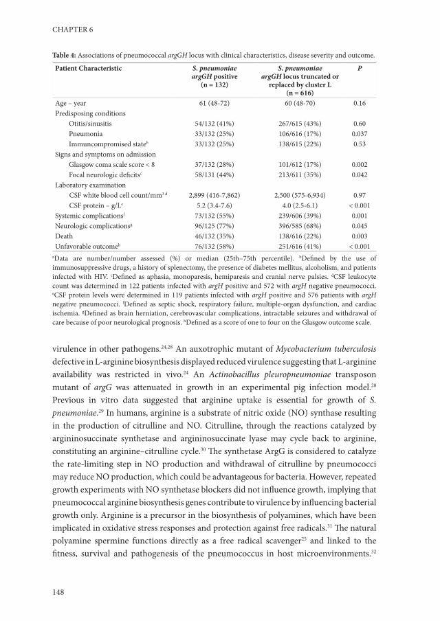
CHAPTER 6
148
Table 4: Associations of pneumococcal argGH locus with clinical characteristics, disease severity and outcome.
Patient Characteristic S. pneumoniaeargGH positive
(n = 132)
S. pneumoniaeargGH locus truncated or
replaced by cluster L(n = 616)
P
Age – year 61 (48-72) 60 (48-70) 0.16Predisposing conditions
Otitis/sinusitis 54/132 (41%) 267/615 (43%) 0.60Pneumonia 33/132 (25%) 106/616 (17%) 0.037Immuncompromised stateb 33/132 (25%) 138/615 (22%) 0.53
Signs and symptoms on admissionGlasgow coma scale score < 8 37/132 (28%) 101/612 (17%) 0.002Focal neurologic deficitsc 58/131 (44%) 213/611 (35%) 0.042
Laboratory examinationCSF white blood cell count/mm3 d 2,899 (416-7,862) 2,500 (575-6,934) 0.97CSF protein – g/Le 5.2 (3.4-7.6) 4.0 (2.5-6.1) < 0.001
Systemic complicationsf 73/132 (55%) 239/606 (39%) 0.001Neurologic complicationsg 96/125 (77%) 396/585 (68%) 0.045Death 46/132 (35%) 138/616 (22%) 0.003Unfavorable outcomeh 76/132 (58%) 251/616 (41%) < 0.001
aData are number/number assessed (%) or median (25th–75th percentile). bDefined by the use of immunosuppressive drugs, a history of splenectomy, the presence of diabetes mellitus, alcoholism, and patients infected with HIV. cDefined as aphasia, monoparesis, hemiparesis and cranial nerve palsies. dCSF leukocyte count was determined in 122 patients infected with argH positive and 572 with argH negative pneumococci. eCSF protein levels were determined in 119 patients infected with argH positive and 576 patients with argH negative pneumococci. fDefined as septic shock, respiratory failure, multiple-organ dysfunction, and cardiac ischemia. gDefined as brain herniation, cerebrovascular complications, intractable seizures and withdrawal of care because of poor neurological prognosis. hDefined as a score of one to four on the Glasgow outcome scale.
virulence in other pathogens.24,28 An auxotrophic mutant of Mycobacterium tuberculosis defective in L-arginine biosynthesis displayed reduced virulence suggesting that L-arginine availability was restricted in vivo.24 An Actinobacillus pleuropneumoniae transposon mutant of argG was attenuated in growth in an experimental pig infection model.28 Previous in vitro data suggested that arginine uptake is essential for growth of S. pneumoniae.29 In humans, arginine is a substrate of nitric oxide (NO) synthase resulting in the production of citrulline and NO. Citrulline, through the reactions catalyzed by argininosuccinate synthetase and argininosuccinate lyase may cycle back to arginine, constituting an arginine–citrulline cycle.30 The synthetase ArgG is considered to catalyze the rate-limiting step in NO production and withdrawal of citrulline by pneumococci may reduce NO production, which could be advantageous for bacteria. However, repeated growth experiments with NO synthetase blockers did not influence growth, implying that pneumococcal arginine biosynthesis genes contribute to virulence by influencing bacterial growth only. Arginine is a precursor in the biosynthesis of polyamines, which have been implicated in oxidative stress responses and protection against free radicals.31 The natural polyamine spermine functions directly as a free radical scavenger25 and linked to the fitness, survival and pathogenesis of the pneumococcus in host microenvironments.32
41973 Geldhoff_v3.indd 148 24-08-16 00:40

6
STREPTOCOCCUS PNEUMONIAE ARGININE SYNTHESIS GENES
149
We showed the ΔargGH strain being less resistant to oxidative stress. Polar effects by the knockout mutation on transcription from genes flanking argGH was excluded by RT-PCR. Transcription from SP_0109 (encoding ApbA) was lower in the argGH mutant, but a deletion of apbA had no effect on growth in low arginine concentration.29 In addition, in D39 the products of abpA, artP and abpB can substitute for each other.29 Antibiotics selectively blocking bacterial argininosuccinate synthetase and argininosuccinate lyase could inhibit pneumococcal growth, reducing disease severity.
Presence of arginine biosynthesis genes was associated with pneumococcal serotype 7F, but the association between these genes and clinical outcome even remained robust in a multivariate analysis including pneumococcal serotype. Individual serotypes and major clones of S. pneumoniae appear to differ markedly in their potential to cause invasive disease (80–120-fold variation).9 Serotype 7F has previously been associated with higher invasive disease potential, but with lower case-fatality rates.33,34 In the Netherlands, 7F has increased during the last decade, being the most frequent serotype in meningitis patients from 2007 onwards.35 Because of lack of heterogeneity between the OR for invasive disease of different clones of the same serotype, and analysis of isolates of the same genotype, but different serotype, it has been suggested that capsular serotype may be more important than genotype in the ability of pneumococci to cause invasive disease.9 The effect of argGH on disease outcome in patients with pneumococcal meningitis remained robust in a multivariate analysis including serotype.
Our findings are in line with the ‘avirulence hypothesis’ assuming parasites (bacteria, viruses, protozoa and helminths) will evolve to become avirulent.36 Pneumococci with the argGH locus truncated or replaced by cluster L are less fit and virulent. Virulence is considered as a maladaptation of new associations between parasites and hosts.36 During co-evolution with their host, microorganism may acquire functions that make them more fit in the interaction with their host.36 Genetic exchange with species sharing the same ecological niche is the main mechanism of evolution of S. pneumoniae.37
Supplementary data
Supplementary materials are available at The Journal of Infectious Diseases online (http://jid.oxfordjournals.org/content/suppl/2013/12/13/jit818.DC1). Supplementary materials consist of data provided by the author that are published to benefit the reader. The posted materials are not copyedited. The contents of all supplementary data are the sole responsibility of the authors. Questions or messages regarding errors should be addressed to the author.
41973 Geldhoff_v3.indd 149 24-08-16 00:40

CHAPTER 6
150
Declarations
Author ContributionsJ. R. P., A. vd. E. and D. vd. B. conceived and designed the experiments. J. R. P., M. G., B. D. v. S., M. C. B., A. vd. E. and D. vd. B. wrote the manuscript. J. R. P., B. D. v. S. and A. v. K. conducted bioinformatics analyses. M. G., and M. V. S. performed the mouse model experiments, A. H. Z. advised in statistical analyses. J. R. P., M. E. J. and F. B. performed the bacterial genome sequencing. All authors discussed the results and implications, and commented on the manuscript at all stages.
Financial supportThis work was supported by grants from the European Research Council (ERC Starting Grant [Proposal/Contract no. 281156] to D. van de Beek), Netherlands Organization for Health Research and Development (ZonMw; NWO-Veni grant 2006 [Proposal/Contract no. 916.76.023], NWO-Vidi grant 2010 [Proposal/Contract no. 016.116.358] to D. van de Beek), the Academic Medical Center (AMC Fellowship 2008 to D. van de Beek, AMC PhD scholarship 2008 to J.R. Piet). D. van de Beek and A. van der Ende participate in the Seventh Framework Programme of the European Commission, Proposal/Contract no.: 279185 (EUCLIDS -The genetic basis of meningococcal and other life threatening bacterial infections of childhood), A. van der Ende participated in the Sixth Framework Programme of the European Commission, Proposal/Contract no.: 512061 (Network of Excellence ‘European Virtual Institute for Functional Genomics of Bacterial Pathogens’.)
Potential conflicts of interestAll authors: No reported conflicts. All authors have submitted the ICMJE Form for Disclosure of Potential Conflicts of Interest. Conflicts that the editors consider relevant to the content of the manuscript have been disclosed.
AcknowledgementsWe are indebted to all Dutch physicians who participated in the study. We thank R. A. G. Huis in ‘t Veld for his technical assistance. We also would like to thank M. Giglio and S. Daugherty at the Institute of Genome Sciences for their support in annotating the genomes and depositing them at GenBank.
41973 Geldhoff_v3.indd 150 24-08-16 00:40

6
STREPTOCOCCUS PNEUMONIAE ARGININE SYNTHESIS GENES
151
References1. Poll Tvd, Opal SM. Pathogenesis, treatment, and prevention of pneumococcal pneumonia. The Lancet
2030;374:1543-56.2. McIntyre PB, O’Brien KL, Greenwood B, van de Beek D. Effect of vaccines on bacterial meningitis
worldwide. Lancet 2012;380:1703-11.3. van de Beek D, de Gans J, Spanjaard L, Weisfelt M, Reitsma JB, Vermeulen M. Clinical features and
prognostic factors in adults with bacterial meningitis. N Engl J Med 2004;351:1849-59.4. Brouwer MC, Tunkel AR, van de Beek D. Epidemiology, diagnosis, and antimicrobial treatment of acute
bacterial meningitis. Clin Microbiol Rev 2010;23:467-92.5. van de Beek D. Progress and challenges in bacterial meningitis. Lancet 2012;380:1623-4.6. Hoogman M, van de Beek D, Weisfelt M, de Gans J, Schmand B. Cognitive outcome in adults after bacterial
meningitis. J Neurol Neurosurg Psychiatry 2007;78:1092-6.7. Brouwer MC, de Gans J, Heckenberg SG, Zwinderman AH, van der Poll T, van de Beek D. Host genetic
susceptibility to pneumococcal and meningococcal disease: a systematic review and meta-analysis. Lancet Infect Dis 2009;9:31-44.
8. Woehrl B, Brouwer MC, Murr C, et al. Complement component 5 contributes to poor disease outcome in humans and mice with pneumococcal meningitis. J Clin Invest 2011;121:3943-53.
9. Brueggemann AB, Griffiths DT, Meats E, Peto T, Crook DW, Spratt BG. Clonal relationships between invasive and carriage Streptococcus pneumoniae and serotype- and clone-specific differences in invasive disease potential. J Infect Dis 2003;187:1424-32.
10. Enright MC, Spratt BG. A multilocus sequence typing scheme for Streptococcus pneumoniae: identification of clones associated with serious invasive disease. Microbiology 1998;144 ( Pt 11):3049-60.
11. Claverys JP, Havarstein LS. Extracellular-peptide control of competence for genetic transformation in Streptococcus pneumoniae. Front Biosci 2002;7:d1798-814.
12. Donati C, Hiller NL, Tettelin H, et al. Structure and dynamics of the pan-genome of Streptococcus pneumoniae and closely related species. Genome Biol 2010;11:R107.
13. Darling AC, Mau B, Blattner FR, Perna NT. Mauve: multiple alignment of conserved genomic sequence with rearrangements. Genome Res 2004;14:1394-403.
14. Kent WJ. BLAT--the BLAST-like alignment tool. Genome Res 2002;12:656-64.15. Trzcinski K, Thompson CM, Lipsitch M. Construction of otherwise isogenic serotype 6B, 7F, 14, and 19F
capsular variants of Streptococcus pneumoniae strain TIGR4. Appl Environ Microbiol 2003;69:7364-70.16. Sung CK, Li H, Claverys JP, Morrison DA. An rpsL cassette, janus, for gene replacement through negative
selection in Streptococcus pneumoniae. Appl Environ Microbiol 2001;67:5190-6.17. Trzcinski K, Thompson CM, Srivastava A, Basset A, Malley R, Lipsitch M. Protection against nasopharyngeal
colonization by Streptococcus pneumoniae is mediated by antigen-specific CD4+ T cells. Infect Immun 2008;76:2678-84.
18. Hartel T, Klein M, Koedel U, Rohde M, Petruschka L, Hammerschmidt S. Impact of glutamine transporters on pneumococcal fitness under infection-related conditions. Infect Immun 2011;79:44-58.
19. Mook-Kanamori B, Geldhoff M, Troost D, van der Poll T, van de Beek D. Characterization of a pneumococcal meningitis mouse model. BMC Infect Dis 2012;12:71.
20. Dessing MC, Florquin S, Paton JC, van der Poll T. Toll-like receptor 2 contributes to antibacterial defence against pneumolysin-deficient pneumococci. Cell Microbiol 2008;10:237-46.
21. Krogh A, Larsson B, von Heijne G, Sonnhammer EL. Predicting transmembrane protein topology with a hidden Markov model: application to complete genomes. J Mol Biol 2001;305:567-80.
22. Lawrence JG, Ochman H. Amelioration of bacterial genomes: rates of change and exchange. J Mol Evol 1997;44:383-97.
23. Bruckner R, Nuhn M, Reichmann P, Weber B, Hakenbeck R. Mosaic genes and mosaic chromosomes-genomic variation in Streptococcus pneumoniae. Int J Med Microbiol 2004;294:157-68.
24. Gordhan BG, Smith DA, Alderton H, McAdam RA, Bancroft GJ, Mizrahi V. Construction and phenotypic characterization of an auxotrophic mutant of Mycobacterium tuberculosis defective in L-arginine biosynthesis. Infect Immun 2002;70:3080-4.
25. Chattopadhyay MK, Tabor CW, Tabor H. Polyamines protect Escherichia coli cells from the toxic effect of oxygen. Proc Natl Acad Sci U S A 2003;100:2261-5.
41973 Geldhoff_v3.indd 151 24-08-16 00:40
CHAPTER 6
152
26. Pozzi G, Masala L, Iannelli F, et al. Competence for genetic transformation in encapsulated strains of Streptococcus pneumoniae: two allelic variants of the peptide pheromone. J Bacteriol 1996;178:6087-90.
27. Burrus V, Pavlovic G, Decaris B, Guedon G. Conjugative transposons: the tip of the iceberg. Mol Microbiol 2002;46:601-10.
28. Sheehan BJ, Bosse JT, Beddek AJ, Rycroft AN, Kroll JS, Langford PR. Identification of Actinobacillus pleuropneumoniae genes important for survival during infection in its natural host. Infect Immun 2003;71:3960-70.
29. Kloosterman TG, Kuipers OP. Regulation of arginine acquisition and virulence gene expression in the human pathogen Streptococcus pneumoniae by transcription regulators ArgR1 and AhrC. J Biol Chem 2011;286:44594-605.
30. Haines RJ, Pendleton LC, Eichler DC. Argininosuccinate synthase: at the center of arginine metabolism. Int J Biochem Mol Biol 2011;2:8-23.
31. Ha HC, Sirisoma NS, Kuppusamy P, Zweier JL, Woster PM, Casero RA, Jr. The natural polyamine spermine functions directly as a free radical scavenger. Proc Natl Acad Sci U S A 1998;95:11140-5.
32. Shah P, Nanduri B, Swiatlo E, Ma Y, Pendarvis K. Polyamine biosynthesis and transport mechanisms are crucial for fitness and pathogenesis of Streptococcus pneumoniae. Microbiology 2011;157:504-15.
33. Jansen AG, Rodenburg GD, van der Ende A, et al. Invasive pneumococcal disease among adults: associations among serotypes, disease characteristics, and outcome. Clin Infect Dis 2009;49:e23-9.
34. Weinberger DM, Harboe ZB, Sanders EA, et al. Association of serotype with risk of death due to pneumococcal pneumonia: a meta-analysis. Clin Infect Dis 2010;51:692-9.
35. Rodenburg GD, de Greeff SC, Jansen AG, et al. Effects of pneumococcal conjugate vaccine 2 years after its introduction, the Netherlands. Emerg Infect Dis 2010;16:816-23.
36. Adiba S, Nizak C, van Baalen M, Denamur E, Depaulis F. From grazing resistance to pathogenesis: the coincidental evolution of virulence factors. PLoS One 2010;5:e11882.
37. Croucher NJ, Walker D, Romero P, et al. Role of conjugative elements in the evolution of the multidrug-resistant pandemic clone Streptococcus pneumoniaeSpain23F ST81. J Bacteriol 2009;191:1480-9.
41973 Geldhoff_v3.indd 152 24-08-16 00:40

6
STREPTOCOCCUS PNEUMONIAE ARGININE SYNTHESIS GENES
153
Supplementary tables and figures
Supplementary Table 1: Genome assembly strategies.
Bacterial genome de novo 0 errors de novo 2 errors Reference mapping CabogAMCSP01 93 80 136 (NC_008533) 80AMCSP02 56 61 105 (NC_011072) 65AMCSP03 75 72 227 (NC_003098) 280AMCSP04 98 87 128 (NC_003098) 112AMCSP05 155 148 198 (NC_008533) 240AMCSP06 83 90 118 (NC_008533) 75AMCSP07 66 70 108 (NC_008533) 92AMCSP08 149 110 119 (NC_003098) 81AMCSP09 144 139 72 (NC_003028) 83AMCSP10 47 46 98 (NC_011072) 72AMCSP11 88 86 117 (NC_003098) 70AMCSP12 87 90 131 (NC_011072) 90AMCSP13 141 149 85 (NC_003098) 100AMCSP14 136 133 150 (NC_008533) 95AMCSP15 64 81 101 (NC_011072) 83AMCSP16 60 57 105 (NC_008533) 81AMCSP17 41 49 100 (NC_008533) 102AMCSP18 104 * 33 (NC_003028) 144AMCSP19 59 65 113 (NC_008533) 86AMCSP20 84 82 135 (NC_008533) 92
The number of contigs is shown per assembly strategy: Newbler de novo assembler with 0 and 2 errors allowed in the MID calling, a Newbler Reference Mapper assembly (all reference strain GenBank accession numbers producing the least amounts of contigs in brackets: S. pneumoniae TIGR4 (NC_003028), S. pneumoniae R6 (NC_003098), S. pneumoniae D39 (NC_008533), S. pneumoniae Hungary 19A-6 (NC_010380), S. pneumoniae CGSP14 (NC_010582), S. pneumoniae G54 (NC_011072), S. pneumoniae Spain23F (NC_011900) S. pneumoniae JJA (NC_012466), S. pneumoniae P1031 (NC_012467), S. pneumoniae 70585 (NC_012468) and S. pneumoniae Taiwan 19F-14 (NC_012469), and a Cabog assembly. *Because this strain was sequenced with MID 16 (TCACGTACTA) which has close resemblance to MID 18, no de novo assembly with 2 errors allowed in the MID calling procedure was made.
Supplementary Table 2: Available at http://jid.oxfordjournals.org/content/209/11/1781/suppl/DC1.
41973 Geldhoff_v3.indd 153 24-08-16 00:40

CHAPTER 6
154
Supplementary Table 3: Primers used for cluster determination and constructing bacterial knock-outs.
Primer name Orientation DNA sequence Target Cluster
Cluster determinationJP_pneu_01 Forward CCCTGACCCTGCTAGCTCTT AJP_pneu_02 Reverse CTGACGGCCTTACTGATTGG AJP_pneu_07 Forward GATTGGGCTACCCTTTGACA FJP_pneu_08 Reverse TGGATAATCAAGTCCATGCTC FJP_pneu_11 Forward ATGTCGCAAAAAGAGGGTCA GJP_pneu_12 Reverse TTTCAACATCTCTGACAAAGTCAA GJP_pneu_15 Forward CTATTGCTGGTTTGGGTGGT HJP_pneu_16 Reverse CAATTGGCATTCCAAAAGAAA HJP_pneu_17 Forward ATGATTTGGAAATCTTAGCGAAA IJP_pneu_18 Reverse TCATTGATCGTTCCCCATTT IJP_pneu_21 Forward TGGCAGCCACTTGTTTATCC BJP_pneu_22 Reverse TGAACGCGAGTAAATGCTTG BJP_pneu_23 Forward TCCATGGCATTACACCTGAA CJP_pneu_24 Reverse GCTTGCTCGTGCATCTGATA CJP_pneu_25 Forward AATCTTGGAGTGGCAACAGG DJP_pneu_26 Reverse TCCTTTTGGAGGACGACTGT DJP_pneu_27 Forward ACAAAGTGGCCGACTACACC JJP_pneu_28 Reverse CAGTGAACTGCCCAATAGCA JJP_pneu_31 Forward TGCTTCGTTTTTGTTGGTTAAA KJP_pneu_32 Reverse CTTGTCACCGGAATCAAACA KJP_pneu_33 Forward GAACTCCCTCTCATGCCATC LJP_pneu_34 Reverse AGAGGCAAAAGCAACGGTTT LJP_pneu_35 Forward CACTACCAGGGAGGGAAACA MJP_pneu_36 Reverse ATCGCAAGTCCCATCACTTC MJP_pneu_37 Forward TTTGTTGTGGTCGATTGTGG EJP_pneu_38 Reverse GTTGCAGAATCTCCCAAGGA EJP_pneu_39 Forward TCTGAGGGACAACGTCAGC NJP_pneu_40 Reverse CCGTACCATCACGTCTTCCT NJP_pneu_43 Forward TGGGTAGAGCGCTTTGGTGCG argHJP_pneu_44 Reverse GCCCATAAACTCTGCCAGTCTTTCC argHJP_pneu_45 Forward TCTGCCTTGAGCCGCCCTCT argGJP_pneu_48 Reverse TTTGCAGTCCCATTGACAAC argG
Constructing bacterial knock-outsKSPNArg2061617F8 GGCTGGAATTGAAGCCKSPNArg2061617R6 CGAAACCGCCACAATAGGATCCAAAATTACKSPNArg2061617F7 ATTAAAGGGCCCATGATGCATATGAGTCGKSPNArg2061617R9_1 CGACTTGGTCAAACCKSPNArg2061617F1KSjanuskanaR1
GCTATCGCAACAGAACTAGGAAAGATTATATCACATTATCCATT
41973 Geldhoff_v3.indd 154 24-08-16 00:40

6
STREPTOCOCCUS PNEUMONIAE ARGININE SYNTHESIS GENES
155
Primer name Orientation DNA sequence Target Cluster
RT PCRKSABCFKSABCRKSarghypFKsarghypRGki upGki dn
GAACGGAGCAAGGTGTTTGAAATAACGGCATCCACTTGCAGATCGTGGTGGGTCTTGAGGGCAAGACAACCCCAAAATAGGCATTGGAATGGGATCACCTCTCCCGCAGCTGACAC
For each gene (rows), the frequency per genome is scored (columns).
41973 Geldhoff_v3.indd 155 24-08-16 00:40

41973 Geldhoff_v3.indd 156 24-08-16 00:40

7Cerebrospinal fluid inflammatory
mediators and outcome in patients with
pneumococcal meningitis
Geldhoff M, Brouwer MC, Valls-Seron M, Ferwerda B, Zwinderman AH, van der Ende A, van de Beek D.
Submitted for publication
41973 Geldhoff_v3.indd 157 24-08-16 00:40

CHAPTER 7
158
Abstract
Background. The severity of the cerebrospinal fluid (CSF) inflammatory response in pneumococcal meningitis has been associated with brain damage and unfavorable outcome.
Methods. We analyzed 88 inflammatory mediators in the CSF of adult patients with pneumococcal meningitis included in a prospective cohort study (MeninGene). We compared CSF levels of inflammatory mediators between patients with favorable and unfavorable outcome, defined as a Glasgow Outcome Scale (GOS) score below 5. We used CSF of patients diagnosed with thunderclap headache as negative controls.
Results. CSF was available from 271 patients with pneumococcal meningitis and 19 controls. Unfavorable outcome was reported in 109 patients (40%), and 35 patients (13%) died. After Bonferroni correction for multiple testing (p <0.0057) 59 mediators were significantly elevated in the CSF of pneumococcal meningitis patients compared to negative controls. CSF levels of sICAM-1, MMP-1, MMP-3, IL-1RA, IL-10, CXCL5 and CCL7 were associated with unfavorable outcome. High CSF levels of sICAM-1 and MMP-1 were associated with mortality.
Conclusion. We identified an adhesion molecule (sICAM-1), two matrix metalloproteinases (MMP-1 and MMP-3), two chemokines (CXCL5 and CCL7) and two anti-inflammatory cytokines (IL-10 and IL-1RA) to be associated with unfavorable outcome in patients with pneumococcal meningitis, which suggests they might be involved in the pathophysiology of this disease.
41973 Geldhoff_v3.indd 158 24-08-16 00:40

7
CEREBROSPINAL FLUID INFLAMMATORY MEDIATORS
159
Introduction
Bacterial meningitis is an infection of the membranes surrounding the brain and is associated with high mortality and morbidity rates.1,2 Streptococcus pneumoniae is the most common causative agent of bacterial meningitis in adults, causing 70% of cases.3,4 The reported incidence of pneumococcal meningitis has lowered to 0.8 per 100,000 persons per year, since routine vaccination of children with the seven- and ten-valent pneumococcal conjugate vaccines was introduced in the Netherlands in 2006 and 2011.4 In low-income countries the incidence remains substantially higher.5 In high-income countries, the mortality rate among patients with pneumococcal meningitis in adults is 18%, and up to half of surviving patients suffer from neurologic sequelae including cognitive impairment. 3,6
Experimental studies showed that brain damage and adverse outcome results mainly from the central nervous system (CNS) inflammatory response caused by the interaction between host and pathogen.7,8,9,10 Several inflammatory mediators have been found elevated in the CSF in patients with pneumococcal meningitis.11,12 Comprehensive prospective studies analyzing the association between large number of different cytokines, chemokines or endothelial growth factors with outcome are not available. In 2006, we started a prospective cohort study to identify and characterize host genetic traits and bacterial genetic factors controlling occurrence and outcome of bacterial meningitis (MeninGene). 4,13-15 In this study we also collected CSF of the diagnostic lumbar puncture of patients with bacterial meningitis. To gain more insight in the pathophysiology of pneumococcal meningitis to identify potential new targets for adjuvant treatments, we performed an explorative study analyzing 88 different inflammatory mediators in the CSF of patients with pneumococcal meningitis.
Material and methods
Dutch bacterial meningitis cohortWe conducted a prospective nationwide cohort study in the Netherlands including adult patients (over 16 years of age) with community acquired bacterial meningitis confirmed by positive CSF cultures. Patients were identified by the Netherlands Reference Laboratory for Bacterial Meningitis (NRLBM) from March 2006 to August 2010, which receives bacterial strains from approximately 85% of all patients with bacterial meningitis in the Netherlands. The treating physician was contacted, and informed consent was obtained from all participating patients or their legally authorized representatives.
Patients with hospital-acquired bacterial meningitis, those with a neurosurgical device or negative CSF cultures were excluded. Clinical information on the disease course was collected in a secured online case-record form. Outcome was assessed at discharge by the treating physician using the Glasgow Outcome Scale (GOS), a 5 point scale in which a
41973 Geldhoff_v3.indd 159 24-08-16 00:40

CHAPTER 7
160
score of 5 indicates mild or no disability (the patient is able to return to work or school), a score of 4 indicates moderate disability (able to live independently), a score of 3 indicates severe disability (unable to live independently), a score of 2 represents a vegetative state and a score of 1 death. A score of 1-4 was defined as unfavorable outcome, a score of 5 as favorable outcome.
When leftover CSF from the first diagnostic lumbar puncture was available, it was spun down and supernatant was locally stored at -80 degrees for study purposes. Participating centers were regularly visited by the investigators to collect the CSF samples and transport them to the Academic Medical Center Amsterdam on dry ice for further storage until measurements were performed. Samples were thawed, spun down and the supernatant was aliquoted in 30 µl volumes. CSF samples from 19 patients with thunderclap headache in whom subarachnoidal hemorrhage was excluded by CSF examination, served as negative controls. All these CSF samples had normal leukocyte count, protein and glucose levels. The study was approved by the Medical Ethics Committee (MEC) of the Academic Medical Center Amsterdam.
Analysis of inflammatory mediatorsWe measured 88 inflammatory mediators in CSF of 271 patients with pneumococcal meningitis and 19 negative controls. Analytes included CCL1 to CCL5, CCL7, CCL8, CCL13, CCL14a, CCL15, CCL17, CCL19 to CCL22, CCL24, CCL26, CCL27, CXCL1, CXCL5 to CXCL7, CXCL9 to CXCL11, CXCL12a and b, CXCL13, XCL1, CX3CL1, interleukin (IL)-1-α, IL-1-β, interleukin-1 receptor antagonist (IL-1RA), IL-2 to IL-11, IL-12p40, IL12p70, IL-13, IL-15 to IL-18, IL-20, IL-21, IL-23, IL-28A, IL-29, IL-33, interferon (IFN)-α2, IFN-γ, leukemia inhibitory factor (LIF), thyroid peroxidase (TPO), tumor necrosis factor –related apoptosis-inducing ligand (TRAIL), stem cell factor (SCF), thymic stromal lymphopoietin (TSLP), macrophage colony stimulating factor (M-CSF), epidermal growth factor (EGF), eotaxin, fibroblast growth factor (FGF)-2, FMS-related tyrosine kinase (Flt)-3 ligand, granulocyte-colony stimulating factor (G-CSF), granulocyte macrophage-colony stimulating factor (GM-CSF), platelet derived growth factor (PDGF)-AA, PDGF-AB/BB, soluble cluster of differentiation 40 ligand (sCD40L), soluble interleukin-2 receptor alpha (sIL-2Ra; sCD25), transforming growth factor alpha (TGF-α), tumor necrosis factor alpha (TNF)-α, TNF-β, vascular endothelial growth factor (VEGF), soluble vascular cell adhesion molecule-1 (sVCAM-1), soluble intercellular adhesion molecule (sICAM)-1, matrix metalloproteinase (MMP)-1 to MMP-3, MMP-7, MMP-9, MMP-10, MMP-12, MMP-13 and macrophage migration inhibitory factor (MIF). This selection was based on the availability of the combination of specific cytokines and chemokines of our interest included in Luminex assays by the manufacturer. All analytes were measured with Luminex® xMAP® technology using Milliplex® map multiplex assay’s (Millipore Billerica, MA, USA). CSF was diluted 1:50 in assay buffer provided with the Multiplex assay and inflammatory mediators were measured according to the manufacturer’s instructions. Plates were read with the Bio-Plex® 200 system array reader.
41973 Geldhoff_v3.indd 160 24-08-16 00:40

7
CEREBROSPINAL FLUID INFLAMMATORY MEDIATORS
161
Bio-Plex Manager™ software was used for data acquisition and analysis (Bio-Rad Hercules, CA, USA). All concentrations are given in pg/ml.
StatisticsData below the lower limit of quantification (LLOQ) of the assay were imputed with a random value between zero and the individual LLOQ of the analyte. Data above the upper limit of quantification (ULOQ) of the assay were assigned the value of the ULOQ of the individual analyte. Levels of inflammatory mediators were compared between groups with a Mann-Whitney U test. Continuous data are presented as median with interquartile range. All statistical tests were 2-tailed, and a Bonferroni-corrected P-value < 0.00057 (0.05/88) was considered significant. Spearman’s correlation test was used to identify correlations between levels of immune mediators in the CSF. Analyses were done using IBM SPSS statistics version 22.0. Statistical correction for batch effect was performed. We conducted a principal component analysis (PCA) and linear determinant analysis (LDA) after log transformation and standardizing the data, to identify clusters of immune mediators correlated to outcome, using R statistics version 3.2.3.16 We conducted a supervised pathway analysis using QIAGEN’s Ingenuity Pathway Analysis (IPA).
Results
Study cohortWe identified 1115 episodes of bacterial meningitis in 1100 patients with bacterial meningitis through the NRLBM between January 2006 and November 2010. Of 1115 episodes, in 75 cases CSF culture was negative, 21 cases had hospital-acquired meningitis and in 63 cases no informed consent was obtained. Of 956 cases included in the study 689 (72%) had S. pneumoniae as a causative agent. CSF from the diagnostic lumbar puncture was available for analysis in 271 of 689 patients (39%) (figure 1). Patients with CSF available, had similar baseline characteristics as patients of patients without CSF available, except for a lower mortality rate (22% versus 13%; p = 0.002; table 1).
Median age of patients with available CSF samples was 61 years, and 128 patients (47%) were male, 126 (48%) had symptoms less than 24 hours and 107 (66%) had a predisposing condition such as sinusitis, otitis, pneumonia, or an immunocompromised state. At presentation 200 patients (80%) had fever, 218 (80%) had an altered mental status, 58 (23%) had focal neurological deficits and 57 (21%) showed signs of septic shock. CSF of patients with pneumococcal meningitis showed an elevated leukocyte count of 2,560 cells/mm3 (IQR 514 – 6,655), high protein content of 4.0 g/L (IQR 2.5 – 6.0), and low glucose CSF-blood ratio 0.023 (IQR <0.001 – 0.233). Outcome was unfavorable in 109 patients (40%) and 35 patients (13%) died (table 1).
41973 Geldhoff_v3.indd 161 24-08-16 00:40

CHAPTER 7
162
Figure 1. Flow chart of patients included in the MeninGene study and negative controls.
CSF inflammatory mediators in patients with pneumococcal meningitisInflammatory mediators were successfully measured in 98-100% of samples. Invalid measurements were due to technical aspects such as too small amounts of CSF available for multiple assays or clogging of the luminex beads. Of the inflammatory mediators measured 59 were elevated in the CSF of patients with pneumocccal meningitis compared to negative controls (supplementary table 1). The ten most significantly elevated analytes included MMP-9 (median 497,193 versus 109 pg/ml; p-value 2.69x10⁻²¹), CCL2 (median 99,802 versus 465 pg/ml; p-value 1.49x10⁻¹⁶), IL-6 (median 78,636 versus 13 pg/ml; p-value 4.97x10⁻¹³), G-CSF (median 16,372 versus 55 pg/ml; p-value 8.59x10⁻¹³), CXCL1 (median 21,574 versus 55 pg/ml; p-value 1.30x10⁻¹²), CXCL8 (median 53,241 versus 27 pg/ml; p-value 1.41x10⁻¹²), sICAM-1 (median 60,375 versus 2,292 pg/ml; p-value 1.75x10⁻¹²), IL-10 (median 1,868 versus 4 pg/ml; p-value 2.14x10⁻¹²), IL-1β (median 1,875 versus 4 pg/ml; p-value 2.21x10⁻¹²) and IL-1RA (median 4,030 versus 22 pg/ml; p-value 3.18x10⁻¹²).
41973 Geldhoff_v3.indd 162 24-08-16 00:40

7
CEREBROSPINAL FLUID INFLAMMATORY MEDIATORS
163
Table 1. Baseline characteristics of patients with CSF sample compared to patients without a CSF sample available.
Characteristic Patients with CSF sample (N = 271)
Patients without CSF sample (N = 418)
P-value
General Age 61 (49 – 70) 61 (50 – 69) 0.89 Male sex 128/271 (47%) 209/418 (50%) 0.48 Duration of symptoms < 24 hours 126/263 (48%) 198/405 (49%) 0.81Predisposing conditions Otitis 90/263 (34%) 139/397 (35%) 0.84 Sinusitis 37/260 (14%) 53/391 (14%) 0.50 Pneumonia 25/262 (10%) 48/402 (12%) 0.34 Immunocompromised statea 82/271 (30%) 105/418 (25%) 0.14Presenting symptoms Temperature > 38 °C 200/249 (80%) 322/392 (82%) 0.56 Focal neurological deficits 58/255 (23%) 96/393 (25%) 0.62 Signs of septic shockb 57/267 (21%) 102/409 (24%) 0.28 Altered mental status (GCS < 14) 218/271 (80%) 329/418 (79%) 0.58Cerebrospinal fluid White blood cell count (/mm3) 2560 (514 – 6655) 2800 (549 – 7339) 0.40 White blood cell count <1000/mm3 124/387 (32%) 0.58 Protein (g/L) 4.0 (2.5 – 6.0) 4.3 (2.5 – 6.3) 0.24 CSF blood glucose ratio 0.023 (0.00 – 0.23) 0.016 (0.00 – 0.15) 0.05Clinical course Neurologic complicationsc 163/207 (79%) 262/324 (81%) 0.55 Systemic complicationsd 117/266 (44%) 181/409 (44%) 0.95 Unfavorable outcome 109/271 (40%) 180/418 (43%) 0.46 Death 35/271 (13%) 92/418 (22%) 0.002
Data are number/number evaluated (%), and median (interquartile range). aImmunocompromised state was defined as the use of immunosuppressive drugs, the presence of diabetes mellitus or alcoholism, and human immunodeficiency virus (HIV) infection. b Signs of septic shock were defined as a systolic blood pressure <90 mmHg, a diastolic blood pressure <60 mmHg and/or heart rate >120/min. cNeurologic complications were defined as an altered mental status, focal neurologic deficits, hydrocephalus, seizures, cerebrovascular events (infarction, hemorrhage, cerebral venous sinus thrombosis), cerebral abscess or empyema during admission. dSystemic complications were defined as septic shock (diastolic blood pressure <60 mmHg), respiratory failure and need for mechanical ventilation during admission.
CSF inflammatory mediators associated with outcome in pneumococcal meningitisAfter Bonferroni correction, unfavorable outcome in pneumococcal meningitis was associated with high CSF levels of MMP-1 (median 4,441 versus 1,530 pg/ml; p-value 4.95x10⁻¹⁰), sICAM-1 (median 78,580 versus 50,530 pg/ml; p-value 9.36x10-07), IL-1RA (median 6,122 versus 2,879 pg/ml; p-value 9.36x10-06), CXCL5 (median 2,410 versus 681 pg/ml; p-value 1.41x10-05), IL-10 (median 2,787 versus 1,204 pg/ml; p-value 2.15x10-05), MMP-3 (median 74,491 versus 21,286 pg/ml; p-value 9.42x10-05) and CCL7 (median 1,694 versus 920 pg/ml; p-value 3.20x10-04) (table 2). All of these analytes were elevated compared to negative controls except CXCL5 (median 854 pg/ml versus 577 pg/ml; p-value 8.89x10-03). CSF levels of CXCL5 were elevated above the LLOQ in only 40% of patients. Results of all inflammatory mediators in patients with favorable versus unfavorable outcome are listed in supplementary table 2.
41973 Geldhoff_v3.indd 163 24-08-16 00:40

CHAPTER 7
164
Mortality in pneumococcal meningitis was associated with high CSF levels of sICAM-1 (median 125,860 versus 55,230 pg/ml; p-value 1.49x10-06) and MMP-1 (median 8,904 versus 2,101 pg/ml; p-value 1.11x10-05) (table 2). Both sICAM-1 (71,875 pg/ml versus 52,810 pg/ml; p-value 1.19x10-04) and MMP-1 (3483 pg/ml versus 1612 pg/ml; p-value 1.40x10-05) levels were higher in patients with systemic complications (defined as septic shock (diastolic blood pressure <60 mmHg), respiratory failure and need for mechanical ventilation during admission). We did not identify CSF inflammatory mediators that were associated with neurologic complications, hearing loss or cognitive impairment (data not shown). Correction for batch effect did not influence results (data not shown).
IL-1RA blocks the binding of IL-1α and IL-1β to the IL-1 receptor. Previous human sepsis studies reported that a 100-1000 fold increase of IL-1RA levels compared to IL-1β levels are needed to antagonize the effect of IL-1β.17,18 In our population IL-1RA/IL-1β ratio was higher in patients with unfavorable outcome compared to patients with favorable outcome (ratio 2.52 versus 1.65; p-value 0.018) and in deceased compared to patients that survived (ratio 3.59 versus 1.73; p-value 0.028). However the IL-1RA/IL-1β ratio was substantially lower than the previously reported 100-fold relative increase of IL-1RA needed to antagonize the effect of IL-1β.
Table 2. CSF concentrations of inflammatory mediators in patients with pneumococcal meningitis.
OUTCOMEAnalyte Favorable (n = 162) Unfavorable (n = 109) p-valueMMP-1 1530 (520 - 3,506) 4,441 (1,822 - 9,578) 4.95x10⁻¹⁰
sICAM-1 50,530 (29,110 - 78,095) 78,580 (47,375 - 135,260) 9.36x10-07
IL-1RA 2,879 (905 - 6,777) 6,122 (3,325 - 11,492) 9.36x10-06
CXCL5 681 (318 - 1,492) 2,410 (659 - 4,536) 1.41x10-05
IL-10 1,204 (464 - 3,270) 2,787 (1,308 - 5,899) 2.15x10-05
MMP-3 21,286 (6,487 - 77,267) 74,491 (16,062 - 159,301) 9.43x10-05
CCL7 920 (377 - 2,042) 1,694 (726 - 3,955) 3.20x10-04
SURVIVALAnalyte Survived (n = 236) Died (n = 35) p-valuesICAM-1 55,230 (34,720 - 84,610) 125,860 (69,300 - 183,480) 1.49x10-06
MMP-1 2,101 (826 - 4,601) 8,904 (2,605 - 12,096) 1.11x10-05
CSF concentrations (pg/ml) of inflammatory mediators in patients with pneumococcal meningitis with favorable (GOS 5) versus unfavorable outcome (GOS ≤4) and patients that survived versus patients that died. Data are given as median with interquartile range.
Correlations with patient characteristics and other inflammatory mediatorsInflammatory mediators in CSF were not substantially correlated to blood or CSF leukocyte count (data not shown). We found moderate to strong positive correlations between the seven inflammatory mediators associated with outcome and CSF protein content, and moderate negative correlations between these inflammatory mediators and CSF blood-glucose ratio (table 3). The seven inflammatory mediators associated with outcome, were not associated with age or GCS on admission (data not shown). Among the inflammatory
41973 Geldhoff_v3.indd 164 24-08-16 00:40
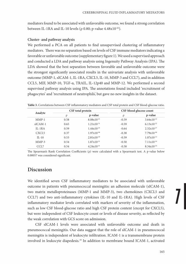
7
CEREBROSPINAL FLUID INFLAMMATORY MEDIATORS
165
mediators found to be associated with unfavorable outcome, we found a strong correlation between IL-1RA and IL-10 levels (ρ 0.80; p-value 4.48x10-62).
Cluster- and pathway analysisWe performed a PCA on all patients to find unsupervised clustering of inflammatory mediators. There was no separation based on levels of CSF immune mediators indicating a favorable or unfavorable outcome (supplementary figure 1). We used a supervised approach and conducted a LDA and pathway analysis using Ingenuity Pathway Analysis (IPA). The LDA showed that the best separation between favorable and unfavorable outcome were the strongest significantly associated results in the univariate analysis with unfavorable outcome (MMP-1, sICAM-1, IL-1RA, CXCL5, IL-10, MMP-3 and CCL7), and in addition CCL5, MIF, MMP-10, TGF-α, TRAIL, IL-12p40 and MMP-12. We performed a second supervised pathway analysis using IPA. The annotations found included ‘recruitment of phagocytes’ and ‘recruitment of neutrophils’, but gave no new insights in the dataset.
Table 3. Correlations between CSF inflammatory mediators and CSF total protein and CSF blood-glucose ratio.
AnalyteCSF total protein CSF-blood glucose count
ρ p-value ρ p-valueMMP-1 0.58 8.08x10⁻²⁵ -0.59 3.64x10⁻²²
sICAM-1 0.65 1.21x10⁻³¹ -0.59 6.13x10⁻²²
IL-1RA 0.59 1.44x10⁻²⁵ -0.64 2.32x10⁻²⁷
CXCL5 0.37 1.97x10-08 -0.38 7.79x10-08
IL-10 0.53 2.81x10⁻²⁰ -0.59 1.07x10⁻²²
MMP-3 0.54 1.87x10⁻²¹ -0.50 7.11x10⁻¹⁶
CCL7 0.54 4.24x10⁻²¹ -0.58 8.34x10⁻²²
The Spearman’s Rank Correlation Coefficients (ρ) were calculated with a Spearman’s test. A p-value below 0.00057 was considered significant.
Discussion
We identified seven CSF inflammatory mediators to be associated with unfavorable outcome in patients with pneumococcal meningitis: an adhesion molecule (sICAM-1), two matrix metalloproteinases (MMP-1 and MMP-3), two chemokines (CXCL5 and CCL7) and two anti-inflammatory cytokines (IL-10 and IL-1RA). High levels of CSF inflammatory mediator levels correlated with markers of severity of the inflammation, such as low CSF blood-glucose ratio and high CSF protein content (except for CXCL5), but were independent of CSF leukocyte count or levels of disease severity, as reflected by the weak correlation with GCS score on admission.
CSF sICAM-1 levels were associated with unfavorable outcome and death in pneumococcal meningitis. Our data suggest that the role of sICAM-1 in pneumococcal meningitis is independent of leukocyte infiltration. ICAM-1 is a transmembrane protein involved in leukocyte diapedesis.19 In addition to membrane bound ICAM-1, activated
41973 Geldhoff_v3.indd 165 24-08-16 00:40

CHAPTER 7
166
cells are able to produce a soluble form of ICAM-1 which is secreted.20 The function of circulating sICAM-1 is yet unknown. Soluble ICAM-1 can bind to the receptor lymphocyte function-associated antigen (LFA)-1, suggesting a role in inhibiting the binding of leukocytes to endothelial cells, but it also may also be released as a result of tissue damage, thereby promoting inflammation.20 In our study we found no correlation between CSF sICAM-1 concentration and CSF leukocyte count. The lack of association between sICAM-1 and leukocyte count is in line with a previous study showing a similar degree of neutrophil infiltration in the lung in ICAM-1 deficient mice compared to wild type mice in a pneumocccal pneumonia model.21
We identified two matrix metalloproteinases (MMP-1 and MMP-3) to be associated with outcome. MMP-1 is a collagenase whose production can be induced by several cytokines such as IL-1β, IL-31 or activation of Toll-like receptor (TLR)2.22,23,24 Substrates of MMP-1 include multiple extracellular components, but also several inflammatory components including L-selectin, pro-TNF, IL-1β, MMP-2, MMP-9 and several chemokines of which some are cleaved into receptor antagonists and may exert an anti-inflammatory effect.25 Not much is known about the role of MMP-1 in pneumococcal disease. A selective MMP blocker of MMP-1, -8 and -13 was used in a pneumococcal meningitis model in rats, showing reduced mortality, CSF TNF-a and IL-1β concentrations and less hippocampal apoptosis and cortical necrosis on brain pathology.26 To what extent blocking of MMP-1 is responsible for these effects is unknown. MMP-3 degrades multiple extracellular matrix components and can activate several other MMP’s such as MMP-1, MMP-7 and MMP-9.25 The function of MMP-3 and its role in invasive pneumococcal disease is unclear. In a rat model of pneumococcal meningitis MMP-3 mRNA was upregulated in brain tissue.27 Furthermore, a role for MMP-3 in the activation of MMP-1 has been described,25 which was also associated with unfavorable outcome in our study.
IL-1RA is a receptor antagonist that prevents binding of both IL-1α and IL-1β to the IL-1 receptor, resulting in a reduced release of IL-6, TNF and IL-1 by monocytes, and subsequent reduced recruitment of neutrophils.18 IL-1RA was previously shown to be present in the CSF of patients with bacterial meningitis and high levels were associated with fatal disease 28. From experimental studies we know that IL-1RA plays a role in the prevention of host tissue damage during pneumococcal infection.29,30 In human sepsis studies and in vitro, a 100-1000 fold increase of IL-1RA levels compared to IL-1β levels was shown to fully antagonize the effect of IL-1β.17,18 In our study we found higher IL-1RA/IL-1β ratio in patients with unfavorable outcome compared to those with favorable outcome. However, IL-1RA/IL-1β ratios were well below the levels needed to antagonize IL-1β. The increased IL-1RA levels in patients with an unfavorable outcome may represent a failing attempt to control the inflammation, as these levels appear to be insufficient to fully antagonize the effect of IL-1β.
IL-10 is an anti-inflammatory cytokine. In experimental studies it was shown that IL-10 reduces the expression of pro-inflammatory cytokines and the recruitment of neutrophils, leading to less collateral tissue damage but also a less efficient clearance of
41973 Geldhoff_v3.indd 166 24-08-16 00:40

7
CEREBROSPINAL FLUID INFLAMMATORY MEDIATORS
167
bacteria.31 In children with bacterial meningitis CSF IL-10 levels were associated with high bacterial loads.32 We found that high CSF IL-10 levels were associated with unfavorable outcome, and that higher levels were strongly associated with co-existing CSF levels of the anti-inflammatory IL-1RA. Similar to IL1-RA the high levels of IL-10 could indicate a failing compensatory mechanism to control the inflammatory response, or high IL-10 levels may lead to over suppression of the immune response and thereby decreased bacterial clearance and adverse outcome.
We found two chemokines, CCL7 and CXCL5, to be elevated in the CSF of pneumococcal meningitis patients with unfavorable outcome. CCL7 is a monocyte and neutrophil chemo-attractant.33,34 CCL7 is modulated by MMP-2 and MMP-12, and both are able to cleave CCL7 into a receptor antagonist, thereby inhibiting chemotaxis.34,35 In pneumococcal disease, CCL7 is upregulated in response to pneumolysin and antibodies against CCL7 reduced neutrophil recruitment in experimental pneumococcal pneumonia. 36 ,37 CXCL5 is a neutrophil attractant and activator, which has shown to be produced by a human alveolar epithelial cell line in response to choline-binding protein A (CbpA), together with ICAM-1.38,39 In previous reports CXCL5 was demonstrated in the CSF of patients with bacterial meningitis where it exerted a chemotactic activity towards neutrophils.40 In our study neither CCL7 nor CXCL5 levels were associated with CSF leukocyte count, which could suggest a different function for these chemokines in the pathophysiology of pneumococcal meningitis.
Our study has several limitations. First, as we only included culture- or PCR proven bacterial meningitis we have a selected group of patients. Negative cerebrospinal fluid cultures occur in 11 to 20% of patients with bacterial meningitis.41 Furthermore, patients of whom CSF was available showed a lower mortality rate than the patients of whom CSF was not obtained for this study. This would probably lead to an underestimation of the associations found rather than an overestimation. The second limitation is the potentially different handling of CSF samples. Our study was a multicenter study, and although we provided and agreed a laboratory protocol with all participating hospitals there might have been differences in handling of the CSF samples in the local hospitals before storage at -80 degrees. Also, the time between CSF collection, storage and analysis of the inflammatory mediators varied between samples. This may specifically have implications for the comparisons between CSF samples of patients with high leukocyte contents and CSF of negative controls with relatively few leukocytes. In the analyses where patients with favorable versus unfavorable outcome were compared, differences in sample handling are expected to have occurred at random in both groups, and therefore it is less likely that they have influenced the results. MMP-9, CCL2 and IL-6 levels were elevated above the ULOQ in a large percentage of samples, an association with unfavorable outcome could therefore be missed. Finally, we cannot draw conclusions from this study about the causality of the increase in inflammatory mediators in patients with an unfavorable outcome. The elevation of CSF inflammatory mediators could for instance be due to higher bacterial loads or uncontrolled inflammatory response. It is unknown whether the inflammatory
41973 Geldhoff_v3.indd 167 24-08-16 00:40

CHAPTER 7
168
mediators measured are in active or inactive state, or are cleaved in agonist or antagonist variant where applicable. Further research is needed to address these questions.
In conclusion, we identified CSF levels of an adhesion molecule (sICAM-1), two matrix metalloproteinases (MMP-1 and MMP-3), two anti-inflammatory cytokines (IL-10 and IL-1RA) and two chemokines (CXCL5 and CCL7) to be associated with unfavorable outcome in patients with pneumococcal meningitis. These results provide new leads for risk assessment for patients with pneumococcal meningitis and may point to the direction of new specific adjuvant treatments aiming at improved outcome of pneumococcal meningitis.
Declarations
Author ContributionsThe work presented here was carried out in collaboration between all authors. Multiplex assays were carried out by MG and MVS. AZ and BF contributed to the statistical analysis. The manuscript was drafted by MG and discussed and edited by MCB, AE and DB. All authors have read and approved the final version of the manuscript.
Financial supportThis work was supported by grants from the European Research Council (ERC Starting Grant [Proposal/Contract no. 281156] to D. van de Beek), Netherlands Organization for Health Research and Development (ZonMw) NWO-Veni grant 2006 [Proposal/Contract no. 916.76.023], NWO-Vidi grant 2010 [Proposal/Contract no. 016.116.358] to D. van de Beek, the Academic Medical Center, Amsterdam. The Netherlands Reference Laboratory for Bacterial Meningitis is supported by the National Institute of Public Health and Environment, Bilthoven.
Potential conflicts of interestThe authors have no conflicts of interest to report.
AcknowledgementsWe thank all Dutch physicians and patients who participated in the MeninGene study. We thank dr. Rene Lutter, Barbara Dierdorp and Tamara Dekker of the Department of Experimental Immunology, Academic Medical Center Amsterdam, for the use of the Luminex machine and kind help and advice on the technical aspects of the Luminex assays
41973 Geldhoff_v3.indd 168 24-08-16 00:40

7
CEREBROSPINAL FLUID INFLAMMATORY MEDIATORS
169
References1. van de Beek D, de Gans J, Spanjaard L, Weisfelt M, Reitsma JB, Vermeulen M. Clinical features and
prognostic factors in adults with bacterial meningitis. N Engl J Med 2004;351:1849-59.2. van de Beek D, de Gans J, Tunkel AR, Wijdicks EF. Community-acquired bacterial meningitis in adults. N
Engl J Med 2006;354:44-53.3. Brouwer MC, Heckenberg SG, de Gans J, Spanjaard L, Reitsma JB, van de Beek D. Nationwide
implementation of adjunctive dexamethasone therapy for pneumococcal meningitis. Neurology 2010.4. Bijlsma MW, Brouwer MC, Kasanmoentalib ES, et al. Community-acquired bacterial meningitis in adults
in the Netherlands, 2006-14: a prospective cohort study. Lancet Infect Dis 2015.5. Scarborough M, Thwaites GE. The diagnosis and management of acute bacterial meningitis in resource-
poor settings. Lancet Neurol 2008;7:637-48.6. van de Beek D, Schmand B, de Gans J, et al. Cognitive impairment in adults with good recovery after
bacterial meningitis. J INFECT DIS 2002;186:1047-52.7. Piet JR, Geldhoff M, van Schaik BD, et al. Streptococcus pneumoniae arginine synthesis genes promote
growth and virulence in pneumococcal meningitis. J Infect Dis 2014;209:1781-91.8. Kasanmoentalib ES, Valls Seron M, Morgan BP, Brouwer MC, van de Beek D. Adjuvant treatment with
dexamethasone plus anti-C5 antibodies improves outcome of experimental pneumococcal meningitis: a randomized controlled trial. J Neuroinflammation 2015;12:149.
9. Geldhoff M, Mook-Kanamori BB, Brouwer MC, et al. Inflammasome activation mediates inflammation and outcome in humans and mice with pneumococcal meningitis. BMC Infect Dis 2013;13:358.
10. Panackal AA, Williamson KC, van de Beek D, Boulware DR, Williamson PR. Fighting the Monster: Applying the Host Damage Framework to Human Central Nervous System Infections. MBio 2016;7.
11. Mook-Kanamori BB, Geldhoff M, van der Poll T, van de Beek D. Pathogenesis and pathophysiology of pneumococcal meningitis. Clin Microbiol Rev 2011;24:557-91.
12. Mook-Kanamori BB, Brouwer MC, Geldhoff M, Ende A, van de Beek D. Cerebrospinal fluid complement activation in patients with pneumococcal and meningococcal meningitis. J Infect 2014;68:542-7.
13. van de Beek D. Progress and challenges in bacterial meningitis. Lancet 2012;380:1623-4.14. Woehrl B, Brouwer MC, Murr C, et al. Complement component 5 contributes to poor disease outcome in
humans and mice with pneumococcal meningitis. J Clin Invest 2011;121:3943-53.15. van de Beek D, Brouwer MC, Thwaites GE, Tunkel AR. Advances in treatment of bacterial meningitis.
Lancet 2012;380:1693-702.16. Team RDC. R: A language and environment for statistical computing. . R Foundation for Statistical
Computing, Vienna, Austria 2008.17. Samson LM, Allen UD, Creery WD, Diaz-Mitoma F, Singh RN. Elevated interleukin-1 receptor antagonist
levels in pediatric sepsis syndrome. J Pediatr 1997;131:587-91.18. Dinarello CA, Thompson RC. Blocking IL-1: interleukin 1 receptor antagonist in vivo and in vitro.
Immunol Today 1991;12:404-10.19. Gorina R, Lyck R, Vestweber D, Engelhardt B. beta2 integrin-mediated crawling on endothelial ICAM-
1 and ICAM-2 is a prerequisite for transcellular neutrophil diapedesis across the inflamed blood-brain barrier. J Immunol 2014;192:324-37.
20. Rothlein R, Mainolfi EA, Czajkowski M, Marlin SD. A form of circulating ICAM-1 in human serum. J Immunol 1991;147:3788-93.
21. Kadioglu A, De Filippo K, Bangert M, et al. The integrins Mac-1 and alpha4beta1 perform crucial roles in neutrophil and T cell recruitment to lungs during Streptococcus pneumoniae infection. J Immunol 2011;186:5907-15.
22. Wang Z, Qiu Y, Lu J, Wu N. Connective tissue growth factor promotes interleukin-1beta-mediated synovial inflammation in knee osteoarthritis. Mol Med Rep 2013;8:877-82.
23. Yagi Y, Andoh A, Nishida A, et al. Interleukin-31 stimulates production of inflammatory mediators from human colonic subepithelial myofibroblasts. Int J Mol Med 2007;19:941-6.
24. Monaco C, Gregan SM, Navin TJ, Foxwell BM, Davies AH, Feldmann M. Toll-like receptor-2 mediates inflammation and matrix degradation in human atherosclerosis. Circulation 2009;120:2462-9.
25. Van Lint P, Libert C. Chemokine and cytokine processing by matrix metalloproteinases and its effect on leukocyte migration and inflammation. J Leukoc Biol 2007;82:1375-81.
41973 Geldhoff_v3.indd 169 24-08-16 00:40

CHAPTER 7
170
26. Liechti FD, Grandgirard D, Leppert D, Leib SL. Matrix metalloproteinase inhibition lowers mortality and brain injury in experimental pneumococcal meningitis. Infect Immun 2014;82:1710-8.
27. Leib SL, Leppert D, Clements J, Tauber MG. Matrix metalloproteinases contribute to brain damage in experimental pneumococcal meningitis. Infect Immun 2000;68:615-20.
28. Grandgirard D, Gaumann R, Coulibaly B, et al. The causative pathogen determines the inflammatory profile in cerebrospinal fluid and outcome in patients with bacterial meningitis. Mediators Inflamm 2013;2013:312476.
29. Kafka D, Ling E, Feldman G, et al. Contribution of IL-1 to resistance to Streptococcus pneumoniae infection. Int Immunol 2008;20:1139-46.
30. Barichello T, Generoso JS, Simoes LR, et al. Interleukin-1beta Receptor Antagonism Prevents Cognitive Impairment Following Experimental Bacterial Meningitis. Curr Neurovasc Res 2015;12:253-61.
31. Penaloza HF, Nieto PA, Munoz-Durango N, et al. Interleukin-10 plays a key role in the modulation of neutrophils recruitment and lung inflammation during infection by Streptococcus pneumoniae. Immunology 2015;146:100-12.
32. Carrol ED, Guiver M, Nkhoma S, et al. High pneumococcal DNA loads are associated with mortality in Malawian children with invasive pneumococcal disease. Pediatr Infect Dis J 2007;26:416-22.
33. Ben-Baruch A, Xu L, Young PR, Bengali K, Oppenheim JJ, Wang JM. Monocyte chemotactic protein-3 (MCP3) interacts with multiple leukocyte receptors. C-C CKR1, a receptor for macrophage inflammatory protein-1 alpha/Rantes, is also a functional receptor for MCP3. J Biol Chem 1995;270:22123-8.
34. McQuibban GA, Gong JH, Tam EM, McCulloch CA, Clark-Lewis I, Overall CM. Inflammation dampened by gelatinase A cleavage of monocyte chemoattractant protein-3. Science 2000;289:1202-6.
35. Dean RA, Cox JH, Bellac CL, Doucet A, Starr AE, Overall CM. Macrophage-specific metalloelastase (MMP-12) truncates and inactivates ELR+ CXC chemokines and generates CCL2, -7, -8, and -13 antagonists: potential role of the macrophage in terminating polymorphonuclear leukocyte influx. Blood 2008;112:3455-64.
36. Rogers PD, Thornton J, Barker KS, et al. Pneumolysin-dependent and -independent gene expression identified by cDNA microarray analysis of THP-1 human mononuclear cells stimulated by Streptococcus pneumoniae. Infect Immun 2003;71:2087-94.
37. Jose RJ, Williams AE, Mercer PF, Sulikowski MG, Brown JS, Chambers RC. Regulation of neutrophilic inflammation by proteinase-activated receptor 1 during bacterial pulmonary infection. J Immunol 2015;194:6024-34.
38. Murdoch C, Read RC, Zhang Q, Finn A. Choline-binding protein A of Streptococcus pneumoniae elicits chemokine production and expression of intercellular adhesion molecule 1 (CD54) by human alveolar epithelial cells. J Infect Dis 2002;186:1253-60.
39. Tester AM, Cox JH, Connor AR, et al. LPS responsiveness and neutrophil chemotaxis in vivo require PMN MMP-8 activity. PLoS One 2007;2:e312.
40. Zwijnenburg PJG, de Bie HMA, Roord JJ, van der Poll T, van Furth AM. Chemotactic activity of CXCL5 in cerebrospinal fluid of children with bacterial meningitis. J Neuroimmunol 2003;145:148-53.
41. Brouwer MC, Tunkel AR, van de Beek D. Epidemiology, diagnosis, and antimicrobial treatment of acute bacterial meningitis. Clin Microbiol Rev 2010;23:467-92.
41973 Geldhoff_v3.indd 170 24-08-16 00:40

7
CEREBROSPINAL FLUID INFLAMMATORY MEDIATORS
171
Supplementary tables and figures
Supplementary table 1: CSF inflammatory mediator levels of patients with pneumococcal meningitis and negative controls.
AnalyteS. pneumoniae meningitis Negative controls
P-value%<LLOQ
%>ULOQ Na Median concentration Median concentration
MMP-9 0 81 268 497,193 (497,193 - 497,193)
109 (59 - 160) 2.69x10⁻²¹
CCL2 1 76 269 99,802 (65,741 - 99,802) 465 (374 - 582) 1.49x10⁻¹⁶
IL-6 1 33 270 78,635 (43,092 - 99,232) 13 ( 9 - 34) 4.97x10⁻¹³
GCSF 2 0 269 16,372 (6,463 - 31,464) 55 (36 - 96) 8.59x10⁻¹³
CXCL1 3 1 270 21,574 (4,973 - 40,859) 55 (21 - 88) 1.30x10⁻¹²
CXCL8 1 0 270 53,241 (18,150 - 116,520) 27 (16 - 39) 1.41x10⁻¹²
sICAM-1 1 0 266 60,375 (37,535 - 93,413) 2,292 (995 - 5,090) 1.75x10⁻¹²
IL-10 2 0 270 1,868 (565 - 3,982) 4 (2 - 6) 2.14x10⁻¹²
IL-1β 2 0 270 1,875 (292 - 5,968) 4 (2 - 6) 2.21x10⁻¹²
IL-1RA 3 0 270 4,030 (1,318 - 8,925) 22 (8 - 31) 3.18x10⁻¹²
IFN-α2 4 0 265 1,182 (626 - 1,763) 67 (7 - 125) 4.56x10⁻¹²
IFN-γ 3 0 268 1,260 (503 - 4,271) 18 (9 - 32) 5.76x10⁻¹²
TNF- α 8 0 269 3,488 (631 - 11,467) 50 (32 - 61) 1.72x10⁻¹¹
IL-17 6 0 270 145 (64 - 269) 5 (3 - 5) 2.25x10⁻¹¹
CCL4 9 0 270 16,440 (4,621 - 39,995) 574 (309 - 811) 2.63x10⁻¹¹
CCL20 4 0 226 6,790 (1,957 - 17,105) 23 (10 - 28) 3.22x10⁻¹¹
GMCSF 6 0 270 895 (313 - 1,768) 18 (9 - 31) 3.26x10⁻¹¹
CXCL10 4 16 270 129,928 (60,277 - 317,858) 425 (115 - 679) 3.86x10⁻¹¹
IL-1α 7 0 270 482 (203 - 1,302) 18 (11 - 21) 4.34x10⁻¹¹
IL-12p70 9 0 269 136 (77 - 205) 19 (13 - 29) 7.02x10⁻¹¹
MMP-3 8 3 270 37,537 (9,408 - 119,713) 1,016 (732 - 1,900) 8.63x10⁻¹¹
Eotaxin 9 0 270 994 (488 - 1,573) 81 (28 - 126) 1.04x10⁻¹⁰
MCSF 1 0 224 37,046 (18,575 - 74,373) 1,477 (1,194 - 2,670) 1.07x10⁻¹⁰
CXCL6 5 0 226 1,579 (379 - 3,426) 18 (11 - 25) 1.22x10⁻¹⁰
CXCL7 6 0 222 11,149 (5,773 - 20,656) 469 (87 - 646) 2.41x10⁻¹⁰
CCL8 9 5 226 5,594 (1,569 - 13,625) 112 (89 - 207) 4.40x10⁻¹⁰
CXCL9 4 1 226 3,896 (1,466 - 14,037) 122 (37 - 169) 5.62x10⁻¹⁰
CCL7 12 0 270 1,138 (444 - 2,706) 114 (59 - 167) 7.53z10-10
IL-12p40 14 0 270 571 (268 - 1,480) 69 (12 - 103) 1.00x10-09
CCL3 14 1 269 6,113 (947 - 23,085) 125 (50 - 1,420) 1.09x10-09
IL-2 7 0 270 60 (31 - 121) 4 (3 - 7) 1.63x10-09
IL-18 9 0 267 12,206 (3,889 - 25,223) 706 (400 - 885) 2.25x10-09
TGF-α 17 0 270 99 (47 - 176) 16 (10 - 26) 2.77x10-09
sVCAM-1 2 0 270 238,145 (137,975 - 381,923)
42,310 (33,930 - 50,610) 3.66x10-09
MIF 38 0 271 3,076 (1,242 - 9,653) 155 (139 - 170) 5.30x10-09
IL-13 22 0 265 67 (38 - 134) 11 (5 - 28) 7.56x10-09
CXCL11 8 2 226 507 (115 - 1,875) 14 (8 - 21) 1.25x10-08
MMP-10 17 0 268 474 (256 - 890) 114 (65 - 190) 1.51x10-08
VEGF 20 0 269 2,423 (1201 - 4,027) 499 (367 - 742) 1.92x10-08
41973 Geldhoff_v3.indd 171 24-08-16 00:40

CHAPTER 7
172
AnalyteS. pneumoniae meningitis Negative controls
P-value%<LLOQ
%>ULOQ Na Median concentration Median concentration
Flt3Lig 21 0 270 382 (210 - 604) 71 (41 - 133) 3.36x10-08
CX3CL1 23 0 269 1,720 (976 - 12,462) 534 (358 - 712) 4.61x10-08
MMP-1 27 0 268 2,384 (871 - 5,891) 333 (158 - 594) 5.57x10-08
TRAIL 31 0 226 57 (25 - 104) 10 (4 - 23) 1.83x10-07
CCL5 16 0 270 148 (55 - 333) 20 (10 - 29) 1.87x10-07
CCL15 25 0 226 372 (157 - 763) 88 (35 - 121) 2.25x10-07
FGF2 27 0 270 1,429 (807 - 1,831) 484 (136 - 731) 3.48x10-07
sCD40L 21 0 270 302 (160 - 598) 76 (50 - 104) 5.12x10-07
sIL2Ra 41 0 270 179 (85 - 364) 55 (24 - 96) 2.05x10-06
TNF-β 40 0 270 50 (24 - 515) 15 (10 - 24) 3.72x10-06
CXCL13 45 0 226 12 (6 -40) 3 (1 - 7) 3.74x10-06
IL-7 43 0 269 255 (89 - 1,424) 92 (25 - 139) 2.18x10-05
CCL14a 7 0 226 4,289 (2,232 - 7,500) 1,180 (938 - 1,803) 3.27x10-05
CCL24 33 0 226 65 (22 - 142) 24 (10 - 29) 4.16x10-05
IL-5 27 0 270 10 (6 - 15) 4 (2 - 7) 4.74x10-05
IL-29 39 0 223 2,944 (1,765 - 4,002) 1,193 (474 - 2,122) 4.84x10-05
PDGF-AB/BB 43 0 264 258 (104 - 570) 73 (48 - 123) 6.50x10-05
IL-15 34 0 270 52 (26 - 83) 22 (15 - 34) 1.09x10-04
CCL27 43 0 221 19 (10 -41) 8 (6 - 12) 1.81x10-04
CCL19 45 0 226 136 (78 - 328) 79 (38 - 98) 3.12x10-04
CCL17 52 0 225 3 (2 - 5) 2 ( 1- 3) 3.47x10-03
EGF 65 0 270 139 (73 - 297) 81 (45 - 126) 4.27x10-03
CCL22 49 0 270 1,031 (450 - 1,499) 707 (230 - 883) 4.59x10-03
CCL26 97 0 226 89 (38 - 130) 41 (27 - 78) 0.01CXCL5 58 0 226 854 (399 - 3,028) 577 (271 - 733) 0.01LIF 100 0 224 426 (237 - 648) 636 (439 - 828) 0.01IL-9 99 0 266 77 (41 - 122) 42 (22 - 83) 0.02SCF 60 0 225 27 (14 - 52) 21 (10 - 27) 0.02IL-21 88 0 224 34 (16 - 51) 20 (3 - 41) 0.02CXCL12ab 89 0 224 2,771 (1,347 - 4,310) 1,992 (927 - 3,177) 0.11XCL1 70 0 226 657 (343 - 1,342) 600 (202 - 750) 0.13TSLP 99 0 224 63 (34 - 94) 45 (23 - 85) 0.24IL-4 87 0 270 87 (45 - 122) 82 (25 - 116) 0.25IL-11 82 0 223 2,524 (1,259 - 3,950) 2,129 (1,297 - 2,744) 0.25IL-3 88 0 270 20 (10 - 34) 15 (7 - 29) 0.25MMP-7 93 0 267 9,709 (4,511 - 15,483) 12,821 (7,112 - 16,053) 0.32IL-16 82 0 224 353 (175 - 553) 280 (176 - 459) 0.38IL-33 99 0 226 103 (48 - 161) 78 (34 - 148) 0.40CCL13 100 0 226 284 (142 - 432) 333 (200 - 436) 0.41IL-28A 95 0 225 71 (36 - 115) 68 (34 - 102) 0.50IL-23 89 0 226 320 (135 - 462) 234 (143 - 443) 0.50PDGF-AA 86 0 269 52 (26 - 73) 50 (21 - 73) 0.52MMP-13 100 0 270 4,906 (2,822 - 7,730) 4,910 (1,618 - 8,411) 0.67TPO 100 0 226 1,166 (629 - 1,694) 968 (328 - 1,747) 0.68CCL1 100 0 226 45 (22 - 69) 47 (29 - 65) 0.71
Supplementary Table 1: Continued
41973 Geldhoff_v3.indd 172 24-08-16 00:40
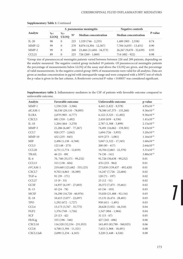
7
CEREBROSPINAL FLUID INFLAMMATORY MEDIATORS
173
AnalyteS. pneumoniae meningitis Negative controls
P-value%<LLOQ
%>ULOQ Na Median concentration Median concentration
IL-20 98 0 225 1,529 (746 - 2,235) 1,489 (905 - 2,558) 0.74MMP-12 99 0 270 8,076 (4,304 - 12,367) 7,784 (4,035 - 13,431) 0.90MMP-2 99 0 268 23,466 (11,604 - 34,373) 26,267 (9,670 - 32,639) 0.93CCL21 89 0 225 726 (269 - 1,069) 714 (482 - 922) 0.98
aGroup size of pneumococcal meningitis patients varied between between 226 and 289 patients, depending on the analyte measured. The negative control group included 19 patients. Of pneumococcal meningitis patients the percentage of measurements below LLOQ of the assay and above the ULOQ are given, and the percentage of valid measurements. In the negative control group 100% of measurements were valid for all analytes. Data are given as median concentration in pg/ml with interquartile range and were compared with a MWU test of which the p-value is given in the last column. A Bonferroni-corrected P-value < 0.00057 was considered significant.
Supplementary table 2. Inflammatory mediators in the CSF of patients with favorable outcome compared to unfavorable outcome.
Analyte Favorable outcome Unfavorable outcome p-valueMMP-1 1,530 (520 - 3,506) 4,441 (1,822 - 9,578) 4.95x10⁻¹⁰
sICAM-1 50,530 (29,110 - 78,095) 78,580 (47,375 - 135,260) 9.36x10-07
IL1RA 2,879 (905 - 6,777) 6,122 (3,325 - 11,492) 9.36x10-06
CXCL5 681 (318 - 1,492) 2,410 (659 - 4,536) 1.41x10-05
IL-10 1,204 (464 - 3,270) 2,787 (1,308 - 5,899) 2.15x10-05
MMP-3 21,286 (6,487 - 77,267) 74,491 (16,062 - 159,301) 9.43x10-05
CCL7 920 (377 - 2,042) 1,694 (726 - 3,955) 3.20x10-04
MMP-10 432 (235 - 845) 619 (273 - 1,081) 1.16x10-03
MIF 2,496 (1,138 - 6,768) 5,047 (1,522 - 17,345) 1.60x10-03
CCL5 122 (48 - 273) 200 (85 - 417) 2.64x10-03
CCL20 4,751 (1,774 - 12,819) 10,704 (2,863 - 22,370) 3.31x10-03
TRAIL 46 (21 - 89( 74 (30 - 141) 3.88x10-03
IL-6 70, 748 (35,571 - 99,232) 91,726 (50,638 - 99,232) 0.01CCL15 315 (138 - 604) 476 (233 - 964) 0.01sVCAM-1 219,440 (121,682 - 335,125) 273,830 (159,437 - 492,420) 0.01CXCL7 9,702 (4,863 - 18,389) 14,247 (7,726 - 22,684) 0.02TGF-α 91 (39 - 171) 120 (71 - 197) 0.02CCL27 15 (9 - 33) 25 (12 - 51) 0.02GCSF 14,937 (6,197 - 27,603) 20,372 (7,475 - 35,661) 0.02IL-15 45 (24 - 78) 63 (36 - 103) 0.03MCSF 34,290 (16,720 - 60,976) 53,020 (21,488 - 92,116) 0.03IL-18 10,415 (3,077 - 22,697) 15,131 (6,474 - 28,401) 0.03TPO 1,292 (672 - 1,727) 958 (611 - 1,491) 0.03CCL4 13,173 (3,767 - 35,775) 20,628 (5,932 - 44,310) 0.04FGF2 1,376 (749 - 1,726) 1,547 (904 - 1,984) 0.04SCF 23 (13 - 42) 31 (15 - 67) 0.05Flt3Lig 335 (196 - 546) 427 (245 - 696) 0.06CXCL10 116,520 (52,534 - 251,953) 163,493 (83,790 - 360,925) 0.06CCL8 4,700 (1,394 - 11,321) 7,652 (1,968 - 18,491) 0.08CXCL12ab 2,690 (1,234 - 4,165) 3,220 (1,448 - 4,526) 0.08
Supplementary Table 1: Continued
41973 Geldhoff_v3.indd 173 24-08-16 00:40

CHAPTER 7
174
Analyte Favorable outcome Unfavorable outcome p-valueCXCL6 1,373 (356 - 3,021) 1,935 (386 - 4,644) 0.08TSLP 70 (37 - 96) 57 (31 - 84) 0.10IL-12p40 530 (247 - 1,351) 627 (282 - 1,607) 0.12CXCL9 3,728 (1,242 - 9,559) 4,206 (1,728 - 17,126) 0.13IL-11 2, 468 (1,221 - 3,682) 2,766 (1,370 - 4,430) 0.14IL-3 22 (12 - 34) 18 (8 - 33) 0.15CCL24 54 (20 - 143) 69 (40 - 128) 0.16PDGF-AA 50 (23 - 70) 52 (29 - 77) 0.17sIL-2Ra 179 (72 - 323) 179 (114 - 413) 0.18VEGF 2,223 (1,130 - 3,806) 2,576 (1,244 - 4,639) 0.20CCL21 713 (235 - 1,011) 746 (302 - 1,151) 0.22CCL19 122 (77 - 297) 156 (78 - 388) 0.22IL-12p70 127 (67 - 195) 150 (85 - 212) 0.22IL-2 57 (26 - 118) 69 (31 - 131) 0.25IFN-γ 1,212 (463 - 3,503) 1,401 (532 - 5,765) 0.25MMP-12 8,461 (4,787 - 12,662) 7,690 (3,606 - 12,211) 0.27TNF-α 2,905 (409 - 11,945) 4,904 (726 - 11,351) 0.29CXCL1 19,805 (4,033 - 39,171) 23,065 (5,836 - 42,783) 0.30IL-21 32 (16 - 49) 37 (16 - 54) 0.31IL-4 92 (51 - 123) 80 (41 - 118) 0.31CXCL13 12 (6 - 31) 14 (6 - 44) 0.32PDGF-AB/BB 258 (94 - 567) 263 (122 - 591) 0.32IL-33 98 (44 - 144) 108 (56 - 172) 0.32IL-17 143 (62 - 283) 169 (64 - 300) 0.37CCL14a 4,295 (2,972 - 7,611) 4,284 (1,117 - 7,257) 0.38CCL3 5,306 (887 - 21,963) 6,963 (1,146 - 25,251) 0.38GMCSF 865 (323 - 1,583) 1,125 (261 - 1,951) 0.39IFNa2 1,151 (653 - 1,679) 1,214 (599 - 1,974) 0.39CXCL11 434 (116 - 1,695) 595 (114 - 2,011) 0.42MMP-7 9,663 (4,166 - 15,237) 9,911 (5,752 - 15,929) 0.43IL-13 67 (31 - 135) 67 (38 - 134) 0.44MMP-13 5,575 (2,759 - 7,910) 4,597 (2,892 - 6,988) 0.45CCL26 89 (39 - 136) 88 (33 - 124) 0.47CCL1 46 (22 - 70) 39 (21 - 69) 0.50CCL2 99,802 (95,276 - 99,802) 99,802 (99,802 - 99,802) 0.50IL-1β 1,612 (258 - 6,022) 2,424 (388 - 5,807) 0.50MMP-9 497,193 (497,193 - 497,193) 497,193 (497,193 - 497,193) 0.52TNF-β 47 (23 - 501) 53 (25 - 547) 0.53Eotaxin 956 (500 - 1,540) 1,065 (414 - 1,672) 0.55CCL17 3 (2 - 5) 3 (1 - 6) 0.56IL-23 311 (134 - 461) 338 (147 - 468) 0.58IL-9 79 (42 - 127) 75 (39 - 114) 0.59LIF 445 (191 - 667) 418 (272 - 638) 0.60sCD40L 271 (158 - 580) 334 (163 - 652) 0.61CCL13 305 (151 - 446) 275 (131 - 403) 0.69CCL22 1,031 (475 - 1,499) 1,031 (387 - 1,499) 0.70IL-29 2,882 (1,732 - 3,909) 3,076 (1,790 - 4,006) 0.73
Supplementary table 2. Continued
41973 Geldhoff_v3.indd 174 24-08-16 00:40

7
CEREBROSPINAL FLUID INFLAMMATORY MEDIATORS
175
Analyte Favorable outcome Unfavorable outcome p-valueIL-20 1,458 (684 - 2,235) 1,674 (796 - 2,354) 0.76XCL1 673 (325 - 1,342) 630 (354 - 1,342) 0.76IL-7 256 (89 - 1,388) 247 (81 - 1,516) 0.83IL-5 10 (6 - 15) 10 (7 - 14) 0.86EGF 137 (69 - 282) 139 (76 - 299) 0.88MMP-2 22,812 (11,610 - 34,305) 24,634 (11,586 - 35,024) 0.88IL-16 371 (175 - 554) 350 (170 - 551) 0.90IL-28A 71 (36 - 115) 71 (32 - 116) 0.92IL-1α 484 (223 - 1,275) 469 (165 - 1,415) 0.94CX3CL1 1,755 (969 - 12,588) 1,678 (976 - 12,368) 0.99CXCL8 53,241 (18,334 - 113,442) 54,127 (18,086 - 117,229) 1.00
Group size for patients with favorable outcome varied between 134 and 162 patients, and for unfavorable outcome between 87 and 109 patients, depending on the analyte measured. Data are given as median concentration in pg/ml with interquartile range and were compared with a MWU test of which the p-value is given in the last column. A Bonferroni-corrected P-value < 0.00057 was considered signifi cant.
Supplementary fi gure 1. Principal component analysis.
-20 -10 0 10
-10
-50
5
Individuals factor map (PCA)
Dim 1 (36.02%)
Dim
2 (6
.61%
)
CRF0013
CRF0018
CRF0029
CRF0032CRF0036
CRF0060
CRF0061
CRF0069
CRF0070
CRF0079CRF0081CRF0085CRF0090
CRF0093
CRF0095CRF0110CRF0121
CRF0123
CRF0126
CRF0127
CRF0134
CRF0136
CRF0143
CRF0149
CRF0152CRF0153
CRF0154CRF0160
CRF0169
CRF0173
CRF0174
CRF0175
CRF0178
CRF0179
CRF0180
CRF0189
CRF0192
CRF0194
CRF0197CRF0203
CRF0220
CRF0222
CRF0224CRF0225
CRF0227
CRF0228
CRF0230
CRF0231
CRF0232
CRF0237CRF0240
CRF0241
CRF0243
CRF0246
CRF0248
CRF0254
CRF0258
CRF0259
CRF0260
CRF0261
CRF0263
CRF0271
CRF0273
CRF0276CRF0293CRF0295
CRF0312CRF0314
CRF0316
CRF0327
CRF0333 CRF0340
CRF0353CRF0360
CRF0364CRF0365
CRF0369CRF0376
CRF0379
CRF0380
CRF0385
CRF0388CRF0390
CRF0394
CRF0397
CRF0398
CRF0403
CRF0405
CRF0406 CRF0408
CRF0410CRF0411
CRF0417
CRF0420
CRF0424
CRF0425
CRF0426
CRF0429
CRF0430
CRF0434
CRF0435
CRF0437CRF0449
CRF0453
CRF0456
CRF0457
CRF0465CRF0466
CRF0475
CRF0495
CRF0512
CRF0516
CRF0517
CRF0521
CRF0523
CRF0527
CRF0539 CRF0542
CRF0545CRF0567
CRF0568CRF0569
CRF0571
CRF0580CRF0596CRF0597
CRF0604
CRF0609
CRF0616
CRF0617
CRF0618CRF0621CRF0622
CRF0626
CRF0632
CRF0633
CRF0634
CRF0639
CRF0640CRF0644
CRF0646
CRF0651CRF0656CRF0657
CRF0661 CRF0677CRF0682
CRF0694
CRF0703CRF0706
CRF0710
CRF0713
CRF0715
CRF0720 CRF0721CRF0736
CRF0739
CRF0743
CRF0746
CRF0747CRF0757
CRF0766
CRF0777
CRF0789
CRF0790
CRF0802
CRF0803
CRF0804CRF0805
CRF0813
CRF0816
CRF0819
CRF0833
CRF0839
CRF0842
CRF0846CRF0848
CRF0849
CRF0850
CRF0851
CRF0855CRF0857
CRF0858
CRF0863
CRF0865
CRF0877
CRF0878
CRF0880CRF0884
CRF0894
CRF0895
CRF0896
CRF0898
CRF0899
CRF0901
CRF0905
CRF0907
CRF0912
CRF0915
CRF0920CRF0924
CRF0926
CRF0929
CRF0930 CRF0931CRF0933
CRF0934
CRF0940
CRF0942
CRF0946CRF0957
CRF0964
Principal component analysis of patients with favorable outcome (blue) and unfavorable outcome (red) based on all 88 measured immune mediators. Th e principal component 1 (dim1) is given on the x-axis and principal component 2 (dim2) on the y-axis. CRF (case record form) are the patients individual study numbers.
Supplementary table 2. Continued
41973 Geldhoff_v3.indd 175 24-08-16 00:40

41973 Geldhoff_v3.indd 176 24-08-16 00:40

8SUMMARY AND GENERAL DISCUSSION
41973 Geldhoff_v3.indd 177 24-08-16 00:40

CHAPTER 8
178
Summary
Pneumococcal meningitis is a serious infectious disease of the meninges surrounding the brain and spinal cord, with high mortality and morbidity.1 Despite treatment with antibiotics, adjuvant dexamethasone and advanced supportive care, 20% of patients do not survive pneumococcal meningitis.2 There is a need for new adjuvant treatments to improve outcome. In this thesis we tried to obtain a better understanding of the pathophysiology of pneumococcal meningitis and to find new potential targets for future research aiming at new adjunctive therapies to improve outcome.
In Chapter 2 we reviewed the literature to date on the pathophysiology of pneumococcal meningitis. An adequate immune response is essential to effectively control infection and limit tissue damage as a result of bacterial growth and release of toxins. 3-5Experimental studies showed that also the immune response itself can cause damage to the tissue of the host.6 Dead pneumococci or purified pneumococcal constituents are able to induce a pronounced cerebrospinal fluid (CSF) inflammatory response, leukocyte influx, cerebral edema and increased intracranial pressure in experimental animals.6,7 To limit host tissue damage as a result of inflammation, the host inflammatory response is controlled by an anti-inflammatory immune response that eventually down-regulates the inflammation. An effective immune response starts with the correct pattern recognition by the host innate immune system, thereby identifying the invading pathogen involved to initiate a tailored immune response.8,9 Toll-like receptors (TLR’s) have been extensively studied in pneumococcal meningitis and were found to play an important role, as TLR2 and 4 deficient mice showed low cytokine responses, higher bacterial loads and worse disease outcome.10-13 A different group of pattern recognition receptors (PRR’s) that was recently described include the Nod-like receptors (NLR’s). NLR’s are intracellular signaling molecules that have been reported to recognize several bacterial toxins, including pneumococcal pneumolysin and bacterial DNA.14-16 In addition, two downstream effectors of inflammasome signaling, caspase-1 and IL-18 were shown to play a role in the brain damage associated with pneumococcal meningitis.17,18 This fuelled our interest in these PRR’s, which resulted in chapter 4 and 5.
In Chapter 3 we established an experimental mouse model of meningitis by intracisternal inoculation of S. pneumoniae. We aimed to develop an animal model that was reproducible, in which features of meningitis were on the foreground compared to systemic infection, and with as little as iatrogenic structural damage as possible. Bacterial meningitis was induced in all mice with good reproducibility and uniform disease course between mice. We measured cytokines and chemokines known to be involved in the immune response against pneumococci in humans and found CXCL1, CXLC2, IL-1β and IL-6 to be elevated in our mouse model. TNF-α and IL-10 were low in all body compartments measured. Furthermore, features of pneumonia and sepsis only became apparent at the end of the disease course. On histopathology of the brain we observed meningeal, parenchymal and perivascular lymphocytic infiltration, and
41973 Geldhoff_v3.indd 178 24-08-16 00:40

8
SUMMARY AND GENERAL DISCUSSION
179
in some cases beginning abscess formation. Furthermore we found signs of microglial activation, hippocampal apoptosis and (micro) hemorrhages, which have also been described in meningitis patients.19-21 However, we observed no signs of stroke, a well-known complication in human disease.22-25
In Chapter 4 we studied the role of the NACHT, LRR and PYD domains-containing protein (NLRP)-3 inflammasome in the pathophysiology of pneumococcal meningitis. We found the inflammasome downstream effector cytokines IL-1β and IL-18 in patients’ CSF to be associated with systemic complications, and observed that cytokine levels were dependent on the causative S. pneumoniae serotype. Then, we induced pneumococcal meningitis in mice deficient for NLRP3, a protein involved in the NLRP3 inflammasome signaling pathway, and in mice lacking Apoptosis-associated Speck-like protein containing CARD (ASC), which is an adaptor protein of multiple inflammasome pathways. Nlrp3-/- mice showed less meningeal neutrophil influx and cerebral hemorrhages, which surprisingly was independent of ASC, IL-1β or IL-18. Both Nlrp3-/- and Asc-/- mice had a reduced systemic inflammatory response. The decrease of systemic inflammation was most pronounced in Asc-/- mice, and was accompanied by lower levels of brain pro-inflammatory cytokines CXCL1, CXCL2 and IL-6 at 30h after infection. We found no differences in IL-1β and IL-18 levels between wild-type (WT) and Nlrp3-/- or Asc-/- mice, which was unexpected. Our results implicate a role for inflammasome proteins NLRP3 and ASC in the regulation of the systemic inflammatory response and the development of cerebral damage during pneumococcal meningitis.
In Chapter 5 we analyzed eight nonsynonymous single nucleotide polymorphisms (SNP’s) in coding regions of in genes involved in the inflammasome-signaling pathway, for which a commercial assay was available and with a reported minor allele frequency of at least 5%. A genetic variant in the CARD8 gene was associated with unfavorable outcome in pneumococcal meningitis, which seemed to be driven by systemic and neurologic complications. The SNP in the CARD8 gene was not associated with CSF IL-1β or IL-18 levels. The role of CARD8 as a component of the NLRP3 inflammasome remains ambiguous, CARD8 independent NLRP3 signaling has been described and recently CARD8 was suggested to have an inhibitory effect on NLRP3 signalling.26,27 Furthermore, CARD8 plays a role in multiple other signaling pathways.28-31 Thus, we found a correlation between a genetic variant in CARD8 and unfavorable outcome, but further research is needed to elucidate the causality of the association and underlying mechanism.
In Chapter 6 we show that the lack of a S. pneumoniae argGH locus is associated with favorable outcome in patients with pneumococcal meningitis. The genes located on this locus are argG and argH, which are involved in the biosynthesis of arginine out of citrulline. Pneumococci are able to take up arginine from their surrounding via an ABC-transporter system that is present in all known pneumococcal strains. Only a minority of strains posses an arginine synthesis system.32 Arginine is used for growth, but is also a precursor in the biosynthesis of polyamines, which have been implicated in oxidative stress responses and protection against free radicals.33 We showed that a serotype 2
41973 Geldhoff_v3.indd 179 24-08-16 00:40

CHAPTER 8
180
pneumococcal knock out strain for the locus (D39ΔargGH) is less resistant to H2O2 in vitro compared to wild-type D39 and has impaired growth in human blood and CSF, but not in growth medium. Bacterial growth was attenuated in a meningitis and pneumonia model in mice inoculated with the D39ΔargGH pneumococcus, and in line with these results, D39ΔargGH inoculated mice had better survival. Thus, defective pneumococcal arginine synthesis results in decreased virulence and bacterial growth in blood and CSF in mice, which may be due to impaired growth as a results of arginine deficiency, decreased resistance to free radicals, or a combination of these.
In Chapter 7 we conducted an explorative study in which we analyzed 88 different inflammatory mediators in the CSF of patients with pneumococcal meningitis and related these to disease outcome. Seven inflammatory mediators were associated with unfavorable outcome, and included two matrix metalloproteinases (MMP-1 and MMP-3), an adhesion molecule (sICAM-1), two chemokines (CCL7 and CXCL5) and two anti-inflammatory cytokines (IL-1RA and IL-10). Of these MMP-1 and sICAM-1 were also associated with mortality and systemic complications. These data suggest a role for these inflammatory mediators in the pathophysiology of pneumococcal meningitis.
Discussion
The main objective of this thesis was to gain more insight in the pathophysiology of pneumococcal meningitis. A more close understanding of the pathophysiology is needed to identify potential new targets for future studies aiming at the development of adjunctive therapies that can reduce brain damage and improve outcome of pneumococcal meningitis.
In chapter 3 we describe how we set up an experimental mouse model of pneumococcal meningitis. We choose for intracisternal inoculation as route of infection. The advantages of this model are its reproducibility, as infection proceeds to meningitis in all mice with similar CSF bacterial loads and disease progression over time. A drawback of this model is that the natural route of infection, via the lungs and blood, is bypassed and the systemic immune response is activated after induction of meningitis rather than before. Other studies using intraperitoneal or intranasal inoculation resemble the natural route of infection more closely, however not all mice develop meningitis and a proportion of animals succumb from sepsis and pneumonia.34-36 In our study, signs of respiratory distress and positive blood cultures only became apparent at the end of the disease course, which makes the model suitable for studying meningitis. On histopathology of the brain we observed multiple features resembling human disease, except for signs of stroke, a well-known complication in human disease and described in 17% of patients with pneumococcal meningitis.22-25 Cortical necrosis has been described in experimental models using rats or infant mice, but not in adult mice.37,38 Thus, our model resembles the human situation in many ways, but also shows several differences, which need to be considered when interpreting the results of future experiments using this mouse model.
41973 Geldhoff_v3.indd 180 24-08-16 00:40

8
SUMMARY AND GENERAL DISCUSSION
181
In Chapter 4 we evaluated the role of the inflammasome pathway in a mouse model of pneumococcal meningitis. First, we showed that in experimental meningitis NLRP3 signaling was responsible for an enhanced neutrophil influx and cerebral hemorrhages, which was independent of ASC, IL-1β or IL-18. Inflammasome independent NLRP3 signaling has been described before in different disease models, but its precise mechanism is not yet clear.39,40 Secondly, in our study both NLRP3 and ASC deficiency resulted in decreased systemic bacterial loads and inflammatory response, which was most pronounced in Asc-/- mice, that also showed decreased brain cytokine and chemokine levels. Surprisingly, we found no differences in IL-1β and IL-18 levels between knock out and wild-type mice. Possible explanations for this unexpected finding can be that we measured at an inadequate time point, as IL-1β in humans is increased in the first 18 hours of infection41, or that the serotype 3 pneumococcus is a relatively weak activator of inflammasome signaling compared to other serotypes. Our results are conflicting with a study that was concurrently done, in which meningitis was induced in Asc-/- and Nlrp3-/- mice with a serotype 2 pneumococcus.42 Asc-/- and Nlrp3-/- mice had a better clinical outcome and decreased brain inflammation, which was dependent on IL1-β and IL-18 signaling.42 These different outcomes may be the result of the different pneumococcal serotypes used in the experiments. Pneumococcal serotype 3 is more heavily encapsulated and more virulent than serotype 2. In line with this hypothesis we show that CSF IL-1β production in patients differs depending on the pneumococcal serotype, and is relatively low with serotype 3. We were unable to show this for serotype 2 as this is not a causative agent of pneumococcal meningitis in humans.1 However, in in vitro experiments high doses of serotype 2 pneumococci were needed to induce IL-β production.43 The inflammasomes are relatively newly discovered pathogen recognition receptors and part of a complicated system of which the knowledge is rapidly expanding. In conclusion, these results implicate a role for inflammasome signaling in the pathogenesis of pneumococcal meningitis, however the mechanism appears to be different than previously described for the NLRP3 signaling pathway, which warrants further research.
In Chapter 5 we analyzed eight SNP’s in genes involved in multiple inflammasome pathways, and correlated these to disease outcome. We found a genetic variant in the CARD8 gene to be associated with unfavorable outcome, also in the subgroup of patients with pneumococcal meningitis. Secondly, we found that a SNP in NLRP1 was associated with mortality in pneumococcal meningitis, although the association was not preserved after correction for multiple testing. Therefore, confirmation of this association in a larger cohort or an experimental study is needed. CARD8 was initially described as a negative regulator of NF-κB, caspase-1 dependent IL-1β production, Nod2 activation and apoptosis.28-31 The role of CARD8 in the NLRP3 inflammasome complex remains ambiguous.44 Some studies described CARD8 as a component of the NLRP3 inflammasome, however CARD8 independent NLRP3 inflammasome activation has also been described and more recent studies showed a possible role for CARD8 as a negative regulator of the NLRP3 inflammasome signaling.28,45,26 The genetic variant described is known to result
41973 Geldhoff_v3.indd 181 24-08-16 00:40
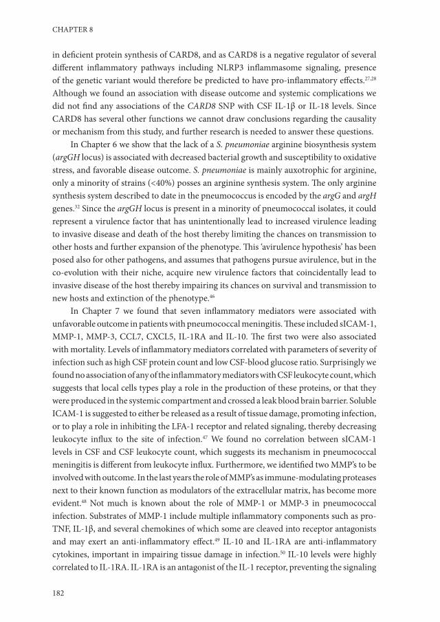
CHAPTER 8
182
in deficient protein synthesis of CARD8, and as CARD8 is a negative regulator of several different inflammatory pathways including NLRP3 inflammasome signaling, presence of the genetic variant would therefore be predicted to have pro-inflammatory effects.27,28 Although we found an association with disease outcome and systemic complications we did not find any associations of the CARD8 SNP with CSF IL-1β or IL-18 levels. Since CARD8 has several other functions we cannot draw conclusions regarding the causality or mechanism from this study, and further research is needed to answer these questions.
In Chapter 6 we show that the lack of a S. pneumoniae arginine biosynthesis system (argGH locus) is associated with decreased bacterial growth and susceptibility to oxidative stress, and favorable disease outcome. S. pneumoniae is mainly auxotrophic for arginine, only a minority of strains (<40%) posses an arginine synthesis system. The only arginine synthesis system described to date in the pneumococcus is encoded by the argG and argH genes.32 Since the argGH locus is present in a minority of pneumococcal isolates, it could represent a virulence factor that has unintentionally lead to increased virulence leading to invasive disease and death of the host thereby limiting the chances on transmission to other hosts and further expansion of the phenotype. This ‘avirulence hypothesis’ has been posed also for other pathogens, and assumes that pathogens pursue avirulence, but in the co-evolution with their niche, acquire new virulence factors that coincidentally lead to invasive disease of the host thereby impairing its chances on survival and transmission to new hosts and extinction of the phenotype.46
In Chapter 7 we found that seven inflammatory mediators were associated with unfavorable outcome in patients with pneumococcal meningitis. These included sICAM-1, MMP-1, MMP-3, CCL7, CXCL5, IL-1RA and IL-10. The first two were also associated with mortality. Levels of inflammatory mediators correlated with parameters of severity of infection such as high CSF protein count and low CSF-blood glucose ratio. Surprisingly we found no association of any of the inflammatory mediators with CSF leukocyte count, which suggests that local cells types play a role in the production of these proteins, or that they were produced in the systemic compartment and crossed a leak blood brain barrier. Soluble ICAM-1 is suggested to either be released as a result of tissue damage, promoting infection, or to play a role in inhibiting the LFA-1 receptor and related signaling, thereby decreasing leukocyte influx to the site of infection.47 We found no correlation between sICAM-1 levels in CSF and CSF leukocyte count, which suggests its mechanism in pneumococcal meningitis is different from leukocyte influx. Furthermore, we identified two MMP’s to be involved with outcome. In the last years the role of MMP’s as immune-modulating proteases next to their known function as modulators of the extracellular matrix, has become more evident.48 Not much is known about the role of MMP-1 or MMP-3 in pneumococcal infection. Substrates of MMP-1 include multiple inflammatory components such as pro-TNF, IL-1β, and several chemokines of which some are cleaved into receptor antagonists and may exert an anti-inflammatory effect.49 IL-10 and IL-1RA are anti-inflammatory cytokines, important in impairing tissue damage in infection.50 IL-10 levels were highly correlated to IL-1RA. IL-1RA is an antagonist of the IL-1 receptor, preventing the signaling
41973 Geldhoff_v3.indd 182 24-08-16 00:40

8
SUMMARY AND GENERAL DISCUSSION
183
of both IL-1α and IL-1β. In our study, IL-1RA/IL-1β ratios were well below the levels needed to antagonize IL-1β. Either this reflects an insufficient attempt to antagonize IL-1R signaling or possibly IL-1RA exerts different functions besides inhibiting IL-1R signaling. Recently, IL-1RA was described to inhibit NLRP3 inflammasome signaling.51 To find high levels of two anti-inflammatory proteins to be associated with unfavorable outcome was unexpected. Possibly this reflects the failing control of the inflammatory response or that an overactive anti-inflammatory response may lead to ineffective infection control and bacterial clearance, and unfavorable outcome. CCL7 and CXCL5 are both chemokines that attract neutrophils, promoting inflammation. In a pneumococcal pneumonia model CCL7 was shown to exhibit a pro-inflammatory effect.52 However, CCL7 can be modulated in a receptor antagonist resulting in an inverse inflammatory effect. The assay used to measure CCL7 does not discriminate between the agonistic and antagonistic variants. In previous reports CXCL5 was demonstrated in the CSF of patients with bacterial meningitis where it exerts a chemotactic activity towards neutrophils.53
The limitations of this study include an inclusion bias (only patients with positive CSF culture, severely ill patients may have a contra-indication for lumbar puncture) and differences in sample handling. We cannot draw conclusions whether these inflammatory mediators are produced intrathecally or pass the blood brain barrier from the systemic compartment, and how these levels are correlated to the CSF bacterial load. For future experiments analysis of inflammatory mediators in paired blood samples and measurement of CSF bacterial loads would be interesting. Furthermore, we cannot draw conclusions from this study about a causative relationship between the inflammatory mediators found and unfavorable outcome. Therefore, further studies on the role of these inflammatory mediators in pneumococcal meningitis are needed.
Future researchFurther research will focus on the hypothesis free identification of pathways and biological processes involved in disease outcome of pneumococcal meningitis. For this purpose patient clinical data, genetic data, bacterial genetic data and CSF inflammatory mediators will be analyzed altogether. As part of the MeninGene study we collected DNA samples of a large proportion of patients, and in 198 cases we have paired CSF samples that were available for analysis of immune mediators and this number is still expanding. We have sequenced a large set of exome variants in these DNA samples and will correlate this data with the levels of CSF inflammatory mediators. In all cases the causative agent was isolated and stored. Genetic analysis of the causative S. pneumoniae strain combined with patient data may lead to a better understanding of host-pathogen interactions. This hypothesis free approach will hopefully lead to the identification of more proteins or pathways involved in the pathophysiology of pneumococcal meningitis.
Secondly, it would be interesting to broaden our research on the inflammatory mediators detected in CSF of patients. Whether they are also expressed in the mouse, in what cell types and to assess whether these inflammatory mediators are involved in, or
41973 Geldhoff_v3.indd 183 24-08-16 00:40

CHAPTER 8
184
are just markers of, unfavorable outcome of disease. The last question could be addressed using a comparative study with knock out mice in our pneumococcal mouse model. Then, it would be interesting to go into more detail and assess whether these inflammatory mediators are in their agonistic or antagonistic state, where applicable, and gain more insight in their function and role in the inflammatory response. Since we found two anti-inflammatory cytokines (IL-10 and IL-1RA) to be associated with unfavorable outcome in patients, it would be interesting to take a specific look at the role of regulatory T-cells, which are known for their ability to suppress the inflammatory response. In past years it became more evident that in the elderly, which have a high incidence of pneumococcal meningitis, regulatory T-cell function and therefore immunosuppression increases.54,55 Furthermore, to gain more insight in the level of bacterial control in patients in relation to the anti-inflammatory mediators that were found in the CSF, and whether the CSF inflammatory mediators are produced in the CSF or enter the CNS from the periphery, it would be relevant to collect data on the CSF bacterial load and peripheral blood levels of inflammatory mediators in our patient cohort.
In conclusion, we found that inflammasome signaling was associated with systemic complications, and that inflammasome independent NLRP3 signaling induced brain damage in our mouse model of pneumococcal meningitis. Furthermore we showed that pneumococcal virulence factors play a role in the clinical outcome of patients with pneumococcal meningitis, as the absence of an arginine synthesis pathway in the causative S. pneumoniae strain is associated with favorable outcome in these patients. Lastly, we found two MMP’s, two anti-inflammatory cytokines, two chemokines and a signaling molecule in the CSF to be associated with disease outcome in patients, suggesting a role for these inflammatory mediators in the pathophysiology of pneumococcal meningitis. Further research will be focusing on combining patient clinical and genetic data with genetic data of the causative S. pneumoniae strains and the levels of CSF inflammatory mediators, to gain more insight in the host-pathogen interaction, and its effects on the inflammatory response and patient outcome. We hope this will lead to the identification of new pathways or biological processes involved in the outcome of pneumococcal meningitis, which can be further explored in the pneumococcal meningitis mouse model to find potential new targets for adjunctive therapies, and will eventually get us back from bench to bedside.
41973 Geldhoff_v3.indd 184 24-08-16 00:40

8
SUMMARY AND GENERAL DISCUSSION
185
References1. van de Beek D, de Gans J, Spanjaard L, Weisfelt M, Reitsma JB, Vermeulen M. Clinical features and
prognostic factors in adults with bacterial meningitis. N Engl J Med 2004;351:1849-59.2. Brouwer MC, McIntyre P, de Gans J, Prasad K, van de Beek D. Corticosteroids for acute bacterial meningitis.
Cochrane Database Syst Rev 2010;9:CD004405.3. Tuomanen E, Liu H, Hengstler B, Zak O, Tomasz A. The induction of meningeal inflammation by
components of the pneumococcal cell wall. J INFECT DIS 1985;151:859-68.4. Wellmer A, Zysk G, Gerber J, et al. Decreased virulence of a pneumolysin-deficient strain of Streptococcus
pneumoniae in murine meningitis. Infection and Immunity 2002;70:6504-8.5. Picard C, von Bernuth H, Ghandil P, et al. Clinical features and outcome of patients with IRAK-4 and
MyD88 deficiency. Medicine (Baltimore) 2010;89:403-25.6. Tuomanen E, Tomasz A, Hengstler B, Zak O. The relative role of bacterial cell wall and capsule in the
induction of inflammation in pneumococcal meningitis. J INFECT DIS 1985;151:535-40.7. Pfister HW, Koedel U, Lorenzl S, Tomasz A. Antioxidants attenuate microvascular changes in the early
phase of experimental pneumococcal meningitis in rats. Stroke 1992;23:1798-804.8. Liu Q, Ding JL. The molecular mechanisms of TLR-signaling cooperation in cytokine regulation. Immunol
Cell Biol 2016.9. Thaiss CA, Levy M, Itav S, Elinav E. Integration of Innate Immune Signaling. Trends Immunol 2016;37:84-
101.10. Echchannaoui H, Frei K, Schnell C, Leib SL, Zimmerli W, Landmann R. Toll-like receptor 2-deficient mice
are highly susceptible to Streptococcus pneumoniae meningitis because of reduced bacterial clearing and enhanced inflammation. J Infect Dis 2002;186:798-806.
11. Koedel U, Angele B, Rupprecht T, et al. Toll-like receptor 2 participates in mediation of immune response in experimental pneumococcal meningitis. J Immunol 2003;170:438-44.
12. Klein M, Obermaier B, Angele B, et al. Innate immunity to pneumococcal infection of the central nervous system depends on toll-like receptor (TLR) 2 and TLR4. J INFECT DIS 2008;198:1028-36.
13. Albiger B, Sandgren A, Katsuragi H, et al. Myeloid differentiation factor 88-dependent signalling controls bacterial growth during colonization and systemic pneumococcal disease in mice. Cell Microbiol 2005;7:1603-15.
14. Opitz B, Püschel A, Schmeck B, et al. Nucleotide-binding oligomerization domain proteins are innate immune receptors for internalized Streptococcus pneumoniae. J Biol Chem 2004;279:36426-32.
15. Gurcel L, Abrami L, Girardin S, Tschopp J, van der Goot FG. Caspase-1 activation of lipid metabolic pathways in response to bacterial pore-forming toxins promotes cell survival. Cell 2006;126:1135-45.
16. Kim S, Bauernfeind F, Ablasser A, et al. Listeria monocytogenes is sensed by the NLRP3 and AIM2 inflammasome. Eur J Immunol 2010;40:1545-51.
17. Leppert D, Leib SL, Grygar C, Miller KM, Schaad UB, Hollander GA. Matrix metalloproteinase (MMP)-8 and MMP-9 in cerebrospinal fluid during bacterial meningitis: association with blood-brain barrier damage and neurological sequelae. Clin Infect Dis 2000;31:80-4.
18. Zwijnenburg PJG, Van Der Poll T, Florquin S, et al. Interleukin-18 gene-deficient mice show enhanced defense and reduced inflammation during pneumococcal meningitis. Journal of Neuroimmunology 2003;138:31-7.
19. Nau R, Soto A, Brück W. Apoptosis of neurons in the dentate gyrus in humans suffering from bacterial meningitis. J Neuropathol Exp Neurol 1999;58:265-74.
20. Vergouwen MD, Schut ES, Troost D, van de Beek D. Diffuse cerebral intravascular coagulation and cerebral infarction in pneumococcal meningitis. Neurocrit Care 2010;13:217-27.
21. Weisfelt M, de Gans J, van der Poll T, van de Beek D. Pneumococcal meningitis in adults: new approaches to management and prevention. Lancet Neurol 2006;5:332-42.
22. Schut ES, Lucas MJ, Brouwer MC, Vergouwen MD, van der Ende A, van de Beek D. Cerebral infarction in adults with bacterial meningitis. Neurocrit Care 2012;16:421-7.
23. Weisfelt M, van de Beek D, Spanjaard L, Reitsma JB, de Gans J. Clinical features, complications, and outcome in adults with pneumococcal meningitis: a prospective case series. Lancet Neurol 2006;5:123-9.
24. Kastenbauer S, Pfister H-W. Pneumococcal meningitis in adults: spectrum of complications and prognostic factors in a series of 87 cases. Brain 2003;126:1015-25.
41973 Geldhoff_v3.indd 185 24-08-16 00:40

CHAPTER 8
186
25. Weststrate W, Hijdra A, de Gans J. Brain infarcts in adults with bacterial meningitis. Lancet 1996;347:399.26. Tangi TN, Elmabsout AA, Bengtsson T, Sirsjo A, Fransen K. Role of NLRP3 and CARD8 in the regulation
of TNF-alpha induced IL-1beta release in vascular smooth muscle cells. Int J Mol Med 2012;30:697-702.27. Razmara M, Srinivasula SM, Wang L, et al. CARD-8 protein, a new CARD family member that regulates
caspase-1 activation and apoptosis. J Biol Chem 2002;277:13952-8.28. Ito S, Hara Y, Kubota T. CARD8 is a negative regulator for NLRP3 inflammasome, but mutant NLRP3 in
cryopyrin-associated periodic syndromes escapes the restriction. Arthritis Res Ther 2014;16:R52.29. von Kampen O, Lipinski S, Till A, et al. Caspase recruitment domain-containing protein 8 (CARD8)
negatively regulates NOD2-mediated signaling. J Biol Chem 2010;285:19921-6.30. Bouchier-Hayes L, Martin SJ. CARDINAL roles in apoptosis and NFkappaB activation. Vitam Horm
2004;67:133-47.31. Blomgran R, Patcha Brodin V, Verma D, et al. Common genetic variations in the NALP3 inflammasome are
associated with delayed apoptosis of human neutrophils. PLoS One 2012;7:e31326.32. Kloosterman TG, Kuipers OP. Regulation of arginine acquisition and virulence gene expression in the
human pathogen Streptococcus pneumoniae by transcription regulators ArgR1 and AhrC. J Biol Chem 2011;286:44594-605.
33. Ha HC, Sirisoma NS, Kuppusamy P, Zweier JL, Woster PM, Casero RA, Jr. The natural polyamine spermine functions directly as a free radical scavenger. Proc Natl Acad Sci U S A 1998;95:11140-5.
34. Tan TQ, Smith CW, Hawkins EP, Mason EO, Kaplan SL. Hematogenous bacterial meningitis in an intercellular adhesion molecule-1-deficient infant mouse model. J INFECT DIS 1995;171:342-9.
35. Zwijnenburg PJ, Van Der Poll T, Florquin S, van Deventer SJ, Roord JJ, van Furth AM. Experimental pneumococcal meningitis in mice: a model of intranasal infection. J INFECT DIS 2001;183:1143-6.
36. Kostyukova NN, Volkova MO, Ivanova VV, Kvetnaya AS. A study of pathogenic factors of Streptococcus pneumoniae strains causing meningitis. FEMS Immunol Med Microbiol 1995;10:133-7.
37. Grandgirard D, Steiner O, Täuber MG, Leib SL. An infant mouse model of brain damage in pneumococcal meningitis. Acta Neuropathol 2007;114:609-17.
38. Ostergaard C, Brandt C, Konradsen HB, Samuelsson S. Differences in survival, brain damage, and cerebrospinal fluid cytokine kinetics due to meningitis caused by 3 different Streptococcus pneumoniae serotypes: evaluation in humans and in 2 experimental models. J INFECT DIS 2004;190:1212-20.
39. Shigeoka AA, Mueller JL, Kambo A, et al. An inflammasome-independent role for epithelial-expressed Nlrp3 in renal ischemia-reperfusion injury. J Immunol 2010;185:6277-85.
40. Iyer SS, Pulskens WP, Sadler JJ, et al. Necrotic cells trigger a sterile inflammatory response through the Nlrp3 inflammasome. Proc Natl Acad Sci U S A 2009;106:20388-93.
41. Engelhard D, Pomeranz S, Gallily R, Strauss N, Tuomanen E. Serotype-related differences in inflammatory response to Streptococcus pneumoniae in experimental meningitis. J Infect Dis 1997;175:979-82.
42. Hoegen T, Tremel N, Klein M, et al. The NLRP3 inflammasome contributes to brain injury in pneumococcal meningitis and is activated through ATP-dependent lysosomal cathepsin B release. J Immunol 2011;187:5440-51.
43. Zysk G, Bruck W, Huitinga I, et al. Elimination of blood-derived macrophages inhibits the release of interleukin-1 and the entry of leukocytes into the cerebrospinal fluid in experimental pneumococcal meningitis. J Neuroimmunol 1997;73:77-80.
44. Paramel GV, Sirsjo A, Fransen K. Role of genetic alterations in the NLRP3 and CARD8 genes in health and disease. Mediators Inflamm 2015;2015:846782.
45. Agostini L, Martinon F, Burns K, McDermott MF, Hawkins PN, Tschopp J. NALP3 forms an IL-1beta-processing inflammasome with increased activity in Muckle-Wells autoinflammatory disorder. Immunity 2004;20:319-25.
46. Adiba S, Nizak C, van Baalen M, Denamur E, Depaulis F. From grazing resistance to pathogenesis: the coincidental evolution of virulence factors. PLoS One 2010;5:e11882.
47. Rothlein R, Mainolfi EA, Czajkowski M, Marlin SD. A form of circulating ICAM-1 in human serum. J Immunol 1991;147:3788-93.
48. Chiang TY, Tsao SM, Yeh CB, Yang SF. Matrix metalloproteinases in pneumonia. Clin Chim Acta 2014;433:272-7.
49. Van Lint P, Libert C. Chemokine and cytokine processing by matrix metalloproteinases and its effect on leukocyte migration and inflammation. J Leukoc Biol 2007;82:1375-81.
41973 Geldhoff_v3.indd 186 24-08-16 00:40

8
SUMMARY AND GENERAL DISCUSSION
187
50. Penaloza HF, Nieto PA, Munoz-Durango N, et al. Interleukin-10 plays a key role in the modulation of neutrophils recruitment and lung inflammation during infection by Streptococcus pneumoniae. Immunology 2015;146:100-12.
51. Borghi M, De Luca A, Puccetti M, et al. Pathogenic NLRP3 Inflammasome Activity during Candida Infection Is Negatively Regulated by IL-22 via Activation of NLRC4 and IL-1Ra. Cell Host Microbe 2015;18:198-209.
52. Jose RJ, Williams AE, Mercer PF, Sulikowski MG, Brown JS, Chambers RC. Regulation of neutrophilic inflammation by proteinase-activated receptor 1 during bacterial pulmonary infection. J Immunol 2015;194:6024-34.
53. Zwijnenburg PJG, de Bie HMA, Roord JJ, van der Poll T, van Furth AM. Chemotactic activity of CXCL5 in cerebrospinal fluid of children with bacterial meningitis. J Neuroimmunol 2003;145:148-53.
54. Schmitt V, Rink L, Uciechowski P. The Th17/Treg balance is disturbed during aging. Exp Gerontol 2013;48:1379-86.
55. Garg SK, Delaney C, Toubai T, et al. Aging is associated with increased regulatory T-cell function. Aging Cell 2014;13:441-8.
41973 Geldhoff_v3.indd 187 24-08-16 00:40

41973 Geldhoff_v3.indd 188 24-08-16 00:40

NEDERLANDSE SAMENVATTING
41973 Geldhoff_v3.indd 189 24-08-16 00:40

190
Samenvatting
Pneumokokken meningitis is een ernstige infectieziekte waarbij de hersenvliezen die de hersenen en het myelum omgeven geïnfecteerd zijn. De ziekte heeft een hoge mortaliteit en morbiditeit1. Twintig procent van de patiënten overlijd, ondanks behandeling met antibiotica in combinatie met dexamethason, en de huidige geavanceerde intensive-care zorg.2 Er is dringend behoefte aan nieuwe adjuvante therapieën om de uitkomst van patiënten met pneumokokken meningitis te verbeteren. In dit proefschrift trachten we een beter inzicht te krijgen in de pathofysiologie van pneumokokken meningitis, met als uiteindelijk doel om nieuwe potentiele behandelstrategieën te identificeren voor de ontwikkeling van een nieuwe adjuvante therapie om de ziekte uitkomst te verbeteren.
Hoofdstuk 2 van dit proefschrift geeft een overzicht van de huidige literatuur over de pathofysiologie van pneumokokken meningitis. Om schade aan het zenuwstelsel te voorkomen als gevolg van de bacteriële infectie en de toxines die hierbij vrijkomen, is een effectieve immuunrespons essentieel.3-5 In eerdere studies is aangetoond dat naast de bacterie ook de immuunrespons van de gastheer weefselschade kan veroorzaken6. Dode pneumokokken of gezuiverde eiwitten of polysaccharides van de pneumokok, kunnen een forse immuunrespons teweeg brengen in de liquor bij proefdieren, die leidt tot leukocyten influx, hersenoedeem en verhoogde intracraniële druk.6,7 Om dit te voorkomen wordt de immuunrespons van nature in bedwang gehouden door een anti-inflammatoire reactie die er uiteindelijk voor zorgt dat de immuunrespons uitdooft. Een effectieve immuunrespons begint bij de juiste patroon herkenning van het infecterende pathogeen door het innate immuun systeem, waardoor de juiste afweerreactie gericht op dat specifieke pathogeen op gang komt.8,9 Toll-like receptoren (TLR’s) zijn pattern recognition receptoren (PRR’s) die een belangrijke rol spelen in pneumokokken meningitis. TLR2 en 4 deficiënte muizen hebben een lagere cytokine response, hogere bacteriële load en slechtere ziekte uitkomst.10-13 Een andere groep PRR’s zijn de Nod-like receptoren (NLR’s) die relatief recent in de literatuur zijn beschreven. Dit zijn intracellulaire signalerings eiwitten die onder andere verschillende bacteriële toxines kunnen herkennen waaronder pneumokokken pneumolysine, maar ook bacterieel DNA.14-16 Er is aangetoond dat twee down-stream effector moleculen van inflammasome activatie, caspase-1 en IL-18 een rol spelen in de hersenschade die ontstaat na pneumokken meningitis.17-18 Hierdoor werd onze interesse gewekt in deze groep PRR’s, wat heeft geresulteerd in hoofdstuk 4 en 5.
In hoofdstuk 3 laten we zien hoe we een experimenteel meningitis muismodel hebben opgezet, waarbij pneumokokken intracisternaal worden geïnoculeerd. Ons doel was een diermodel te ontwikkelen dat reproduceerbaar is, en waarbij de meningitis component van de infectie op de voorgrond staat, in tegenstelling tot de systemische infectie, en dit door zo min mogelijk iatrogene schade te veroorzaken. Intracisternale injectie van pneumokken leidde tot bacteriële meningitis in alle muizen, met een goede reproduceerbaarheid een uniform ziektebeloop tussen de muizen per groep. In ons model hebben we cytokines en chemokines gemeten waarvan bekend is dat ze een rol spelen in de immuun reactie tegen pneumokokken,
41973 Geldhoff_v3.indd 190 24-08-16 00:40

8
191
NEDERLANDSE SAMENVATTING
en we vonden verhoogde concentraties CXCL1, CXLC2, IL1β en IL-6. Echter, TNF-α en IL-10 waren niet verhoogd in de verschillende organen die we hebben bekeken. Tekenen van pneumonie en sepsis kwamen pas later in het ziekte beloop. Histopathologie (PA) van de hersenen toonde meningeale, parenchymateuze en perivasculaire lymfocyten infiltratie, en in enkele gevallen beginnende abces vorming. Verder waren er aanwijzingen voor microglia activatie, apoptose in de hippocampus en (micro) bloedingen, alle drie kenmerken die ook in patiënten met pneumokokken meningitis zijn beschreven.19-21 Echter, we vonden geen herseninfarcten bij PA in ons muismodel, wat een belangrijke complicatie is in patiënten.22-25
In hoofdstuk 4 hebben we gekeken naar het NACHT, LRR and PYD domains-containing protein (NLRP)-3 inflammasome, en de rol hiervan in pneumokokken meningitis. In patiënten met meningitis bleken concentraties van IL-1β en IL-18 in de liquor geassocieerd te zijn met systemische complicaties, en deze concentraties waren gerelateerd aan het serotype pneumokok dat uit de liquor werd geïsoleerd. Vervolgens hebben we gekeken naar muizen met een deficiëntie voor NLRP3, een eiwit dat betrokken is in de NLRP3 signaling pathway, en in muizen met een deficiëntie voor Apoptosis-associated Speck-like protein containing CARD (ASC), wat een onderdeel is van meerdere inflammasome signaling pathways. Nlrp3-/- muizen hadden minder meningeale neutrofielen influx en hersenbloedingen op PA, wat onafhankelijk leek te zijn van ASC, IL-1β en IL-18. Zowel Nlrp3-/- en Asc-/- muizen produceerden lagere concentraties systemische inflammatoire cytokines. Dit was het meest uitgesproken in de Asc-/- muizen, en ging samen met een lagere concentratie CXCL1, CXCL2 en IL-6 in de hersenen op 30 uur na infectie. Tegen alle verwachting in werden geen verschillen gevonden in IL-1β en IL-18 concentraties tussen wild-type (WT) en Nlrp3-/- of Asc-/- muizen. Deze resultaten suggereren een rol voor NLRP3 en ASC in de systemische immuunrespons en het ontstaan van hersenschade bij pneumokokken meningitis.
In hoofdstuk 5 hebben we acht nonsynonymous single nucleotide polymorfismen (SNP’s) geanalyseerd in de coderende regionen van genen die geassocieerd zijn met de inflammasome signalerings cascade, en voor welke een commerciële assay beschikbaar was en met een gerapporteerde minor allel frequentie van minimaal 5%. Een genetische variant in het CARD8 gen bleek geassocieerd te zijn met slechte uitkomst in patiënten met pneumokokken meningitis, wat leek gedreven te worden door zowel systemische als neurologische complicaties. Deze SNP was niet geassocieerd met concentraties IL-1β en IL-18 in de liquor. De rol van CARD8 als onderdeel van de NLRP3 signalerings cascade is onduidelijk, en ook CARD8 onafhankelijke NRLP3 signalering en recent zelfs inhibitie van NLRP3 signalering door CARD8 zijn in de literatuur beschreven.26,27 Daarbij speelt CARD8 ook een rol in verschillende andere signalerings cascades.28-31 Concluderend vonden we een correlatie tussen een genetische variant in het CARD8 gen met slechte ziekte uitkomst, verder onderzoek is nodig om de causaliteit van deze relatie en het onderliggend mechanisme aan te tonen.
In hoofdstuk 6 laten we zien dat S. pneumoniae isolaten die het argGH locus missen zijn geassocieerd met een relatief goede ziekte uitkomst bij de patiënt. De genen op het argG
41973 Geldhoff_v3.indd 191 24-08-16 00:40

192
en argH locus zijn betrokken bij de biosynthese van citrulline in arginine. Pneumokokken kunnen arginine opnemen uit hun omgeving via een ABC-transporter systeem dat in alle pneumokokken stammen die beschreven zijn aanwezig is. Slechts een minderheid van de pneumokokken stammen hebben een systeem waarmee arginine kan worden geproduceerd.32 Arginine wordt door de pneumokok gebruikt voor groei, maar is ook een voorloper van de biosynthese van polyamines, die een rol spelen in de oxidatieve stress reactie en de bescherming tegen vrije radicalen.33 We toonden aan dat een serotype 2 knock out pneumokok voor het argGH locus (D39ΔargGH) gevoeliger is voor H2O2 in vitro vergeleken met een WT D39 en dat deze knock out variant een slechtere overleving had in bloed en liquor, maar niet in kweekmedium. Verder groeide de knock out variant slechter in een muizen meningitis en pneumonie model, en de muizen geïnoculeerd met de knock variant hadden een betere overleving. Concluderend, leidt een defect in het pneumokokken arginine synthetase system tot verminderde virulentie en bacteriegroei in bloed en liquor in muizen, wat mogelijk wordt veroorzaakt door een slechtere groei als gevolg van arginine deficiëntie, verhoogde gevoeligheid voor oxidatieve stress of een combinatie van beiden.
In hoofdstuk 7 hebben we in een exploratieve studie 88 inflammatoire mediatoren geanalyseerd in de liquor van patiënten met pneumokokken meningitis en gerelateerd aan ziekte uitkomst. Zeven inflammatoire mediatoren waren geassocieerd met slechte ziekte uitkomst, waaronder twee matrix metalloproteinases (MMP-1 en MMP-3), een adhesie molecuul (sICAM-1), twee chemokines (CCL7 en CXCL5) en twee anti-inflammatoire cytokines (IL-1RA en IL-10). Hiervan waren MMP-1 en sICAM-1 ook geassocieerd met mortaliteit en systemische complicaties. Deze resultaten suggereren een rol in de pathofysiologie van pneumokokken meningitis voor deze inflammatoire mediatoren.
Discussie
Het doel van dit proefschrift was een beter inzicht te krijgen in de pathofysiologie van pneumokokken meningitis. Een beter begrip van de pathofysiologie is belangrijk om zo nieuwe potentiele behandelstrategieën voor de toekomst te identificeren, met als uiteindelijk doel om nieuwe adjuvante behandelingen te ontwikkelen en zo de uitkomst van patiënten met pneumokokken meningitis te verbeteren.
In hoofdstuk 3 beschrijven we het opzetten van een experimenteel muismodel voor pneumokokken meningitis. We kozen voor intracisternale inoculatie als techniek om meningitis te induceren. De voordelen van dit model zijn de reproduceerbaarheid, de infectie veroorzaakt meningitis in alle muizen met een gelijke bacterie load in de liquor en ziekte progressie in de tijd. Een nadeel van dit model is dat de natuurlijke infectieroute, via de longen en bloed worden gepasseerd, en dat de systemische immuun respons daardoor pas op gang komt na het ontstaan van de meningitis in plaats van ervoor. In andere studies waarin intraperitoneale of intranasale inoculatie wordt gebruikt hebben meer
41973 Geldhoff_v3.indd 192 24-08-16 00:40

8
193
NEDERLANDSE SAMENVATTING
overeenkomsten met de natuurlijke route van infectie, echter hebben als nadeel dat niet alle muizen meningitis ontwikkelen en een deel bezwijkt aan een pneumokokken pneumonie of sepsis.34-36 In onze studie komen respiratoire insufficiëntie en positieve bloedkweken pas later in het ziektebeloop, wat dit model geschikt maakt voor het bestuderen van meningitis. PA van de hersenen liet meerdere kenmerken zien die ook bij patiënten worden gevonden, behalve herseninfarcten wat een belangrijke complicatie is in patiënten en in 17% van de patiënten met pneumokokken meningitis wordt beschreven.22-25 Corticale necrose is wel eerder beschreven in meningitis modellen in ratten of zeer jonge muizen, maar niet in volwassen muizen.37,38 Concluderend, zijn er verschillende kenmerken van ons muismodel die overeenkomen met de kenmerken die worden beschreven in patiënten, maar zijn er ook enkele verschillen die in acht moeten worden genomen wanneer men de resultaten interpreteert die verkregen zijn met dit muismodel.
In hoofdstuk 4 evalueren de rol van de inflammasome signaleringscascade in een pneumokokken meningitis muis model. Eerst tonen we aan dat in experimentele meningitis NLRP3 signalering verantwoordelijk is voor een verhoogde neutrofielen influx en cerebrale bloedingen, wat onafhankelijk bleek te zijn van ASC, IL-1β or IL-18. Inflammasome onafhankelijke NLRP3 signalering is eerder in de literatuur beschreven, in verschillende ziekte modellen, maar het onderliggende mechanisme is nog niet geheel opgehelderd.39,40 Ten tweede, toonden we aan dat zowel NLRP3 als ASC deficiëntie leiden tot minder systemische bacteriële uitgroei en inflammatie wat het meest uitgesproken was in de Asc-/- muizen, waarin ook de cerebrale cytokine levels lager waren. Verassend genoeg vonden we geen verschillen in IL-1β en IL-18 concentraties in knock out en WT muizen. Een mogelijke verklaring voor dit onverwachte resultaat is dat we niet op het juiste tijdstip hebben gemeten, gezien bekend is uit de literatuur dat IL-1β in patiënten in de eerste 18 uur van infectie verhoogd is.41 Of dat serotype 3 pneumokokken relatief zwakke activators zijn van de inflammasome signalerings cascade in vergelijking met andere serotypen. De resultaten zijn tegenstrijdig met de resultaten van een studie die gelijktijdig is gedaan, en waarin meningitis werd geïnduceerd in Asc-/- en Nlrp3-/- muizen met een serotype 2 pneumokok.42 Asc-/- en Nlrp3-/- muizen hadden een betere klinische uitkomst en minder tekenen van inflammatie in de hersenen, welke afhankelijk waren van IL1-β en IL-18.42 De verschillen tussen de studies zouden kunnen worden verklaard door de verschillende pneumokokken serotypen die zijn gebruikt. Serotype 3 heeft een dikker kapsel en is virulenter dan de serotype 2 pneumokok. We toonden aan dat IL-1β concentraties in de liquor van patiënten verschilt met het serotype pneumokok dat de meningitis veroorzaakt, en relatief laag is bij serotype 3 pneumokokken, wat deze hypothese ondersteund. Voor serotype 2 kon dit niet worden aangetoond omdat dit geen verwekker is van pneumokokken meningitis in mensen.1 Echter, in in vitro experimenten waren hoge doseringen serotype 2 pneumokokken nodig om IL-1β productie te induceren.43 De inflammasomes zijn relatief nieuw ontdekte PRR’s, en onderdeel van een gecompliceerd signalerings systeem waar snel veel nieuwe literatuur over beschikbaar komt. Concluderend, impliceren deze resultaten een rol voor de
41973 Geldhoff_v3.indd 193 24-08-16 00:40

194
inflammasome signalerings cascade in de pathogenese van pneumokokken meningitis, echter het mechanisme lijkt anders te zijn dat de meest gangbare beschreven NLRP3 signalerings cascade, en vervolg onderzoek is nodig om dit verder uit te diepen.
In hoofdstuk 5 hebben we acht SNP’s bepaald in genen die betrokken zijn bij inflammasome signalering, en we hebben deze gecorreleerd aan de ziekte uitkomst in patiënten. We vonden een genetische variant in het CARD8 gen welke was geassocieerd met slechte ziekte uitkomst, ook in de subgroep van patiënten met pneumokokken meningitis. Ten tweede vonden we een SNP in het NLRP1 gen welke was geassocieerd met mortaliteit in pneumokokken meningitis, echter de associatie bleef niet significant na correctie voor multipele testen. Bevestiging van een associatie is nodig in een grotere groep patiënten of een experimentele studie. CARD8 is initieel beschreven als een negatieve regulator van NF-κB, caspase-1 afhankelijke IL-1β productie, Nod2 activatie en apoptose.28-31 De rol die CARD8 speelt in het inflammasome complex is nog niet helemaal opgehelderd.44 Er zijn studies die CARD8 beschrijven als onderdeel van het NLRP3 inflammasome, maar ook CARD8 onafhankelijke NLRP3 signalering is beschreven, en een recente studie toonde CARD8 als een mogelijke negatieve regulator van het NLRP3 inflammasome.28,45,26 De genetische variant die in onze studie is beschreven, resulteert in een eiwit deficiëntie, en als CARD8 een negatieve regulator is van meerdere inflammatie cascades, inclusief het NLRP3 inflammasome, zou deze genetische variant een pro-inflammatoir effect moeten hebben.27,28 We vonden een associatie met ziekte uitkomst en systemische complicaties, echter we vonden geen associatie tussen de CARD8 SNP en IL-1β en IL-18 concentraties in de liquor. Aangezien CARD8 ook meerdere andere functies heeft, kunnen we geen conclusie trekken uit deze studie over de causaliteit van de associatie of het mechanisme, daarvoor is verder onderzoek nodig.
In hoofdstuk 6 laten we zien dat afwezigheid van een S. pneumoniae arginine biosynthese systeem (het argGH locus) geassocieerd is met een verlaagde bacteriële uitgroei, verhoogde gevoeligheid voor oxidatieve stress en een relatief goede ziekte uitkomst van de patiënt. S. pneumoniae is auxotroof wat betreft arginine, en slechts een minderheid van het aantal pneumokokken stammen (<40%) heeft een arginine synthetase systeem. De argG en argH genen zijn tot op heden het enige arginine synthetase systeem beschreven in pneumokokken.32 Omdat het argGH locus aanwezig is in de minderheid van de pneumokokken isolaten, zou het een virulentie factor kunnen betreffen die onbedoeld heeft geleid tot een verhoogde neiging tot invasieve ziekte en mortaliteit van de host. Hierdoor benadeelt de pneumokok zichzelf omdat de kans op transmissie naar een volgende gastheer en verdere expansie van het fenotype wordt verhinderd. Deze ‘a-virulentie hypothese’ is van toepassing op meerdere pathogenen en gaat ervanuit dat deze pathogenen a-virulentie nastreven, maar in de co-evolutie met hun niche nieuwe virulentiefactoren bemachtigen die per toeval leiden tot invasieve ziekte van de gastheer en daarmee de kans op overleving en transmissie van het pathogeen kleiner maken, wat uiteindelijk leidt tot uitsterven van het fenotype.46
In hoofdstuk 7 tonen we zeven inflammatoire mediatoren aan in de liquor die geassocieerd zijn met slechte ziekte uitkomst bij patiënten met pneumokokken
41973 Geldhoff_v3.indd 194 24-08-16 00:40

8
195
NEDERLANDSE SAMENVATTING
meningitis. Dit zijn sICAM-1, MMP-1, MMP-3, CCL7, CXCL5, IL-1RA en IL-10. De eerst twee genoemde waren ook geassocieerd met mortaliteit. Concentraties van deze inflammatoire mediatoren correleerden met parameters voor ernst van de infectie zoals een hoog liquor eiwit en lage liquor-bloed glucose ratio. Tot onze verbazing vonden we geen associatie tussen de inflammatoire mediatoren en liquor leukocyten getal, wat de suggestie wekt dat lokale cellen in de subarachnoïdale ruimte en het ventrikelsysteem een rol spelen in de productie van deze inflammatoire mediatoren, of dat ze in het systemische compartiment zijn geproduceerd en door een lekke bloed-hersenbarrière naar de liquor zijn gediffundeerd. Over soluble ICAM-1 wordt gesuggereerd dat het mogelijk vrijkomt bij weefselschade als gevolg van infectie of dat het een rol speelt bij de inhibitie van de LFA-1 receptor, en er voor zorgt dat er minder leukocyten influx is naar de plaats van infectie.47 Wij vonden geen correlatie tussen sICAM-1 concentratie en het leukocyten getal in de liquor, wat suggereert dat de functie van sICAM-1 in pneumokokken meningitis anders is dan de inhibitie van leukocyten influx. Verder vonden we twee MMP’s geassocieerd met ziekte uitkomst. In de laatste jaren is steeds meer bekend geworden over de rol van MMP’s als immuno-modulerende proteases, naast hun bekende functie als modulatoren van de extracellaire matrix.48 Er is nog weinig bekend over de rol van MMP-1 of MMP-3 in pneumokokken infecties. Onder de bekende substraten van MMP-1 vallen onder andere meerdere inflammatoire mediatoren zoals pro-TNF, IL-1β en meerdere chemokines waarvan bekend is dat sommigen in receptor antagonisten worden gekliefd en dus een anti-inflammatoir effect hebben.49 IL-10 en IL-1RA zijn anti-inflammatoire cytokines, die betrokken zijn bij het beperken van weefselschade als gevolg van een infectie.50 IL-10 concentraties in onze studie zijn sterk gecorreleerd aan IL-1RA concentraties. IL-1RA is een antagonist van de IL-1 receptor, waardoor binding van IL-1α en IL-1β wordt bemoeilijkt. In onze studie zijn IL-1RA/IL-1β ratio’s fors lager dan de levels die volgens de literatuur nodig zijn om IL-1β effectief te antagoneren. Dus de IL-1RA concentraties zijn of een ontoereikende poging om IL-1R signalering te antagoneren of IL-1RA heeft een andere nog onbekende functie naast het antagoneren van de IL-1R. Recent is beschreven dat IL-1RA in staat is NLRP3 signalering te inhiberen.51 Het was onverwacht dat we twee anti-inflammatoire mediatoren vonden die geassocieerd waren met slechte uitkomst. Mogelijk duidt dit op een falende onderdrukking van de pro-inflammatoire respons, of een overactieve anti-inflammatoire respons zou ook kunnen leiden tot ineffectieve controle van de infectie en bestrijden van de bacteriën, en daardoor leiden tot slechte ziekte uitkomst.
CCL7 en CXCL5 zijn beiden chemo-attractanten van neutrofielen, en daarmee pro-inflammatoir. In een pneumokokken pneumonie model had CCL7 een pro-inflammatoir effect.52 Echter, CCL7 kan ook worden gemoduleerd in een receptor antagonist, resulterend in een anti-inflammatoir effect. De assay die wij hebben gebruikt maakt geen onderscheid tussen de pro- en anti-inflammatoire variant van CCL7. In eerdere studies is aangetoond dat CXCL5 is verhoogd in de liquor van patiënten met bacteriële meningitis en dat het chemo-attractief is voor neutrofielen.53
41973 Geldhoff_v3.indd 195 24-08-16 00:40

196
Limitaties van onze studie zijn onder andere een inclusie bias (alleen patiënten met een positieve liquor kweek zijn geïncludeerd, waardoor ernstig patiënten met een contra-indicatie voor een lumbaalpunctie zijn geëxcludeerd), verder zijn er verschillen in de behandeling van de samples in de verschillende ziekenhuizen. Uit deze studie kunnen geen conclusies worden getrokken of de gevonden inflammatoire mediatoren intrathecaal worden geproduceerd of de bloed-hersen barrière passeren vanuit het systemische compartiment, of hoe deze levels zich verhouden tot de bacterie load in de liquor. Voor toekomstige experimenten waarbij inflammatoire mediatoren worden geanalyseerd in de liquor zou het interessant zijn om dit te combineren met gepaarde bloedsamples, en de bacteriële load in de liquor te meten. Verder kunnen geen conclusies uit deze studie worden getrokken ten aanzien van een causale relatie tussen de inflammatoire mediatoren en de ziekte uitkomst. Hiervoor zijn vervolgstudies nodig naar de rol van deze inflammatoire mediatoren in de pathofysiologie van pneumokokken meningitis.
Toekomstig onderzoek
Toekomstig onderzoek naar pneumokokken meningitis zal zich vooral richten op het hypothese vrij vinden van nieuwe pathways en biologische processen die betrokken zijn bij de ziekte uitkomst. In het verlengde hiervan worden klinische patiënt data, genetische patiënt data, bacteriële genetische data en liquor inflammatoire mediatoren en hun onderlinge relatie met elkaar geanalyseerd. Als onderdeel van de MeninGene studie is DNA verzameld van een groot cohort patiënten, en in 198 gevallen zijn zowel liquor samples als DNA van de patiënt beschikbaar voor analyse, en dit aantal groeit nog steeds. In deze patiënten is een grote set exoom varianten bepaald, en deze data worden momenteel gecorreleerd aan de concentraties inflammatoire mediatoren in de liquor. In alle gevallen is de verwekker geïsoleerd en opgeslagen. Genetische analyse van de geïsoleerde S. pneumoniae stam in combinatie met de patiënten data zal leiden tot nieuwe inzichten in de host-pathogeen interactie. Deze hypothese vrije aanpak zal hopelijk leiden tot de identificatie van nieuwe eiwitten, pathways of processen die betrokken zijn in de pathofysiologie van pneumokokken meningitis.
Ten tweede, is het interessant om het onderzoek naar de gevonden inflammatoire mediatoren in hoofdstuk 7 verder voort te zetten. Daarbij zou dan eerst gekeken moeten worden of deze inflammatoire mediatoren tot expressie komen in ons pneumokokken muis model, en zoja in welke cel typen, en of deze inflammatoire mediatoren betrokken, of slechts markers zijn van een slechte ziekte uitkomst. Deze laatste vraag kan worden beantwoord door bijvoorbeeld een vergelijkend onderzoek te doen met knock out muizen in ons pneumokokken meningitis model. Vervolgens zou het interessant zijn om te kijken of de inflammatoire mediatoren een agonist of antagonistische werking hebben, en om meer inzicht te krijgen in hun functie in de immuun respons. Omdat we twee anti-inflammatoire cytokines geassocieerd met uitkomst vonden (IL-10 en IL-1RA), spelen
41973 Geldhoff_v3.indd 196 24-08-16 00:40

8
197
NEDERLANDSE SAMENVATTING
mogelijk regulatoire T-cellen een rol, waarvan bekend is dat ze de immuun respons in bedwang houden. In de laatste jaren is duidelijk geworden dat in ouderen, met een hoge incidentie pneumokokken meningitis, regulatoire T-cellen actiever zijn, en dus een groter immunosuppressief effect hebben.54,55 Om meer inzicht te krijgen in de mate van controle van de bacteriële groei gedurende de infectie in relatie met de inflammatoire mediatoren die zijn gevonden in de liquor, en of deze inflammatoire mediatoren geproduceerd worden in het centraal zenuwstelsel of in het systemische compartiment, zou data moeten worden verzameld ten aanzien van de bacteriële load in de liquor en concentraties van inflammatoire mediatoren in het bloed in ons patiënt cohort.
Concluderend, hebben we aangetoond dat inflammasome signalering geassocieerd is met systemische complicaties, en dat inflammasome onafhankelijke NLRP3 signalering hersenschade induceert in ons pneumokokken meningitis muis model. We vonden dat virulentie factoren van de pneumokok een rol spelen in de klinische uitkomst van de patiënt. Afwezigheid van een arginine synthetase systeem in de geïsoleerde pneumokokken stam was geassocieerd met goede uitkomst in deze patiënten. Ten slotte, hebben we een aantal nieuwe inflammatoire mediatoren geassocieerd met ziekte uitkomst, waaronder twee MMP’s, twee anti-inflammatoire cytokines, twee chemokines en een adhesie molecuul, wat suggereert dat deze betrokken zijn bij de pathofysiologie van pneumokokken meningitis. Vervolgonderzoek zal zich richten op het combineren van klinische en genetische patiënt data, genetische data van de geïsoleerde S. pneumoniae stam, en de concentratie inflammatoire mediatoren in de liquor, om zo meer inzicht te krijgen in de gastheer-pathogeen interactie, en het effect op de inflammatoire respons en ziekte uitkomst van de patiënt. We hopen dat dit leidt tot de identificatie van nieuwe pathways of biologische processen die betrokken zijn bij de ziekte uitkomst van pneumokokken meningitis, die dan vervolgens kunnen worden onderzocht in ons meningitis muis model om zo nieuwe potentiele behandelingen te vinden die ons uiteindelijk terugbrengen van de laboratorium tafel naar de patiënt.
41973 Geldhoff_v3.indd 197 24-08-16 00:40

41973 Geldhoff_v3.indd 198 24-08-16 00:40

LIST OF ABBREVIATIONS
41973 Geldhoff_v3.indd 199 24-08-16 00:40

200
List of abbreviations:
Adr: acetyltransferaseAIM2: absent in melanoma 2APC: antigen presenting cellArgG: argininosuccinate locusArgH: argininosuccinate lyase locusASC: apoptosis-associated speck-like proteinC1q: complement factor 1qCARD: caspase activation and recruitment domainCBF: cerebral blood flowCbpA: choline-binding protein A CFU: colony forming unitsCI: confidence intervalCNS: central nervous system CRP: C-reactive proteinCSF: cerebrospinal fluidECM: extracellular matrixENA-78: epithelial derived neutrophil activating peptide-78ERK: extracellular signal-regulated kinaseGAPDH: Glyceraldehyde 3-phosphate dehydrogenaseGCS: Glasgow Coma ScoreG-CSF: granulocyte-colony stimulating factorGM-CSF: granulocyte macrophage-colony stimulating factor GOS: Glasgow Outcome Scale ICAM-1: soluble intercellular adhesion moleculeICP: intracranial pressureIFN: interferonIL-1RA: interleukin-1 receptor antagonistIL: interleukinIQR: interquartile rangeKC: keratinocyte-derived chemokineLDA: linear determinant analysisLFA: lymphocyte function-associated antigen LLOQ: lower limit of quantificationLPS: lipopolysaccharideLTA: lipoteichoic acidMBL: mannose binding lectinMCP: monocyte chemoattractant proteinM-CSF: macrophage colony stimulating factorMIF: macrophage migration inhibitory factor
41973 Geldhoff_v3.indd 200 24-08-16 00:40

8
201
LIST OF ABBREVIATIONS
MIP: macrophage inflammatory proteinMMP: matrix metalloproteinase MPO: mouse myeloperoxidaseNF-κB: nuclear factor kappa BNLR: Nod-like receptorNLRC: Nod-like receptor family, CARD containingNLRP: Nod-like receptor family, pyrin domain containingNod: nucleotide oligomerization domainOR: odds ratioPAF: platelet activating factorPAI: plasminogen activator inhibitorPAMP: pathogen associated molecular pattern PavA: pneumococcal adhesion and virulence protein APCW: pneumococcal cell wallPIgR receptor: polymeric immunoglobulin receptorPly: pneumolysinPspA: pneumococcal surface protein APspC: pneumococcal surface protein CRNS: reactive nitrogen speciesROS: reactive oxygen speciessCD40L: soluble cluster of differentiation 40 ligandSCF: stem cell factor sIgA: secretory IgATACE: TNF-α converting enzymeTAFI: thrombin activatable TF: tissue factor fibrinolysis inhibitorTFPI: tissue factor pathway inhibitorTGF: transforming growth factorTLR: Toll-like receptorTNF: tumor necrosis factor tPA: tissue-type plasminogen activatorTreg: regulatory T-cellsULOQ: upper limit of quantificationuPA: urokinase-type plasminogen activatorVCAM-1: soluble vascular cell adhesion molecule-1VEGF: vascular endothelial growth factorWT: wild-type
41973 Geldhoff_v3.indd 201 24-08-16 00:40

41973 Geldhoff_v3.indd 202 24-08-16 00:40

DANKWOORD
41973 Geldhoff_v3.indd 203 24-08-16 00:40

204
Dankwoord
Dit proefschrift is tot stand gekomen door de samenwerking van veel verschillende mensen. Graag wil ik iedereen bedanken die heeft bijgedragen, zonder jullie was dit proefschrift er niet geweest. Een aantal mensen wil ik in het bijzonder bedanken.
Promotor prof. dr. Diederik van de Beek. Beste Diederik, onderzoek naar meningitis was iets wat ik langere tijd graag wilde doen, dus toen ik als onderzoeker aan de slag mocht bij de meningitis groep ervaarde ik dat als een enorme buitenkans. En het werd een geweldige tijd! Met Barry hebben we het experimentele meningitis onderzoek opgezet in het AMC onder jou supervisie. Mede door jou vooruitstrevendheid en doelgerichtheid (de zin ‘laten we de vaart erin houden’ kwam vaak langs in mailtjes) hadden we na een half jaar al een lopend meningitis muismodel in het AMC. Je gaf me het gevoel dat ik vrij was in het vinden van mijn eigen weg in het onderzoek, maar als ik er niet uit kwam kon ik ook altijd bij je terecht voor overleg. Daarnaast heb ik van je geleerd om me niet te veel te verliezen in details. Om strakke artikelen te schrijven en presentaties te maken, waar ik nog steeds veel profijt van heb. Niet alleen als wetenschapper maar ook als neuroloog ben je een voorbeeld. In het begin van mijn opleiding heb ik meerdere gesprekken met je gehad over het leren van het vak, dit heeft mij enorm geholpen in mijn dagelijks werk en daar ben ik je erg dankbaar voor.
Promotor prof. dr. Tom van der Poll. Beste Tom, door jou enorme kennis en ervaring op het gebied van experimenteel onderzoek naar infectieziekten is het gelukt om ons meningitis model snel en efficiënt lopende te krijgen. Maar ook daarna, toen bleek dat onze resultaten niet altijd even eenduidig waren, wist jij daar de juiste sturing aan te geven. Onze gesprekken over welke kant we op moesten met ons onderzoek waren voor mij altijd erg verhelderend. Je gaf vaak geen direct advies wat we moesten doen, maar door de lijn in de discussie liet je ons zelf tot de juiste conclusies komen. Ik heb veel van je geleerd op wetenschappelijk gebied. Je positieve en toch kritische reacties op de (vervolgens afgewezen) manuscripten werkten enorm motiverend. En de infectieuze partijtjes waren groot feest!
Co-promotor dr. Matthijs Brouwer. Beste Matthijs, initieel werkten we veel aan inclusies in de databases en de analyses van de liquor samples, en later werd je mijn copromotor. Je snelle werken en scherpe blik op de manuscripten waren erg fijn (en soms ook frustrerend als je toch nog een typo wist te vinden in een poster die inmiddels al bij de drukker lag). Door je uitgebreide kennis van cohort- en genetisch onderzoek heb ik veel van je geleerd. Je voorliefde voor de foute humor van onder andere de Mannen van de Radio en Radio Bergeijk zorgden voor de dagelijkse dosis gezonde humor op de meningitis kamer. Bedankt voor je begeleiding!
41973 Geldhoff_v3.indd 204 24-08-16 00:40

8
205
DANKWOORD
De overige leden van de leescommissie wil ik hartelijk bedanken voor het op wetenschappelijke waarde schatten van dit proefschrift.
Mijn paranimfen Hanneke en Kirsten, ik vind het ontzettend fijn dat jullie aan mijn zijde willen staan tijdens deze gedenkwaardige gebeurtenis. Lieve Hanneke, we zijn tegelijk met onderzoek en met de opleiding tot neuroloog gestart. Met je humor en relativeringsvermogen konden we om de meest gekke situaties lachen. Daarbij staat bij jou de nodige ontspanning buiten werktijd hoog in het vaandel, en door je voorliefde voor muziek ben je altijd in voor een goed concert, feestje of festival. Het verbaasde me dan ook niet dat de DJ voor het feest, al ver voor ik een locatie had, was geregeld! Lieve Kirsten, ik leerde je in het begin van mijn onderzoeksperiode vooral kennen omdat je als jonge moeder af en toe de consultenpieper kwijt moest in de meningitiskamer. Later begreep ik pas goed wat een organisatie en tomeloze energie het vergt om het moederschap te combineren met opleiding en promotie onderzoek. Ik vind het bewonderenswaardig hoe je dit al die jaren hebt aangepakt en hoe je ook als neuroloog je ambitie om onderzoek te blijven doen hebt voortgezet. Er is nu de bijzondere situatie ontstaan waarbij ik onder jou supervisie werk in het OLVG, waar ik hoop nog veel van je te leren!
De onderzoekers van de neuro-infectie groep. Beste Barry, het grootste deel van het onderzoek hebben we samen gedaan. Jij lijkt nooit beren op de weg te zien en door je positieve instelling waren de tegenslagen in het onderzoek een stuk makkelijker. Zo combineerde je op een gegeven moment het onderzoek met kliniek, en tijdens je nachtdienst vond je het dan geen probleem om even wat pneumokokken in te zetten in het lab! Tijdens het schrijven van de verschillende artikelen vulden we elkaar goed aan, en door jou kennis van het Engels werd het al snel een soepel verhaal. Dank voor de samenwerking en leuke tijd in de meningitis groep.
Verder wil ik alle andere onderzoekers van de neuro-infectie groep bedanken, en een aantal in het bijzonder. Merche, we leerden elkaar kennen tijdens het saaie coupes snijden bij de pathologie, tijdens het werk hadden we uren de tijd om te praten over allerlei leuke dingen! Daarna kwam je als postdoc bij de meningitis groep en heb ik veel steun aan je gehad tijdens het laatste deel van mijn promotie. Jurgen, dank voor de leuke samenwerking, je bevlogenheid en onze discussies over pneumokokken werkten zeer motiverend! De halloween party in Philly waar we per ongeluk in terecht kwamen en de trip naar New York met Willeke zal ik niet snel vergeten! Sebastiaan en Ewout, voor de leuke tijd in de meningitis kamer (het afreageren op het wuppie plankje, voetballen op 8m2 en Pearl Jam in Philadelphia), Inge voor de lol die je kan hebben over een aardgasaansluiting, en je gezelschap in het lab, Willeke, Jan-Dirk, Dunja, Philip, Bart, Merel, Soemirien, Anne, Marjolein, Daan, Merijn en Joost, dank voor de gezelligheid op de werkvloer en congressen. Verder wil ik de toenmalig medisch studenten bedanken die hebben meegewerkt aan het onderzoek, Daan Fritz en Kin Ki Jim die inmiddels zelf als onderzoeker zijn begonnen bij de neuro-infectie groep, en Rutger Top voor het snijden en analyseren van heel veel coupes.
41973 Geldhoff_v3.indd 205 24-08-16 00:40

206
De onderzoekers van het CEMM, oftewel ‘de Tommies’, Miriam, Floor, Jolanda, Tim, Tijmen, Arjan, Liesbeth, Achmed, Anne Jan, Sacha, Dana, Katja, Tassili, Lonneke, Adam, Brendon en Jan Willem. Dank voor jullie hulp bij het opzetten van mijn onderzoek in jullie lab, ik kon zonder problemen bij jullie terecht met praktische vragen of voor het meekijken met een proef. Ik heb me altijd erg thuis gevoeld in jullie groep met als hoogtepunt het TLR congres in Riva del Garda! De meesten van jullie zijn inmiddels gepromoveerd en een deel in opleiding tot internist, waardoor we elkaar nog regelmatig tegenkomen in de kliniek. Beste Alex, dank voor je adviezen bij het opzetten van ons onderzoek, je kritische vragen bij de jelly-belly’s en je niet aflatende geduld bij het opzetten van de FACS analyses. Joost en Marieke, dank voor jullie hulp bij onze dierexperimenten en het delen van jullie kennis en kunde van proefdieren. Danielle, Regina en Monique, dank voor jullie gezellige aanwezigheid, hulp en ondersteuning in het lab.
De medewerkers van de afdeling medische microbiologie, dank dat ik in jullie lab mocht werken en voor de tips and tricks hoe die gekke pneumokokken op commando te laten groeien. Beste Arie, Robert en Kim, dank voor de fijne samenwerking in de verschillende projecten. Ik heb veel van jullie geleerd over de voor mij geheel nieuwe wereld van de bacteriële genetica.
De medewerkers van het Nederlands Referentie Laboratorium, de afdeling pathologie, het laboratorium voor genoomanalyse en experimentele immunologie dank voor jullie inzet en adviezen bij het opzetten en uitvoeren van de experimenten.
Alle mede auteurs, dank voor de samenwerking en jullie kritische blik.
Al mijn vriendinnen uit mijn studententijd, en in het bijzonder Mariëlle, Corona, Simone en Sis, bedankt dat jullie altijd een luisterend oor hebben geboden en sorry als ik te veel heb gesproken over de perikelen in mijn onderzoek! Het goede nieuws is dat ik vanaf nu meer tijd heb om af te spreken en dat we het weer vooral over andere leuke dingen kunnen hebben!
Mijn schoonouders, lieve Wim en Ineke, dank voor jullie interesse in de afgelopen jaren in mijn wel en wee in het onderzoek en opleiding. Dank dat jullie zulke leuke schoonouders zijn en voor de indirecte ondersteuning bij dit proefschrift door regelmatig op jullie twee kleinkinderen te passen, iets waar ze altijd erg naar uitkijken!
Mijn zusje, lieve Suzan, we lijken meer op elkaar dan ik vaak besef, ondanks dat onze werelden erg van elkaar verschillen begrijp je me dan ook altijd opvallend goed. Als we elkaar spreken is het altijd weer gezellig en vertrouwd. Ik ben ontzettend trots op je en hoop op nog vele lunch afspraken in de toekomst!
41973 Geldhoff_v3.indd 206 24-08-16 00:40

8
207
DANKWOORD
Mijn ouders Loet en Tineke, lieve pap en mam, dankzij jullie liefdevolle en vrije opvoeding en het feit dat ik altijd de keuzes heb kunnen maken die ik wilde, ben ik geworden wie ik ben. Jullie hebben me altijd onvoorwaardelijk gesteund. De laatste jaren vooral doordat jullie wekelijks oppassen op jullie kleinkinderen, en dat vinden ze geweldig! Maar daarnaast kunnen we bij ziekte of problemen ook altijd op jullie rekenen. Ik vind het heel erg bijzonder dat jullie zo betrokken zijn, en hoop dat dit nog lange tijd zo mag blijven!
Lieve Jan Willem, we kennen elkaar nu veertien jaar en nog steeds is het niet saai! Je hebt me altijd gesteund in mijn werk, van mijn stage in Boston tot het zoveelste weekend dat ik aan mijn proefschrift moest werken of dienst had. We complementeren elkaar op vele vlakken. Ik weet veel van het ziekenhuis maar jij snapt hoe de wereld daarbuiten in elkaar zit, iets waar ik vaak je advies voor nodig heb en nog veel van je kan leren. Je advies om ‘Let’s make America great again’ als een van mijn stellingen op te nemen heb ik echter wel naast me neergelegd. Ik hou ontzettend veel van je en heb zin in onze tijd samen na het proefschrift!
Lieve Miep en Brecht, jullie zijn de liefste en leukste kinderen die ik me had kunnen voorstellen! Sinds jullie er zijn neem ik mijn werk niet meer mee naar huis, want als jullie de sleutel in de voordeur horen roepen jullie ‘mama is thuis!’ en word ik overspoeld met knuffels en verhalen. Ik ben ontzettend blij dat jullie er zijn, en kijk uit naar de komst van jullie broertje en om jullie verder op te zien groeien.
41973 Geldhoff_v3.indd 207 24-08-16 00:40

41973 Geldhoff_v3.indd 208 24-08-16 00:40

CURRICULUM VITAE
41973 Geldhoff_v3.indd 209 24-08-16 00:40

210
Curriculum vitae
Madelijn Geldhoff was born on April 22nd 1980, in Lienden, the Netherlands. She graduated from secondary school (Christelijk Lyceum Veenendaal) in 1999. In the same year she started a study on Biomedical Sciences at Utrecht University and in 2001 she started Medical School. During her studies she participated as a student member in the Educational Committee of the faculty of Biomedical Sciences of Utrecht University from 2000 to 2002. Along the way she got interested in both neurology and infectious diseases and did a 6-month research internship on temporal lobe epilepsy at the Brain Center Rudolph Magnus in Utrecht. In 2007 she started a 9-month research internship at the Department of Infectious Diseases at Children’s Hospital, Harvard University, Boston (USA) focusing on an intranasal pneumococcal vaccine. In 2007 she earned her Doctor of Medicine degree and in 2008 a Master’s degree in Biomedical Sciences.
In 2008 she worked as a resident not in training at the Intensive Care Unit of the Diakonessen Hospital Utrecht, and in 2009 she started as a PhD candidate at the Department of Neurology of the Academic Medical Center (AMC), Amsterdam (under supervision of prof. dr. D. van de Beek and prof. dr. T. van der Poll) which lead to the publication of this thesis. In 2012 she started her residency in Neurology at the AMC under supervision of prof. dr. I.N. van Schaik, prof. dr. J. Stam and dr. J.H.T. Koelman. Currently she is situated at the OLVG Oost under supervision of dr. V.I.H. Kwa.
Madelijn lives together with Jan Willem van den Besselaar, their daughter Miep and son Brecht, and are expecting a second son in November 2016.
41973 Geldhoff_v3.indd 210 24-08-16 00:40

LIST OF PUBLICATIONS
41973 Geldhoff_v3.indd 211 24-08-16 00:40

212
List of publications
1. Geldhoff M, Mook-Kanamori BB, van de Beek D. Macrophage migration inhibitory factor, infection, the brain, and corticosteroids. Crit Care 2009;13:170.
2. Mook-Kanamori B, Geldhoff M, Troost D, van der Poll T, van de Beek D. Characterization of a pneumococcal meningitis mouse model. BMC Infect Dis 2012;12:71.
3. Geldhoff M, Mook-Kanamori BB, van der Poll T, van de Beek D. Pathogenesis and pathophysiology of pneumococcal meningitis. Clin Microbiol Rev 2011;24:557-91.
4. Geldhoff M, Mook-Kanamori BB, Brouwer MC, Valls Seron M, Baas F, van der Ende A, van de Beek D. Genetic variation in inflammasome genes is associated with outcome in bacterial meningitis. Immunogenetics 2013;65:9-16.
5. Geldhoff M, Mook-Kanamori BB, Brouwer MC, Troost D, Leemans JC, Flavell RA, Van der Ende A, Van der Poll T, Van de Beek D. Inflammasome activation mediates inflammation and outcome in humans and mice with pneumococcal meningitis. BMC Infect Dis 2013;13:358.
6. Piet JR, Geldhoff M, van Schaik BD, Brouwer MC, Valls Seron M, Jakobs ME, Schipper K, Pannekoek Y, Zwinderman AH, van der Poll T, van Kampen AH, Baas F, van der Ende A, van de Beek D. J Infect Dis 2014;209:1781-91.
7. Adriani KS, Brouwer MC, Geldhoff M, Baas F, Zwinderman AH, Paul Morgan B, Harris CL, van der Ende A, van de Beek D. Common polymorphisms in the complement system and susceptiblity to bacterial meningitis. J Infect 2013;66:255-62.
8. Mook-Kanamori BB, Brouwer MC, Geldhoff M, Ende A, van de Beek D. Cerebrospinal fluid complement activation in patients with pneumococcal and meningococcal meningitis. J Infect 2014;68:542-7.
9. Koopmans MM, Brouwer MC, Geldhoff M, Seron MV, Houben J, van der Ende A, van de Beek D. Cerebrospinal fluid inflammatory markers in patients with Listeria monocytogenes meningitis. BBA Clin 2014;1:44-51.
10. Klein M, Brouwer MC, Angele B, Geldhoff M, Marquez G, Varona R, Häcker G, Schmetzer H, Häcker H, Hammerschmidt S, van der Ende A, Pfister HW, van de Beek D, Koedel U. Leukocyte attraction by CCL20 and its receptor CCR6 in humans and mice with pneumococcal meningitis. PLoS One 2014;9:e93057.
41973 Geldhoff_v3.indd 212 24-08-16 00:40

8
213
LIST OF PUBLICATIONS
11. Valls Serón M, Duitman J, Geldhoff M, Engelen-Lee J, Havik SR, Brouwer MC, van de Beek D, Spek CA. CCAAT/enhancer-binding protein delta (C/EBPdelta) aggravates inflammation and bacterial dissemination during pneumococcal meningitis. J Neuroinflammation 2015;12:88.
12. Mook-Kanamori BB, Valls Serón M, Geldhoff M, Havik SR, van der Ende A, Baas F, van der Poll T, Meijers JC, P Morgan B, Brouwer MC, van de Beek D. Thrombin-activatable fibrinolysis inhibitor influences disease severity in humans and mice with pneumococcal meningitis. J Thromb Haemost 2015;13:2076-86.
13. Ferwerda B, Valls Serón M, Jongejan A, Zwinderman AH, Geldhoff M, van der Ende A, Baas F, Brouwer MC, van de Beek D. Variation of 46 Innate Immune Genes Evaluated for their Contribution in Pneumococcal Meningitis Susceptibility and Outcome. EBioMedicine 2016.
14. Valls Serón M, Ferwerda B, Engelen-Lee J, Geldhoff M, Jaspers V, Zwinderman AH, Tanck MW, Baas F, van der Ende A, Brouwer MC, van de Beek D. V-akt murine thymoma viral oncogene homolog 3 (AKT3) contributes to poor disease outcome in humans and mice with pneumococcal meningitis. Acta Neuropathol Commun 2016;4:50.
41973 Geldhoff_v3.indd 213 24-08-16 00:40

214
Research portfolio
Name PhD student: M. GeldhoffPhD period: 2009 - 2016Name PhD supervisor: prof. dr. van de Beek
PhD training year Workload (ECTS)
General courses
Graduate School course Infectious Diseases 2010 1.3
Graduate School course on clinical epidemiology 2011 0.9
Graduate School course practical biostatistics 2011 1.1
Specific courses
ESCMID postgraduate technical workshop histopathology in experimental neuroinfection
2009 1
Seminars, workshops and masterclasses
Masterclass by Charles Dinarello, 2011, Amsterdam, The Netherlands
2011 0.5
Weekly research meetings of the Center of Experimental and Molecular Medicine (CEMM), Academic Medical Center Amsterdam, The Netherlands
2009-2011 5.6
Weekly journal club of the Center of Experimental and Molecular Medicine (CEMM), Academic Medical Center Amsterdam, The Netherlands
2009-2011 5.6
Two-weekly research meetings, Department of Neurology, Academic Medical Center Amsterdam
2009-2016 6.5
Monthly research meetings of the Center for Infection and Immunity Amsterdam (CINIMA), Academic Medical Center Amsterdam, The Netherlands
2009-2011 1.5
41973 Geldhoff_v3.indd 214 24-08-16 00:40

8
215
RESEARCH PORTFOLIO
PhD training year Workload (ECTS)
Presentations
The inflammasome in experimental pneumococcal meningitis, poster presentation, ICAAC 2010, Boston, United States
2010 0.5
The inflammasome, oral presentation, 2-weekly research meetings, Department of Neurology, Academic Medical Center Amsterdam
2011 0.5
The inflammasome in experimental pneumococcal meningitis, oral presentation, Masterclass Charles Dinarello, Amsterdam The Netherlands
2011 0.5
Cerebrospinal fluid chemokine levels and outcome in bacterial meningitis, poster presentation 51th ICAAC, Chicago, United States
2011 0.5
The inflammasome in experimental pneumococcal meningitis, poster presentation TOLL2011 meeting
2011 0.5
Cerebrospinal fluid inflammatory mediators in patients with pneumococcal meningitis, oral presentation ECCMID 2016, Amsterdam, The Netherlands
2016 0.5
International conferences
47th annual meeting of the Infectiuos Diseases Society of America (IDSA), Philadelphia, United states
2009 1.25
50th Interscience Conference on Antimicobial Agents and Chemotherapy (ICAAC), Boston, United States
2010 1.25
TOLL 2011 meeting, Riva del Garda, Italy 2011 1.25
26th annual meeting of the European Congress of Clinical Microbioloy and Infectious Diseases (ECCMID) 2016, Amsterdam, The Netherlands
2016 1.25
Parameters of Esteem
Grants
Student travel grant ICAAC 2010, Boston United States 2010
41973 Geldhoff_v3.indd 215 24-08-16 00:40

41973 Geldhoff_v3.indd 216 24-08-16 00:40


Pathophysiology of
pneumococcal meningitis
Madelijn Geldho�
Pathophysiology of pneumococcal m
eningitis M
adelijn Geldho� 2016
41937 Geldhoff, Madelijn Cover en Uitn Diner font.indd 1 16-08-16 11:40