
UvA-DARE (Digital Academic Repository) Bacterial ... filedecreased level of consciousness, brainstem...
14
UvA-DARE is a service provided by the library of the University of Amsterdam (http://dare.uva.nl) UvA-DARE (Digital Academic Repository) Bacterial meningitis in adults: clinical characteristics, risk factors and adjunctive treatment Brouwer, M.C. Link to publication Citation for published version (APA): Brouwer, M. C. (2010). Bacterial meningitis in adults: clinical characteristics, risk factors and adjunctive treatment. General rights It is not permitted to download or to forward/distribute the text or part of it without the consent of the author(s) and/or copyright holder(s), other than for strictly personal, individual use, unless the work is under an open content license (like Creative Commons). Disclaimer/Complaints regulations If you believe that digital publication of certain material infringes any of your rights or (privacy) interests, please let the Library know, stating your reasons. In case of a legitimate complaint, the Library will make the material inaccessible and/or remove it from the website. Please Ask the Library: http://uba.uva.nl/en/contact, or a letter to: Library of the University of Amsterdam, Secretariat, Singel 425, 1012 WP Amsterdam, The Netherlands. You will be contacted as soon as possible. Download date: 31 May 2019
Transcript of UvA-DARE (Digital Academic Repository) Bacterial ... filedecreased level of consciousness, brainstem...

UvA-DARE is a service provided by the library of the University of Amsterdam (http://dare.uva.nl)
UvA-DARE (Digital Academic Repository)
Bacterial meningitis in adults: clinical characteristics, risk factors and adjunctive treatmentBrouwer, M.C.
Link to publication
Citation for published version (APA):Brouwer, M. C. (2010). Bacterial meningitis in adults: clinical characteristics, risk factors and adjunctivetreatment.
General rightsIt is not permitted to download or to forward/distribute the text or part of it without the consent of the author(s) and/or copyright holder(s),other than for strictly personal, individual use, unless the work is under an open content license (like Creative Commons).
Disclaimer/Complaints regulationsIf you believe that digital publication of certain material infringes any of your rights or (privacy) interests, please let the Library know, statingyour reasons. In case of a legitimate complaint, the Library will make the material inaccessible and/or remove it from the website. Please Askthe Library: http://uba.uva.nl/en/contact, or a letter to: Library of the University of Amsterdam, Secretariat, Singel 425, 1012 WP Amsterdam,The Netherlands. You will be contacted as soon as possible.
Download date: 31 May 2019
8C h a p t e r
Delayed cerebral thrombosis after initial good recovery from pneumococcal meningitis
Ewout S Schut, Matthijs C Brouwer, Jan de Gans, Sandrine Florquin, Dirk Troost, Diederik van de Beek
Neurology 2009; 73; 1988-1995

140
Chapter 8
AbstractWe report an unusual clinical course in 6 patients with community-acquired acute bacterial meningitis from Dutch hospitals from 2003 to 2008 and compare clinical features with cases reported in the literature. Five out of six patients were male, age ranged from 30 to 73 years (mean age, 47 years). All patients had pneumococcal meningitis, received adjunctive dexamethasone treatment on admission, and made a good or excellent initial recovery. After 7 to 19 days, patients suddenly deteriorated, developing headache, fever, a decreased level of consciousness, brainstem signs, or hemiparesis. Imaging studies showed infarctions involving the thalamus or brainstem in all patients. Repeated lumbar puncture showed a pleocytosis, but CSF cultures were sterile. Five patients were treated with high-dose steroids on deterioration. Outcome was poor: 4 patients died and 2 remained disabled. Autopsies, performed in 2 patients, showed infarctions predominantly involving the posterior circulation territory, thrombosis in penetrating arteries, but no evidence of vasculitis. We identified 5 meningitis cases with delayed vasculopathy in the literature, but these patients did not exactly resemble the clinical course of our patients. Delayed cerebral thrombosis may occur in patients with excellent recovery from pneumococcal meningitis. All patients were treated initially with adjunctive dexamethasone therapy, suggesting a dexamethasone-associated effect. Pathology suggests an immunologic reaction targeting cerebral blood vessels.

141
Delayed cerebral thrombosis in pneumococcal meningitis
IntroductionCerebrovascular complications are common in bacterial meningitis.1,2 Cerebral infarction is especially common in patients with pneumococcal meningitis and typically develops in the first few days of disease when CNS inflammation is most severe.3,4 In a retrospective analysis of 87 patients with pneumococcal meningitis admitted in a neurologic intensive care unit (ICU) between 1984 and 2002, arterial cerebrovascular complications were detected in 19 patients (22%); CT-, magnetic resonance-, or digital subtraction angiography was performed in 17 of these patients and were compatible with vasculitis in 11 patients.3 A rare cerebrovascular complication of meningitis is delayed cerebral vasculitis or vasculopathy. The etiology is unknown and few cases have been described.5-9 We recently encountered 6 patients with a devastating delayed vasculopathy complicating bacterial meningitis, which we will refer to as delayed cerebral thrombosis. Herein, we describe these cases with this new type of vasculopathy complicating bacterial meningitis, review the literature, and discuss the possible underlying mechanism and treatment options.
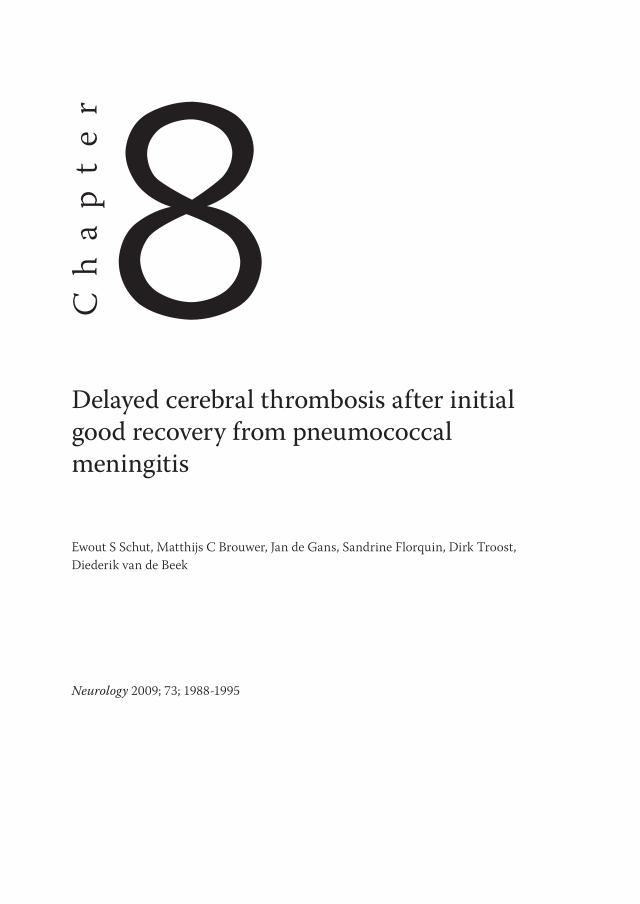
Case reportsCase 1. A 38-year-old man with otitis presented with headache, fever, and altered consciousness. Neurologic examination showed neck stiffness and decreased level of consciousness (Glasgow Coma Scale [GCS] score, E4M5V1). Treatment with penicillin and dexamethasone was started and cranial CT showed no abnormalities. Subsequent CSF examination yielded 1,787 leukocytes/mm3 and a high protein and low glucose level (Table 1). CSF gram stain showed gram-positive diplococci. Cultures of blood and CSF yielded Streptococcus pneumoniae sensitive to penicillin. On day 2, aphasia and a mild right hemiparesis were noted. Cranial MRI showed a left parietal T2 hyperintense lesion with restricted diffusion consistent with infarction. He showed excellent recovery. On day 10, he was fit for discharge. However, on day 11, he developed progressive headache and deteriorated into coma within hours. Cranial CT now showed slight communicating hydrocephalus and the previously identified left parietal infarct. An external lumbar drain was placed and CSF examination showed 5,933 leukocytes/mm3 (100% granulocytes). CSF gram stain and cultures yielded no bacteria. Therapy was restarted with ceftriaxone, metronidazole, and dexamethasone. Cranial MRI showed infarction of the thalamus bilaterally (Figure 1A), midbrain, pons (Figure 1B), and medulla oblongata. Despite supportive care, no neurologic improvement occurred. Care was withdrawn on day 32 and the patient died. Autopsy showed areas of infarction involving the left parietal lobe, and thalamus, midbrain, pons (Figure 1C), and medulla oblongata bilaterally. Macroscopically, the basilar artery was normal, but perforating arteries supplying the brainstem showed focal thrombi, without evidence of vasculitis (Figure 1D).
Case 2. A 46-year-old man presented with headache and a temperature of 38.6 °C. Neurologic examination showed neck stiffness, decreased attention span, and disorientation. No other neurologic abnormalities were found. Cranial CT was normal. Community-acquired
142
Chapter 8
bacterial meningitis was suspected and treatment with ceftriaxone and dexamethasone was started. CSF examinations showed 87 leukocytes/mm3 (91% monocytes) and high protein and low glucose levels. CSF gram stain revealed gram-positive diplococci and culture of blood and CSF yielded S. pneumoniae, sensitive to penicillin. The patient made a full recovery in the next few days.
Figure 1. MRI and pathology images posterior circulation infarction Case 1.
Axial T2-weighted MRI showing bilateral thalamic (A, arrow) and pontine (B, arrow) infarcts in case 1. Recent pontine infarction (C, Hematoxylin and Eosin, magnification:100X). A single asterisk indicates normal transverse pontine fibers; a double asterisk indicates infarction. Perforating brainstem artery with subtotal stenosis caused by thrombus (D, Hematoxylin and Eosin, magnification:400X). Vessel wall shows no evidence of vasculitis
143
Delayed cerebral thrombosis in pneumococcal meningitis
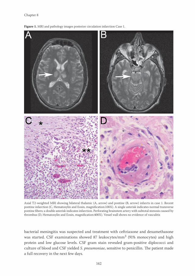
However, on day 10, his level of consciousness suddenly decreased and he developed a left hemiplegia. Cranial MRI showed bilateral thalamic infarctions (Figure 2A). He was immediately treated with prednisone 60 mg daily and ceftriaxone. His condition stabilized and his level of consciousness slowly improved. On day 84, he was discharged to a rehabilitation facility. On discharge, he had marked abulia and left hemiparesis. After several months of rehabilitation, he was able to return home. Currently, he is ambulating but unable to resume his previous occupation.
Case 3. A 55-year-old man with a history of idiopathic epilepsy presented with fever, diarrhea, and altered consciousness following 1 week of cough and general malaise. He had a temperature of 41.3 °C and was disorientated. Pneumonia was diagnosed and he was intubated because of respiratory failure. Treatment with ceftriaxone was initiated and blood cultures yielded S. pneumoniae. On day 2, a neurology consultation was requested. He had a GCS score of E4M5Vtube and there was neck stiffness. Cranial CT showed no abnormalities. Nevertheless, lumbar puncture was deferred and dexamethasone was started for 4 days. On day 10, he was awake, alert, and oriented, and ready for discharge.
Figure 2. MRI’s of posterior circulation infarction due to delayed cerebral thrombosis.
Axial T2-weighted MRI showing bilateral thalamic infarcts in case 2 (A). Axial T2-weighted MRI showing infarction of both thalami, left internal capsule (B) and left pons (C) in case 3. Axial diffusion-weighted MRI showing infarct of the right thalamus in case 5 (D). Axial (E) and coronal (F) fluid-attenuated inversion recovery (FLAIR) images showing infarction of medulla oblongata, cerebellum (E) and basal ganglia bilaterally (F) in case 6.

144
Chapter 8
However, on day 11, fever recurred and the patient rapidly became unresponsive. A cranial CT showed no abnormalities. A lumbar puncture revealed 166 leukocytes/mm3 (98% granulocytes), elevated protein, and normal glucose levels, and ceftriaxone was restarted. CSF gram stain and cultures of blood and CSF revealed no bacteria. Cranial MRI showed widespread infarcts, involving the thalamus bilaterally, left internal capsule (Figure 2B), and left pons (Figure 2C). Transesophageal echocardiography was unremarkable. On day 19, care was withdrawn and the patient died. No autopsy was performed.
Case 4. A 73-year-old man with a 2-day history of otitis presented with headache, altered consciousness, and a temperature of 39 °C. Neurologic examination showed neck stiffness, global aphasia, and gaze deviation to the left. Treatment with penicillin and dexamethasone was started. CSF examination yielded 17,700 leukocytes/mm3 (100% granulocytes), high protein, and low glucose levels. CSF gram stain showed gram-positive diplococci and cultures of blood and CSF yielded S. pneumoniae, sensitive to penicillin. Cranial CT scan showed no abnormalities. Over the next few days the patient improved. On day 6, he was awake, alert, and ambulating. However, on day 7, fever recurred and he developed gait impairment. A urinary tract infection was diagnosed and treatment with ciprofloxacin was started. On day 8, his level of consciousness started to fluctuate, with intermittent dysconjugate eye movements. Cranial CT scans again showed no abnormalities. A
Table 1. Characteristics of 6 patients with delayed cerebral intravascular thrombosis complicating bacterial meningitisCharacteristic Case 1 (2003) Case 2 (2004) Case 3 (2004) Case 4 (2008) Case 5 (2008) Case 6 (2008)Sex (age) M, 38 yr M, 46 yr M, 55 yr M, 73 yr F, 30yr M, 40yrAdmission GCS E4M4V1 E4M6V4 E2M5V2 E3M5V1 E4M6V4 E1M3V1Neurologic deficits Aphasia No No Aphasia, gaze deviation No NoCSF WBC (cells/mm3) 1,787 87 NP 17,7 2 80CSF protein (mg/dL) 101 610 NP 526 290 600CSF glucose (mg/dL) 61 <1,8 NP <1,8 <1,8 <1,8Causative organism S. pneumoniae S. pneumoniae S. pneumoniae S. pneumoniae S. pneumoniae S. pneumoniaeDexamethasone 10mg every 4 hours,
day 1 through 4 10mg every 4 hours, day 1 through 4
10mg every 4 hours, day 1 through 4
10mg every 4 hours, day 1 through 4
10mg every 4 hours, day 1 through 4
10mg every 4 hours, day 1 through 4
Cranial CT Otitis Normal Normal Normal Normal NormalStatus after first week Good recovery Excellent recovery Excellent recovery Good recovery Excellent recovery Good recoveryDeterioration second week Day 11: fever, headache, coma Day 10: fever, headache, coma Day 11: fever, hemiparesis,
comaDay 7: fever, seizures, brainstem signs, coma
Day 13: fever, headache, coma, hemiparesis
Day 19: fever, coma
CSF WBC 2nd LP (cells/mm3) 5,933 NP 166 500 1,100 1,080CSF protein 2nd LP (mg/dL) 774 NP 170 296 130 239CSF glucose 2nd LP (mg/dL) <1,8 NP 25 72 29 59CSF culture 2nd LP Negative NP Negative Negative Negative NegativeMRI Infarction left parietal lobe,
bilateral thalamus, brainstemInfarction thalamus bilaterally Infarction basal ganglia,
brain stemInfarction left frontal lobe, brain stem
Infarction left corona radiate, right thalamus, brain stem
Infarction left basal ganglia, medulla, cerebellum
Treatment Ceftriaxon, metroni-dazole, dexamethasone
Ceftriaxone, prednisolone Cefotaxim Ceftriaxone, prednisolone Penicillin, dexamethasone Ceftriaxone, prednisolone
Outcome Death, day 32 Survival, moderately disabled Death, day 19 Death, day 25 Death, day 22 Survived, severely disabledGCS Glasgow Coma Scale, CSF cerebrospinal fluid, NP not performed, WBC white blood cells 2nd CSF of patient one was obtained from a external ventricular catheter

145
Delayed cerebral thrombosis in pneumococcal meningitis
second lumbar puncture yielded 500 leukocytes/ mm3 (90% granulocytes); CSF gram stain and cultures remained negative. On day 12, cranial MRI showed multiple areas of cerebral infarction in brainstem and hemispheres. Transesophageal echocardiography was unremarkable. On day 18, he became comatose. A third lumbar puncture was performed; CSF cultures remained sterile. On day 25, supportive care was withdrawn and the patient died. Autopsy showed areas of granulocyte infiltration of brain parenchyma along the convexity and widespread areas of infarction and gliosis involving the cerebral hemispheres (Figure 3A), brainstem, and cerebellum. Microscopically, vessel walls showed minor infiltration by granulocytes, thickening and fibrosis of the intima, but the main findings were thrombi inside vessel lumina, resulting in subtotal stenosis (Figure 3B).
Case 5. A 30-year-old woman presented with fever, malaise, and headache. She had a temperature of 41 °C. Neurologic examination showed neck stiffness and disorientation, but was otherwise normal. Treatment with ceftriaxone and dexamethasone was started. A lumbar puncture yielded 2,000 leukocytes/mm3 (90% granulocytes), high protein, and low glucose levels. CSF gram stain showed gram-positive diplococci and culture of blood and CSF yielded S. pneumoniae, sensitive to penicillin. The patient improved and was ready for discharge on day 12. However, on day 13, fever, headache, and neck pain recurred. A lumbar puncture revealed 1,100 leukocytes/mm3 and CSF gram stain showed no bacteria.
Table 1. Characteristics of 6 patients with delayed cerebral intravascular thrombosis complicating bacterial meningitisCharacteristic Case 1 (2003) Case 2 (2004) Case 3 (2004) Case 4 (2008) Case 5 (2008) Case 6 (2008)Sex (age) M, 38 yr M, 46 yr M, 55 yr M, 73 yr F, 30yr M, 40yrAdmission GCS E4M4V1 E4M6V4 E2M5V2 E3M5V1 E4M6V4 E1M3V1Neurologic deficits Aphasia No No Aphasia, gaze deviation No NoCSF WBC (cells/mm3) 1,787 87 NP 17,7 2 80CSF protein (mg/dL) 101 610 NP 526 290 600CSF glucose (mg/dL) 61 <1,8 NP <1,8 <1,8 <1,8Causative organism S. pneumoniae S. pneumoniae S. pneumoniae S. pneumoniae S. pneumoniae S. pneumoniaeDexamethasone 10mg every 4 hours,
day 1 through 4 10mg every 4 hours, day 1 through 4
10mg every 4 hours, day 1 through 4
10mg every 4 hours, day 1 through 4
10mg every 4 hours, day 1 through 4
10mg every 4 hours, day 1 through 4
Cranial CT Otitis Normal Normal Normal Normal NormalStatus after first week Good recovery Excellent recovery Excellent recovery Good recovery Excellent recovery Good recoveryDeterioration second week Day 11: fever, headache, coma Day 10: fever, headache, coma Day 11: fever, hemiparesis,
comaDay 7: fever, seizures, brainstem signs, coma
Day 13: fever, headache, coma, hemiparesis
Day 19: fever, coma
CSF WBC 2nd LP (cells/mm3) 5,933 NP 166 500 1,100 1,080CSF protein 2nd LP (mg/dL) 774 NP 170 296 130 239CSF glucose 2nd LP (mg/dL) <1,8 NP 25 72 29 59CSF culture 2nd LP Negative NP Negative Negative Negative NegativeMRI Infarction left parietal lobe,
bilateral thalamus, brainstemInfarction thalamus bilaterally Infarction basal ganglia,
brain stemInfarction left frontal lobe, brain stem
Infarction left corona radiate, right thalamus, brain stem
Infarction left basal ganglia, medulla, cerebellum
Treatment Ceftriaxon, metroni-dazole, dexamethasone
Ceftriaxone, prednisolone Cefotaxim Ceftriaxone, prednisolone Penicillin, dexamethasone Ceftriaxone, prednisolone
Outcome Death, day 32 Survival, moderately disabled Death, day 19 Death, day 25 Death, day 22 Survived, severely disabledGCS Glasgow Coma Scale, CSF cerebrospinal fluid, NP not performed, WBC white blood cells 2nd CSF of patient one was obtained from a external ventricular catheter

146
Chapter 8
Penicillin was restarted but cultures of blood and CSF remained sterile. Subsequently, she developed a hemiparesis and impaired level of consciousness. On day 17, cranial MRI showed multiple infarctions in the pons, right thalamus (Figure 2D), and left corona radiata. Transesophageal echocardiography was unremarkable. Dexamethasone was restarted but no clinical improvement occurred. On day 22, care was withdrawn and the patient died. No autopsy was performed.
Case 6. A 40-year-old man presented with fever, headache, and a decreased level of consciousness (GCS score E1M3V1). Cranial CT showed no abnormalities and lumbar puncture yielded 80 leukocytes/mm3 (90% granulocytes), high protein, and low glucose levels. CSF gram stain showed grampositive diplococci and culture of blood and CSF yielded S. pneumoniae, sensitive to penicillin. He was treated with penicillin and dexamethasone, made a good recovery, and was discharged on day 17. However, on day 19, he was readmitted because of fever and developed a coma within hours. Cranial CT showed a hypodense lesion in the left basal ganglia. Transesophageal echocardiography was unremarkable. Cranial MRI showed evidence of infarcts in the medulla oblongata and cerebellum bilaterally (Figure 2E), and basal ganglia bilaterally (Figure 2F). Reexamination of CSF was not performed. The patient was treated with prednisolone 30 mg twice daily. His condition stabilized and his level of consciousness slowly improved. On day 92, he was discharged to a rehabilitation facility. On discharge he had marked abulia and was wheelchair-bound. Currently, he uses a walker, but still has severe cognitive defects.
Cortical wedge-shaped infarct in case 4 (A, Hematoxylin and Eosin, magnification:20X). A single asterisk indicates normal brain parenchyma; a double asterisk indicates infarction. Perforating artery with subtotal stenosis caused by thrombus (B, Hematoxylin and Eosin, magnification:400X ). Vessel wall shows no evidence of vasculitis.
Figure 3. Pathology images of cortical infarction and intravascular thrombus.

147
Delayed cerebral thrombosis in pneumococcal meningitis
Review of the literaturePubMed was searched through May 2009 using search terms “meningitis,” “vasculitis,” “vasculopathy,” “stroke,” and “cerebral infarction” in the English, French, and German language. We only included adult cases with secondary deterioration after 5 days. We did not evaluate cases with early deterioration and vasculitis or vasculopathy. Five patients were reported between 1992 and 2007 (Table 2); age varied from 19 to 53 years.5-7,9,10 Four patients had streptococcal meningitis (S. pneumoniae in 3, S. milleri in 1); 1 patient had meningitis due to Staphylococcus aureus. Empirical treatment consisted of third-generation cephalosporin-based regimens in 4 patients and penicillin monotherapy in 1 patient. Two patients were initially treated with adjunctive dexamethasone therapy. All cases were admitted with severe illness. Secondary deterioration occurred after 5 to 9 days after admission. Importantly, at time of deterioration, 3 of 5 patients were severely ill and still admitted to the ICU. One patient was admitted to the ward and suddenly became unresponsive due to a subarachnoid hemorrhage. One patient developed gait impairment on day 9, while out of the ICU. It is unknown whether this patient initially received dexamethasone. She developed a vasculopathy resembling moyamoya syndrome. CSF examination was repeated in 2 patients; CSF leukocyte counts were up to 500 mm3 (65% to 74% neutrophils); CSF cultures remained sterile. Neuroimaging showed multiple areas of infarction in the posterior circulation territory in 4 patients; 1 patient also had infarction in the MCA territory. Three patients were treated with steroids after clinical deterioration
Table 2. Patients reported in the literature with delayed vasculopathy complicating bacterial meningitisCharacteristic Case 1 (1992) Case 2 (2005) Case 3 (2006) Case 4 (2007) Case 5 (2007) Sex (age) M, 34 yr F, 20 yr M, 53 yr M, 19 yr M, 49yrCausative organism S. milleri S. pneumoniae S. pneumoniae S. aureus S. pneumoniaeDexamethasone Yes Unknown No No YesStatus after first week
Hemiparesis Out of ICU ICU: intubated, awake, hemiplegia
ICU: intubated, awake
ICU: intubated, awake
Secondary deterioration
Day 8: coma, fixed pupils
Day 9: 3rd nerve palsy, hemiparesis
Day 10: coma, hemiplegia
Day 5: 3rd nerve palsy, hemiparesis
Day 9: coma
CSF WBC on deterioration (cells/mm3)
NP 400 57 NP NP
Repeat CSF culture NP Negative Negative NP NPNeuroimaging Subarachnoid
hemorrhageMoyamoya syndrome, Infarction hemispheres, brain stem.
Infarction hemispheres, basal ganglia, brain stem
Infarction basal ganglia
Hemorrhagic infarction both hemispheres
Treatment Supportive care Dexamethasone Methylprednisolone Dexamethasone Supportive care, antibiotics
Outcome Death Death (day 260) Severe disability Moderate disability
Death
ICU intensive care unit, NP denotes not performed, WBC white blood cells

148
Chapter 8
occurred. Three patients died and the remaining 2 patients had poor outcomes. The patient with moyamoya syndrome died after 260 days. Autopsy revealed no systemic vasculitis or arteriopathy. The proximal cerebral vasculature exhibited severe luminal narrowing. Autopsy of patient 1 showed an angiodestructive inflammatory process in all wall layers; arteritis was a prominent feature. Interestingly, intracellular gram-positive cocci were present, indicating suboptimal antibiotic therapy. Both surviving patients were treated with high-dose steroids after clinical deterioration.
DiscussionWe present 6 patients with delayed cerebral thrombosis complicating bacterial meningitis. All had pneumococcal meningitis and made a good or excellent recovery in the first week of admission. After 7 to 19 days they suddenly deteriorated and developed multiple infarctions primarily located in the posterior circulation territory. We have not come across similar reports describing this devastating phenomenon in patients with initial excellent recovery from bacterial meningitis. Autopsy showed thrombosis mainly involving penetrating arteries supplying the thalamus and brainstem, indicating an immunologic reaction specifically targeting cerebral blood vessels. There were no inflammatory cells in any of the vessel wall layers. These findings are consistent with cerebral thrombosis. We estimate that this complication occurs in approximately 1 out of 100 cases with pneumococcal meningitis.
Very few patients reported in the literature resemble our patients: we did not identify patients with a similar degree of initial recovery or severity of clinical deterioration. One case has been published with mild secondary deterioration after initial recovery. This patient developed a moyamoya-like syndrome, most likely immune-mediated since symptoms reappeared after tapering immunosuppressive therapy. This patient died after cerebral hemorrhage more than 250 days after developing meningitis. Autopsy showed no vasculitis. It is unknown whether this patient was initially treated with dexamethasone.
We did not encounter cases with delayed cerebral thrombosis in our prospective nationwide cohort evaluating 696 cases with community-acquired bacterial meningitis (1998-2002, D. van de Beek).11 This cohort study was performed in the period before routine use of adjunctive dexamethasone therapy. Adjunctive dexamethasone treatment has been shown to decrease mortality in community-acquired bacterial meningitis in adults in a randomized clinical trial and meta-analysis.12,13 In this trial, the effect on mortality was most apparent in the subgroup of patients with pneumococcal meningitis.12 A post hoc analysis showed that mortality in this group was decreased by prevention of systemic complications rather than neurologic complications.13
In patients with bacterial meningitis, significant reduction in cerebrospinal white blood cell counts is usually found by day 3 of therapy and glucose levels usually return to normal more rapidly.14 In our case series, 3 out of 4 patients with repeated examination of CSF had no substantial reduction of white blood cell counts and 3 out of 5 patients had persisting low

149
Delayed cerebral thrombosis in pneumococcal meningitis
glucose levels. All had negative CSF cultures. This suggests a mechanism of persisting or recurrent inflammation without persistent bacterial infection.
Dexamethasone is a glucocorticoid, which has been shown to attenuate the inflammatory response and coagulation caused by the release of bacterial endotoxins in the acute phase of disease.15 An interesting parallel to that of thrombosis in bacterial meningitis is found in the field of renal transplantation. OKT3, an anti-CD3 monoclonal antibody used to prevent allograft rejection, is associated with a massive release of proinflammatory cytokines and carries an increased risk of intragraft thrombosis.16 Although few patients develop thrombosis after OKT3, those patients who had been pretreated with high-dose methylprednisolone had a significantly higher risk of thrombosis compared to those who had received lower doses of methylprednisolone. It has been suggested that high-dose glucocorticoids can tip the balance toward sustained coagulation and platelet aggregation by influencing the tissue factor/ factor VII pathway.17 Another study showed that in subjects who were pretreated with glucocorticoids 12 to 144 hours before endotoxin exposure, significantly higher levels of proinflammatory cytokines and coagulation were found.18
Dexamethasone stimulates macrophages by upregulation of endocytic receptors and increases their phagocytic potential. In a recent in vitro study, macrophages stimulated by dexamethasone and interleukin-4 for 4 days were compared to those stimulated by interleukin-4 only. Stimulation by dexamethasone enhanced the surface expression of transforming growth factor beta receptor II (TGF-β RII) in a time-dependent manner by 700% over 4 days.19 TGF-β plays an important role in the development of cerebral necrotizing vasculitis in pneumococcal meningitis. Deletion of the TGF-β R II gene in polymorphonuclear leukocytes enhances polymorphonuclear leukocytes recruitment into the CNS of mice with S. pneumoniae meningitis. This was associated with more efficient clearance of bacteria, and almost complete prevention of cerebral necrotizing vasculitis.20 An additional explanation could be a rebound effect of the primary inflammatory reaction initially suppressed by dexamethasone. Adjunctive dexamethasone may not cause deterioration but merely delays it.
All our patients were initially treated with adjunctive dexamethasone therapy, suggesting an association with this newly introduced adjunctive therapy in bacterial meningitis. Nevertheless, a causal relation between delayed cerebral thrombosis and dexamethasone remains difficult to establish. Delayed cerebral thrombosis may occur in patients with pneumococcal meningitis irrespective of steroid therapy.
The outcome of delayed cerebral thrombosis in our patients was devastating. Four patients died and 2 remained disabled. Combining our patients and those reported, all who survived received prolonged treatment with high-dose steroids. This supports the hypothesis of an immunologic basis of this disease. Patients with bacterial meningitis with clinical deterioration after initial improvement should undergo further investigations without delay, including MRI, and repeated CSF examination. If delayed cerebral thrombosis is suspected, immediate high-dose steroids should be started. These patients typically have multiple infarctions on MRI, predominantly in the posterior circulation territory. Lumbar puncture may reveal substantially elevated leukocyte counts. Although no relapse of bacterial

150
Chapter 8
meningitis was proven in our cases, antibiotic therapy should be prolonged or restarted until CSF cultures remain negative. Once cerebral infarctions have been demonstrated by MRI, a cardioembolic cause should be ruled out by echocardiography, since bacterial meningitis and endocarditis may coexist.21 Tapering of steroids may lead to new clinical deterioration. Although based on speculation, we suggest prompt reinitiation of immunosuppressive therapy. Future case series are needed to determine whether this will improve the outcome of this devastating complication.
References 1. Pfister HW, Borasio GD, Dirnagl U, Bauer M, Einhaupl KM. Cerebrovascular complications of
bacterial meningitis in adults. Neurology 1992; 42: 1497-504.
2. van de Beek D, de Gans J, Tunkel AR, Wijdicks EF. Community-acquired bacterial meningitis in adults. N Engl J Med 2006; 354: 44-53.
3. Kastenbauer S, Pfister HW. Pneumococcal meningitis in adults: spectrum of complications and prognostic factors in a series of 87 cases. Brain 2003; 126: 1015-25.
4. Weisfelt M, de Gans J, van der Poll T, van de Beek D. Pneumococcal meningitis in adults: new approaches to management and prevention. Lancet Neurol 2006; 5: 332-42.
5. Bentley P, Qadri F, Wild EJ, Hirsch NP, Howard RS. Vasculitic presentation of staphylococcal meningitis. Arch Neurol 2007; 64: 1788 -9.
6. Czartoski T, Hallam D, Lacy JM, Chun MR, Becker K. Postinfectious vasculopathy with evolution to moyamoya syndrome. J Neurol Neurosurg Psychiatry 2005; 76: 256-9.
7. Lefebvre N, Carre AC, Delabranche X, Guiot P, Mootien Y. [Implication of dexamethasone adjunctive therapy after the onset of cerebral vasculitis in Streptococcus pneumoniae meningitis.] Med Mal Infect 2007; 37: 118-20.
8. Palacio S, Hart RG, Vollmer DG, Kagan-Hallet K. Late developing cerebral arteriopathy after pyogenic meningitis. Arch Neurol 2003; 60: 431-3.
9. Pugin D, Copin JC, Goodyear MC, Landis T, Gasche Y. Persisting vasculitis after pneumococcal meningitis. Neurocrit Care 2006; 4: 237-40.
10. Perry JR, Bilbao JM, Gray T. Fatal basilar vasculopathy complicating bacterial meningitis. Stroke 1992; 23: 1175-8.
11. van de Beek D, de Gans J, Spanjaard L, Weisfelt M, Reitsma JB, Vermeulen M. Clinical features and prognostic factors in adults with bacterial meningitis. N Engl J Med 2004; 351: 1849-59.
12. de Gans J, van de Beek D. Dexamethasone in adults with bacterial meningitis. N Engl J Med 2002; 347: 1549-56.
13. van de Beek D, de Gans J, McIntyre P, Prasad K. Steroids in adults with acute bacterial meningitis: a systematic review. Lancet Infect Dis 2004; 4: 139-143.
14. Fishman RA. Cerebrospinal Fluid in Diseases of the Nervous System, 2nd ed. Philadelphia: Saunders; 1992.
15. van der Poll T. Coagulation and inflammation. J Endotoxin Res 2001; 7: 301-4.
16. Abramowicz D, Pradier O, Marchant A, et al. Induction of thromboses within renal grafts by high-dose prophylactic OKT3. Lancet 1992; 339: 777-8.

151
Delayed cerebral thrombosis in pneumococcal meningitis
17. Abramowicz D, Pradier O, de Pauw L, et al. High-dose glucocorticosteroids increase the procoagulant effects of OKT3. Kidney Int 1994; 46: 1596-602.
18. Barber AE, Coyle SM, Marano MA, et al. Glucocorticoid therapy alters hormonal and cytokine responses to endotoxin in man. J Immunol 1993; 150: 1999-2006.
19. Gratchev A, Kzhyshkowska J, Kannookadan S, et al. Activation of a TGF-beta-specific multistep gene expression program in mature macrophages requires glucocorticoidmediated surface expression of TGF-beta receptor II. J Immunol 2008; 180: 6553-65.
20. Malipiero U, Koedel U, Pfister HW, et al. TGFbeta receptor II gene deletion in leucocytes prevents cerebral vasculitis in bacterial meningitis. Brain 2006; 129: 2404-15.
21. Salgado AV. Central nervous system complications of infective endocarditis. Stroke 1991; 22: 1461-3.


