UvA-DARE (Digital Academic Repository) … 2 PetrosiaPetrosia (Petrosia) alfiani spec , no v . (Fig...
19
UvA-DARE is a service provided by the library of the University of Amsterdam (http://dare.uva.nl) UvA-DARE (Digital Academic Repository) Indonesian sponges : biodiversity and mariculture potential de Voogd, N.J. Link to publication Citation for published version (APA): de Voogd, N. J. (2005). Indonesian sponges : biodiversity and mariculture potential. Amsterdam: UvA-IBED. General rights It is not permitted to download or to forward/distribute the text or part of it without the consent of the author(s) and/or copyright holder(s), other than for strictly personal, individual use, unless the work is under an open content license (like Creative Commons). Disclaimer/Complaints regulations If you believe that digital publication of certain material infringes any of your rights or (privacy) interests, please let the Library know, stating your reasons. In case of a legitimate complaint, the Library will make the material inaccessible and/or remove it from the website. Please Ask the Library: http://uba.uva.nl/en/contact, or a letter to: Library of the University of Amsterdam, Secretariat, Singel 425, 1012 WP Amsterdam, The Netherlands. You will be contacted as soon as possible. Download date: 09 Apr 2019
Transcript of UvA-DARE (Digital Academic Repository) … 2 PetrosiaPetrosia (Petrosia) alfiani spec , no v . (Fig...

UvA-DARE is a service provided by the library of the University of Amsterdam (http://dare.uva.nl)
UvA-DARE (Digital Academic Repository)
Indonesian sponges : biodiversity and mariculture potentialde Voogd, N.J.
Link to publication
Citation for published version (APA):de Voogd, N. J. (2005). Indonesian sponges : biodiversity and mariculture potential. Amsterdam: UvA-IBED.
General rightsIt is not permitted to download or to forward/distribute the text or part of it without the consent of the author(s) and/or copyright holder(s),other than for strictly personal, individual use, unless the work is under an open content license (like Creative Commons).
Disclaimer/Complaints regulationsIf you believe that digital publication of certain material infringes any of your rights or (privacy) interests, please let the Library know, statingyour reasons. In case of a legitimate complaint, the Library will make the material inaccessible and/or remove it from the website. Please Askthe Library: http://uba.uva.nl/en/contact, or a letter to: Library of the University of Amsterdam, Secretariat, Singel 425, 1012 WP Amsterdam,The Netherlands. You will be contacted as soon as possible.
Download date: 09 Apr 2019

IndonesianIndonesian sponges of the genus Petrosia
CHAPTERR 2
INDONESIANN SPONGES OF THE GENUS PETROSIA
VOSMAERR (DEMOSPONGIAE: HAPLOSCLERIDA)
N I C O L EE J. D E V O O G D & R O B W . M . V A N S O E S T
2\ 2\

ChapterChapter 2
A B S T R A C T T
Spongess of the genus Petrosia (Demospongiae: Haplosclerida) are large and characteristicc components of many Indonesian reefs. We identify and provide descriptionss of seven species of Petrosia collected recently on Sulawesi reefs (Eastern Indonesia),, two of which are new to science: Petrosia (Petrosia) alfiani spec.nov. and PetrosiaPetrosia (Petrosia) hoeksemai spec. nov. Additional species recorded are Petrosia (Petrosia)(Petrosia) lignosa Wilson, 1925, Petrosia (Petrosia) nigricans Lindgren, 1897, Petrosia (Petrosia)(Petrosia) plana Wilson, 1925, Petrosia (Strongylophora) corticata (Wilson, 1925) and PetrosiaPetrosia (Strongylophora) strongylata Thiele, 1903. The species are discussed and comparedd with Petrosia records from other Indonesian and neighbouring Indo-West Pacificc areas. We also present a key to the common Petrosia species of Sulawesi.
I N T R O D U C T I O N N
Iff trends observed in other marine benthic groups also apply to sponges, Indonesian waterss may be expected to contain the world's highest sponge biodiversity. Van Soestt (1989, 1990, 1994) and Hooper et al. (2000) reviewed the sponge fauna of this areaa on the basis of literature and informal database records, and these reviews confirmm the richness. However, the published knowledge base of Indonesian spongess is woefully incomplete: much of the collected material still awaits formal descriptionn and many locations in the area remain to be explored. The present study iss induced by the search for sponge secondary metabolites with properties useful to mankind,, financed under the EC-MAS3 project' SYMBIOSPONGE' and the NWO-WOTRO-projectt (W84-474) 'Sponges as a potential resource of Eastern Indonesia'. Itt is also one of a recently started ongoing series of genus-by-genus revisions of thee Indonesian sponge fauna (Hofman & Van Soest 1995; Van Soest, 1998), which eventuallyy wil l result in a much greater accessibility of the Indonesian sponges for interestedd ecologists and chemists.
M A T E R I A L SS A N D M E T H O D S
Thee material was collected by B.W. Hoeksema (fieldnumbers # BH97/xxxx/xx and 98/NSS or SS/xxxx/BH/xx), N.J. de Voogd (NV/97/xx/xx; NV/xx/xx00/xx), H. Moll and R.W.M.. van Soest from various locations in Indonesia. The specimens are preserved inn 70% ethylalcohol and deposited in the sponge collection of the Zoological Museum Amsterdamm (ZMA) and the National Museum of Natural History of Leiden (RMNH). Thee descriptions presented below are based on external morphology, skeletal architecturee and shape and size of the spicules. For study of the skeletal architecture hand-cutt tangential sections of the ectosome and perpendicular sections of the choanosomee were made. The sections were air-dried, mounted in Canada-balsam onn a microscope slide, and studied under a Leitz high power light microscope.
22 2

IndonesianIndonesian sponges of the genus Petwsia
Spiculee preparations were made by dissolving a small piece of the specimen in 100%% nitric acid (HN03), after which the residue was rinsed four times with water, oncee with hydrogen peroxide (H,OJ and finally once with 96% ethylalcohol. The spiculess were air-dried on microscopic slides and prepared for study with the light microscope,, as well as put on aluminium stubs and coated with gold for study with aa Jeol Scanning Electron Microscope (SEM).
SYSTEMATICA A
PHYLUMM PORIFERA GRANT, 1835
CLASSS DEMOSPONGIAE SOLLAS, 1885
ORDERR HAPLOSCLERIDA TOPSENT, 1928
SUBORDERR PETROSINA BOURY -ESNAULT & VAN BEVEREN, 1982
FAMIL YY PETROSIIDAE VAN SOEST, 1980
Definition.. — Haploscerida with ectosomal skeleton consisting of an isotropic reticulationn of single spicules or spicule tracts, and a choanosomal skeleton verging towardss an isotropic reticulation of spicule tracts, in which primary and secondary tractss are indistinct.
Genuss Petrosia Vosmaer, 1887
Definition.. — Petrosiidae with an ectosomal triangular or polygonal reticulation of spiculee tracts or single spicules, usually echinated at the nodes or along the tracts by aa smaller category of spicules. Choanosomal skeleton basically a lamellate-isotropic reticulationn of spicule tracts, and an interstitial unispicular reticulation. Megascleres withh distinct size categories of strongyles or oxeas, often with a special category of ectosomall microxeas or microstrongyles. N.B.. This definition includes sponges referable to the genus Strongylophora Dendy, 1905.. Its separate generic status vis-a-vis Petrosia is doubtful, and in a forthcoming revisionn (Desqueyrouz-Faündez in litteris) both are treated as subgenera of Petrosia. Thiss is followed here. Reviewss in Dendy (1905); van Soest (1980); Bergquist & Warne (1980); De Weerdt (1985);; Desqueyroux-Faundez (1987), Desqueyroux-Faundez & Valentine (2002); Fromont(1991). .
S U B G E N USS PETROSIA V O S M A E R, 1887
Definition.. — Spicule complement includes two or three size categories of oxeas or strongyles,, in which the smallest is concentrated at the surface.
23 3

ChapterChapter 2
PetrosiaPetrosia (Petrosia) alfiani spec, no v.
(Figss IA-C, 5A)
Materiall examined. - Holotype: ZMA POR. 15992, NW Kudingareng Keke, Spermonde Archipelago,
SWW Sulawesi, 5°09'S 119°16'E, reef slope, on coral sand, 15 m, coll. B.W. Hoeksema, # BH97/0206/007,
6.vi.l997;; Paratypes: ZMA POR. 13178, Bone Tambung, Spermonde Archipelago, SW Sulawesi, 5°02'S
119°18'E,, reef slope, on coral sand, 20 m, coll. NJ. de Voogd ft NV/180697/K, 18.vi.1997; ZMA POR. 14499,
NWW Kapoposang, Spermonde Archipelago, SW Sulawesi, 4°60'S 118°98'E, vertical wall, 26 m, coll. B.W.
Hoeksemaa 9 98/SS/MAY01/BH/052,1.v. 1998; ZMAPOR.15995, NW Kapoposang, Spermonde Archipelago,
SWW Sulawesi, 4°60'S 118°98'E, reef slope, on coral sand, 20 m, coll. B.W. Hoeksema # BH97/2606/013,
26.vi.1997. .
Shape.. — massive, globular, or thick undividing arm-like branches, of a maximum lengthh of 20 cm, 10 cm in width and 4 cm in height. Holotype ZMA 15992 measures 99 x 7.5 x 2 cm, paratype ZMA 14499 is 8 x 4 * 3 cm. Numerous small oscules are scatteredd across the sponge body, 2-6 mm in diameter. The surface is smooth and microscopicallyy hispid. The consistency varies from stony hard to very slightly compressible. . Colour.. —Bright canary yellow, turns cherry red-brown exposed to air. In spirit light too dark chocolate brown. Preservative turns very dark red-brown. Skeleton.. — The ectosomal skeleton is an isodictyal reticulation of multispicular tractss (2-6 um), forming irregular round meshes (200-250 urn), echinated at the nodes byy brushes of the smaller sized spicules. The smaller sized spicules are only present inn the ectosomal skeleton. The ectosomal skeleton is obscured by abundant pigment cells.. The choanosomal skeleton is more compact with thick multispicular tracts (7-155 spicules) forming round meshes (200-400 um), regularly arranged parallel to the surface.. Pigment cells are still present in the choanosome, but not so dense as in the ectosome.. Spongin is scarce, causing the stony texture of the sponge. Spicules.. — The species has three sizes of abruptly pointed oxeas or strongyles with smoothh rounded ends: 183-253 * 10-15 urn, 106-153 * 7-14 pm, and 60-70 * 6-7 urn. Etymology.. — The species is named after Prof. dr. Alfian Noor. He is co-ordinator of thee Buginesia Programme and Head of the Radiation Chemistry Laboratory of the Hasanuddinn University, Makassar. Habitat.. — On reef slopes, from shallow waters down to 40m; growing as thick encrustingg masses on coral blocks and rubble or coral sand. Distribution.. — Spermonde Archipelago, SW Sulawesi. Remarks.. — The sponge resembles Petrosia similis var. compacta (Ridley & Dendy, 1887)) sensu Hentschel (1912); however the smaller oxeas and the changing from the brightt yellow to red brown are not mentioned. The original description by Ridley && Dendy concerns a sponge from Kerguelen in the subantarctic Indian Ocean. It sharess the bright yellow colour and it turns likewise to brown when exposed to air. Howeverr our newly described species does not exude abundant mucus mentioned byy Ridley & Dendy, and the habitus is also very different. Itt is unusual for a petrosiid to be of a vivid yellow colouration as most of the speciess are beige, red, brown or black. Desqueyroux-Faündez (1987) described a
24 4

IndonesianIndonesian sponges of the genus Petrosia
lemon-yelloww species, Petrosia capsa from New Caledonia, but this species differs substantiallyy from the new species in oxea sizes and classes (two classes: 140-210 * 2.5-100 urn and 40-60 x 6-8 um), habitus (massive-cylindrical) and skeleton (thicker fibers).. No colour change was noted for P. capsa.
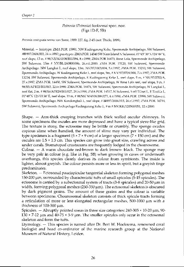
Fig.. 1 A-C. Petrosia (Petrosia) alfiani spec, nov., holotype ZMA POR. 15992, A. tangential view of ecto-
somall skeleton (scale— 250 |_im), B. cross section of choanosomal skeleton (scale— 250 um), C. spicules
(scale—— 250 pm). D-F. Petrosia (Petrosia) hoeksemai spec, nov., D. spicules (scale— 250 pm), E. tangential
vieww of ectosomal skeleton (scale— 250 pm), F. ditto at larger magnification to show brushes of microxeas
(scale-- 100 um).
25 5
ChapterChapter 2
PetrosiaPetrosia (Petrosia) hoeksemai spec. nov. (Figss 1D-F, 5B)
PetrosiaPetrosia contignata sensu van Soest, 1989: 227, fig. 2-45 (not: Thiele, 1899).
Material.. — holotype: ZMAPOR. 15991, NW Kudingareng Keke, Spermonde Archipelago, SW Sulawesi,
#BH97/2604/002,24.iv.1997;; paratypes: ZMAPOR. 14540 SW Nain Island N Sulawesi, 01°45"30'N 124°46"E,
reeff slope, 13 m, # 98/NS/MAY08/BH/094, 8.V.1998; ZMA POR 16473, Bone Lola, Spermonde Archipelago,
SWW Sulawesi, 15m # NV/BL/260800/086, 26.viii.2000; ZMA POR. 13120, SW Sulawesi, Spermonde
Archipelago,, NW Langkai I., reef slope, 15m. NV/97/1005/004, 5.V.1997; ZMA POR. 13210, SW Sulawesi,
Spermondee Archipelago, W Kudingareng Keke I., reef slope, 9m, # NV/97/0704/008, 7.iv.l997; ZMAPOR.
13214,, SW Sulawesi, Spermonde Archipelago, E Kudingareng Keke I., reef slope, 9 m, I NV/97/2705/A,
27.V.1997;; ZMA POR. 14450, SW Sulawesi, Spermonde Archipelago, W Bone Lola reef, reef slope, 9 m, #
98/SS/APR22/BH/003,, 22.iv.1998; ZMA POR. 14474, SW Sulawesi, Spermonde Archipelago, W Langkai I.,
reeff flat, 2 m, # 98/SS/APR29/BH/027, 29.iv.1998; ZMA POR. 14517, N Sulawesi, S off Tilisei I., E Tindila I.,
01°48'NN 125°03'30"E, reef slope, 19 m, # 98/NS/ MAY06/BH/071, 6.V.1998; ZMA POR. 15998, SW Sulawesi,
Spermondee Archipelago, NW Kondongbali I., reef slope, # BH97/2606/015, 26.vi.1997; ZMA POR. 16744,
SWW Sulawesi, Spermonde Archipelago Kudingareng Keke I., 9 m m # NV/KK/120500/051,12.V.2000.
Shape.. — Arm-thick creeping branches with thick walled oscular chimneys. In somee specimens the oscules are more depressed and have a typical sieve-like grid. Thee texture is stony, the ectosome may be brittle or crumbly. The sponge exudes copiouss slime when handled, the amount of slime may vary per individual. The typee specimen is a fragment (5 * 7 * 9 cm) of a larger specimen (7 * 150 cm) and the osculess are 1.5 * 1.5 cm. This species can grow into great size, crawling across and underr corals. Stomatopod crustaceans are frequently lodged in the choanosome. Colour.. — A warm chocolate red-brown to dark brown- black. The sponge may bee very pale in colour (e.g. like in Fig. 5B) when growing in caves or underneath overhangs,, this species clearly derives its colour from symbionts. The inside is lighter,, almost greyish. The colour persists more or less in spirit, but a greyish tinge predominates. . Skeleton.. — Ectosomal paucispicular tangential skeleton forming polygonal meshes 150-2000 um, surrounded by characteristic tufts of small spicules (5-15 spicules). The ectosomee is carried by a subectosmal system of tracts (3-8 spicules) and 20-50 um in width,, forming polygonal meshes (250-350 um). The ectosomal skeleton is obscured byy dark pigment grains. The amount of these grains and the colour is variable betweenn specimens. Choanosomal skeleton consists of thick spicule tracts forming aa reticulation of more or less elongated rectangular meshes, 500-1000 um with a thicknesss of 100-500 um. Spicules.. — Abruptly pointed oxeas in three size categories: 240-305 * 10-20 um, 90-1300 x 7-12 um and 40-75 * 5-9 um. The smaller spicules only occur in the ectosomal skeletonn and form the tufts. Etymology.. — This species is named after Dr. Bert W. Hoeksema, renowned coral biologistt and head co-ordinator of the marine research group at the National Museumm of Natural History, Leiden.
26 6

IndonesianIndonesian sponges of the genus Petrosia
Habitat.. — Creeping across live and dead corals, cryptic underneath coral blocks. Distribution.. — Throughout Indonesia. Remarks.. — Previous records of this species were under the name Petrosia contignata Thiele,, 1899 (e.g. Van Soest, 1989), but subsequent examination of the type specimen fromm the Basel Museum (Nr. 31) showed that this is not a Petrosia, but a Xestospongia (noo size categories of the oxeas). There is also a superficial resemblance to P. pigmentosapigmentosa Fromont, 1991 described from the Great Barrier Reef. We re-examined thee type specimen, Museum for Tropical Queensland QM 925020, Whitsunday Island,, 20°48'S 149°16'E. This is a cake-shaped fragment with small more or less flushh oscules. The skeleton consists of shorter and thinner spicules than those of P.. hoeksemai, and these are also predominantly strongyles. The species P. hoeksemai, P.P. lignosa and P. plana (cf. below) are easily distinguished from each other by habit (respectivelyy repent-branching, cup-shaped and tubular), but their skeletons are veryy much alike. The meshes of the choanosomal skeleton of P. lignosa are somewhat smallerr and the overall compactness of the skeleton is looser because of the lack of spongin.. P. plana appears to be restricted to the Togian Islands and North Sulawesi, whilee P. lignosa is also a widespread species, but occurs in vertical reef habitats, rather thann on shallow reef slopes. P. hoeksemai spec. nov. is the most common species in the Spermondee Archipelago, and is found in every shallow reef monitored.
PetrosiaPetrosia (Petrosia) lignosa Wilson, 1925 (Figss 2A-C, 5C)
PetrosiaPetrosia lignosa Wilson, 1925: 403, pi. 41 fig. 3, pi. 48, fig. 9.
PetrosiaPetrosia spec. Colin & Arneson, 1995: 47, fig. 153.
Material.. - ZMA POR. 14485, SW Sulawesi, Spermonde Archipelago, Kapoposang I., 4°60'S 118°98'E,
verticall wall at 33 m, » 98/SS/MAY01/BH/038, l.v.1998; ZMA POR. 14554, N Sulawesi, S Tetapaan I.,
01°18'NN 124°30'30"E, reef slope 26 m, # 98/NS/MAY12/BH/108,12.V.1998; ZMA POR. 16745, SW Sulawesi,
Spermondee Archipelago, Kapoposang I., vertical wall at 25 m, # NV/KP/020900/106,2. viii.2000; ZMA POR.
16746,, SW Sulawesi, Spermonde Archipelago, Kapoposang I., vertical wall at 30m # NV/KP/130900/134,
13.viii.2000. .
Shape.. — Wide flaring vasiform bowls with a characteristic thickened rim. Outer surfacee irregular, with blunt projections or bumps. Size may be considerable, up to 11 m in diameter. Inner surface smooth with characteristics annulated ridges, with scatteredd small oscules. Texture is incompressible, stony. Colour.. — Dull golden brown, inside cream or slighter lighter. Skeleton.. — Ectosomal paucispicular tangential skeleton forming meshes 150-200 urn,, the sides of which are characteristic tufts of small spicules (5-10 spicules), consistingg of an intermediately sized spicule carrying a bouquet of the smallest spicules.. Pigment grains are adhering to the binding spongin, not dominating or obscuringg the skeleton. The ectosomal skeleton in its turn carried by a subectosmal systemm of tracts of 3-8 spicules and 20-50 um in width, forming polygonal meshes of 250-3500 [tm. Choanosomal skeleton consists of thick spicule tracts with a thickness
27 7
ChapterChapter 2
off 250-450 \im forming a reticulation of more or less elongated rectangular meshes, 500-15000 urn. Spicules.. — The spicules are smooth, slightly curved, and range from oxeas to real strongyles;; this may vary individually; some contain only strongylotes. The size rangess from large to very small ones, in which 3 categories may be distinguished, 230-3000 x 14-18 (am, 75 -150 * 10-13 urn and 35-65 * 7 * 10 urn. Distribution.. — Sulawesi, Bali. Habitat.. — Vertical reef slopes, 25 m and deeper. Remarks.. — As mentioned above, P. lignosa is similar to P. hoeksemai and P. plana inn skeletal features, but they differ strongly in habit. Desqueyroux-Faündez (1987) describedd a petrosiid sponge, P. capsa, from New Caledonia which is very similar to P.. lignosa in habit. She decided, that her species was different from Wilson's species basedd on skeletal features (much smaller, tighter choanosomal meshes of 300-400 \im\im only) and spicule dimensions (megascleres only up to 210 x 10 um), and this is confirmedd by our material.
PetrosiaPetrosia (Petrosia) nigricans Lindgren, 1897 (Figss 2D-H, 5D)
PetrosiaPetrosia nigricans Lindgren, 1897: 5, pi. 17 fig. 5, pi. 19 fig. 4; Van Soest, 1989: 226.
PetrosiaPetrosia imperforate! Thiele, 1899: 20, pi. 2 fig. 7, pi. 5 fig. 12.
PetrosiaPetrosia cancellata Thiele, 1903: 938, fig. 3.
PetrosiaPetrosia nigricans var irregularis Hentschel, 1912: 405.
Material.. — holotype: Uppsala University Zoological Museum, Nr. 391, Java.
ZMAA POR. 09610, SW Sulawesi, Spermonde Archipelago, Samalona I., 18 m, coll H. Moll , 18.X.1980;
ZMAA POR. 14448, SW Sulawesi, Spermonde Archipelago, Bone Lola reef, reef slope 7 m., coll B.W.
Hoeksemaa # 98/SS/APR22/BH/001, 22.iv,.1998; ZMA POR. 14491, SW Sulawesi, Spermonde Archipelago,
Kapoposangg L, 4°60'S 118°98'E, vertical wall, 19 m. coll B.W. Hoeksema, # 98/SS/May01/BH/044,1.v. 1998;
ZMAA POR. 14511, N Sulawesi, South off Bangka I., rocks off Tanjung Sahaong, 01°45"N 125°9"3(rE, 12 m. coll.
B.W.. Hoeksema # 98/NS/May06/BH/064, ó.v.1998; ZMA POR. 15988, SW Sulawesi, Spermonde Archipelago,
Samalonaa I., coll B.W. Hoeksema # BH97/0405/008, 4.V..1997; ZMA POR. 15989, SW Sulawesi, Spermonde
Archipelago,, Samalona I., coll B.W. Hoeksema # BH97/1205/001,12.V.1997; ZMA POR. 15993, SW Sulawesi,
Spermondee Archipelago, Samalona I., coll B.W. Hoeksema # BH97/1205/007, 12.V.1997
Shape.. — Lamellate, flabelliform, cup-shaped or broadly tubular sponges. The size mayy be considerable, up to 150 cm in height, 200 cm in diameter and with a lateral thicknesss of up to 10 cm. The outside surface is undulate or irregularly sharply ridged,, almost honeycombed, while the inner side is smooth. Oscules, other than an eventuall vent in more tubular specimens are not very obvious, but are 0.5-1 cm in diameterr and are slightly elevated. Copious slime is exuded when the sponge is cut orr put in alcohol. The amount of slime may be variable per specimen. Colour.. — Chocolate-brown to dark, almost black-brown; choanosome is lighter coloured.. These colours more or less persist in spirit, although a greyish tinge predominated. .
28 8

IndonesianIndonesian sponges of the genus Petrosia
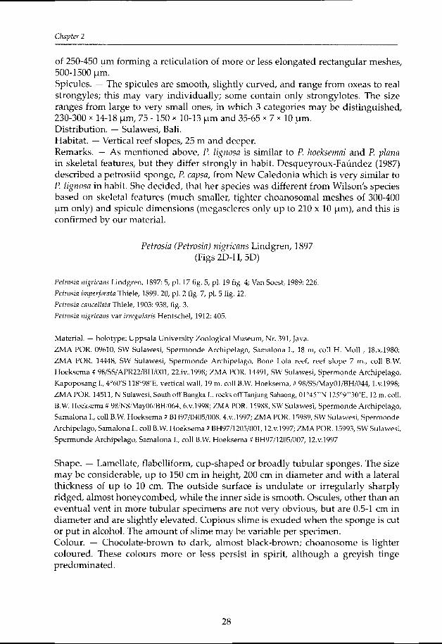
Fig.. 2. A-C. Petrosia (Petrosia) Ugnosa, A. cross section of choanosomal skeleton (scale— 500 pm), B. ver-
vieww of spicules (scale— 250 pm), C. detail of spicules (scale— 100 pm). D-H. Petrosia nigricans, D. spic-
uless of holotype Uppsala Mus. nr. 391 (scale— 100 pm), E. holotype of P. nigricans Lindgren, Uppsala
Mus.. nr. 391 (scale— lcm), F. spicules of ZMA POR. 15989 (scale— 100 pm), G. tangential view of ecto-
somall skeleton of ZMA POR. 15989 (scale- 250 pm), H. detail of ectosomal skeleton of ZMA POR. 15989
(scale—— 100 pm).
29 9
ChapterChapter 2
Skeleton.. — The ectosomal skeleton is a tangential reticulation of bundles and singlee larger megascleres, on the nodes of which short bushes of small megascleres aree erected. Brushes consist of 6-8 spicules which are intertwining. The ectosomal skeletonn often contains large pigment grains. The subectosomal tracts consist of 10-200 spicules and are 40-100 pm in diameter. The hexagonal subectosomal meshes are 100-4500 pm in diameter. This region is independent from the choanosomal skeleton andd is easily detachable. The choanosome is a system of thick spicule tracts forming largee almost rectangular meshes, 500-900 pm in diameter. The tracts consist of 20 or moree spicules and are 100-500 pm in diameter, spongin is not visible, but the tracts aree distinct. Pigmentt grains are also present in the choanosome, but not so abundant ass in the ectosome.
Spicules.. — Oxeas and strongylote modifications, predominantly bluntly pointed, butt occasionally sharply pointed or even rounded, in 3 distinct size categories: 240-3055 x 8-16 pm, 120-188 * 9-10 pm, and 57-85 * 5 pm. The smaller category only occurss in the ectosomal tufts. Habitat.. — From 3 to 45 m. Smaller specimens grow attached to coral rubble or more cryptic,, whereas the larger sized specimens may grow on sand slopes. Lionfishes aree often associated with this sponge and it provides shelter for large variety of reef animals.. Crinoids and large numbers of holothurians of the genus Synaptula are oftenn present on the outside of the sponges. Distribution.. — Apparently widely distributed in the Indo-Australian area. Remarks.. — The type specimen of Lindgren (Fig. 2E) originated from the Java Sea. A smalll thick plate of 7 * 7 * 3 cm is the remnant of an originally larger specimen. The consistencyy is stony, the colour greyish brown and no oscules are visible. The oxeas of thee type (Fig. 2D) are obviously thicker (20 pm), than the specimens from the material wee examined (up to 14 pm), but this may be caused by different environmental silica levels.. We re-examined Thiele's (1899) P. imperforata (Basel Museum nr. 28) and found thee skeleton to be in complete accordance with our own specimens. The fragment off Thiele had an undulating but essentially smooth surface like Lindgren's fragment. Fremont'ss (1991) P. pigmentosa from the Great Barrier Reef, which is a massive sponge withh short protuberances, shows some similarity to P. nigricans. After examination wee concluded that this is a different species, based on the differences in habit, texture andd the size of the spicules (these are shorter and thinner strongyles in P. pigmentosa). Osculess are also not conspicuous. The importance of mucus is stressed here, but accordingg to our observations, mucus excretion may vary between specimens of the samee species. Thiele (1903) described P. cancellata from Ternate (Indonesia, northern Moluccas)) as a small fragment of an apparently bigger specimen. He mentioned that hiss specimen may very well be P. nigricans, but that the surface of Lindgren's type iss completely smooth and even, while his specimen's surface is more undulating. Thee oxeas are of various sizes, but he gives only the largest measurements, 250 * 166 pm. The habit is described ambiguously, but because he mentions the resemblance to P. nigricans,nigricans, and because in our experience the surface features show great variation fromm smooth to almost honeycombed, we suggest that P. cancellata is a synonym off P. nigricans. Hentschel (1912) described P. nigricans var. irregularis from the Aru Islandss (Indonesia, eastern Moluccas). He mentioned the similarities to Lindgren's material,, but its skeleton is more confused and the oxeas are different in form.
30 0

IndonesianIndonesian sponges of the genus Petrosia
Wee believe this to fall within the variation of the nominal species. P.P. nigricans is a common, prominent, large species in the Spermonde Archipelago andd elsewhere in Eastern Indonesia, and we think this species could hardly have beenn overlooked in the past by collectors. Thee skeleton and spicule sizes often differ only slightly between different Petrosia species,, thus we stress the importance of the habit in designating the different species. .
PetrosiaPetrosia (Petrosia) plana Wilson, 1925 (Figss 3A-D, 5E)
PetrosiaPetrosia lignosa var. plana Wilson, 1925: 404, pi. 41 figs 4-5.
Material.. - ZMA POR. 14516, N Sulawesi, S off Tilisei I., E slope Tindila I., 01°48'N 125°03'30"E, reef
slopee 19 m, t) 98/NS/MAY06/BH/070, 6.V.1998; ZMA FOR. 14520, N Sulawesi, Tanjung Torowitan, 01°45'N
124°58'30"E,, steep slope 29 m, # 98/NS/MAY06/BH/074, 6.V.1998; ZMA POR. 14541 SW, N Sulawesi, Nain
I.,, 01°45'30"N 124'46'E, reef slope 13 m, » 98/NS/MAY08/BH/095, S.v.1998. The specimens were collected
byy B.W. Hoeksema.
Fig.. 3. Petrosia I Petrosia) plana, A. tangential view of ectosomal skeleton (scale— 250 um), B. at greater
magnificationn (scale— 100 um), C. cross section of choanosomal skeleton (scale— 500 urn), D. spicules
(scale—— 100 um).
31 1

ChapterChapter 2
Shape.. — Massive cylindrical tube, several tubes may be fused together. The texture iss stony and the surface is slightly roughened. Colour.. — Grey-brown to dark brown, choanosome lighter in colour. The colour persistss more or less in spirit. Copious slime is exuded when handled. Skeleton.. — Ectosomal paucispicular tangential skeleton forming meshes 60-150 urn, thee sides of which consist of characteristic tufts of small spicules with intermediate sizedd spicules carrying a bouquet of 5-10 of the smallest spicules. The ectosome iss carried by a subectosmal system of tracts, 3-8 spicules and 15-50 um in width, formingg polygonal meshes of 150-250 um. Choanosomal skeleton consists of thick spiculee tracts, 100-400 um in width, forming a reticulation of more or less elongated, rectangularr meshes, 500-1000 um in size. Choanosomal tracts contain more spongin thann those of the ectosome. Pigment grains are present throughout the body. Spicules.. — These are abruptly pointed oxeas in 3 size categories, 190-290 x 7-14 um, 95-1300 x 7-9.5 um and 43-75 * 5-9 um. Habitat.. — Reef slopes from 15 m to deeper waters. Distribution.. — North Sulawesi, Togian Islands. Remarks.. — Wilson (1925) described two new petrosiid species from the Togian Islands,, P. lignosa and P. lignosa var. plana. He emphasised the similarities in habit andd skeletal features, but distinguished a cylindrical var. plana with a smooth surface, fromm the cup-shaped nominal variety with irregular protuberances on the outside. P.. plana is also similar in many features to P. nigricans, but that species has obviously thickerr subectosomal tracts, consisting of 10-20 spicules, whereas those of P. plana havee only 3-8 spicules.
SUBGENUSS STRONGYLOPHORA DENDY, 1905
Definition:: Petrosia with 4 or 5 categories of strongyles including sharp angled microxeass concentrated at the surface.
PetrosiaPetrosia (Strongylophora) corticata (Wilson, 1925) (Figss 4A-C, 5F)
StrongylophoraStrongylophora corticata Wilson, 1925: 392, pi. 40 fig.7, pi. 48 figs 2,7.
StrongylophoraStrongylophora strongylata; Colin & Arneson, 1995: 48, fig. 157 (not: Thiele, 1903)
TabulocalyxTabulocalyx corticatus; Pulitzer-Finali, 1996: 126, figs 23-24.
Material.. — ZMA POR. 16748, SW Sulawesi, Spermonde Archipelago, Barang Lompo I., 15 m # NV/
BA/061000/151,, 6.X.2000; ZMA POR. 08254, Tukang Besi Islands, southern reef of Karang Kaledupa, east of
entrance,, 05°56'S 123°48'E, 4-10 m, 6.viii.l984, coll. R.W.M. van Soest, Dutch-Indonesian 'Snellius II ' Exped.
stat.. 016/HI/44.
Shape.. — Undulating smooth branches, 3-10 cm diameter, up to 30 cm long; numerouss small typical sieve-like oscules (4-8 mm) are scattered across the surface. Thee ectosome forms a distinct, firm rind; is slightly transparent and easily detachable fromm subdermal regions. The inside is pulpy.
32 2

IndonesianIndonesian sponges of the genus Petrosia
Fig.. 4. A-C. Petrosia (Strongylophora) corticata, A. tangential view of ectosomal skeleton (scale— 100 pm),
B.. subectosomal skeleton (scale— 250 |-im), C. spicule overview and detail of microxea/microstrongyle
(scalee overview— 100 pm, inset— 25 pm). D-F. Petrosia (Strongylophora) strongylata, D. spicules (microxeas
lacking)) (scale— 100 pm), E. tangential view of ectosomal skeleton (scale— 100 pm), F. cross section of
choanosomall skeleton (scale— 250 pm).
Colour.. — Ochre-greenish tinge, in spirit dull brown. Skeleton.. — The ectosome is a paucispicular reticulation of the larger spicules formingg irregular triangular meshes, with perpendicular tufts of the intermediate spicules,, and with moderate amounts of echinating microxeas and microstrongyles. Subectosomall tracts form regular polygonal meshes of 100-300 pm. The tracts of 2-4 spiculess are 30 pm in width, and consist mainly of the larger spicules.
33 3

ChapterChapter 2
Thee choanosomal skeleton is dense, tracts are 250-300 |am in width, and many spiculess are scattered loosely and singly. Spicules.. — The dominating spicules are strongyles, smooth, slightly curved with evenlyy rounded ends. The strongyles appear to occur in 3 sizes, 300-360 * 11-14 urn,, 80-200 * 11-14 yim, and 21-50 * 3-9 \im. Many immature spicules of these are oxeas,, 70-300 * 6 um, and these lie in between the meshes. Sharp angled ectosomal microxeass are 30-45 * 1-3 um. Habitat.. — Shallow reefs. Distribution.. — Philippines, Eastern Indonesia, Papua New Guinea. Remarks.. — Pulitzer-Finali (1996) assigned this species to the genus Tabulocalyx Pulitzer-Finalii (Phoeodictyidae), because of the difference in the skeletal structure withh other species of Petrosia (Strongylophora), but admitted that the spiculation is indistinguishablee from Strongylophora. The pulpy ectosome is clearly unusual for the normall stony texture of the family Petrosiidae; this feature together with the easily detachablee ectosome fits better in the family Phloeodictyidae. However, the spicules aree unmistakably those of Petrosia (Strongylophora). Moreover, typical phloeodictyid spongess have fistules issuing from a turnip-shaped body.
Colinn & Arnesen (1995) mistook this species for the closely related P. (S.) strongylata.strongylata. This is a dark coloured tube-shaped species, which also has clearly smallerr ectosomal strongyles (see below).
PetrosiaPetrosia (Strongylophora) strongylata (Thiele 1903) (Fig.. 4D-F, 5G)
StrongylophoraStrongylophora strongylata Thiele, 1903: 938, fig. 2.
{not:: S. strongylata Colin & Arneson, 1995: 48, fig. 157 = P. corticata)
Material.. — ZMA POR. 13155, SW Sulawesi, Spermonde Archipelago, Bone Baku reef, 15 m, coll. N.J. de
Voogd,, # NV/97/2005/030, 20.V.1997; ZMA POR. 16747, SW Sulawesi, Spermonde Archipelago, Samalona
I.,, 9 m, # NV/SA/241100/189, 24.xi. 2000; ZMA POR. 9011, NE coast of Sumba, E of Melolo, 09°54.2'S
120°43.5'E,, coll. R.W.M. van Soest, 15.viii.1984, dredged at 50 m, Dutch-Indonesian 'Snellius II ' Exped.
stat.. 4.061/V/12.
Shape.. — Smooth tube, up to 4 cm diameter, up to 6 cm long, rising from a broader base;; conspicious concentric rings inside the tube. Consistency extremely hard and stony. . Colour.. — Dark-brown to black, choanosome lighter in colour. Skeleton.. — The ectosome is a tangential skeleton of single spicules arranged in triangularr neshes forming a larger hexagonal system. Vague brushes of middle sized sizedd spicules are arranged on the nodes of the triangular meshes. Microxeas and microstrongyless echinate the single spicules. The subectosomal skeleton consists off larger spicules forming polygonal meshes, diameter 50-250 |am, of bundles consistingg of 2-4 spicules and with diameter 40-50 um. A very dense choanosomal skeletonn consists of a reticulation of spicule tracts, cored by upto 20 larger and smallerr spicules, diameter 70-150 um, forming polygonal meshes, 150-250 um.
34 4

IndonesianIndonesian sponges of the genus Petrosia
Loosee single spicules are scattered in between the meshes. Spicules.. — True strongyles, isodiametric, some are curved, many juvenile stages aree thin blunt oxeas; 3 size categories can be distinguished; 326 * 18 um, 95-145 * 10-122 urn and 44-60 x 8-12 um. Abruptly pointed, curved microxeas; 28-32 * 1-2 um. Habitat... — Cryptic, in caves and under coral overhangs. Distribution.. — Throughout Indonesia, Papua New Guinea. Remarks.. — This species was originally assigned to the genus Strongylophora, based onn the presence of microstrongylote spicules and characteristic sausage-shaped microstrongyles;; however this feature is now included in the genus Petrosia. Both P.P. corticata and P. strongylata have the characteristic echinating micro-oxeas in the ectosomall skeleton, but the species are easily distinguishable from each other on basiss of habit. In addition to this, the ectosomal microstrongyles of P. corticata (21-50 ** 3-9 um) are much smaller in size than those of P. strongylata (44-60 * 8-12um).
K E YY T O THE PETROSIA SPECIES OF E A S T E R N I N D O N E S I A
11 Creeping arms or thickly encrusting with prominent oscules 2 -- Tubes, vases or lamellate 4 22 Bright yellow PetrosiaiPetrosia) alfiani -- Brown or ochre-greenish 3 33 Detachable ectosome and pulpy from the inside
PetrosiaPetrosia (Strongylophora) corticata -- Stony texture, pigment grains abundant Petrosia (Petrosia) hoeksemai 44 Microxeas echinating ectosomal tracts PetrosiaiStrongylophora) strongylata -- Megascleres strongyles to oxeas, no microxeas 5 55 Cup/bowl-shaped with narrow base and a thickened rim
PetrosiaPetrosia (Petrosia) lignosa Tubess or vases with broad base, or upright plates 6 66 Cylindrical smooth tubes Petrosia (Petrosia) plana -- Lamellate, flabelliform, or broadly tubular sponge Petrosia (Petrosia) nigricans
D I S C U S S I ON N
Thee Petrosia species treated here form a common and striking complement of Indo-nesiann reefs. Nevertheless, there is a considerable list of additional Petrosia species recordedd from this area in the literature. Based on re-examination of type material or onn published descriptions, many of these do not conform to the definition of Petrosia employedd here, but rather are valid species or junior synonyms of species belonging too the genus Xestospongia. Examples are P. chaliniformis Thiele, 1899, P. contignata Thiele,, 1899, P. expansa Thiele, 1903, P. pulvilla Thiele, 1899, P. rava Thiele, 1899, P. se-riatariata Hentschel, 1912, P. similis granulosa Wilson, 1925, P. truncata aruensis Hentschel, 1912,, and P. densissima Dendy, 1905. Remainingg species recorded from the Indo-Malayan area which are likely to be truee Petrosia are: Petrosia (Petrosia) brachysclera Levi & Levi, 1989, Petrosia (Petrosia) hebeshebes Von Lendenfeld, 1888, and Petrosia (Strongylophora.) durissima Dendy, 1905.
35 5

ChapterChapter 2
Fig.. 5. In situ photos of Indonesian Petrosia species, A. paratvpe of Petrosia (Petrosia) alfiani spec,
nov.. (ZMA POR. 14999), photo B.W. Hoeksema; B. holotype of Petrosia (Petrosia) hoeksemai spec,
nov.. (ZMA POR. 15991), photo B.W. Hoeksema; C. Petrosia (Petrosia) lignosa (ZMA POR. 14554),
photoo B.W. Hoeksema; D. Petrosia (Petrosia) nigricans (POR.15989), photo B.W. Hoeksema; E. Petro-
siasia (Petrosia) plana (POR. 14516 and POR. 14520), photo B.W. Hoeksema; F. Petrosia (Strongylophora)
corticatacorticata (ZMA POR. 16478 / NV/BA/061000/151), photo N.J. de Voogd; G. Petrosia (Strongylophora)
strongylatastrongylata (ZMA POR. 16747), photo N.J. de Voogd.
36 6

IndonesianIndonesian sponges of the genus Petrosia
A C K N O W L E D G E M E N TS S
Manyy specimens and photographs were contributed by our colleague Dr Bert W. Hoeksemaa (Naturalis, Leiden) and we are also grateful for his help with logistics andd field supervision. Prof. Alfian Noor (Hasanuddin University, Makassar) kindly providedd access to his laboratory and supported in many other ways. The cura-torr of the Basel Museum, Mrs. Stockman, is thanked for the loan of Thiele's Cele-bess material. The curator of the Uppsala Museum, Mr. Mats Eriksson, is thanked forr the loan of Lindgren's P. nigricans. The curator of the Museum for Tropi-call Queensland, Mr. Peter Arnold, is thanked for the loan of P. pigmentosa. Prof. Svenn Zea (Universidad Nacional, Colombia) kindly reviewed the manuscript and madee useful corrections. Field work of NdV was financed by NWO-WOTRO un-derr grant nr. W84-474. The EC-MAS3 grant CT97-0144 ('SYMBIOSPONGE') to RvSS provided funds for field collecting and photographing of Dr B.W. Hoeksema.
37 7