
Use of the Functional Movement Screen TM Corrective ...
46
Use of the Functional Movement Screen TM Corrective Exercises to Address Strength Deficits in a 55 Year Old Female Status Post L5-S1 Discectomy A Capstone Project for PTY 768 Presented to the Faculty of the Department of Physical Therapy Sage Graduate School In Partial Fulfillment of the Requirements of the Degree of Doctor of Physical Therapy Hannah Solomon May, 2010 Approved: _________________________________ Gabriele Moriello, PT, PhD, MS, GCS Research Advisor _________________________________ Marjane Selleck, PT, DPT, MS, PCS Program Director, Doctor of Physical Therapy Program
Transcript of Use of the Functional Movement Screen TM Corrective ...

Use of the Functional Movement Screen TM
Corrective Exercises to Address Strength
Deficits in a 55 Year Old Female Status Post L5-S1 Discectomy
A Capstone Project for PTY 768
Presented to the Faculty of the Department of Physical Therapy
Sage Graduate School
In Partial Fulfillment
of the Requirements of the Degree of
Doctor of Physical Therapy
Hannah Solomon
May, 2010
Approved:
_________________________________
Gabriele Moriello, PT, PhD, MS, GCS
Research Advisor
_________________________________
Marjane Selleck, PT, DPT, MS, PCS
Program Director, Doctor of Physical Therapy Program

SAGE GRADUATE SCHOOL
I hereby give permission to Sage Graduate School to use my work,
Use of the Functional Movement Screen TM
Corrective Exercises to Address Strength
Deficits in a 55 Year Old Female Status Post L5-S1 Discectomy
For the following purposes:
- Place in the Sage Colleges Library collection and reproduce for Interlibrary Loan.
- Keep in the Program office or library for use by students, faculty,
or staff.
- Reproduce for distribution to other students, faculty, or staff.
- Show to other students, faculty or outside individuals, such as accreditors
or licensing agencies, as an example of student work.
- Use as a resource for professional or academic work by faculty or staff.
Hannah Solomon May, 20010

I represent to The Sage Colleges that this project and abstract are the original work of the
author, and do not infringe on the copyright or other rights of others.
Use of the Functional Movement Screen TM
Corrective Exercises to Address Strength
Deficits in a 55 Year Old Female Status Post L5-S1 Discectomy
________________________________________________________________________
________________________________________________________________________
Hannah Solomon May, 2010

Use of the Functional Movement Screen TM
Corrective Exercises to
Address Strength Deficits in a 55 Year Old Female Status Post L5-S1
Discectomy
Hannah Solomon, SPT

Abstract
Background and Purpose: This case report describes the use of the Functional Movement
Screen™ (FMS) corrective exercises in addition to a traditional strengthening and
rehabilitation program in the treatment of a 55 year old female status post L5-S1 discectomy.
The aim of this case report was to determine if the FMS corrective exercises in addition to a
traditional rehabilitative program are useful in increasing trunk strength and stability,
flexibility, ROM, and ADL’s in a person post L5-S1 lumbar discectomy. Case Description:
The individual in this case report is a 55 year old female department of transportation laborer.
She received outpatient physical therapy 3 times per week for a duration of 11 weeks. A
dynamic strengthening program was provided using several of the FMS corrective exercises
in conjunction with other exercises to provide a comprehensive rehabilitative program that
focused on improving strength and return to work. Outcomes: At the completion of treatment
improvements were noted in strength, flexibility, activities of daily living, and special and
functional testing. Her lower body strength improved from 0.5 to 1.5 manual muscle testing
grades and trunk range of motion improved from 20% to 55%. She also had significant
improvements in ADL’s such as tolerance to sitting, walking, and bending at the trunk yet
was not able to return to work. She was discharged to a local fitness club. Discussion: The
findings suggest that FMS corrective exercises when used in adjunct to a traditional
treatment program may be useful in the rehabilitation of individuals status post L5-S1
discectomy. However further experimental research is needed utilizing the corrective
exercises to determine the efficacy and usefulness of the FMS corrective exercises in the
physical therapy setting.

3
Introduction
Lumbar disc herniations are the most common cause of lumbar radiculopathy. In
North America, lumbar disc herniations affect 1% to 2% of the population,1 with ninety-five
percent occurring in one of the lower intervertebral discs.2 A disc herniation at the level of
L5 and S1 affects the corresponding nerve roots resulting in sciatica, bowel and bladder
dysfunction, postural control, and lower extremity impairments.3,4
Discectomies are currently the most frequently utilized surgical intervention to
address spinal nerve and nerve root compression resulting from intervertebral disc herniation.
Surgical intervention has been reported in 2% to 10% of people with lumbar disc herniation,
with approximately 200,000 discectomies performed annually in North America.1 A study by
Lurie et al3 explored the differences in outcomes over a 2 year period between upper and
lower lumbar spine discectomies finding that individuals with upper spine discectomies (L2-
L3 and L3-L4) had better outcomes after surgical procedure as compared to lower spine
discectomies (L4- L5 and L5-S1). Outcomes in individuals with lower spine discectomies
were worse in all aspects of treatment after surgery with regards to pain, quality of life, and
disability after operative treatment.
Following surgery, residual sciatic pain is reported by 10% to 30% of individuals,5
whereas residual back pain is reported by 30% to 40%.6,7
Only 80% of those having
undergone discectomy return to work within 12 months after surgery.8,9
Approximately half
of those who undergo disc herniation experience a preoperative as well as postoperative
reduction of muscle strength and endurance corresponding to the affected nerve root
distribution which is one of the reasons that the return to work rate is so low after undergoing
discectomy surgery.2,10,11
The aforementioned low return to work rate after surgical

4
discectomy implicates the necessity for an intense and comprehensive rehabilitative program
for return to work and functional independence.
The Functional Movement ScreenTM
(FMS) is an assessment tool developed by Gray
Cook and Lee Burton as a screening tool for fitness professionals such as physical therapists
and athletic trainers to gather objective data on human movement patterns during the
performance of functional activities for injury prevention. It is also widely used in many
colleges and professional sports teams for injury prevention in athletes. Each of the testing
criteria in the FMS was created to exacerbate the individual’s compensatory movement
patterns, allowing for easy identification by the examiner. By identifying movement flaws it
is expected that fitness professionals can assist individuals by preventing eventual breakdown
and trauma during activity.12
One study by Hoover et al13
found that the FMS’ specificity for
injury prediction was 97.2% in a sample of 49 recreational runners training for a half-
marathon.
The FMS consists of 7 fundamental movement tests to identify abnormal movement
patterns and impaired mobility and stability. The 7 testing categories consist of a deep squat,
hurdle step, in-line lunge, shoulder mobility, active straight leg-raise, trunk stability push-up,
and rotary stability. Scoring is performed on a 0 to 3 point system. A score of zero is given if
the individual has pain at any point during the movement; a 1 is given if the individual is
unable to be complete the activity even with compensations; a 2 is given if the individual is
able to perform the movement but uses poor mechanics or compensatory methods; and a 3 is
given if the individual can perform the movement without any compensations.12
Three of the
tests: shoulder mobility, trunk stability push-up, and rotary stability have clearing tests
associated with them that are pass/fail to determine if the actual FMS tests in these sections is

5
safe to perform. For example, the clearing test for the trunk stability push up test is a prone
press up. If the press up elicits pain in the individual, a score of zero is given for that section
of the screen the trunk stability push up test is not attempted.
Corrective exercises were also developed as a supplement to the FMS. The corrective
exercises are based on dysfunction in the dynamic motor learning, mobility and movement,
stability and static exercise categories. The exercises then coordinate to specific movement
dysfunction that the individual displays during the testing scenarios. The corrective exercises
developed to accompany the FMS are based on incorporating proprioceptive neuromuscular
facilitation (PNF) and motor learning.14,15
At this time there is no research available to
support these corrective exercises however PNF and motor learning are both well established
and accepted principles for rehabilitation.
PNF exercises were developed to enhance the body’s neuromuscular response
through stimulating proprioceptors in the joint. PNF movement patterns are performed on a
diagonal and often have a spiral component. The usage of PNF patterns has been suggested
to allow muscle strengthening in functional movements patterns such as those found in sports
and daily activities.16
Kofotolis et al16
utilized both static and dynamic PNF exercise
programs in women with chronic low back pain. Finding that both methods of PNF training
were highly effective in decreasing back pain, improving trunk musculature strength and
endurance and decreasing disability as measured by the Oswestry disability index. PNF
exercises are also widely used in rehabilitative programs in both the upper and lower
extremities in addition to the trunk and have been demonstrated to be an effective method of
strengthening and gaining flexibility.17,18

6
Motor learning is defined as "a set of processes associated with practice or experience
leading to relatively permanent changes in the capability for producing skilled action.”19, Pg 22
It is the necessary process that allows individuals to learn new skills and improve the
smoothness and accuracy of movements.19
Physical therapists routinely utilize the theory of
motor learning for rehabilitative treatment. Through the utilization of practice and intensity,
the hallmarks of successful motor learning, physical therapists enable patients to produce
motor patterns that are beyond their current capabilities.20
The use of practice and intensity
during training periods has been demonstrated to improve motor learning in individuals
learning or relearning a task. Training that emphasizes these principles has been
demonstrated to improve the quality of motor learning and reproducibility of the skill in
future practice sessions and improve outcomes in both children and adults.21
At this time there is no research available to determine the appropriateness of the
FMS corrective exercises in the rehabilitative setting. Therefore, the purpose of this case
report was to determine if the FMS corrective exercises are useful in increasing trunk
strength and stability, flexibility, ADL’s and functional activities in a person post L5-S1
lumbar discectomy.
Case Description
The individual in this case study was a 55 year old female department of
transportation laborer. Her job entails driving machinery such as a snow plow, heavy lifting,
sitting and twisting at the trunk. She initially injured her low back while shoveling dirt from
a washout at work on May 1, 2004. After this injury her symptoms resolved, in that she was
able to return to work and athletic/recreational activities. The medical management to treat
her back pain at this time is unknown. She had a second injury on March 26, 2009 when
7
seated bending over to tie her boot at work. Shortly after her injury she was seen by her
chiropractor who recommended she see a local orthopedic surgeon. She had an MRI which
was positive for a herniated nucleus pulposis at the L5-S1 intervertebral disc. She was then
admitted to the hospital for pain reduction several days prior to surgery, where she was
treated with morphine. The L5-S1 lumbar discectomy was performed on April 20, 2009. She
rated her pain as a 10/10 on the verbal analogue scale up until after the surgery was
performed. The participant reported constant pain located down the posterior aspect of her
right lower extremity extending to her foot. After the L5-S1 discectomy she was treated in
the hospital by a physical therapist until she was discharged at which time she returned home
with a home exercise program.
She presented to outpatient physical therapy for an evaluation and strengthening
program after referral from her orthopedic surgeon. At the initial interview she rated her low
back pain as a 0-1/10 according to the verbal analogue scale. Her low back was “sore” in
nature, with constant numbness in her posterior thigh, leg, lateral foot and heel. At the initial
evaluation she reported no bowel or bladder dysfunction. She was independent in activities of
daily living (ADL) including self care and driving, however she was unable to perform other
tasks such as cleaning her home, walking, hiking, hunting, camping and competitive archery.
She had limitations in sitting, bending, trunk rotation, and lifting as depicted in table 1. Her
goals for physical therapy treatment were to return to work, and activities of daily living
unrestricted and without symptoms.
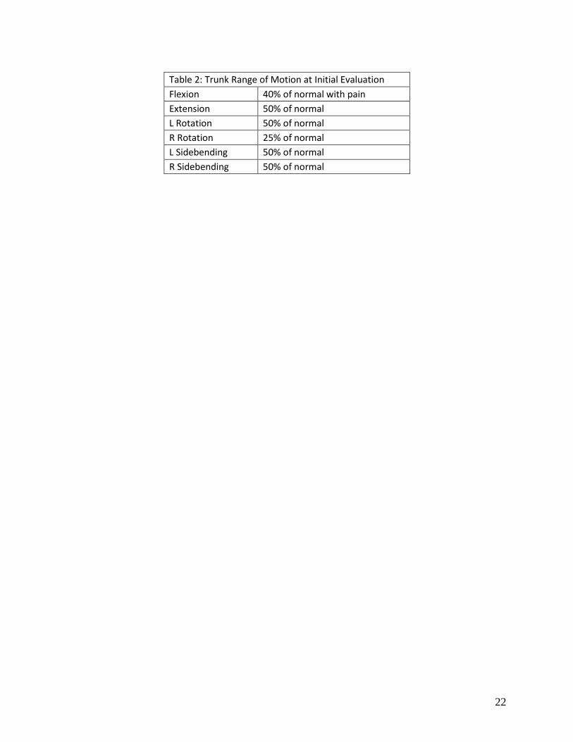
Upon physical examination, she had limited trunk range of motion (ROM) in all
directions most notably rotation to the right. See Table 2 for specific measurements. Manual
muscle testing was performed according to the method determined by Hislop.22
She exhibited

8
muscle strength deficits bilaterally with increased deficits noted in her right lower extremity.
See Table 3 for specific measurements.
She presented with impaired light touch sensation at her L4-S1 dermatomes and her
Achilles deep tendon reflex was impaired. See Table 4 for specific measurements. Special
testing demonstrated a positive straight leg raise and short sitting slump test on the right. The
aforementioned special tests were used to assess neural tension, and functional strength that
is frequently affected in those having undergone discectomy. See Table 5 for specific
measurements. She also demonstrated deficits in functional testing. See Table 6 for specifics.
Joint mobility was then tested showing bilaterally decreased anterior hip and lumbo-sacral
joint mobility. Her flexibility testing demonstrated bilateral restriction of the iliopsoas,
piriformis, hamstrings and gastrocnemius. See Table 7 for specific measurements.
Outcome Measures
Manual muscle testing was employed using the “break test” method in positions
against gravity as described by Daniel and Worthingham.22
Gross ROM was performed by
observing her stand with feet shoulder width apart and instructing her to rotate side to side as
much as possible without twisting her hips, side bend bilaterally as much as possible, forward
flex at the trunk to reach her toes and then extend backwards as much as possible. She was
instructed that she should not move past the point where pain is induced. The slump test was
performed with the procedure described by Maitland.23
Active straight leg raises were
performed as described by Cook.24
The Functional squat was also performed where
instructions were given to keep feet shoulder width apart and to squat down as far as she can.
For reliability and validity of the testing of all outcome measures, see Table 8.
Reliability and validity was not available for the single leg stance balance test however, Lin

9
et al25
performed a study in which it was determined that a shorter stance time for the single
leg stance was significant in predicting a decline in ADL’s however it did not significantly
predict the occurrence of falls in elderly adults.
Evaluation
Based on the information gathered at the initial evaluation session, it was determined
that the participant presented to physical therapy with residual numbness and myotomal
weakness in her right leg affecting the L4-S2 nerve root consistent with her post surgical
status. Her limitations in sitting were caused by increased neural tension and decreased
flexibility. Limitations in bending, trunk rotation, transfers, lifting and ADL’s and
independent activities of daily living (IADL’s) were due to increased neural tension,
weakness, decreased flexibility and limited ROM. Her heel and toe walks were also impaired
due to weakness and balance deficits. Her impairments limited her in ADL’s, pain free
mobility and vocational activities. It was determined that she would benefit from physical
therapy treatment to restore her functional mobility.
According to the Guide to Physical Therapist Practice26
the appropriate Primary
Preferred Practice Pattern was 4F: Impaired joint mobility, motor function, muscle
performance, range of motion, and reflex integrity associated with spinal disorders. The
physical therapy diagnosis for this individual corresponds with a diagnosis of intervertebral
disc disorder with myelopathy and postsurgical status, ICD-9 codes 722.7 and V45.89
respectively. The prognosis was determined to be good with expected return to work,
activities of daily living and functional mobility consistent with prior level of function within
a 1 year time period from the date or surgery, with significant improvement in function and

10
activities of daily living to occur within 3-4 months. Return to work is not typically expected
until 10 months to 1 year after surgery.
The short term physical therapy goals for this individual were to (1) Improve
flexibility and neural mobility from moderate-severely restricted to mildly to moderately
restricted (2) Improve myotomal strength of right LE by 1/3 grade (3) Restore functional
squatting, bending and twisting for light housework. Each of these goals was to be attained
within 4 to 6 weeks.
The long term physical therapy goals for this individual were to (1) Improve sciatic
nerve mobility and general flexibility to allow for asymptomatic tolerance of prolonged
sitting for 1-2 hours to allow for return to work (2) Improve trunk strength 4- to 4/5 for being
able to tolerate prolonged sitting for 1-2 hours for return to work and restore ADL’s for
vacuuming and light lifting (3) Achieve an ADL functional status to meet occupational
requirements such as trunk twisting, reaching overhead, reaching below the waist, forward
bending, squatting, crouching, sustained forward bending and driving vehicles such as a
forklift or plow. Each of these goals was to be attained within 11 weeks. The frequency of
physical therapy treatment was 3 times per week for a total of 11 weeks. Criterion for
discharge was completion of the long term physical therapy goals, which were to return to
ADL’s, and go to a gym for return to work preparation.
Interventions used during physical therapy treatment of this individual were
musculoskeletal re-education, stretching, therapeutic exercise, cryotherapy, manual therapy,
mobilization techniques of the joints, soft tissue and neural tissue, strength training, balance
exercises, range of motion exercises, and home exercise program (HEP). These were given to

11
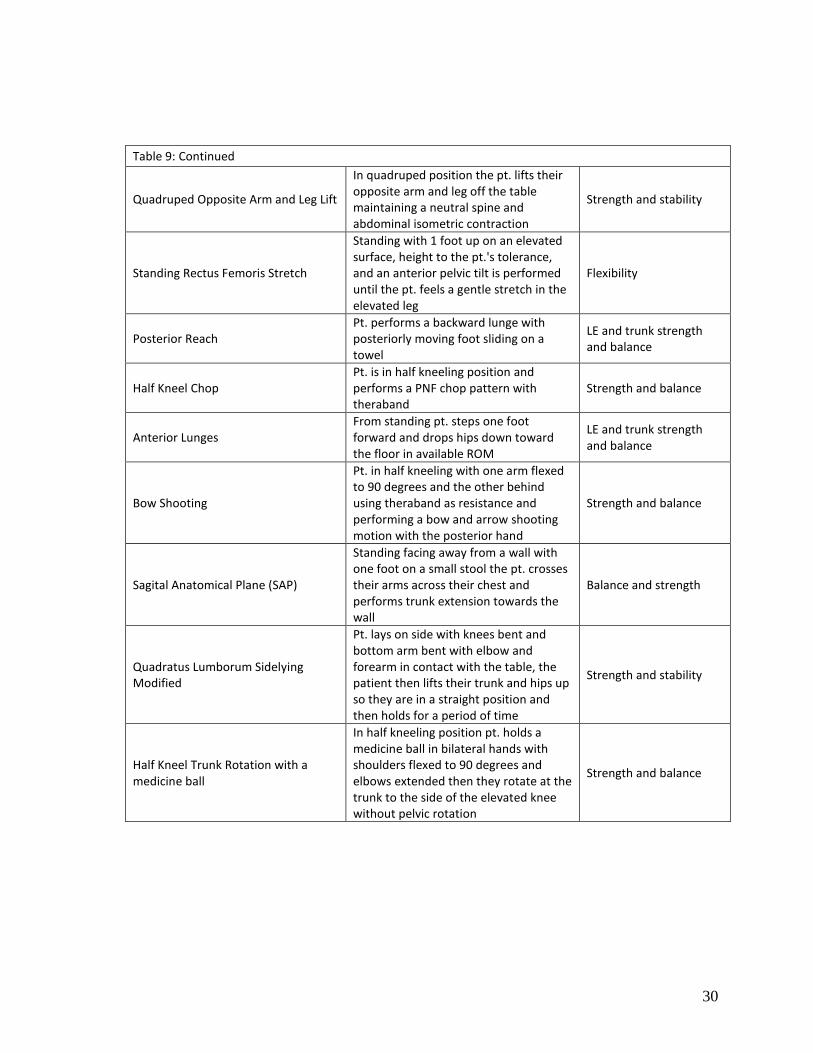
address the functional limitations and impairments of this individual. See Table 9 for the
specific exercises performed and Table 10 for the progression of exercises.
Additionally the purpose of each intervention is listed in Table 9. Some of the
exercises listed in Table 9 are specific corrective exercises from the FMS, and variations of
certain FMS corrective exercises were also included in her intervention program. The
purpose of the variations were to decrease the difficulty of an exercise to make it appropriate
for the specific needs of the individual being treated. For specifics on FMS based exercises
see Table 11.
Outcomes
Significant improvements were noted in functional activities, range of motion, muscle
strength, flexibility, and special and functional testing. All measures were not taken at
discharge (ROM, strength, neurological testing and flexibility) however there was a re-
evaluation which occurred approximately one month after beginning physical therapy
treatment.
At discharge the individual in this case rated her low back pain as 0-1/10 according to
the verbal analogue scale, which is the same rating that she provided at her initial evaluation.
At her initial evaluation she was unable to perform tasks such as cleaning her home, walking,
hiking, hunting, camping and competitive archery. At discharge she was able to perform all
of the aforementioned activities. She had increased tolerance for sitting, standing, walking,
bending, trunk rotation, vacuuming, sleeping and lifting as depicted in Table 12.
Upon her re-evaluation 4 weeks after starting physical therapy treatment, trunk ROM
improved to 70-95% of normal whereas at her initial evaluation it was 25-50% of normal.
She made significant improvements in strength at the one month re-evaluation although she

12
still exhibited some muscle strength deficits especially in her right lower extremity. At re-
evaluation she presented with impaired light touch sensation in only the S1 dermatome as
well as some hypersensitivity at the L4-L5 dermatomes which was positive for numbness at
the initial evaluation. Her Achilles deep tendon reflexes were unchanged from initial
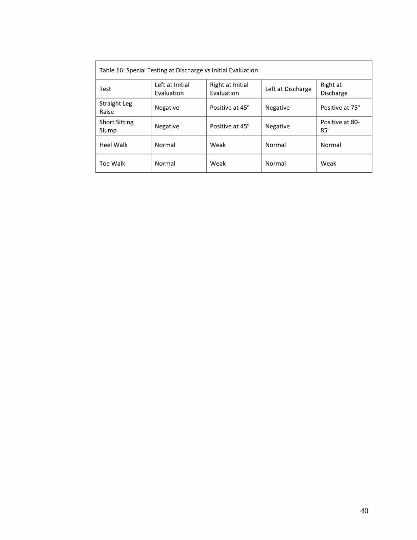
evaluation. At discharge special testing demonstrated a positive straight leg raise; at 75º
improving from the initial evaluation which was positive at 45º. The short sitting slump test
did not aggravate symptoms until 80-85º at discharge where at initial evaluation it was
positive at 45º. At discharge she demonstrated an unchanged toe walk on the right, and a
bilaterally normal heel walk. See Tables 13-16 for specific measurements.
At discharge she demonstrated improved results in functional testing particularly in
balance, the squat test, heel walking, and transfers. See Table 17 for specifics. At re-
evaluation joint mobility was improved however still slightly decreased from normal at her
anterior hip and lumbo-sacral joint mobility. Her flexibility testing at re-evaluation
demonstrated improved flexibility particularly in her iliopsoas and piriformis. See Table 18
for specific measurements. All physical therapy goals were achieved except she was unable
to return to work.
Discussion
This case report has shown how a restorative exercise program utilizing components
of the corrective exercises for the FMS was used in a 55 year old female laborer status post
L5-S1 discectomy for repair of a herniated nucleus pulposis. The individual in this case
demonstrated improvements in ADL’s, strength, range of motion, flexibility, special tests and
functional testing. The improvements in testing seen in this individual are likely due to a
progressive and frequently updated exercise program. Additionally the individual in this case

13
was extremely dedicated to the therapeutic process and was compliant in every aspect of
treatment including her HEP.
Her Achilles tendon DTR remained absent at the completion of physical therapy
treatment. It is possible this may not return. A study by Astrand et al27
observed that 2 years
post surgery Achilles tendon DTR’s were absent in 35% the status post discectomy.
Additionally Astrand et al27
also found that 40% of individuals had impaired sensation 2
years post operatively.27
The individual in this case report did not have an improvement in
toe walking prior to discharge Astrand et al27
also found that 11% of patients had decreased
planterflexor strength 2 years after undergoing lumbar discectomy.
Although her verbal analogue pain scale rating remained consistent from her initial
evaluation to discharge, she was able to greatly increase her activity without increasing her
pain. She was able to tolerate ADL’s which she was unable to perform at the initial
evaluation such as outdoors walking, lengthy sitting and standing, bending at the trunk, trunk
rotation, lifting, vacuuming and archery before experiencing the same level of pain.
The individual in this case demonstrated improvements in flexibility of her bilateral
illiopsoas, piriformis, and left hamstrings and gastrocnemius due to a progressive and
strategic stretching program addressing limited muscle groups. Improvements in flexibility
allowed her to improve ADL’s by increasing her ability to squat, ambulate greater distances
and sit in a chair for longer periods of time with less pain.
Additionally improvements in strength of her abdominals, and bilateral lower
extremities was due to a strategic and comprehensive strengthening program which was
frequently progressed to decrease plateau effects. Several of the corrective exercises were
adapted slightly so that they were appropriate for the functional level of the individual and

14
progressed appropriately. The FMS corrective exercises that were utilized in the
rehabilitation of this individual aided in improving her balance and functional abilities
through strategic and functional strengthening of the above mentioned muscle groups which
demonstrated limitations at initial evaluation. The FMS corrective exercises utilized are
dynamic in nature and require the individual performing them to improve their muscle
stability and balance to allow them to perform the dynamic and functional components of the
exercises. This aided in her improvements in performance of ADL’s and functional testing as
well as beginning a return to her previous functional status.
The individual in this case was able to return to nearly all ADL’s and functional
activities however she was unable to return to work at the completion of physical therapy
treatment. This occurred due to the physically demanding nature of her occupation as a
department of transportation laborer. She was unable to meet the occupational requirements
of her job such as trunk twisting, reaching overhead, reaching below the waist, forward
bending, squatting, crouching, sustained forward bending and driving vehicles such as a
forklift or plow. She was able to perform many of the above mentioned occupational
requirements for the completion of ADL’s however she was still unable to perform several of
these activities in the occupational context. This is due to the additional physical
requirements of her occupation beyond those for ADL completion. For example, one
occupational duty is that she has to sit on the back of a moving truck and put out road cones.
In order to do this she would have to rotate at her trunk across her body and place the road
cones on the road requiring her to have the flexibility to reach below her feet while carrying
an object weighing greater than 5 lbs. This action would then need to be completed
repetitively. Several other activities such as squatting, bending forward and reaching

15
overhead would also have to be completed while lifting potentially heavy equipment. At
discharge from physical therapy she was able to transition to utilizing a home exercise
program at a local gym to further increase her strength until she was able to return to work.
This case report had several limitations. Re-evaluation testing was not performed
every 30 days resulting in several measures not being collected immediately prior to
discharge. Additionally the corrective exercises of the FMS were not exclusively used with
this individual limiting the ability to determine a direct correlation between the efficacy of
the FMS corrective exercises in the treatment of an individual status post L5-S1 discectomy.
Additionally, a cause and effect relationship cannot be inferred due to this study not having a
large sample size without the presence of a control group.
Several of the outcome measures utilized demonstrate low sensitivity, specificity or
have no research available on reliability and validity. The straight leg raise test has a
sensitivity of .78-.98 and varied report of specificity throughout research of this test of .11-
.84. These values indicate that the straight leg raise is better at ruling in pathology than ruling
it out. The short sitting slump test has a specificity of .55, which is relatively low.24
The
statistics on reliability and validity of the functional squat were based on a study where the
participants had knee osteoarthritis, not lumbar discectomy limiting its value in this case
report. Additionally, there was no available research on the reliability or validity of gross
observational trunk ROM.
Other tests and measures may have been more appropriate for use in this case. The
FMS was also not utilized as an outcome measure in this case report due to the researcher
being unaware of its existence until after the treatment was already initiated. However the
FMS would have been a useful outcome measure to correlate the FMS corrective exercises

16
with progress made in outcomes as the FMS is entirely functionally based and correlates to
the high level of functional ability needed for the individual to return to work.
Use of the EquiTest System may have been more useful than the single leg balance
screen. The EquiTest System consists of a support surface with sensors at the corners below
the surface and a visual surround. The EquiTest device performs a sensory organization test
(SOT) with six conditions which provides a useful objective measure for identifying balance
deficits for individuals with real world high level balance requirements.28
Additionally, a back range of motion (BROM) instrument would have provided more
precise ROM measurements than use of gross ROM. The BROM is used to measure lumbar
spine active planar motions. A study by Kachingwe et al29
demonstrated that intrarater
reliability is good for side bending (ICC=.85), lumbar forward flexion and pelvic inclination
was (ICC=.84) and extension and rotation was (ICC=.76).
The findings of this case report support that the corrective exercises of the FMS may
be helpful in the rehabilitation of this individual status post L5-S1 discectomy. Further
experimental research is needed utilizing the corrective exercises to determine the efficacy
and usefulness of the FMS corrective exercises in the physical therapy setting.

17
References
1. Wera GD, Marcus RE, Ghanayem AJ, Bohlman HH. Failure within one year
following subtotal lumbar discectomy. J Bone Joint Surg Am. 2008;90(1):10-15.
2. Millisdotter M, Stromqvist B, Jonsson B. Proximal neuromuscular impairments in
lumbar disc herniation. Spine. 2003;28(12):1281-1289.
3. Lurie JD, Faucett SC, Hanscom B, Tosteson TD, Ball PA, Abdu WA, Frymoyer JW,
Weinstein JN. Lumbar discectomy outcomes vary by herniation level in the spine
patient outcomes research trial. J Bone Joint Surg Br. 2008;90(9):1811-1819.
4. Bouche K, Stevens V, Cambier D, Caemaert J, Danneels L. Comparison of postural
control in unilateral stance between healthy controls and lumbar discectomy patients
with and without pain. Eur Spine J. 2006;15(4):423-432.
5. Tullberg T, Isacson J, Weidenhielm L. Does microscopic removal of the lumbar disc
herniation lead to a better result than the standard procedure? Results of a one-year
randomized study. Spine. 1993;18(11):24–27.
6. Weber H. Lumbar disc herniation: a controlled, prospective study with ten years of
observation. Spine. 1983;8(2):131–140.
7. Dvorak J, Gauchat MH, Valach L. The outcome of surgery for lumbar disc herniation
A 4-17 years’ follow-up with emphasis on somatic aspects. Spine.
1988;13(12):1418–1422.
8. Donceel P, Du Bois M. Fitness for work after surgery for lumbar disc herniation: a
retrospective study. Eur Spine J. 1998;7(1):29–35.
9. Nygaard OP, Romner B, Trumpy JH. Duration of symptoms as a predictor of
outcome after lumbar disc surgery. Acta Neurochir. 1994;128(1-4):53–56.
18
10. Kortelainen P, Puranen J, Koivisto, et al. Symptoms and signs of sciatica and their
relation to the localization of the lumbar disc herniation. Spine. 1985;10(1):88–92.
11. Woertgen C, Rothoerl RD, Breme K, Altmepprn J, Holzschuh M, Brawanski A.
Variability of outcome after lumbar disc surgery. Spine. 1999;24(8):807–811.
12. FMS History. http://www.functionalmovement.com/SITE/aboutfms/fmshistory.php.
Accessed September 5, 2009.
13. Hoover D, Killian CB, Bourcier B, Lewis S, Thomas J, Willis R. Predictive validity
of the functional movement screen TM in a population of recreational runners
training for a half marathon. Med Sci Sports Exerc.2008;40(5):219.
14. Cook G, Burton L, Hoogenboom B. Pre-participation screening: the use of
fundamental movmements as an assessment of function- part 1. N Am J Sports Phys
Ther. 2006;1(2):62-72.
15. Cook G, Burton L, Hoogenboom B. Pre-participation screening: the use of
fundamental movmements as an assessment of function- part 2. N Am J Sports Phys
Ther. 2006;1(3):132-139.
16. Kofotolis N, Kellis E. Effects of two 4-week proprioceptive neuromuscular
facilitation programs on muscle endurance, flexibility, and functional performance in
women with chronic low back pain. Phys Ther. 2006;86(7):1001-1012.
17. Higgs F, Winter SL. The effect of a four-week proprioceptive neuromuscular
facilitation stretching prorgam on isokinetic torque production. J Strength Condition
Res. 2009;23(5):1442-1447.

19
18. Kofotolis ND, Kellis E. Effects of proprioceptive neuromuscular facilitation
stretching on stiffness and force-producing characteristics of the ankle in active
women. Phys Ther Sport. 2007; 8(3): 109-116.
19. Schmidt RA, Lee TD. Motor control and learning: A behavioral emphasis. 4th ed.
Champaign, IL: Human Kinetics;2005.
20. Schmidt RA. Contemporary management of motor control problems: Proceedings of
the II STEP Conference. Alexandria,VA: Foundation for Physical Therapy; 1991:49–
63.
21. Sullivan KJ, Kantak SS, Burtner PA. Motor learning in children: feedback effects on
skill acquisition. Phys Ther. 2008;88(6):720-733.
22. Hislop H, Montgomery J. Daniels and Worthington’s muscle testing: techniques of
manual examination. 8th
ed. St. Louis, MO: Saunders; 2007.
23. Johnson EK, Chiarello CM. The slump test: the effects of head and lower extremity
position on knee extension. J Orthop Sports Phys Ther. 1997;26(6):310-317.
24. Cook CE, Hegedus EJ. Orthopedic physical examination tests: an evidence-based
approach. Upper Saddle River, NJ: Pearson Prentice Hall Inc.; 2008.
25. Lin MR, Hwang HF, Hu MH, Wu HD, Wang YW and Huang FC. Psychometric
comparisons of the timed up and go, one-leg stand, functional reach, and tinetti
balance measures in community-dwelling older people. J Am Geriatr Soc.
2004;52(8):1343-1348.
26. Bohmert J, Moffat M, Zadai C, eds. Guide to Physical Therapist Practice. 2nd
ed.
Alexandra, VA: American Physical Therapy Association; 2003.

20
27. Astrand P, Maattanen H, Vucetic N, Svensson O. Pain and orthopedic and
neurological signs after lumbar discectomy: a 2-year follow up. Clin Orthop Relat
Res. 2000;(379):154-160.
28. Chaudhry H, Bukied B, Ji Z, Findley T. Measure of balance in computer
posturography: comparison of methods a brief review. J Bodyw Mov Ther. 2009;1-10.
29. Kachingwe AF; Phillips BJ. Inter-and intrarater reliability of a back range of motion
instrument. Arch Phys Med Rehabil. 2005;86(12): 2347-2353.

21
Table 1: Activities of Daily Living at Initial Evaluation
Activity Tolerance
Sitting Aggravates symptoms with duration = 1 hour
Standing > 1 hour without symptom aggravation
Bending Unable to perform
Trunk rotation Unable to perform
Vacuuming Unable to perform
Lifting Tolerates lifting up to 5 lbs
Sleeping Able to sleep on affected side
22
Table 2: Trunk Range of Motion at Initial Evaluation
Flexion 40% of normal with pain
Extension 50% of normal
L Rotation 50% of normal
R Rotation 25% of normal
L Sidebending 50% of normal
R Sidebending 50% of normal

23
Table 3: Muscle Strength at Initial Evaluation
Spinal Innervation Muscle Group Left Right
L1-L2 Hip flexion 4-/5 4-/5
L3 Knee extension 4+/5 4+/5
L4 ankle dorsiflexion 5-/5 4/5
L5 Great toe extension 4/5 3+/5
S1 Ankle plantarflexion 4+/5 3/5
S2 Knee flexion 5-/5 3+/5
L4-L5 Hip extension 4/5 4/5
L4-S1 Hip abducton 4+/5 4/5
T4-L3 Abdominals 3/5 3/5

24
Table 4: Neurologic Testing at Initial Evaluation
Test Left Right
Light Touch Normal Positive Numbness L4-S1
Achilles DTR 2+ 0

25
Table 5: Special Testing at Initial Evaluation
Test Left Right
Straight Leg Raise Negative Positive at 45º
Short Sitting Slump Negative Positive at 45º
Heel Walk Normal Weak
Toe Walk Normal Weak

26
Table 6: Functional Testing at Initial Evaluation
Test Left Right
Squat Approximately 50% of normal with left shift
Single Leg Balance 15 seconds 15 seconds
Heel Walk Normal Weak
Toe Walk Normal Weak
Rolling transfer Guarded but proper technique
Sit to supine transfer Guarded but proper technique

27
Table 7: Flexibility at Initial Evaluation
Muscle Left Right
Iliopsoas Moderate Restriction Moderate Restriction
Piriformis >90º Slight Restriction Moderate Restriction
Hamstring Moderate Restriction Mild Restriction
Gastrocnemius Mild Restriction Mild Restriction

28
Table 8: Reliability and Validity of Special Tests
Test Validity Inter-rater Reliability
Test Retest Reliability
Sensitivity Specificity
Manual Muscle Testing N/A .97 .98 N/A N/A
Range of Motion via observation N/A N/A N/A N/A N/A
Straight Leg Raise N/A N/A N/A 0.78-0.98 0.11-0.84
Short Sitting Slump N/A N/A N/A 0.83 0.55
*Functional Squat N/A .92 in pts
with knee OA
N/A .23 .86

29
Table 9: Physical Therapy Intervention Program
Exercise Name Description of Exercise Purpose of exercise
Slump Slider
Position of the short slump test where patient performs a self neural mobilization by extending their leg to the point of tension and then releasing
Neural tension along sciatic tract
Slump Tensioner Same as slump slider however patient keeps head flexed down
Neural tension along sciatic tract
Isometric Abdominal Bracing
Lay in supine hooklying position keeping a neutral spine patient is verbally cued to "bring their belly button into their spine" and hold the contraction
Abdominal strength
Lower Trunk rotation Supine hooklying position pt. then allows both knees to fall to one side and then the other
Trunk flexibility
Upper Body Ergometer (standing)
Pt. stands and pedals the hand crank forward for a period of time and then backwards
Trunk mobility and strength
Supine Hamstring Stretch Pt. lays supine with hip and knee at 90-90 position the pt. then extends knee until a gentle stretch is felt
Flexibility
Bridging
Pt. lays supine in hooklying, braces abdominals and then lifts buttocks off the table so that their trunk and knees are a straight line
LE and trunk strength and stability
Functional Squat
Standing with feet shoulder width apart a belt is tied around the pt.'s thighs to prevent compensation and the pt. squats as low as they can up to 90degrees
LE and trunk strength, flexibility and balance
Retro Treadmill Walking Backwards walking on a level treadmill Strength of gluteal and hip extensors
Piriformis Stretch
Pt. lays in supine hooklying position and brings one ankle across the other knee making a figure four appearance then the pt. places their hand on the lateral portion of the elevated thigh and pulls the knee to their opposite shoulder until a gentle stretch is felt
Flexibility
Manual Therapy Supine piriformis stretch, sciatic nerve mobilization, sideling illiopsoas stretch
Flexibility and neural mobility
Cryotherapy Ice pack applied in supine with hips and knees propped up into a 90-90
Pain and inflammation reduction
Isometric Abdominal Bracing with Marching
Same as above with alternating hip flexion with instruction to maintain a neutral and unmoving pelvis
Abdominal strength
30
Table 9: Continued
Quadruped Opposite Arm and Leg Lift
In quadruped position the pt. lifts their opposite arm and leg off the table maintaining a neutral spine and abdominal isometric contraction
Strength and stability
Standing Rectus Femoris Stretch
Standing with 1 foot up on an elevated surface, height to the pt.'s tolerance, and an anterior pelvic tilt is performed until the pt. feels a gentle stretch in the elevated leg
Flexibility
Posterior Reach Pt. performs a backward lunge with posteriorly moving foot sliding on a towel
LE and trunk strength and balance
Half Kneel Chop Pt. is in half kneeling position and performs a PNF chop pattern with theraband
Strength and balance
Anterior Lunges From standing pt. steps one foot forward and drops hips down toward the floor in available ROM
LE and trunk strength and balance
Bow Shooting
Pt. in half kneeling with one arm flexed to 90 degrees and the other behind using theraband as resistance and performing a bow and arrow shooting motion with the posterior hand
Strength and balance
Sagital Anatomical Plane (SAP)
Standing facing away from a wall with one foot on a small stool the pt. crosses their arms across their chest and performs trunk extension towards the wall
Balance and strength
Quadratus Lumborum Sidelying Modified
Pt. lays on side with knees bent and bottom arm bent with elbow and forearm in contact with the table, the patient then lifts their trunk and hips up so they are in a straight position and then holds for a period of time
Strength and stability
Half Kneel Trunk Rotation with a medicine ball
In half kneeling position pt. holds a medicine ball in bilateral hands with shoulders flexed to 90 degrees and elbows extended then they rotate at the trunk to the side of the elevated knee without pelvic rotation
Strength and balance

31
Table 9: Continued
Overhead Reach Added to Anterior Lunges
Same as above with bilateral arm lift into forward flexion to full shoulder range of motion
Strength, flexibility and balance
Lunge Matrix Anterior lunge with overhead reach, lateral and posterior lunges
Strength, flexibility and balance
Prone Ball Opposite Arm and Leg lift
Prone over a physioball with opposite arm and leg lift maintaining a neutral and stabilized spine and trunk
Strength, stability and balance
Standing Hamstring Stretch
Pt. stands with one foot placed on a stool with knee extended pt. then forward flexes at the hip until a gentle stretch is felt
Flexibility
Toe Touch FAP and SAP Progressed to toe touching without foot on stool
Balance and strength
Quadruped Opposite Arm and Leg Lift with stick
Same as above with addition of dowel rod between shoulders and buttocks to maintain neutral spine position and trunk stability
Strength and stability
Single Leg Balance with Chop Pt. stands on a single leg while performing the PNF chopping pattern using theraband for resistance
Strength and balance
Single Leg Balance with Trunk Rotation
Pt. stands on a single leg with the elevated leg held at a 90-90 position with bilateral arms extended into 90 degrees of flexion pt. rotates at the trunk to the side of the elevated leg keeping pelvis neutral
Strength and balance
Bridge with Leg Lift and Core Activation
Pt. lays in supine hooklying position and extends one knee out while performing a bridge and lifting the opposite arm into shoulder flexion
Strength, stability, and balance
Bridge with Ball and LE Flexion/Extension
Bilateral feet on a physioball pt. performs a bridge then flexes the knees up while feet remain on ball and then extends knees back to original position
Strength and stability
FAP and SAP Altered to single leg balance Balance and strength
Cybex Functional Squat Altered to use Cybex cable machine with a bar held at the shoulders
Strength
Bridge with Ball and LE Flexion/Extension Sustained
Altered to sustained bridge with LE flexion/extension
Strength and stability
Ball Bridge Sit
Pt. sits on a physioball with hands over chest and walks their legs out until their shoulders only remain on the ball and they are in a bridge position then they walk their body back up until they are in the start of exercise position
Strength

32
Table 9: Continued
Latisimus Dorsi Pull Down
Using a Cybex machine the pt. sits facing the machine using a pulldown bar the patient reaches overhead and grabs the bar. They then pull the bar down towards the sternoclavicular notch and then brings hands back up overhead
Strength
Frontal Anatomical Plane (FAP)
Standing next to a wall with the foot nearest the wall up on a small stool the pt. crosses their arms across their chest and performs a lateral bend at the trunk towards the wall
Balance and strength
Modified Plank
Pt. in prone on a table propped on elbows with knees bent, pt. then lifts hips so that trunk and lower extremities are aligned then the position is held
Stability and strength
Figure Skater
Single leg stance with pt. holding a dowel rod to their shoulders and buttocks then the patient forward flexes at the hip maintaining contact with the dowel rod and extends one leg posteriorly keeping the knee straight
Strength, flexibility and balance
Aquatic Lateral Walking Laterally walking from one end of the pool to the other
Strength
Aquatic Circle walking Walk in a circle in pool until a current is produced then turn around and switch directions walking in the other direction
Strength
Aquatic Diagonal Leg kicks
Stand on one leg and with leg extended perform diagonal kicks in diagonals into D1 flexion and extension and hip abduction starting with leg maximally adducted
Strength and balance
Aquatic Noodle Sword Fighting With both hands below the water surface the pt. has a noodle sword fight using trunk rotation
Strength and trunk ROM
Aquatic Supine Snow angels Pt. floats on her back in the pool and makes snow angles
Strength
Aquatic Squat and lift Pt. squats in the pool and performs a PNF lift while extending the lower extremities and rotating at the trunk
Strength, ROM and balance

33
Table 10: Exercise Progression
Exercise Week 1 Week 2 Week 3 Week 4 Week 5 Week 6 Week 7 Week 8 Week 9 Week 10 Week 11
Slump Slider *, HEP *, HEP *, HEP *, HEP *, HEP *, HEP *, HEP
Slump Tensioner *, HEP *, HEP *, HEP *, HEP
Isometric Abdominal Bracing
*, HEP
Lower Trunk Rotation
*, HEP *, HEP *, HEP HEP HEP HEP HEP HEP HEP HEP HEP
Standing Upper Body Ergometer
* * * * * * * * * *
Supine Hamstring Stretch
*, HEP *, HEP *, HEP HEP HEP
Bridging * * *, HEP HEP HEP
Functional Squat * * * * * * *, HEP HEP HEP HEP HEP
Retro Treadmill Walking
* * * * * * * * * * *
Piriformis Stretch *, HEP *, HEP *, HEP HEP HEP HEP HEP HEP
Manual Therapy * * * * * * * * * * *
Cryotherapy * * * * * * * * * * *
Isometric Abdominal Bracing with Marching
*, HEP *, HEP HEP HEP HEP HEP HEP HEP HEP HEP
FAP * * * * *
Quadruped Opposite Arm and Leg Lift
* * * *
Standing Rectus Femoris Stretch
* * * * HEP HEP HEP HEP HEP
Posterior Reach * * *
Posterior Lunge *
Half Kneel Chop * * * *
Anterior Lunge * * *
Bow Shooting * * * * *
SAP * * *
Quadratus Lumborum Sidelying Modified
* * * * * * * *
Half Kneel Trunk Rotation with Medicine Ball
* * *
Modified Plank * * * * * *

34
Table 10: Continued
Figure Skater * * * * * *
Anterior Lunge with Overhead Reach
* * * * * *
Lunge Matrix * * * * *
Prone Ball Opposite Arm and Leg Lift
* * * * * *
Standing Hamstring Stretch
*, HEP *, HEP *, HEP *, HEP *, HEP *, HEP
Toe Touch SAP * *
Quadruped Opposite Arm and Leg Lift with Stick
*, HEP *, HEP *, HEP *, HEP *, HEP
Single Leg Balance with Chop
* * * * *
Single Leg Balance with Trunk Rotation
* * * * *
Bridge with Leg Lift and Core Activation
*, HEP *, HEP * * *
Bridge with Ball and LE Flexion/Extension
*, HEP *, HEP *, HEP HEP HEP
Single Leg FAP * * * * *
Single Leg SAP * *
Bridge with Ball and LE Flexion/Extension Sustained
* *
Cybex Functional Squat * * * *
Ball Bridge Sit * * *
Latisimus Dorsi Pull Down
*
Aquatic Lateral Walking HEP HEP HEP HEP
Aquatic Circle walking HEP HEP HEP HEP
Aquatic Diagonal Leg kicks
HEP HEP HEP HEP
Aquatic Noodle Sword Fighting
HEP HEP HEP HEP
Aquatic Supine Snow angels
HEP HEP HEP HEP
Aquatic Squat and Lift HEP HEP HEP HEP
* Indicates an exercise being performed during PT interventions, HEP indicates an exercise in the home
exercise program and a blank box indicates the discontinuation of the exercise

35
Table 11: FMS Corrective Exercises
FMS Exercise Corresponding exercise
Resisted quadruped diagonals neutral spine
Quadruped opposite arm and leg lift and with stick
Single leg dead lift Figure skater, same as FMS activity but with dowel to improve spinal positioning
Overhead deep squat Cybex squat, pt. was unable to perform deep squat activity was performed in available range
Single leg bridge with core activation
Same as described
Half kneel chop Same as described
Backward lunge Posterior lunge performed without resistance bands for patient level
Split stance chop Difficulty increased to single leg balance
Ball roll with core activation
Difficulty increased by adding bridge
Squat stance lift Aquatic squat and lift same as FMS activity however in the aquatic environment and without a theraband

36
Table 12: Activities of Daily Living at Discharge vs Initial Evaluation
Activity Tolerance at Initial Evaluation Tolerance at Discharge
Sitting Aggravates symptoms with duration = 1 hour
Able to tolerate duration = 2 hours
Standing > 1 hour without symptom aggravation > 2 hours without symptom aggravation
Walking Unable to perform 3 Miles per day
Bending Unable to perform Avoids but able to perform
Trunk Rotation
Unable to perform Avoids but able to perform
Vacuuming Unable to perform Able to perform
Lifting Tolerates lifting up to 5 lbs Tolerates lifting light objects
Sleeping Able to Sleep on affected side Able to sleep on affected side

37
Table 13: Trunk Range of Motion at 1st Re-evaluation vs Initial Evaluation
Motion Range of Motion at Initial Evaluation Range of Motion at 1st Re-evaluation
Flexion 40% of normal with pain 75% of normal with pain at end range
Extension 50% of normal 70% of normal
L Rotation 50% of normal 80% of normal
R Rotation 25% of normal 80% of normal
L Sidebending 50% of normal 95% of normal
R Sidebending 50% of normal 95% of normal

38
Table 14: Muscle Strength at 1st
Re-evaluation vs Initial Evaluation
Spinal Innervation
Muscle Group Left at Initial Evaluation
Right at Initial Evaluation
Left at 1st
Re-evaluation
Right at 1st
Re-evaluation
L1-L2 Hip Flexion 4-/5 4-/5 5-/5 4+/5
L3 Knee Extension
4+/5 4+/5 5/5 5-/5
L4 Ankle Dorsiflexion
5-/5 4/5 5/5 4+/5
L5 Great Toe Extension
4/5 3+/5 5-/5 4+/5
S1 Ankle Plantarflexion
4+/5 3/5 5/5 4+/5
S2 Knee Flexion 5-/5 3+/5 5/5 4+/5
L4-L5 Hip Extension 4/5 4/5 5-/5 4+/5
L4-S1 Hip Abducton 4+/5 4/5 5/5 5-/5
T4-L3 Abdominals 3/5 3/5 4/5 4/5

39
Table 15: Neurologic Testing at 1st Re-evaluation vs Initial Evaluation
Test Left at Initial Evaluation
Right at Initial Evaluation
Left at 1st Re-evaluation
Right at 1st Re-evaluation
Light Touch Normal Positive Numbness L4-S1
Normal Positive Numbness S1, Hypersensitivity L4-L5
Achilles DTR 2+ 0 2+ 0
40
Table 16: Special Testing at Discharge vs Initial Evaluation
Test Left at Initial Evaluation
Right at Initial Evaluation
Left at Discharge Right at Discharge
Straight Leg Raise
Negative Positive at 45º Negative Positive at 75º
Short Sitting Slump
Negative Positive at 45º Negative Positive at 80-85º
Heel Walk Normal Weak Normal Normal
Toe Walk Normal Weak Normal Weak

41
Table 17: Functional Testing at Discharge vs Initial Evaluation
Test Left at Initial Evaluation
Right at Initial Evaluation
Left at Discharge Right at Discharge
Squat Approximately 50% of normal with left shift
Approximately 70% of normal with slight left shift
Single Leg Balance
15 seconds 15 seconds > 30 seconds with good trunk control
30 seconds with poor trunk control
Heel Walk Normal Weak Normal Normal
Toe Walk Normal Weak Normal Weak
Rolling Transfer Guarded but proper technique Slightly guarded but proper technique
Sit to Supine Transfer
Guarded but proper technique Slightly guarded but proper technique

42
Table 18: Flexibility at 1st
Re-evaluation vs Initial Evaluation
Muscle Left at Initial Evaluation
Right at Initial Evaluation
Left at 1st
Re-evaluation
Right at 1st
Re-evaluation
Iliopsoas Moderate Restriction
Moderate Restriction
Slight Restriction Mild Restriction
Piriformis >90º Slight Restriction Moderate Restriction
No Restriction Mild Restriction
Hamstring Moderate Restriction
Mild Restriction Slight Restriction Mild Restriction
Gastrocnemius Mild Restriction Mild Restriction Slight Restriction Mild Restriction


