Use of Pedicle Flaps for Skull Base Reconstruction after ...
6
324 Use of Pedicle Flaps for Skull Base Reconstruction after Expanded Endonasal Approaches Carlos Diógenes Pinheiro Neto*, Sebastião Diógenes Pinheiro**. * ENT Physician ** Head of ENT Service of Universitário da Universidade Federal do Ceará. Professor of Otorhinolaryngology at Universidade Federal do Ceará. Institution: Universidade Federal do Ceará. Federal University – Ceará. Fortaleza / CE – Brazil. Address for correspondence: Prof. Sebastião Diógenes Pinheiro – Universidade Federal do Ceará Disciplina de Otorrinolaringologia – Rua Alexandre Baraúna, 949 – Rodolfo Teófilo – Fortaleza / CE – Brazil – Zip code: 60430-160 – Fax: (+55 85) 3262-7297 – Email: [email protected] Article received on Auguts 1 st 6 th , 2007. Article approved on September 21 st , 2007. S UMMARY Introduction: Reconstruction of the skull base after an expanded endonasal approach is critical to achieve a good outcome. Techniques based on the use of pedicle flaps have proven to be a reliable and versatile reconstructive option for extensive defects of skull base. Objective: To describe and discuss two techniques for endoscopic skull base reconstruction after expanded endonasal approaches. Techniques: Description of the pedicle nasoseptal flap and the posterior pedicle flap of inferior turbinate. Conclusion: Skull base defects after expanded endonasal approaches can be reconstructed using pedicle flaps. These techniques must be considered mainly for the reconstruction of major defects. Key words: skull base, reconstruction, surgical flaps, endoscopy. Revision Article Intl. Arch. Otorhinolaryngol., São Paulo, v.11, n.3, p. 324-329, 2007.
Transcript of Use of Pedicle Flaps for Skull Base Reconstruction after ...
324
Use of Pedicle Flaps for Skull Base Reconstruction after
Expanded Endonasal Approaches
Carlos Diógenes Pinheiro Neto*, Sebastião Diógenes Pinheiro**.
* ENT Physician** Head of ENT Service of Universitário da Universidade Federal do Ceará. Professor of Otorhinolaryngology at Universidade Federal do Ceará.
Institution: Universidade Federal do Ceará.Federal University – Ceará.Fortaleza / CE – Brazil.
Address for correspondence: Prof. Sebastião Diógenes Pinheiro – Universidade Federal do Ceará Disciplina de Otorrinolaringologia – Rua Alexandre Baraúna, 949 –Rodolfo Teófilo – Fortaleza / CE – Brazil – Zip code: 60430-160 – Fax: (+55 85) 3262-7297 – Email: [email protected] received on Auguts 1st 6th, 2007. Article approved on September 21st, 2007.
SUMMARY
Introduction: Reconstruction of the skull base after an expanded endonasal approach is critical to achieve a good
outcome. Techniques based on the use of pedicle flaps have proven to be a reliable and versatile
reconstructive option for extensive defects of skull base.
Objective: To describe and discuss two techniques for endoscopic skull base reconstruction after expanded
endonasal approaches.
Techniques: Description of the pedicle nasoseptal flap and the posterior pedicle flap of inferior turbinate.
Conclusion: Skull base defects after expanded endonasal approaches can be reconstructed using pedicle flaps.
These techniques must be considered mainly for the reconstruction of major defects.
Key words: skull base, reconstruction, surgical flaps, endoscopy.
Revision Article
Intl. Arch. Otorhinolaryngol.,
São Paulo, v.11, n.3, p. 324-329, 2007.

325
INTRODUCTION
Advanced endonasal approaches (AEA) for treatinglesions of skull base have been quickly developed (1). Themain factors for such development are: better understanding ofthe endoscopic anatomy of skull base, proper tools productionand the development of vascularized flaps for endoscopicreconstruction of skull base after such approaches (2).
Besides proper exposition and lesion resection, theendoscopic reconstruction of skull base defects is essential toachieve a good outcome. The targets of endonasalreconstruction and the targets of reconstruction after craniofacialapproaches are the same, i.e. isolation of cranial cavity in orderto prevent intracranial infections, liquoric fistula and andPneumocephalus (3). The extension and localization ofresidual defect; previous endonasal surgery history orradiotherapy; age and patient’s health condition as well assurgery team experience are important factor which mightinfluence when choosing the reconstruction approach (4,5).
Endoscopic reconstruction of small fistula can beperformed with graft of different types of tissues such asfat, fascia lata, medium turbinate mucosa, among others,presenting high level of success (6). Due to the expansionof endoscopic access to greater lesions on the skull base,the resulting fistula becomes massive to be reconstructedonly with grafts. In this manner, the endoscopicreconstruction of such approaches has become a challengefor professionals. The rate of liquoric fistula in the post-operative period of advanced endonasal approach washigh when using only grafts for reconstruction.
The use of a posterior pedicle nasoseptal flaps hasbeen recently described for endoscopic reconstruction ofthe skull base, Hadad-Bassagasteguy flap (3). Since then,fistula rate on post-operative period of endoscopic approachto skull base was considerably reduced and has becomesimilar to cranial-facial approaches rate. Special attentionhas been given to reconstruction with pedicle flaps and,only in the beginning of this year, two new flaps which canbe used were described. The present study aims to reviewthe present knowledge state concerning endoscopicreconstructive techniques for skull base after AEA. Twoendoscopic reconstruction techniques are described, inwhich posterior nasal-septal flap and inferior turbinateposterior flap are used.
SURGERY APPROACHES
Nasal-septal posterior flap
This technique was described in 2006 by HADAD-
BASSAGASTEGUY and consists of the use of a septal-nasal flapof mucoperiosteum and mucoperichondrium based on theposterior septal artery that is the posterior branch of thesphenopalatine artery (3).
Initially a vasoconstriction of the nasal cavity withoxymetazoline 0.05% is performed and the nasal septumis infiltrated with 0.5% to 1% Lidocaine with adrenalin 1/100,000 to 1/200.000. Usually the right medium concha isremoved so that the skull base approach is performed,which makes the ipsilateral visibility of vascular pedicleand the rise of the septal flap easy. The flap is drawnaccording to the size and the form of the expected defect,however it is better to overestimate the size and, later, toremove the excesses of the flap if necessary.
Two parallel incisions are performed in the septal-nasal mucosa through right nasal fossa. The first incision(inferior) must be parallelly made to the nasal fossa floorfrom posterior to anterior between septum and the floor.The second incision (superior) must be parallelly carriedthrough to the first incision respecting 1 to 2 cm of distanceof the nasal fossa ceiling for the preservation of theolfactory neuroepithelium. The two horizontal incisionsmust previously be joined by a vertical incision. Theseincisions can be modified in accordance with the specificarea to be reconstructed. The two incisions are posteriorlyextended in direction to rostrum sphenoidale.
The inferior incision is continued with a verticalincision in the posterior free edge of nasal septum and,after that, laterally in the superior edge of the choana in alevel which is a little below the sphenoid sinus floor. Thesuperior incision follows with an incision which isimmediately inferior to the sphenoid sinus ostium indirection to the sidewall of the nose. In this way, a narrowposterior pedicle is drawn containing the posterior septalartery. The rise of the flap of mucoperichondrium andmucoperiosteum is initiated anteriorly with a Cottle elevatoror another similar instrument.
The elevation of the sphenoid anterior wall flap iscarried through with the posterior-lateral preservation ofvascular pedicle. Multiple variations are possible in termsof the size and form of the flap. In the case of biggerdefects, the inferior incision can be extended laterally inthe nasal fossa floor, or bilateral flaps can be confectioned(Picture 1). Once it is elevated from the septum, the flapcan be placed in nasopharynx until the surgical phase ofapproach and removal of the tumor have been concluded.In these endoscopic approaches to the skull base, theposterior portion of nasal septum is removed after theconfection of the flap. During the surgery one must becareful with the bone removal in the lateral region to thepterygoid canal, since there can be injury of vascular
Pinheiro Neto CD
Intl. Arch. Otorhinolaryngol.,
São Paulo, v.11, n.3, p. 324-329, 2007.

326
pedicle of the flap in this region. According literature theaverage area of this flap is approximately 25 cm2 (7).
The reconstruction with the use of multiple layers isin general the most accepted one. A collagen matrix isplaced in the subdural space (in lay). After that, one fasciaor free fat graft is located on the dura-máter (on lay). Thefat is preferred in the cases in which the obliteration ofspaces such as in the defects of clivus, for example, isnecessary. The nasal-posterior flap is, then, placed on thedura or the fat. Fibrin glue is used to help in the stabilizationof the flap in the proper position. A Foley catheter (number12 or 14), in general, is insufflated with saline solutioninside the nasal cavity to perform a compression of the flapon the dural defect.
The insufflation must be monitored under endoscopicvisibility, since the hyper-insufflation can result intocompression of intra-craniaal structures, harm the flapvascular pedicle, or dislocate the tissues used for thereconstruction. The probe must be kept for 4 to 7 daysdepending on the difficulty of the reconstruction and the
liquoric risk on fistula. Splints of silicon are used to re-coverand to protect the naked remaining portion of nasalseptum, being left there for 10 to 14 days.
Inferior turbinate posterior flap
This flap was described in July 2007 by Fortes-Carrau. The pedicle of this flap is based on the artery ofinferior turbinate that is the terminal branch of the lateralnasal artery which is branch of the sphenoplatine artery(8).
Initially a vasoconstriction of the nasal cavity ismade with 0.05% oxymetazoline and nasal septum isinfiltrated with 0.5% to 1% lidocaine with adrenalin 1/100,000 to 1/200.000. After the accomplishment of theapproach endoscopic and exeresis of the injury of skullbase, the flap is confectioned as much as possible in thesame side of the defect. Bilateral flaps of inferior turbinatecan be made, in case they are necessary. Initially inferiorturbinate is placed in the middle to display the meatussurface of inferior turbinate and to allow the visibility of themucosa of the inferior meatus. The flap can be elevatedaccording to the size of the defect in the skull base.However, is preferable to elevate the entire inferiorturbinate to assure a complete covering of the defect.
Initially, the sphenopalatine artery is identified inthe sphenopalatine foramen following it distally untilfinding the lateral nasal artery. The vascular pedicle of theflap is based on the artery of inferior turbinate that is branchof the lateral nasal artery. Under endoscopic visibility, twoparallel incisions are made following the sagittal plan ofinferior turbinate. The superior incision must immediatelybe made above inferior turbinate (maxillary sinus fontanel)and the inferior incision must be made in the caudal edgeof inferior turbinate. A vertical anterior incision is carriedthrough in the head of turbinate to join the two parallelincisions previously described. These incisions can bemade with endonasal shears or, preferentially, with a tip ofelectrocautery.
After that, the mucoperiosteum is elevated initiallyby the anterior aspect of turbinate. A variable amount ofbone can be elevated together with the mucoperiosteum.Care must be taken to prevent the injury of vascularpedicle, since the artery enters the inferior turbinate about1 to 1.5 cm anteriorly to the posterior extremity ofturbinate, in its superior and lateral aspect. Special attentionmust be given to the lateral nasal artery, since the injury ofthis artery will harm the viability of the flap.
The lateral nasal artery goes down vertically on theascending process of the palatal bone. Its passage is
Picture 1. Nasal-septal posterior flap (sagittal incision, corpse
dissection). (a) In prominence, septal area to be elevated for
the confection of the flap. (b) Demonstration of the flap after
the elevation (arrow). Attention: The visibility of flap pedicle
in the images is not possible.
Pinheiro Neto CD
Intl. Arch. Otorhinolaryngol.,
São Paulo, v.11, n.3, p. 324-329, 2007.
327
anterior to the posterior wall of the maxillary sinus,consequently, care must be taken during the posteriorextension of the maxillary antrostomy to maxillary in theattempt of localize the sphenoplatine artery, for example.Once it is completely elevated, the flap is located to coverthe defect of the skull base (Picture 2). The approach areaof the posterior flap of inferior turbinate is about 4.97 cm2
(9).
The flap can be applied directly over the dura-materor naked bone, or can be used re-covering fat graft. It is
mandatory, however, that the flap is in direct contact withedges of the defect and that all non-vascularized tissuepresent between the flap and edges of the defect isremoved. Ibrine glue is used to help in the stabilization ofthe flap in the adequate position. A Foley catheter (number12 or 14), in general, is insufflated with saline solutioninside of the nasal cavity to exert a compression of the flapon the dural defect.
The insufflation must be monitored under endoscopicvisibility, since the hyper-insufflation can result incompression of intra-cranial structures, harm the vascularpedicle of the flap, or dislocate the tissues used for thereconstruction. The probe must be kept for 4 to 7 daysdepending on the difficulty of the reconstruction and onliquoric fistula risk. Splints of silicon are used to re-cover andto protect the sidewall of the naked nose, being left for 10to 14 days.
DISCUSSION
Small defects in the skull base are repairedsuccessfully through a variety of techniques (6,10). Resultantdefects of advanced endoscopic approaches (AEA) to theskull base present a series of challenges that increase theliquoric risk of fistula in the postoperative period (11).Despite the success of approximately 95% in theendoscopic treatment of traumatic liquoric fístula, the riskof fistula in the postoperative period of AEA to the skullbase was of about 20 to 30% with the use of grafts andcollagen matrix (11). Rates considered unacceptable forthese approaches (8).
The advantages of the use of flaps for thereconstruction of the skull base after skull-face traditionalapproaches are well established. They promote a comple-te and fast cicatrization, speed up the postoperativerecovery and prevent liquoric fistula and ascendingmeningitis (8). These advantages are important especiallyin the cases of great defects and patients that had beensubmitted to daily post-operative radiotherapy.
Based on the concept of the use of flaps for thereconstruction of the defects of the skull base, the nasal-septal posterior flap (HADAD-BASSAGASTEGUY) was developed.With the use of this flap the fistula rate in the postoperativeAEA period presented a significant reduction beingcompared with the fistula rate of the traditional cranial-facial approaches (3).
The posterior nasal-septal flap is versatile, presentsa good area of surface and a good arc of rotation (7). Theformation of new mucosa on the donator area occurs insome weeks, and no case of previous septal perforation
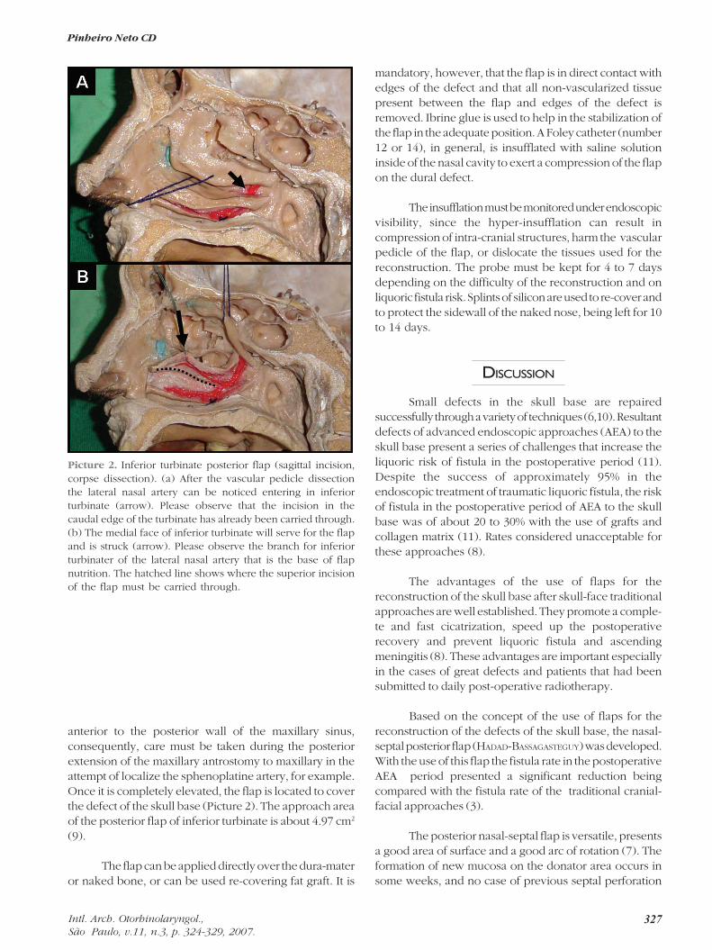
Picture 2. Inferior turbinate posterior flap (sagittal incision,
corpse dissection). (a) After the vascular pedicle dissection
the lateral nasal artery can be noticed entering in inferior
turbinate (arrow). Please observe that the incision in the
caudal edge of the turbinate has already been carried through.
(b) The medial face of inferior turbinate will serve for the flap
and is struck (arrow). Please observe the branch for inferior
turbinater of the lateral nasal artery that is the base of flap
nutrition. The hatched line shows where the superior incision
of the flap must be carried through.
Pinheiro Neto CD
Intl. Arch. Otorhinolaryngol.,
São Paulo, v.11, n.3, p. 324-329, 2007.
328
has been reported. However, some disadvantages of thisflap can be observed: it must be elevated before thesurgeon has the real dimension of the defect in the skullbase and it cannot be used in patients who were submittedto posterior septectomy or ample sphenoidoctomies. Theseprocedures interrupt the sanguine flow for the flap, makingits use impracticable (3, 8).
In these cases, the inferior turbinate posterior flapcan be used, which is based on the artery of the inferiorturbinate, which is branch of the lateral nasal artery that,in turn, is branch of sphenopalatine artery (8). This flappresents good arc of rotation that reaches most of theventral-caudal defects of the skull base and is elevatedafter the complete removal of the injury of the skullbase. However, the area of surface of this flap is not soample as the one of the posterior nasoseptal flap.According to the literature, the average area of theinferior turbinate posterior flap is about 4.97 cm2 (9),while the area of the posterior nasoseptal flap isapproximately 25 cm2 (7). Inferior turbinate bilateralflap can be used in the cases of defects which are biggerthan the surface of a flap.
Another disadvantage of the inferior turbinate pos-terior flap is the formation of crusts on the turbinate donatorarea. The formation of new mucosa on the donator areaoccurs in about 3 or 4 weeks. Patients with previousturbinectomy or atrophic rhinitis can present an insufficientarea of flap surface. These cases are considered a relativecontra-indication for this type of flap.
Recently, a new technique of use of the flap oftemporal-parietal fascia for the endoscopic reconstructionof the skull base has been described. This flap is alreadywell-established for the reconstruction after traditionalskull-face approaches and is based on the superficialtemporal artery. FORTS et al [ 2007 ] described this newtechnique for the transposition of this flap for the interiorof the nasal cavity so that this can after be used inendoscopic AEA reconstruction. Through the confection ofa tunnel temporal-infratemporal and an endonasaltranspterigoid approach, the transposition of the flap forthe interior of the nasal cavity is possible.
The endonasal transpterigoid approach is carriedthrough through the removal of the posterior wall of themaxillary sinus, dissection of pterigopalatine fossa andpartial removal of the medial and lateral pterigoidsprocesses. These maneuvers are carried through withintention to open a bone window so that the flap canpass from the temporal region, infrasecular fossa,pterigopalatina fossa and nasal cavity. The passage ofthe flap by soft tissues is opened in the use ofpercutaneous dilatators of tracheostomy. This flap
presents a bigger morbidity when compared to theinferior turbinate flap and the nasoseptal flap. Howeverit must be considered in cases in which theaccomplishment of the two last flaps is not possible (12)
CONCLUSIONS
The defects in the skull after advanced endonasalapproaches can be reconstructed with pedicle flaps. Theseflaps must be considered mainly for the reconstruction ofgreat defects. Initial studies have demonstrated that theuse of flaps is associated with a lesser liquoric fistula ratewhen compared to the use of free grafts. The nasal-septalposterior flap of Hadad-Bassagasteguy seems to be the firstoption of endoscopic reconstruction with flaps. However,in cases of posterior septectomy or any another conditionthat makes its use impracticable, the inferior turbinateposterior flap can be an excellent option.
REFERENCES
1. Kassam A, Carrau RL, Snyderman CH, Gardner P, MintzA. Evolution of reconstructive techniques followingendoscopic expanded endonasal approaches. NeurosurgFocus. 2005, 19(1):E8.
2. Nameki H, Kato T, Nameki I, Ajimi Y. Selectivereconstructive options for the anterior skull base. Int J ClinOncol. 2005, 10:223-28.
3. Hadad G, Bassagasteguy L, Carrau RL, Mataza JC, KassamA, Snyderman CH, Mintz A. A novel reconstructive techniqueafter endoscopic expanded endonasal approaches: vascularpedicle nasoseptal flap. Laryngoscope. 2006, 116:1882-86.
4. Carrau RL, Snyderman CH, Kassam AB. The managementof CSF leaks in patients at risk for high-pressurehydrocephalus. Laryngoscope. 2005, 115:205-12.
5. Goel A, Muzumdar DP. Recontruction of the sellar floorusing vascularized pedicle mucosal flap. Br J Neurosurg.2003, 17(6):553-55.
6. Hegazy HM, Carrau RL, Snyderman CH, Kassam A, ZweigJ. Transnasal endoscopic repair of cerebrospinal fluidrhinorrhea: a meta-analysis. Laryngoscope. 2000, 110:1166-72.
7. Pinheiro-Neto CD, Prevedello DM, Carrau RL, SnydermanCH, Mintz A, Gardner P, Kassam A.Improving the Designof the Pedicle Nasoseptal Flap for Skull Base Reconstruction:A Radioanatomic Study. Laryngoscope. 2007, Jun 27; [Epubahead of print].
Pinheiro Neto CD
Intl. Arch. Otorhinolaryngol.,
São Paulo, v.11, n.3, p. 324-329, 2007.
329
8. Fortes FS, Carrau RL, Snyderman CH, Prevedello D, VescanA, Mintz A, Gardner P, Kassam AB.The Posterior PedicleInferior Turbinate Flap: A New Vascularized Flap for SkullBase Reconstruction. Laryngoscope. 2007, Jun 27; [Epubahead of print].
9. Murakami CS, Kriet D, Ierokomos A. Nasal reconstructionusing the inferior turbinate mucosal flap. Arch Facial PlastSurg. 1999, 1:97-00.
10. Zweig JL, Carrau RL, Celin SE, Schaitkin BM, Pollice PA,Snyderman CH, Kassam A, Hegazy H.Endoscopic repair ofcerebrospinal fluid leaks to the sinonasal tract: predictors of
success. Otolaryngol Head Neck Surg. 2000, 123(3):195-01.
11. Snyderman CH, Kassam AB, Carrau R, Mintz A. EndoscopicReconstruction of Cranial Base Defects following EndonasalSkull Base Surgery. Skull Base. 2007, 17(1):73-8.
12. Fortes FS, Carrau RL, Snyderman CH, Kassam A,Prevedello D, Vescan A, Mintz A, Gardner P.Transpterygoidtransposition of a temporoparietal fascia flap: a new methodfor skull base reconstruction after endoscopic expandedendonasal approaches. Laryngoscope. 2007, 117(6):970-6.
Pinheiro Neto CD
Intl. Arch. Otorhinolaryngol.,
São Paulo, v.11, n.3, p. 324-329, 2007.