USE OF LIMBERG FLAP FOR PILONIDAL SINUS–A VIABLE OPTION
3
J Ayub Med Coll Abbottabad 2009;21(4) http://www.ayubmed.edu.pk/JAMC/PAST/21-4/Nadeem.pdf 31 USE OF LIMBERG FLAP FOR PILONIDAL SINUS–A VIABLE OPTION Muhammad Nadeem Aslam, Sidra Shoaib, Abdul Majeed Choudhry* Department of Surgery, Mayo Hospital, King Edward Medical University, *Sir Ganga Ram Hospital. Lahore, Pakistan Background: Pilonidal sinus disease has been treated for a long time with conventional open excision technique. The rhomboid flap of Limberg is a transposition flap that has been pleaded for treatment of this condition. Methods: We present our experience with the Limberg technique for both primary and recurrent pilonidal sinuses. One hundred and ten patients, with pilonidal sinus disease were treated with rhombic excision and Limberg transposition flaps. All sinus tracts were resected en bloc, and the fasciocutaneous Limberg flap was prepared from the gluteal region and closed it with a suction drain. Results: Full primary healing was obtained in 110 patients, 1 patient had minimal necrosis of flap and 2 had gaping of flap. Minor infection occurred in 3 patients. But all these complications healed uneventfully. The average hospital stay was 3 days. Follow up period was 1 year and 1 recurrence occurred. Conclusions: Limberg transposition flap is a promising surgical technique to treat pilonidal sinus. Keywords: Pilonidal sinus, Limberg flap, rhomboid transposition flap, fasciocutaneous flap, primary INTRODUCTION Intergluteal pilonidal disease is a commonly encountered condition in adult primary care, and it causes significant morbidity. The estimated incidence is 26 per 100,000, people. 1,2 Pilonidal disease generally presents as a cyst, abscess, or one or more sinus tracts with or without discharge in the upper part of the natal cleft. 3 Men are affected twice as often as women 1 and the condition is most frequent in the third decade of life. 4 However, there appears to be a small group that develops the condition for the first time in their 4 th decade. 4 The aetiology of pilonidal cysts has been a matter of debate. The condition was probably first described by Mayo in 1833, and it was felt to result from the congenital disposition but with time the view shifted towards acquired theory. 5 A widely acceptable view is that they are caused by local trauma, poor hygiene, excessive hairiness, and presence of deep natal cleft. 6,7 The management of pilonidal sinus disease is frequently unsatisfactory. Many surgical and non- surgical treatment modalities have been suggested, but the ideal and widely accepted treatment has still not yet been established. 8 In this regard, low recurrence rate, shorter hospital stay, less cost, minimal inconvenience and time off work are important considerations. Surgical techniques include laying the track open, wide excision with open wound, wide excision with marsupialisation, excision with primary midline or asymmetric closure and techniques involving various flaps procedures. All the surgical procedures have their pros and cons. The rhomboid flap of Limberg is a transposition flap that has been advocated for treatment of this condition. 9,10 In 1946, Limberg first described a technique for closing a 60º rhombus shaped defect with a transposition flap. 11 It is a series of communicating equilateral triangles, with all angles meeting at 60º. The advantage of this reconstruction is that it is very easy to perform and design. It flattens the natal cleft with a wide and well- vascularised pedicle that can be sutured without tension. That eventually helps in maintaining local hygiene, avoids hair insertion by reducing the friction between buttocks, reducing humidity, maceration, erosions and scar formation at the natal cleft. Limberg procedure is a safe and reliable technique in the treatment of sacrococcygeal pilonidal sinus disease, with low complication and recurrence rates if performed according to appropriate surgical principles. 12 This study was carried out to evaluate the usefulness of Limberg Flap technique in treatment of pilonidal sinus in our setup. MATERIAL AND METHODS We performed this procedure on 110 patients between Jan 2006 to Dec 2007. Patients having primary or recurrent pilonidal sinus disease underwent this operation. Patients who had pilonidal abscess had incision and drainage first with before the definite treatment. These patients were advised to return to normal activities after removal of stitches, after about 10 days, but to avoid excessive physical strain and strenuous sports for following 3 to 4 weeks. Follow up of all patients was performed on outpatient basis, every month for first six months and then six monthly for a period of twelve months. Surgery is performed either in general or spinal anesthesia. Patient is placed in jackknife position with buttocks strapped for wide exposure. After adequate shaving and skin preparation, area to be excised is carefully marked and flap lines are mapped on the skin (Figure-1). The rhomboid incision including the sinus and its extensions is made down to the pre-sacral fascia. Flap is constructed by extending
Transcript of USE OF LIMBERG FLAP FOR PILONIDAL SINUS–A VIABLE OPTION
J Ayub Med Coll Abbottabad 2009;21(4)
http://www.ayubmed.edu.pk/JAMC/PAST/21-4/Nadeem.pdf 31
USE OF LIMBERG FLAP FOR PILONIDAL SINUS–A VIABLE OPTION
Muhammad Nadeem Aslam, Sidra Shoaib, Abdul Majeed Choudhry* Department of Surgery, Mayo Hospital, King Edward Medical University, *Sir Ganga Ram Hospital. Lahore, Pakistan
Background: Pilonidal sinus disease has been treated for a long time with conventional open excision technique. The rhomboid flap of Limberg is a transposition flap that has been pleaded for treatment of this condition. Methods: We present our experience with the Limberg technique for both primary and recurrent pilonidal sinuses. One hundred and ten patients, with pilonidal sinus disease were treated with rhombic excision and Limberg transposition flaps. All sinus tracts were resected en bloc, and the fasciocutaneous Limberg flap was prepared from the gluteal region and closed it with a suction drain. Results: Full primary healing was obtained in 110 patients, 1 patient had minimal necrosis of flap and 2 had gaping of flap. Minor infection occurred in 3 patients. But all these complications healed uneventfully. The average hospital stay was 3 days. Follow up period was 1 year and 1 recurrence occurred. Conclusions: Limberg transposition flap is a promising surgical technique to treat pilonidal sinus. Keywords: Pilonidal sinus, Limberg flap, rhomboid transposition flap, fasciocutaneous flap, primary
INTRODUCTION Intergluteal pilonidal disease is a commonly encountered condition in adult primary care, and it causes significant morbidity. The estimated incidence is 26 per 100,000, people.1,2 Pilonidal disease generally presents as a cyst, abscess, or one or more sinus tracts with or without discharge in the upper part of the natal cleft.3 Men are affected twice as often as women1 and the condition is most frequent in the third decade of life.4 However, there appears to be a small group that develops the condition for the first time in their 4th decade.4
The aetiology of pilonidal cysts has been a matter of debate. The condition was probably first described by Mayo in 1833, and it was felt to result from the congenital disposition but with time the view shifted towards acquired theory.5 A widely acceptable view is that they are caused by local trauma, poor hygiene, excessive hairiness, and presence of deep natal cleft.6,7
The management of pilonidal sinus disease is frequently unsatisfactory. Many surgical and non- surgical treatment modalities have been suggested, but the ideal and widely accepted treatment has still not yet been established.8 In this regard, low recurrence rate, shorter hospital stay, less cost, minimal inconvenience and time off work are important considerations. Surgical techniques include laying the track open, wide excision with open wound, wide excision with marsupialisation, excision with primary midline or asymmetric closure and techniques involving various flaps procedures. All the surgical procedures have their pros and cons.
The rhomboid flap of Limberg is a transposition flap that has been advocated for treatment of this condition.9,10 In 1946, Limberg first described a technique for closing a 60º rhombus shaped defect with a transposition flap.11 It is a series
of communicating equilateral triangles, with all angles meeting at 60º. The advantage of this reconstruction is that it is very easy to perform and design. It flattens the natal cleft with a wide and well-vascularised pedicle that can be sutured without tension. That eventually helps in maintaining local hygiene, avoids hair insertion by reducing the friction between buttocks, reducing humidity, maceration, erosions and scar formation at the natal cleft.
Limberg procedure is a safe and reliable technique in the treatment of sacrococcygeal pilonidal sinus disease, with low complication and recurrence rates if performed according to appropriate surgical principles.12 This study was carried out to evaluate the usefulness of Limberg Flap technique in treatment of pilonidal sinus in our setup.
MATERIAL AND METHODS We performed this procedure on 110 patients between Jan 2006 to Dec 2007. Patients having primary or recurrent pilonidal sinus disease underwent this operation. Patients who had pilonidal abscess had incision and drainage first with before the definite treatment.
These patients were advised to return to normal activities after removal of stitches, after about 10 days, but to avoid excessive physical strain and strenuous sports for following 3 to 4 weeks. Follow up of all patients was performed on outpatient basis, every month for first six months and then six monthly for a period of twelve months.
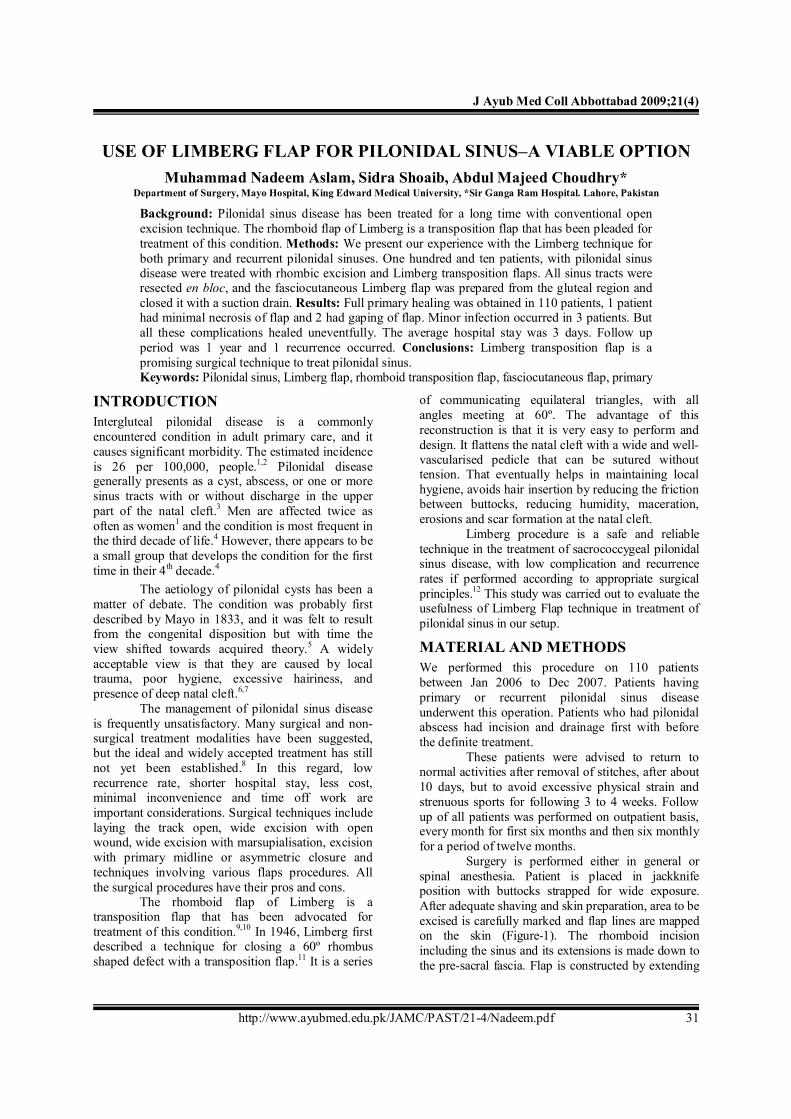
Surgery is performed either in general or spinal anesthesia. Patient is placed in jackknife position with buttocks strapped for wide exposure. After adequate shaving and skin preparation, area to be excised is carefully marked and flap lines are mapped on the skin (Figure-1). The rhomboid incision including the sinus and its extensions is made down to the pre-sacral fascia. Flap is constructed by extending
J Ayub Med Coll Abbottabad 2009;21(4)
http://www.ayubmed.edu.pk/JAMC/PAST/21-4/Nadeem.pdf 32
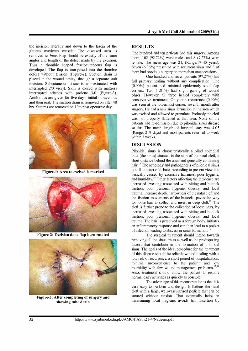
the incision laterally and down to the fascia of the gluteus maximus muscle. The diseased area is removed en bloc. Flap should be exactly of the same angles and length of the defect made by the excision. Thus a rhombic shaped fasciocutaneous flap is developed. The flap is transposed into the rhombic defect without tension (Figure-2). Suction drain is placed in the wound cavity, through a separate stab incision. Subcutaneous tissue is approximated with interrupted 2/0 vicryl. Skin is closed with mattress interrupted stitches with prolene 3/0 (Figure-3). Antibiotics are given for five days, initial intravenous and then oral. The suction drain is removed on after 48 hrs. Sutures are removed on 10th post operative day.
Figure-1: Area to excised is marked
Figure-2: Excision done flap been rotated
Figure-3: After completing of surgery and showing tube drain
RESULTS One hundred and ten patients had this surgery. Among them, 102 (92.72%) were males and 8 (7.27%) were female. The mean age was 21, (Range:17–45 years). Seven (6.36%) presented with recurrent sinus and 3 of them had previous surgery on more than one occasions.
One hundred and seven patients (97.27%) had full primary healing without any complication. One (0.90%) patient had minimal epidermolysis of flap corners. Two (1.81%) had slight gaping of wound edges. However all three healed completely with conservative treatment. Only one recurrence (0.90%) was seen at the lowermost corner, seventh month after surgery. He had a new sinus formation in the area which was excised and allowed to granulate. Probably the cleft was not properly flattened at that area. None of the patients had re-admission due to pilonidal sinus disease so far. The mean length of hospital stay was 4.05 (Range: 2–9 days) and most patients returned to work within 3 weeks.
DISCUSSION Pilonidal sinus is characteristically a blind epithelial tract (the sinus) situated in the skin of the natal cleft, a short distance behind the anus and generally containing hair.13 The aetiology and pathogenesis of pilonidal sinus is still a matter of debate. According to present view it is basically caused by excessive hairiness, poor hygiene, and humidity.14 Other factors affecting the incidence are increased sweating associated with sitting and buttock friction, poor personal hygiene, obesity, and local trauma, Increase depth, narrowness of the natal cleft and the friction movements of the buttocks paves the way for loose hair to collect and insert in deep cleft.15 The cleft is further prone to the collection of loose hairs, by increased sweating associated with sitting and buttock friction, poor personal hygiene, obesity, and local trauma. The hair is perceived as a foreign body, initiates an inflammatory response and can then lead to a pocket of infection leading to abscess or sinus formation.16
The surgical treatment should intend towards removing all the sinus tracts as well as the predisposing factors that contribute in the formation of pilonidal sinus. The goals of the ideal procedure for the treatment of this disease should be reliable wound healing with a low risk of recurrence, a short period of hospitalization, minimal inconvenience to the patient, and low morbidity with few wound-management problems.17,18 Also, treatment should allow the patient to resume normal daily activities as quickly as possible.
The advantage of this reconstruction is that it is very easy to perform and design. It flattens the natal cleft with a large, well-vascularised pedicle that can be sutured without tension. That eventually helps in maintaining local hygiene, avoids hair insertion by

J Ayub Med Coll Abbottabad 2009;21(4)
http://www.ayubmed.edu.pk/JAMC/PAST/21-4/Nadeem.pdf 33
reducing the friction between buttocks, reducing humidity, maceration, erosions and scar formation at the natal cleft. Any midline dead space is eliminated and a midline scar is avoided. It is a particularly useful technique for complex sinuses with multiple pits and extended tracts where radical excision leaves a large defect.18 The healing with secondary intention would require prolonged supervised wound care. This operation is also suitable for cases where other simpler operations have failed. The use of local flap accelerates healing.
Several series have been reported recently using this technique with minimal complications (Table-1). Our results with the Limberg flap are therefore comparable with other series that have shown wound complication and recurrence rates of 0–16% and 0–5% respectively.14,16,18,19
The importance of the post-operative wound care should also be stressed. Exercise or sitting down on
the wound should be avoided for two weeks and the patient has to return slowly to normal activities. Hair removal either by shaving the edges of the wound is mandatory.3,10 This has to be continued at least until complete healing of the wound, but preferably on a long-term basis.
CONCLUSION The results of our series support the wide excision and Limberg flap rotation as a preferred treatment of the disease. The technique can be mastered easily and provides an effective procedure for primary as well as recurrent disease. Few complications associated with it can further be reduced by meticulous skin closure and preventing skin edge inversion, especially at the lower midline. Also the flap should be wide enough to completely obliterate the midline natal cleft.
Table-1: Summary of results with Limberg flap technique in various series Author Patients (n) Hospital stay (days) Complication (%) Recurrence (%)
Katsoulis IE et al 200614 25 4.0 16 - Akin M et al 200816 411 3.2 15.75 2.91 Urhan MK et al 200218 102 3.7 7 4.9 Mentes BB et al 200419 238 2–3 2 1.26 Aslam MN 2007 (Present study) 110 3.0 5 1
REFERENCES 1. McCallum I, King PM, Bruce J. Healing by primary versus
secondary intention after surgical treatment for pilonidal sinus. Cochrane Database Syst Rev 2007(4):CD006213.
2. Sondenaa K, Andersen E, Nesvik I, Soreide JA. Patient characteristics and symptoms in chronic pilonidal sinus disease. Int J Colorectal Dis 1995;10(1):39–42.
3. Hull TL, Wu J. Pilonidal disease. Surg Clin North Am 2002;82:1169–85.
4. Clothier PR, Haywood IR. The natural history of the post anal (pilonidal) sinus. Ann R Coll Surg Engl 1984;66(3):201–3.
5. Brearley R. Pilonidal sinus; a new theory of origin. Br J Surg 1955;43:62–8.
6. Bascom J. Pilonidal disease: origin from follicles of hairs and results of follicle removal as treatment. Surgery. 1980;87:567–72.
7. Patel MR, Bassini L, Nashad R, Anselmo MT. Barber's interdigital pilonidal sinus of the hand: a foreign body hair granuloma. J Hand Surg Am 1990;15:652–5.
8. Chiedozi LC, Al-Rayyes FA, Salem MM, Al-Haddi FH, Al-Bidewi AA. Management of pilonidal sinus. Saudi Med J 2002;23:786–8.
9. Mohamed HA, Kadry I, Adly S. Comparison between three therapeutic modalities for non-complicated pilonidal sinus disease. Surgeon 2005;3(2):73–7.
10. Jamal A, Shamim M, Hashmi F, Qureshi MI. Open excision with secondary healing versus rhomboid excision with Limberg transposition flap in the management of sacrococcygeal pilonidal disease. J Pak Med Assoc 2009;59:157–60.
11. Azab AS, Kamal MS, Saad RA, Abou al Atta KA, Ali NA. Radical cure of pilonidal sinus by a transposition rhomboid flap. Br J Surg 1984;71(2):154–5.
12. Kapan M, Kapan S, Pekmezci S, Durgun V. Sacrococcygeal pilonidal sinus disease with Limberg flap repair. Tech Coloproctol 2002;6(1):27–32.
13. Ertan T, Koc M, Gocmen E, Aslar AK, Keskek M, Kilic M. Does technique alter quality of life after pilonidal sinus surgery? Am J Surg 2005;190:388–92.
14. Katsoulis IE, Hibberts F, Carapeti EA. Outcome of treatment of primary and recurrent pilonidal sinuses with the Limberg flap. Surgeon 2006;4(1):7–10, 62.
15. Mentes O, Bagci M, Bilgin T, Ozgul O, Ozdemir M. Limberg flap procedure for pilonidal sinus disease: results of 353 patients. Langenbecks Arch Surg 2008;393:85–9.
16. Akin M, Gokbayir H, Kilic K, Topgul K, Ozdemir E, Ferahkose Z. Rhomboid excision and Limberg flap for managing pilonidal sinus: long-term results in 411 patients. Colorectal Dis 2008;10:945–8.
17. Ersoy E, Onder Devay A, Aktimur R, Doganay B, Ozdogan M, Gundogdu RH. Comparison of the short-term results after Limberg and Karydakis procedures for pilonidal disease: Randomized prospective analysis of 100 patients. Colorectal Dis 2008;11:705–10.
18. Urhan MK, Kucukel F, Topgul K, Ozer I, Sari S. Rhomboid excision and Limberg flap for managing pilonidal sinus: results of 102 cases. Dis Colon Rectum 2002;45:656–9.
19. Mentes BB, Leventoglu S, Cihan A, Tatlicioglu E, Akin M, Oguz M. Modified Limberg transposition flap for sacrococcygeal pilonidal sinus. Surg Today 2004;34:419–23.
Address for Correspondence: Dr. Muhammad Nadeem Aslam, Associate Professor of Surgery, King Edward Medical University, Lahore, Pakistan. Postal Address: 261-F2, Johar Town, Lahore, Pakistan. Cell: +92-321-4787507 Email: [email protected]