Urological trauma during O/G procedures
46
ACUTE UROLOGICAL INJURIES DURING OBSTETRIC & GYNECOLOGICAL PROCEDURES Gaurav Nahar, Deptt.of Urology, MMHRC
-
Upload
gaurav-nahar -
Category
Health & Medicine
-
view
318 -
download
0
Transcript of Urological trauma during O/G procedures

ACUTE UROLOGICAL INJURIES DURING OBSTETRIC
& GYNECOLOGICAL PROCEDURES
Gaurav Nahar,Deptt.of Urology,
MMHRC

INTRODUCTION
• Urological injuries to urinary bladder & ureter- uncommon but imp.surgical complications during various obstetric & gynecological procedures.
• Anatomic proximity of ureters & bladder to genital tract.• Bladder injuries- most frequent urologic injury.• Bladder injuries usually recognized and repaired immediately,
and potential complications are typically minor.• But ureteral injuries(70%) typically are not recognized
immediately & can lead to long term complications.

URETERAL INJURIES• Iatrogenic trauma is the commonest cause of
ureteral injury.
• Two-thirds of injuries- during gynecologic surgeries.
• Most occur through abdominal than vaginal route.

ANATOMIC COURSE OFPELVIC URETER
• Ureter- 25-30 cm length.• Abdominal part lies
anteriorly on psoas muscle.• At the level of common iliac
artery bifurcation, it crosses internal iliac vessels to enter the pelvis.
• Ovarian vessels travel in infundibulo-pelvic ligament of the ovary and cross ureters anteriorly and laterally to the iliac vessels.
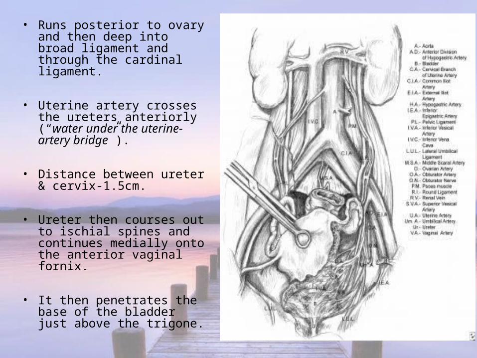
• Runs posterior to ovary and then deep into broad ligament and through the cardinal ligament.
• Uterine artery crosses the ureters anteriorly (“water under the uterine-artery bridge”).
• Distance between ureter & cervix-1.5cm.
• Ureter then courses out to ischial spines and continues medially onto the anterior vaginal fornix.
• It then penetrates the base of the bladder just above the trigone.

• Blood supply:1. Upper third- renal and ovarian arteries,2. Middle third-aortic branches and common
iliac arteries,3. Lower third- uterine, vaginal, middle
haemorrhoidal, vesical and hypogastric vessels.
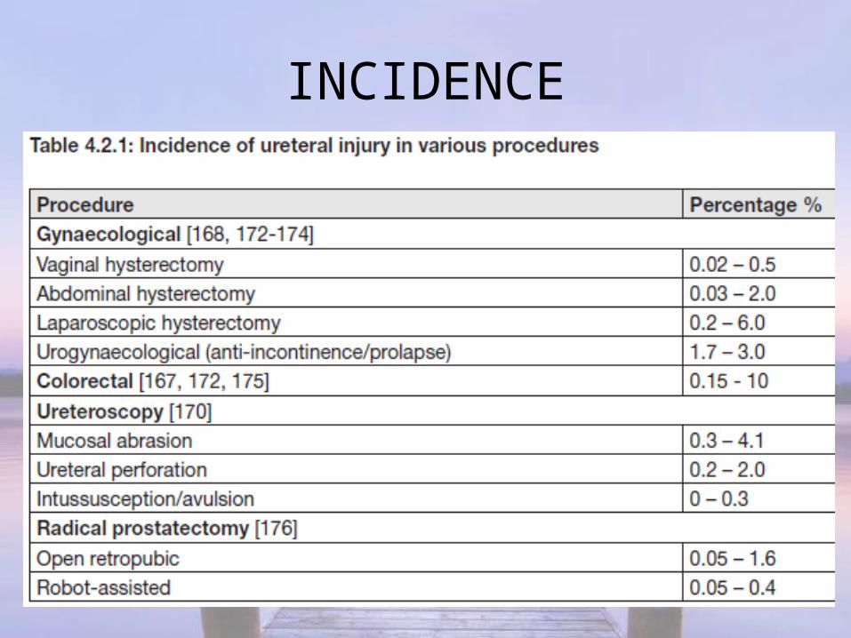
INCIDENCE


• Most ureteral injuries occur during technically straightforward hysterectomies for benign disease.
• Ureteral injuries from gynecologic surgery: 50% during radical hysterectomy, 40% from abdominal hysterectomy and <5% result from vaginal hysterectomy.

ETIOLOGYRISK FACTORS:• Enlarged uterus• Previous pelvic surgery or radiation• Advanced malignancy• Endometriosis, PID• Pelvic adhesions• Distorted pelvic anatomy• Coexistent bladder injury• Massive intraoperative haemorrhage

SITES OF INJURY• Injury: most frequently lower third of ureter(51%),
f/b upper third(30%) and middle third(19%).Most common sites of injury are:Lateral to uterine vesselsArea of ureterovesical junction close to cardinal
ligamentsBase of the infundibulopelvic ligament as ureters
cross pelvic brim at ovarian fossa at the level of uterosacral ligament.• Most common site of injury during laparoscopy-
adjacent to Uterosacral ligaments(USL).

Ureteral injuries during Laparoscopic gynec surgeries: during
• Laser ablative endometriosis surgery• Laparoscopic-assisted vaginal hysterectomy
(LAVH)• Laparoscopic tubal ligation,• Laparoscopic adnexectomy (removal of one of
the uterine tubes and an ovary) and• Laparoscopic uterosacral ligament ablation.

• Most LAVH ureteral injuries occur near cardinal and uterosacral ligaments.
• Caused by either thermal-electrocautery or sharp dissection, CO2 laser, endoscopic linear stapler and loop ligature.

MECHANISMS OF INJURY
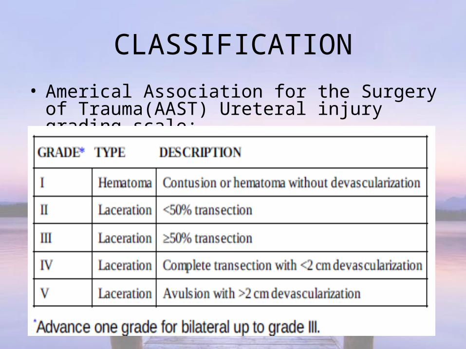
CLASSIFICATION• Americal Association for the Surgery of
Trauma(AAST) Ureteral injury grading scale:

PREVENTION

ROLE OF PREOPERATIVE PROPHYLACTIC URETERAL STENTING:
Assists in visualisation and palpation in complicated cases.
Also advantageous in making it easier to detect ureteral injury.
But it does not decrease the rate of injury.
Lighted fibreoptic ureteral catheters(5Fr)- new introduction.

• Haematuria is an unreliable & poor indicator of ureteral injury, present in only 50-75%.
• Cystoscopy without indigo carmine/methylene blue administration, used to document the absence of hematuria and the presence of bilateral ureteral jets, is a poor predictor of injury.
MANAGEMENT• No specific medical therapy for ureteric injury.• Immediate repair of ureteral injury advisable.• Optimal time for repair of a ureteral injury is during the
operation; the tissues are in their best condition, options and likelihood for success are greatest.
• Immediate repair provides better results and fewer complications than in a delayed fashion.
• Management depends on nature, severity, length & location of injury.

Reconstructive Procedures
• Ureteral resection & Uretero-ureterostomy• Trans-uretero-ureterostomy• Uretero-neo-cystostomy/Reimplantation• Ureterocalycostomy• Psoas hitch• Boari flap• Ileal interposition graft/Ileal ureter• Renal autotransplantation

Uretero-ureterostomyTechnique of uretero
ureterostomy after traumatic disruption.
A, Injury site definition by ureteral mobilization.
B, Debridement of margins and spatulation.
C, Stent placement.D, Approximationwith 5-0 absorbable suture.E, Final result.
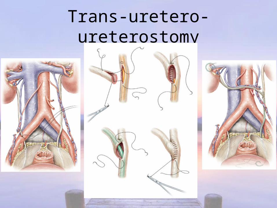
Trans-uretero-ureterostomy

Uretero-neo-cystostomy

Uretero-calycostomy

Psoas-hitch

Boari flap
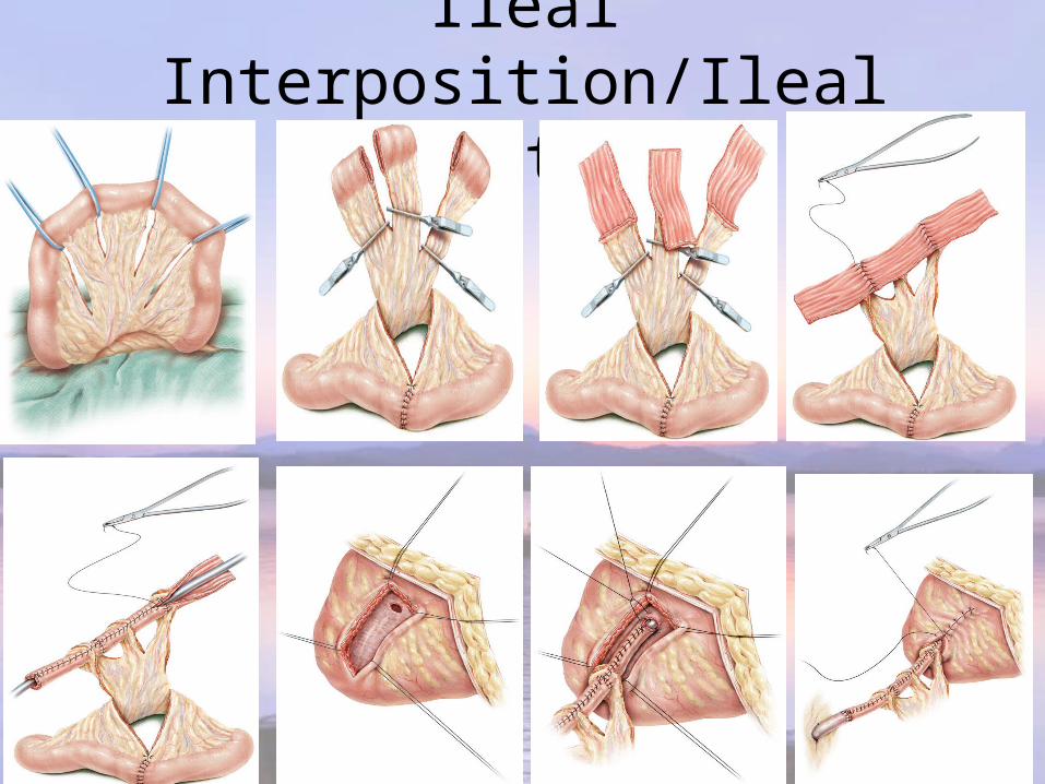
Ileal Interposition/Ileal ureter

Auto-transplantation

Principles of surgical repair of ureteral injury:• Debridement of necrotic tissue.• Ureteric dissection preserving adventitial sheath and its blood
supply.• Spatulation of ureteral ends.• Tension-free, watertight mucosa-to-mucosa anastomosis with
absorbable sutures.• Internal stenting.• External drain.• Isolation of injury with peritoneum or omentum.

M/m Based on Type of injury

• Immediate diagnosis of a ligation injury intraoperatively can be managed by de-ligation & stent placement.
• Partial injuries can be repaired immediately with a stent.
• Stenting is helpful because it provides canalisation and may decrease the risk of stricture.

M/m Based on Site of injury


PROXIMAL & MID-URETERAL INJURY:• Injuries shorter than 2-3 cm- Primary uretero-
ureterostomy.• When not feasible- Uretero-calycostomy.• Extensive ureteral loss- Transuretero-
ureterostomy(proximal stump of ureter is transposed across the midline and anastomosed to the contralateral ureter).
DISTAL URETERAL INJURY:• Ureteroneocystostomy/Ureteric reimplantion- best.• Refluxing vs.non-refluxing--controversial.• Psoas hitch- relieves tension off the anastomosis,
bridges gap.• Boari flap- for extensive mid-lower ureteral injury.

COMPLETE URETERAL INJURY:• Replacement using Ileal interposition
graft(Ileal ureter)- Yang-Monti principle.• Extensive ureteral loss or after multiple
attempts of ureteral repair- Autotransplantation.

COMPLICATIONS

Ureteral Injuries: Pearls of Wisdom
• Iatrogenic ureteral trauma gives rise to the commonest cause of ureteral injury.
• Haematuria is an unreliable and poor indicator of ureteral injury.
• The diagnosis of ureteral trauma is often delayed.• Preoperative prophylactic stents do not prevent ureteral injury,
but may assist in its detection.• Visually identify the ureters and meticulously dissect in their
vicinity to prevent ureteral trauma.• Use preoperative prophylactic stents only in selected cases
based on risk factors.

BLADDER INJURIES• Urinary bladder- urological
organ most often suffering iatrogenic injury.
• Occurred mostly during separation of bladder from lower segment of uterus in patients with previous cesarean sections.
• Primary repair during operation has excellent results.

INCIDENCE

CLASSIFICATION• Location of bladder injury is important to
guide further management:Intraperitoneal;Extraperitoneal;Combined intra-extraperitoneal.

INTRAOPERATIVE DETECTION• Signs of external iatrogenic bladder trauma: extravasation of
urine, visible laceration, clear fluid in surgical field, appearance of bladder catheter, and blood and/or gas in urine bag during laparoscopy.
• Direct inspection is the most reliable method of assessing bladder integrity.
• Large cystotomy is easily detected while smaller tears can be detected by Intravesical instillation of methylene blue. If bladder perforation is close to the trigone, the ureteric orifices should be inspected.

CYSTOSCOPY:• Preferred method for detection of intra-
operative bladder injuries, as it may directly visualise the laceration.
• Cystoscopy can localise the lesion in relation to the position of trigone and ureteral orifices.
• Lack of bladder distension during cystoscopy suggests a large perforation.

MANAGEMENT
• Uncomplicated extraperitoneal injury: Clinical observation, continuous bladder drainage and antibiotic prophylaxis.
• Complicated extraperitoneal injuries with symptomatic extravesical collection: Exploration, drainage & repair.
• Intraperitoneal injuries: surgical exploration with repair(double-layer closure with absorbable suture + Foley's catheter drainage).

FOLLOW-UP:• Simple injuries- Continuous F.C. drainage for
7-10 days f/b CR without cystogram.• Complex injuries(repaired)- Continuous F.C.
drainage for 14 days f/b control cystogram & CR.

CONCLUSION
• Iatrogenic urologic injuries can be prevented by adequate pre-operative assessment, good surgical technique, and visualisation of the bladder & ureters.
• Anticipation and high index of suspicion, early urological referral, and appropriate investigation of suspected urologic injury is of paramount importance.

Thank you...