Urolithiasis (kidney stones)
38
UROLITHIASIS Mwashambwa
-
Upload
ivan-kato -
Category
Health & Medicine
-
view
297 -
download
5
Transcript of Urolithiasis (kidney stones)

UROLITHIASIS
Mwashambwa

OVERVIEW
• INTRODUCTION• EPIDEMIOLOGY• CLASSIFICATION• PATHOGENESIS• CLINICAL FEATURES• INVESTIGATIONS• TREATMENT MODALITIES• COMPLICATIONS• PREVENTION

INTRODUCTION
• Urolithiasis, kidney stones, renal stones, and renal calculi are used interchangeably to refer to the accretion of hard, solid, nonmetallic minerals in the urinary tract
• Passage of a urinary stone is the most common cause of acute ureteral obstruction
• The pain may be some of the most severe pain that humans experience
• Complications of stone disease may result in severe infection; renal failure; or, in rare cases, death.
• Urinary stones have afflicted humankind since antiquity• The earliest recorded example being bladder and kidney stones detected
in Egyptian mummies dated to 4800 BC• The specialty of urologic surgery was recognized even by Hippocrates, who
wrote, in his famous oath for the physician,"I will not cut, even for the stone, but leave such procedures to the practitioners of the craft“ (obviously, Hippocrates was not a urologist!!)

EPIDEMIOLOGY
• The prevalence of urinary tract stonedisease is estimated to be 2% to 3%.• Rare in Blacks; Commoner in Whites and Asians• The likelihood that a white man will develop stone disease by age 70 years
is about 1 in 8. • The recurrence rate without treatment for calcium oxalate renal stones is
about – 10% at 1 year– 35% at 5 years, and– 50% at 10 years
• Male : Female ratio is 3:1• Peak at 20-40 years old• Ingestion of excessive amounts of purines ,oxalates,calcium, phosphate,
and other elements often results in excessive excretion of these components in urine
• A low fluid intake, with a subsequent low volume of urine production, produces high concentrations of stone-forming solutes in the urine.
This is an important environmental factor in stone formation.

EPIDEMIOLOGY …ctd
Disease associated with stone formation:• Hyperparathyroidism• renal tubular acidosis
(partial/complete)• jejunoileal bypass• Crohn’s disease,• intestinal resection• malabsorptive conditions• sarcoidosis• Hyperthyroidism
Medication associated with stone formation:
• calcium supplements• vitamin D supplements• Acetazolamide• ascorbic acid in megadoses ( > 4 g/day),• Sulphonamides• Triamterene• indinavir
Anatomical abnormalities associated with stoneformation:•tubular ectasia (medullary sponge kidney)•pelvo-ureteral junction obstruction•calix diverticulum•calix cyst•ureteral stricture•vesico-ureteral reflux•horseshoe kidney•ureterocele

CLASSIFICATION
• Calcium Stones 70-80%
– Ca Phosphate 5-10%
– Ca Oxalate/Phosphate 30-45% (Mixed)
– Ca Oxalate 20-30%
• Struvite stones 15-20%
• Cystine stones -3%
• Uric acid stones

CLASSIFICATION…ctdCOMPARATIVE INCIDENCES OF FORMS OF URINARY LITHIASIS
Stone analysis in Percentage
Form of Lithiasis India USA Japan UK
Pure Calcium Oxalate 86.1 33 17.4 39.4
Mixed Calcium Oxalate and 4.9 34 50.8 20.2
Phosphate
Magnesium Ammonium 2.7 15 17.4 15.4
Phosphate (Struvite )
Uric Acid 1.2 8.0 4.4 8.0
Cystine 0.4 3.0 1.0 2.8

CLASSIFICATION…ctd
Oxalate (Calcium Oxalate)
• Also Called Mulberry Stone
• Covered With Sharp Projections
• Sharp Makes Kidney Bleed (Haematuria)
• Very Hard
• Radio – Opaque
• Under microscope looks like Hourglass or Dumbbell shape if
monohydrate and Like an Envelope if Dihydrate

CLASSIFICATION…ctd
Phosphate stones
• Usually Calcium Phosphate
• Sometimes Calcium Magnesium Ammonium Phosphate Or Triple
Phosphate
• Smooth Minimum Symptoms
• Dirty White
• Radio – Opaque
• Calcium Phosphate also called ‘Brushite’ appears like Needle shape under
microscope
• In Alkaline urineEnlarges rapidlyTake the shape of CalycesStaghorn

CLASSIFICATION…ctd
Uric Acid & Urate Stone
• Hard & Smooth
• Multiple
• Yellow or Red-brown
• Radio - Lucent (Use Ultrasound)
• Under microscope appear like irregular plates or rosettes

CLASSIFICATION…ctd
Cystine Stone• Autosomal recesive disorder
• Usually in Young Girls
• Due To Cystinuria -
• Cystine Not Absorbed by Tubules
• Multiple
• Soft or Hard – can form stag-horns
• Pink or Yellow
• Radio-opaque
• Under microscope appears like hexagonal or benzene ring

PATHOGENESIS
• more than 1 of 3 general mechanisms is likely to be active– the possible presence or abundance of substances
that promote crystal and stone formation
– a possible relative lack of substances to inhibit crystal formation;
– a possible excessive excretion or concentration of salts in the urine, which leads to supersaturation of the crystallizing salt.
The greater the degree of supersaturation, the greater the rate of growth of the calculi

PATHOGENESIS…ctd
• Stasis or anatomic factors can also contribute to the development of stone disease.
• ~ 85% of calcium stones are idiopathic, or primary. – Idiopathic hypercalciuria occurs in more than one half of
patients with calcium oxalate stones.
– The remaining 15% of calcium stones are secondary to some discernible etiology, most commonly, hyperparathyroidism
– Renal tubular acidosis (RTA) is an additional fairly common secondary cause of calcium stones
– Immobilization of an individual causes rapid mobilization of the calcium in bones, and this is an important mechanism in patients with spinal cord injury

PATHOGENESIS…ctd
• Magnesium ammonium phosphate (struvite) stones account for approximately 10-20% of urinary stones.
– Sometimes they form complex with calcium phosphate.
– Struvite stones are caused by urea-splitting bacteria such as Proteus, Klebsiella, and Pseudomonas species.
– Combined obstruction and infection frequently cause renal destruction and, potentially, renal failure if both kidneys are affected

PATHOGENESIS…ctd
• Uric acid stones account for 5-10% of urinary stones, Predisposing factors include – acidic concentrated urine,– excess urinary uric acid, – small-bowel disease or resection, – gout, and cell lysis – Treatment and prevention for these stones is
alkalinization and dilution of the urine. • Cystine stones account for only approximately 1%
of urinary stones.– result from cystinuria (a rare autosomal recessive
metabolic disorder),

PATHOGENESIS…ctd
• Miscellaneous Stones– Triamterene Stones
• potassium sparing diuretic
• 70% excreted in urine
• pure stone or nidus for CaOx/UA
– Indinavir Stones• greatest incidence of protease inhibitors
• mean duration to stone 21.5 wks (6-50)
• 19% unchanged in urine
• fan shaped or starburst crystals
• not seen on IVU or CT
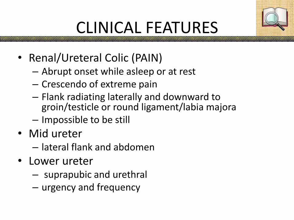
CLINICAL FEATURES
• Renal/Ureteral Colic (PAIN)– Abrupt onset while asleep or at rest– Crescendo of extreme pain– Flank radiating laterally and downward to
groin/testicle or round ligament/labia majora– Impossible to be still
• Mid ureter– lateral flank and abdomen
• Lower ureter– suprapubic and urethral– urgency and frequency

CLINICAL FEATURES…ctd
• GI Symptoms– Nausea and vomiting – autonomic n.s.– Ileus or diarrhea– DDX: gastroenteritis, appendicitis, colitis, diverticular
disease and salpingitis
• Hematuria– gross or microscopic– 15% no hematuria!
• Pyuria/Fever– Pyuria even without infection– Infection especially in females

CLINICAL FEATURES…ctd
• History
– Duration, characteristics, and location of pain
– History of urinary calculi
– Prior complications related to stone manipulation
– Urinary tract infections
– Loss of renal function
– Family history of calculi

INVESTIGATIONS
• Urinalysis- haematuria ~ 85% of pts
• FBP
– elevated WBC = renal/ systemic inf.
– low RBC= xnic d’se/ sev. haematuria
• serum eletrolytes, creatinine, calcium, uric acid, phosphorus: to asses renal function and metabolic risk factors for stone formation
• 24 hr urine collection for pH, Ca, oxalate, uric acid, Na, phosphorus, citrate, magnesium, creatinine and total volume

INVESTIGATION…ctd
• Plain abdominal radiograph– KUB for assessing total stone burden ,the size, shape, and
location of urinary calculi in some patients.
– Calcium-containing stones (~85% of all upper urinary tract calculi) are radiopaque,
– Pure uric acid, indinavir-induced, and cystine calculi are relatively radiolucent on plain radiography
• Renal ultrasound
• IVU– determine the size & location
– anatomical & functional assessment
• Helical CT-scan without contrast

INVESTIGATIONS…ctd
CALCULUS IN LT
KIDNEY LOWER POLE

INVESTIGATIONS…ctd
STAGHORN CALCULUS

TREATMENT MODALITIES
MEDICAL
SURGICAL

MEDICAL RX
• The cornerstone of management of ureteral colic is analgesia
• Morphine sulfate is the narcotic analgesic drug of choice for parenteral use.
• Antiemetic agents [metoclopramide ] may also be added as needed.
• The calcium channel blocker[ nifedipine] relaxes ureteral smooth muscle and enhances stone passage
• The alpha blockers, [ terazosin], also relax musculature of the ureter and lower urinary tract, markedly facilitating passage of ureteral stones
• Uric acid and cystine calculi can be dissolved with medical therapy
• stones are dissolved with alkalinization of the urine.
• Sodium bicarbonate can be used as the alkalinizing agent

MEDICAL RX…ctd
• High Fluid Intake and Alkalinized Urine – dissolve most of the
smaller cystine stones
• D-Pencillamine or MPG (Mercaptopropionylglycine) binds to
cystine that is soluble in urine
• Side effects of Pencillamine restricts it use – Allergic rashes,
GI problems- Nausea, Vomiting, Diarrhoea
• MPG better tolerated
• Large obstructive stones – Surgery required first

SURGERY
•Extracorporeal Shock Wave Lithotripsy (ESWL)
•Percutaneous Nephrolithotomy (PNL)
•Ureteroscopy
•Open surgery
Choice of approach depends on stone burden (size and number), stone composition, and stone location.
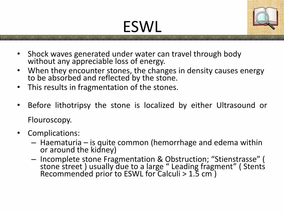
ESWL
• Shock waves generated under water can travel through body without any appreciable loss of energy.
• When they encounter stones, the changes in density causes energy to be absorbed and reflected by the stone.
• This results in fragmentation of the stones.
• Before lithotripsy the stone is localized by either Ultrasound or
Flouroscopy.
• Complications: – Haematuria – is quite common (hemorrhage and edema within
or around the kidney)– Incomplete stone Fragmentation & Obstruction; “Stienstrasse” (
stone street ) usually due to a large “ Leading fragment” ( Stents Recommended prior to ESWL for Calculi > 1.5 cm )

ESWL

Steinstrasse (Stone street)- post ESWL
PNL • Percutaneous approach allows stone removal with less morbidity, shorter
convalescence, and reduced cost compared with open techniques
• PNL has replaced open surgical procedures for removal of large or complex renal calculi at most institutions
• PNL can be performed with general, epidural, or local anesthesia
• The kidney should be approached from below the 12th rib to reduce the risk of pleural complications
• The position of the retroperitoneal colon is usually anterior or anterolateral to the lateral renal border. Therefore, risk of colon injury is minimal
• The liver and spleen may also be at risk of injury during percutaneous access. However, in the absence of splenomegaly or hepatomegaly, injury to these organs is extremely rare with a puncture below the 12th rib
• Once the point of puncture and the preferred calyx have been selected, a C-arm fluouroscope is entered. The tract is dilated by special dilators
• The urologist can proceed with stone removal using endoscopic techniques e.g with Randall's forceps, a grssper or stone baskets under fluoroscopic guidance

PNL…. Ureteroscope
• There is a concurrence in the literature regarding the need for postoperative drainage with a nephrostomy tube after percutaneous procedures.
• The main function of a nephrostomy tube is the drainage of urine and possibly the tamponade of bleeding originating from the structures acutely expanded during dilatation.
URETEROSCOPY:• A ureteroscope is passed through the ureteral orifices• It is performed under general or regional anaesthesia• Once the stone is visualized, fragmentation with of the stone can be done
with laser, or mechanically• If significant ureteral edema or manipulation occurs, a stent should be
placed to prevent colic and obstruction

Open surgery
• Generally indicated for large stones that would require multiple ESWL or PNL
• obese patients are poor candidates for ESWL and may be difficult to manage with PNL; Open surgery might be the best option
• Open surgery may be
– Pyelolithotomy
– Nephrolithotomy
– Ureterolithotomy
– Cystolithotomy

Summary
• Depending on the location of the stone, various procedures are done for stone extraxtion– In the kidney
• ESWL• PNL• Open methods
– Pyelolithotomy for a stone in the extrarenal pelvis– Nephrolithotomy for a stone deep into the renal parenchyma– Partial nephrectomy if there is a stone impacted into the lower most
calyx
– In the ureter• Upper ureter: ESWL is ideal• Mid ureter: ESWL, ureteroscopy or ureterolithotomy• Lower Ureter: Ureteroscope or ureterolithotomy

Summary
– In the Bladder
• Litholapaxy:
through a cystoscopy, the stone is grasped firmly and broken.
Small fragments are evacuated by evacuator
• Suprapubic cystolithotomy if the stone is too big or too hard

Complications
• Ureteral scarring and stenosis
• Nidus for infectionserious infection of the kidney that diminishes renal function
• Urinary fistula formation
• Ureteral perforation
• Extravasation
• Urinary outflow obstructionhydronephrosisCRF

Prevention
• High Fluid Intake
• Restrict Salt
• Avoid high intake of purine food
• Increased citrus fruits may help
• If hypercalciuria restrict Ca intake

Thanks