Urol Res 1996 Paper
8
Urol Res (1996) 24:305~3J 1 ORIGINAL PAPER F. Grases· L. García-Ferragut· A. Costa-Bauzá Study of the early stages of renal stone formation: experimental model using urothelium of pig urinary bladder Received: 30 August 1995 / Accepted: 8 March 1996 © Springer-Verlag 1996 Abstract An experimental model that enables the close simulation of conditions prevailing in the kidney dur- ing the formation of stones, using living tissue of pig urinary bladder, was developed. The results obtained clearly confirmed the importance of the antiadherent glycosaminoglycan (GAG) layer in preventing the de- velopment of solid concretions on the urothelium. Of importance was the capacity of the necrosed urothelium to act as a heterogeneous nucleant of cal- cium oxalate monohydrate (COM) and dihydrate (COD) crystals, demonstrating the major urolithiasic risk factor that alterations of the healthy epithelium covering the renal papilla may pose in humans. The significant increase in brushite and hydroxyapatite crystals detected on the urothelium when the pH of the artificial urine was 6.5, and the protective GAG layer was reduced or the tissue was necrosed, was also no- table. The crystallization inhibitory effects caused by citrate and phytate were also studied. It was found that whereas citrate, at normal urinary concentrations, caused a slight reduction in crystallization, with phytate there was total elimination of crystallization- when it was present at very low concentrations such as 1.0 ~lg/ml. Key words Calculogenesis' Calcium oxalate' Calcium phosphates' Crystallization inhibitors' Stone development Introduction It seems clear that at least a significant number of papillary calculi have their origin on the surface of the papillary tip as a consequence of uroepithelial alter- F. Grases (B)' L. García-Ferragut . A. Costa-Bauzá Faculty of Science, University of the Balearic Islands, E-07071 Palma de Mallorca, Spain ation, or alterations, combined with other factors. The main alterations which must be considered are the re- duction of the antiadherent glycosaminoglycan (GAG) layer [12J, focused necrosis, i.e., beca use of extended analgesic consumption [IJ, and microinfections. Other major factors which must also be taken into account are a persistent urinary pH below 5.5 (which favors the formation of uric acid crystals) or above 6.5 (which favors the formation of calcium phosphate crystals) and a deficit of crystallization inhibitory substances [5]. Most papillary calculi acquire a typical shape, exhi- biting a concave zone that corresponds to the point of attachment to the papilla [2,4]. Calcium phosphate and organic matter are frequently found in this area. Nevertheless, these substances can hardly be con- sidered as a stone nidus responsible for the origin of uroliths. If these substances were a stone nidus, then they would have played a relevant role in the calculus crystalline organization; this has seldom been observed. A core developed around a nidus is always situated inside the calculus and from its fine structure it may be deduced that the stone body grows on this coreo Basic geometrical considerations indicate that a nidus was originally located on the papilla surface and not deep in the collecting duct or in a subepithelial position. Consequently, it is very important to study the early stages of the development of attached crystals using models that try to reproduce the conditions found in vivo [6,9, 10]. This is why in this study we employed an experimental device using living pig bladder tissue to study the initial stages of calcium oxalate mono- hydrate (COM) stone formation. Materials and methods Synthetic urine The synthetic urine was prepared immediately before use by mixing equal volumes of solulions A and B in a T-lype mixing chamber.
-
Upload
anjanet-loon -
Category
Documents
-
view
232 -
download
1
description
chemistry lab sample
Transcript of Urol Res 1996 Paper
Urol Res (1996) 24:305~3J 1
ORIGINAL PAPER
F. Grases· L. García-Ferragut· A. Costa-Bauzá
Study of the early stages of renal stone formation:experimental model using urothelium of pig urinary bladder
Received: 30 August 1995 / Accepted: 8 March 1996
© Springer-Verlag 1996
Abstract An experimental model that enables the closesimulation of conditions prevailing in the kidney during the formation of stones, using living tissue of pigurinary bladder, was developed. The results obtainedclearly confirmed the importance of the antiadherentglycosaminoglycan (GAG) layer in preventing the development of solid concretions on the urothelium. Ofimportance was the capacity of the necrosedurothelium to act as a heterogeneous nucleant of calcium oxalate monohydrate (COM) and dihydrate(COD) crystals, demonstrating the major urolithiasicrisk factor that alterations of the healthy epitheliumcovering the renal papilla may pose in humans. Thesignificant increase in brushite and hydroxyapatitecrystals detected on the urothelium when the pH of theartificial urine was 6.5, and the protective GAG layerwas reduced or the tissue was necrosed, was also notable. The crystallization inhibitory effects caused bycitrate and phytate were also studied. It was found thatwhereas citrate, at normal urinary concentrations,caused a slight reduction in crystallization, withphytate there was total elimination of crystallizationwhen it was present at very low concentrations such as1.0 ~lg/ml.
Key words Calculogenesis' Calcium oxalate' Calciumphosphates' Crystallization inhibitors' Stonedevelopment
Introduction
It seems clear that at least a significant number ofpapillary calculi have their origin on the surface of thepapillary tip as a consequence of uroepithelial alter-
F. Grases (B)' L. García-Ferragut . A. Costa-BauzáFaculty of Science, University of the Balearic Islands,E-07071 Palma de Mallorca, Spain
ation, or alterations, combined with other factors. Themain alterations which must be considered are the reduction of the antiadherent glycosaminoglycan (GAG)layer [12J, focused necrosis, i.e., beca use of extendedanalgesic consumption [IJ, and microinfections. Othermajor factors which must also be taken into accountare a persistent urinary pH below 5.5 (which favors theformation of uric acid crystals) or above 6.5 (whichfavors the formation of calcium phosphate crystals) anda deficit of crystallization inhibitory substances [5].
Most papillary calculi acquire a typical shape, exhibiting a concave zone that corresponds to the point ofattachment to the papilla [2,4]. Calcium phosphateand organic matter are frequently found in this area.Nevertheless, these substances can hardly be considered as a stone nidus responsible for the origin ofuroliths. If these substances were a stone nidus, thenthey would have played a relevant role in the calculuscrystalline organization; this has seldom been observed.A core developed around a nidus is always situatedinside the calculus and from its fine structure it may bededuced that the stone body grows on this coreo Basicgeometrical considerations indicate that a nidus wasoriginally located on the papilla surface and not deep inthe collecting duct or in a subepithelial position.
Consequently, it is very important to study the earlystages of the development of attached crystals usingmodels that try to reproduce the conditions found invivo [6,9, 10]. This is why in this study we employedan experimental device using living pig bladder tissueto study the initial stages of calcium oxalate monohydrate (COM) stone formation.
Materials and methods
Synthetic urine
The synthetic urine was prepared immediately before use by mixingequal volumes of solulions A and B in a T-lype mixing chamber.
306
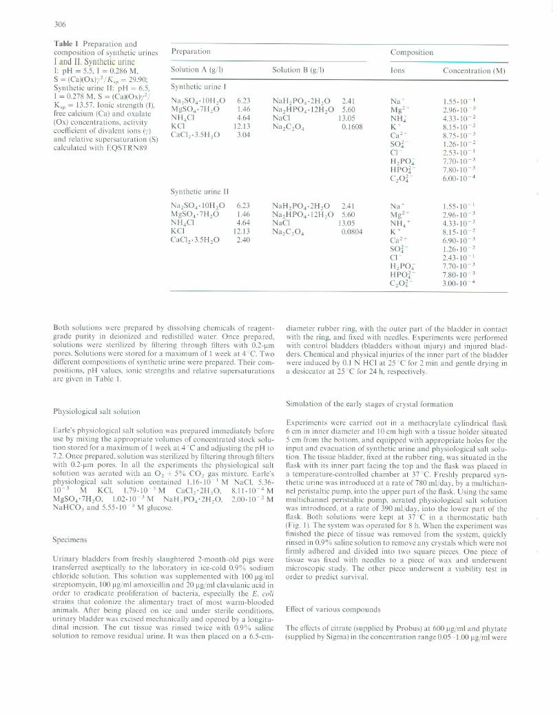
Table I Preparation andcomposition of synthetic urines
1 and 11. Synthetic mine1: pH = 5.5, 1 = 0.286 M,
S = (Ca)(Ox)yl I K,p = 29.90;Synthetic urine [1: pH = 6.5,1 = 0.278 M, S = (Ca)(Ox)y21
K,p = 13.57. [onic strength (1),free caIcium (Ca) and oxalate
(Ox) concentrations, activitycoefficient of divaIcnt ions (y)
and relative supersaturation (S)calculated with EQSTRN89
Preparation
Solution A (gil)
Synthetic urine [
Na1S04·10H10MgS04·7H20NH4CIKCl
CaCI2·3.5H20
Synthetic urine II
Na2S04·IOH20MgS04·7H20NH4CIKCI
CaCI2·3.5H20
6.231.464.64
12.133.04
6.231.464.64
12.132.40
Solution B (gil)
NaH2P04·2H20Na1HP04·12H20NaCI
Na2C204
NaH2P04·2H20Na2HP04·12H20NaCl
Na1C104
2.415.60
13.050.1608
2.415.60
13.050.0804
Composition
lons
Na+
Mg2+NH:K+Ca2+
SO,¡Cl-H2PO"HPO'¡-C2°'¡-
Na+
Mg2+NH4 +K+Ca2+
SO,¡Cl-H2PO"HPO'¡-C2°'¡-
Concentration (M)
1.55.10-12.96.10-3
4.33.10-28.15.10-28.75.10-31.26.10-22.53.10-17.70.10-37.80.10-36.00.10-4
1.55.10-12.96.10-34.33.10- 2
8.15.10-26.90.10-31.26.10-22.43.10-17.70.10-37.80.10-33.00.10-4
Both solutions were prepared by dissolving chemicals of reagentgrade purity in deionized and redistilled water. Once prepared,solutions were sterilized by filtering through lilters with 0.2-Jlmpores. Solutions were stored for a maximum of 1 week at 4 0e. Two
different compositions of synthetic urine were prepared. Their compositions, pH values. ionic strengths and relative supersaturationsare given in Table 1.
Physiological salt solution
Earle's physiological salt solution was prepared immediately beforeuse by mixing the appropriate volumes of concentrated stock solu
tion stored for a maximum of 1 week at 4 °C and adjusting the pH to7.2. Once prepared, solution was sterilized by filtering through filterswith 0.2-Jlm pores. In all the experiments the physiological saltsolution was aerated with an O2 + 5°Ir, CO2 gas mixture. Earle'sphysiological salt solution contained 1.16.10- 1 M NaCl, 5.36·10-3 M KCl, 1.79·1O-3M CaCI2'2H20, 8.11·1O-4MMgS04'7H20, 1.02.10-3 M NaH2P04'2H20, 2.00.10-1 MNaHC03 and 5.55.10-3 M glucose.
Specimens
Urinary bladders from freshly slaughtered 2-month-old pigs weretransferred aseptically to the laboratory in ice-cold 0.9% sodium
chloride solution. This solution was supplemented with 100 ~lg/mlstreptomycin, 100 Jlg/ml amoxicillin and 20 Jlg/ml clavulanic acid inorder to eradicate proliferation of bacteria, especially the E. colistrains that colonize the alimentary tract of most warm-bloodedanimals. After being placed on ice and under sterile conditions,urinary bladder was excised mechanically and opened by a longitudinal incision. The cut tissue was rinsed twice with 0.9% saline
solution to remove residual urine. It was then placed on a 6.5-cm-
di ame ter rubber ring, with the outer part of the bladder in contactwith the ring, and fixed with needles. Experiments were performedwith control bladders (bladders without injury) and injured bladders. Chemical and physical injuries of the inner part of the bladderwere induced by 0.1 N H CI at 25 cC for 2 min and gentle drying ina desiccator at 25°C for 24 h, respectively.
Simulation of the early stages of crystal formation
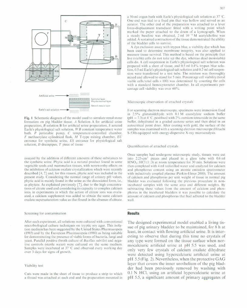
Experiments were carried out in a methacrylate cylindrical ftask6 cm in inner diameter and 10 cm high with a tissue holder situated5 cm from the bottom, and equipped with appropriate holes for theinput and evacuation of synthetic urine and physiological salt sol ution. The tissue bladder, fixed at the rubber ring, was situated in the
tlask with its inner part facing the top and the tlask was placed ina temperature-controlled chamber at 37°e. Freshly prepared synthetic urine was introduced at arate of 780 ml/day, by a multichannel peristaltic pump, into the upper par! of the flask. Using the samemultichannel peristaltic pump, aerated physiological salt solutionwas introduced, at arate of 390 ml/day, into the lower part of theflask. Both solutions were kept at 37 DC in a thermostatic bath(Fig. 1). The system was operated for 8 h. When the experiment was
finished the piece of tissue was removed from the system, quicklyrinsed in 0.9% saline solution to remove any crystals which were notfirmly adhered and divided into two square pieces. One piece oftissue was lixed with needlcs to a piece of wax and underwentmicroscopic study. The other piece underwent a viability test inorder to predict survival.
Effect of various compounds
The efTects of citrate (supplied by Probus) at 600 Jlg/ml and phytate(supplied by Sigma) in the concentration range 0.05-1.00 ~lg/ml were
oe
M
307
a 50-mI organ bath with Earle's physiological salt solution at 37 ae.One end was ticd to a fixed pin that was hollow and servcd as anaerator. The other end 01' the preparation was attached to a leverforce-displacement transducer fitted with a writing point whichmarked the paper attached to the drum 01' a kymograph. Whena steady baseline was obtained, 2 mI 10-6 M acetylcholine wasadded. A sustained contraction 01' the tissue demonstrated the ability01' the bladder cells to survive.
A dye exclusion assay with trypan blue, a viability dye which hasbeen used to determine membrane integrity, was also applied tomeasure tissue survival. This method is based on the principIe thatlive (viable) cells do not take up the dye, whereas dead (nonviable)cells do. A cell suspension in Earle's physiological salt solution wasprepared with a sheet 01' tissue, and 0.5 mI O.4'Yo trypan blue sol ution, 0.5 mi Earle's physiological salt solution and 0.2 mI cell suspension were transferred to a test tube. The mixture was thoroughlymixed and allowed to stand for 5 mino Percentage cell viability (totalviable cells/total cells x 100) was determined by counting the cellswith a standard hemocytometer chamber. In all experiments percentage cell viability was over 60%.
Epj¡he1iallayerPropria mucosal ¡ayer
Smooth muscle ¡ayer
Fig. 1 Schematic diagram 01' the model used to simula te renal stoneformation on pig bladder tissue. A Solution A for artificial urinepreparation, B solution B for artificial urine preparation, S aeratedEarlc's physiological salt solution, W B constant temperature waterbath. P peristaltic pump, e temperature-controlled chamber,F methacrylate cylindrical !1ask, M T-type mixing chamber, EUen trance for synthctic urine, ES en trance for physiological saltsolution, D drainpipes, T piece 01' tissue
assayed by the addition 01' dilTerent amounts 01' these substances tothe synthetic urine. Phytic acid is a natural product found in somevegetable seeds and mammalian tissues, with noteworthy effects onthe inhibition 01' calcium oxalate crystallization which were recentlydescribed [4,7J and, for this reason, phytic acid was included in thepresent study. Considering the normal range 01' urinary pH values,phytic acid is mainly found in the urine as the dissociatcd form, i.e.,as phytate. As eXplained previously [7J, due to the high concentrations 01' citrate used and considering its capacity to complex calciumions, in experiments in which the action 01' citrate ions was evaluated, a calcium supplement was added to obtain the same calciumoxalate supersaturation value as that found in the absence 01' citrate.
Screening for contamination
Arter each experiment, all solutions were cultured with conventionalmicrobiological culture techniques on tryptic soy agar. This isolation medium has been suggested by the United States Pharmacopeia(1985) and by the European Pharmacopeia (1980) as being suitablefor demonstrating the presence 01' viable forms 01' bacteria, fungi andyeast. Parallel positive (broth culture 01' Bacil/us subtilis) and negative controls .(sterile water) were cultured on the same medium.Samples were incubated at 37 ac and observed every working dayover 3 days for signs 01' growth.
Viability test
Cuts were made in the sheet 01' tissue to produce a strip to whicha thread was attached at each end and the preparation mounted in
Microscopic observation 01' attached crystals
For scanning electron microscopy, specimens were immersion fixedin 2.5% glutaraldehyde with 0.1 M cacodylate sodium buffer(pH = 7.3) at 4°C, postfixed with 2% osmium tetraoxide in the samebuffer, dehydrated in a graded acetone series and then dried in anautocritical point drier. Al'ter coating with gold, the surface 01' thesamples was examined with a scanning electron microscope (HitachiS-530) equipped with energy-dispersive X-ray microanalysis.
Quantification 01' attached crystals
Once samples had undergone microscopic study, tissues were cutinto 2.25-cm2 pieces and placed in a glass tube with 0.6 miHN03: HCl (1 : 3) at room temperature for 30 mino Solutions werethen completed with 4 mI redistilled water and analyzed for calciumand phosphorus content using an atomic emission spectrometerwith inductively coupled plasma (Perkin-Elmer 2000). The amount01' calcium and phosphorus per unit weight 01' tissue in normal pigbladder was evaluated following the previous procedure in nonincubated samples with the same area and different weights. Bysubtracting these values from the amount 01' calcium and phosphorus in the incubated bladders it was possible to calculate theamount 01' calcium and phosphorus that had adhered to the bladderwall.
Results
The designed experimental model enabled a living tissue of pig urinary bladder to be maintained, for 8 h atleast, in contact with flowing artificial urine. It is interesting to observe that during this time no crystals ofany type were formed on the tissue surface when normocalciuric artificial urine at pH 5.5 was used, andonly very few crystals of calcium oxalate dihydratewere detected using hypercalciuric artificial urine atpH 5.5 (Fig. 2). Nevertheless, when the protective GAGlayer that covers the inner urothelium of the pig bladder had been previously removed by washing with0.1 N HC!, using an artificial hypercalciuric urine atpH 5.5, a significant amount of primary aggregates of

308
Fig.2 Calcium oxalate dihydrate crystals observed on the surface ofhealthy tissue when using hypercalciuric artificial urine at pH 5.5(SEM image)
Fig. 3 Primary aggregates of calcium oxalate monohydrate anddihydrate observed on the surface of a tissue with injuries induced by0.1 N HCl when using hypercalciuric artificial urine at pH 5.5 (SEMimage)
eOM and eoo crystals were observed on the apicalside (Fig. 3). Using totally necrosed urothelial tissueand an artificial hypercalciuric urine at a pH of 5.5, allthe necrosed surface appeared fully covered with eOMand eoo crystals (Fig. 4).These experiments cleariydemonstrated that crystals that appeared on theurothelial tissue, when it was damaged, were directlyformed on this tissue as a consequence of interactionsbetween normally supersaturated urine and tissue, anddid not appear as a result of interactions betweenpreformed crystals and tissue, due to the deposition ofpreviously formed crystals.
Experiments performed using noninjured pig bladdel' urothelium and normocalciuric artificial urine witha pH of 6.5 showed the formation of some isolatedaggregates of brushite crystals (Fig. 5). When thesame experiment was performed using a pigbladder urothelium with previous elimination of theprotective GAG layer, a significant quantity ofbrushite
Fig. 4A, B Primary aggregates of calcium oxalate monohydrate anddihydrate observed on the surface of a tissue with injuries induced bydrying in a desiccator when using hypercalciuric artificial urine atpH 5.5. A General view, B detail (SEM images)
Fig.5 Isolated aggregates of brushite crystals observed on the surface of healthy tissue when using normocalciuric artificial urine witha pH of 6.5 (SEM image)
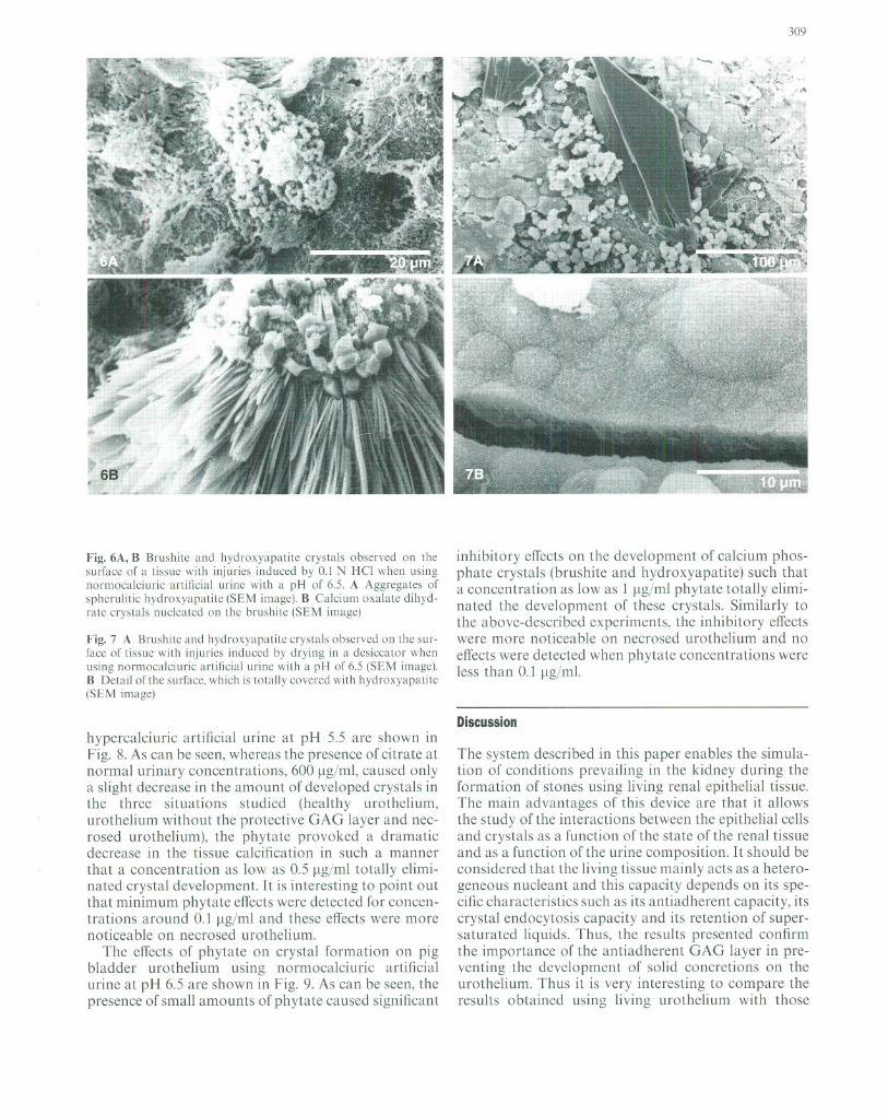
and hydroxyapatite was detected (Fig. 6). It is interesting to observe the presence of eoo crystals nucleatedon the brushite. Using totally necrosed urothelial tissueand a normocalciuric artificial urine at a pH of 6.5, allthe surface appeared covered with brushite and hydroxyapatite (Fig. 7).
The effects of citrate and phytate on the crystal development on pig bladder urothelium using
309
Fig. 6A, B Brushite and hydroxyapatite crystals observed on thesurface of a tissue with injuries induced by 0.1 N HCI whcn usingnormocalciuric artificial urine with a pH of 6.5. A Aggregates ofspherulitic hydroxyapatitc (SEM image). B Calcium oxalate dihydrate crystals nucleated on the brushite (SEM image)
Fig. 7 A Brushite and hydroxyapatite crystals observed on the surface of tissue with injuries induced by drying in a desiccator whenusing normocalciuric artificial urine with a pH of 6.5 (SEM image).B Oetail of (he surface. which is totally covered with hydroxyapatite(SEM image)
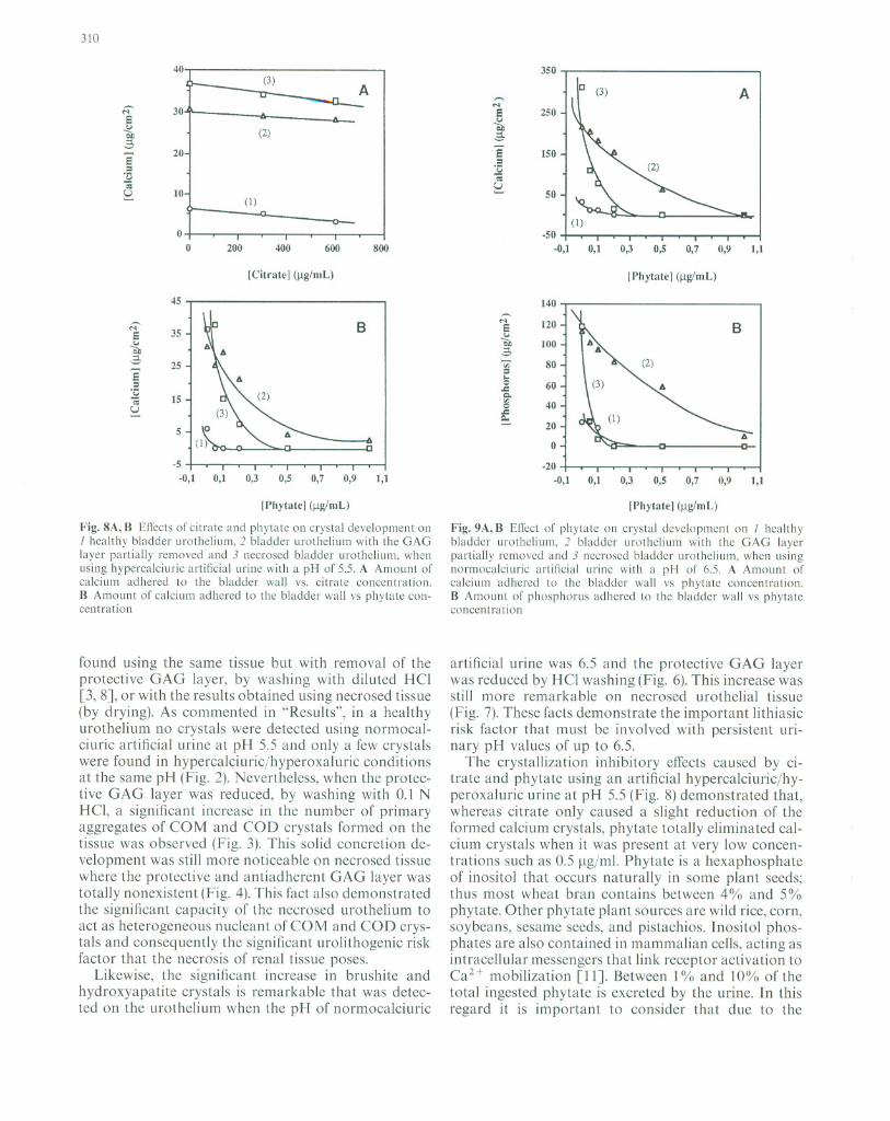
hypercalciuric artificial urine at pH 5.5 are shown inFig. 8. As can be seen. whereas the presence of citrate atnormal urinary concentrations, 600 ~lg/ml. caused onlya slight decrease in the amount of developed crystals inthe three situations studied (healthy urothelium,urothelium without the protective GAG layer and necrosed urothelium), the phytate provoked a dramaticdecrease in the tissue calcification in such a manner
that a concentration as low as 0.5 ~g/ml totally eliminated crystal development. 1t is interesting to point outthat minimum phytate effects were detected for concentrations around 0.1 ~lg/ml and these effects were morenoticeable on necrosed urothelium.
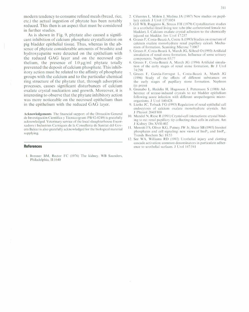
The effects of phytate on crystal formation on pigbladder urothelium using normocalciuric artificialurine at pH 6.5 are shown in Fig. 9. As can be seen. thepresence of small amounts of phytate caused significant
inhibitory effects on the development of calcium phosphate crystals (brushite and hydroxyapatite) such thata concentration as low as 1 ~g/ml phytate totally eliminated the development of these crystals. Similarly tothe above-described experiments, the inhibitory effectswere more noticeable on necrosed urothelium and no
effects were detected when phytate concentrations wereless than 0.1 ~g/ml.
Discussion
The system described in this paper enables the simulation of conditions prevailing in the kidney during theformation of stones using living renal epithelial tissue.The main advantages of this device are that it allowsthe study of the interactions between the epithelial cellsand crystals as a function of the state of the renal tissueand as a function of the urine composition. It should beconsidered that the living tissue mainly acts as a heterogeneous nucleant and this capacity depends on its specific characteristics such as its antiadherent capacity, itscrystal endocytosis capacity and its retention of supersaturated liquids. Thus, the results presented confirmthe importance of the antiadherent GAG layer in preventing the development of solid concretions on theurothelium. Thus it is very interesting to compare theresults obtained using living urothelium with those
310
40
350(3) A uc(3) A""'¡:¡'
-~...•30 S250
Sc,.¡
~ (2)e.
OJO
ÓÓ S15020
S=
=.;:¡
·u-¡-¡ UU
10 ~SO- (1)
O
-SO
O
200400600800 -0,10,10,30,50,70,91,1
[Citrate] (¡.tg/mL)
lPhytate] (¡.tg/mL)
45 ,
.140
B
...•120¡:¡' SS
35c,.¡
c,.¡
e.100b¡¡ Ó::i ~80~
25 =S ...o 60= .c.;:¡
15Q..
-¡ '"40oU .c- Q.- 20S
O-5
-20-0,1
0,10,30,50,70,91,1 -0,10,10,30,50,70,91,1
lPhytate] (¡.tg/mL)
Fig. 8A, B EfTcctsof citrate and phytatc on crystal development on1 healthy bladder urothelium, 2 bladdcr urothelium with the GAGlayer partially removed and 3 nccroscd bladder urothelium, whcnusing hypercalciuric artificial urine with a pH of 5.5. A Amount ofcalcium adhered to the bladder wall vs. citrate concentration.B Amount of calcium adhered to the bladder wall vs phytate concentration
found using the same tissue but with removal of theprotective GAG layer, by washing with diluted HCl[3, 8J, or with the results obtained using necrosed tissue(by drying). As commented in "Results", in a healthyurothelium no crystals were detected using normocalciuric artificial urine at pH 5.5 and only a few crystalswere found in hypercalciuric/hyperoxaluric conditionsat the same pH (Fig. 2). Nevertheless, when the protective GAG layer was reduced, by washing with 0.1 NHCl, a significant increase in the number of primaryaggregates of COM and COO crystals formed on thetissue was observed (Fig. 3). This sol id concretion development was still more noticeable on necrosed tissuewhere the protective and antiadherent GAG layer wastotally nonexistent (Fig. 4). This fact also demonstratedthe significant capacity of the necrosed urothelium toact as heterogeneous nucleant ofCOM and COO crystals and consequently the significant urolithogenic riskfactor that the necrosis of renal tissue poses.
Likewise, the significant increase in brushite andhydroxyapatite crystals is remarkable that was detected on the urothelium when the pH of normocalciuric
[Phytate] (¡.tg/mL)
Fig. 9A, B EfIect of phytate on crystal dcvclopment on 1 healthybladder urothelium, 2 bladder urothelium with the GAG layerpartially removed and 3 necrosed bladder urothelium, when usingnormocalciuric artificial urine with a pH of 6.5. A Amount ofcalcium adhered to the bladder wall vs phytate concentration.B Amount of phosphorus adhered to the bladder wall vs phytateconcentration
artificial urine was 6.5 and the protective GAG layerwas reduced by HCl washing (Fig. 6). This increase wasstill more remarkable on necrosed urothelial tissue(Fig. 7). These facts demonstrate the important lithiasicrisk factor that must be involved with persistent urinary pH values of up to 6.5.
The crystallization inhibitory effects caused by citrate and phytate using an artificial hypercalciuric/hyperoxaluric urine at pH 5.5 (Fig. 8) demonstrated that,whereas citrate only caused a slight reduction of theformed calcium crystals, phytate totally eliminated calcium crystals when it was present at very low concentrations such as 0.5 ¡.tg/ml. Phytate is a hexaphosphateof inositol that occurs naturally in some plant seeds;thus most wheat bran contains between 4% and 5%phytate. Other phytate plant sources are wild rice, corn,soybeans, sesame seeds, and pistachios. lnositol phosphates are also contained in mammalian cells, acting asintracellular messengers that link receptor activation toCaH mobilization [11]. Between 1°;;, and 10% of thetotal ingested phytate is excreted by the urine. In thisregard it is important to consider that due to the
modern tendency to consume refined meals (bread, rice,etc.) the actual ingestion of phytate has been notablyreduced. This then is an aspect that must be consideredin further studies.
As is shown in Fig. 9, phytate al so caused a significant inhibition of calcium phosphate crystallization onpig bladder epithelial tissue. Thus, whereas in the absence of phytate considerable amounts of brushite andhydroxyapatite were detected on the epithelium withthe reduced GAG layer and on the necrosed epithelium, the presence of 1.0 ~lg/ml phytate totallyprevented the deposit of calcium phosphate. This inhibitory action must be related to the affinity of phosphategroups with the calcium and to the particular chemicalring structure of the phytate that, through adsorptionprocesses, causes significant disturbances of calciumoxalate crystal nucleation and growth. Moreover, it isinteresting to observe that the phytate inhibitory actionwas more noticeable on the necrosed epithelium thanin the epithelium with the reduced GAG layer.
Acknowledgements The financial support of the Dirección Generalde Investigación Cientifica y Técnica (grant PB 92-0249) is gratefullyacknowledged. Veterinary service of the local slaughterhouse Escorxadors i Industries Carniques de la Conselleria de Sal1itat del Govern Balear is also gratefully acknowledged for the biological materialsupplying.
References
1. Brenner BM, Rector FC (1976) The kidney. WB Saunders,Philadel phia, 11:1640
311
2. Cifuentes L, Miñón 1, Medina lA (1987) New studies 011 papillary calculi. 1 U rol 137: I024
3. Gill WB, Ruggiero K, Straus FH (1979) Crystallization studiesin a llrothelial-lined living test tube (the catheterized female ratbladder). 1. Calcium oxalate crystal adhesion to the chemicallyinjllred rat bladder. Inv Urol 17:257
4. Grases F, Costa-Bauzá A, Conte A (1993) Stlldies on structure of
calcium oxalate monohydrate renal papillary calcllli. Mechanism of formation. Scanning Microsc 7: 1067
5. Grases F, Costa-Bauzá A, March lG, Sohnel O (1993) Artificial
simlllation of renal stone formation. Influence of some llrinarycomponents. Nephron 65:77
6. Grases F, Costa-Bauzá A, March lG (1994) Artificial simula
tion of the early stages of renal stone formation. Br 1 Urol74:298
7. Grases F, Garcia-Ferragut L, Costa-Bauzá A, March lG(1996) Study of the elfects of different sllbstances onthe early stages of papillary stone formation. Nephron(in press)
8. Grenabo L, Hedelin H, Hugosson 1, Peltersson S (1988) Adherence of urease-indllced crystals to rat bladder epithelillmfollowing acute infection with dilTerent uropathogenic mieroorganisms.l Urol 140:428
9. Lieske lC, Toback FG (1993) Regulation of renal epithelial cellendocytosis of calcium oxalate monohydrate crystals. Am1 Physiol 264:F800
10. Mandel N, Riese R (1991) Crystal-cell interactions: crystal binding to ral renal papillary tip collecting dllct cells in clllture. Am1 Kidney Dis XV¡I:402
11. Menniti FS, Oliver KG, Plltney lW 11', Shear SB (1993) ¡nositolphosphates and cell signaling: new views of InsPs and InsP6.Trends Biochem Sci 18:53
12. See WA, Williams RD (1992) Urothelial injury and clottingcascade activalion: common denominators in particulate adherence lo urothelial surfaces. 1 Urol 147:541
Urol Res (1996) 24: 312
ANNOUNCEMENTS
25 January-l February 1997Beaver Creek, Colorado, USAWinter Urologic Forum
Faculty ineludes: D. F. Paulson (Director), R. deVere White, R.1.Krane, C. A. Olsson, G. D. Webster, C. N. Robertson, C. F. Donatucci, G. M. Preminger, E. Mazeman.Information: Linda Mace, Meeting Coordinator, Box 3707, DukeMedical Center, Durham, NC 27710, USA, Te!. 919-684-2033,Fax: 919-684-4611
6-8 March 1997
Esscn, Germany12th Congress of the European Society for Urological Oncology
and Endocrinology
Information: Prof. Dr. H. Riibben, Urologische Klinik und Poliklinik, Medizinische Einrichtungen der Universitiit - GH Essen,Hufclandstr. 55, D-45122 Esscn, Germany, Te!. +201/723-3210,Fax: + 2 01/7 23-59 02
© Springer- Verlag 1996
20-23 March 1997Barcelona, Spain5th Mediterranean Congress of Urology
Information: GRUP SERVEIS, Valencia, 261, Entlo. 1a, E-08007Barcelona, Spain. Phone: (343) 48811 77, Fax: (343) 4881279
25-29 May 1997Salzburg, AustriaThe Vlth International Congress of Andrology
Information: Dr. Julian Frick, Secretariat VIth International Congress of Andrology, Department of Urology, Salzburg GeneralHospital, Miillner Hauptstrasse 48, A-5020 Salzburg, Austria.Phone: + 43 6624482 Ext. 2950 or 2951, Fax: + 43 6624482880