URL - IRUCAA@TDC : ホームir.tdc.ac.jp/irucaa/bitstream/10130/3986/1/8_22.pdf22 Clinical report...
8
Posted at the Institutional Resources for Unique Collection and Academic Archives at Tokyo Dental College, Available from http://ir.tdc.ac.jp/ Title Morphological analysis of relationship between oral cytology and biopsy in diagnoses of leukoplakia or oral lichen planus Author(s) Matsuzaka, K; Hashimoto, K; Nakajima, K; Horikawa, T; Kokubun, K; Yano, H; Sakamoto, M; Murakami, S; Yakushiji, T; Kasahara, K; Katakura, A; Shibahara, T; Hashimoto, S; Inoue, T Journal ����������, 8(1): 22-28 URL http://hdl.handle.net/10130/3986 Right
Transcript of URL - IRUCAA@TDC : ホームir.tdc.ac.jp/irucaa/bitstream/10130/3986/1/8_22.pdf22 Clinical report...

Posted at the Institutional Resources for Unique Collection and Academic Archives at Tokyo Dental College,
Available from http://ir.tdc.ac.jp/
Title
Morphological analysis of relationship between oral
cytology and biopsy in diagnoses of leukoplakia or
oral lichen planus
Author(s)
Matsuzaka, K; Hashimoto, K; Nakajima, K; Horikawa,
T; Kokubun, K; Yano, H; Sakamoto, M; Murakami, S;
Yakushiji, T; Kasahara, K; Katakura, A; Shibahara,
T; Hashimoto, S; Inoue, T
Journal 日本口腔検査学会雑誌, 8(1): 22-28
URL http://hdl.handle.net/10130/3986
Right
22
Clinical report
Morphological analysis of relationship between oral cytology and
biopsy in diagnoses of leukoplakia or oral lichen planusMatsuzaka K1)*, Hashimoto K2), Nakajima K1), Horikawa T1), Kokubun K1), Yano H1),
Sakamoto M1), Murakami S1), Yakushiji T3), Kasahara K4), Katakura A4), Shibahara T3),
Hashimoto S5), Inoue T1)
1) Department of Clinical Pathophysiology, Tokyo Dental College
2) Department of Pathology and Laboratory Medicine, Tokyo Dental College Ichikawa General Hospital
3) Department of Oral and Maxillofacial Surgery, Tokyo Dental College
4) Department of Oral Pathobiological Science and Surgery, Tokyo Dental College
5) Department of Biology, Tokyo Dental College
*:2-9-18, Misaki-cho, Chiyoda-ku, Tokyo, 101-0061 JAPAN
TEL: +81-3-6380-9252 FAX : +81-3-6380-9606
e-mail: [email protected]
Abstract
Squamous cell carcinoma (SCC) is the most common cancer among oral malignant
tumors. Oral lichen planus and leukoplakia are known as precancerous conditions /
lesions. Recently, oral cytology has been incorporated in the examination to improve
early detection and early treatment. The purpose of this study was to investigate the
cytological diagnosis based on the final diagnosis of SCC when the clinical diagnosis was
leukoplakia or oral lichen planus. Fifty-nine cases of clinically diagnosed leukoplakia
or oral lichen planus from the Department of Oral Surgery, Hospital of Tokyo Dental
College, with cytological examination and a biopsy were analyzed. The ratios of cases
of diagnosed SCC in cytologically negative, doubtful or positive cases were calculated.
Further, eight distinguishing cases were introduced. The ratio of histologically SCC in
cytological negative cases was 18%, that in cytological doubtful cases was 78%, and that
in cytological positive cases was 84%. Morphologically, most cytological negative cases
showed a histologically surface differentiated SCC. However, in cytological positive cases,
various atypical cells in the superficial area of SCC could be observed. In conclusion,
although the cytology of oral precancerous lesions / conditions plays an important role
and is a useful technique in view of screening and minimally-invasive procedures, some
cases show a false-negative. The frequency of false-negative cases is higher in the cases
of surface differentiated SCC.
Key Words: Leukoplakia, Oral lichen planus, cytology, biopsy
Received:December 20th 2015 accepted:February 2nd 2016

JJ S E D P Vol. 8 No. 1: , 2016
23
Introduction
Although it is possible to observe the oral mucosa
by direct observation, oral exploration is important
for the early detection of oral cancers while general
dental treatment or care is needed to address the
social problem of oral cancer. However, the complex
morphological configuration of the oral cavity
often delays the detection of oral cancers, which
can interrupt functions relevant to the quality of
life such as talking, smiling, eating food, and so on.
Oral lichen planus and leukoplakia are the most
common lesions in oral mucosal diseases 1 - 3). They
are known as precancerous conditions / lesions.
Squamous cell carcinoma (SCC) is most common
type of cancer among oral malignant tumors.
Recently, oral cytology has been incorporated in
the examination to improve early detection and
early treatment. Cytology is advantageous, as it is a
painless, bloodless, non-invasive, quick and simple
procedure 4, 5). The cytology of mucosa has been
developed in the field of gynecology. The cytology
of uterine cancer has an established role, but that of
oral SCC has not been characterized yet. Although
cervical cytology is a standardized diagnosis for
dysplasia, intraepithelial neoplasm, SCC, and so on,
oral SCC is unstandardized because of the presence
of a superficial differentiation type in some cases. It
is very important to understand the concordance rate
for the diagnosis between cytology and biopsy.
So, the purpose of this study is to investigate
the cytological diagnosis based on the final diagnosis
of SCC when the clinical diagnosis was leukoplakia or
oral lichen planus.
Materials and methods
From cases clinically diagnosed as leukoplakia or
oral lichen planus in the Department of Oral Surgery,
Suidobashi and Chiba Hospitals of the Tokyo Dental
College from April 2014 to March 2015, fifty-
nine cases, which had carried out a cytological
examination and a biopsy, were analyzed. The
ratios of cases of diagnosed SCC in cytologically
negative, doubtful or positive cases were calculated.
Further, morphological observations of the clinical
features and the cytological and histological findings
were observed. Further, eight cases were shown
with clinical, cytological and histological findings.
Cytological observation was carried out using a
liquid based cytological (LBC) system (BD SurePathTM,
Tokyo, Japan). Cells that were obtained by LBC
were stained by Papanicolaou staining, and biopsy
specimens which were fixed with 20% formalin were
paraffin sectioned at approximately 5 μm in thickness
and stained with hematoxylin and eosin.
Results
The numbers of cytological diagnosis while clinical
diagnosed as leukoplakia or oral lichen planus from
April 2014 to March 2015 are shown in table 1. The
ratio of histologically diagnosed SCC in cytological
negative cases was 18%, that in cytological doubtful
cases was 78%, and that in cytological positive
cases was 84%. Morphologically, most cases that
were cytological negative showed a histologically
surface differentiated SCC. However, in cases that
were cytological positive, various atypical cells in the
surface area of the SCC could be observed. Further,
eight typical cases are presented below; the clinical
diagnosis was oral lichen planus in cases 1 and 2,
and was leukoplakia in cases 3 to 8.
clinical diagnosis cytological diagnosis number of cases
leukoplakia (275 cases)negative 256doubtful 14positive 5
oral lichen planus (137 cases)negative 124doubtful 9positive 4
table 1: Number of cytological diagnosis in leukoplakia and oral lichen plnaus
22 - 28
24
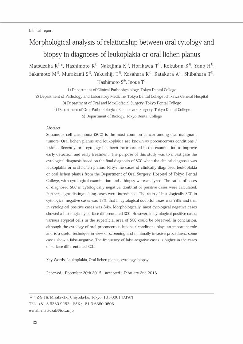
Case 1 (Fig. 1)
Gender: male
Age: 75-year-old
Region: left margin of the tongue to the oral floor
Clinical diagnosis: oral lichen planus
Cytological diagnosis: negative
Pathological diagnosis: squamous cell carcinoma
First visit: April, 2014
History of present illness: aware of haphalgesia 20
years ago, and disappeared a bit later
Date of cytology: April, 2014
Date of biopsy: May, 2014
Induration: negative
Fig. 1: Case of a 75-year-old male clinically diagnosed with oral lichen planus arising from the left margin of the tongue to the oral floor. a: intraoral photograph, b: cytological finding (Original magnification X100), c: histochemical finding (Original magnification X20)
Fig. 2: Case of a 44-year-old female clinically diagnosed with oral lichen planus arising from the right buccal gingiva of the premolar to molar region. a: intraoral photograph, b: cytological finding, c: histochemical finding (Original magnification X20)
Case 2 (Fig. 2)
Gender: female
Age: 44-year-old
Region: right buccal gingiva of the premolar to molar
region
Clinical diagnosis: oral lichen planus
Cytological diagnosis: positive
Pathological diagnosis: squamous cell carcinoma
First visit: April, 2014
History of present illness: smarting pain from 2013
Date of cytology: April, 2014
Date of biopsy: May, 2014
Induration: negative
Matsuzaka et al. Morphological analysis of relationship between oral cytology and biopsy in diagnoses of leukoplakia or oral lichen planus
a
b c
a
b c
JJ S E D P Vol. 8 No. 1: , 2016
25
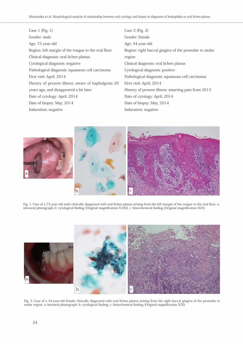
Fig. 3: Case of a 61-year-old female clinically diagnosed with leukoplakia arising from the right margin of the tongue. a: intraoral photograph, b: cytological finding (Original magnification X100), c: histochemical finding (Original magnification X20)
Fig. 4: Case of a 57-year-old female clinically diagnosed with leukoplakia arising from the right margin of the tongue. a: intraoral photograph, b: cytological finding, c: histochemical finding (Original magnification X20)
Case 3 (Fig. 3)
Gender: female
Age: 61-year-old
Region: right margin of the tongue
Clinical diagnosis: leukoplakia
Cytological diagnosis: negative
Pathological diagnosis: squamous cell carcinoma
First visit: November, 2014
History of present illness: white lesion noticed by
primary care stomatologist
Date of cytology: January, 2015
Date of biopsy: February, 2015
Induration: negative
Case 4 (Fig. 4)
Gender: female
Age: 57-year-old
Region: right margin of the tongue
Clinical diagnosis: leukoplakia
Cytological diagnosis: negative
Pathological diagnosis: squamous cell carcinoma
First visit: May, 2014
History of present illness: conscious of white lesion
from 2014
Date of cytology: May, 2014
Date of biopsy: June, 2014
Induration: doubtful
a
b c
a
b c
22 - 28

26
Case 5 (Fig. 5)
Gender: male
Age: 67-year-old
Region: right margin of the tongue
Clinical diagnosis: leukoplakia
Cytological diagnosis: doubtful
Pathological diagnosis: Verrucous carcinoma
First visit: October, 2014
History of present illness: conscious of mass from
September 2014
Date of cytology: October, 2014
Date of biopsy: January, 2015
Induration: negative
Case 6 (Fig. 6)
Gender: male
Age: 54-year-old
Region: left margin to the under-surface of the
tongue
Clinical diagnosis: leukoplakia
Cytological diagnosis: doubtful
Pathological diagnosis: squamous cell carcinoma
First visit: September, 2014
History of present illness: white lesion noticed by
primary care stomatologist in July 2014
Date of cytology: September, 2014
Date of biopsy: September, 2014
Induration: negative
Fig. 6: Case of a 54-year-old male clinically diagnosed with leukoplakia arising from the left margin to the under-surface of the tongue. a: intraoral photograph, b: cytological finding (Original magnification X100), c: histochemical finding (Original magnification X20)
Fig. 5: Case of a 67-year-old male clinically diagnosed with leukoplakia arising from the right margin of the tongue. a: intraoral photograph, b: cytological finding (Original magnification X100), c: histochemical finding (Original magnification X20)
Matsuzaka et al. Morphological analysis of relationship between oral cytology and biopsy in diagnoses of leukoplakia or oral lichen planus
a
b c
a
b c
JJ S E D P Vol. 8 No. 1: , 2016
27
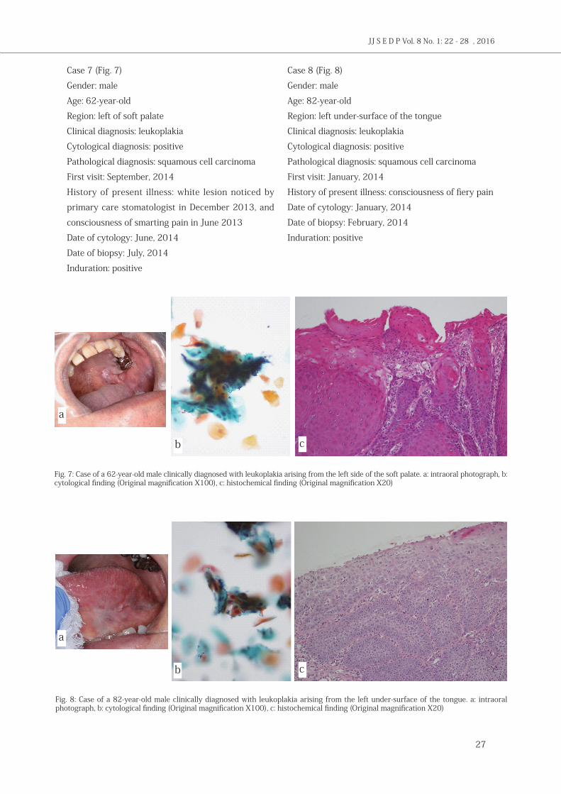
Case 7 (Fig. 7)
Gender: male
Age: 62-year-old
Region: left of soft palate
Clinical diagnosis: leukoplakia
Cytological diagnosis: positive
Pathological diagnosis: squamous cell carcinoma
First visit: September, 2014
History of present illness: white lesion noticed by
primary care stomatologist in December 2013, and
consciousness of smarting pain in June 2013
Date of cytology: June, 2014
Date of biopsy: July, 2014
Induration: positive
Case 8 (Fig. 8)
Gender: male
Age: 82-year-old
Region: left under-surface of the tongue
Clinical diagnosis: leukoplakia
Cytological diagnosis: positive
Pathological diagnosis: squamous cell carcinoma
First visit: January, 2014
History of present illness: consciousness of fiery pain
Date of cytology: January, 2014
Date of biopsy: February, 2014
Induration: positive
Fig. 7: Case of a 62-year-old male clinically diagnosed with leukoplakia arising from the left side of the soft palate. a: intraoral photograph, b: cytological finding (Original magnification X100), c: histochemical finding (Original magnification X20)
Fig. 8: Case of a 82-year-old male clinically diagnosed with leukoplakia arising from the left under-surface of the tongue. a: intraoral photograph, b: cytological finding (Original magnification X100), c: histochemical finding (Original magnification X20)
a
b c
a
b c
22 - 28

28
Discussion
White lesions in the oral cavity are common, and
while most intraoral white lesions are benign, some
are premalignant and/or malignant at the time of
clinical presentation, making it extremely important
to accurately identify and appropriately manage
those lesions 6). It is known that oral lichen planus
is a precancerous condition and that leukoplakia is
a precancerous lesion. The manifestations of oral
lichen planus, which is an inflammatory dermatosis
of the stratified squamous cell epithelium, have
been reported around the world 7, 8). In 1978, the
WHO classified oral lichen planus as a potentially
malignant disorder, since it had been associated with
a significantly increased risk of developing cancer.
Based on a 12-year retrospective study, Bardellini
et al. reported that patients with oral lichen planus
should be followed-up for all their lives by clinicians
for the potential risk of malignant transformation 3). Leukoplakia is a premalignant lesion that has
long been considered to confer an increased risk
for the development of oral cancer 9, 10). Leukoplakia
is a diagnosis made by excluding all other known
diseases or disorders, and may be characterized
by a range of disorders of epithelial renewal and
maturation, such as hyperkeratosis, acanthosis or
epithelial dysplasia 9, 11, 12).
Oral mucosal cytology, which is a minimally-
invasive procedure compared to a biopsy, could
be useful for the detection of oral cancer in oral
mucosal exploration 13). Precancerous lesions or
conditions are necessary to be followed-up for early
detection and early treatment of SCC. The rate of
malignant transformation in leukoplakia has been
reported as 5 to 10 percent, and that in lichen planus
has been reported as a few percent 3). In this study,
the technique of cytology used was liquid-based
cytology. Navone reported about the comparison
of conventional cytology and liquid-based cytology,
which gives better results than conventional
cytology for oral SCC 13). The false-negative rate in
the cytological diagnosis of oral cancer was shown
to exceed 30%, while that of oral precancerous/
dysplasia was found to be 63% 14). Our study
reveals that the false-negative rate, which is the
ratio of histologically SCC in cytologically negative
specimens, is 18%. This investigation found a similar
rate compared with the previous report 14).
In conclusion, although the cytology of
oral precancerous lesions/conditions, such as oral
lichen planus, leukoplakia and erythroplakia, plays
an important role and is a useful technique for
minimally-invasive screening. The frequency of
false-negative cases is higher in the case of surface
differentiated SCC.
References1) Lee JJ, Hong WK, Hittelman WN, Mao L, Lotan R, Shin DM,
Benner SE, Xu XC, Lee JS, Papadimitrakopoulou VM, Geyer C, Perez C, Martin JW, El-Naggar AK, Lippman SM: Predicting cancer development in oral leukoplakia: ten years of translational research, Clin Cancer Res 6:1702-1710, 2000.
2) Hildebrand LD, Carrard VC, Lauxen ID, de Quadros OF, Chaves AC, Sant' Ana-Filho M: Evaluation of cell proliferation rate in non-dysplastic leukoplakias, Med Oral Patol Oral Cir Bucal 15:e328-e334, 2010.
3) Bardellini, E, Amadori F, Flocchini P, Bonadeo S, Majorana A: Clinicopathological features and malignant transformation of oral lichen planus: A 12-year retrospective study, Acta Odontol Scand 71: 834-840, 2013.
4) Verma R, Singh A, Badni M, Chandra A, Gupta S, Verma R: Evaluation of exfoliative cytology in the diagnosis of oral premalignant and malignant lesions: A cytomorphometric analysis, Dent Res J 12: 83-88, 2015.
5) Nanayakkara PGCL, Dissanayaka WL, Nanayakkara BG, Amaratunga EAPD: Camparison of spatula and cytobrush cytological techniques in early detection of oral malignant and premalignant lesions: a prospective and blinded study, Oral Pathol Med 1-7, 2015.
6) Jones KB, Jordan R: White lesions in the oral cavity: clinical presentation, diagnosis, and treatment, Semin Cutan Med Surg 34: 161-170, 2015.
7) Carbone M, Arduino PG, Carrozzo M, Gandolfo S, Argiolas MR, Bertolusso G et al.: Course of oral lichen planus: a retrospective study of 808 northern Italian patients. Rofal Dis 15: 235-243, 2009.
8) Eisen D: The clinical features, malignant potential, and systemic associations of oral lichen planus: a study of 723 patients. J Am Acad Dermatol 46: 207-214, 2002.
9) Kramer IR, Lucas RB, Pindborg JJ Sobin LH: Definition of leukoplakia and related lesions: an aid to studies on oral precancer, Oral Surg Oral Med Oral Pathol 46: 518-539, 1978.
10) Silverman S Jr ed: Oral cancer ED 3. Atlanta, CA: American Cancer Society, 1990.
11) Waldron CA, Shafer WG: Leukoplakia revisited. A clinicopathlogic study 3256 oral leukoplakias, Cancer 36: 1386-1392, 1975.
12) van der Waal I: Potentially malignant disorders of the oral and oropharyngeal mucosa: terminology, classification and present concepts of management. Oral Oncol 45: 317-323, 2009.
13) Navone R: Cytology of the oral cavity: a re-evaluation, Pathlogica 101: 6-8, 2009.
14) Shingh A, Carrol DJ, Mehrotra R: Pitfalls and limitations of oral cytopathology. In: Mehrotra R, ed. Oral cytology: a concise guide, New York: Springer Science+Business Media, 147-156, 2013.
Matsuzaka et al. Morphological analysis of relationship between oral cytology and biopsy in diagnoses of leukoplakia or oral lichen planus