Urinary System Diseases Pathophysiology. Review of Urinary Anatomy & Physiology Located: –Under...
31
Urinary System Diseases Urinary System Diseases Pathophysiology Pathophysiology
-
Upload
linda-farmer -
Category
Documents
-
view
222 -
download
0
Transcript of Urinary System Diseases Pathophysiology. Review of Urinary Anatomy & Physiology Located: –Under...

Urinary System DiseasesUrinary System Diseases
PathophysiologyPathophysiology


Review of Urinary Anatomy & PhysiologyReview of Urinary Anatomy & Physiology
• Located:– Under back muscles
– Behind peritoneum
• Thus: retroperitoneal
– Below level of lowest ribs
– Right lower than left
– Adrenal gland on top of kidney
• Cortex
• Medulla– Contains Pyramids &
Papilla
• Pelvis– Calyx = division of pelvis
• Pleural = calyces

• Bladder– Lined with transitional
epithelium
• Can stretch
– Lined with rugae
– Trigone
• On posterior wall
• Where ureters & urethra open
• Rigid area with NO rugae
– Micturition (voiding, urination)
• Internal urinary sphincter– Involuntary
• External urinary sphincter– Voluntary
• Stretch receptors in bladder wall

• Nephron = functional unit– Consists of:
• Renal Corpuscle
• Renal Tubules
• Renal Corpuscle contains:– Bowman’s capsule
• Part of collecting system
– Glomerulus
• Afferent arteriole
• Efferent arteriole
• Renal Tubules1. Proximal convoluted tubule
2. Loop of Henle
3. Distal convoluted tubule
4. Collecting tubule

• Key point:
– The cortex contains all the structures of the nephron
– The medulla contains only the collecting ducts & the loop of Henle

• Functions of the kidney1. Removes nitrogenous wastes
– Urea– Uric acid– Creatinine– Ammonia
2. Maintains homeostasis– Fluid balance– Electrolyte balance– Acid-base balance
3. Excretory Organ– Via blood filtration &
formation of urine
4. Regulation of Blood Pressure– Juxtaglomerular apparatus– RAA system
» Renin» Angiotensin» Aldosterone
• Urine formation1. Filtration
– Occurs in renal corpuscle
2. Reabsorption– Occurs in proximal
convoluted tubule– Also occurs in distal
convoluted tubule– It takes things back into
blood
3. Secretion– Occurs in distal
convoluted tubule– Blood gives things up to
the urine
4. Concentration– Occurs in collecting
tubules
See next slide


Some Key Points of Renal PhysiologySome Key Points of Renal Physiology
• Nitrogenous wastes primarily come from breakdown of proteins
• Aging & renal function• By age 35, one begins to lose nephrons
• By age 80, one has approx. 30% reduction in nephron capacity
• GFR = glomerular filtration rate– Normal = 125cc/min (7500cc/hour)
– 99% of filtered product is reabsorbed
» Normal urine output = 60cc/hour (1500cc/day)
• All along the duct system water is reabsorbed– Includes the prox. conv. tubule, loop of Henle, distal conv. Tubule, & collecting tubule
– Sodium follows water
• Key elements involved in each process– Reabsorption = H2O(Na), proteins (amino acids), & sugars (glucose)
– Secretion = ions(K+), drugs, ammonia
– Concentration = more reabsorption of H2O

• 2 key factors determine volume of urine produces
1. Glomerular filtration rate (GFR)
– Determined by the unique arrangment of blood vessels
2. Hormonal secretion
– Determined by fluid & electrolyte balance

Volume of urine also controlled by glomerular filtration rate
• Unique arrangment of blood vessels
– Afferent arteriole -----to----capillary bed-----to----efferent arteriole -----to-----capillary bed ----to---- veins
• First capillary bed = glomerular capillaries
• Second capillary bed = peritubular capillaries
• Purpose of this = to control the pressure in the glomerular capillaries & consequently the glomerular filtration pressure
• 3 factors control this:
• (1) autoregulation
• Local feedback from muscle tension in afferent arteriole
• Local feedback from DCT at JGA
• Mediated via endothelial
secretions of glomerular capillaries
• (2) sympathetic nervous system
• (3) renin

• B = increase fluid volume; overhydration; high output heart failure
• C = kidney pathology
• D = hypertension; arteriolar spasm

– Hormones help control the volume of urine via fluid & electrolyte balance
• The concentration factor essentially deals with urine volume– Usually more the volume = more the dilution [a direct proportion]
1. Aldosterone » From adrenal cortex
» Works on distal convoluted tubule
» Causes H2O & Na+ retention
2. Atrial natriuretic hormone(ANH)» From atrial wall of heart
» Works on distal convoluted tubule
» Works in opposition to aldosterone
» Causes H2O & Na+ loss
3. Antidiuretic hormone» From posterior pituitary
» Works on collecting tubules
» Causes reabsorption of H2O (Na+ goes with it)

Diagnostic Tests - UrinalysisDiagnostic Tests - Urinalysis
1. Physical Characteristics & Measurements
– appearance
– color
– odor
– volume
– specific gravity
2. Chemical Measurements
– pH
– protein; glucose
– ketones
– bilirubin; urobilinogen
– leukocytes; nitrite
– blood
3. Microscopic
– cells (wbc, rbc, sperm)
– casts
– crystals
– bacteria
4. Detection of Bacteriuria– nitrite test
• qualitative or screening test– C & S
• Colony Count, if done, make this a quantitative test
• NOTE: Step 4, qualitatively, is done as part of step 2

• Appearance – Clear = normal– Cloudy = ? Infection – If sediment = kidney disease– Dark = ?blood, ?bilirubin,
?concentrated• Color
– Urochrome pigment = yellow• comes from breakdown of
hemoglobin– Concentration
• More Concentrated = Deeper Yellow
– Change of Color From:• Meds
– Vitamin = yellow• Diseases
– Blood = red-brown– Liver = Orange
• Foods– Rhubarb = red-brown
• Odor– Normal = ammonia-like smell
• from breakdown of urea– Unpleasant = ? infection
• Quantity
– Average per 24 hours = 1500 cc• 60 cc per hour
• GFR = 125 cc/min
– Thus, 7500 cc/ hour
• Urine Made Per Hour = 60 cc
• Urine GFR Per Hour = 7500 cc
– KEY: 1 % of filtered urine remains urine; 99 % becomes reabsorbed back into blood
– Oliguria = 100 - 400 cc per day
– Anuria = less than 100 cc per day
– Polyuria = diabetes, nerves, diuretics

• Specific Gravity– Determines Concentration– Compares Test Liquid to H2O– Normal = 1010 - 1030– First AM Specimen = > 1020– In many kidney diseases, one
loses the ability to concentrate urine
– 3 ways to do it:1. Reagent Strip2. Refractometer3. Urinometer
• pH – Determines Acidity or
Alkalinity– Normal = 6.0– Range = 4.5 - 8.0
• Acidity example = diabetes• Alkaline example = UTI
• Protein– OK to have a Trace in the urine
– Benign Conditions:
• exercise
• exposure to cold protein consumption
– Generally Means Kidney Disease
• Glucose – Will only be in urine if exceed Renal
Threshold (160 - 180 mg/dl)
• Ketone (note Acetone is a Ketone)– Ketones are products of Fat
Metabolism
– If cant breakdown Sugars for energy, the body will begin using Fat
– Seen in:
• Uncontrolled Diabetes
• Starvation
• Hi-Fat Diet

• Bilirubin & Urobilinogen Formation
– When used-up RBC’s are broken down by R-E System, a by-product is Bilirubin
– Bilirubin removed from blood by liver & excreted into intestine
– Bacteria in intestine convert Bilirubin into Urobilinogen
– Some Urobilinogen reabsorbed into blood
• Of this amount reabsorbed some my be normally passed in urine
• Bilirubin– Normally None in Urine
– Found in urine if it can’t get from the liver into G-I tract
• From Obstruction of Bile Ducts
– Found in urine if have:
• Liver Disease (hepatitis)
• Blood Disease (hemolysis)
• Urobilinogen – generally follows whatever happens
to bilirubin
– may get none in urine if on antibiotics (destruction of gut flora)
– usually get small amount in urine
• Blood – None is normal
– But may see some if female is menstruating
• Leukocytes– from inflammation of kidney or
lower G-U tract
• Nitrites– screening test for bacteriuria
– bacteria convert nitrate to nitrite

Other Diagnostic TestsOther Diagnostic Tests
• Blood tests• BUN / creatinine• CBC ------ anemia if decreased EPO production• Renin • Antistreptolysin titers
• Urine culture & sensitivity (C&S)• Include colony count
• Imaging• IVP• Retrograde pyelography• CAT/ MRI
• Surgical procedures• Cystoscopy• Biopsy

• Incontinence & retention• UTI’s• Inflammatory disorders• Nephrotic syndrome• Urinary tract obstruction
• Stones• Hydronephrosis• Tumors
– Renal cell carcinoma– Bladder cancer
• Congenital disorders• Polycystic kidneys• Wilm’s tumor (nephroblastoma)
• Renal failure• Acute• Chronic• Dialysis
Urinary Tract Disordersoverall outline

• Urinary Incontinence– Loss of voluntary control of bladder– Frequently called “neurogenic bladder”– Many causes – Enuresis = involuntary control after age 4 or 5– Types:
– Stress– Urge– Overflow
• Urinary retention– Called “residual urine”– Causes :
– Anatomical defects– Neurogenic defects
• Treated with “catheterization”– Foley– French
Incontinence, retention, & catheters

Urinary Tract InfectionsUrethritis; Cystitis; Pyelonephritis
• Etiology
– Ascending infection ----- women > men
– Prostatic hypertrophy with urinary retention
– Incomplete emptying of bladder with urinary stasis
– Pregnancy associated with stasis
– Blood borne pathogens
• Pathophysiology of UTI’s ----- see next slide
• Dx
• Dysuria, urgency, & nocturia
• Systemically get fever & malaise
• CVA tenderness in pyelonephritis
• Note glomerulonephritis is vastly different with regards etiology & pathophysiology

• Note etiologies– Inflammation of
mucosa– Trauma of mucosa– Obstruction– Vesicoureteral reflux– Immobility– Blood-borne
pathogens• TB• HIV• Septicemia

• Glomerulonephritis
– Acute
• Sx = proteinuria, edema, oliguria
• Etiol = 1-2 weeks post strept infect.
– Chronic
• Etiol = autoimmune disease– e.g. lupus, diabetes, hepatitis C
• Can lead to irreversible kidney damage
Inflammatory disorders(1) glomerulonephritis
(2) nephrotic syndrome

• Nephrotic Syndrome– Glomerular disorder where one loses the capacity
to retain protein, especially albumin
– Sx
– severe edema (anasarca)
* can get skin breakdown since impaired arterial flow
– proteinuria
– hypoalbuminemia
– oliguria
– Etiol:
» Toxic agents (lead, mercury)
» Toxic drugs (aminoglycosides)
» Diseases (diabetes, lupus
» Key = any significant problem with
glomerulus can lead
to nephrotic syndrome

• Tumors– Note that primary
symptom = hematuria
– Renal Cell Ca = most common, unilateral, adeno Ca from tubular epithelium
• See picture
– Bladder Ca = usually from transitional epithelium
• Neurogenic bladder
• Renal Calculi– Etiology: Calcium,
Uric acid, Urine crystals
– Symptoms: renal colic, N&V, chills, fever
– Risk factors: prolong dehydration, prolong immobilization, infection
– Treatment: surgery,lithotripsy
• Anomalies– Strictures
– Kinks
– Ptosis
– Pelvic kidney
Obstructive Disorders

Major sites of urinary tract obstruction
– These result in:
– Hydronephrosis
– Hydroureter
• If these conditions exist longer than 2 months get destruction of kidney

Congenital Diseases
• Vesicoureteral reflux • Due to ectopic insertion of ureter into bladder. If far away from
trigone, do not get adequate compression of ureter when voiding & get reflux
• Incidence: 1/1000• If one gets it each sibling(to be) has 50% incidence• Girls> boys; 10:1 ratio
• Ectopic kidney• May get kinking of ureter• Usually in pelvis• Asymptomatic
• Renal agenesis • Usually unilateral & left kidney• 2 types: (1) occurs randomly (2) genetic• Asymptomatic • Remaining kidney becomes large since compensatory hypertrophy
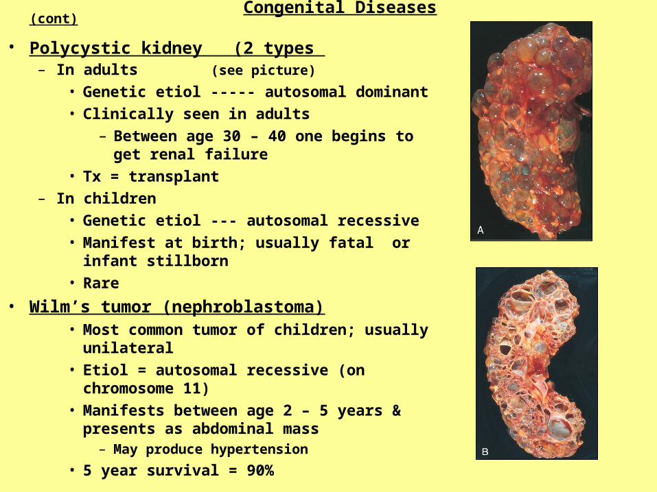
Congenital Diseases (cont)
• Polycystic kidney (2 types – In adults (see picture)
• Genetic etiol ----- autosomal dominant
• Clinically seen in adults
– Between age 30 – 40 one begins to get renal failure
• Tx = transplant
– In children
• Genetic etiol --- autosomal recessive
• Manifest at birth; usually fatal or infant stillborn
• Rare
• Wilm’s tumor (nephroblastoma)• Most common tumor of children; usually
unilateral
• Etiol = autosomal recessive (on chromosome 11)
• Manifests between age 2 – 5 years & presents as abdominal mass
– May produce hypertension
• 5 year survival = 90%

Renal Failure
• Acute renal failure– Abrupt decrease in renal function
• Nitrogenous wastes accumulate– Usually reversible– Sx:
• Oliguria• Drowsiness• Altered levels of consciousness
– Etiol:• Glomerular disease• Severe pyelonephritis• Nephrotoxins that damages
tubular epithelium• Ischemic causes
– shock• ATN (acute tubular necrosis)
» e.g. burns(hgb accumulates)
» e.g. trauma (myoglobin accumulates)
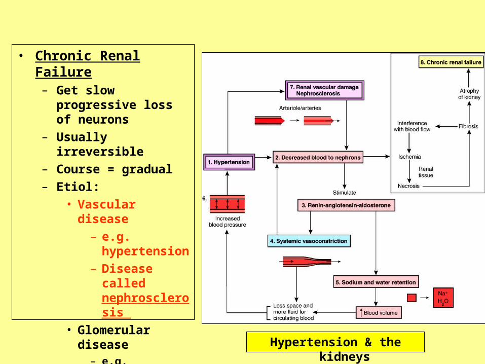
• Chronic Renal Failure– Get slow progressive loss
of neurons
– Usually irreversible
– Course = gradual
– Etiol:
• Vascular disease
– e.g. hypertension
– Disease called nephrosclerosis
• Glomerular disease– e.g. diabetes
• Tubular disease– e.g. toxins
Hypertension & the kidneys

Dialysis in renal failure
• 2 types:– Hemodialysis– Peritoneal dialysis
• Mechanism– Simple diffusion for
wastes & electrolytes
– Osmosis for water balance