Urinary lithiasis 6
65
-
Upload
ahmed-eliwa -
Category
Health & Medicine
-
view
643 -
download
0
Transcript of Urinary lithiasis 6


Urinary Lithiasis

EPIDEMIOLOGY OF RENAL CALCULI• Prevalence 10% to 15%.
• Men > women.
• Highest prevalence is in whites, followed by Hispanics, Asians,and African-Americans.
• Uncommon before age 20 but peaks in fourth to sixthdecades of life.
• A higher prevalence is found in hot and dry climates.
• Directly correlated with body mass index (BMI).

PHYSICOCHEMISTRY

State of Saturation• concentration product is a mathematic expression of the product
of the concentrations of the pure chemical components (ions ormolecules) of the salt. For example, the concentration product(CP) expression for sodium chloride is CP = [Na+][Cl−].
• A pure aqueous solution of a salt is considered saturated when itreaches the point at which no further added salt crystals willdissolve.
• The concentration product at the point of saturation is called thesolubility product, Ksp.

• In urine, despite concentration products of stone formingsalt exceed the solubility product, crystallization does notnecessarily occur because of the presence of crystalizationinhibitors.
• As concentrations of the salt increase further, the point at whichit can no longer be held in solution is reached and crystals form.
• The concentration product at this point is called the formationproduct, Kf.
• The solubility product and the formation product differentiate thethree major states of saturation in urine: undersaturated,metastable, and unstable.

• Nucleation will occur• Inhibitors not generally effective
• Crystal growth will occur• Crystal aggregation will occur• Inhibitors will impede or prevent
crystallization• De novo nucleation is very slow• Heterogeneous nucleation may occur• Matrix may be involved
• Crystals will not form• Existing stones may dissolve
0
Concentrationproduct
Solubilityproduct
Formationproduct

Randall plaque
• Randall (1937) first observed areas of damage associated withsubepithelial plaques on the renal papillae.
• Randall's plaques are soft tissue calcifications found in the deeprenal medulla skirting the surface of the epithelium of the papilla.
• Act as nucleating elements for renal calculi or stones.
• Crystal component of plaque was determined to be calcium apatite(Evan, 2003).

In the photo the Randall plaque can be clearly seen as the white part at the top of the renal papilla. Copyright: X. Carpentier et al., 2010.


Figure 45–3. Endoscopic (A) and histologic (B) images of Randall plaques in calcium oxalatepatients. In A, sites of Randall’s plaques (arrows) appear as irregular white areas beneath theurothelium. In B, a low-magnification light-microscopic image of a papillary biopsy specimen isshown. Sites of calcium deposits were stained black by the Yasue metal substitution method forcalcium histochemistry. (From Evan AP, Lingeman JE, Coe FL, et al. Randall’s plaque of patientswith nephrolithiasis begins in basement membranes of thin loops of Henle. J Clin Invest2003;111:607–16.)

Urolithiasis: Crystal growth
• Homogenous nucleation: crystals precipitate spontaneously(unlikely in urine)
• Heterogenous nucleation: another substance acts as a nidusfor crystal precipitation (likely in urine)
• Epitaxy: Precipitation of one crystal on the surface of another

Inhibitors of crystallization and aggregation• Pyrophosphates
• Diphosphonates
• Citrate
• Some cations (e.g. Mg+2)
• Nephrocalcin
• Uropontin
• Tamm-Horsfall protein

Matrix
• Renal calculi consist of both crystalline and noncrystallinecomponents.
• The noncrystalline component is termed matrix, which typicallyaccounts for about 2.5% of the weight of the stone.
• In some cases, matrix comprises the majority of the stone (up to65%), usually with chronic urinary tract infection.
• Among the proteins incorporated into the matrix substance areTamm-Horsfall protein, nephrocalcin, a γ-carboxyglutamic acid–rich protein, renal lithostathine, albumin, glycosaminoglycans,free carbohydrates, and a mucoprotein called matrix substanceA.

Urolithiasis: Naming of stones
• When >70% of the urolith is composed of one type of crystal it is named for the crystal
• Mixed urolith < 70% one crystal; no identifiable nidus or shell
• Compound urolith Identifiable nidus of one crystal with surrounding layers of another crystal
• Matrix urolith Matrix without appreciable crystalloid

Classification of stones1- X-ray characteristics
• Radiolucent• Poor radiopaque• Radiopaque
• Uric acid• Ammonium urate• Xanthine• 2,8-
dihydroxyadenine• ‘Drug-stones’
• Magnesium ammonium phosphate
• Apatite• Cystine
• Calcium oxalate dihydrate
• Calcium oxalate monohydrate
• Calcium phosphates

2- Aetiology of stone formation
‘Drug stones’Genetic causesInfection stones
Non-infection stones
Substances impairing urine composition• Acetazolamide• Allopurinol• Aluminiummagnesium hydroxide• Ascorbic acid• Calcium• Furosemide• Laxatives• Methoxyflurane• Vitamin D
Active compounds crystallizing in urine• Allopurinol / oxypurinol• Amoxicillin / ampicillin• Ceftriaxone• Ciprofloxacin• Ephedrine• Indinavir• Magnesium trisilicate• Sulfonamide• Triamterene
• Cystine• Xanthine• 2,8-
dihydroxyadenine
• Magnesium ammonium phosphate
• Apatite• Ammonium
urate
• Calcium oxalates
• Calcium phosphates
• Uric acid

3- Stone composition (mineralogy)
MineralChemical composition
whewelliteCalcium oxalate monohydrate
wheddeliteCalcium-oxalate-dihydrate
uriciteUric acid dihydrate
Ammonium urate
struviteMagnesium ammonium phosphate
dahlliteCarbonate apatite (phosphate)
brushiteCalcium hydrogenphosphate
Cystine
Xanthine
2,8-dihydroxyadenine
‘Drug stones’

4-Diagnostic Classification of Nephrolithiasis
PREVALENCE (%)CONDITION
20-40 %Absorptive hypercalciuria
5-8 %Renal hypercalciuria
3-5 %Resorptive hypercalciuria
10-40 %Hyperuricosuric calcium nephrolithiasis
10-50 %Hypocitraturic calcium nephrolithiasis
2-15 %Hyperoxaluric calcium nephrolithiasis
5-10 %Hypomagnesiuric calcium nephrolithiasis
15-30 %Gouty diathesis
< 1 %Cystinuria
1-5 %Infection stones
10-50 %Low urine volume
< 3 %Miscellaneous or no abnormality

•High risk stone formers1-General factors• Early onset of urolithiasis in life (especially children and
teenagers)
• Familial stone formation
• Brushite containing stones (calcium hydrogen phosphate;CaHPO4 . 2H2O)
• Uric acid and urate containing stones
• Infection stones
• Solitary kidney (The solitary kidney itself does not have aparticular increased risk of stone formation, but the preventionof a potential stone recurrence is of more importance)

2-Diseases associated with stone formation
• Hyperparathyroidism
• Nephrocalcinosis
• Gastrointestinal diseases or disorders (i.e. jejuno-ileal bypass,intestinal resection, Crohn’s disease,
• malabsorptive conditions)
• Sarcoidosis

3-Genetically determined stone formation• Cystinuria (type A, B, AB)
• Primary hyperoxaluria (PH)
• Renal tubular acidosis (RTA) type I
• 2,8-dihydroxyadenine
• Xanthinuria
• Lesh-Nyhan-Syndrome
• Cystic fibrosis

4-Anatomical and urodynamic abnormalities associated withstone formation
• Medullary sponge kidney (tubular ectasia)
• Ureteropelvic junction (UPJ) obstruction
• Calyceal diverticulum, calyceal cyst
• Ureteral stricture
• Vesico-uretero-renal reflux
• Horseshoe kidney
• Ureterocele
• Urinary diversion (via enteric hyperoxaluria)
• Neurogenic bladder dysfunction

A-Calcium Stones
• Hypercalciuria is the most common abnormality identified incalcium stone formers.
• The normal kidney filters approximately 270 mmol of calcium dailyand reabsorbs all but 4 mmol.
• Definition: Parks and Coe (1986) defined hypercalciuria as excretionof greater than 4 mg/kg/day or greater than 7 mmol/day in menand 6 mmol/day in women.
• Historically, the term idiopathic hypercalciuria was applied to stoneformers for whom classification of their metabolic abnormality wasdifficult.
• Types: 1-Absorptive 2-Renal 3-Resorptive
a-Hypercalciuria

1-Absorptive Hypercalciuria(AH).• Defined as increased urinary calcium excretion (>0.2 mg/mg
creatinine) after an oral calcium load.
• Type I: when urinary calcium remains high despite a low calciumdiet (400 mg dietary calcium daily).
• Type II: when urinary calcium normalizes with a restricted calciumintake.
• The added systemic load of calcium due to intestinal calciumhyperabsorption results in a transient increase in serum calcium,which suppresses serum PTH and results in increased renalfiltration of calcium, ultimately leading to hypercalciuria.

2-Renal Hypercalciuria
• In renal hypercalciuria, impaired renal tubular reabsorptionof calcium results in elevated urinary calcium levels leading tosecondary hyperparathyroidism.
• High fasting urinary calcium levels (>0.11 mg/dL glomerularfiltration) with normal serum calcium values are characteristicof renal hypercalciuria.
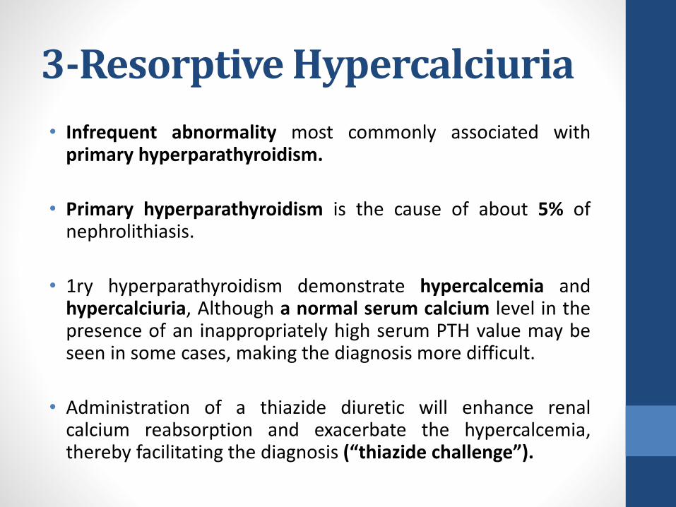
3-Resorptive Hypercalciuria
• Infrequent abnormality most commonly associated withprimary hyperparathyroidism.
• Primary hyperparathyroidism is the cause of about 5% ofnephrolithiasis.
• 1ry hyperparathyroidism demonstrate hypercalcemia andhypercalciuria, Although a normal serum calcium level in thepresence of an inappropriately high serum PTH value may beseen in some cases, making the diagnosis more difficult.
• Administration of a thiazide diuretic will enhance renalcalcium reabsorption and exacerbate the hypercalcemia,thereby facilitating the diagnosis (“thiazide challenge”).

3-Resorptive Hypercalciuria
• Suspicious in patients with nephrolithiasis and serum calciumlevels greater than 10.1 mg/dL.
• Measurement of serum ionized calcium may help in equivocalcases.
• Urinary cyclic AMP levels have been used to diagnose primaryhyperparathyroidism because PTH promotes release of cyclicAMP from the kidney, leading to increased urinary cyclic AMPlevels. However, this test has been largely abandoned in favorof direct measurement of serum PTH.

Sarcoid and Granulomatous Disease
• Production of 1,25(OH)2D3 from 1-α hydroxylase present inmacrophages of the sarcoid granuloma, causing increasedintestinal absorption of calcium, hypercalcemia, andhypercalciuria.
• Sarcoidosis can also be differentiated from other diagnoses bythe rapid resolution of hypercalcemia with initiation ofcorticosteroid therapy.

Malignancy-Associated Hypercalcemia
• Malignancy is the main cause of hypercalcemia in hospitalizedpatients.
• An assay for intact PTH can help distinguish patients withhyperparathyroidism from those with other causes of hypercalcemia.
• Tumors in patients with humoral hypercalcemia produce a PTHrelatedprotein (PTHrP) whose production is regulated by calcium sensingreceptors on the cell surface.
• Lung and breast cancers account for about 60% of malignancy-associated hypercalcemia, whereas renal cell (10% to 15%), head andneck (10%), and hematologic cancers such as lymphoma and myeloma(10%) account for the rest.

Glucocorticoid-Induced Hypercalcemia
• Glucocorticoids can significantly alter calcium metabolism throughtheir actions on bone, intestine, and parathyroid glands.
• Their most potent effect is related to calcium metabolism in bones,where glucocorticoids promote bone resorption and reduce boneformation, ultimately leading to osteopenia with chronic use.
• Additionally, they stimulate release of PTH.
• On the other hand, glucocorticoids inhibit intestinal absorption ofcalcium, which accounts for their effectiveness in preventinghypercalciuria induced by sarcoidosis.

b-Hyperoxaluria
• Defined as urinary oxalate greater than 40 mg/day.
• leads to increased urinary saturation of calcium oxalate andsubsequent promotion of calcium oxalate stones.
• Additionally, oxalate has been implicated in crystal growth andretention by means of renal tubular cell injury.
• Causes of hyperoxaluria include
1. Primary hyperoxaluria: disorders in biosynthetic pathways
2. Enteric hyperoxaluria: Intestinal malabsorptive states
3. Dietary hyperoxaluria: Excessive dietary intake or highsubstrate levels (vitamin C)

Primary Hyperoxaluria• A rare autosomal recessive disorder in glyoxylate metabolism by
which the normal conversion of glyoxylate to glycine is prevented.
• Type 1: The primary enzyme catalyzing glyoxylate conversion toglycine is alanine/glyoxylate aminotransferase, which issynthesized in the liver.
• Type2: a defect in glyoxylate reductase/hydroxypyruvatereductase in the liver, less aggressive course with regard to renalfailure than PH1.
• Inevitably leads to end stage renal failure, which occurs by age 15in 50% of affected patients and is associated with an overall deathrate of approximately 30%.
• Combined liver kidney transplant is accepted treatment.

Enteric Hyperoxaluria• The most common cause of acquired hyperoxaluria.
• Malabsorption of any cause can lead to increased intestinalabsorption of oxalate.
• Fat malabsorption results in saponification of fatty acids withdivalent cations such as calcium and magnesium, thereby reducingcalcium oxalate complexation and increasing the pool of availableoxalate for reabsorption.
• As such diarreal diseases, small bowel resection intrinsic disease,and jejunoileal bypass have all been associated with hyperoxaluria

Dietary Hyperoxaluria• Overindulgence in oxalate-rich foods such as nuts, chocolate,,
spinach, and strawberries can result in hyperoxaluria in otherwisenormal individuals.
• Increased animal protein can also increase urinary levels ofcalcium and oxalate.
• Severe calcium restriction.
• Recent studies have also implicated Oxalobacter formigenes, anoxalate-degrading intestinal bacterium, as a potential modulator ofintestinal oxalate levels (Duncan et al, 2002).

c-Hyperuricosuria• Defined by urinary uric acid exceeding 600 mg/day.
• Up to 10% of calcium stone formers have high urinary uric acidlevels as the only abnormality.
• Hyperuricosuria increases urinary levels of monosodium urate,which in turn promotes calcium oxalate stone formation throughheterologous nucleation.
• Uric acid may also reduce the effectiveness of naturally occurringinhibitors of crystallization

d-Hypocitraturia• Defined as a urinary citrate level less than 320 mg/day.
• Hypocitraturia is an important and correctable abnormalityassociated with nephrolithiasis
• Exists as an isolated abnormality in up to 10% of calcium stoneformers and is associated with other abnormalities in 20% to 60%.
• Metabolic acidosis reduces urinary citrate levels secondary toenhanced renal tubular reabsorption and decreased synthesis ofcitrate in peritubular cells.
• Causes: Distal renal tubular acidosis (RTA), Chronic diarrheal States,Diuretics such as thiazides, Enalapril and strenuous exercise.

e-Low Urine pH• At pH (<5.5), the undissociated form of uric acid predominates,
leading to uric acid and/or calcium stone formation.
• Calcium oxalate stones form as a result of heterologousnucleation with uric acid crystals.
• Chronic metabolic acidosis can lead to low urine pH,hypercalciuria, and hypocitraturia.
• Acidosis increases bone resorption and produces renal calciumleak.
• “Gouty diathesis” refers to a stone-forming propensitycharacterized by low urine pH of unknown etiology with orwithout associated gouty arthritis

f-Hypomagnesuria• A rare cause of nephrolithiasis, affecting less than 1% of stone
formers as an isolated abnormality, with other abnormalities in 6%to 11% of cases.
• Magnesium complexes with oxalate and calcium salts, andtherefore low magnesium levels result in reduced inhibitoryactivity.
• Low urinary magnesium is also associated with decreased urinarycitrate levels, which may further contribute to stone formation.
• Low magnesium levels occur with poor dietary intake or as a resultof reduced intestinal absorption associated with intestinalabnormalities producing chronic diarrheal syndrome

g-Renal Tubular Acidosis• Clinical syndrome characterized by metabolic acidosis resulting
from defects in renal tubular hydrogen ion secretion orbicarbonate reabsorption.
• There are three types of RTA: 1, 2, and 4.
• Type 1 (distal) RTA is of particular significance to urologists notonly because it is the most common form of RTA but alsobecause it is the form of RTA most frequently associated withstone formation.

Type 1 (Distal) RTA.• Syndrome of abnormal collecting duct function characterized by
inability to acidify the urine in the presence of systemic acidosis.
• Distal RTA occurs as a consequence of dysfunction of the α-typeintercalated cells, which secrete protons into the urine via anapical H+ ATPase.
• The classic findings include hypokalemic, hyperchloremic, non–anion gap metabolic acidosis along with nephrolithiasis,nephrocalcinosis, and elevated urine pH (>6.0).
• Incomplete RTA Demonstrate failure to lower urine pH below 5.5after an acid load, but they do not manifest metabolic acidosisand consequently have normal serum electrolytes.

α-intercalcated cells
HCo3-
H+
K+
K+/H+ – ATPase
H+ – ATPase
ATP6V1B1
H+
SLC4AE1
BloodLumen
H2o+Co2
H2Co3
HCo3-+H+
ATP6V0A4
CA II3CI–
Mechanism of acidification in the collecting duct α-intercalated cell. Enzymes areshown in red and their corresponding genes in italics. The α-intercalcated cells secreteH+ into the lumen of the distal tubule and collecting duct by way of an apical H+ATPase and possibly an H+/K+ ATPase exchanger. Bicarbonate is transported into theblood via a Cl−/HCO3 − anion exchanger on the basolateral membrane. Defects in theAE1 Cl−/ HCO3 − anion exchanger or the H+ ATPase result in failure to acidify theurine in distal renal tubular acidosis.

Type 1 (Distal) RTA
• The metabolic acidosis promotes bone demineralization, whichleads to secondary hyperparathyroidism and hypercalciuria.
• Commonly present as adults with symptoms of nephrolithiasis
• The most common stone composition is calcium phosphate as aresult of hypercalciuria, hypocitraturia, and increased urinary pH.
• Profound hypocitraturia, perhaps the most important factor instone formation.

Type 1 (Distal) RTA• Distal RTA is a heterogeneous disorder that may be hereditary,
idiopathic, or acquired.
• Autosomal recessive distal RTA also tends to occur earlier in life andis additionally associated with mental retardation and deafness.
• Secondary distal RTA in sporadic cases is commonly associated withautoimmune diseases such as Sjogren syndrome and systemic lupuserythematosus.
• Secondary RTA is also associated with obstructive uropathy,pyelonephritis, acute tubular necrosis, hyperparathyroidism, andidiopathic hypercalciuria.

Type 2 (Proximal) RTA• Defect in HCO3 − reabsorption associated with initial high urine pH that
normalizes as plasma HCO3 − decreases and the amount of filteredHCO3− falls.
• With reduced capacity of the proximal tubule to reclaim filtered HCO3−,more HCO3 − is delivered to the distal tubule, which has a limitedcapacity for bicarbonate reabsorption.
• Consequently, bicarbonaturia ensues, resulting in reduced net acidexcretion and metabolic acidosis.
• As the filtered HCO3 − load declines with progressive metabolic acidosis,less bicarbonate reaches the distal tubule, until eventually the capacityof the distal tubule is sufficient to handle the load and no furtherbicarbonate is lost.
• At steady state, serum HCO3 − is low (15 to 18 mEq/L) and urine pH isacid (<5.5).

Type 2 (Proximal) RTA• Generalized defects in proximal tubule function similar to
Fanconi syndrome, with loss of glycogen, protein, uric acid, andphosphate.
• Nephrolithiasis is uncommon due to normal urinary citrate.
• Most cases are sporadic, but inherited diseases have beendescribed.
• present as Growth retardation and hypokalemia in children dueto metabolic acidosis.
• Metabolic bone disease is seen more frequently due toassociated abnormalities in vitamin D metabolism andhypophosphatemia.

Type 4 (Distal) RTA.• Associated with chronic renal damage as interstitial renal disease
and diabetic nephropathy.
• Reduction in glomerular filtration results in hyperkalemic,hyperchloremic metabolic acidosis due to loss of HCO3 − in theurine and decreased excretion of ammonium.
• Aldosterone resistance is commonly occurs results in decreasedammonia generation and further exacerbates hyperkalemia.
• Nephrolithiasis is uncommon due to reduced renal excretion ofcalcium and uric acid owing to impaired renal function

•Ca Oxalate stones

Calcium oxalate monohydrates
Calcium oxalate dihydrates

B-Uric Acid Stones• Uric acid is a weak acid with a pKa of 5.35 at 37° C. At that pH,
half of the uric acid is present as the urate salt and half as freeuric acid.
• Because sodium urate is approximately 20 times more solublethan the free acid, the relative proportion present as free uricacid strongly determines the risk of stone formation.
• Urine pH is a critical factor in determining uric acid solubility.
• The three main determinants of uric acid stone formation are lowpH, low urine volume, and hyperuricosuria (Fig. 45–11).
• The most important pathogenetic factor is low urine pH.

Uric acid nephrolithiasis
Low urine volume Low urinary pH Hyperuricosuria
Diarrhealstates
High animalprotein diet
Primary gout
Congenitaldisorders
Uricosuricmedications
Myeloproliferativedisorders
Obesity Insulin resistance
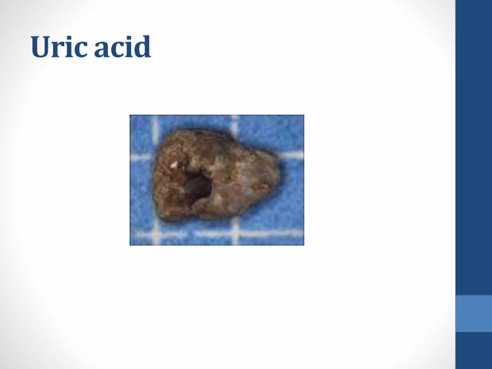
Uric acid

C-Cystine Stones• Cystinuria is an inherited autosomal recessive disorder characterized by
a defect in intestinal and renal tubular transport of dibasic amino acids.
• Types: type A (chromosome 2), type B (chromosome 19), and type AB(both chromosomes).
• Although the defect also results in high urinary concentrations of lysine,ornithine, and arginine, the poor solubility of cystine leads to stoneformation.
• Cystine is a dimer, composed of two cysteine molecules linked via adisulfide bond.
• Cystine is much less soluble than cysteine and is responsible for cystinestone formation.

Cystine Stones• Cystine stones are rare, only 1 in 1000 to 1 in 17,000.
• In children, cystinuria is the cause of up to 10% of all stones.
• Several factors determine the solubility of cystine including cystineconcentration, pH, ionic strength, and urinary macromolecules.
• The main contributor to cystine crystallization is supersaturationbecause no specific inhibitor of cystine crystallization in the urine.
• The solubility of cystine is highly pH dependent, with solubilities of300 mg/L, 400 mg/L, and 1000 mg/L at pH levels of 5, 7, and 9,respectively

Cystine

D-Infection Stones• Infection stones are composed primarily of magnesium
ammonium phosphate hexahydrate (MgNH4PO4 •6H2O) butmay in addition contain calcium phosphate in the form ofcarbonate apatite (Ca10[PO4]6 • CO3).
• Struvite stones (magnesium ammonium phosphate) occuronly in association with urinary infection by urea-splittingbacteria.

Epidemiology• 5% to 15% of all stones.
• However, struvite/carbonate apatite was the most commonstone composition among African-American stone formers.
• Women > men by a ratio of 2 : 1
• Other populations at risk of recurrent infection include theelderly, premature infants or infants born with congenitalurinary tract malformation, diabetics, and those with urinarystasis as a result of urinary tract obstruction, urinary diversion,or neurologic disorders.

Pathogenesis• Urealysis provides an alkaline urinary environment and sufficient
concentrations of carbonate and ammonia to induce theformation of infection stones.
• Because urease is not present in sterile human urine, infection withurease-producing bacteria is a prerequisite for the formation ofinfection stones.
• Urea is hydrolyzed to ammonia and carbon dioxide by bacterialurease:
(NH2 )2CO+H2O NH3 + CO2
• The alkaline urine that results from this reaction (pH 7.2 to 8.0)favors the formation of ammonium:
NH3 + H2O NH4+ +OH− pK = 9.0

Pathogenesis• The dissociation of hydrogen phosphate under alkaline conditions
provides phosphate:
H2PO4- H+ + HPO4
-2 pK= 7.2
HPO4-2 H+ + PO4
-3 pK= 12.4
• This chemical cascade, along with physiologic concentrations ofmagnesium, provides the constituents necessary for precipitationof struvite.
• In addition, the concentrations of calcium, phosphate, andcarbonate allow precipitation of carbonate apatite andhydroxyapatite, thereby comprising the components of infectionstones.

Bacteriology• The most common urease-producing organisms are Proteus,Klebsiella, Pseudomonas, and Staphylococcus species.
• Proteus mirabilis the most common organism associated withinfection stones.
• Although Escherichia coli is a common cause of urinary tractinfections, only rare species of E. coli produce urease.
• Bacteria damages the mucosal layer of the urinary tract, resultingin both increased bacterial colonization and crystal adherence.

Struvite

F-Miscellaneous Stones1.Xanthine and Dihydroxyadenine Stones:• Inherited disorder in the enzyme xanthine dehydrogenase (XDH) or
xanthine oxidase, which catalyzes the conversion of xanthine to uricacid.
• Allopurinol, which inhibits XDH and is consequently used to treathyperuricemia and hyperuricosuria, can, at high levels, predispose toxanthine stones.
• Adenine phosphoribosyl transferase deficiency is a rare metabolicabnormality presenting with 2,8 dihydroxyadenine urolithiasis.
• 2,8 dihydroxyadenine urolithiasis are characteristically radiolucent andtherefore need to be differentiated from uric acid stones which arealso radiolucent and have identical chemical reactivity.
• Xanthine stones, 2,8-DHA stones are extremely insoluble at any pH.

2-Ammonium Acid Urate Stones:• Ammonium acid urate stones represent less than 1% of all
stones.
• In developing countries, however, endemic ammonium acidurate urolithiasis is still observed because it comprises bladdercalculi in children.
• Conditions associated with ammonium acid urate crystallizationinclude laxative abuse, recurrent urinary tract infection,recurrent uric acid stone formation, and inflammatory boweldisease.

• Patients with ileostomy after colectomy have markedly reducedurinary volume, pH, and sodium and are not prone tohyperoxaluria like other individuals with bowel disease becausethe colon is the main site of dietary oxalate absorption.
• As such, these patients are prone to ammonium acid urate anduric acid stones rather than calcium oxalate stones.
• Laxative abuse has been postulated to be the result ofdehydration due to gastrointestinal fluid loss causingintracellular acidosis and enhanced ammonia excretion.
• Because urinary sodium is low in the setting of laxative use,urate complexes with abundant ammonia, thereby leading tourinary supersaturation of ammonium acid urate.

Thank you