Urethral catheterization – male dog
33
Urethral catheterization Dr Amit Singla Surgery & Radiology, DGCN COVAS, CSKHPKV, Palampur Himachal Pradesh (INDIA)
-
Upload
vikash-babu-rajput -
Category
Education
-
view
462 -
download
2
Transcript of Urethral catheterization – male dog

Urethral catheterization
Dr Amit SinglaSurgery & Radiology,DGCN COVAS, CSKHPKV,PalampurHimachal Pradesh (INDIA)

Male Dog Indications/Use
• To collect urine for urinalysis
• To empty the urinary bladder
• To administer radiographic contrastmedia into the lower urinary tract

Indwelling
• To maintain constant, controlledbladder drainage
• To maintain a patent urethra
• To monitor urine output
• To assist with the nursing care of therecumbent patient and of the patientthat is unable to urinate voluntarily

Contraindications
• Pre-existing urethral trauma
• Large space-occupying urethral mass orurethral stricture
• Urine collected by catheterization is notsuitable for microbiology, as the samplemay be contaminated. Cystocentesis ispreferred

Equipment
• Catheters
• Sterile soft gauze swabs
• Gloves
• Sterile aqueous lubricant
• Plain sample pot or Kidney Tray
• 5–10 ml syringe

The relative size of a Foley catheter is described using French units (F). The most common sizes are 10 F to
28 F. 1 F is equivalent to 0.33 mm

If the catheter is to be indwelling
• For silicone Foley catheters, water sufficient tofill balloon
• For standard flexible nylon (polyamide)catheters: suture materials; Adhesive tape
• Sterile intravenous fluid administration set andempty fluid bag or commercial closed urinecollection system, with appropriate adaptersfor attachment to selected urinary catheter
• Elizabethan collar

Patient preparation and positioning
• Gentle physical restraint
• Sedation
• General anaesthesia
• Lateral recumbency,
• The prepuce should be cleaned with theantiseptic solution,
• The area around the prepuce can beclipped, especially in long-hairedbreeds.


Sample handling
For urinalysis:
• Approximately 5 ml is required
• Samples should be collected into a plaintube.

Potential complications
• Trauma to the urethra or urinarybladder
• Iatrogenic urinary tract infection

Urethral catheterization –Bitch

Patient preparation and positioning
• Bitches may require sedation or General anaesthesia.
• The patient may be positioned in dorsal recumbencywith the hind-limbs held cranially (for directvisualization)
• In lateral recumbency (right lateral recumbency for aright handed operator; left lateral recumbency for aleft-handed operator) (for digital palpation)
• In sternal recumbency with the hind-limbs over theedge of the table.
• Clean the vulva with the antiseptic to remove anydischarge and surface dirt.
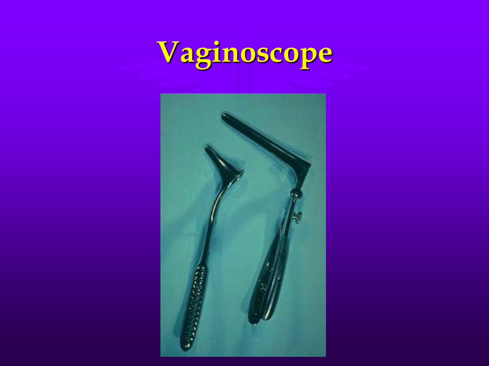
Vaginoscope

TechniqueDirect visualization of urethral orifice

Urethral catheterization –Horse
• Bladder catheterization in male horses isaccomplished using a male equine urinarycatheter (stallion catheter).
• The glans penis should be cleaned withchlorhexidine scrub and sterile irrigationsolution and the catheter handled usingsterile technique.
• A small amount of sterile lubricant mayfacilitate catheter passage.
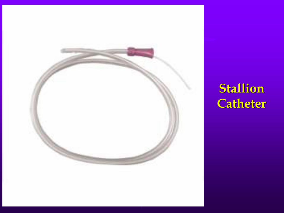
Stallion Catheter


Urethral catheterization –Mare
• cleaning the perineum with chlorhexidine scruband sterile irrigation solution.
• A sterile gloved hand should be placed into thevaginal vault and the index finger advanced intothe opening of the urethra (located on the floor ofthe vaginal vault).
• The tip of the catheter should pass under the indexfinger, and the catheter should advance through theinternal sphincter into the bladder until urineflows.

Mare Catheter


Urinary Catheters in Adult Cattle

Urethral Retrograde Urohydropulsion In Male Dog
• Indications/Use
• To flush uroliths lodged in the urethrainto the bladder, to restore urethralpatency. Uroliths flushed into the bladderlumen can then be removed via cystotomy.
• Urethral obstruction with uroliths shouldbe confirmed with radiography prior toperforming retrograde urohydropulsion.
Equipment
• Flexible nylon urinary catheters are preferred to Foleycatheters for urohydropulsion
• Hypodermic needles: 21 G, 1.5 inch
• 3-way taps
• Intravenous extension tubing
• 20–35 ml syringes
• Sterile intravenous fluid administration set and emptyfluid bag or commercial closed urine collection system
• Sterile isotonic fluids (e.g. saline, lactated Ringer’ssolution)
• Sterile water-soluble lubricant

Technique1. Decompress the bladder if overdistended by cystocentesis.
2. Insert a lubricated large-bore male dog urinary catheter intothe urethra
3. Instil 3–8 ml of the local anaesthestic around the uroliths.
The tip of the catheter should remain distal to the uroliths.
Never attempt to force uroliths retrograde with the tip of thecatheter.
4. Insert a gloved index finger into the rectum and occlude theurethral lumen by compressing the urethra against the floorof the bony pelvis.
5. With a moistened gauze swab, occlude the distal urethra bycompressing the distal tip of the penis around the urinarycatheter.

6. Fill a large syringe with sterile isotonicsolution. As a guide, the normal bladder willaccommodate approximately 7–11 ml/kgbodyweight, but this volume is most oftennot required.
7. Attach the syringe to the urinary catheter.
8. Push sterile isotonic solution into the urethra,with the goal of dilating the urethral lumenaround the uroliths.
9. Once the urethra is dilated, immediatelyrelease digital compression of the pelvicurethra.


11. Continue flushing fluid through the urinary catheterand urethral lumen to propel uroliths into the urinarybladder. Repeated occlusion of the pelvic urethra andflushing of the urethra may be required. Use cautionnot to overdistend the bladder lumen with fluid.Palpate the bladder regularly to check foroverdistension and repeat bladder decompression ifrequired.
12. Confirm successful retrograde urohydropulsion ofuroliths into the bladder by retrograde urethrography.
13. If the animal is not taken to surgery immediately forremoval of uroliths from the bladder, placement of anindwelling urethral catheter is recommended pendingsurgery, to maintain urine flow.

Preventable causes of failure of retrograde urohydropropulsion
Technique factors:
• Inadequate pressure and/or insufficient urethral dilation
• Urinary catheter size too small
• Saline volume too small
• Insufficient pressure applied to syringe plunger
• Lack of complete digital occlusion of urethra
• Resistance to retrograde flushing caused by over-distended bladder
• Lack of coordination of individuals performing technique
• Inadequate pharmacologic control of patient discomfort
• Discontinuation of procedure before stones are flushed into bladder lumen

Patient factors:
• Discomfort resulting in impaired ability toproperly restrain the patient
• Rough surface of uroliths (silica; calciumoxalate dihydrate) impairing attempts tomove them into the bladder lumen
Potential complications
• Urethral rupture (rare)
• Urinary tract infection

Thanks