Ureteral protection during microwave ablation of renal...
4
Ureteral protection during microwave ablation of renal cell carcinoma: combined use of pyeloperfusion and hydrodissection Katayoun Samadi Ronald S. Arellano 388 Diagn Interv Radiol 2018; 24:388–391 © Turkish Society of Radiology 2018 INTERVENTIONAL RADIOLOGY TECHINCAL NOTE ABSTRACT A 56-year-old female with past medical history of thrombotic microangiopathy presented to her physician with nonspecific abdominal pain. A magnetic resonance imaging scan was obtained, which revealed a 3.1 cm mass arising from medial lower pole of the left kidney that was sub- sequently shown to be renal cell carcinoma by percutaneous biopsy. Because of her history of thrombotic microangiopathy and other comorbidities, she was deemed a nonsurgical candidate and was therefore referred to interventional radiology for thermal ablation. Computed tomogra- phy (CT)-guided microwave ablation was performed with the combined use of pyeloperfusion and hydrodissection for maximal ureteral protection. Follow-up unenhanced CT scan obtained one month after ablation showed a normal collecting system without evidence of hydronephro- sis or urinoma. You may cite this article as: Arellano RS, Samadi K. Ureteral protection during microwave ablation of renal cell carcinoma: combined use of pyeloperfusion and hydrodissection. Diagn Interv Radiol 2018; 24:388–391. From the Division of Interventional Radiology, Department of Radiology (R.S.A rarellano@ mgh.harvard.edu) Massachusetts General Hospital, Boston, Massachusetts, USA. Received 5 April 2018; revision requested 30 April 2018; last revision received 1 June 2018; accepted 14 June 2018. DOI 10.5152/dir.2018.18137 P ercutaneous image-guided thermal ablation is a widely accepted treatment option for renal tumors (1). Of the thermal ablation methods currently available, including microwave ablation (MWA), cryoablation, and radiofrequency ablation (RFA), MWA has potential advantages of shortest treatment time and higher thermal efficacy, and it is less affected by heat sink phenomenon compared with RFA and cryoablation (2). Nontarget organ injury is one of the most dreaded complications of any thermal ablation procedure. Given the anatomical location of kidneys, structures at risk of nontarget thermal injury can include the duodenum, colon, small bowel, pancreas, adrenal gland, psoas muscle, and ureter (3). To help lessen the risk of injury to these structures during renal ablations, sever- al adjunctive maneuvers including gas insufflation, electrode torqueing, hydrodissection, and ureteral pyeloperfusion have been described (4, 5). Hydrodissection describes the process of physically displacing structures away from renal tumors, while pyeloperfusion is used to cool or maintain body temperature within the ipsilateral ureter during thermal ablations of tumors that arise from the medial lower pole and keep ureter from untargeted thermal injury. In this report, we present a case of CT-guided MWA of a renal cell carcinoma for which the combined use retrograde pyeloperfusion for ureteral cooling and hydrodissection for displacement of the ureter from the tumor was performed to minimize the risk of thermal injury. Technique Our hospital does not require Institutional Review Board approval for retrospective case reports. A 56-year-old female with a complex medical history of thrombotic microangiop- athy which required chronic anticoagulation, anemia, and chronic renal failure presented with nonspecific abdominal pain. An informed consent was obtained from the patient. MRI revealed left renal lower pole 3.1 cm tumor abutting the proximal left ureter (Fig. 1). Subse- quent CT-guided biopsy confirmed clear cell renal cell carcinoma. She was not considered a suitable surgical candidate due to her medical comorbidities and was therefore referred to Interventional Radiology for percutaneous thermal ablation. Percutaneous MWA was chosen over RFA and cryoablation due to the potentially relative- ly short treatment time and exposure of the left ureter to thermal energy compared with
-
Upload
nguyenliem -
Category
Documents
-
view
216 -
download
0
Transcript of Ureteral protection during microwave ablation of renal...

Ureteral protection during microwave ablation of renal cell carcinoma: combined use of pyeloperfusion and hydrodissection
Katayoun Samadi Ronald S. Arellano
388
Diagn Interv Radiol 2018; 24:388–391
© Turkish Society of Radiology 2018
I N T E R V E N T I O N A L R A D I O LO G YT E C H I N C A L N OT E
ABSTRACT A 56-year-old female with past medical history of thrombotic microangiopathy presented to her physician with nonspecific abdominal pain. A magnetic resonance imaging scan was obtained, which revealed a 3.1 cm mass arising from medial lower pole of the left kidney that was sub-sequently shown to be renal cell carcinoma by percutaneous biopsy. Because of her history of thrombotic microangiopathy and other comorbidities, she was deemed a nonsurgical candidate and was therefore referred to interventional radiology for thermal ablation. Computed tomogra-phy (CT)-guided microwave ablation was performed with the combined use of pyeloperfusion and hydrodissection for maximal ureteral protection. Follow-up unenhanced CT scan obtained one month after ablation showed a normal collecting system without evidence of hydronephro-sis or urinoma.
You may cite this article as: Arellano RS, Samadi K. Ureteral protection during microwave ablation of renal cell carcinoma: combined use of pyeloperfusion and hydrodissection. Diagn Interv Radiol 2018; 24:388–391.
From the Division of Interventional Radiology, Department of Radiology (R.S.A [email protected]) Massachusetts General Hospital, Boston, Massachusetts, USA.
Received 5 April 2018; revision requested 30 April 2018; last revision received 1 June 2018; accepted 14 June 2018.
DOI 10.5152/dir.2018.18137
Percutaneous image-guided thermal ablation is a widely accepted treatment option for renal tumors (1). Of the thermal ablation methods currently available, including microwave ablation (MWA), cryoablation, and radiofrequency ablation (RFA), MWA
has potential advantages of shortest treatment time and higher thermal efficacy, and it is less affected by heat sink phenomenon compared with RFA and cryoablation (2).
Nontarget organ injury is one of the most dreaded complications of any thermal ablation procedure. Given the anatomical location of kidneys, structures at risk of nontarget thermal injury can include the duodenum, colon, small bowel, pancreas, adrenal gland, psoas muscle, and ureter (3). To help lessen the risk of injury to these structures during renal ablations, sever-al adjunctive maneuvers including gas insufflation, electrode torqueing, hydrodissection, and ureteral pyeloperfusion have been described (4, 5). Hydrodissection describes the process of physically displacing structures away from renal tumors, while pyeloperfusion is used to cool or maintain body temperature within the ipsilateral ureter during thermal ablations of tumors that arise from the medial lower pole and keep ureter from untargeted thermal injury.
In this report, we present a case of CT-guided MWA of a renal cell carcinoma for which the combined use retrograde pyeloperfusion for ureteral cooling and hydrodissection for displacement of the ureter from the tumor was performed to minimize the risk of thermal injury.
Technique Our hospital does not require Institutional Review Board approval for retrospective case
reports. A 56-year-old female with a complex medical history of thrombotic microangiop-athy which required chronic anticoagulation, anemia, and chronic renal failure presented with nonspecific abdominal pain. An informed consent was obtained from the patient. MRI revealed left renal lower pole 3.1 cm tumor abutting the proximal left ureter (Fig. 1). Subse-quent CT-guided biopsy confirmed clear cell renal cell carcinoma. She was not considered a suitable surgical candidate due to her medical comorbidities and was therefore referred to Interventional Radiology for percutaneous thermal ablation.
Percutaneous MWA was chosen over RFA and cryoablation due to the potentially relative-ly short treatment time and exposure of the left ureter to thermal energy compared with
Ureteral protection during microwave ablation of renal cell carcinoma • 389
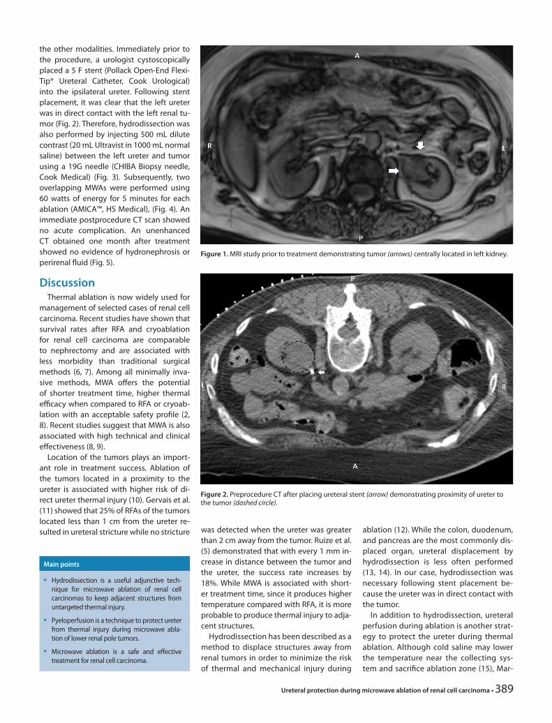
the other modalities. Immediately prior to the procedure, a urologist cystoscopically placed a 5 F stent (Pollack Open-End Flexi-Tip® Ureteral Catheter, Cook Urological) into the ipsilateral ureter. Following stent placement, it was clear that the left ureter was in direct contact with the left renal tu-mor (Fig. 2). Therefore, hydrodissection was also performed by injecting 500 mL dilute contrast (20 mL Ultravist in 1000 mL normal saline) between the left ureter and tumor using a 19G needle (CHIBA Biopsy needle, Cook Medical) (Fig. 3). Subsequently, two overlapping MWAs were performed using 60 watts of energy for 5 minutes for each ablation (AMICA™, HS Medical), (Fig. 4). An immediate postprocedure CT scan showed no acute complication. An unenhanced CT obtained one month after treatment showed no evidence of hydronephrosis or perirenal fluid (Fig. 5).
DiscussionThermal ablation is now widely used for
management of selected cases of renal cell carcinoma. Recent studies have shown that survival rates after RFA and cryoablation for renal cell carcinoma are comparable to nephrectomy and are associated with less morbidity than traditional surgical methods (6, 7). Among all minimally inva-sive methods, MWA offers the potential of shorter treatment time, higher thermal efficacy when compared to RFA or cryoab-lation with an acceptable safety profile (2, 8). Recent studies suggest that MWA is also associated with high technical and clinical effectiveness (8, 9).
Location of the tumors plays an import-ant role in treatment success. Ablation of the tumors located in a proximity to the ureter is associated with higher risk of di-rect ureter thermal injury (10). Gervais et al. (11) showed that 25% of RFAs of the tumors located less than 1 cm from the ureter re-sulted in ureteral stricture while no stricture was detected when the ureter was greater
than 2 cm away from the tumor. Ruize et al. (5) demonstrated that with every 1 mm in-crease in distance between the tumor and the ureter, the success rate increases by 18%. While MWA is associated with short-er treatment time, since it produces higher temperature compared with RFA, it is more probable to produce thermal injury to adja-cent structures.
Hydrodissection has been described as a method to displace structures away from renal tumors in order to minimize the risk of thermal and mechanical injury during
ablation (12). While the colon, duodenum, and pancreas are the most commonly dis-placed organ, ureteral displacement by hydrodissection is less often performed (13, 14). In our case, hydrodissection was necessary following stent placement be-cause the ureter was in direct contact with the tumor.
In addition to hydrodissection, ureteral perfusion during ablation is another strat-egy to protect the ureter during thermal ablation. Although cold saline may lower the temperature near the collecting sys-tem and sacrifice ablation zone (15), Mar-
Main points
• Hydrodissection is a useful adjunctive tech-nique for microwave ablation of renal cell carcinomas to keep adjacent structures from untargeted thermal injury.
• Pyeloperfusion is a technique to protect ureter from thermal injury during microwave abla-tion of lower renal pole tumors.
• Microwave ablation is a safe and effective treatment for renal cell carcinoma.
Figure 1. MRI study prior to treatment demonstrating tumor (arrows) centrally located in left kidney.
Figure 2. Preprocedure CT after placing ureteral stent (arrow) demonstrating proximity of ureter to the tumor (dashed circle).
gulis et al. (16) showed in an animal study that pyeloperfusion did not significantly affect the ablation size when using RFA. Similarly, Cantwell et al. (17) demonstrat-ed 100% effectiveness of pyeloperfusion with RFA for renal tumors within 1.5 cm to ureter without any urologic complica-tions. While several studies have assessed the efficacy of pyeloperfusion with RFA, studies investigating pyeloperfusion in MWA are lacking.
Because the primary mechanism of ac-tion of MWA is agitation of water mole-cules within tissue, the presence of a flu-id-filled ureter adjacent to tumors treated with MWA would theoretically be at higher risk of thermal injury. However, the patient discussed in this report was treated suc-cessfully with pyeloperfusion, hydrodis-section, and MWA.
In conclusion, this case demonstrates the combined value of hydrodissection with pyeloperfusion to successfully per-form CT-guided MWA for a renal cell carci-noma arising from the medial lower pole. By physically displacing the ureter away from the tumor and active ureteral cooling with pyeloperfusion, MWA could be com-pleted without acute or delayed complica-tion of the collecting system. Larger series with longer follow-up are necessary to as-sess the efficacy of this technique.
Conflict of interest disclosureThe authors declared no conflicts of interest.
References1. Gao Y, Liang P, Yu X, et al. Microwave treatment
of renal cell carcinoma adjacent to renal sinus. Eur J Radiol 2016; 85:2083–2089. [CrossRef]
2. J. Yu, P. Liang, X. Yu, et al. A comparison of mi-crowave ablation and bipolar radiofrequency ablation both with an internally cold probe: results in ex vivo and in vivo porcine livers. Eur J Radiol 2011; 79:124–130. [CrossRef]
3. Young JL, Kolla SB, Pick DL, et al. In vitro, ex vivo and in vivo isotherms for renal cryotherapy. J Urol 2010; 183:752–758. [CrossRef]
4. Mauri G, Nicosia L, Varano GM, et al. Tips and tricks for a safe and effective image-guided percutaneous renal tumour ablation. Insights Imaging 2017; 8:357–363. [CrossRef]
5. Ruiz YA, Lönnemark M, Brekkan E, et al. Predictive factors for complete renal tumor ablation using RFA. Acta Radiol 2015; 57:886–893. [CrossRef]
6. Huang WC, Levey AS, Serio AM, et al. Chronic kidney disease after nephrectomy in patients with renal cortical tumours: a retrospective co-hort study. Lancet Oncol 2006; 7:735e40.
7. Yu J, Liang P, Yu XL, et al. US-guided percuta-neous microwave ablation versus open radical nephrectomy for small renal cell carcinoma: intermediate-term results. Radiology 2014; 3:880–887. [CrossRef]
390 • November–December 2018 • Diagnostic and Interventional Radiology Samadi and Arellano
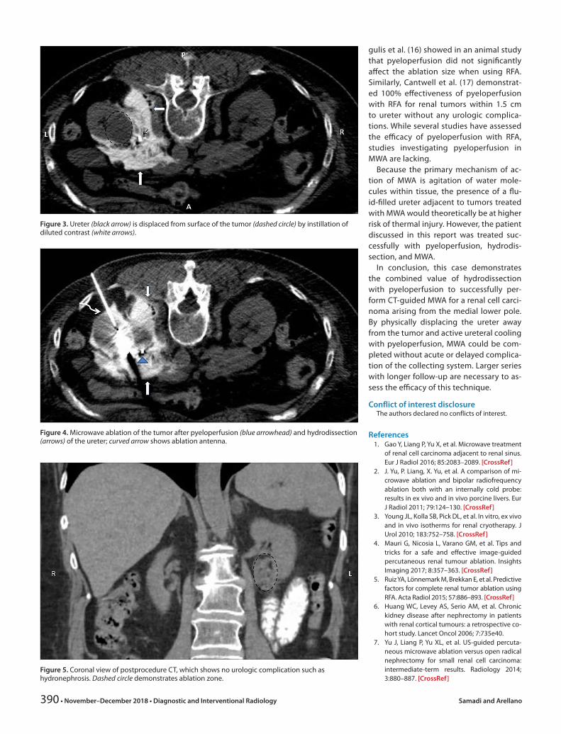
Figure 3. Ureter (black arrow) is displaced from surface of the tumor (dashed circle) by instillation of diluted contrast (white arrows).
Figure 4. Microwave ablation of the tumor after pyeloperfusion (blue arrowhead) and hydrodissection (arrows) of the ureter; curved arrow shows ablation antenna.
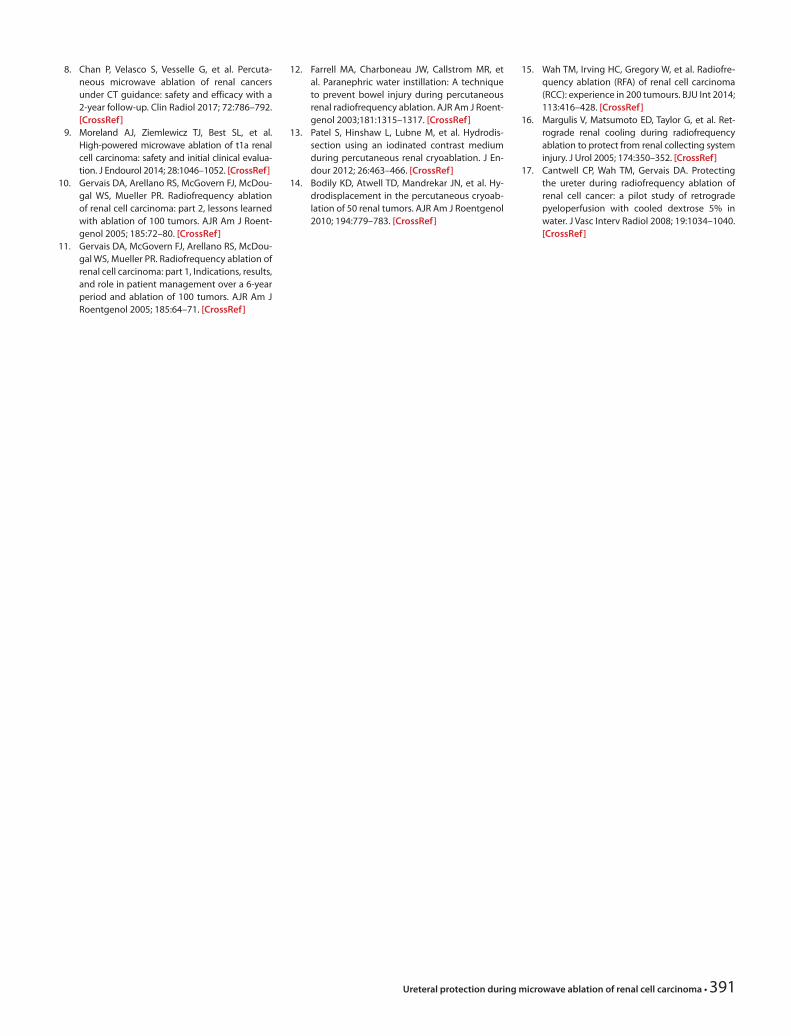
Figure 5. Coronal view of postprocedure CT, which shows no urologic complication such as hydronephrosis. Dashed circle demonstrates ablation zone.
Ureteral protection during microwave ablation of renal cell carcinoma • 391
8. Chan P, Velasco S, Vesselle G, et al. Percuta-neous microwave ablation of renal cancers under CT guidance: safety and efficacy with a 2-year follow-up. Clin Radiol 2017; 72:786–792. [CrossRef]
9. Moreland AJ, Ziemlewicz TJ, Best SL, et al. High-powered microwave ablation of t1a renal cell carcinoma: safety and initial clinical evalua-tion. J Endourol 2014; 28:1046–1052. [CrossRef]
10. Gervais DA, Arellano RS, McGovern FJ, McDou-gal WS, Mueller PR. Radiofrequency ablation of renal cell carcinoma: part 2, lessons learned with ablation of 100 tumors. AJR Am J Roent-genol 2005; 185:72–80. [CrossRef]
11. Gervais DA, McGovern FJ, Arellano RS, McDou-gal WS, Mueller PR. Radiofrequency ablation of renal cell carcinoma: part 1, Indications, results, and role in patient management over a 6-year period and ablation of 100 tumors. AJR Am J Roentgenol 2005; 185:64–71. [CrossRef]
12. Farrell MA, Charboneau JW, Callstrom MR, et al. Paranephric water instillation: A technique to prevent bowel injury during percutaneous renal radiofrequency ablation. AJR Am J Roent-genol 2003;181:1315–1317. [CrossRef]
13. Patel S, Hinshaw L, Lubne M, et al. Hydrodis-section using an iodinated contrast medium during percutaneous renal cryoablation. J En-dour 2012; 26:463–466. [CrossRef]
14. Bodily KD, Atwell TD, Mandrekar JN, et al. Hy-drodisplacement in the percutaneous cryoab-lation of 50 renal tumors. AJR Am J Roentgenol 2010; 194:779–783. [CrossRef]
15. Wah TM, Irving HC, Gregory W, et al. Radiofre-quency ablation (RFA) of renal cell carcinoma (RCC): experience in 200 tumours. BJU Int 2014; 113:416–428. [CrossRef]
16. Margulis V, Matsumoto ED, Taylor G, et al. Ret-rograde renal cooling during radiofrequency ablation to protect from renal collecting system injury. J Urol 2005; 174:350–352. [CrossRef]
17. Cantwell CP, Wah TM, Gervais DA. Protecting the ureter during radiofrequency ablation of renal cell cancer: a pilot study of retrograde pyeloperfusion with cooled dextrose 5% in water. J Vasc Interv Radiol 2008; 19:1034–1040. [CrossRef]