Upper GI Bleed: Clinical Case Presentation Lisa Philipose 4 / 25/ 06.
21
Upper GI Bleed : Clinical Case Presentation Lisa Philipose 4 / 25/ 06
-
Upload
austin-howard -
Category
Documents
-
view
241 -
download
2
Transcript of Upper GI Bleed: Clinical Case Presentation Lisa Philipose 4 / 25/ 06.
Upper GI Bleed:
Clinical Case Presentation
Lisa Philipose4 / 25/ 06
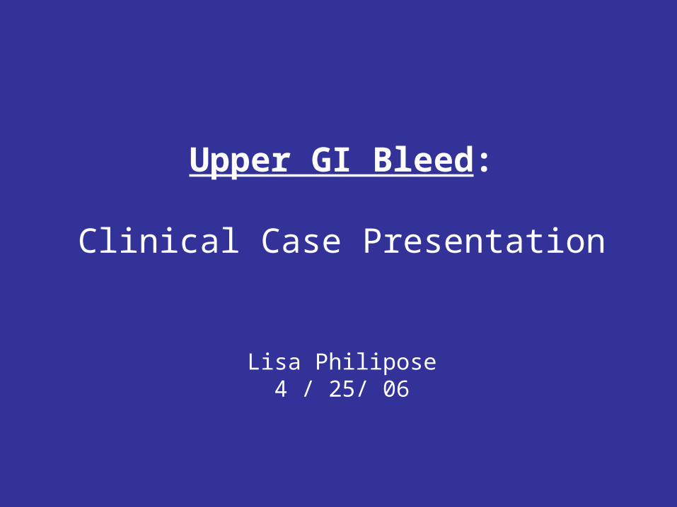
History
• CC: 79 y.o. white male presents via EMS to the Bayview E.D. with two days of loose black tarry stools.
• HPI: On the morning PTA, patient felt weak and light-headed, so wife called EMS. VS in the field were: HR:136; BP: 82/48; RR:18; O2sat: 98% on RA; D-stick:150.
• 600 cc bolus was administered by EMS and BP increased to 107/61 and HR decreased to 96.
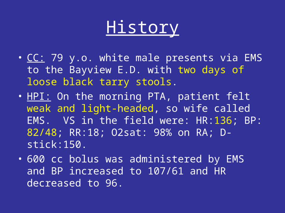
History
• ROS: Pt denies N/V/D/C, and denies chest/abdominal/ back/flank/rectal pain.
• PMH: HTN, DM, no h/o bleeding d/o; no h/o GI disorders– Last colonoscopy 3 yrs ago reported normal per
patient.• PSH: None• Meds: Lisinopril, Metformin, Glucophage, HCTZ,
ASA• All: NKDA• SH: No h/o tobacco/alcohol/illicits
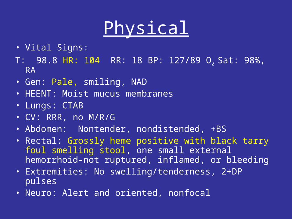
Physical• Vital Signs:
T: 98.8 HR: 104 RR: 18 BP: 127/89 O2 Sat: 98%, RA• Gen: Pale, smiling, NAD• HEENT: Moist mucus membranes• Lungs: CTAB• CV: RRR, no M/R/G• Abdomen: Nontender, nondistended, +BS• Rectal: Grossly heme positive with black tarry foul
smelling stool, one small external hemorrhoid-not ruptured, inflamed, or bleeding
• Extremities: No swelling/tenderness, 2+DP pulses• Neuro: Alert and oriented, nonfocal
E.D. course
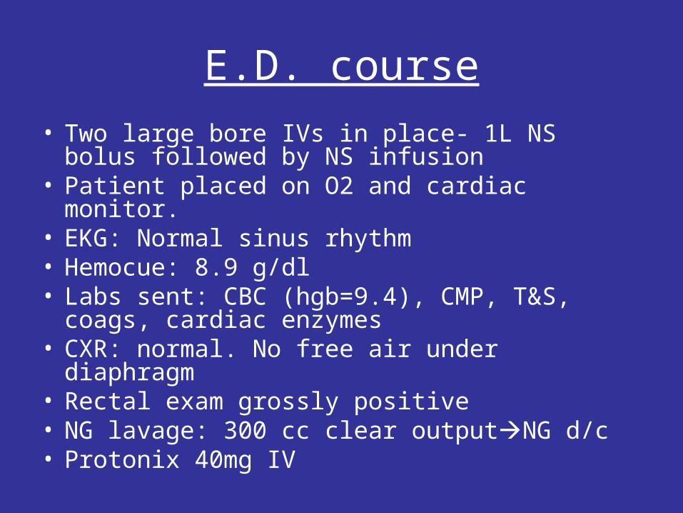
• Two large bore IVs in place- 1L NS bolus followed by NS infusion
• Patient placed on O2 and cardiac monitor.• EKG: Normal sinus rhythm • Hemocue: 8.9 g/dl• Labs sent: CBC (hgb=9.4), CMP, T&S, coags,
cardiac enzymes• CXR: normal. No free air under diaphragm• Rectal exam grossly positive• NG lavage: 300 cc clear outputNG d/c• Protonix 40mg IV
E.D. course
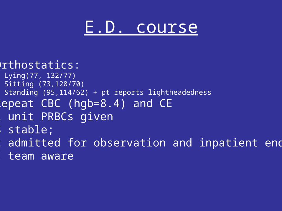
• Orthostatics:• Lying(77, 132/77) • Sitting (73,120/70) • Standing (95,114/62) + pt reports lightheadedness
• Repeat CBC (hgb=8.4) and CE• 1 unit PRBCs given•VS stable; •Pt admitted for observation and inpatient endoscopy•GI team aware
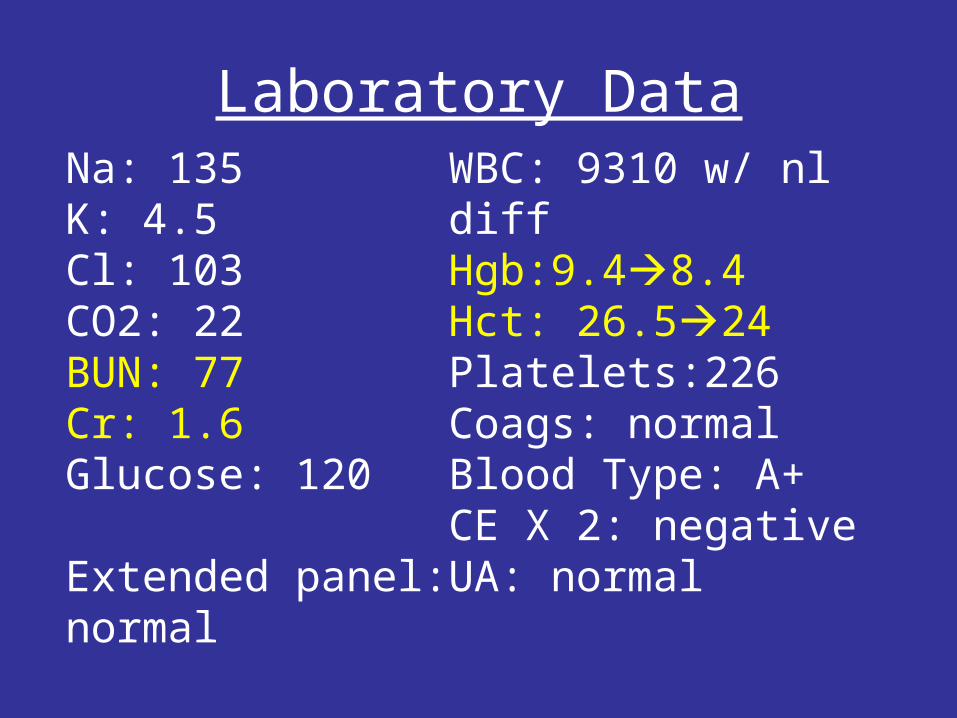
Laboratory DataWBC: 9310 w/ nl diffHgb:9.48.4Hct: 26.524Platelets:226Coags: normalBlood Type: A+CE X 2: negativeUA: normal
Na: 135K: 4.5Cl: 103CO2: 22BUN: 77Cr: 1.6Glucose: 120
Extended panel: normal
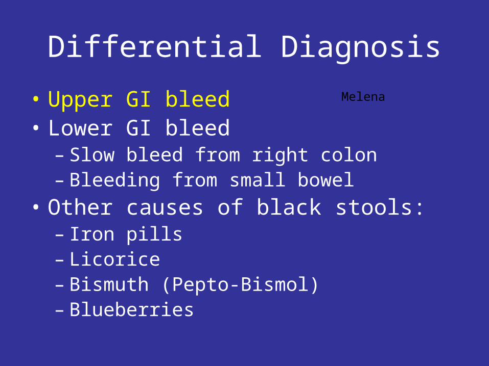
Differential Diagnosis
• Upper GI bleed • Lower GI bleed
– Slow bleed from right colon– Bleeding from small bowel
• Other causes of black stools:– Iron pills– Licorice– Bismuth (Pepto-Bismol)– Blueberries
Melena
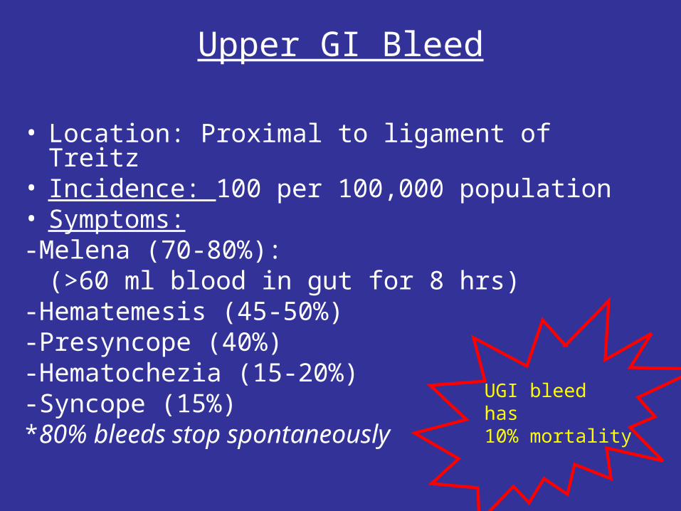
Upper GI Bleed
• Location: Proximal to ligament of Treitz• Incidence: 100 per 100,000 population• Symptoms:-Melena (70-80%):
(>60 ml blood in gut for 8 hrs)-Hematemesis (45-50%)-Presyncope (40%)-Hematochezia (15-20%)-Syncope (15%)*80% bleeds stop spontaneously
UGI bleedhas 10% mortality
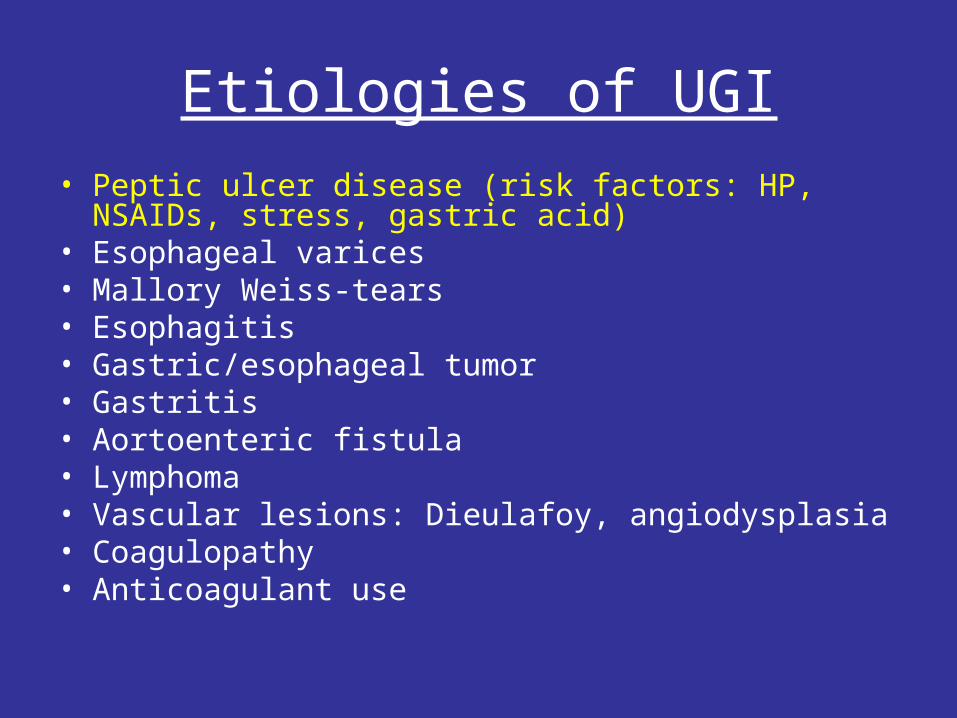
Etiologies of UGI
• Peptic ulcer disease (risk factors: HP, NSAIDs, stress, gastric acid)
• Esophageal varices• Mallory Weiss-tears• Esophagitis• Gastric/esophageal tumor• Gastritis• Aortoenteric fistula• Lymphoma• Vascular lesions: Dieulafoy, angiodysplasia• Coagulopathy• Anticoagulant use
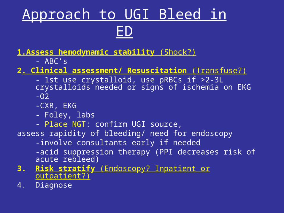
1.Assess hemodynamic stability (Shock?)- ABC’s
2. Clinical assessment/ Resuscitation (Transfuse?)- 1st use crystalloid, use pRBCs if >2-3L crystalloids needed or signs of ischemia on EKG-O2 -CXR, EKG- Foley, labs- Place NGT: confirm UGI source,
assess rapidity of bleeding/ need for endoscopy-involve consultants early if needed-acid suppression therapy (PPI decreases risk of acute rebleed)
3. Risk stratify (Endoscopy? Inpatient or outpatient?)4. Diagnose
Approach to UGI Bleed in ED
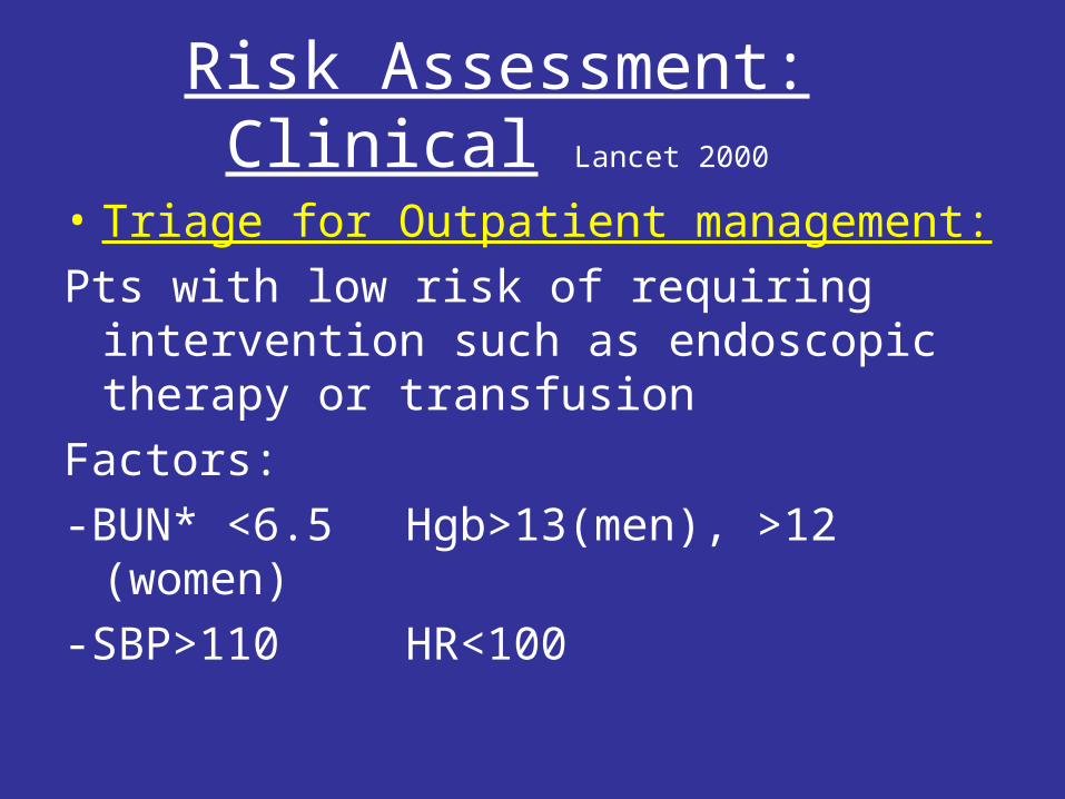
Risk Assessment: Clinical Lancet 2000
• Triage for Outpatient management:
Pts with low risk of requiring intervention such as endoscopic therapy or transfusion
Factors:
-BUN* <6.5 Hgb>13(men), >12 (women)
-SBP>110 HR<100
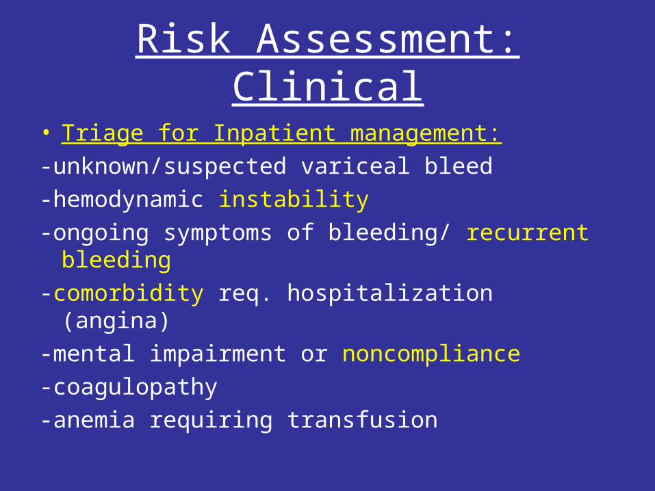
Risk Assessment: Clinical
• Triage for Inpatient management:
-unknown/suspected variceal bleed
-hemodynamic instability
-ongoing symptoms of bleeding/ recurrent bleeding
-comorbidity req. hospitalization (angina)
-mental impairment or noncompliance
-coagulopathy
-anemia requiring transfusion
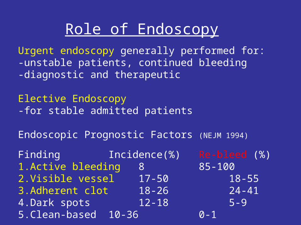
Role of EndoscopyUrgent endoscopy generally performed for:-unstable patients, continued bleeding-diagnostic and therapeutic
Elective Endoscopy -for stable admitted patients
Endoscopic Prognostic Factors (NEJM 1994)
Finding Incidence(%) Re-bleed (%)1.Active bleeding 8 85-1002.Visible vessel 17-50 18-553.Adherent clot 18-26 24-414.Dark spots 12-18 5-95.Clean-based 10-36 0-1
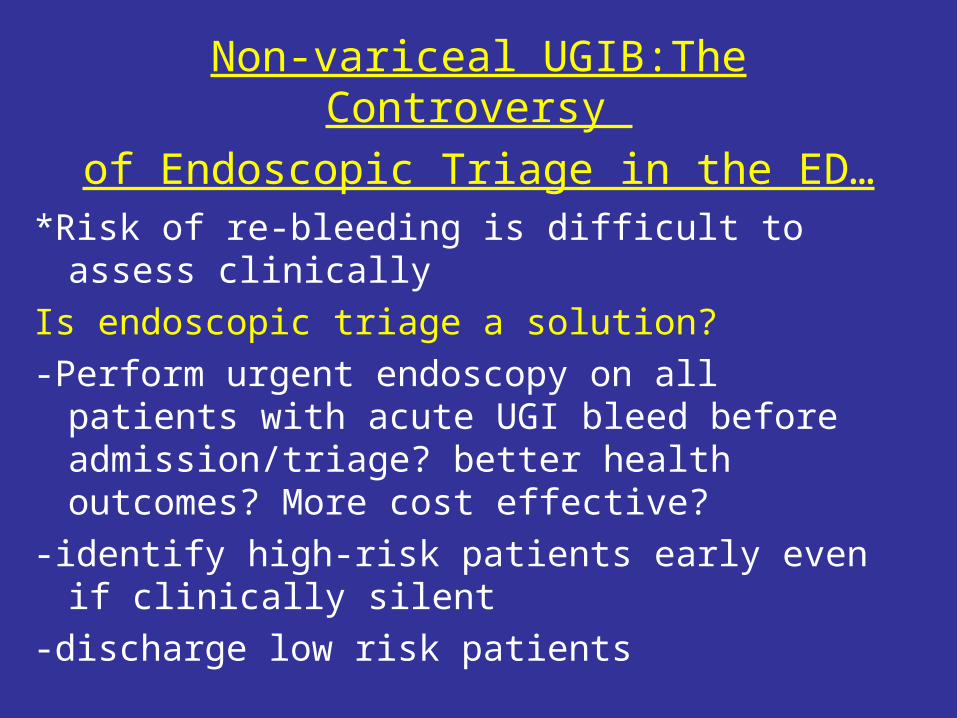
*Risk of re-bleeding is difficult to assess clinically
Is endoscopic triage a solution?
-Perform urgent endoscopy on all patients with acute UGI bleed before admission/triage? better health outcomes? More cost effective?
-identify high-risk patients early even if clinically silent
-discharge low risk patients
Non-variceal UGIB:The Controversy
of Endoscopic Triage in the ED…
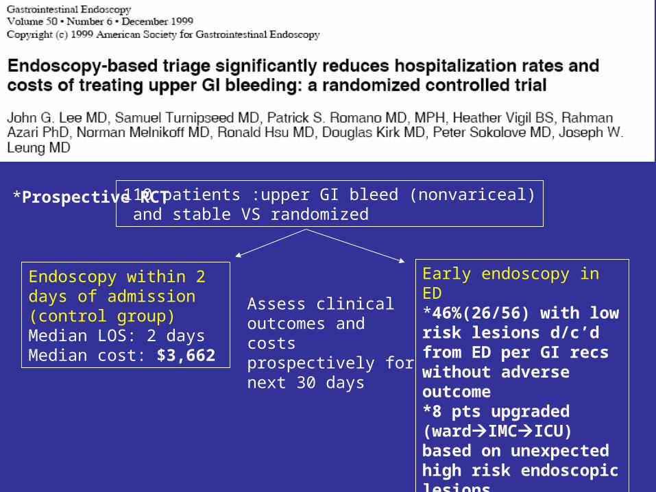
*Prospective RCT
Endoscopy within 2 days of admission (control group)Median LOS: 2 daysMedian cost: $3,662
Early endoscopy in ED*46%(26/56) with low risk lesions d/c’d from ED per GI recs without adverse outcome*8 pts upgraded (wardIMCICU) based on unexpected high risk endoscopic lesionsMedian LOS: 1 daysMedian cost: $2,068
Assess clinical outcomes and costs prospectively for next 30 days
110 patients :upper GI bleed (nonvariceal) and stable VS randomized
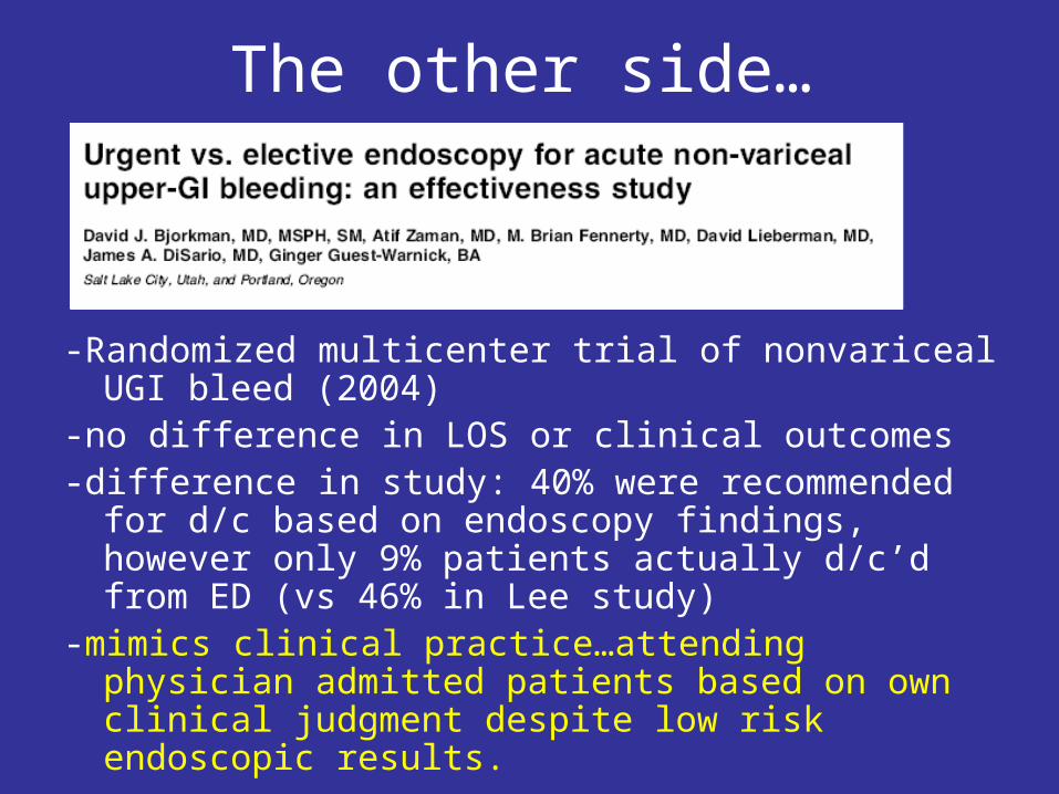
The other side…
-Randomized multicenter trial of nonvariceal UGI bleed (2004)
-no difference in LOS or clinical outcomes -difference in study: 40% were recommended for d/c based
on endoscopy findings, however only 9% patients actually d/c’d from ED (vs 46% in Lee study)
-mimics clinical practice…attending physician admitted patients based on own clinical judgment despite low risk endoscopic results.
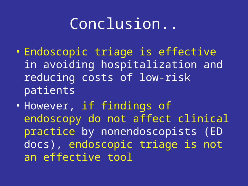
Conclusion..
• Endoscopic triage is effective in avoiding hospitalization and reducing costs of low-risk patients
• However, if findings of endoscopy do not affect clinical practice by nonendoscopists (ED docs), endoscopic triage is not an effective tool
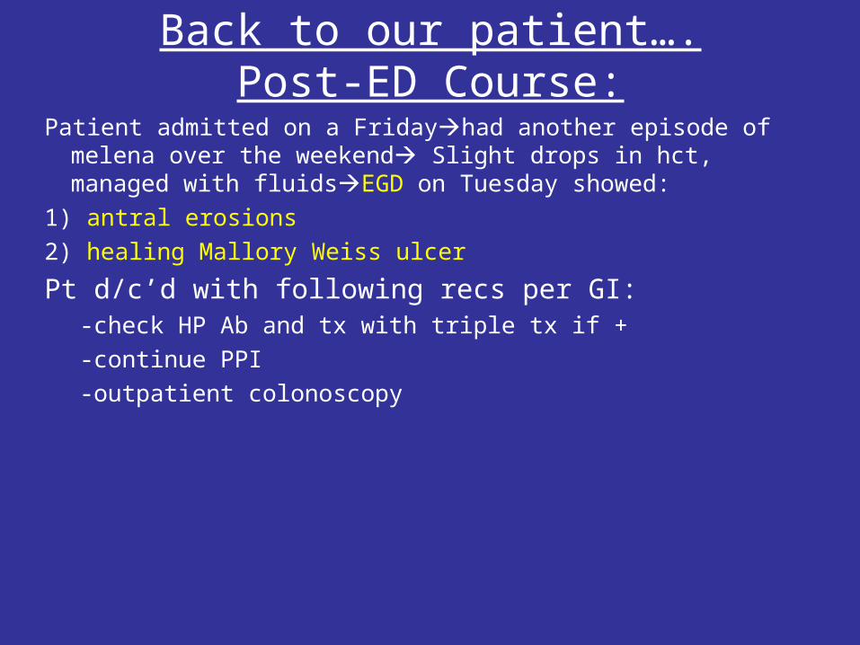
Back to our patient….Post-ED Course:
Patient admitted on a Fridayhad another episode of melena over the weekend Slight drops in hct, managed with fluidsEGD on Tuesday showed:
1) antral erosions
2) healing Mallory Weiss ulcer
Pt d/c’d with following recs per GI:-check HP Ab and tx with triple tx if +
-continue PPI
-outpatient colonoscopy
Summary
• Assess hemodynamic stability
• Resuscitate
• History/physical: risk factors?
• Re-assess need for resuscitation often
• NG lavage
• Endoscopy?
• All bleeding stops…eventually
References• Bjorkman DJ. Endoscopic triage for nonvariceal upper gastrointestinal
bleeding: the optimal approach in 2001? ASGE wesbite, 2001.
• Bjorkman DJ et al., Urgent vs elective endoscopy for acute nonvariceal upper GI bleeding: an effectiveness study. Gastrointest endosc 2004; 60:94-95.
• Blatchford O et al., A risk score to predict need for treatment for upper-gastrointestinal hemorrhage. Lancet 2000; 356:1318-21.
• Eisen GM et al., Guidelines: An annotated algorithmic approach to gastrointestinal bleeding. Gastro Endo 2001; 53:853.
• Jutabha R, Jensen D. Approach to the Adult patient with upper gastro-intestinal bleeding In: UpToDate, Wellesley, MA, 2006.
• Laine L, Peterson WL. Bleeding peptic ulcer. NEJM 1994; 331:717-27.
• Lee JG, et al., Endoscopy-based traige significantly reduces hospitalization rates and costs of upper GI bleeding: a randomized controlled trial. Gastrointest Endosc 1999;50:755-61.
• Peter DJ and Daughtery JM, Evaluation of the patient with gastrointestinal bleeding: An evidence-based approach. Emerg Med Clin NA 17:239, 1999.