Upper Gastrointestinal Bleed
3
Author: Natalie Voorn, MS IV | Editor: Andrea Sarchi, DO ; Jason Mansour, MD October 2016 | Vol 3 | Issue 6 Upper GI Bleed Upper Gastrointestinal Bleed A 64-year-old Haitian female with a past medical history of GERD presents to the ED with blood-tinged sputum for 5 days along with two episodes of bloody vomiting. This has happened several times before in Haiti for whom she saw several doctors with no diagnosis made. This sputum production is associated with 7/10 sharp epigastric pain that is non-radiating. She takes a daily antacid with no relief of symptoms. The patient also admits to nausea, fatigue, weight loss and odynophagia. She recently traveled to Haiti, arriving back in the states two months prior. Previous TB testing performed within the last three months came back negative. She has never had an endoscopy or colonoscopy. What is the most appropriate next step in management of this patient? A. Placement of a nasogastric tube B. Assess for hemodynamic instability C. Obtain a chest X-ray D. Perform an upper endoscopy E. Referral to outpatient gastroenterologist via pathology.jhu.edu A gastrointestinal bleed is defined as intraluminal blood loss anywhere from the oropharynx to the anus. Upper GI bleed occurs when the source of bleeding is above the ligament of Treitz Lower GI bleed occurs when the source is below the ligament of Treitz
Transcript of Upper Gastrointestinal Bleed

Author: Natalie Voorn, MS IV | Editor: Andrea Sarchi, DO ; Jason Mansour, MD
October 2016 | Vol 3 | Issue 6
Upper GI Bleed
Upper Gastrointestinal Bleed A 64-year-old Haitian female with a past medical history of GERD presents to the ED with blood-tinged sputum for 5 days along with two episodes of bloody vomiting. This has happened several times before in Haiti for whom she saw several doctors with no diagnosis made. This sputum production is associated with 7/10 sharp epigastric pain that is non-radiating. She takes a daily antacid with no relief of symptoms. The patient also admits to nausea, fatigue, weight loss and odynophagia. She recently traveled to Haiti, arriving back in the states two months prior. Previous TB testing performed within the last three months came back negative. She has never had an endoscopy or colonoscopy. What is the most appropriate next step in management of this patient?
A. Placement of a nasogastric tube
B. Assess for hemodynamic instability
C. Obtain a chest X-ray
D. Perform an upper endoscopy
E. Referral to outpatient gastroenterologist
via pathology.jhu.edu
A gastrointestinal bleed is defined as intraluminal blood loss anywhere from the oropharynx to the anus. Upper GI bleed occurs when the source of bleeding is above the ligament of Treitz Lower GI bleed occurs when the source is below the ligament of Treitz

October 2016 | Vol 3 | Issue 6
Upper GI Bleed
The correct answer is B. The most important first step is to for assess hemodynamic instability and resuscitate if neded. Look for tachycardia (10% volume loss), orthostatic hypotension (20% volume loss), or shock (>30% volume loss). Upper gastrointestinal bleeding includes hemorrhage originating from the esophagus to the ligament of Treitz. Peptic ulcer bleeding is the most common cause of upper GI bleeding, accounting for more than 60 percent of cases, whereas esophageal varices cause approximately 6 percent. Other etiologies include AV malformations, gastritis, Mallory-Weiss tear, malignancy, esophagitis and duodenitis. Patients can present with a variety of clinical manifestations such as hematemesis, melena, syncope, dizziness, dyspepsia, hematochezia, epigastric pain, diffuse abdominal pain, and weight loss. Obtaining a good history is essential in the initial evaluation of those with GI bleeding. It is important to ask about comorbid conditions (malignancy/ liver disease), medication history (frequent NSAID use/ anticoagulants), previous episodes, and toxic exposures, as well as the severity, timing, duration and amount of bleeding. Physical exam should assess for guarding, rebound tenderness, prior surgical scars, and sequelae of chronic liver disease. Rectal examination should be performed to assess stool color and fecal occult blood testing should be obtained. Workup Initial laboratory tests include measuring the hemoglobin, hematocrit, BUN and creatinine; platelet count; PT, PTT, and INR; liver function tests; blood type and crossmatch. Nasogastric tube can help determine location. For example, if it yields bright red blood or coffee-ground material, it implies that an upper GI bleed is more likely; however, studies show no benefit in regard to clinical outcome using this method. It is associated with shorter time to endoscopy but no difference in mortality, length of hospital stay, surgery or transfusion requirement.
Treatment and Management
Initial management begins by assessing severity of the bleed. Patients may be hypotensive or tachycardic, or may exhibit orthostatic hypotension. Begin resuscitation by establishing two large bore IV lines and administering a bolus of colloid or crystalloid solution. Monitor vital signs, urine output, and mental status for signs of shock. The patient should receive supplemental oxygen and nothing by mouth. Blood transfusions should be administered to those with a hemoglobin level of 7 g/dL or less. Monitory H&H every 4-8 hours until hemoglobin is stable for at least 24 hours. Consider transfusing fresh frozen plasma for those with coagulopathy and platelets for those with thrombocytopenia.
Upper endoscopy within 24 hours of presentation is recommended because it confirms the diagnosis and allows for targeted endoscopic treatment, resulting in reduced morbidity, hospital stays, risk of recurrent bleeding, and need for surgery. Endoscopic therapies include epinephrine injection, thermocoagulation, application of clips, and banding. Patients with low-risk ulcer bleeding (clean ulcer base) can be discharged on the same day as endoscopy. Those with high-risk peptic ulcer bleeding and recent hemorrhage (active arterial bleed, visible vessel, adherent clot) who have undergone endoscopic hemostasis should receive IV PPIs and remain hospitalized for 72 hours. Despite successful endoscopic therapy, rebleeding can occur in 10 to 20 percent of patients; a second attempt at endoscopic therapy is recommended in these patients. Arteriography with embolization or surgery may be needed if there is persistent and severe bleeding.
A. Gastric ulcer with vessel B. Following thermocoagulation Via http://www.aafp.org/afp/2012/0301/p469.html
w
October 2016 | Vol 3 | Issue 6
Upper GI Bleed
Cerulli MA. Upper Gastrointestinal Bleeding. [Internet]. New York (NY): Medscape Reference; [updated 2016 Mar 21]. Available from: http://emedicine.medscape.com/article?. 187857-overview Saltzman JR. Approach to acute upper gastrointestinal bleeding in adults. In: UpToDate, Basow, DS (Ed), UpToDate, Waltham, MA, 2016. Thad Wilkins, MD. Diagnosis and Management of Upper GI Bleed. Am Fam Physician. 2012 Mar 1;85(5):469-476.
This month’s case was written by Natalie Voorn. Natalie is a 4th year medical student from NSU-COM. She did her emergency medicine rotation at BHMC in September 2016. Natalie plans on pursuing a career in Internal Medicine after graduation.
Prevention H.pylori infection and NSAIDs are the major causes of peptic ulcer bleeding in the United States; therefore, preventive strategies should focus on these etiologies. Patients should be counseled about smoking cessation and limiting alcohol use as both of these impair ulcer healing. In patients with a history of peptic ulcer bleeding, aspirin, clopidogrel, and NSAIDs should be avoided if possible. In those with H.pylori, eradication is essential and should be confirmed by urea breath test, stool antigen test, or biopsy urease test. A repeat upper endoscopy in 8-12 weeks is recommended for patients with peptic ulcer bleeding secondary to gastric ulcers to assess for healing and to exclude malignancy, and for patients with severe esophagitis to exclude Barrett esophagus.
Take Home Points • Upper gastrointestinal bleeding causes significant morbidity and mortality in the
United States. Peptic ulcer bleeding is the most common cause and has been associated with NSAID use and H.pylori infection.
• Assessment of hemodynamic stability and resuscitation is critical prior to diagnostic evaluation in unstable patients with severe bleeding.
• Blood transfusions are recommended to obtain a Hgb > 7 in those with clinical evidence of intravascular volume depletion or comorbid conditions such as CAD.
• In acute cases of a GI Bleed, an upper endoscopy should be performed within 24 hours to determine the source of bleeding.
• Eradicating H.pylori infection and limiting NSAID use are essential in treating PUD
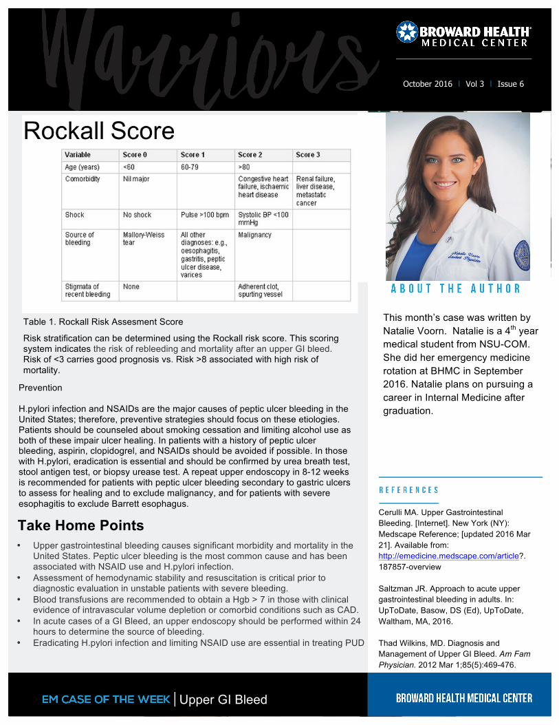
Risk stratification can be determined using the Rockall risk score. This scoring system indicates the risk of rebleeding and mortality after an upper GI bleed. Risk of <3 carries good prognosis vs. Risk >8 associated with high risk of mortality.
Table 1. Rockall Risk Assesment Score