Upper Airway Obstruction Ibrahim Alsaif Consultant Pediatrician Pediatric Emergency Consultant Al...
64
Upper Airway Obstruction Ibrahim Alsaif Consultant Pediatrician Pediatric Emergency Consultant Al Yamammah Hospital 9/15/2015 1 Ped.emergency.Dr.Alsaif
-
Upload
robert-henry -
Category
Documents
-
view
219 -
download
2
Transcript of Upper Airway Obstruction Ibrahim Alsaif Consultant Pediatrician Pediatric Emergency Consultant Al...

Ped.emergency.Dr.Alsaif 1
Upper Airway Obstruction
Ibrahim AlsaifConsultant Pediatrician
Pediatric Emergency ConsultantAl Yamammah Hospital
9/15/2015

Ped.emergency.Dr.Alsaif 2
Learning Objectives
Differentiate between upper and lower respiratory problems based on clinical basis.
Know the “ABCD” (the priorities of airway, breathing, circulation) assessment.
Recognized clinical presentation, radiological features, and management of foreign body aspiration.
Know the initial emergency management of upper airway obstruction.
Recognize the signs and symptoms associated with croup and epiglottitis.
Know the management of croup and epiglottitis. 9/15/2015

Ped.emergency.Dr.Alsaif 3
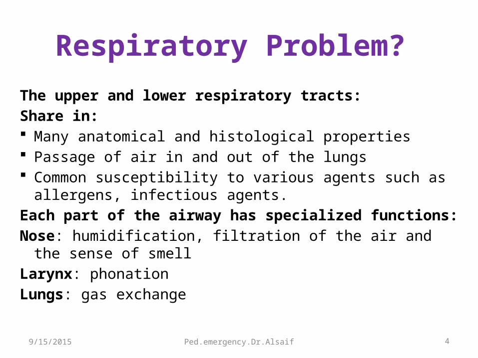
Respiratory Problem?
First question: upper or lower?The upper respiratory tract:Nose, nasal cavity, sinuses, pharynx, larynx, and the upper portion of the trachea.
The lower respiratory tract:Lower portion of the trachea, the bronchial tree, and the lungs.
9/15/2015
Ped.emergency.Dr.Alsaif 4
Respiratory Problem?
The upper and lower respiratory tracts:Share in: Many anatomical and histological properties Passage of air in and out of the lungs Common susceptibility to various agents such as allergens,
infectious agents.Each part of the airway has specialized functions:Nose: humidification, filtration of the air and the sense of smellLarynx: phonationLungs: gas exchange
9/15/2015
Ped.emergency.Dr.Alsaif 5
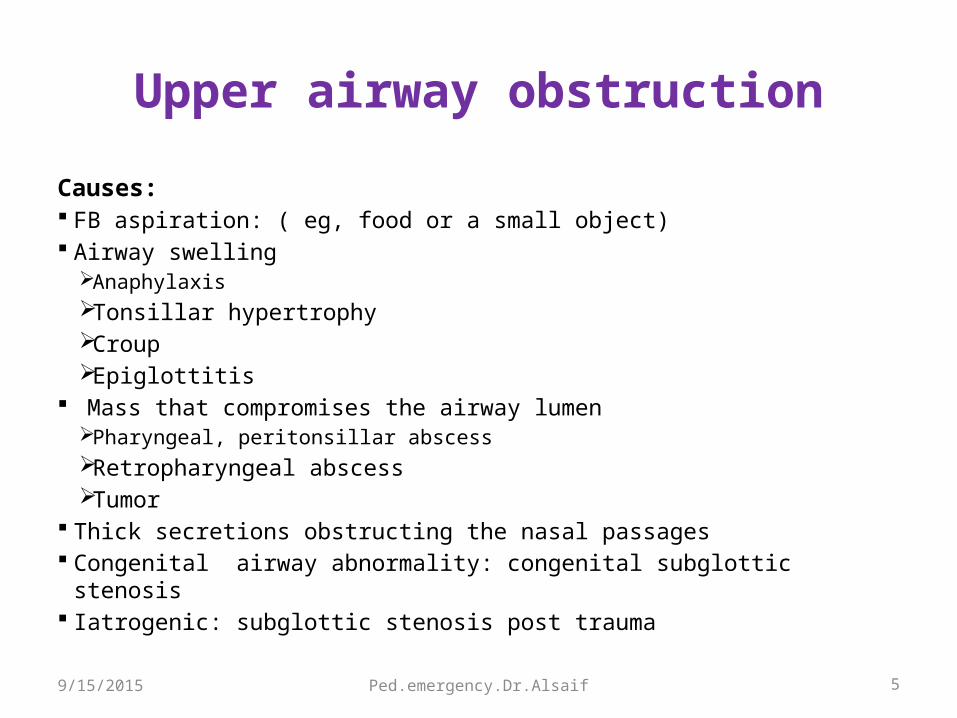
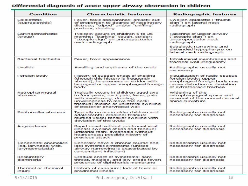
Upper airway obstruction
Causes: FB aspiration: ( eg, food or a small object) Airway swelling
Anaphylaxis Tonsillar hypertrophy Croup Epiglottitis
Mass that compromises the airway lumen Pharyngeal, peritonsillar abscess Retropharyngeal abscess Tumor
Thick secretions obstructing the nasal passages Congenital airway abnormality: congenital subglottic stenosis Iatrogenic: subglottic stenosis post trauma
9/15/2015

Ped.emergency.Dr.Alsaif 6
Upper airway obstruction
Signs: Mostly during inspiration Change in voice: hoarseness, barking cough. Inspiratory stridor. Cyanosis, drooling. Nasal flaring. Tachypnea ( mild). Retractions: suprasternal, supraclavicular. Poor chest expansion. Poor air entry on auscultation. Prolonged inspiratory phase.
9/15/2015
Ped.emergency.Dr.Alsaif 7
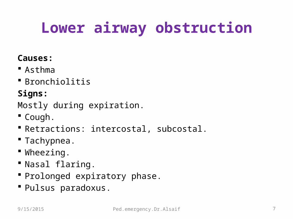
Lower airway obstruction
Causes: Asthma BronchiolitisSigns:Mostly during expiration. Cough. Retractions: intercostal, subcostal. Tachypnea. Wheezing. Nasal flaring. Prolonged expiratory phase. Pulsus paradoxus.
9/15/2015

Ped.emergency.Dr.Alsaif 8
Lung tissue disease
Causes: Pneumonia( viral, bacterial, chemical) Pulmonary edema ( heart failure, ARDS) Pulmonary contusion( trauma ) Allergic reaction Toxins Vasculitis TumorSigns: Marked tachypnea Retractions, nasal flaring Grunting Crackles Decrease breath sound Tachycardia Hypoxemia
9/15/2015


Ped.emergency.Dr.Alsaif 10
ANY LIFE-THREATENING
Always----Emergency, get consultant Universal Precautions ABCDE approach
9/15/2015

Ped.emergency.Dr.Alsaif 11
ABCDE Approach
Airway
Breathing
Circulation
9/15/2015

Ped.emergency.Dr.Alsaif 12
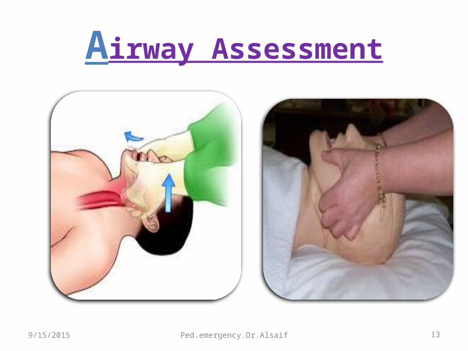
ABCDE approachAirway Assessment
Patent Maintainable
9/15/2015
Ped.emergency.Dr.Alsaif 13
Airway Assessment
9/15/2015
Ped.emergency.Dr.Alsaif 14
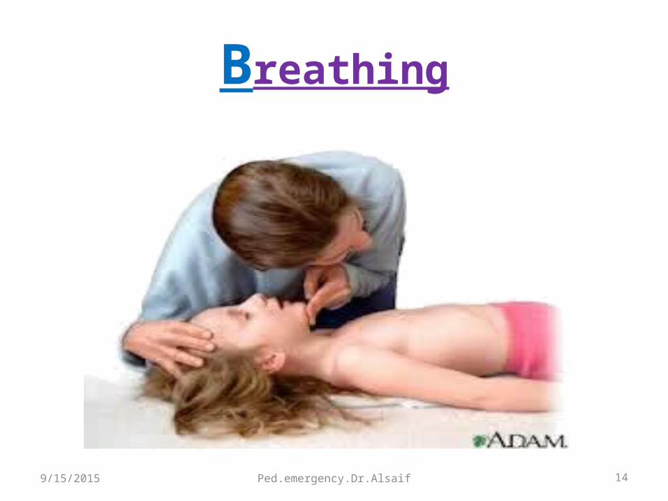
Breathing
9/15/2015
Ped.emergency.Dr.Alsaif 15
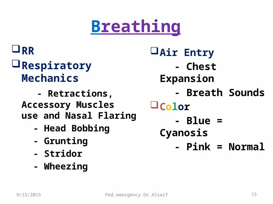
BreathingRR Respiratory Mechanics - Retractions, Accessory Muscles
use and Nasal Flaring - Head Bobbing - Grunting - Stridor - Wheezing
Air Entry - Chest Expansion - Breath Sounds Color - Blue = Cyanosis - Pink = Normal
9/15/2015
Ped.emergency.Dr.Alsaif 16
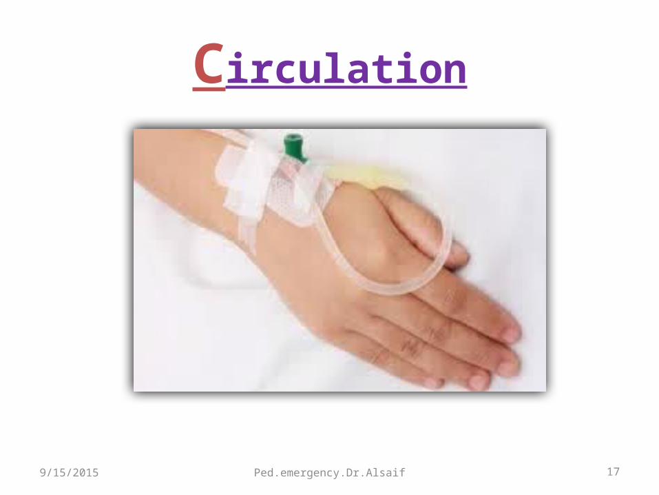
CirculationHeart rateBPPeripheral pulsesSkin perfusion Cap. refill timeColor
9/15/2015
Ped.emergency.Dr.Alsaif 17
Circulation
9/15/2015

Ped.emergency.Dr.Alsaif 189/15/2015
Ped.emergency.Dr.Alsaif 199/15/2015

Ped.emergency.Dr.Alsaif 20
Foreign Body Aspiration
(FBA )
Life-threatening 80 % of pediatric FBA episodes < 3 years of age Peak incidence 1- 2 years Aspirated FBs in children: Peanuts, seeds, popcorn, food
particles, hardware, and pieces of toys, coins, paper clips, pins, pen caps.
Location: Bronchial- common Laryngo-tracheal Tracheal
Morbidity and mortality is high with L.T9/15/2015
Ped.emergency.Dr.Alsaif 21
FBAClinical Presentation
Depends on: History of choking(witnessed) Age of the child Type of object aspirated Degree of airway blockage Location of the object.50-75% of cases will present and diagnosed
within 24 hs of aspiration.
9/15/2015

Ped.emergency.Dr.Alsaif 22
FBAClinical Presentation
Symptoms and Signs Choking: ( Sudden onset of cough +- dyspnea +- cyanosis).Laryngotracheal: acute respiratory distress, stridor, hoarseness, or
complete airway obstructionTracheal FBs: stridor, wheeze, and dyspnea.Bronchial FBs: coughing and wheezing, hemoptysis, dyspnea,
respiratory distress, decreased breath sounds, fever, and cyanosis.If Delayed diagnosis (days or weeks after the aspiration) Symptoms due to complications: infection and inflammation of
the airway.
9/15/2015

Ped.emergency.Dr.Alsaif 23
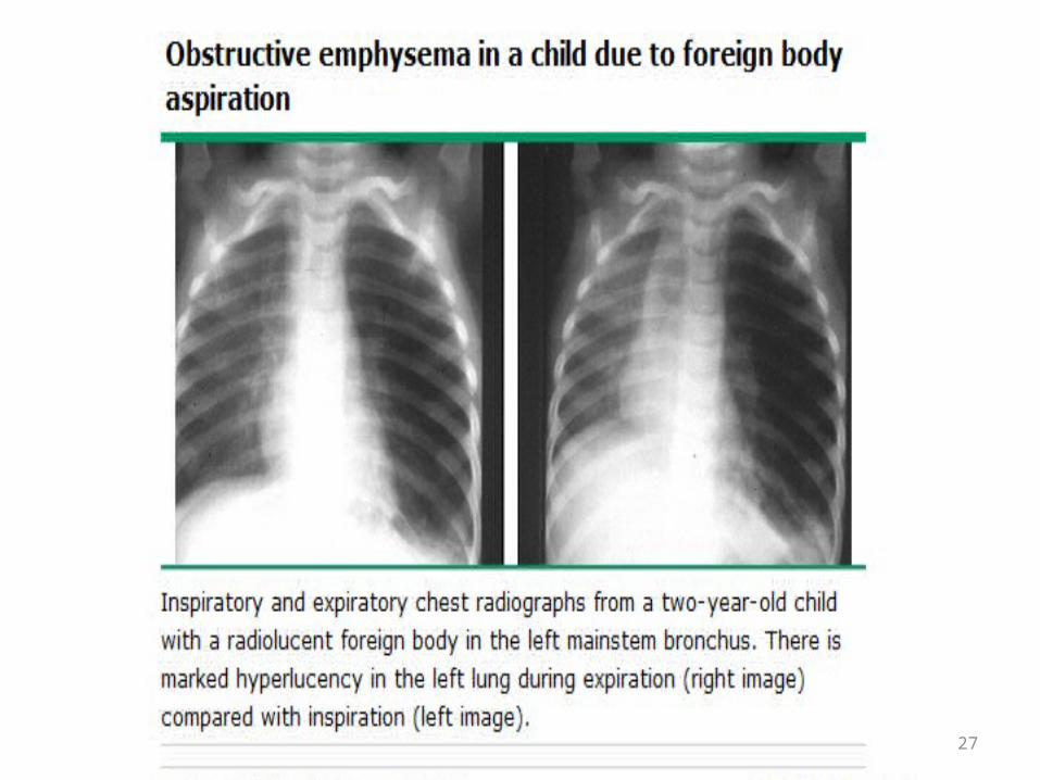
FBADiagnosis
Inspiratory chest + Lateral soft tissue x-ray A normal chest radiograph does not rule out FBA Radioopaque (10 % of FBs) Radiolucent (eg, nuts, food particles) Expiratory chest x-ray or fluoroscopyFor children with a suggestive presentation and normal inspiratory chest x-ray.
9/15/2015

Ped.emergency.Dr.Alsaif 24
FBADiagnosis
Bronchial FBAFindings in chest x-ray:
Hyperinflated lungAtelectasisMediastinal shiftPneumonia Pulmonary abscesses and bronchiectasis (late)
9/15/2015
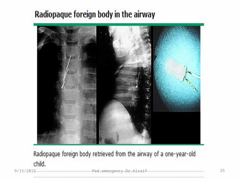
Ped.emergency.Dr.Alsaif 259/15/2015

26
27
Ped.emergency.Dr.Alsaif 28
FBADiagnosis
Suspected FBA Rigid bronchoscopy with ventilation under
general anesthesia. Flexible bronchoscopy.
9/15/2015

299/15/2015
Ped.emergency.Dr.Alsaif 30
MANAGEMENT OF FBAChoking
Life-threatening FBA (complete UAO….unable to speake or cough). Visualize remove No finger sweep
Infant5 back blow follow 5 chest thrust
9/15/2015
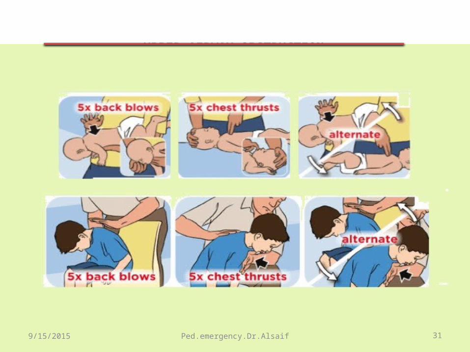
Ped.emergency.Dr.Alsaif 31
UPPER AIRWAY OBSTRUCTION FB
9/15/2015
Ped.emergency.Dr.Alsaif 32
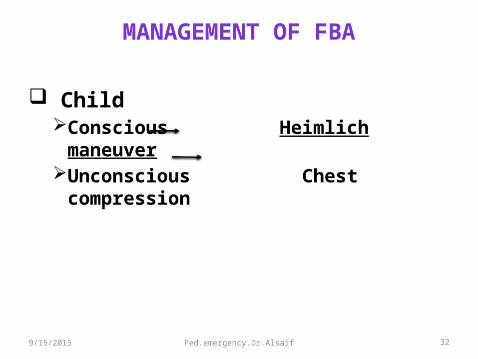
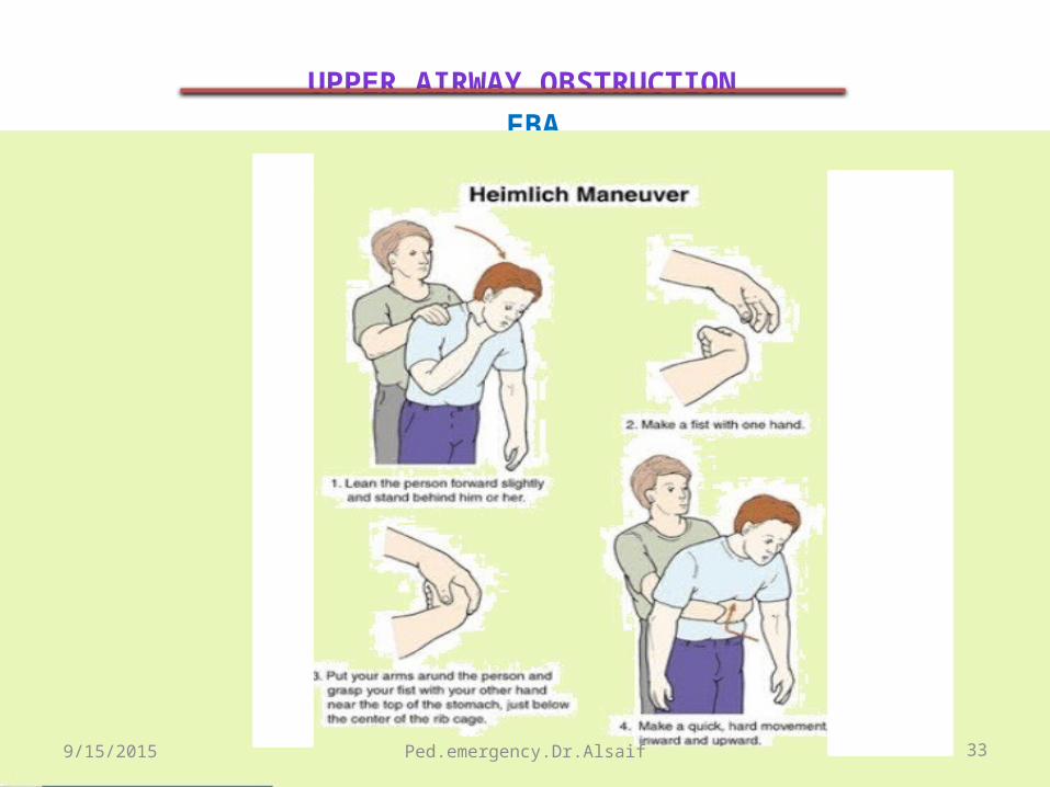
MANAGEMENT OF FBA
Child Conscious Heimlich maneuverUnconscious Chest compression
9/15/2015
Ped.emergency.Dr.Alsaif 33
UPPER AIRWAY OBSTRUCTION FBA
9/15/2015
Ped.emergency.Dr.Alsaif 34
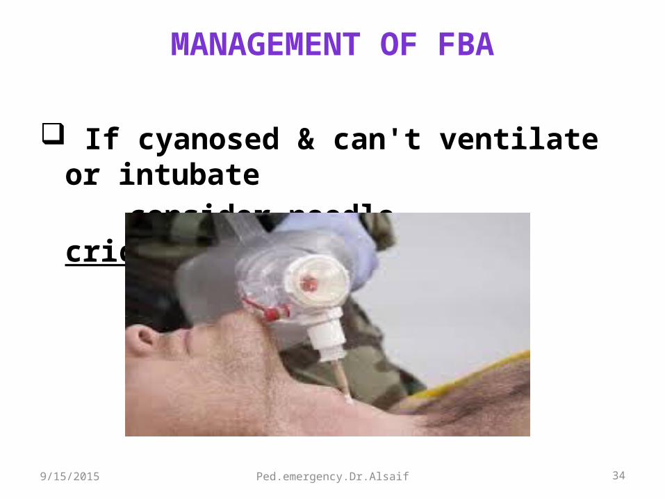
MANAGEMENT OF FBA
If cyanosed & can't ventilate or intubate consider needle cricothyrotomy
9/15/2015
Ped.emergency.Dr.Alsaif 35
EPIGLOTTITIS
• Acute Epiglottitis was most common in children aged 2-4 years.
• Since the Hib vaccine(1991), Epiglottitis become rare.
• Streptococci (strept pneum+ group A strept) are the major pathogens.
• Incidence in adult has remained constant and still Haem Inf is the most common organisms.
9/15/2015

Ped.emergency.Dr.Alsaif 36
EPIGLOTTITIS Clinical Presentation
Febrile toxic child Sore throat Drooling Can’t talk, can’t swallow No cough Respiratory distress Stridor is a late presentation indicating Advanced
Airway obstruction. Sniffing / Tripod posture9/15/2015

f 37

389/15/2015 Ped.emergency.Dr.Alsaif

39

Ped.emergency.Dr.Alsaif 40
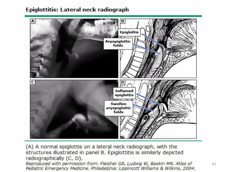
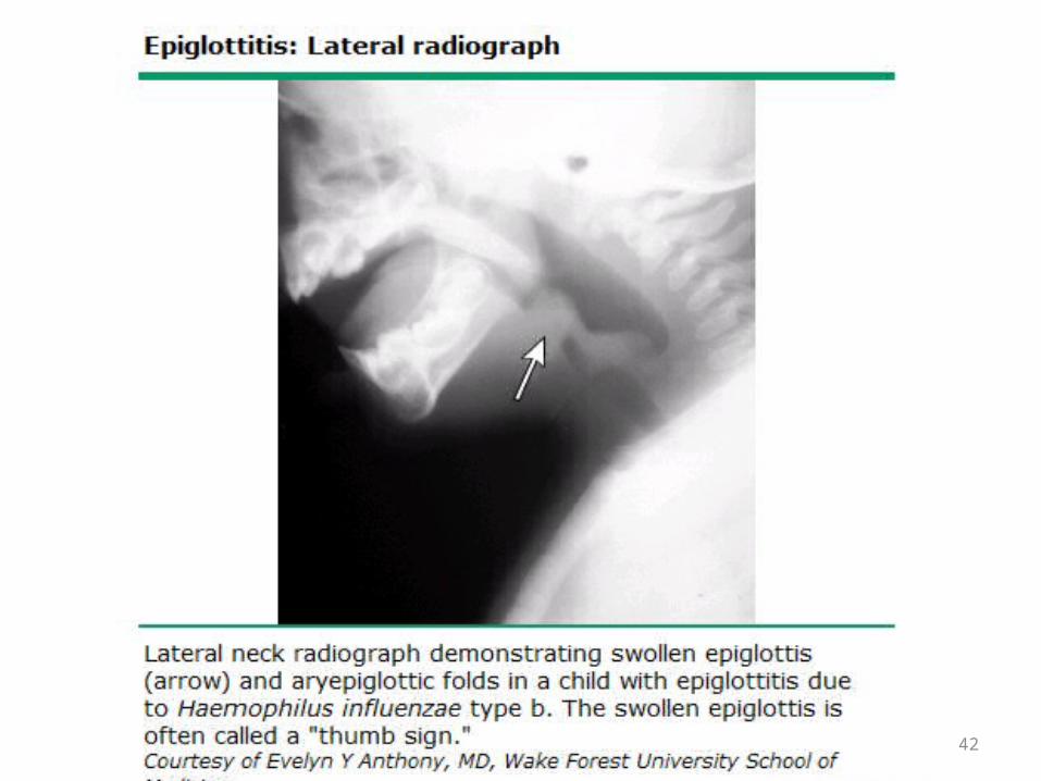
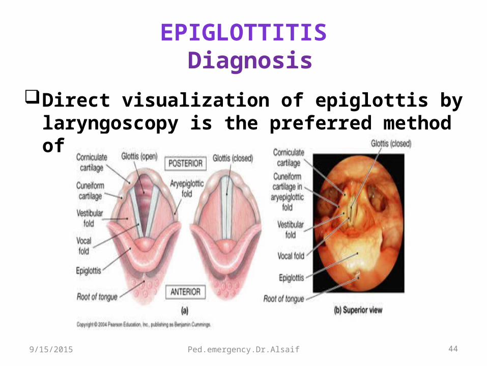
EPIGLOTTITIS Diagnosis
Lat neck soft- tissue x-ray ( portable)Positive in 80%Unnecessary if clinically is suspectedThumb sign
9/15/2015
41
42

Ped.emergency.Dr.Alsaif 439/15/2015
Ped.emergency.Dr.Alsaif 44
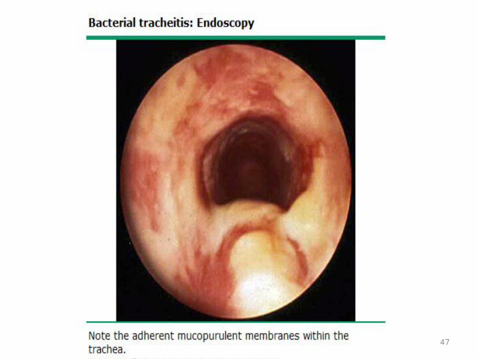
EPIGLOTTITIS Diagnosis
Direct visualization of epiglottis by laryngoscopy is the preferred method of diagnosis.
9/15/2015

Ped.emergency.Dr.Alsaif 459/15/2015

46
47

48

49

Ped.emergency.Dr.Alsaif 50
EPIGLOTTITIS Diagnosis
Bedside U/S is an alternative.Blood + epiglottic cultures if the airway is secured.
9/15/2015
Ped.emergency.Dr.Alsaif 51
EPIGLOTTITISManagement
Advice to not irritate the child, keep on parent's lap Avoid therapy – sedation, inhalers or neubulizer Humidified O2 if possible Airway management is the most urgent consideration:
Assess for level of distress before any other workupEnsure that ENT, Anesthesiologist are available before tracheal
IntubationAirway equipments including that for cricothyrotomy and
tracheotomy
9/15/2015
Ped.emergency.Dr.Alsaif 52
EPIGLOTTITISManagement
All patients should be monitored in ICU Ceftriaxone or cefotaxime + clindamycin or vancomycin If: community or hospital Staph Aureus Refampin for close contacts
9/15/2015

Ped.emergency.Dr.Alsaif 53
Croup (Laryngotracheitis)
Inflammation of the larynx and trachea characterized by: Inspiratory stridor Barking cough Hoarseness.
Children 6 mo - 3 years of age. Usually is a mild and self-limited illnessEtiology Parainfluenza virus type 1 is the most common cause. Respiratory syncytial virus and influenza virus.
9/15/2015

Ped.emergency.Dr.Alsaif 54
Croupclinical presentation
Symptoms The onset is usually gradual, beginning with nasal irritation,
congestion, and coryza. Symptoms generally progress over 12 to 48 hours:
Fever, hoarseness, barking cough, and stridor.Respiratory distress increases as upper airway obstruction
becomes more severe.
9/15/2015

Ped.emergency.Dr.Alsaif 55
Croupclinical presentation
Points in the history that are helpful in distinguishing croupfrom other causes of acute upper airway obstruction: Absence of fever from onset of symptoms to the time of
presentation is suggestive of: Spasmodic croup or Noninfectious etiology (eg, FBA)
Absence of Hoarseness and barking cough Acute epiglottitis FBA Angioneurotic edema.
9/15/2015
Ped.emergency.Dr.Alsaif 56
CroupClinical Presentation
Points in the history Difficult swallowing
Acute epiglottitis FBA.
Drooling Peritonsill arabscesse Retropharyngeal abscesses Retropharyngeal cellulitis Epiglottitis.
Throat pain and dysphagia Common in epiglottitis.
9/15/2015

Ped.emergency.Dr.Alsaif 57
Croup Assessment of severity
Clinical scoring systems (the Westley croup score). Level of consciousness: Normal = 0 disoriented = 5Cyanosis: None = 0 With agitation = 4 At rest = 5Stridor: None = 0 With agitation = 1 At rest = 2Air entry: Normal = 0 Decreased =1 Markedly
decreased= 2Retractions: None=0 Mild= 1 Moderate =2 Severe = 3
9/15/2015

Ped.emergency.Dr.Alsaif 58
Assessment of severity
Mild croup Westley croup score of ≤ 2 barking cough and hoarse cry No stridor at rest.Moderate croup Westley croup score of 3 -7 Stridor at rest Mild retractions.Severe croup Westley croup score of ≥ 8 Significant stridor at rest Decreased air entry Severe retractions Anxious, agitated, or fatigued. Cyanosis
9/15/2015
Ped.emergency.Dr.Alsaif 59
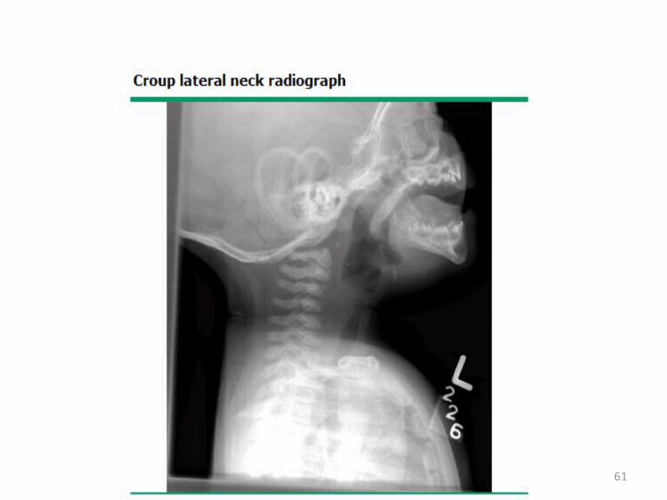
Diagnosis
Clinical diagnosis:Presence of a barking cough and stridor Neither radiographs nor laboratory tests are
necessary to make the diagnosis. Radiographs may be helpful in excluding other
causes.
9/15/2015

60
61

Ped.emergency.Dr.Alsaif 62
Treatment
Mild symptoms Managed at home Single dose of oral dexamethasone (0.6 mg/kg)
Moderate to severe symptoms Supportive care: humidified air or oxygen, intravenous fluids. Racemic epinephrine as nebulizer over 15 min 0.05 mL/kg per dose (maximum of 0.5 mL) of a 2.25 % in 3 ml of NS Nebulized epinephrine 0.5 mL/kg per dose (maximum of 5 mL) of a 1:1000 dilution. Nebulized epinephrine can be repeated every 15 to 20 min. Dexamethasone (0.6 mg/kg) Observed for three to four hours after intervention. Monitoring for worsening respiratory distress
9/15/2015

Ped.emergency.Dr.Alsaif 63
Any question?
9/15/2015

Ped.emergency.Dr.Alsaif 649/15/2015