Update on GI Imaging - Einstein Health · Update on GI Imaging ... •Recurrent or persistent...
35
6/1/2014 1 Update on GI Imaging Susan Summerton, MD Department of Radiology Director, GI Imaging Einstein Healthcare Foundation
-
Upload
duongtuong -
Category
Documents
-
view
213 -
download
0
Transcript of Update on GI Imaging - Einstein Health · Update on GI Imaging ... •Recurrent or persistent...

6/1/2014
1
Update on GI Imaging
Susan Summerton, MD Department of Radiology
Director, GI Imaging
Einstein Healthcare Foundation

6/1/2014
2
Disclosure of Conflicts of Interest
• I have no affiliations with commercial
interests to disclose.
Overview
• GI bleeding evaluation
• Chronic/obscure versus Active
• Diagnostic Imaging Modalities
• Evaluation of Crohn’s Disease
• SBFT/Enteroclysis
• CT Enterography
• MR Enterography
6/1/2014
3
Obscure GI bleeding (OGIB)
• Recurrent or persistent bleeding with no source
found at initial (upper and lower) endoscopy nor
with radiologic evaluation of the small bowel
(SBFT, enteroclysis)
• UGI: Cameron’s erosions in HH, Fundic varices,
PUD, angioectasias, Dieulafoy’s lesion, gastric antral
vascular ectasia
• Colon: angioectasia/neoplasm
Obscure GI bleeding (OGIB):
Small Bowel
• Younger patients (<40 yo)
• Small intestinal tumors
• Meckel’s diverticulum
• Dieulafoy’s lesion
• Crohn’s Disease
• Older patients (>40 yo)
• Vascular lesions (40%)
• NSAID-induced small bowel disease
• Tumors

6/1/2014
4
Obscure GI bleeding
• Upper GI Bleeding EGD
• Mid GI Bleeding capsule endoscopy and
double-balloon enteroscopy
• Colonic Bleeding colonoscopy
• Still no etiology?....
• CT Angiography (Active)
• CT Enterography
Active GI Bleeding:
Diagnostic Imaging Options • Nuclear Medicine GI Bleed Study
• CT Angiography GI Bleed Study
• Conventional Angiography
0.1 mL/min 0.3 mL/min 0.5 mL/min

6/1/2014
5
Nuclear Medicine:
Advantages
• Image over time
• Can use if IV contrast allergy
• Can use with elevated GFR
• Active arterial or venous bleeding rates as
low as 0.1 mL/min
• 93% sensitive
• 95% specific
Nuclear Medicine:
Limitations
• Limited resolution
• Limited availability
• Length of time required to
perform study
6/1/2014
6
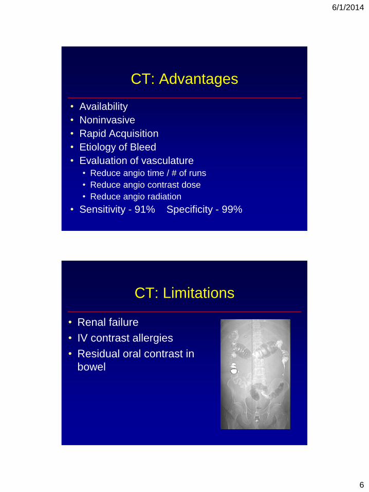
CT: Advantages
• Availability
• Noninvasive
• Rapid Acquisition
• Etiology of Bleed
• Evaluation of vasculature • Reduce angio time / # of runs
• Reduce angio contrast dose
• Reduce angio radiation
• Sensitivity - 91% Specificity - 99%
CT: Limitations
• Renal failure
• IV contrast allergies
• Residual oral contrast in
bowel

6/1/2014
7
CT Angiography for Active Bleed
• No oral contrast
• Triple Phase CT Abdomen and Pelvis
• Unenhanced (Low dose)
• Arterial Phase - 30 sec
100-150 cc contast at 4 cc/sec
• Venous Phase - 70 sec delay
*Alternatively: arterial/venous/delayed
Diagnosis of Active Bleeding
(look for something bright) • High attenuation material in bowel lumen at CTA,
not present at unenhanced CT performed
immediately prior
• Linear, jetlike, pooled or swirled or ellipsoid focal
collection of arterial density contrast material within
bowel lumen
Tew K, et al: MDCT of Acute Lower Gastrointestinal Bleeding. AJR 2004
Laing CF et al. Acute Gastrointestinal Bleeding: Emerging Role of Multidetector CT
Angiography and Review of Current Imaging Techniques. Radiographics 2007.

6/1/2014
8
No IV
Arterial
Venous

6/1/2014
9

6/1/2014
10
False Positives for GI Bleeding
• Residual contrast in bowel/diverticula
• Foreign bodies
• Sutures

6/1/2014
11
False positives: Oral Contrast
• Unchanged between phases
• Often with sharp edges
• Density often brighter than vascular structures
Medication
Evaluation of Occult/Chronic
GI Bleeding
• CT Enterography
• Noninvasive, easy to perform
• View extra-enteric structures
as well as bowel wall
• Detect small bowel tumors
• Detect occult GI bleeding--
hemodynamically stable
• KEY: Negative oral contrast
AND IV contrast

6/1/2014
12
Negative oral contrast vs
Positive oral contrast
Negative Positive
CT Enterography:
Contrast
• ORAL:
• Volumen® (low density barium)
• 3 bottles, 450 cc each = 1.35 L total
• 60, 45, 30 minutes prior
• IV: ~ 150 cc @ 4cc/sec
• Triphasic Acquisition
• Arterial phase: ~ 20 seconds
• Enteric phase: 50 seconds after start
• Delayed phase: 90 seconds after start

6/1/2014
13
CT Enterography:
What to look for… • High attenuation structure
• Tumors (carcinoid)/angiodysplasia
• Intraluminal filling defects/masses
• polyps
• Focal bowel wall thickening
• Neoplasm/vascular
• Dilated feeding artery or early draining vein
• Sign of vascular malformation
• Progressive accumulation of intraluminal contrast
• Bleeding
Intraluminal Masses: Peutz Jeghers Polyps
High attenuation Mass:
Carcinoid

6/1/2014
14
High Attenuation Structure:
Angiodysplasia
Half time quiz….

6/1/2014
15
Crohn’s Disease
• Endoscopy/Capsule Endoscopy
• Small bowel follow through
• Peroral pneumocolon
• SB enteroclysis
• CT Enterography/Enteroclysis
• MR Enterography/Enteroclysis
2-5-2013
5-20-2014

6/1/2014
16
Capsule Endoscopy
• Excellent depiction of mucosa
• Limited ability to localize disease
• Limited submucosal evaluation
• No extraluminal evaluation
• Cannot use with bowel stenosis
35 yo female history of Crohn’s Disease: 3 weeks S/P capsule endoscopy

6/1/2014
17
Role of CTE in Crohn’s Disease
• Differentiate active inflammatory strictures
from fibrotic strictures to guide therapy
• Active bowel disease medical therapy
• Fibrotic strictures surgery, strictureplasty
CTE findings of active mucosal
and mural inflammation
• Mural hyperenhancement
• Mural stratification
• Bowel wall thickening
• Soft-tissue stranding in perienteric fat
• Engorged vasa recta

6/1/2014
18
Mural Hyperenhancement
• Segmental: relative to
nearby normal loops
• Correlates with
histologic findings of
active Crohn’s disease
39 yo with Crohn’s Disease,
presents with vomiting
• Mucosal hyperenhancement active inflammatory disease

6/1/2014
19
Potential Pitfalls
• In late arterial phase:
jejunum enhances > ileum
• Collapsed bowel loops- denser than
distended ones
• Compare SB loops with increased attenuation
with normal-appearing distended loops in same
bowel segment
Normal
Jejunum
Hyperenhancement
Ileum
Normal
Ileum

6/1/2014
20
Mural Stratification on CTE
• Visualization of layers of bowel wall
• Mucosa and serosa enhance avidly,
intervening bowel wall: variable
• fat: indicates past or chronic inflammation
• edema (water): active inflammation
• soft-tissue attenuation: may represent an
inflammatory infiltrate
Fat: chronic Fluid:
Active
Fluid:
Active
Soft Tissue:
Inflammatory
infiltrate
6/1/2014
21
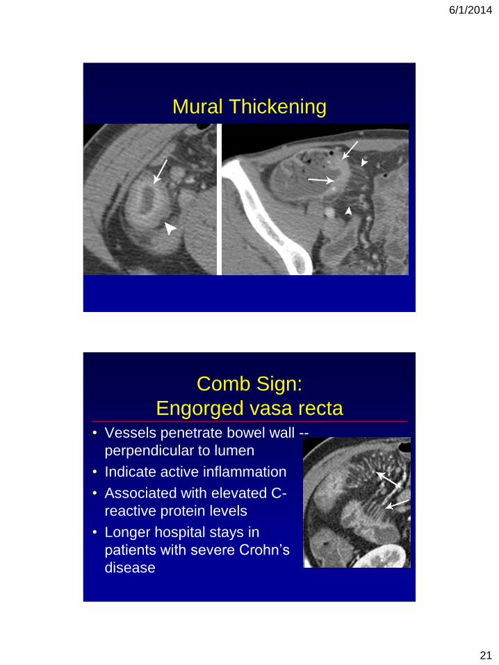
Mural Thickening
Comb Sign:
Engorged vasa recta • Vessels penetrate bowel wall --
perpendicular to lumen
• Indicate active inflammation
• Associated with elevated C-
reactive protein levels
• Longer hospital stays in
patients with severe Crohn’s
disease

6/1/2014
22
Fibrofatty Proliferation
• Occurs along mesenteric border of bowel
• Considered surgically pathognomonic for
Crohn’s disease
• Perienteric fat in patients with Crohn’s
disease not simply result of inflammation
• Is hormonally active
• May help drive inflammatory process
Fistulas
• Hyperenhancing tracts originating from bowel
loops that exhibit signs of active inflammation
• Exception: perianal fistula, often isodense to
anorectum

6/1/2014
23
• Enterovesical fistula in 61 yo man with CD, recurrent
UTI’s and pneumaturia
• Ileoileal fistula arising from ileal loop with acute
asymmetric bowel inflammation with wall thickening
and enhancement
True
lumen
ileum

6/1/2014
24
MRI
• MR Enterography, MR Enteroclysis
• No ionizing radiation
• Excellent tissue contrast
• Intrinsic bowel disease
• Surrounding edema
• Real-time functional imaging
• Evaluation of intra and extra-luminal
pathology
MR Enteroclysis/Enterography
True FISP T1 fat sat post contrast

6/1/2014
25
MRE Technique
• Glucagon: Reduce peristalsis
• VoLumen: Oral Contrast
• 1-2 liters
• (PEG, methylcellulose, water)
• Gadolinium: IV Contrast
• Active inflammation, Fistulas, Abscesses
• MR Protocol:
• Multiple pulse sequences, pre & post contrast
MR Findings and
Disease Subtypes Disease Stage Balanced GRE T2 FSE Post contrast T1
Active Fold thickening,
mural ulcers, comb
sign, mesenteric
lymphadenopathy
Fold thickening,
mural ulcers,
mural edema
Mucosal hyperemia,
mural thickening, mural
edema, comb sign,
mesenteric lymph
nodes
Fibrostenotic Stricture Stricture, mural
fibrosis
Stricture, mural fibrosis
Penetrating Deep fissuring
ulcers, fistulas,
abscess
Deep fissuring
ulcers, fistulas,
abscess
Fistulas, abscess
Regenerative Regenerative
polyps, decreased
luminal diameter
Regenerative
polyps,
decreased
luminal diameter
Decreased luminal
diameter
More than one process may be in a segment or multiple adjacent segments in the same patient

6/1/2014
26
Active Inflammation in Crohn’s Disease
SSFP
Wall thickening
Post-Contrast T1
Enhancement, comb sign
DWI
Restricted diffusion
Ulcerations in
terminal ileum
Fibrofatty mesentery
Ileal wall thickening

6/1/2014
27
Stratified enhancement
Transient bowel collapse mimicking active inflammation
Potential Pitfall

6/1/2014
28
Penetrating Disease in CD
• Transmural extension of inflammatory process
• Deep fissuring ulcers
• Longitudinal or transverse thin high intensity lines in
bowel wall
• Sinus tracts
• Fistulae
• Thicker than adhesions
• Enhance earlier than adhesions
• Abscesses
• Extraintestinal involvement
Enteroenteric fistulas
Enterovesicle fistula

6/1/2014
29
Fibrostenotic
• Fixed mural thickening and luminal narrowing
• Mural fibrosis Strictures Bowel obstruction
• Aperistaltic on cine
• Prestenotic dilatation • Less likely to respond to medical therapy
• Lack mural inflammation and edema
• Usually little to no increased T2 signal
• Reduced enhancement
• Homogenous, not stratified
Fibrostenosing strictures
Prestenotic dilatation
Homogeneous
enhancement

6/1/2014
30
ACR Appropriateness Criteria…
• Evidence-based
guidelines
• Developed to help
referring clinicians
• Choose appropriate
diagnostic imaging test
• ACR.org
• FREE!!!

6/1/2014
31

6/1/2014
32
Final Quiz: Name that foreign
body:
Thank you!
Questions?

6/1/2014
33

6/1/2014
34

6/1/2014
35