Update in the Diagnosis of Gastroesophageal Reflux Disease · gastroesophageal reflux disease the...
5
J Gastrointestin Liver Dis September 2006 Vol.15 No.3, 243-247 Address for correspondence: Radu Tutuian, MD Div. Gastroenterol.Hepatol. University Hospital Ramistrasse 100 CH-8091 Zurich, Switzerland E-mail: [email protected] Update in the Diagnosis of Gastroesophageal Reflux Disease Radu Tutuian Division of Gastroenterology and Hepatology, University Hospital Zurich, Switzerland Abstract Clinical manifestations of gastroesophageal reflux disease (GERD) include heartburn, regurgitation, dysphagia, chest pain, cough and other extraesophageal symptoms. GERD is known to cause erosive esophagitis, Barrett esophagus and has been linked to the development of adenocarcinoma of the esophagus. Currently upper gastrointestinal endoscopy is the main clinical tool for visualizing esophageal lesions. Since the majority of GERD patients do not have endoscopic visible lesions other methods are required to document the abnormal acid exposure in the distal esophagus. For many clinicians ambulatory esophageal pH monitoring is the gold standard in diagnosing GERD since it quantifies distal esophageal acid exposure and allows the evaluation of the relationship between symptoms and acid reflux. The availability of highly selective gastric acid suppressive therapy led to the introduction of short trials of proton pump inhibitors (PPI) to diagnose GERD. PPI trials are often used as a first line diagnostic tool in clinical practice and in particular in the primary care settings. This development has a major influence in the type of patients referred to gastrointestinal specialists, the current trend being that gastroenterologists are asked to evaluate an increasing number of patients with persistent GERD symptoms while on PPI therapy. In these patients the question is whether the persistent symptoms are or not associated with reflux (acid or non-acid). In the recent years combined multichannel intraluminal impedance and pH (MII- pH) monitoring has become a clinical tool that permits the clarification of the mechanisms underlying the persistent symptoms on acid suppressive therapy. Key words Gastroesophageal reflux disease - upper GI endoscopy - pH-metry - multichannel intraluminal impedance and pH monitoring - proton pump inhibitors Introduction Gastroesophageal reflux disease (GERD) is a common, chronic gastrointestinal disorder in Europe and the United States (1). Epidemiologic studies indicate that approximately 40% of the adult US population experiences occasionally heartburn, the most common GERD symptom (2), while 20% of the US population reports heartburn weekly (3). In Romania, the prevalence of gastroesophageal reflux disease is estimated to be around 15-25%. The diagnosis of GERD has evolved over the years influenced by technologic and therapeutic progresses. The clinical armamentarium for diagnosing GERD includes radiographic examinations using contrast dyes, endoscopy, catheters and capsules measuring intraesophageal pH and empiric treatment trials. The indications for various procedures have adapted to the clinical challenges and the type of GERD patients referred to gastrointestinal specialists. The purpose of this review is to discuss the time-honored diagnostic tools as well as the latest developments in the field of gastroesophageal reflux disease. Upper GI series (barium esophagogram) In the beginning of the 20th century physicians recognized the ability of visualizing internal organs using X-Rays. In time the methods have become more sophisticated and are currently an indispensable part of the clinical diagnostic armamentarium. For the evaluation of the hollow organs of the digestive tract radiologists use radio-opaque agents to enhance the contrast of these organs from other structures. Barium esophagograms have been used for many years to evaluate alterations of the esophageal structures.
Transcript of Update in the Diagnosis of Gastroesophageal Reflux Disease · gastroesophageal reflux disease the...

Update in the diagnosis of GERD
J Gastrointestin Liver Dis
September 2006 Vol.15 No.3, 243-247
Address for correspondence: Radu Tutuian, MD
Div. Gastroenterol.Hepatol.
University Hospital
Ramistrasse 100
CH-8091 Zurich, Switzerland
E-mail: [email protected]
Update in the Diagnosis of Gastroesophageal Reflux Disease
Radu Tutuian
Division of Gastroenterology and Hepatology, University Hospital Zurich, Switzerland
Abstract
Clinical manifestations of gastroesophageal reflux
disease (GERD) include heartburn, regurgitation, dysphagia,
chest pain, cough and other extraesophageal symptoms.
GERD is known to cause erosive esophagitis, Barrett
esophagus and has been linked to the development of
adenocarcinoma of the esophagus.
Currently upper gastrointestinal endoscopy is the main
clinical tool for visualizing esophageal lesions. Since the
majority of GERD patients do not have endoscopic visible
lesions other methods are required to document the abnormal
acid exposure in the distal esophagus. For many clinicians
ambulatory esophageal pH monitoring is the gold standard
in diagnosing GERD since it quantifies distal esophageal
acid exposure and allows the evaluation of the relationship
between symptoms and acid reflux.
The availability of highly selective gastric acid
suppressive therapy led to the introduction of short trials
of proton pump inhibitors (PPI) to diagnose GERD. PPI trials
are often used as a first line diagnostic tool in clinical practice
and in particular in the primary care settings. This
development has a major influence in the type of patients
referred to gastrointestinal specialists, the current trend
being that gastroenterologists are asked to evaluate an
increasing number of patients with persistent GERD
symptoms while on PPI therapy. In these patients the
question is whether the persistent symptoms are or not
associated with reflux (acid or non-acid). In the recent years
combined multichannel intraluminal impedance and pH (MII-
pH) monitoring has become a clinical tool that permits the
clarification of the mechanisms underlying the persistent
symptoms on acid suppressive therapy.
Key words
Gastroesophageal reflux disease - upper GI endoscopy -
pH-metry - multichannel intraluminal impedance and pH
monitoring - proton pump inhibitors
Introduction
Gastroesophageal reflux disease (GERD) is a common,
chronic gastrointestinal disorder in Europe and the United
States (1). Epidemiologic studies indicate that approximately
40% of the adult US population experiences occasionally
heartburn, the most common GERD symptom (2), while 20%
of the US population reports heartburn weekly (3). In
Romania, the prevalence of gastroesophageal reflux disease
is estimated to be around 15-25%.
The diagnosis of GERD has evolved over the years
influenced by technologic and therapeutic progresses. The
clinical armamentarium for diagnosing GERD includes
radiographic examinations using contrast dyes, endoscopy,
catheters and capsules measuring intraesophageal pH and
empiric treatment trials. The indications for various
procedures have adapted to the clinical challenges and the
type of GERD patients referred to gastrointestinal specialists.
The purpose of this review is to discuss the time-honored
diagnostic tools as well as the latest developments in the
field of gastroesophageal reflux disease.
Upper GI series (barium esophagogram)
In the beginning of the 20th century physicians
recognized the ability of visualizing internal organs using
X-Rays. In time the methods have become more sophisticated
and are currently an indispensable part of the clinical
diagnostic armamentarium. For the evaluation of the hollow
organs of the digestive tract radiologists use radio-opaque
agents to enhance the contrast of these organs from other
structures. Barium esophagograms have been used for many
years to evaluate alterations of the esophageal structures.

Tutuian244
While Barium esophagograms can reveal mucosal lesion
defects they have been replaced to a great extent by methods
allowing direct visualization of the esophagus (i.e.
endoscopy). Still, barium esophagogram remains an
important tool in diagnosing primary esophageal disorders
such as achalasia, evaluated extrinsic compressions on the
esophagus and evaluating the motility of the upper
esophageal sphincter. In the diagnosis of GERD Barium
esophagograms are helpful in diagnosing hiatus hernias,
peptic esophageal strictures, the opening of the lower
esophageal sphincter and allows the appreciation of
esophageal peristalsis. The limitations of this method are
related to the use of radiating energy and to providing only
a certain degree of diagnostic information. With regards to
gastroesophageal reflux disease the one finding reported
by radiologists that should always be interpreted cautiously,
is the presence of gastroesophageal reflux during
videofluoroscopy. The main reason for this caution is the
limited sensitivity and specificity of these findings. Limited
amounts of gastroesophageal reflux can occur even in normal
healthy volunteers and the radiographic examination
evaluates only a short period of time.
Upper GI endoscopy
In 1946 Allison introduced the term “reflux esophagitis”
as the consequence of gastric acid injury to the esophagus
(4) and for many years this was considered the evidence of
gastroesophageal disease. Esophagogastroduodenoscopy
(EGD) is superior to upper GI series in order to identify
erosive esophagitis and allows the grading of the severity
of esophagitis. Most commonly used scales to grade erosive
esophagitis are the Savary-Miller and the Los Angeles (LA)
classification and the more recently proposed MUSE
classification. In addition to identifying erosive esophagitis,
EGD with biopsies is the method of choice in diagnosing
Barrett esophagus, esophageal erosions due to causes other
than GERD (i.e. viral esophagitis, eosinophilic esophagitis,
Crohn’s disease etc.). Esophagogastroscopy also allows
treating complications of reflux disease such as peptic
strictures, esophageal hemorrhage. In the last years several
endoscopic antireflux procedures have been developed for
the treatment of GERD (i.e. Stretta, Endocinch, Gatekeeper
etc.). Despite these advantages the use of upper GI
endoscopy in the diagnosis of GERD is losing ground and
the main reason for this trend is the availability of highly
active acid suppressive therapy. Proton pump inhibitors
(PPIs) are highly effective in treating erosive esophagitis
healing up to 90% of patients according to large, double-
blind randomized studies (5). Due to the favorable side-
effect profile PPIs are frequently prescribed by primary care
physicians for patients complaining of heartburn, acid
regurgitation or other symptoms attributed to GERD and
refer patients to gastrointestinal specialists mainly when
they do not respond to acid suppressive therapy. Therefore
by the time gastroenterologists perform an upper endoscopy
in GERD patients on PPI therapy the exam is likely to reveal
normal appearing mucosa.
Ambulatory esophageal pH-metry
Systems measuring the intraesophageal acid
concentration (i.e. esophageal pH-metry) were developed
in the 1960’s allowing to quantify the duration of esophageal
acid exposure and to evaluate the relationship between
symptoms and acid reflux. Esophageal pH monitoring is
considered by many investigators the gold-standard to
diagnose GERD. Currently the most accepted criterion to
identify a gastroesophageal reflux during pH monitoring is
a sudden decrease in intraesophageal pH from above to
below 4.0. In the mid 1970’s Johnson and DeMeester
published a set of normal values for pH monitoring including
cut-offs to separate normal from pathologic esophageal acid
exposure which are still accepted by many centers (6).
Measuring esophageal acid exposure in the distal esophagus
allows diagnosing gastroesophageal reflux disease even in
the absence of endoscopic visible lesions. Esophageal pH-
metry has been used over the years to evaluate the effects
of acid suppressive medications and to perform comparisons
between various agents. More recently wireless systems
have become clinically available, improving the patients’
acceptance of this test and allowing esophageal pH
monitoring for extended (up to 96 hours) periods of time
(Fig.1). Currently the most accepted definition of a
gastroesophageal reflux episode during esophageal pH
monitoring is a sudden drop in pH from above to below 4.0
(Fig.2).
Fig.1 Esophageal pH monitoring using either catheter-based
systems or wireless pH capsules.
The limits of esophageal pH monitoring are reached when
it comes to evaluating patients with persistent symptoms
on acid suppressive therapy. These limitations are the result
of the facts that (1) PPI reduce the amount of esophageal
acid exposure and (2) pH criteria for identifying
gastroesophageal reflux episodes with a pH above 4.0 (i.e.
non-acid or weakly acidic reflux episodes) do not have a
good sensitivity and specificity. Therefore conventional pH
Update in the diagnosis of GERD 245
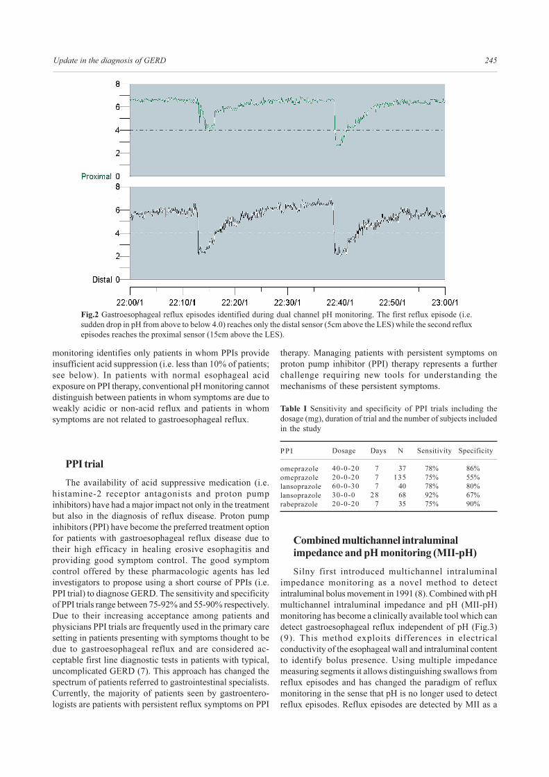
Fig.2 Gastroesophageal reflux episodes identified during dual channel pH monitoring. The first reflux episode (i.e.
sudden drop in pH from above to below 4.0) reaches only the distal sensor (5cm above the LES) while the second reflux
episodes reaches the proximal sensor (15cm above the LES).
monitoring identifies only patients in whom PPIs provide
insufficient acid suppression (i.e. less than 10% of patients;
see below). In patients with normal esophageal acid
exposure on PPI therapy, conventional pH monitoring cannot
distinguish between patients in whom symptoms are due to
weakly acidic or non-acid reflux and patients in whom
symptoms are not related to gastroesophageal reflux.
PPI trial
The availability of acid suppressive medication (i.e.
histamine-2 receptor antagonists and proton pump
inhibitors) have had a major impact not only in the treatment
but also in the diagnosis of reflux disease. Proton pump
inhibitors (PPI) have become the preferred treatment option
for patients with gastroesophageal reflux disease due to
their high efficacy in healing erosive esophagitis and
providing good symptom control. The good symptom
control offered by these pharmacologic agents has led
investigators to propose using a short course of PPIs (i.e.
PPI trial) to diagnose GERD. The sensitivity and specificity
of PPI trials range between 75-92% and 55-90% respectively.
Due to their increasing acceptance among patients and
physicians PPI trials are frequently used in the primary care
setting in patients presenting with symptoms thought to be
due to gastroesophageal reflux and are considered ac-
ceptable first line diagnostic tests in patients with typical,
uncomplicated GERD (7). This approach has changed the
spectrum of patients referred to gastrointestinal specialists.
Currently, the majority of patients seen by gastroentero-
logists are patients with persistent reflux symptoms on PPI
therapy. Managing patients with persistent symptoms on
proton pump inhibitor (PPI) therapy represents a further
challenge requiring new tools for understanding the
mechanisms of these persistent symptoms.
Table I Sensitivity and specificity of PPI trials including the
dosage (mg), duration of trial and the number of subjects included
in the study
PPI
omeprazole
omeprazole
lansoprazole
lansoprazole
rabeprazole
Dosage
40-0-20
20-0-20
60-0-30
30-0-0
20-0-20
Days
7
7
7
28
7
N
37
135
40
68
35
Sensitivity
78%
75%
78%
92%
75%
Specificity
86%
55%
80%
67%
90%
Combined multichannel intraluminal
impedance and pH monitoring (MII-pH)
Silny first introduced multichannel intraluminal
impedance monitoring as a novel method to detect
intraluminal bolus movement in 1991 (8). Combined with pH
multichannel intraluminal impedance and pH (MII-pH)
monitoring has become a clinically available tool which can
detect gastroesophageal reflux independent of pH (Fig.3)
(9). This method exploits differences in electrical
conductivity of the esophageal wall and intraluminal content
to identify bolus presence. Using multiple impedance
measuring segments it allows distinguishing swallows from
reflux episodes and has changed the paradigm of reflux
monitoring in the sense that pH is no longer used to detect
reflux episodes. Reflux episodes are detected by MII as a

Tutuian246
drop in impedance advancing in time from distally to
proximally and pH data are used to classify the reflux episode
as acid (pH <4) or non-acid (pH >4). Since acid suppressive
therapy is thought to change primarily the pH of the refluxate
without decreasing the total number of reflux episodes
combined MII-pH is the ideal technique to monitor
gastroesophageal reflux on acid suppressive therapy (10).
Fig.3 Combined MII-pH probe with 6 impedance measuring
segments and 2 pH sensors (esophageal and gastric) placed in the
esophagus for reflux monitoring on acid suppressive therapy.
Fig.4 Reflux episodes during combined multichannel intraluminal impedance and pH monitoring. Reflux episodes
are detected by impedance as a drop in impedance starting distally and over time advancing proximally. Data
from the pH sensor are used to classify reflux episodes as acid (i.e. drop in esophageal pH from above to below
4.0) or non-acid (i.e. esophageal pH remains above 4.0).
Among patients who have failed to respond to an empiric
trial of PPI ambulatory, patients can be separated in whom
symptoms are associated with acid reflux and non-acid reflux
from those in whom symptoms are not associated with reflux
(Fig.5).
In a large multicenter study including 168 patients referred
for persistent symptoms on twice daily PPI (11) we analyzed
the mechanism of persistent symptoms using combined
MII-pH monitoring. On the day of monitoring, 144 (86%)
patients recorded symptoms allowing the evaluation of the
relationship between symptoms and reflux using the
symptom index (i.e. number of symptoms preceded by a
reflux within 5 minutes divided by the total number of
symptoms (12). A positive symptom index (i.e. SI >50%) for
acid reflux was found in 16 (11%) patients, a positive SI for
non-acid reflux was noted in 53 (37%) patients while in the
remaining 75 (52%) patients the persistent symptoms were
not associated with reflux (i.e. negative SI). Conventional
pH would have been able to identify the positive association
of symptoms and acid reflux (i.e. in 11% of patients)
However, it would not have been able to distinguish, in the
remaining 89% of patients, those with symptoms associated
with non-acid reflux from those with symptoms not related
to reflux. Outcome studies are currently underway evaluating
the clinical significance of the information provided by
combined MII-pH monitoring.
Summary
Clinical tools to diagnose gastroesophageal reflux have
evolved parallel to our understanding of the disease and

Update in the diagnosis of GERD 247
Fig.5 Suggested diagnostic algorithm for GERD.
MII-pH = combined multichannel intraluminal impedance and pH
GER = gastroesophageal reflux
PPI = proton pump inhibitor
References
1. Sandler RS, Everhart JE, Donowitz M et al. The burden of se-
lected digestive diseases in the United States. Gastroenterology
2002; 122:1500-1511
2. Locke GR, Talley NJ, Fett SL, et al. Prevalence and Clinical
spectrum of gastroesophageal reflux: a population based study
in Olmsted County, Minnesota. Gastroenterology 1997;
112:1448-1456
3. Nebel OT, Fornes MF, Castell DO. Symptomatic gastro-
esophageal reflux: incidence and precipitating factors. Am J
Dig Dis 1976;21:953– 956
4. Allison PR. Peptic ulcer of the esophagus. J Thorac Cardiovasc
Surg 1946; 15:308-317
5. Castell DO, Kahrilas PJ, Richter JE, et al. Esomeprazole (40
mg) compared with lansoprazole (30 mg) in the treatment of
erosive esophagitis. Am J Gastroenterol. 2002; 97:575-583
6. Johnson LF, Demeester TR. Twenty-four-hour pH monitoring
of the distal esophagus. A quantitative measure of gastro-
esophageal reflux. Am J Gastroenterol. 1974; 62:325-332
7. DeVault KR, Castell DO. American College of Gastroentero-
logy. Updated guidelines for the diagnosis and treatment of
gastroesophageal reflux disease. Am J Gastroenterol. 2005;
100:190-200
8. Silny J. Intraluminal multiple electric impedance procedure
for measurement of gastrointestinal motility. J Gastrointest
Motil 1991; 3:151-162
9. Tutuian R, Vela MF, Shay SS, Castell DO. Multichannel
intraluminal impedance in esophageal function testing and
gastroesophageal reflux monitoring. J Clin Gastroenterol. 2003;
37:206-215
10. Vela MF, Camacho-Lobato L, Srinivasan R, Tutuian R, Katz
PO, Castell DO. Simultaneous intraesophageal impedance and
pH measurement of acid and nonacid gastroesophageal reflux:
effect of omeprazole. Gastroenterology 2001; 120:1599-1606
11. Mainie I, Tutuian R, Shay S, Vela M, Zhang X, Sifrim D, Castell
D. Acid and non-acid reflux in patients with persistent symptoms
despite acid suppressive therapy. A multicenter study using
combined ambulatory impedance- pH monitoring Gut 2006
Mar 23; (Epub ahead of print)
12. Wiener GJ, Richter JE, Copper JB, Wu WC, Castell DO. The
symptom index: a clinically important parameter of ambulatory
24-hour esophageal pH monitoring. Am J Gastroenterol. 1988;
83:358-361
therapeutic options. While endoscopy remains the main
modality of identifying mucosal or intraluminal lesions and
pH-metry is the main modality to quantify esophageal acid
exposure in untreated patients, these clinical tools are of
limited value in evaluating patients with persistent
symptoms on acid suppressive therapy. Combined
impedance-pH monitoring helps clarifying the mechanisms
of persistent symptoms in patients on PPI therapy and allows
separating patients with symptoms due to persistent acid
or non-acid reflux from those in whom persistent symptoms
are not related to reflux. Rather than being a replacement for
currently available techniques, combined impedance-pH
monitoring should be regarded as an addition to the clinical
armamentarium in the diagnosis of GERD, with excellent
chances of becoming the gold standard for evaluating
patients with persistent symptoms on acid suppressive
therapy.