Unveiling Crucivirus Diversity by Mining Metagenomic Data
19
Portland State University Portland State University PDXScholar PDXScholar Biology Faculty Publications and Presentations Biology 2020 Unveiling Crucivirus Diversity by Mining Unveiling Crucivirus Diversity by Mining Metagenomic Data Metagenomic Data Ignacio de la Higuera Portland State University George Kasun Portland State University Ellis L. Torrance Portland State University Alyssa A. Pratt Portland State University Amberlee Maluenda Portland State University See next page for additional authors Follow this and additional works at: https://pdxscholar.library.pdx.edu/bio_fac Part of the Genetics and Genomics Commons, and the Virology Commons Let us know how access to this document benefits you. Citation Details Citation Details de la Higuera I, Kasun GW, Torrance EL, Pratt AA, Maluenda A, Colombet J, Bisseux M, Ravet V, Dayaram A, Stainton D, Kraberger S, Zawar-Reza P, Goldstien S, Briskie JV, White R, Taylor H, Gomez C, Ainley DG, Harding JS, Fontenele RS, Schreck J, Ribeiro SG, Oswald SA, Arnold JM, Enault F, Varsani A, Stedman KM. 2020. Unveiling crucivirus diversity by mining metagenomic data. mBio 11:e01410-20. https://doi.org/ 10.1128/mBio.01410-20. This Article is brought to you for free and open access. It has been accepted for inclusion in Biology Faculty Publications and Presentations by an authorized administrator of PDXScholar. Please contact us if we can make this document more accessible: [email protected].
Transcript of Unveiling Crucivirus Diversity by Mining Metagenomic Data

Portland State University Portland State University
PDXScholar PDXScholar
Biology Faculty Publications and Presentations Biology
2020
Unveiling Crucivirus Diversity by Mining Unveiling Crucivirus Diversity by Mining
Metagenomic Data Metagenomic Data
Ignacio de la Higuera Portland State University
George Kasun Portland State University
Ellis L. Torrance Portland State University
Alyssa A. Pratt Portland State University
Amberlee Maluenda Portland State University
See next page for additional authors Follow this and additional works at: https://pdxscholar.library.pdx.edu/bio_fac
Part of the Genetics and Genomics Commons, and the Virology Commons
Let us know how access to this document benefits you.
Citation Details Citation Details de la Higuera I, Kasun GW, Torrance EL, Pratt AA, Maluenda A, Colombet J, Bisseux M, Ravet V, Dayaram A, Stainton D, Kraberger S, Zawar-Reza P, Goldstien S, Briskie JV, White R, Taylor H, Gomez C, Ainley DG, Harding JS, Fontenele RS, Schreck J, Ribeiro SG, Oswald SA, Arnold JM, Enault F, Varsani A, Stedman KM. 2020. Unveiling crucivirus diversity by mining metagenomic data. mBio 11:e01410-20. https://doi.org/10.1128/mBio.01410-20.
This Article is brought to you for free and open access. It has been accepted for inclusion in Biology Faculty Publications and Presentations by an authorized administrator of PDXScholar. Please contact us if we can make this document more accessible: [email protected].

Authors Authors Ignacio de la Higuera, George Kasun, Ellis L. Torrance, Alyssa A. Pratt, Amberlee Maluenda, Jonathan Colombet, Maxime Bisseux, Viviane Ravet, Anisha Dayaram, Daisy Stainton, Simona Kraberger, Peyman Zawar-Reza, Sharyn Goldstien, James V. Briskie, Robyn White, Helen Taylor, Christopher Gomez, David G. Ainley, Jon S. Harding, Rafaela S. Fontenele, Joshua Schreck, Simone Ribeiro, Stephen A. Oswald, Jennifer M. Arnold, François Enault, Arvind Varsani, and Kenneth M. Stedman
This article is available at PDXScholar: https://pdxscholar.library.pdx.edu/bio_fac/307

Unveiling Crucivirus Diversity by Mining Metagenomic Data
Ignacio de la Higuera,a George W. Kasun,a Ellis L. Torrance,a Alyssa A. Pratt,a Amberlee Maluenda,a Jonathan Colombet,b
Maxime Bisseux,b Viviane Ravet,b Anisha Dayaram,c Daisy Stainton,d Simona Kraberger,e Peyman Zawar-Reza,f
Sharyn Goldstien,g James V. Briskie,g Robyn White,g Helen Taylor,h Christopher Gomez,i David G. Ainley,j Jon S. Harding,g
Rafaela S. Fontenele,e Joshua Schreck,e Simone G. Ribeiro,k Stephen A. Oswald,l Jennifer M. Arnold,l François Enault,b
Arvind Varsani,e,g,m Kenneth M. Stedmana
aDepartment of Biology, Center for Life in Extreme Environments, Portland State University, Portland, Oregon, USAbUniversité Clermont Auvergne, CNRS, Laboratoire Microorganismes: Génome et Environnement, UMR 6023, Clermont–Ferrand, FrancecInstitut für Neurophysiology, Charité-Universitätsmedizin, Berlin, GermanydDepartment of Entomology and Plant Pathology, Division of Agriculture, University of Arkansas System, Fayetteville, Arkansas, USAeThe Biodesign Center for Fundamental and Applied Microbiomics, Center for Evolution and Medicine, School of Life Sciences, Arizona State University, Tempe, Arizona,USA
fSchool of Earth and Environment, University of Canterbury, Christchurch, New ZealandgSchool of Biological Sciences, University of Canterbury, Christchurch, New ZealandhDepartment of Anatomy, University of Otago, Dunedin, New ZealandiGraduate School of Maritime Sciences, Laboratory of Sediment Hazards and Disaster Risk, Kobe University, Kobe City, JapanjHT Harvey and Associates, Los Gatos, California, USAkEmbrapa Recursos Genéticos e Biotecnologia, Brasília, DF, BrazillDivision of Science, Pennsylvania State University, Reading, Pennsylvania, USAmStructural Biology Research Unit, Department of Clinical Laboratory Sciences, University of Cape Town, Rondebosch, Cape Town, South Africa
ABSTRACT The discovery of cruciviruses revealed the most explicit example of acommon protein homologue between DNA and RNA viruses to date. Crucivirusesare a novel group of circular Rep-encoding single-stranded DNA (ssDNA) (CRESS-DNA) viruses that encode capsid proteins that are most closely related to those en-coded by RNA viruses in the family Tombusviridae. The apparent chimeric nature ofthe two core proteins encoded by crucivirus genomes suggests horizontal genetransfer of capsid genes between DNA and RNA viruses. Here, we identified andcharacterized 451 new crucivirus genomes and 10 capsid-encoding circular geneticelements through de novo assembly and mining of metagenomic data. These ge-nomes are highly diverse, as demonstrated by sequence comparisons and phyloge-netic analysis of subsets of the protein sequences they encode. Most of the variationis reflected in the replication-associated protein (Rep) sequences, and much of thesequence diversity appears to be due to recombination. Our results suggest that re-combination tends to occur more frequently among groups of cruciviruses with rela-tively similar capsid proteins and that the exchange of Rep protein domains be-tween cruciviruses is rarer than intergenic recombination. Additionally, we suggestmembers of the stramenopiles/alveolates/Rhizaria supergroup as possible crucivirushosts. Altogether, we provide a comprehensive and descriptive characterization ofcruciviruses.
IMPORTANCE Viruses are the most abundant biological entities on Earth. In addi-tion to their impact on animal and plant health, viruses have important roles in eco-system dynamics as well as in the evolution of the biosphere. Circular Rep-encodingsingle-stranded (CRESS) DNA viruses are ubiquitous in nature, many are agriculturallyimportant, and they appear to have multiple origins from prokaryotic plasmids. Asubset of CRESS-DNA viruses, the cruciviruses, have homologues of capsid proteinsencoded by RNA viruses. The genetic structure of cruciviruses attests to the transferof capsid genes between disparate groups of viruses. However, the evolutionary his-
Citation de la Higuera I, Kasun GW, TorranceEL, Pratt AA, Maluenda A, Colombet J, BisseuxM, Ravet V, Dayaram A, Stainton D, Kraberger S,Zawar-Reza P, Goldstien S, Briskie JV, White R,Taylor H, Gomez C, Ainley DG, Harding JS,Fontenele RS, Schreck J, Ribeiro SG, Oswald SA,Arnold JM, Enault F, Varsani A, Stedman KM.2020. Unveiling crucivirus diversity by miningmetagenomic data. mBio 11:e01410-20.https://doi.org/10.1128/mBio.01410-20.
Editor Stephen J. Giovannoni, Oregon StateUniversity
Copyright © 2020 de la Higuera et al. This is anopen-access article distributed under the termsof the Creative Commons Attribution 4.0International license.
Address correspondence to Kenneth M.Stedman, [email protected].
Received 29 May 2020Accepted 27 July 2020Published
RESEARCH ARTICLEEcological and Evolutionary Science
crossm
September/October 2020 Volume 11 Issue 5 e01410-20 ® mbio.asm.org 1
1 September 2020
on Septem
ber 8, 2020 by guesthttp://m
bio.asm.org/
Dow
nloaded from
tory of cruciviruses is still unclear. By collecting and analyzing cruciviral sequencedata, we provide a deeper insight into the evolutionary intricacies of cruciviruses.Our results reveal an unexpected diversity of this virus group, with frequent recom-bination as an important determinant of variability.
KEYWORDS crucivirus, CRESS-DNA viruses, gene transfer, recombination, virusevolution, environmental virology
In the last decade, metagenomics has allowed for the study of viruses from a newangle; viruses are not merely agents of disease but abundant and diverse members
of ecosystems (1, 2). Viruses have been shaping the biosphere probably since the originof life, as they are important drivers of the evolution of the organisms they infect (3–5).However, the origin of viruses is not entirely clear. Viruses, as replicons and mobileelements, are also subject to evolution. Virus variability is driven by various mutationrates, recombination, and reassortment of genetic components (6). These attributes,coupled with many types of genomes (RNA or DNA, single or double stranded, andcircular or linear), lead to a large genetic diversity in the “viral world.”
Viruses are generally classified based on the nature of their transmitted geneticmaterial (7). Viral genetic information is coded in either RNA or DNA. Moreover, thesegenomes can be single (positive or negative sense) or double stranded, or linear orcircular, and can be comprised of a single or multiple molecules of nucleic acid(monopartite or multipartite, respectively). These different groups of viruses havedifferent replication strategies, and they harbor distinct taxa based on their genomearrangement and composition (1). The striking differences between viral groups withdisparate genome types suggest polyphyletic virus origins (8).
For example, the highly abundant circular Rep-encoding single-stranded DNA(CRESS-DNA; Rep being the replication-associated protein) viruses may have beenderived from plasmids on multiple occasions by acquiring capsid genes from RNAviruses (9–11). Eukaryotic CRESS-DNA viruses, recently classified into the phylum Cress-dnaviricota (12), constitute a diverse and widespread group of viruses with circulargenomes—some of them multipartite—that contains the families Geminiviridae, Circo-viridae, Nanoviridae, Alphasatellitidae, Genomoviridae, Bacilladnaviridae, Smacoviridae,and Redondoviridae, in addition to vast numbers of unclassified viruses (13, 14).Universal to all CRESS-DNA viruses is the Rep protein, which is involved in the initiationof the virus’ rolling-circle replication. Rep homologues are also encoded in plasmids (14,15). Some pathogenic CRESS-DNA viruses are agriculturally important, such as porcinecircoviruses, and nanoviruses and geminiviruses that infect a wide range of plant hosts(13). However, many CRESS-DNA viruses have been identified in apparently healthyorganisms, and metagenomic studies have revealed their presence in most environ-ments (13).
In 2012, a metagenomic survey of a hot and acidic lake in the volcanic CascadeRange of the western United States uncovered a new type of circular DNA virus (16).The genome of this virus appears to make it a CRESS-DNA virus based on the circularityof its sequence, the presence of a rep gene, and a predicted stem-loop structure witha conserved nucleotide sequence (ori) that serves as an origin for CRESS-DNA virusrolling-circle replication (reviewed in references 17 and 18). Interestingly, the aminoacid sequence of the capsid protein encoded by this genome resembles those encodedby RNA viruses in the family Tombusviridae (16). It was hypothesized that this virusoriginated by the acquisition of a capsid gene from an RNA virus through a yet-to-be-demonstrated RNA-DNA recombination event (16, 19). Since the discovery of thisputatively “chimeric virus,” 80 circular sequences encoding a Rep that shares homologyto ssDNA viruses and a capsid protein that shares homology to tombusvirus capsidproteins have been found in different environments around the globe (20–32). Thisgrowing group of viruses have been branded “cruciviruses,” as they imply the crossingbetween CRESS-DNA viruses and RNA tombusviruses (28). Cruciviruses have beenfound associated with forams (21), alveolates hosted by isopods (27), arthropods (20,
de la Higuera et al. ®
September/October 2020 Volume 11 Issue 5 e01410-20 mbio.asm.org 2
on Septem
ber 8, 2020 by guesthttp://m
bio.asm.org/
Dow
nloaded from

23) and in peatland ecosystems (28), but no host for cruciviruses has been elucidatedto date.
The circular genome of known cruciviruses is variable in size, ranging from 2.7 to5.7 kb, and often contains open reading frames (ORFs) in addition to the Rep and capsidgenes, which have been found in either a unisense or an ambisense orientation (21, 28).The function of additional crucivirus ORFs is unclear due to their lack of sequencesimilarity with any characterized protein. The genome replication of CRESS-DNA virusesis initiated by the Rep protein, which binds to direct repeats present just downstreamof the stem of the ori-containing stem-loop structure and nicks the ssDNA (33, 34). Theexposed 3= OH serves as a primer for cellular enzymes to replicate the viral genome viarolling-circle replication (34–36). The exact terminating events of CRESS-DNA virusreplication are poorly understood for most CRESS-DNA viruses, but Rep is known to beinvolved in the sealing of newly replicated genomes (34, 36–38).
Rep has a domain in the N terminus that belongs to the HUH endonucleasesuperfamily (39). This family of proteins is characterized by a HUH motif (motif II), inwhich two histidine residues are separated by a bulky hydrophobic amino acid, and aTyr-containing motif (motif III) that catalyzes the nicking of the ssDNA (33, 39–41).CRESS-DNA virus Reps also contain a third conserved motif in the N-terminal portion ofthe protein (motif I), likely responsible for double-stranded DNA (dsDNA) bindingspecificity (42). In many CRESS-DNA viruses, the HUH motif has been replaced with asimilar motif that lacks the second histidine residue (e.g., circoviruses have replacedHUH with HLQ) (10, 39). The C-terminal portion of eukaryotic CRESS-DNA virus Repscontains a superfamily 3 helicase domain (S3H) that may be responsible for unwindingdsDNA replicative intermediates (43, 44). This helicase domain is characterized byWalker A and B motifs, motif C, and an Arg finger. Previous studies have identifiedevidence of recombination in the endonuclease and helicase domains of Rep, whichcontributes to the potential ambiguity of Rep phylogenies (45). Interestingly, the Repproteins of different cruciviruses have been shown to be similar to CRESS-DNA virusesin different families, including circoviruses, nanoviruses, and geminiviruses (21, 28). Insome cruciviruses, these differences in phylogeny have been observed between theindividual domains of a single Rep protein (22, 28). The apparent polyphyly of crucivirusReps suggests recombination events involving cruciviruses and other CRESS-DNAviruses, even within Reps (21, 22).
All characterized CRESS-DNA viruses package their DNA into small capsids withicosahedral symmetry or their geminate variants, built from multiple copies of thecapsid protein encoded in their genome (13). The capsid protein of these CRESS-DNAviruses appears to fold into an eight-strand �-barrel that conforms to the single jelly-roll(SJR) architecture, which is also commonly found in eukaryotic RNA viruses (46). Thecapsid protein of cruciviruses has no detectable sequence similarity with the capsid ofother CRESS-DNA viruses and is predicted to adopt the SJR conformation found in thecapsid protein of tombusviruses (16, 21, 22). Three domains can be distinguished intombusviral capsid proteins (47, 48). From the N to the C terminus, they are (i) theRNA-interacting or R-domain, a disordered region that faces the interior of the viralparticle to interact with the nucleic acid through abundant basic residues (49, 50); (ii)the shell or S-domain containing the single jelly-roll fold and the architectural base ofthe capsid (48); and (iii) the protruding or P-domain, which decorates the surface of thevirion and is involved in host transmission (51). In tombusviruses, the S-domains of 180capsid protein subunits interact with each other to assemble around the viral RNA in aT�3 fashion, forming an �35-nm virion (48, 52).
The study of cruciviruses suggests evidence for the transfer of capsid genes betweendisparate viral groups, which can shed light on virus origins and the phenotypicplasticity of virus capsids. Here, we document the discovery of 461 new crucivirus(CruV) genomes and cruci-like circular genetic elements (CruCGEs) identified in met-agenomic data obtained from different environments and organisms. This study pro-vides a comprehensive analysis of this greatly expanded data set and explores the
Unveiling Crucivirus Diversity ®
September/October 2020 Volume 11 Issue 5 e01410-20 mbio.asm.org 3
on Septem
ber 8, 2020 by guesthttp://m
bio.asm.org/
Dow
nloaded from

extent of cruciviral diversity—mostly due to Rep heterogeneity—impacted by rampantrecombination.
RESULTS AND DISCUSSIONExpansion of the crucivirus group. To broaden our understanding of the diversity
and relationships of cruciviruses, 461 uncharacterized circular DNA sequences contain-ing predicted coding sequences (CDSs) with sequence similarity to the capsid proteinof tombusviruses were compiled from metagenomic sequencing data (see Table S1 inthe supplemental material). The data came from published and unpublished metag-enomic studies, carried out in a wide variety of environments, from permafrost totemperate lakes, and on various organisms from red algae to invertebrates (metag-enomes and their metadata are provided in Table S2 in the supplemental material).
The cruciviral sequences were named sequentially, beginning with the smallestgenome, which was named CruV-81 to account for the 80 crucivirus genomes reportedin prior literature (16, 20–32). The average GC content of the newly described cruciviralsequences is 42.9% � 4.9% (Fig. 1B) with genome lengths spanning from 2,474 to 7,947bases (Fig. 1A), some exceeding the size of described bacilladnaviruses (�6,000 nucle-otides [nt] [53]), the largest CRESS-DNA viruses known (12).
Of the 461 sequences that contain a capsid protein ORF, 451 have putative codingregions with sequence similarity to Rep of CRESS-DNA viruses (10). The capsid proteinand Rep ORFs are encoded in a unisense orientation in 40% of the genomes and an
FIG 1 Genome properties of 461 new cruciviral circular sequences. (A) Histogram of cruciviral genomelengths categorized in 50-nt bins. (B) Percentage of G�C content versus A�T in each of the sequencesdescribed in this study. (C) Relative abundance of nucleotides in the conserved nonanucleotide sequenceof the 211 stem-loops and putative origins of replication represented predicted with StemLoop-Finder(A. A. Pratt et al., unpublished) in Sequence Logo format.
de la Higuera et al. ®
September/October 2020 Volume 11 Issue 5 e01410-20 mbio.asm.org 4
on Septem
ber 8, 2020 by guesthttp://m
bio.asm.org/
Dow
nloaded from

ambisense orientation in 58% of the genomes. The remaining �2% correspond to 10CruCGEs with no clear Rep CDS. Five of these CruCGEs contain a predicted origin ofrolling-circle replication (Table S1), indicating that they are circular genomes thatundergo rolling-circle replication characteristic of other CRESS-DNA virus genomes(17, 18).
One possible reason for the lack of a Rep ORF in certain sequences is that some ofthese may be subgenomic molecules or possible components of multipartite viruses(54). Some CRESS-DNA viruses, such as geminiviruses and nanoviruses, have multipar-tite genomes (55). Moreover, some ssRNA tombunodaviruses, including Plasmoparahalstedii virus A and Sclerophthora macrospora virus A—viruses that contain the capsidsequences most similar to cruciviral capsids (16, 28)—also have multipartite genomes(56). Unfortunately, no reliable method yet exists to match different sequences belong-ing to the same multisegmented virus in metagenomes, making identification ofmultipartite or segmented viruses from metagenomic data challenging (55).
Stem-loop structures with conserved nonanucleotide motifs as putative origins ofreplication were predicted and annotated in 277 cruciviral sequences with StemLoop-Finder (A. A. Pratt, I. de la Higuera, E. L. Torrance, G. W. Kasun, and K. M. Stedman,unpublished data). In some cases, more than one nonanucleotide motif with similarscores were found for a single genome, resulting in more than one stem-loop anno-tation. Of the annotated genomes, 223 contain a stem-loop with a nonanucleotide witha NANTANTAN pattern, with the most common sequence being the canonical circovi-rus motif TAGTATTAC, found in 64 of the genomes (Table S1) (57). The majority of the54 sequences that do not correspond to NANTANTAN contain a TAWWDHWAN non-anucleotide motif, typical of genomoviruses (58). The frequency of bases at eachposition in the nonanucleotide sequence is given in Fig. 1C and reflects similarity tomotifs found in other CRESS-DNA viruses (10).
Crucivirus capsid protein. The capsid protein of cruciviruses is predicted to have asingle jelly-roll (SJR) architecture, based on its homology to tombusvirus capsid pro-teins, for which three-dimensional (3D) structures have been determined (Fig. 2A)(59–61). The SJR conformation is found in capsid proteins of both RNA and DNA viruses(46). The SJR capsid protein of tombusviruses and cruciviruses contains three distinctdomains: the RNA-binding or R-domain, the shell or S-domain, and the protruding orP-domain (Fig. 2A). All 461 crucivirus capsid proteins analyzed in this study contain acomplete S-domain. This domain contains a distinct jelly-roll fold and interacts with theS-domain of other capsid subunits in the virion of related tombusviruses (48). TheS-domain of these new crucivirus sequences has greater sequence conservation thanthe remaining regions of the capsid protein (Fig. 2A), likely due to its functionalimportance in capsid structure. In tombusviruses, the S-domain contains a calcium-binding motif (DxDxxD), which was not identified in previously described cruciviruses(62). However, we detected this Ca-binding motif in 68 capsid proteins of the newlyidentified cruciviral sequences. These crucivirus sequences form a distinct cluster,shown in red in Fig. 3B. The S-domain is flanked on the N terminus by the R-domain,which in cruciviruses appears variable in size (up to 320 amino acids long) and appearsto be truncated in some of the capsid protein sequences (e.g., CruV-386 and CruV-493).The R-domain is characterized by an abundance of basic residues at the N terminus,followed by a Gly-rich tract (Fig. 2A). The P-domain, on the C-terminal end of the capsidprotein sequence, is generally the largest domain, with the exception of CruV-385,where it appears to be truncated. The conservation of the capsid protein suggests asimilar structure for all cruciviruses. However, those cruciviruses with larger genomesmay assemble their capsids in a different arrangement to accommodate their genome.While the capsids of tombusviruses have been shown to adopt a T�1 icosahedralconformation, rather than the usual T�3, when the R-domain is partially or totallyremoved (61), we have not seen a correlation between the length of capsid proteindomains and genome size in our data set that could be indicative of alternative capsidarrangements. Furthermore, no packaging dynamics relating genome size and virion
Unveiling Crucivirus Diversity ®
September/October 2020 Volume 11 Issue 5 e01410-20 mbio.asm.org 5
on Septem
ber 8, 2020 by guesthttp://m
bio.asm.org/
Dow
nloaded from

T-number arrangement have been determined in CRESS-DNA viruses, although sub-genomic elements of geminiviruses can be packaged in nongeminate capsids (63, 64).
Interestingly, CruV-420 contains not one but two different tombusvirus-relatedcapsid proteins. A recent compilation of CRESS-DNA viruses from animal metagenomesalso contains four genomes with two different capsid proteins each (32). Whether theseviruses use two different capsid proteins in their capsid (as some RNA viruses do [65]),or whether these are intermediates in the exchange of capsid genes, as predicted fromthe gene capture mechanism proposed by Stedman (19), is unclear. If the latter is true,capsid gene acquisition by CRESS-DNA viruses may be much more common thanpreviously thought.
Crucivirus Rep. The Reps of CRESS-DNA viruses typically contain an endonucleasedomain characterized by conserved motifs I, II, and III and a helicase domain withWalker A and B motifs, motif C, and an Arg finger (Fig. 2B) (13). The majority (85.9%) ofthe crucivirus genomes described in this data set contain all of the expected Rep motifs(Table S4). However, five genomes (CruCGE-110, CruCGE-296, CruCGE-436, CruCGE-471,and CruCGE-533) with overall sequence homology to other Reps (35.8, 32.7, 49.7, 60.2,and 57.2% pairwise identity with other putative Reps in the databases, respectively)lack any detectable conserved motifs within their sequence. Thus, these sequences areconsidered capsid-encoding crucivirus-like circular genetic elements (CruCGEs).
The endonuclease catalytic domain of Rep (motif II), including HUH, was identifiedin 441 of the genomes, 95.2% of which had an alternative HUH, with the most commonarrangement being HUQ (70.0%), also found in circoviruses and nanoviruses (10, 25, 39)(Fig. 2B). Crucivirus motif II deviates from the HUH motif by additionally replacing thesecond hydrophobic residue (U) with a polar amino acid in 26.2% of genomes (Fig. 2B;Table S4), with 53 Reps with the sequence HYQ (12.0%) also found in smacoviruses (10,24, 45).
FIG 2 Protein conservation in cruciviruses. (A) (Top) Distribution of domains, isoelectric point, and conservation in a consensus capsid protein. Four hundredsixty-one capsid protein sequences were aligned in Geneious 11.0.4 with MAFFT (G-INS-i, BLOSUM 45, open gap penalty 1.53, offset 0.123) and trimmedmanually. The conservation of the physicochemical properties at each position was obtained with Jalview v2.11.0 (88), and the isoelectric point was estimatedin Geneious 11.0.4. The region of the capsid protein rich in glycine is highlighted with a green bar. (Bottom) Structure of a cruciviral capsid protein (CruV-359)as predicted by Phyre2 showing sequence conservation based on an alignment of the 47 capsid protein sequences from the capsid protein-based clusters. (B)Conserved motifs found in cruciviral Reps after aligning all the extracted Rep protein sequences using PSI-Coffee (94). Sequence logos were generated athttp://weblogo.threeplusone.com to indicate the frequency of residues at each position.
de la Higuera et al. ®
September/October 2020 Volume 11 Issue 5 e01410-20 mbio.asm.org 6
on Septem
ber 8, 2020 by guesthttp://m
bio.asm.org/
Dow
nloaded from

We identified 13 putative Reps in these crucivirus genomes that lack all four motifstypically found in S3H helicases (e.g., CruV-166, CruV-202, and CruV-499 [Table S4]).Recent work has shown that the deletion of individual conserved motifs in the helicasedomain of the Rep protein of beak and feather disease virus does not abolish ATPaseand GTPase activity (66). The absence of all four motifs may prevent these putative Repsfrom performing helicase and ATPase activity using previously characterized mecha-nisms. However, it is possible that crucivirus Reps that lack these motifs are still capableof ATP hydrolysis and associated helicase activity. Alternatively, these activities may beprovided by host factors (67), or by a viral replication-enhancer protein—as is the casewith the AC3 protein of begomoviruses (68).
We identified 36 crucivirus genomes whose putative rep genes contain in-framestop codons or in which the HUH and SF3 helicase are in different frames, suggestingthat their transcripts may require intron splicing prior to translation. Acceptor anddonor splicing sites identical to those found in maize streak virus (69) were found in allthese sequences, and the putatively spliced Reps were annotated accordingly. In five ofthe 36 spliced Reps, we were unable to detect any of the four conserved motifsassociated with helicase/ATPase activity, which are encoded in the predicted secondexon in most cases. CruV-513 and CruV-518 also contain predicted splicing sites in theircapsid gene.
FIG 3 Diversity of cruciviral proteins. (A) Capsid protein diversity. Pairwise amino acid identity (%PI) between the capsid proteins predicted for 461 cruciviralsequences. The alignment and analysis were carried out with SDT, using the integrated MAFFT algorithm. (B) S-domain diversity. (Left) Pairwise identity matrixbetween the capsid protein predicted S-domains of the 461 sequences described in this study. The alignment and analysis were carried out with SDT, usingthe integrated MAFFT algorithm (87). The colored boxes indicate the different clusters of sequences used to create the capsid protein-based cluster sequencesubset. (Right) Unrooted phylogenetic tree obtained with FastTree from a manually curated MAFFT alignment of the translated sequences of the S-domain(G-INS-i, BLOSUM 45, open gap penalty 1.53, offset 0.123) (93, 96). The colored branches represent the different clusters observed in the matrix. Scale barindicates substitutions per site. (C) Rep diversity. (Left) Pairwise identity matrix between all Reps found in cruciviral genomes in this study. The alignment andanalysis were carried out with SDT, using the integrated MUSCLE algorithm (87). (Right) Unrooted phylogenetic tree obtained with FastTree from a PSI-Coffeealignment of the translated sequences of Rep trimmed with TrimAl v1.3 (93–96). The colored branches represent the different clusters that contain theRep-based cluster sequence subset. Scale bar indicates substitutions per site. (D) Pairwise identity frequency distribution. The frequency of pairwise identityvalues for each of the putative proteins or domains analyzed is shown.
Unveiling Crucivirus Diversity ®
September/October 2020 Volume 11 Issue 5 e01410-20 mbio.asm.org 7
on Septem
ber 8, 2020 by guesthttp://m
bio.asm.org/
Dow
nloaded from

No geminivirus Rep sequence (GRS) motifs—which have been identified as neces-sary for geminivirus replication (70) and have also been found in genomoviruses(58)—were detected in Reps in our data set. We were unable to detect any conservedRep motifs present in cruciviruses that are absent in other CRESS-DNA viruses. Giventhe conservation of Rep motifs in these newly described cruciviruses, we expect mostto be active in rolling-circle replication.
Crucivirus capsid proteins share higher genetic identity than their Rep pro-teins. To assess the diversity in the proteins of cruciviruses, the percent pairwiseidentity between the protein sequences was calculated for capsid protein and Repusing SDTv1.2 (Fig. 3). The average pairwise identity for the capsid protein was foundto be 33.1% � 4.9% (mean � SD) (Fig. 3A and D), likely due to the high levels ofconservation found in the S-domain (40.5% � 8.4%) (Fig. 3B and D), while the averagepairwise identity for Rep is quite low at 24.7% � 5.6% (Fig. 3C and D). The differencesin average pairwise identities between Rep, capsid protein, and S-domain are statisti-cally significant (one-way analysis of variance [ANOVA]; P � 0.0001). The high variationof the Rep protein sequence relative to the capsid protein in cruciviruses correlates witha previous observation on a smaller data set (21).
To compare cruciviruses to other viral groups with homologous proteins, sequencesimilarity networks were built for the capsid protein and Rep (Fig. 4). For the capsidprotein, related protein sequences from tombusviruses and unclassified RNA viruseswere included. The virus sequences were connected when the similarity between theirprotein sequence had an E value of �10�20, sufficient to connect all cruciviruses andtombusviruses, with the exception of CruV-523 (Fig. 4A). However, using BLASTp,CruV-523 showed similarity to other RNA viruses with an E value of �10�9, which werenot included in the analysis. The capsid protein sequence similarity network analysisdemonstrates the apparent homology of the capsid proteins in our data set with thecapsid protein of RNA viruses: specifically, to unclassified RNA viruses that haveRNA-dependent RNA polymerases (RdRPs) similar to those of either tombusviruses—also described as tombus-like viruses (56, 71, 72)— or nodaviruses. The latter RNAviruses are proposed to belong to a chimeric group of viruses named tombunodavi-ruses (73).
For sequence similarity network analysis of Rep, sequences from CRESS-DNA virusesbelonging to the families Circoviridae, Nanoviridae, Alphasatellitidae, Geminiviridae,Genomoviridae, Smacoviridae, and Bacilladnaviridae were used (Fig. 4B). Due to theheterogeneity of Rep (Fig. 3C), the score cutoff for the network was relaxed to an Evalue of �10�10; nonetheless, 10 divergent sequences lacked sufficient similarity toform connections within the network. While the Reps of the different viral familiesclustered in specific regions of the network, the similarity of cruciviral Reps spans thediversity of all CRESS-DNA viruses and blurs the borders between them. Though thereare cruciviruses that appear to be closely related to geminiviruses and genomoviruses,these connections are less common than with other classified CRESS-DNA families(Fig. 4B). While still highly divergent from each other, the conserved motifs in the Repstill share the most sequence similarity with CRESS-DNA viruses (Fig. 2B).
The broad sequence space distribution of cruciviral Rep sequences has been pro-posed to reflect multiple Rep acquisition events through recombination with virusesfrom different CRESS-DNA viral families (21). However, the apparent larger diversity ofcruciviral Reps relative to classified CRESS-DNA viruses can be due to the method ofstudy, as most classified CRESS-DNA viruses have been discovered from infectedorganisms and are grouped mainly based on Rep similarity (1). In contrast, herecrucivirus sequences are selected according to the presence of a tombusvirus-likecapsid protein. Moreover, the Rep of cruciviruses could be subject to higher substitu-tion rates than the capsid protein (27). It is possible that sequence divergence in capsidprotein is more limited than in the Rep due to structural constraints.
Horizontal gene transfer among cruciviruses. To gain insight into the evolution-ary history of cruciviruses, we carried out phylogenetic analyses of their capsid proteins
de la Higuera et al. ®
September/October 2020 Volume 11 Issue 5 e01410-20 mbio.asm.org 8
on Septem
ber 8, 2020 by guesthttp://m
bio.asm.org/
Dow
nloaded from

and Reps. Due to the high sequence diversity in the data set, two smaller subsets ofsequences were analyzed.
(i) Capsid protein-based clusters. Clusters with more than six nonidentical capsidprotein sequences whose S-domains share a pairwise identity greater than 70% werevisually identified from Fig. 3B. This resulted in the identification of seven clusters, and
FIG 4 Similarity networks of cruciviral proteins with related viruses. (A) Capsid proteins represented by coloreddots are connected with a solid line when the pairwise similarity, as assessed by the EFI-EST web server (100), hasan E value of �10�20. The dashed line represents an E value of 6 � 10�7 between the nodes corresponding to thecapsid protein of CruV-523 and turnip crinkle virus, as given by BLASTp. (B) Replication-associated protein (Rep)translations, represented by colored dots, are connected with a solid line when the pairwise similarity has an Evalue of �10�10. The eight nodes at the bottom left did not connect to any other node. All networks were carriedout with pairwise identities calculated in the EFI–EST web server and visualized in Cytoscape v3.7.2 (100, 101).
Unveiling Crucivirus Diversity ®
September/October 2020 Volume 11 Issue 5 e01410-20 mbio.asm.org 9
on Septem
ber 8, 2020 by guesthttp://m
bio.asm.org/
Dow
nloaded from

one more divergent, yet clearly distinct, cluster was included (pink in Fig. 3B). A total of47 genomes from the eight different clusters were selected for sequence comparison.The protein sequences of capsid and Rep were extracted and aligned, and theirphylogenies were inferred and analyzed using tanglegrams (Fig. 5A). The capsid proteinphylogeny shows that the sequences from the eight capsid protein-based clusters formseparate clades (Fig. 5A). On the other hand, the phylogeny of Rep shows a differentpattern of relatedness between those genomes (Fig. 5A). This suggests differentevolutionary histories for the capsid and Rep proteins, which could be due torecombination events between cruciviruses, as previously proposed with smallerdata sets (21, 22).
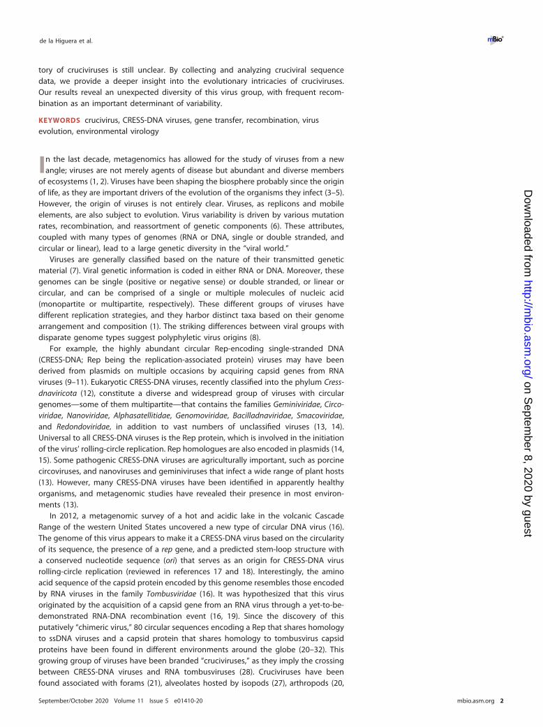
(ii) Rep-based clusters. To account for the possible bias introduced by selectinggenomes from capsid protein cluster groups and to increase the resolution in thephylogeny of the Rep sequences, clusters of crucivirus genomes with more than six Repsequences sharing pairwise identity of 45% and �98% were identified. The cutoffvalues were chosen to allow for the selection of six clusters containing a total of 53genomes (Fig. 3C), whose capsid and Rep protein sequences were analyzed. Thephylogeny of Reps shows distinct clades between the sequences from different Rep-based clusters (Fig. 5B). When the phylogeny of Rep was compared to that of theircorresponding capsid proteins, we observed cruciviruses that group together in bothRep and capsid protein phylogenies. Discrepancies in topology between Rep andcapsid protein trees were observed as well, particularly in the capsid protein clademarked with an asterisk in Fig. 5B. This clade corresponds to the highly homogeneousred capsid protein-based cluster shown in Fig. 3B and suggests that gene transfer ismore common in cruciviruses with a more similar capsid protein, likely infecting thesame type of organism. On the other hand, the presence of cruciviral groups with notrace of genetic exchange may indicate that lineages within the cruciviral group mayhave undergone speciation in the course of evolution.
FIG 5 Comparison of phylogenies of capsid and Rep proteins of representative cruciviruses. (A) Tanglegram calculated with Dendroscope v3.5.10 fromphylogenetic trees generated with PhyML from capsid protein (PhyML automatic model selection LG�G�I�F) and Rep (PhyML automatic model selectionRtREV�G�I) alignments (97, 99). The tips corresponding to the same viral genome are linked by lines that are color coded according to the clusters obtainedfrom Fig. 3A (capsid protein-based clusters). (B) Tanglegram calculated with Dendroscope v3.5.10 from phylogenetic trees generated with PhyML from capsidprotein (PhyML automatic model selection LG�G�I�F) and Rep (PhyML automatic model selection RtREV�G�I) alignments (99). The tips corresponding tothe same viral sequence are linked by lines that are color coded according to the clusters obtained from Fig. 3B (Rep-based clusters). The clade marked witha red asterisk is formed by members of the red capsid protein-based cluster. Branch support is given according to aLRT SH-like (97). All nodes with an aLRTSH-like branch support inferior to 0.8 were collapsed with Dendroscope prior to constructing the tanglegram.
de la Higuera et al. ®
September/October 2020 Volume 11 Issue 5 e01410-20 mbio.asm.org 10
on Septem
ber 8, 2020 by guesthttp://m
bio.asm.org/
Dow
nloaded from
To investigate possible exchanges of individual Rep domains among cruciviruses,the Rep alignments of the analyses of the capsid protein-based and Rep-based clusterswere split at the beginning of the Walker A motif to separate endonuclease andhelicase domains. From the analysis of the capsid protein-based clusters, we observedincongruence in the phylogenies between endonuclease and helicase domains(Fig. 6A), suggesting recombination within crucivirus Reps, as has been previouslyhypothesized with a much smaller data set (22). This incongruency is not observed inthe analyzed Rep-based clusters (Fig. 6B). This is likely due to the higher similaritybetween Reps in this subset of sequences, biased by the clustering based on Rep. Wedo observe different topologies between the trees, which may be a consequence ofdifferent evolutionary constraints to which the endonuclease and helicase domains aresubjected. The detection of capsid protein/Rep exchange and not of individual Repdomains in Rep-based clusters suggests that the rate of intergenic recombination ishigher than intragenic recombination in cruciviruses.
Members of the stramenopiles/alveolates/Rhizaria (SAR) supergroup are po-tential crucivirus hosts. While no crucivirus host has been identified to date, thearchitecture of the Rep protein found in most cruciviruses, as well as the presence ofintrons in some of the genomes, suggests a eukaryotic host. The fusion of an endo-nuclease domain to an S3H helicase domain is observed in other CRESS-DNA viruseswhich are known to infect eukaryotes (39). This is distinct from Reps found inprokaryote-infecting CRESS-DNA viruses—which lack a fused S3H helicase domain(74)—and other related HUH endonucleases involved in plasmid rolling-circle replica-tion and HUH transposases (39). Additionally, the capsid protein of cruciviruses, asuggested determinant of tropism (75, 76), is homologous to the capsid of RNA virusesknown to infect eukaryotes. The RNA viruses with a known host with capsids most
FIG 6 Comparison of phylogenies between the endonuclease and helicase domains of Reps from representative cruciviruses. (A) Tanglegram calculated withDendroscope v3.5.10 from phylogenetic trees generated with PhyML from separate alignments of Rep endonuclease and helicase domains (97, 99). The tipscorresponding to the same viral genome are linked by lines that are color coded according to the clusters obtained from Fig. 3A (capsid protein-based clusters).(B) Same as panel A but with sequences from the clusters obtained from Fig. 3B (Rep-based clusters). All nodes with an aLRT SH-like branch support inferiorto 0.8 were collapsed with Dendroscope v3.5.10 prior to constructing the tanglegram (99).
Unveiling Crucivirus Diversity ®
September/October 2020 Volume 11 Issue 5 e01410-20 mbio.asm.org 11
on Septem
ber 8, 2020 by guesthttp://m
bio.asm.org/
Dow
nloaded from
similar to cruciviral capsids (tombunodaviruses) infect oomycetes, a group of filamen-tous eukaryotic stramenopiles (56).
Cruciviruses have been found as contaminants of spin columns made of diatoma-ceous silica (22), in aquatic metagenomes enriched with unicellular algae (21), in themetagenome of Astrammina rara—a foraminiferan protist part of the Rhizaria (21)—and associated with epibionts of isopods, mainly comprised of apicomplexans andciliates, both belonging to the alveolates (27). These pieces of evidence point towardthe stramenopiles/alveolates/Rhizaria (SAR) supergroup as a candidate taxon to containpotential crucivirus hosts (77). No host prediction can be articulated from our sequencedata. However, at least five of the crucivirus genomes render complete translatedcapsid protein and Rep sequences only when using a relaxed genetic code. Suchalternative genetic codes have been detected in ciliates, in which the hypotheticaltermination codons UAA and UAG encode a glutamine (78). The usage of an alternativegenetic code seems evident in CruV-502—found in the metagenome from seawatercollected above diseased coral colonies (79) that uses a UAA codon for a glutamine ofthe S-domain conserved in 33.5% of the sequences. While the data accumulatedsuggest unicellular eukaryotes and SAR members as crucivirus-associated organisms,the host of cruciviruses remains elusive, and further investigations are necessary.
Classification of cruciviruses. Cruciviruses have circular genomes that encode aRep protein probably involved in rolling-circle replication. The single-stranded nature ofpackaged crucivirus genomes has not been demonstrated experimentally; however, theoverall genomic structure and sequence similarity underpin the placement of crucivi-ruses within the CRESS-DNA viruses.
The classification of the CRESS-DNA viruses is primarily based upon the phylogenyof the Rep proteins, although commonalities in capsid protein and genome organiza-tion are also considered (14). This taxonomic criterion is challenging in cruciviruses,whose Rep proteins are highly diverse and apparently paralogous. Whether the use ofproteins involved in replication for virus classification should be preferred over struc-tural proteins has been previously questioned (80).
The capsid of cruciviruses, as well as the capsid of other CRESS-DNA virus familieslike circoviruses, geminiviruses, and bacilladnaviruses, possesses the single jelly-rollarchitecture (46). However, there is no obvious sequence similarity between the capsidprotein of cruciviruses and that of classified CRESS-DNA viruses. The crucivirus capsidprotein— homologous to the capsid of tombusviruses—is an orthologous trait withinthe CRESS-DNA viruses. Hence, the capsid protein constitutes a synapomorphic char-acter that demarcates this group of viruses from the rest of the CRESS-DNA viralfamilies.
CRESS-DNA viruses appear to have multiple origins from plasmids. Their Rep pro-teins appear to have arisen from these plasmids, and the viruses have diverged intodifferent ssDNA virus groups on acquisition of nonorthologous capsid proteins fromRNA viruses (10, 81). Cruciviruses, however, are classified as such due to shared capsidprotein genes but encode Rep proteins that span many different viral clusters withinthe phylum Cressdnaviricota, as we have shown. Thus, it is unlikely that cruciviruses willform a formal taxon, as they appear to be a collection of viruses from multipleCressdnaviricota groups. However, like Baltimore classes, the label crucivirus can aid inunderstanding virus evolution, particularly the transfer of capsid protein genes, whichappears to have been rampant not only in ssDNA viruses but throughout the viro-sphere (46, 81).
Concluding remarks. Cruciviruses are a growing group of CRESS-DNA viruses thatencode capsid proteins that are homologous to those encoded by tombusviruses. Over500 crucivirus genomes have been recovered from various environments across theglobe. These genomes vary in size, sequence, and genome organization. While cruci-virus capsid proteins are relatively homogeneous, the Reps are relatively diverse amongthe cruciviruses, spanning the diversity of all classified CRESS-DNA viruses. It has beenhypothesized that cruciviruses emerged from the recombination between a CRESS-
de la Higuera et al. ®
September/October 2020 Volume 11 Issue 5 e01410-20 mbio.asm.org 12
on Septem
ber 8, 2020 by guesthttp://m
bio.asm.org/
Dow
nloaded from

DNA virus and a tombus-like RNA virus (16, 19). Furthermore, cruciviruses seem to haverecombined with each other to exchange functional modules between themselves, andprobably with other viral groups, which blurs their evolutionary history. Crucivirusesshow evidence of genetic transfer, not just between viruses with similar genomicproperties but also between disparate groups of viruses such as CRESS-DNA and RNAviruses.
MATERIALS AND METHODSAssembly and recovery of viral genomes. A total of 461 crucivirus-related sequences were
identified from 1,168 metagenomic surveys (see Tables S1 and S2 in the supplemental material). Onethousand one hundred sixty-seven viromes from 57 published data sets and one unpublished viromewere obtained from different types of environments: (i) aquatic systems (freshwater, seawater, hypersa-line ponds, thermal springs, and hydrothermal vents), (ii) engineered systems (bioreactor and foodproduction), and (iii) eukaryote-associated flora (human, insect and other animal feces, human saliva andfluids, cnidarians, and plants). The raw reads from metagenomes were assembled using multiple differentprograms (for details see Table S1), except for the sequences from the work of de Cárcer et al. (82), whichwere already assembled. New cruciviral sequences were identified in these viromes by screening circularcontigs for the presence of capsid proteins from previously known cruciviruses (21) and tombusviruses,using a BLASTx bit-score threshold of 50. The selected genomes are assumed to be complete and circularbased on the terminal redundancy identified in de novo-assembled genomes.
Additionally, sequences CruV-240, CruV-300, CruV-331, CruV-338, and CruV-367 were retrieved asassembled contigs from the Joint Genome Institute (JGI)’s IMG/VR repository (83), by searching scaffoldswith a function set including the protein family pfam00729, corresponding to the S-domain of tombus-virus capsids. The sequences with an RdRP coding region were excluded, and the circularity of thesequences, as well as the presence of an ORF encoding a tombusvirus-like capsid, was confirmed withGeneious 11.0.4 (Biomatters, Ltd.).
Annotation of crucivirus putative genes. The 461 cruciviral sequences were annotated andanalyzed in Geneious 11.0.4. Coding sequences (CDSs) were semiautomatically annotated from a customdatabase (Table S3) of protein sequences of published cruciviruses and close homologues obtained fromGenBank, using Geneious 11.0.4’s annotation function with a 25% nucleotide similarity threshold.Annotated CDSs were rechecked with the GenBank database using BLASTx to identify sequences similarto previously described cruciviruses and putative relatives. Sequences containing in-frame stop codonswere checked for putative splicing sites (69) or translated using a ciliate genetic code only when usagerendered a complete ORF with similarity to other putative crucivirus CDSs. Predicted ORFs longer than300 bases with no obvious homologues and no overlap with capsid protein or Rep-like ORFs wereannotated as “putative ORFs.”
Putative stem-loop annotation. Stem-loop structures that could serve as an origin of replication forcircular ssDNA viruses were identified and annotated using StemLoop-Finder (34, 84; A. A. Pratt et al.,unpublished data). The 461 cruciviral sequences were scanned for the presence of conserved nonanucle-otide motifs described for other CRESS-DNA viruses (NANTANTAN, NAKWRTTAC, TAWWDHWAN, andTRAKATTRC) (13). The integrated ViennaRNA 2.0 library was used to predict secondary structures of DNAaround the detected motif, including the surrounding 15 to 20 nucleotides on either side (85, 86).Predicted structures with a stem longer than 4 bp and a loop including seven or more bases weresubjected to the default scoring system, which increases the score by one point for each deviation fromideal stem lengths of 11 bp and loop lengths of 11 nucleotides. A set of annotations for stem-loops andnonanucleotides was created with StemLoop-Finder for those with a score of 15 or below. Putativestem-loops were excluded from annotation when a separate stem-loop was found with the same firstbase, but they attained a greater score, as well as those that appeared to have a nonanucleotide withinfour bases of their stem-loop structure’s first or last nucleotide.
Conservation analysis and visualization. (i) Pairwise identity matrices. The pairwise identitybetween the protein sequence from translated cruciviral genes was calculated with SDTv1.2 (87), withMAFFT alignment option for capsid proteins and S-domains and MUSCLE alignment options for Reps. Theraw data were further analyzed with Prism v8.4.3.
(ii) Sequence conservation annotation. Capsid protein sequence conservation represented inFig. 2A was generated with Jalview v2.11.0 (88) and reflects the conservation of the physicochemicalproperties for each column of the alignment (89).
(iii) Sequence logos. Sequence logos showing frequency of bases in nonanucleotides at the originof replication or residue in conserved Rep motifs were made using the WebLogo server (http://weblogo.threeplusone.com/) (90).
(iv) Structural representation of capsid conservation. The 3D structure of the capsid protein wasmodeled with Phyre2 (91). The generated graphic was colored by sequence conservation with Chimerav.1.13 (92), from the alignment of the 47 capsid sequences from each of the capsid protein-based clusters(Fig. 3B).
Phylogenetic analyses. (i) Multiple sequence alignments. Capsid protein sequences were alignedusing MAFFT (93) in Geneious 11.0.4, with a G-INS-i algorithm and BLOSUM 45 as exchange matrix, withan open gap penalty of 1.53 and an offset value of 0.123, and manually curated. Rep protein sequenceswere aligned using PSI-Coffee (http://tcoffee.crg.cat/) (94). Rep alignments were manually inspected andcorrected in Geneious 11.0.4 and trimmed using TrimAI v1.3 with a strict plus setting (95). To produce
Unveiling Crucivirus Diversity ®
September/October 2020 Volume 11 Issue 5 e01410-20 mbio.asm.org 13
on Septem
ber 8, 2020 by guesthttp://m
bio.asm.org/
Dow
nloaded from

individual alignments of the endonuclease and helicase domains, the full-length trimmed alignmentswere split at the Walker A motif (45).
(ii) Phylogenetic trees. Phylogenetic trees containing the entire data set of cruciviral sequenceswere built in Geneious using the FastTree plugin (96). For the analysis of sequence subsets, trees wereinferred with the PhyML 3.0 web server (http://www.atgc-montpellier.fr/phyml/) (97), using an aLRTSH-like support (98). The substitution model for each analysis was automatically selected by the program.
(iii) Intergene and interdomain comparison. Tanglegrams were made using Dendroscope v3.5.10(99) to compare the phylogenies between different genes or domains within the same set of crucivirusgenomes.
(iv) Sequence similarity networks. A total of 540 capsid amino acid sequences and 600 Rep aminoacid sequences were uploaded tothe EFI–EST web server for the calculation of pairwise identities(https://efi.igb.illinois.edu/efi-est/) (100). A specific alignment score cutoff was established for each dataset analyzed, and xgmml files generated by EFI-EST were visualized and edited in Cytoscape v3.7.2 (101).
Data availability. Accession numbers are provided in Table S1, and all sequences are provided inText S1.
SUPPLEMENTAL MATERIALSupplemental material is available online only.TEXT S1, DOCX file, 1.9 MB.TABLE S1, PDF file, 1.9 MB.TABLE S2, PDF file, 1.1 MB.TABLE S3, PDF file, 0.1 MB.TABLE S4, PDF file, 0.3 MB.
ACKNOWLEDGMENTSThis work was supported by the NASA Exobiology Program, grant 80NSSC17K0301
(I.D.L.H., G.W.K., E.L.T., A.A.P., and K.M.S.) and the NIH BUILD EXITO Program (A.M.).BUILD EXITO was supported by grants from the National Institutes of Health(UL1GM118964, RL5GM118963, and TL4GM118965) and the Portland State UniversityRonald E. McNair Scholars Program (E.L.T.), supported by grants from the U.S. Depart-ment of Education and Portland State University. The Antarctic field work was sup-ported by the US National Science Foundation (NSF) under grant ANT-0944411, withlogistics supplied by the US Antarctic Program. The freshwater work in New Zealandwas supported by a grant (UC-E6007) from the American New Zealand Association(USA) awarded to P.Z.-R., C.G., J.S.H., and A.V. The green-lipped mussel work wassupported by a grant from the Brian Mason Scientific & Technical Trust of New Zealandawarded to S.G. and A.V. EU-s Horizon 2020 Framework Program for Research andInnovation (‘Virus-X’, project no. 685778) supported F.E.
REFERENCES1. Simmonds P, Adams MJ, Benk M, Breitbart M, Brister JR, Carstens EB,
Davison AJ, Delwart E, Gorbalenya AE, Harrach B, Hull R, King AMQ,Koonin EV, Krupovic M, Kuhn JH, Lefkowitz EJ, Nibert ML, Orton R,Roossinck MJ, Sabanadzovic S, Sullivan MB, Suttle CA, Tesh RB, Van DerVlugt RA, Varsani A, Zerbini FM. 2017. Consensus statement: virustaxonomy in the age of metagenomics. Nat Rev Microbiol 15:161–168.https://doi.org/10.1038/nrmicro.2016.177.
2. Chow C-ET, Suttle CA. 2015. Biogeography of viruses in the sea.Annu Rev Virol 2:41– 66. https://doi.org/10.1146/annurev-virology-031413-085540.
3. Koonin EV, Dolja VV. 2013. A virocentric perspective on the evolution oflife. Curr Opin Virol 3:546 –557. https://doi.org/10.1016/j.coviro.2013.06.008.
4. Koonin EV, Krupovic M. 2018. The depths of virus exaptation. Curr OpinVirol 31:1– 8. https://doi.org/10.1016/j.coviro.2018.07.011.
5. Berliner AJ, Mochizuki T, Stedman KM. 2018. Astrovirology: viruses atlarge in the universe. Astrobiology 18:207–223. https://doi.org/10.1089/ast.2017.1649.
6. Domingo E, Sheldon J, Perales C. 2012. Viral quasispecies evolution.Microbiol Mol Biol Rev 76:159 –216. https://doi.org/10.1128/MMBR.05023-11.
7. Baltimore D. 1971. Expression of animal virus genomes. Bacteriol Rev35:235–241. https://doi.org/10.1128/MMBR.35.3.235-241.1971.
8. Koonin EV, Senkevich TG, Dolja VV. 2006. The ancient virus world and
evolution of cells. Biol Direct 1:29. https://doi.org/10.1186/1745-6150-1-29.
9. Krupovic M, Ravantti JJ, Bamford DH. 2009. Geminiviruses: a tale of aplasmid becoming a virus. BMC Evol Biol 9:112. https://doi.org/10.1186/1471-2148-9-112.
10. Kazlauskas D, Varsani A, Koonin EV, Krupovic M. 2019. Multiple originsof prokaryotic and eukaryotic single-stranded DNA viruses from bac-terial and archaeal plasmids. Nat Commun 10:3425. https://doi.org/10.1038/s41467-019-11433-0.
11. Krupovic M. 2012. Recombination between RNA viruses and plasmidsmight have played a central role in the origin and evolution of small DNAviruses. Bioessays 34:867–870. https://doi.org/10.1002/bies.201200083.
12. Krupovic M, Varsani A, Kazlauskas D, Breitbart M, Delwart E, Rosario K,Yutin N, Wolf YI, Harrach B, Zerbini FM, Dolja VV, Kuhn JH, Koonin EV.2020. Cressdnaviricota : a virus phylum unifying seven families ofRep-encoding viruses with single-stranded, circular DNA genomes. JVirol 94:e00582-20. https://doi.org/10.1128/JVI.00582-20.
13. Zhao L, Rosario K, Breitbart M, Duffy S. 2019. Eukaryotic circular Rep-encoding single-stranded DNA (CRESS DNA) viruses: ubiquitous viruseswith small genomes and a diverse host range. Adv Virus Res 103:71–133. https://doi.org/10.1016/bs.aivir.2018.10.001.
14. Rosario K, Duffy S, Breitbart M. 2012. A field guide to eukaryotic circularsingle-stranded DNA viruses: insights gained from metagenomics. ArchVirol 157:1851–1871. https://doi.org/10.1007/s00705-012-1391-y.
de la Higuera et al. ®
September/October 2020 Volume 11 Issue 5 e01410-20 mbio.asm.org 14
on Septem
ber 8, 2020 by guesthttp://m
bio.asm.org/
Dow
nloaded from

15. Cheung AK. 2015. Specific functions of the Rep and Rep’ proteins ofporcine circovirus during copy-release and rolling-circle DNA replica-tion. Virology 481:43–50. https://doi.org/10.1016/j.virol.2015.01.004.
16. Diemer GS, Stedman KM. 2012. A novel virus genome discovered in anextreme environment suggests recombination between unrelatedgroups of RNA and DNA viruses. Biol Direct 7:13. https://doi.org/10.1186/1745-6150-7-13.
17. Cheung AK. 2012. Porcine circovirus: transcription and DNA replication.Virus Res 164:46 –53. https://doi.org/10.1016/j.virusres.2011.10.012.
18. Laufs J, Jupin I, David C, Schumacher S, Heyraud-Nitschke F, Gronen-born B. 1995. Geminivirus replication: genetic and biochemical charac-terization of rep protein function, a review. Biochimie 77:765–773.https://doi.org/10.1016/0300-9084(96)88194-6.
19. Stedman K. 2013. Mechanisms for RNA capture by ssDNA viruses: grandtheft RNA. J Mol Evol 76:359 –364. https://doi.org/10.1007/s00239-013-9569-9.
20. Rosario K, Dayaram A, Marinov M, Ware J, Kraberger S, Stainton D,Breitbart M, Varsani A. 2012. Diverse circular ssDNA viruses discoveredin dragonflies (Odonata: Epiprocta). J Gen Virol 93:2668 –2681. https://doi.org/10.1099/vir.0.045948-0.
21. Roux S, Enault F, Bronner G, Vaulot D, Forterre P, Krupovic M. 2013.Chimeric viruses blur the borders between the major groups of eukary-otic single-stranded DNA viruses. Nat Commun 4:2700. https://doi.org/10.1038/ncomms3700.
22. Krupovic M, Zhi N, Li J, Hu G, Koonin EV, Wong S, Shevchenko S, ZhaoK, Young NS. 2015. Multiple layers of chimerism in a single-strandedDNA virus discovered by deep sequencing. Genome Biol Evol7:993–1001. https://doi.org/10.1093/gbe/evv034.
23. Hewson I, Ng G, Li WF, LaBarre BA, Aguirre I, Barbosa JG, Breitbart M,Greco AW, Kearns CM, Looi A, Schaffner LR, Thompson PD, Hairston NG.2013. Metagenomic identification, seasonal dynamics, and potentialtransmission mechanisms of a Daphnia-associated single-strandedDNA virus in two temperate lakes. Limnol Oceanogr 58:1605–1620.https://doi.org/10.4319/lo.2013.58.5.1605.
24. Steel O, Kraberger S, Sikorski A, Young LM, Catchpole RJ, Stevens AJ,Ladley JJ, Coray DS, Stainton D, Dayaram A, Julian L, van Bysterveldt K,Varsani A. 2016. Circular replication-associated protein encoding DNAviruses identified in the faecal matter of various animals in New Zea-land. Infect Genet Evol 43:151–164. https://doi.org/10.1016/j.meegid.2016.05.008.
25. McDaniel LD, Rosario K, Breitbart M, Paul JH. 2014. Comparativemetagenomics: natural populations of induced prophages demon-strate highly unique, lower diversity viral sequences. Environ Microbiol16:570 –585. https://doi.org/10.1111/1462-2920.12184.
26. Dayaram A, Galatowitsch ML, Argüello-Astorga GR, van Bysterveldt K,Kraberger S, Stainton D, Harding JS, Roumagnac P, Martin DP, LefeuvreP, Varsani A. 2016. Diverse circular replication-associated protein en-coding viruses circulating in invertebrates within a lake ecosystem.Infect Genet Evol 39:304 –316. https://doi.org/10.1016/j.meegid.2016.02.011.
27. Bistolas K, Besemer R, Rudstam L, Hewson I. 2017. Distribution andinferred evolutionary characteristics of a chimeric ssDNA virus associ-ated with intertidal marine isopods. Viruses 9:361. https://doi.org/10.3390/v9120361.
28. Quaiser A, Krupovic M, Dufresne A, Francez A-J, Roux S. 2016. Diversity andcomparative genomics of chimeric viruses in Sphagnum- dominated peat-lands. Virus Evol 2:vew025. https://doi.org/10.1093/ve/vew025.
29. Salmier A, Tirera S, de Thoisy B, Franc A, Darcissac E, Donato D, BouchierC, Lacoste V, Lavergne A. 2017. Virome analysis of two sympatric batspecies (Desmodus rotundus and Molossus molossus) in French Guiana.PLoS One 12:e0186943. https://doi.org/10.1371/journal.pone.0186943.
30. de la Higuera I, Torrance EL, Pratt AA, Kasun GW, Maluenda A, StedmanKM. 2019. Genome sequences of three cruciviruses found in the Wil-lamette Valley (Oregon). Microbiol Resour Announc 8:e00447-19. https://doi.org/10.1128/MRA.00447-19.
31. Kraberger S, Argüello-Astorga GR, Greenfield LG, Galilee C, Law D,Martin DP, Varsani A. 2015. Characterisation of a diverse range ofcircular replication-associated protein encoding DNA viruses recoveredfrom a sewage treatment oxidation pond. Infect Genet Evol 31:73– 86.https://doi.org/10.1016/j.meegid.2015.01.001.
32. Tisza MJ, Pastrana DV, Welch NL, Stewart B, Peretti A, Starrett GJ, PangY-YS, Krishnamurthy SR, Pesavento PA, McDermott DH, Murphy PM,Whited JL, Miller B, Brenchley J, Rosshart SP, Rehermann B, Doorbar J,Ta’ala BA, Pletnikova O, Troncoso JC, Resnick SM, Bolduc B, Sullivan MB,
Varsani A, Segall AM, Buck CB. 2020. Discovery of several thousandhighly diverse circular DNA viruses. Elife 9:555375. https://doi.org/10.7554/eLife.51971.
33. Brown DR, Schmidt-Glenewinkel T, Reinberg D, Hurwitz J. 1983. DNAsequences which support activities of the bacteriophage phiX174 geneA protein. J Biol Chem 258:8402– 8412.
34. Steinfeldt T, Finsterbusch T, Mankertz A. 2006. Demonstration ofnicking/joining activity at the origin of DNA replication associated withthe Rep and Rep’ proteins of porcine circovirus type 1. J Virol 80:6225– 6234. https://doi.org/10.1128/JVI.02506-05.
35. Gassmann M, Focher F, Buhk HJ, Ferrari E, Spadari S, Hübscher U. 1988.Replication of single-stranded porcine circovirus DNA by DNA poly-merases � and �. Biochim Biophys Acta 951:280 –289. https://doi.org/10.1016/0167-4781(88)90098-x.
36. Roth MJ, Brown DR, Hurwitz J. 1984. Analysis of bacteriophage phiX174gene A protein-mediated termination and reinitiation of phiX DNAsynthesis. II. Structural characterization of the covalent phiX A protein-DNA complex. J Biol Chem 259:10556 –10568.
37. Cheung AK. 2007. A stem-loop structure, sequence non-specific, at theorigin of DNA replication of porcine circovirus is essential for termina-tion but not for initiation of rolling-circle DNA replication. Virology363:229 –235. https://doi.org/10.1016/j.virol.2007.01.017.
38. Stenger DC, Revington GN, Stevenson MC, Bisaro DM. 1991. Replica-tional release of geminivirus genomes from tandemly repeated copies:evidence for rolling-circle replication of a plant viral DNA. Proc NatlAcad Sci U S A 88:8029 – 8033. https://doi.org/10.1073/pnas.88.18.8029.
39. Chandler M, de la Cruz F, Dyda F, Hickman AB, Moncalian G, Ton-HoangB. 2013. Breaking and joining single-stranded DNA: the HUH endonu-clease superfamily. Nat Rev Microbiol 11:525–538. https://doi.org/10.1038/nrmicro3067.
40. Ilyina TV, Koonin EV. 1992. Conserved sequence motifs in the initiatorproteins for rolling circle DNA replication encoded by diverse repliconsfrom eubacteria, eucaryotes and archaebacteria. Nucleic Acids Res20:3279 –3285. https://doi.org/10.1093/nar/20.13.3279.
41. Koonin EV, Ilyina TV. 1993. Computer-assisted dissection of rolling circleDNA replication. Biosystems 30:241–268. https://doi.org/10.1016/0303-2647(93)90074-m.
42. Londoño A, Riego-Ruiz L, Argüello-Astorga GR. 2010. DNA-bindingspecificity determinants of replication proteins encoded by eukaryoticssDNA viruses are adjacent to widely separated RCR conserved motifs.Arch Virol 155:1033–1046. https://doi.org/10.1007/s00705-010-0674-4.
43. Gorbalenya AE, Koonin EV. 1993. Helicases: amino acid sequence com-parisons and structure-function relationships. Curr Opin Struct Biol3:419 – 429. https://doi.org/10.1016/S0959-440X(05)80116-2.
44. Clerot D, Bernardi F. 2006. DNA helicase activity is associated with thereplication initiator protein Rep of tomato yellow leaf curl geminivirus.J Virol 80:11322–11330. https://doi.org/10.1128/JVI.00924-06.
45. Kazlauskas D, Varsani A, Krupovic M. 2018. Pervasive chimerism in thereplication-associated proteins of uncultured single-stranded DNA vi-ruses. Viruses 10:187. https://doi.org/10.3390/v10040187.
46. Krupovic M, Koonin EV. 2017. Multiple origins of viral capsid proteinsfrom cellular ancestors. Proc Natl Acad Sci U S A 114:E2401–E2410.https://doi.org/10.1073/pnas.1621061114.
47. Hopper P, Harrison SC, Sauer RT. 1984. Structure of tomato bushy stuntvirus. V. Coat protein sequence determination and its structural implica-tions. J Mol Biol 177:701–713. https://doi.org/10.1016/0022-2836(84)90045-7.
48. Sherman MB, Guenther R, Reade R, Rochon D, Sit T, Smith TJ. 2019.Near-atomic-resolution cryo-electron microscopy structures of cucum-ber leaf spot virus and red clover necrotic mosaic virus: evolutionarydivergence at the icosahedral three-fold axes. J Virol 94:e01439-19.https://doi.org/10.1128/JVI.01439-19.
49. Alam SB, Reade R, Theilmann J, Rochon DA. 2017. Evidence for the roleof basic amino acids in the coat protein arm region of Cucumbernecrosis virus in particle assembly and selective encapsidation of viralRNA. Virology 512:83–94. https://doi.org/10.1016/j.virol.2017.09.003.
50. Park SH, Sit TL, Kim KH, Lommel SA. 2013. The red clover necroticmosaic virus capsid protein N-terminal amino acids possess specificRNA binding activity and are required for stable virion assembly. VirusRes 176:107–118. https://doi.org/10.1016/j.virusres.2013.05.014.
51. Ohki T, Akita F, Mochizuki T, Kanda A, Sasaya T, Tsuda S. 2010. Theprotruding domain of the coat protein of Melon necrotic spot virus isinvolved in compatibility with and transmission by the fungal vector
Unveiling Crucivirus Diversity ®
September/October 2020 Volume 11 Issue 5 e01410-20 mbio.asm.org 15
on Septem
ber 8, 2020 by guesthttp://m
bio.asm.org/
Dow
nloaded from

Olpidium bornovanus. Virology 402:129 –134. https://doi.org/10.1016/j.virol.2010.03.020.
52. Llauró A, Coppari E, Imperatori F, Bizzarri AR, Castón JR, Santi L,Cannistraro S, De Pablo PJ. 2015. Calcium ions modulate the mechanicsof tomato bushy stunt virus. Biophys J 109:390 –397. https://doi.org/10.1016/j.bpj.2015.05.039.
53. Kazlauskas D, Dayaram A, Kraberger S, Goldstien S, Varsani A, KrupovicM. 2017. Evolutionary history of ssDNA bacilladnaviruses features hor-izontal acquisition of the capsid gene from ssRNA nodaviruses. Virology504:114 –121. https://doi.org/10.1016/j.virol.2017.02.001.
54. Sicard A, Michalakis Y, Gutiérrez S, Blanc S. 2016. The strange lifestyleof multipartite viruses. PLoS Pathog 12:e1005819. https://doi.org/10.1371/journal.ppat.1005819.
55. Varsani A, Lefeuvre P, Roumagnac P, Martin D. 2018. Notes on recom-bination and reassortment in multipartite/segmented viruses. CurrOpin Virol 33:156 –166. https://doi.org/10.1016/j.coviro.2018.08.013.
56. Grasse W, Spring O. 2017. ssRNA viruses from biotrophic Oomycetesform a new phylogenetic group between Nodaviridae and Tombusviri-dae. Arch Virol 162:1319 –1324. https://doi.org/10.1007/s00705-017-3243-2.
57. Rosario K, Mettel KA, Benner BE, Johnson R, Scott C, Yusseff-VanegasSZ, Baker CCM, Cassill DL, Storer C, Varsani A, Breitbart M. 2018. Virusdiscovery in all three major lineages of terrestrial arthropods highlightsthe diversity of single-stranded DNA viruses associated with inverte-brates. PeerJ 6:e5761. https://doi.org/10.7717/peerj.5761.
58. Varsani A, Krupovic M. 2017. Sequence-based taxonomic framework forthe classification of uncultured single-stranded DNA viruses of thefamily Genomoviridae. Virus Evol 3:vew037. https://doi.org/10.1093/ve/vew037.
59. Harrison SC, Olson AJ, Schutt CE, Winkler FK, Bricogne G. 1978. Tomatobushy stunt virus at 2.9 Å resolution. Nature 276:368 –373. https://doi.org/10.1038/276368a0.
60. Chelvanayagam G, Heringa J, Argos P. 1992. Anatomy and evolution ofproteins displaying the viral capsid jellyroll topology. J Mol Biol 228:220 –242. https://doi.org/10.1016/0022-2836(92)90502-b.
61. Katpally U, Kakani K, Reade R, Dryden K, Rochon D, Smith TJ. 2007.Structures of T�1 and T�3 particles of cucumber necrosis virus: evi-dence of internal scaffolding. J Mol Biol 365:502–512. https://doi.org/10.1016/j.jmb.2006.09.060.
62. Campbell JW, Clifton IJ, Greenhough TJ, Hajdu J, Harrison SC, Lidding-ton RC, Shrive AK. 1990. Calcium binding sites in tomato bushy stuntvirus visualized by Laue crystallography. J Mol Biol 214:627– 632.https://doi.org/10.1016/0022-2836(90)90278-T.
63. Casado CG, Javier Ortiz G, Padron E, Bean SJ, McKenna R, Agbandje-McKenna M, Boulton MI. 2004. Isolation and characterization of sub-genomic DNAs encapsidated in “single” T � 1 isometric particles ofMaize streak virus. Virology 323:164 –171. https://doi.org/10.1016/j.virol.2004.02.014.
64. Bennett A, Rodriguez D, Lister S, Boulton M, McKenna R, Agbandje-McKenna M. 2018. Assembly and disassembly intermediates of maizestreak geminivirus. Virology 525:224–236. https://doi.org/10.1016/j.virol.2018.09.011.
65. Luque D, Mata CP, González-Camacho F, González JM, Gómez-Blanco J,Alfonso C, Rivas G, Havens WM, Kanematsu S, Suzuki N, Ghabrial SA,Trus BL, Castón JR. 2016. Heterodimers as the structural unit of the T�1capsid of the fungal double-stranded RNA Rosellinia necatrix quadrivi-rus 1. J Virol 90:11220 –11230. https://doi.org/10.1128/JVI.01013-16.
66. Chen JK, Hsiao C, Wu JS, Lin SY, Wang CY. 2019. Characterization of theendonuclease activity of the replication-associated protein of beak andfeather disease virus. Arch Virol 164:2091–2106. https://doi.org/10.1007/s00705-019-04292-z.
67. Rizvi I, Choudhury NR, Tuteja N. 2015. Insights into the functionalcharacteristics of geminivirus rolling-circle replication initiator proteinand its interaction with host factors affecting viral DNA replication.Arch Virol 160:375–387. https://doi.org/10.1007/s00705-014-2297-7.
68. Pasumarthy KK, Choudhury NR, Mukherjee SK. 2010. Tomato leaf curlKerala virus (ToLCKeV) AC3 protein forms a higher order oligomer andenhances ATPase activity of replication initiator protein (Rep/AC1). VirolJ 7:128. https://doi.org/10.1186/1743-422X-7-128.
69. Wright EA, Heckel T, Groenendijk J, Davies JW, Boulton MI. 1997.Splicing features in maize streak virus virion- and complementary-sense gene expression. Plant J 12:1285–1297. https://doi.org/10.1046/j.1365-313x.1997.12061285.x.
70. Nash TE, Dallas MB, Reyes MI, Buhrman GK, Ascencio-Ibañez JT, Hanley-
Bowdoin L. 2011. Functional analysis of a novel motif conserved acrossgeminivirus Rep proteins. J Virol 85:1182–1192. https://doi.org/10.1128/JVI.02143-10.
71. Shi M, Lin XD, Tian JH, Chen LJ, Chen X, Li CX, Qin XC, Li J, Cao JP, EdenJS, Buchmann J, Wang W, Xu J, Holmes EC, Zhang YZ. 2016. Redefiningthe invertebrate RNA virosphere. Nature 540:539 –543. https://doi.org/10.1038/nature20167.
72. Dolja VV, Koonin EV. 2018. Metagenomics reshapes the concepts ofRNA virus evolution by revealing extensive horizontal virus transfer.Virus Res 244:36 –52. https://doi.org/10.1016/j.virusres.2017.10.020.
73. Greninger AL, DeRisi JL. 2015. Draft genome sequence of tombunoda-virus UC1. Genome Announc 3:e00655-15. https://doi.org/10.1128/genomeA.00655-15.
74. Krupovic M. 2013. Networks of evolutionary interactions underlying thepolyphyletic origin of ssDNA viruses. Curr Opin Virol 3:578 –586. https://doi.org/10.1016/j.coviro.2013.06.010.
75. Allison AB, Organtini LJ, Zhang S, Hafenstein SL, Holmes EC, Parrish CR.2016. Single mutations in the VP2 300 loop region of the three-foldspike of the carnivore parvovirus capsid can determine host range. JVirol 90:753–767. https://doi.org/10.1128/JVI.02636-15.
76. Carbonell A, Maliogka VI, Pérez JDJ, Salvador B, León DS, García JA,Simón-Mateo C. 2013. Diverse amino acid changes at specific positionsin the N-terminal region of the coat protein allow Plum pox virus toadapt to new hosts. Mol Plant Microbe Interact 26:1211–1224. https://doi.org/10.1094/MPMI-04-13-0093-R.
77. Beakes GW, Glockling SL, Sekimoto S. 2012. The evolutionary phylog-eny of the oomycete “fungi.” Protoplasma 249:3–19. https://doi.org/10.1007/s00709-011-0269-2.
78. Hanyu N, Kuchino Y, Nishimura S, Beier H. 1986. Dramatic events inciliate evolution: alteration of UAA and UAG termination codons toglutamine codons due to anticodon mutations in two TetrahymenatRNAs Gln. EMBO J 5:1307–1311. https://doi.org/10.1002/j.1460-2075.1986.tb04360.x.
79. Soffer N, Brandt ME, Correa AMS, Smith TB, Thurber RV. 2014. Potentialrole of viruses in white plague coral disease. ISME J 8:271–283. https://doi.org/10.1038/ismej.2013.137.
80. Krupovic M, Bamford DH. 2010. Order to the viral universe. J Virol84:12476 –12479. https://doi.org/10.1128/JVI.01489-10.
81. Koonin EV, Dolja VV, Krupovic M, Varsani A, Wolf YI, Yutin N, Zerbini FM,Kuhn JH. 2020. Global organization and proposed megataxonomy ofthe virus world. Microbiol Mol Biol Rev 84:e00061-19. https://doi.org/10.1128/MMBR.00061-19.
82. De Cárcer DA, López-Bueno A, Pearce DA, Alcamí A. 2015. Biodiversityand distribution of polar freshwater DNA viruses. Sci Adv 1:e1400127.https://doi.org/10.1126/sciadv.1400127.
83. Paez-Espino D, Chen I-MA, Palaniappan K, Ratner A, Chu K, Szeto E,Pillay M, Huang J, Markowitz VM, Nielsen T, Huntemann M, Reddy TBK,Pavlopoulos GA, Sullivan MB, Campbell BJ, Chen F, McMahon K, HallamSJ, Denef V, Cavicchioli R, Caffrey SM, Streit WR, Webster J, Handley KM,Salekdeh GH, Tsesmetzis N, Setubal JC, Pope PB, Liu W-T, Rivers AR,Ivanova NN, Kyrpides NC. 2017. IMG/VR: a database of cultured anduncultured DNA viruses and retroviruses. Nucleic Acids Res 45:D457–D465. https://doi.org/10.1093/nar/gkw1030.
84. Hafner GJ, Dale JL, Harding RM, Wolter LC, Stafford MR. 1997. Nickingand joining activity of banana bunchy top virus replication protein invitro. J Gen Virol 78:1795–1799. https://doi.org/10.1099/0022-1317-78-7-1795.
85. Lorenz R, Bernhart SH, Höner zu Siederdissen C, Tafer H, Flamm C,Stadler PF, Hofacker IL. 2011. ViennaRNA package 2.0. Algorithms MolBiol 6:26. https://doi.org/10.1186/1748-7188-6-26.
86. Mathews DH, Sabina J, Zuker M, Turner DH. 1999. Expanded sequencedependence of thermodynamic parameters improves prediction ofRNA secondary structure. J Mol Biol 288:911–940. https://doi.org/10.1006/jmbi.1999.2700.
87. Muhire BM, Varsani A, Martin DP. 2014. SDT: a virus classification toolbased on pairwise sequence alignment and identity calculation. PLoSOne 9:e108277. https://doi.org/10.1371/journal.pone.0108277.
88. Waterhouse AM, Procter JB, Martin DMA, Clamp M, Barton GJ. 2009.Jalview version 2—a multiple sequence alignment editor and analysisworkbench. Bioinformatics 25:1189 –1191. https://doi.org/10.1093/bioinformatics/btp033.
89. Livingstone CD, Barton GJ. 1993. Protein sequence alignments: a strat-egy for the hierarchical analysis of residue conservation. Bioinformatics9:745–756. https://doi.org/10.1093/bioinformatics/9.6.745.
de la Higuera et al. ®
September/October 2020 Volume 11 Issue 5 e01410-20 mbio.asm.org 16
on Septem
ber 8, 2020 by guesthttp://m
bio.asm.org/
Dow
nloaded from

90. Crooks GE, Hon G, Chandonia JM, Brenner SE. 2004. WebLogo: asequence logo generator. Genome Res 14:1188 –1190. https://doi.org/10.1101/gr.849004.
91. Kelley LA, Mezulis S, Yates CM, Wass MN, Sternberg MJE. 2015. ThePhyre2 web portal for protein modeling, prediction and analysis. NatProtoc 10:845– 858. https://doi.org/10.1038/nprot.2015.053.
92. Pettersen EF, Goddard TD, Huang CC, Couch GS, Greenblatt DM, MengEC, Ferrin TE. 2004. UCSF Chimera—a visualization system for explor-atory research and analysis. J Comput Chem 25:1605–1612. https://doi.org/10.1002/jcc.20084.
93. Katoh K, Standley DM. 2013. MAFFT multiple sequence alignmentsoftware version 7: improvements in performance and usability. MolBiol Evol 30:772–780. https://doi.org/10.1093/molbev/mst010.
94. Floden EW, Tommaso PD, Chatzou M, Magis C, Notredame C, ChangJ-M. 2016. PSI/TM-Coffee: a web server for fast and accurate multiplesequence alignments of regular and transmembrane proteins usinghomology extension on reduced databases. Nucleic Acids Res 44:W339 –W343. https://doi.org/10.1093/nar/gkw300.
95. Capella-Gutiérrez S, Silla-Martínez JM, Gabaldón T. 2009. trimAl: atool for automated alignment trimming in large-scale phylogeneticanalyses. Bioinformatics 25:1972–1973. https://doi.org/10.1093/bioinformatics/btp348.
96. Price MN, Dehal PS, Arkin AP. 2009. Fasttree: computing large minimumevolution trees with profiles instead of a distance matrix. Mol Biol Evol26:1641–1650. https://doi.org/10.1093/molbev/msp077.
97. Guindon S, Dufayard JF, Lefort V, Anisimova M, Hordijk W, Gascuel O.2010. New algorithms and methods to estimate maximum-likelihoodphylogenies: assessing the performance of PhyML 3.0. Syst Biol 59:307–321. https://doi.org/10.1093/sysbio/syq010.
98. Anisimova M, Gascuel O. 2006. Approximate likelihood-ratio test forbranches: a fast, accurate, and powerful alternative. Syst Biol 55:539 –552. https://doi.org/10.1080/10635150600755453.
99. Huson DH, Scornavacca C. 2012. Dendroscope 3: an interactive tool forrooted phylogenetic trees and networks. Syst Biol 61:1061–1067.https://doi.org/10.1093/sysbio/sys062.
100. Zallot R, Oberg N, Gerlt JA. 2019. The EFI web resource for genomicenzymology web tools: leveraging protein, genome, and metagenomedatabases to discover novel enzymes and metabolic pathways. Biochem-istry 58:4169–4182. https://doi.org/10.1021/acs.biochem.9b00735.
101. Shannon P, Markiel A, Ozier O, Baliga NS, Wang JT, Ramage D, Amin N,Schwikowski B, Ideker T. 2003. Cytoscape: a software environment forintegrated models of biomolecular interaction networks. Genome Res13:2498 –2504. https://doi.org/10.1101/gr.1239303.
Unveiling Crucivirus Diversity ®
September/October 2020 Volume 11 Issue 5 e01410-20 mbio.asm.org 17
on Septem
ber 8, 2020 by guesthttp://m
bio.asm.org/
Dow
nloaded from