Unusual Sights Unusual Appearances or Behaviors Unusual Odors Unusual Noises
Unusual Behavior of the Cytoplasmic Transcript of hsro:: an Abundant, Stress-inducible RNA that Is Translated But Yields No Detectable Protein Product M. El izabe th F in i , Wi l l i am G. B e n d e n a , a n d M a r y L o u P a r d u e
Department of Biology, Massachusetts Institute of Technology, Cambridge, Massachusetts 02139
Abstract. Although a major site of transcription in heat shock, the Drosophila hsroJ gene does not encode any known heat shock proteins. Instead, studies of the hsroJ transcripts suggest that the RNA molecules, rather than encoded proteins, are the active products of this gene. The cytoplasmic RNA, c03, is spliced and polyadenylated and yet has only very small open read- ing frames (ORFs), and these are poorly conserved in different Drosophila species. Surprisingly, the work reported here leads us to conclude that one of the tiny
ORFs in this RNA is translated. This ORE designated ORF-o:, is notable in being the only ORF that shows sequence conservation in the three Drosophila species examined. However, translation of this ORF does not lead to detectable accumulation of the protein product. We suggest that ORF-~ may be an example of an un- usual type of translated ORE The act of translation it- self may be important rather than the generation of a functional protein product. This nonproductive transla- tion may play a role in regulation of cellular activities.
TIVATION of genes by temperature shifts, the "heat shock response, was first identified in Drosophila by the cytological techniques that allowed visualization
of gene activity as specific puffs on polytene chromosomes (Ritossa, 1962). As biochemical methods became available, the heat shock response was found to be inducible in cells of all tissues and to be common to all organisms, from bacte- ria to plants and animals (for review, see Craig, 1985; Lind- quist, 1986). Furthermore, the set of heat shock genes was also found to be induced by a number of other stimuli that, like a heat shock, could be classified as cellular stress agents. Studies on many organisms have now suggested that the re- sponse is a general cellular homeostatic mechanism. Heat shock-induced proteins have amino acid sequences that have been extremely conserved during evolution. The evolution- ary conservation of the heat shock response argues for its importance to cells of all types, yet little is known about the function of the heat shock proteins.
The first five Drosophila melanogaster heat shock loci to be cloned encoded all of the major heat shock proteins, rais- ing questions about the role of the last major heat shock- induced locus, in polytene region 93D (Craig, 1985). In addition to its lack of identification with a protein product, the 93D puff has other unusual properties (for review, see Lakhotia, 1987; Bendena et al., 1989a). A major portion of the transcripts from this gene are found in the nucleus (Len-
Dr. Fini's present address is Eye Research Institute of the Retina Founda- tion, 20 Staniford Street, Boston, MA 02114. Dr. Bendena's present address is Department of Biology, Queens University, Kingston Ontario K7L 3N6, Canada.
gyel et al., 1980). In polytene chromosomes, the 93D puff contains large RNP granules not seen in other puffs (Dangli et al., 1983). The 93D puff is induced independently of the rest of the heat shock set with agents such as benzamide and colchicine, suggesting that its activity is more sensitive to other stimuli than the rest of the heat shock genes (Lakhotia and Singh, 1982).
Recently, the 93D gene has been cloned from D. melano- gaster, as have the equivalent genes from D. hydei (2-48B) and D. pseudoobscura (58C) (Walldorf et al., 1984; Garbe and Pardue, 1986; Ryseck et al., 1987; Garbe et al., 1986; Garbe et al., 1989). The DNA sequences of these three loci are highly diverged, and yet the loci share a unique molecu- lar structure and have small regions of conserved sequence at several places in the transcription unit. In addition, in each species, this is a major heat shock puff, and it is also induced by the other agents that induce 93D (e.g., benzamide and colchicine), binds antibodies that bind 93D uniquely, and contains large RNP granules (for review, see Lakhotia, 1987). Each of the loci is the single locus in the species that shares these phenotypic and molecular characteristics, giving strong evidence that 93D, 2-48B, and 58C are homologous. Because of this homology each of the loci has been named the hsro~ locus for its species. (Although it is now known that the hsro~ locus is constitutively expressed, the name hsr6o reflects its original identification as the locus encoding the 60 set of heat shock RNAs.)
The hsr~o gene, like the gene for hsp82, is a constitutive gene whose expression is elevated, rather than newly in- duced, by heat shock. In both normal and stressed cells, the locus produces three transcripts, all from the same start site.
© The Rockefeller University Press, 0021-9525/89/06/2045/13 $2.00 The Journal of Cell Biology, Volume 108, June 1989 2045-2057 2045
on January 7, 2019jcb.rupress.org Downloaded from http://doi.org/10.1083/jcb.108.6.2045Published Online: 1 June, 1989 | Supp Info:

The largest transcript of hsr,,,, wl, which encompasses the entire transcription unit of ~10 kb, is marked by some 7-8 kb of a short tandem repeat at the 3' end. This is the transcript that accumulates to very high levels in the nucleus. A smaller nuclear transcript (,x,l.9 kb), 602, appears to be the result of alternate termination at a polyadenylation site just upstream of the tandem repeats. The 1.9-kb RNA species is not as abundant as the 601 transcript and apparently acts as the splic- ing precursor to an abundant cytoplasmic RNA, oJ3. The o~3 transcript is 1.2-1.3 kb (depending on the Drosophila spe- cies) and is formed from 602 by removal of a single intron (Garbe et at., 1986).
The cytoplasmic RNA, 603, has unusual properties. All previously known RNAs of this size that are spliced and polyadenylated act as messages and encode proteins. Sur- prisingly, the sequences of the hsr60 loci from D. melanogas- ter, D. hydei, and D. pseudoobscura do not show any long open reading frames (ORFs).' In fact, the short ORFs in this transcript are all within the range that might occur by chance alone and are shorter than some of the ORFs that are found in the nontranscribed strand of the hsr60 sequence. Furthermore, comparison of the sequences from the three Drosophila species show no conservation of either the posi- tions or the amino acid sequences of all but one of these ORFs. The single exception would yield only a small poly- peptide of 23-27 amino acids, depending on the Drosophila species (Garbe et at., 1986; Garbe et al., 1989). Therefore, it appears likely that the cytoplasmic RNA, ~3, does not serve simply to provide the information for a protein but, in- stead, has some other function in and of itself.
To look for clues to the function of hsro:, we investigated the properties of the hsr60 cytoplasmic RNA in cultured cells. Contrary to expectation, these studies, as reported here, have led us to conclude that the 603 RNA has an ORF that is trans- lated. The ORF, which is translated and now called ORF-o~, is the only one showing sequence conservation in the three Drosophila species examined. However, translation of this ORF does not lead to detectable accumulation of the protein product. We suggest that ORF-60 may be an example of an unusual type of translated ORE The act of translation itself, rather than the generation of a functional protein product, may be of importance in this case. Other studies have shown that 603 transcripts turn over rapidly in control cells but ac- cumulate to high levels whenever protein synthesis is in- hibited (Bendena et al., 1989b). We suggest that nonproduc- tive translation of ORF-60 allows the level of co 3 to reflect the level of protein synthesis in the cell and thus provides infor- mation that is used in coordinating intracellular activities.
Materials and Methods
Cell Growth and Preparation of CeU Lysates
D. melanogaster cells of the Schneider line 2-L (Storti et al., 1980) were grown in DME supplemented with 10% FCS, 0.5% lactalbumin hydroly- sate, and MEM nonessential amino acids. Schneider line 3 ($3) cells (D. melanogaster) (Schneider and Blumenthal, 1978) and/9, hydei cells of the KUN-DH-15 line (Sondermeijer et al., 1980) were grown in Schneider's medium supplemented with 10% FCS, 50 U/mi penicillin, and 50/~g/ml streptomycin. (All cell growth materials from Gibco Laboratories, Grand
1. Abbreviations used in this paper: CAT, chloramphenicol acetyltransfer- ase; ORF, open reading frame; $3 Schneider line 3.
Island, NY) Melanogaster line 2-L cells were maintained in spinner culture at 3-15 x 106 cells/ml by dilution every 1-3 d. D. melanogaster $3 cells and hydei cells were maintained at similar levels by 5-10-fold dilutions every 3-7 d. For use in an experiment, line 2-L cells were maintained at 3-6 x 10⁶ cells/ml by daily dilution for at least 3 d. Line 3 cells and hydei cells were diluted 5-10-fold, allowed to grow to 6 × 10⁶ cells/ml, or 1.5 x 10⁷, respectively, then diluted a second time, and used when they had grown to 3-6 x 10~/ml or 1-1.5 x 10⁷, respectively (mid to late log phase of growth).
Sucrose Gradient Sedimentation
A cytoplasmic fraction of cell lysates was prepared and analyzed on 15-50% sucrose gradients exactly as described by Ballinger and Pardue (1983), ex- cept that lysates were prepared using 0.1% Triton to prevent nuclear leakage and lysis. However, high salt conditions (0.5 M KCI) were maintained to minimize nonspecific molecular associations and to dissociate "vacant cou- ples" (monosomes not associated with mRNA). Identical conditions were used for preparation and centrifugation of cell lysates on 5-20% sucrose gradients. For each gradient, a cell lysate prepared from 1-3 x l0 S cells of the 2-L line or 0.5-1 x l0 s of line 3 cells or D. hydei cells was used for a gradient of 27 ml volume. In some experiments, cleared cell lysates were treated for 15 min with 25 mM EDTA on ice before sedimentation on su- crose gradients supplemented with 10 mM EDTA. For studies requiring in- hibition of translation, cells were treated at 25°C with cycloheximide at 10 -4 M for 2 h (Lodish, 1971), with pactamycin at 10 -7 M for 2 h (Stewart- Blair et al., 1971), or with 120 #g/ml L-l-p-tosylamino-2-phenylethyl chlo- romethyl ketone (Pong et al., 1975) for 15 min at 25°C before preparation of cell lysates.
Metrizamide Gradient Sedimentation
Cell lysates of Schneider line 2-L cells were analyzed by equilibrium gra- dient centrifugation on metrizamide exactly as described by Ballinger and Pardue (1983). Gradients were 5 ml in volume and 20% of the amount of cell lysate was loaded on metrizamide gradients as compared to the amount used for sucrose gradients.
Cell Labeling and Protein Gel Electrophoresis
One milliliter of cells was concentrated into 25/zl of Shields and Sang salts (Schneider and Blumenthal, 1978) and labeled with 5 t~l of [35S]methio- nine (15 #Ci/t~l, sp act, 800 Ci/mM) for 10 or 30 min. In some experiments, cells were lysed in 100 ttl of lysis buffer (100 mM Tris, pH 6.8, 1% SDS) and boiled immediately for 5 min. Proteins were separated from free amino acids by precipitation in 10 vol of acetone. Proteins were then dissolved in sample buffer containing 0.1% SDS and boiled for 3 min with or without the addition of ~-mercaptoethanol to 5 %. In other experiments, cells were lysed in sample buffer containing 0.l % SDS and loaded on gets directly after boiling for 3 min with or without #-mercaptoethanol added to 5 %. Samples were electrophoresed on acrylamide gels containing 7.5 M urea, 0.1% SDS (Garbe and Pardue, 1986) or on standard Laemmli gels (Laemmli, 1970).
Cloning Vector A derivative of pSVOSma-CAT obtained from M. Gilman (Massachusetts Institute of Technology), was used as the cloning vector for assay of hsr~o gene fragments. This vector was constructed by replacement of the pBR322 sequences found in pSVOSma-CAT (Gorman, 1986) with the vector, pUC13 (Yanisch-Perron et al., 1985). Such replacement incorporates the pUCI3 polycloning site directly 5' to the bacterial gene for chloramphenicol acetyltransferase (CAT). About 150 bases around the polylinker of this vec- tor construct were sequenced independently by this lab to establish corre- spondence to the expected sequence deduced from published results.
Establishment of Transformed Cell Lines and CAT Assay $3 cells were transformed with construct DNA and heterogeneous cell lines were established as described (Bourouis and Jarry, 1983). Transformed cell lines were sufficiently amplified for use in an experiment within 4-5 wk. The level of CAT activity was analyzed in cell extracts prepared as described (Gorman, 1986), but extracts from these established, transformed cell lines needed to be diluted ,~100-fold over amounts required for assay of transient CAT expression.
The Journal of Cell Biology, Volume 108, 1989 2046
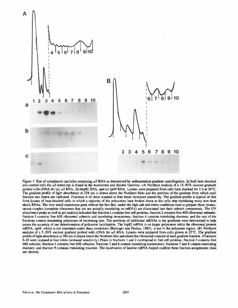
Figure L Size of cytoplasmic particles containing ~03 RNA as determined by sedimentation gradient centrifugation. In both heat-shocked and control cells the c03 transcript is found in the monosome and disome fractions. (A) Northern analysis of a 15-50% sucrose gradient probed with cDNA for (a) -,3 RNA, (b) hsp82 RNA, and (c) rp49 RNA. Lysates were prepared from cells heat shocked for 1 h at 36°C. The gradient profile of light absorbance at 254 nm is drawn above the Northern blots and the portions of the gradient from which each fraction was drawn are indicated. Fractions 4-10 were scanned at four times increased sensitivity. The gradient profile is typical of that from lysates of heat-shocked cells in which a majority of the polysomes have broken down as the cells stop translating many non-heat shock RNAs. The very small monosome peak reflects the fact that, under the high salt and triton conditions used to prepare these lysates, vacant couples (complete ribosomes that are not actually translating on mRNA) are dissociated into their subunit components. The UV absorbance peaks as well as gel analysis indicated that fraction I contains free cell proteins; fraction 2 contains free 40S ribosomal subunits; fraction 3 contains free 60S ribosomal subunits and translating monosomes; fraction 4 contains translating disomes; and the rest of the fractions contain translating polysomes of increasing size. The positions of additional mRNAs in the gradients were determined to help assess the accuracy of our determination of polysome localization. The hsp82 mRNA is on larger polysomes while the ribosomal protein mRNA, rp49, which is not translated under these conditions (Ballinger and Pardue, 1983), is not in the polysome region. (B) Northern analysis of a 5-20% sucrose gradient probed with cDNA for o~3 RNA. Lysates were prepared from cells grown at 25°C. The gradient profile of light absorbance at 280 nm is drawn above the Northern blot and shows the ribosomal contents of each gradient fraction. (Fractions 6-10 were scanned at four times increased sensitivity.) Peaks in fractions 1 and 2 correspond to free cell proteins; fraction 3 contains free 40S subunits; fraction 4 contains free 60S subunits; fractions 5 and 6 contain translating monosomes; fractions 7 and 8 contain translating disomes; and fraction 9 contains translating trisomes. The localization of historic mRNA helped confirm these fraction assignments (data not shown).
Fini et al. The Cytoplasmic RNA of hsrw Is Translated 2047

Protein Gel Blotting and Antibody Probing
A crude protein preparation was made from the pelleted material obtained from 1 rnl of tissue culture cells by lysis in 100/~1 of Laemmli sample buffer. ~mercaptoethanol was then added to some samples to 5 %, the samples were boiled for 5 min, and 25 #1 of the lysate was loaded onlo a single lane of a standard 15 % acrylamide gel. Gels were electroblotted to nitrocellulose (electroblot) and probed with a rabbit antibody against bacterial CAT (5 Prime ~ 3 Prime, Westchester, PA) at an antibody dilution of 1:50000.
Results
Localization of hsro~ Cytoplasmic RNP, ~03, to a Distinct Cytoplasmic Fraction
The ORFs in the hsrw transcript would encode only small polypeptides (Garbe et al., 1986; 1989). Searches for pro- teins of the appropriate size, in cells containing abundant hsr60 transcripts, have been unsuccessful (Garbe and Pardue, 1986; Lakhotia, 1987; see below). These results support the hypothesis, discussed above, that the cytoplasmic transcript of hsr60 acts as a functional molecule in itself. To provide clues to possible functions, the hsr60 cytoplasmic RNA, 603, was examined in its native RNP form. Tissue culture cells of the D. melanogaster line Schneider 2-L or of the Drosoph- ila hydei cell line, KUN-DH-15, were lysed by treatment with 0.1% Triton X-100 in the presence of an RNAse inhibitor and fractionated by differential centrifugation. These initial crude cell fractionation experiments demonstrated that much of the 603 RNA was located in the postmitochondrial supernatant. To examine the size of native 603 RNA-containing particles, a postmitochondrial supernatant was prepared and sediment- ed on 15-50% or 5-20% sucrose gradients. Gradients were collected in 10 fractions and the position of 603 RNA in the gradient was determined by Northern blotting of total RNA extracted from each fraction. A tracing of the UV absor- bance profile, showing ribosome distribution (the predomi- nant particle in these lysates), was used to obtain the approxi- mate sedimentation coefficient of each fraction. Fig. 1 shows an example of one such experiment. On 15-50% sucrose gradients (Fig. 1 A), the 603 transcript exhibited a wide distri- bution across the top of the gradient appearing strongly in the second, third, and fourth fractions. This distribution in- dicated sedimentation with particles between 40S and 120S. Expansion of the area between these sedimentation values by sedimentation of cell lysates on 5-20 % gradients (Fig. 1 B) showed 603 RNA distributed most strongly in particles of a size between 80S and 120S, precisely in the fractions corre- sponding with single complete ribosomes (monosomes) and disomes. Identical results were obtained whether lysates were from cells grown at ambient temperatures or cells that had been heat shocked for 1 h at 36°C. Also, no differences were seen in sedimentation behavior between the cell lines of the two Drosophila species examined.
The position of 60 3 sedimentation is that expected if this RNA were being translated on polysomes of monosome and disome size, consistent with the small size of 60 30RFs. Be- cause translation of ~0 3 had not been expected, a comparison of the gradient positions of several other translatable RNAs was made to ensure the accuracy of our polysomal localiza- tion. The comparisons were accomplished by stripping the Northern blot of 60 3 probe and rehybridizing with additional probes. An example of this type of experiment is shown in Fig. 1 A. For the experiment shown, the cells were heat
shocked before lysis. The RNA for hsp82, which is trans- lated under heat shock conditions, was distributed across the gradient beginning at about 40S, but was most prevalent in fractions containing larger polysomes, consistent with the fairly sizable ORF of this message. Conversely, the RNA for the ribosomal protein, rp49, is found only in the first two fractions, containing only particles smaller than mono- somes. This is the result expected since rp49 RNA is not translated during heat shock and has been shown to leave the polysomes completely (Ballinger and Pardue, 1983). There- fore, the conclusion that particles containing 603 RNA colocalize with monosomes and disomes, that had been based on the identification of polysome peaks by UV light absorbance, is supported by localization of other RNAs known to be associated with polysomes.
The hsr~o Cytoplasmic Transcript ~o3 Is Associated with Polysomes of a Size Consistent with Translation of Its Small ORF
Polysomes of monosome and disome size are fully consistent with those expected for translation of the small ORFs found in the 603 transcript, and thus 603 RNP could colocalize with these particles because it is translated. However, untrans- lated, free cytoplasmic RNP particles of a size as large as monosomes and disomes have been described (McCarthy et al., 1983). To determine whether the 603 cytoplasmic RNA was actually associated with polysomes or in a free RNP par- ticle, we have tested other attributes of bona fide polysomes. The detergent sodium deoxycbolate, which is known to dis- rupt the structure of some free RNPs, but not polysomes (Faiferman et al., 1971), did not disturb the colocalization of 603 RNA and monosomes/disomes in the Schneider 2-L cells. In addition, increasing monovalent cation concentra- tion (K ÷ or Na +) in the gradient over a broad range of 10-500 mM (which would be expected to strip loosely as- sociated proteins from RNPs differentially according to their individual structures) had no effect on this colocalization (data not shown).
Density gradient centrifugation fractionates RNPs on the basis of their protein/RNA ratio, while sedimentation gra- dient centrifugation fractionates on the basis of molecular size. Therefore, cell lysates were banded on metrizamide density gradients as an alternate method of testing the colocalization of 60 3 RNA-containing particles with translat- ing polysomes. As shown in Fig. 2, the 603 cytoplasmic RNP was found in the region of the gradient that contained poly- somes. Lysates of Schneider 2-L cells that had been heat shocked at 36°C for 1 h were used for the experiment shown; the 603 RNA had the same density when obtained from cells held at ambient temperature. An example of a translating RNA, coding for hsp82, was found near the bottom of the gradient (fraction 3), consistent with polysomal localization (Ballinger and Pardue, 1983). In contrast, the RNA coding for the ribosomal protein, rp49, which becomes released from polysomes during heat shock (Ballinger and Pardue, 1983), was found distributed much more broadly in the gra- dient, at densities that might be expected for free RNPs (McCarthy et al., 1983). The 603 RNA was found near the bottom of the gradient in the same fractions as hsp82 RNA, suggesting again that 003 is on polysomes.
Another test for the polysomal association of 603 is the
The Journal of Cell Biology, Volume 108, 1989 2048

Figure 2. Density of cytoplasmic particles containing ~03 RNA. The o:3 RNP has the buoyant density of a polysome. Northern analysis of fractions from metrizamide density gradients probed with cDNA for (A) o~3 RNA, (B) hsp82 RNA, and (C) rp49 RNA. Lysates were prepared from cells heat shocked for 1 h at 36°C. The density of gradient fractions is indicated below the gel lane. Fraction 1 is from the bottom of the gradient. The ¢03 RNA and the hsp82 RNA band at the density of polysomes. The rp49 RNA, most of which sedi- ments as free RNP in sucrose (Fig. I), is found at densities appro- priate for free RNP.
analysis of o: 3 behavior when polysomes are dissociated by different techniques. For example, EDTA treatment is known to disrupt polysomes since Mg +2 is required to hold to- gether the ribosomal subunits (Lee and Brawerman, 1971).
If¢03 RNA is associated with polysomes, then, when cell ly- sates are treated with EDTA before sedimentation on sucrose gradients, the position of o~ 3 RNA should show a shift out of the polysome region of sucrose gradients to the free RNP region at the top of the gradient. In these experiments, oJ3 RNA from untreated lysates colocalized with monosomes and disomes as expected; after EDTA treatment, it shifted to the free RNP region. To demonstrate complete polysome disruption with EDTA, the gradients were probed for several mRNAs. Fig. 3 shows results for ~03, hsp 82 mRNA, and his- tone mRNA (a message that codes for a small protein and thus is found on small polysomes). The o:3 cytoplasmic RNP behaved the same way as the known messenger RNAs: it shifted to the top of the gradient with EDTA treatment, sug- gesting that it indeed is associated with polysomes (although EDTA can also dissociate other types of RNPs).
Another way to test for polysome association is to deter- mine the localization of the RNA in cells treated with either an inhibitor of translation elongation or an inhibitor of trans- lational initiation. A typical experiment is shown in Fig. 4. Polysome disruption was monitored, as before, in two ways: by the UV absorbance profile generated from sucrose gra- dients during collection, and by analyzing the shift in gra- dient position of RNAs known to be translating on polysomes under normal conditions. Sucrose gradients of 5-20 % were used to allow visualization of polysomes containing up to three ribosomes.
For elongation inhibition, cells were treated with cyclohexi- mide (Lodish, 1971), at 10 -4 M for 2 h at 25°C, a treatment that we have found blocks up to 95 % of protein synthesis. Cycloheximide treatment freezes polysomes by preventing the continuation of translations that had already begun, but it does not disrupt polysomes. Fig. 4 shows that, after cells have been treated with cycloheximide, o~3 RNA continues to colocalize with polysomes in much the same way as seen in untreated cells. This distribution is similar to that of repre- sentative, translating mRNAs, the histone transcripts.
In contrast to the effects of elongation inhibitors, inhibitors of initiation will cause mRNA to accumulate in the mono-
Figure 3. Size of particles con- raining o~3 RNA in EDTA- treated cell lysates. Northern analysis of 15-50% sucrose gradients probed with cDNAs for (A) o~3 RNA, (B) total his- tone RNAs, and (C) hsp82 RNA. Lysates were made from cells grown at 25°C. EDTA was added to one of the lysates just before loading on the su- crose gradient as described in Materials and Methods. In the untreated lysates, each RNA is found with polysomes of the appropriate size. After EDTA treatment, RNAs are removed from polysomes and move to the top of the gradient. The ¢03 RNA behaves as do the mRNAs for histones and hsp82.
Fini et al. The Cytoplasmic RNA of hsroJ Is Translated 2049

Figure 4. Size of particles containing o:3 RNA in cell lysates treated with two inhibitors of translation. Northern analysis of 5-20% su- crose gradients probed with cDNA for (A and C) ~03 RNA, and (B and D) total histone RNAs. Arrows indicate the position of the monosome peak (fraction 5) and the disome peak (fraction 7) as determined by the UV absorbance profile taken during gradient col- lection. Beneath the 003 gradient fractions is shown the densitome- try reading of the autoradiograph that gives a more quantitative in- dication that can be determined by eye of the relative amounts of ¢o3 RNA in each fraction. A and B represent RNA from a single gradient made from a cell lysate that had been treated with cyclo- heximide at 10 4 M for 2 h at 25°C. C and D represent RNA from a single gradient made from a cell lysate that had been treated with pactamycin at 10 -7 M for 2 h at 25°C. Both RNAs accumulate in the region of the polysomes after cycloheximide treatment, which freezes translation. Pactamycin inhibits initiation, thus disrupting polysomes. This treatment causes both RNAs to accumulate in the monosome and lighter fractions, as expected of RNAs that are being translated.
some fraction since the initiation complexes will not begin translation, but ribosomes that have already initiated con- tinue to translate until they run off of their messages, ulti- mately disrupting all disomes and larger polysomes. Cells were treated with the inhibitor of translation initiation, pac- tamycin, at 10 -7 M for 2 h before analysis of cell lysates on sucrose gradients (Stewart-Blair et al., 1971). After treat- ment with pactamycin, many of the polysomes were dis- rupted and the proportion of monosomes significantly in- creased. Fig. 4 shows that much of the mRNA for histone was seen to shift out of the trisome and disome fractions to the 40S subunit and monosome fraction after pactamycin treatment. The o:3 RNA behaved in a similar way; it showed
a shift of RNA out of the disome peak and into the 40S subunit and monosome peak. Densitometry of the autoradio- graph shown in Fig. 4 confirms the visually apparent shift in the position of histone mRNA and 603 RNA in pactamycin gradients relative to cycloheximide gradients. The ratio of the relative value for autoradiographic density produced by 0:3 RNA in the monosome versus disome peak (fractions 5 and 7) is 2.4 (7.8:3.3) for the cycloheximide gradients, but is 4.0 (15:3.8) for the pactamycin gradients. Similar results were obtained using a different initiation inhibitor, TPCK (Pong et al., 1975), which, when used at high concentration, disrupted polysomes in only 15 min.
These studies with inhibitors are consistent with the as- sociation of o~ 3 RNA with polysomes. The localization of o: 3 RNA responds to the different inhibitors of protein synthesis as do known translating RNAs, remaining unchanged when translation is frozen by elongation inhibitors, and shifting to lighter fractions when initiation is prevented by the inhibitor.
The Conserved ORF of hsro:, ORF-o~, Is Translated
Collectively, our data leads to the conclusion that the oJ3 RNA is associated with polysomes. Furthermore, in both D. melanogaster and D. hydei, this RNA associates only with monosomes and disomes, consistent with translation of one of the small ORFs in the.o:3 sequence. Comparison of the o~3 sequences in these two species show only one ORF that shows any conservation in sequence or position (Fig. 5). It is important to note that this ORE which we now call ORF-o:, is the first ORF in either species that is in a sequence context of the type that has been shown to be necessary for efficient initiation, at least for mammalian cells (Kozak, 1986). More recently the sequence of co3 from D. pseudoobscura has shown that, in this species also, ORF-~0 is the only ORF showing any sequence conservation (Garbe et al., 1989). Even though the sequence conservation does not extend through the entire ORE the absence of other conserved ORFs make ORF-~o a strong candidate for translation.
Synthetic gene constructs were prepared to test whether ORF-~0 is the ORF that is translated in vivo. Two recom- binant genes were constructed for the initial experiments (Fig. 5). ORF-oJ-CAT contains the entire hsro~ promotor with its heat shock-inducible element followed by the transcribed portion of the gene to a point 30 bases past the end of ORF-60 (base 229 of the hsro~ sequence). This base is linked directly to the bacterial gene for CAT, which has its own translational start. The transcript from this construct contains 5' to 3': a leader sequence, a nine codon ORF in an unfavorable con- text, the complete ORF-t0, and then the CAT coding region. A second construct, ~leader-CAT, was made by generating a deletion in ORF-oJ-CAT that completely removes ORF-o~ but leaves the sequence 5' to ORF-o:, including the first, un- favorable ORF, intact. Each construct was introduced into D. melanogaster $3 cells, along with the bacterial gene for dihydrofolate reductase which allows selection of trans- formed cells with the drug, methotrexate. Both of the result- ing, drug-resistant cell lines were shown by Southern blot- ting to contain an average of 10 copies/haploid genome of the transforming construct in unrearranged form.
Since both constructs have the hsroJ promoter and its 5' transcribed sequences, both would be expected to confer on the transcript the ability to be induced and translated during
The Journal of Cell Biology, Volume 108, 1989 2050

big A ~ ¢~ leader CAT CAT coding ~ .
Rsa J Xho I ATG ATG ORF- ¢~ CAT
I Xho I 0 I TPG
1 . . . . . . AC TC TCAAATGAAAAGTGT TCAAGTGCATTCAAAGTGAAGCTGAAAAAATAACCAGTTAAAAATA~
MetLysSerValGlnValHlsSerLys?? ?
118. ~GAAAT TTT6 TTTC TTGC~TT TGCAAGCAG TAGC TACAAC CAAAAATGGRAAAGTGTAAA~TCGT GT
MetGluLysCysLysAsnAr gVa
141 CC C AGC AGACGAGCAGCAGC/%GT AC GAGTATT GCAAAATGC AGGGGCAAC~ GCCCACGTAGTATT T TT C C ip roAlaAspGluGlnGlnGlnTy rGluTyrCysLysMetClnG lyGl nG lyP r oTh r ? ? ?
210 ACGTCGGGCATTTAATGCTC
ORF- o~ CAT ATG A1 I I i Fus,on B ~i CAT coding ' ~
I // ' ECO R I Sal I
1 . . . . . . ATT CGAAAAAAATAAGTGTTGAAGTGAAGTTCTCTAGTGAAACTTAAAACTGTTTGAAGAATAAAATAT A
TTITcTGGATAAGTGCTGTTTTGCATTGTTTCTACATAAATAATATACAA&GCAATGGAGAAGTGTACAG MetGluLysCysThrV
141 187 . TATTTGTTGCGAAGGCGAGATTGCGGAGCTATAATATGTCACGTCGAggga cct tggcgagat t t t cagg alPheValAlaLysAlaArgLeuArgSerTyrAsnMetSerArgArgGlyThrLeuAlaArgPheSerG1
2 0 ] . . 217 . agctaaggaagct aaaATGGAGAAAAAA ....... yAlaLysGluAlaLySMetGluLysLys .......
Figure 5. Structure and sequence ofhsrw/CAT constructs. (A) ORF- w-CAT was made by ligation of a 1.5-kb Xho I fragment from the cloned hsrw gene of D. melanogaster into the Sal I site of the poly- linker of the vector, p106 (see Materials and Methods). This di- rectly apposes the D. melanogaster DNA to the gene for CAT. The Drosophila Xho fragment contains, in 5' to 3' order: the 5' flanking portion of the hsroJ gene with the promoter sequences for gene ex- pression; the start site for transcription of the hsrw gene (base 1 of the sequence); a short ORF (beginning at base 9 of the sequence and not in an optimal context for translation according to the Kozak theory); ORF-~ with its translation start and stop codons (extending between bases 118 and 201 of the sequence and colored in black on the diagram); and 29 additional bases of sequence. The CAT gene translational leader is colored in gray on the diagram and the start codon for CAT translation is indicated, o~-leader-CAT is a dele- tion of ORF-o~-CAT from the Sma I site in the polylinker (just 3' to the Xho I site) to the Rsa I site at base 67 of the hsroJ gene frag- ment. This deletion removes ORF-o:, but leaves the first ORE The sequence of the resulting transcripts from these constructs up to the beginning of the portion of the construct contributed by the CAT gene is shown below the diagrams. The start site for transcription of the hsroJ gene of D. melanogaster has been previously deter- mined (Garbe et al., 1986; Ryseck et al., t987). (B) The ORF- o:/CAT fusion construct was made by ligation ofa 1.2-kb Eco RI/Sal I fragment of the hsrw gene from D. hydei into the Sma I site of the polylinker in the vector, p106. The Drosophila fragment was blunt-ended by a fill-in reaction using T4 DNA polymerase. The Drosophila fragment contains 5' to 3': the 5' flanking portion of the hsrw gene with the promoter sequences for gene expression; the start site for transcription of the hsra: gene (base 1 of the sequence previously determined for the D. hydei gene [Garbe et al., 1986; Ryseck et al., 1987]); and the majority of ORF-~ (21 out of 23
heat shock. The o~-leader-CAT construct should further al- low the translation of the CAT polypeptide since the CAT coding region is the first ORF that begins in a favorable con- text for translation. According to our hypothesis, the first tiny ORF should not be translated because of the unfavorable se- quence context. In contrast, if ORF-t0 were translated, the mRNA produced from the ORF-o~-CAT construct should be- gin translation in ORF-o: and terminate at its stop codon. In this case, ribosomes should never reach the CAT start codon to translate CAT protein. An assay of CAT activity in the two transformed cell lines supports these predictions (Fig. 6). A moderate amount of CAT activity was seen in the cell line S3-oJ-leader-CAT at ambient temperature (as expected be- cause hsro~ is also a constitutive gene). A significant increase in the activity was induced by heat shock for 1 h at 36°C. In contrast, in the cell line S3-ORF-60-CAT, only a very low level of CAT activity was found at any temperature. Exami- nation of the level of CAT protein by gel electrophoresis provided further confirmation of the results of enzymatic ac- tivity assay. The cells carrying the ORF-o~ construct indeed had very little CAT protein (data not shown). The low level of CAT protein in the S3-ORF-o~-CAT cell line was not be- cause of a low expression of CAT RNA. In fact, Northern blotting showed that the steady-state level of ORF-o~ CAT mRNA was 7.3 times greater than that found in the line S3-o:- leader-CAT at 25°C. RNA from the S3-ORF-w-CAT cell line was found to be induced 8.4-fold at 33°C and 12.6-fold at 36°C over constitutive amounts compared to 10.3-fold at 33°C and 16.2-fold at 36°C for the RNA from S3-~0-1eader- CAT cells. Thus, in spite of the consistently greater amounts of the transcript carrying the ORF-~0, very little CAT protein was detected at any temperature in cells with the ORF-60- CAT. These results are consistent with the hypothesis that ORF-~ is translated.
The ORF-o: just in front of the CAT gene should impede CAT translation because the translation of ORF-~o itself should inhibit reinitiation on the CAT sequence. (The results above could also be obtained if the placement of ORF-o~ adja- cent to the CAT gene causes some other problem for transla- tion, possibly because of structural considerations such as hairpin formation.) If translation of the ORF-o: were inhibit- ing translation of the CAT gene, we would expect to find the ORF-oJ-CAT RNA on monosomes and disomes and the o~-leader-CAT (the construct lacking ORF-o:), on polysomes of the size appropriate for the 24.1-kD CAT protein. Fig. 7 shows that this was the result obtained. In cells of the S3- ORF-~0-CAT line, ORF-o~-CAT RNA was found on polysomes of monosome and disome size, while polysomes translating the 60-leader CAT RNA were slightly larger, as predicted by
codons), including its translational start codon but missing its last two amino acid codons and its translational stop codon. Base 187 of the sequence marks the end of the Drosophila DNA and base 188 is the beginning of the CAT leader (in italics in the sequence below the diagram), and is followed by the translational start codon for CAT (base 225). Translation that begins at ORF-o: should con- tinue through the CAT leader (italicized amino acid sequence) and arrive at the CAT translational start in frame for continued transla- tion through the CAT gene. It is noteworthy that there are no methi- onine codons in any reading frame in the CAT leader, so that any larger CAT protein should start within the hsroJ sequence.
Fini et al. The Cytoplasmic RNA of hsro~ Is Translated 2051

Figure 6. Assay of CAT activ- ity in cell lines transformed with hsro~-CAT constructs. Thin layer chromatogram dem- onstrating the level of CAT activity found in equivalent amounts of extract from cell lines carrying different CAT constructs. The origin of mi- gration is at the bottom of the photo; the autoradiographic densities in order of increas- ing distance from the origin
represent p4C]chloramphenicol substrate followed by the acetylated products. (Lanes 1, 3, and 5) CAT activity at 25°C. (Lanes 2, 4, and 6) CAT activity at 36°C. (Lanes 1 and 2) CAT activity in the cell line S3-o:-leader-CAT. (Lanes 3 and 4) CAT activity in the cell line S3-ORF-00-CAT. (Lanes 5 and 6) CAT activity in the cell line S3-hsp22-1eader-CAT. This last construct was made by linkage of the promoter/leader of the D. melanogaster gene for hsp22 to the CAT gene in the vector, p106 (Bendena, unpublished data). Cell lines carrying both the o:-leader-CAT and the hsp-22-1eader-CAT show significant CAT activity at 25°C, and this activity is increased after heat shock induction of the CAT transcript. Cells carrying the ORF-o:-CAT construct show very little CAT activity at any temper- ature.
the size of the CAT protein. The results are fully consistent with our hypothesis that ORF-o~ is being translated and block- ing the translation of the 3' CAT sequence. If the translation were inhibited simply by structural aspects of the ORF-o:- containing RNA, we would expect much, if not all, of the RNA to be not on polysomes, but on some smaller particle. Additional evidence that CAT translation is not inhibited by the structure of the ORF-00 sequence comes from translation of a construct in which the ORF-~o sequence is fused in frame to CAT (see below).
Translation of the Cytoplasmic RNA o~3 Cannot Be Linked to a Small Protein Product
The evidence presented above for translation of ORF-w seems to contradict the failure of previous work to link a pro- tein product with the hsro: gene (Garbe and Pardue, 1986; Lakhotia, 1987). Therefore, an additional effort was made to search for the translation product of ORF-oJ by examining small cellular proteins under diverse conditions of stress (when ~3 is abundant). Conceptual translation of the D. me- lanogaster ORF-o~ yields a protein product of • 3 kD con- raining two methionine residues. Fig. 8 shows an example of our experiments to search for such a protein. The autoradio- gram of the 10 % polyacrylamide gel in A shows the spectrum of the peptides synthesized in each culture. The autoradio- gram of the 20% gel shown in B displays the same samples in a way that maximizes resolution of the small peptides. No protein of the size predicted from the ORF-~o sequence can be visualized after any of the cell treatments, even when the gel is exposed for times much longer than required to easily demonstrate the 20-kD family of heat shock proteins. In con- trast, another small protein, ubiquitin, which is the product of a minor heat shock puff (Arribas et al., 1986), can be clearly visualized on these gels. Ubiquitin migrates on these gels with the 6-kD size standard and was identified by immu- noprecipitation. A shorter labeling time (10 min) as well as
a different label (complete 3H-labeled amino acids) was tried, but in no case was there a detectable heat shock synthe- sis of any small polypeptide other than ubiquitin. These results suggest that, if ORF-o~ is translated, the protein is ei- ther turning over rapidly or being sequestered.
If the product of ORF-00 is turned over very rapidly, it might still be possible to visualize the resulting peptide by examining labeled proteins from a cell line that is overex- pressing RNA containing ORF-o~. The cell line S3-ORF-w- CAT synthesizes at least ten times more transcripts contain- ing ORF-o~ (in the construct that also has the CAT gene) than do untransformed cells. The additional transcripts are on monosomes and disomes and apparently translated, and yet, even in these cells, no new proteins of the appropriate size were found.
Visualization of the hsro: Translation Product as Part of a Fusion Protein
Our inability to find a small protein product of the hsro~ cyto- plasmic transcript suggests that the protein may be consider- ably more labile than the other heat shock proteins. If the protein is, in fact, labile, it might be possible to stabilize it by covalent linkage to another protein and thus visualize it directly One way to obtain such a covalent linkage is by a translational fusion between ORF-w and the CAT ORE We therefore created a construct similar to ORF-o~-CAT but with the 21 ~c codon of ORF-o~ linked directly to the CAT ORF, in frame. For this construct we used the D. hydei hsroJ gene since it has a convenient restriction site for fusing the ORF-o~ to the CAT gene in frame. (As stated earlier, the D. hydei o~3 shows the same monosome/disome association as the D. me- lanogaster 0:3.) The D. hydei ORF-o~ has 23 codons; thus, the ORF-~o in the construct is complete except for its last two codons. In this new gene construct, the ORF-00/CAT fusion, transcription of the RNA is still dependent on the Drosophila promoter, as it was with all the other constructs. If the ORF- o~ is translated in vivo, we would expect that, in this fusion construct, synthesis of the fusion protein would begin at the ORF-o: start codon and proceed through the ORF-oJ into the CAT sequence, producing a CAT protein that is larger by the size of the linked ORF-o~ protein.
The ORF-oJ/CAT construct does direct the translation of the expected fusion protein. The fusion protein encoded by the ORF-w-CAT gene was identified and visualized by bind- ing to anti-CAT antibody on Western blots (Fig. 9). Verifica- tion that the correct fusion protein was made is based on the apparent molecular size of the protein as compared to the un- altered CAT gene product. The resulting ORF-o:/CAT fusion protein would be 253 amino acids in length, which is 34 amino acids longer than unaltered CAT protein. This would cause an approximate increase in apparent molecular weight of CAT protein from 24.1 kD to 27.8 kD. The putative fusion protein was indeed larger than the unaltered CAT protein by the amount expected if translation began at the methionine start codon in ORF-~, using the natural Drosophila sequence signals for initiation, and continued through the CAT se- quence.
The translation of this fusion protein in the Drosophila cell under natural conditions provides strong evidence that ORF-o~ is indeed translated in vivo. Translation of the fusion protein also indicates that the ORF-w does not impede the translation
The Journal of Cell Biology, Volume 108. 1989 2052

Figure 7. Position of particles containing hsroJ-CAT transcripts in polysome gradients. (A) Northern analysis of 15-50% sucrose gradients generated from lysates of the cell lines; (a, c, and d) S3-w-leader-CAT; and (b) S3-ORF-o:-CAT. The Northern blots were probed with cDNAs for (a and b) CAT RNA; (c) hsp82 RNA; and (d) ,,,3 RNA. Lysates from equal numbers ofceUs grown at 25°C were loaded on each gradient in (a and b) and Northern analysis on both was performed at the same time. Above the blots is the UV absorbance profile of the gradient determined as it was collected with four times increase in sensitivity after fraction 3. The portions of the gradient in each Northern blot fraction are indicated. Fraction 3 contains translating monosomes; fraction 4 contains disomes and trisomes; fraction 5 con- tains trisomes and quadrisomes; and higher number fractions contain larger polysomes. In the cell line that is making CAT protein (a) o:-leader-CAT RNA is on polysomes containing up to 4-5 ribosomes, although oJ3 RNA is in the monosome and disome region (d). In the cell line that is not making CAT protein the ORF-o:-CAT transcripts are in the monosome and disome fractions, as are the transcript for endogenous ~03 RNA. (B) Northern analysis of a 5-20% sucrose gradient of S3-ORF-~o-CAT cells probed with cDNA for o:3 RNA, showing the monosome and disome regions at greater resolution. This lysate was prepared from the same number of cells and analyzed at the same time as a and b above. Drawn over the blot is the UV absorbance profile that was determined as the gradient fractions were collected. The portions of the gradient from which each fraction was taken are indicated. Fractions 1-4 represent prepolysomal RNP parti- cles; fractions 5 and 6 contain translating monosomes; and fractions 7 and 8 contain translating disomes.
of sequences 3' to it simply by forming hairpins or some other structure.
Discussion
The hsro: locus is distinctly different from the other major
heat shock loci (for review, see Pardue et al., 1987; Bendena et al., 1989a). It does not encode a known heat shock pro- tein, and yet the size of the puff in polytene cells and the level of hsrco transcripts induced in diploid cells suggest that hsro~ is an important locus. The hsr6o locus has heat shock promo- tor elements and is induced by heat shock just as are the other
Fini et al. The Cytoplasmic RNA of hsro: Is Translated 2053
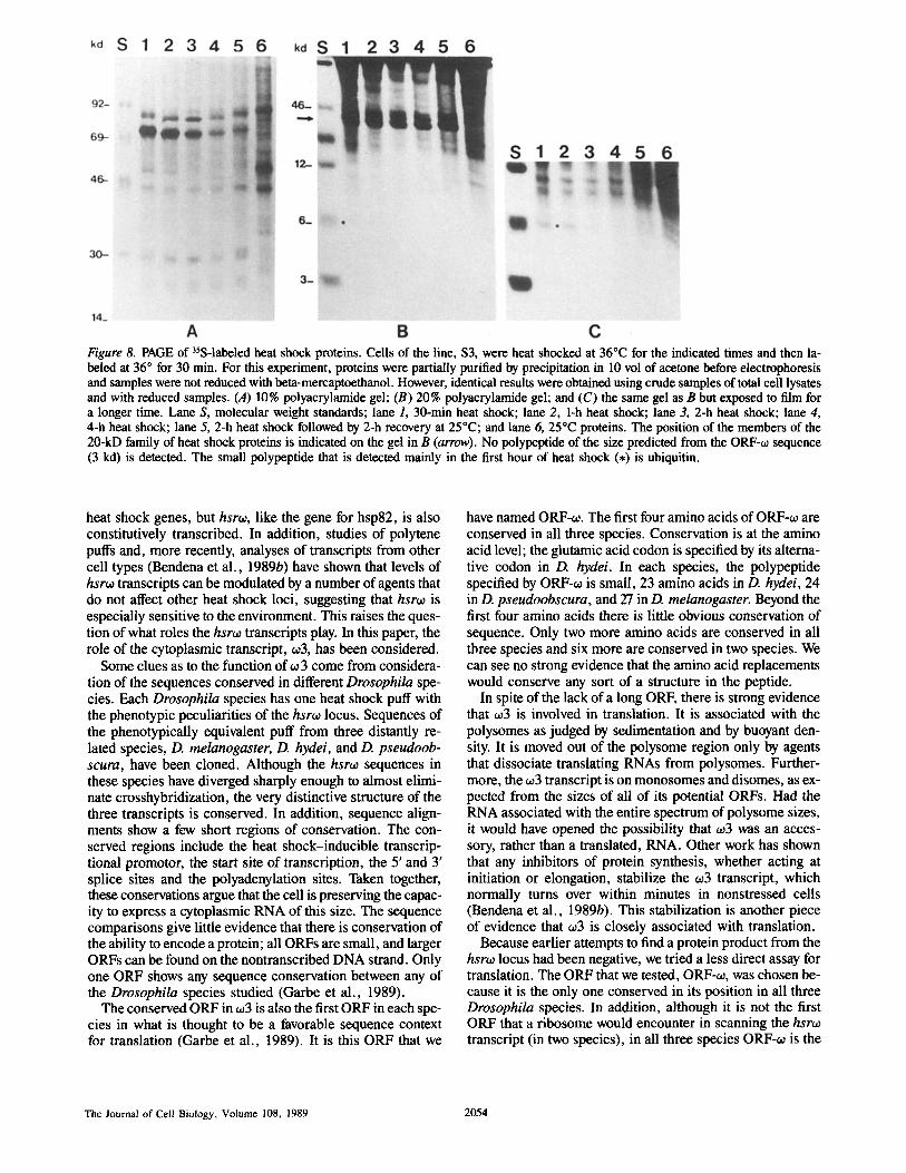
Figure 8. PAGE of 35S-labeled heat shock proteins. Cells of the line, $3, were heat shocked at 36°C for the indicated times and then la- beled at 36 ° for 30 min. For this experiment, proteins were partially purified by precipitation in 10 vol of acetone before electrophoresis and samples were not reduced with beta-mercaptoethanol. However, identical results were obtained using crude samples of total cell lysates and with reduced samples. (A) 10% polyacrylamide gel; (B) 20% polyacrylamide gel; and (C) the same gel as B but exposed to film for a longer time. Lane S, molecular weight standards; lane 1, 30-min heat shock; lane 2, 1-h heat shock; lane 3, 2-h heat shock; lane 4, 4-h heat shock; lane 5, 2-h heat shock followed by 2-h recovery at 25°C; and lane 6, 25°C proteins. The position of the members of the 20-kD family of heat shock proteins is indicated on the gel in B (arrow). No polypeptide of the size predicted from the ORF-~0 sequence (3 kd) is detected. The small polypeptide that is detected mainly in the first hour of heat shock (,) is ubiquitin.
heat shock genes, but hsro~, like the gene for hsp82, is also constitutively transcribed. In addition, studies of polytene puffs and, more recently, analyses of transcripts from other cell types (Bendena et al., 1989b) have shown that levels of hsro~ transcripts can be modulated by a number of agents that do not affect other heat shock loci, suggesting that hsro~ is especially sensitive to the environment. This raises the ques- tion of what roles the hsro~ transcripts play. In this paper, the role of the cytoplasmic transcript, o~3, has been considered.
Some clues as to the function of o~ 3 come from considera- tion of the sequences conserved in different Drosophila spe- cies. Each Drosophila species has one heat shock puff with the phenotypic peculiarities of the hsro~ locus. Sequences of the phenotypically equivalent puff from three distantly re- lated species, D. melanogaster, D. hydei, and D. pseudoob- scura, have been cloned. Although the hsroo sequences in these species have diverged sharply enough to almost elimi- nate crosshybridization, the very distinctive structure of the three transcripts is conserved. In addition, sequence align- ments show a few short regions of conservation. The con- served regions include the heat shock-inducible transcrip- tional promotor, the start site of transcription, the 5' and 3' splice sites and the polyadenylation sites. Taken together, these conservations argue that the cell is preserving the capac- ity to express a cytoplasmic RNA of this size. The sequence comparisons give little evidence that there is conservation of the ability to encode a protein; all ORFs are small, and larger ORFs can be found on the nontranscribed DNA strand. Only one ORF shows any sequence conservation between any of the Drosophila species studied (Garbe et al., 1989).
The conserved ORF in 603 is also the first ORF in each spe- cies in what is thought to be a favorable sequence context for translation (Garbe et al., 1989). It is this ORF that we
have named ORF-~0. The first four amino acids of ORF-o~ are conserved in all three species. Conservation is at the amino acid level; the glutamic acid codon is specified by its alterna- tive codon in D. hydei. In each species, the polypeptide specified by ORF-oJ is small, 23 amino acids in D. hydei, 24 in D. pseudoobscura, and 27 in D. melanogaster. Beyond the first four amino acids there is little obvious conservation of sequence. Only two more amino acids are conserved in all three species and six more are conserved in two species. We can see no strong evidence that the amino acid replacements would conserve any sort of a structure in the peptide.
In spite of the lack of a long ORF, there is strong evidence that o~3 is involved in translation. It is associated with the polysomes as judged by sedimentation and by buoyant den- sity. It is moved out of the polysome region only by agents that dissociate translating RNAs from polysomes. Further- more, the ~o3 transcript is on monosomes and disomes, as ex- pected from the sizes of all of its potential ORFs. Had the RNA associated with the entire spectrum of polysome sizes, it would have opened the possibility that w3 was an acces- sory, rather than a translated, RNA. Other work has shown that any inhibitors of protein synthesis, whether acting at initiation or elongation, stabilize the ~o3 transcript, which normally turns over within minutes in nonstressed cells (Bendena et al., 1989b). This stabilization is another piece of evidence that o~3 is closely associated with translation.
Because earlier attempts to find a protein product from the hsro~ locus had been negative, we tried a less direct assay for translation. The ORF that we tested, ORF-co, was chosen be- cause it is the only one conserved in its position in all three Drosophila species. In addition, although it is not the first ORF that a ribosome would encounter in scanning the hsro~ transcript (in two species), in all three species ORF-~o is the
The Journal of Cell Biology, Volume t08, 1989 2054

Figure 9. Molecular size of the ORF-o~/CAT fusion protein. (A) 15% polyacrylamide gel showing cell proteins labeled with [35S]methionine for 30 min after heat shock at the indicated tem- peratures for 30 min. Samples were reduced with beta-mercapto- ethanol (5 %) before electrophoresis. (B) Western blot of a duplicate gel to that shown in A probed with anti-CAT sera. Lanes 1, 2, and 3, proteins from the S3-ORF-oJ/CAT fusion cell line. Lanes 4, 5, and 6, proteins from the S3-~-leader CAT cell line. Lanes 1 and 4, 25°C proteins; lanes 2 and 5, 33°C proteins; and lanes 3 and 6, 36°C proteins. On A, the position of the CAT protein (23 kD) and the ORF-~/CAT fusion protein (26 kD) are indicated (arrowheads). The ORF-o~/CAT fusion protein coelectrophoreses with the 20-kD family of hsps in this gel system.
first ORF that has its initiation codon in a favorable sequence context, as determined by Kozak (1986). (A survey of se- quence conservation around start codons in Drosophila mes- sages [Cavener, 1987] suggests that translation initiation in this genus requires a similar sequence context.) There is a 5' ORF in an unfavorable context in D. melanogaster and D. pseudoobscura; the lack of conservation of the first ORF in D. hydei also argues against its importance to the cell. Our experiments support the idea that this first ORF is untrans- lated.
Our indirect assay for translation was based on the hypoth- esis that if ORF-60 were translated, most ribosomes would be impeded from progressing on to the translation start site for the CAT protein. The results of our experiments are entirely consistent with the hypothesis. Although the ORF-to-CAT construct differs from the 60-leader-CAT construct only by having ORF-to inserted in front of the CAT gene, the differ- ence in the level of CAT activity and CAT protein produced by the two constructs was dramatic. Cells with ORF-to made almost no CAT protein although they contained even more CAT mRNA than cells with the other construct. As expected of an RNA in which the ORF-to was being translated, the ORF-60-CAT transcript was on monosomes and disomes. This eliminates the alternative possibility, that structural considerations in the ORF portion of the transcript inhibited translational initiation. A more direct demonstration of the translation of ORF-to comes from the construct in which ORF-60 was fused in frame to the CAT ORE Cells with this construct produced CAT protein that was larger by the amount predicted from the extended coding region, showing that the ORF-60 initiation site was functional in vivo.
The evidence that 603 was translated led us to extend the earlier search for a small polypeptide that might be encoded by one of the 603 ORFs. We have looked throughout the period of maximal heat shock-induced increase in the 603
levels and have seen no polypeptides of the appropriate size (3 kD) in either heat shock or control cells. Thus, if to 3 is translated, the product must be either rapidly turned over or rapidly sequestered, perhaps by linking to other proteins. Al- though ORF-to has a conserved cysteine, we do not think that its linkage to other proteins by disulfide bonds has prevented its identification here, because the conditions of electropho- resis should have reduced such bonds. It is possible that the hsrto peptide is normally covalently linked to other cellular proteins, much like ubiquitin, so that there is little of the un- modified form to be found (Finley and Varshavsky, 1985). In fact, one of the conserved residues of ORF-to is a lysine, the amino acid by which ubiquitin forms bonds to other proteins; however, such posttranslational linkage also seems unlikely. We are easily able to identify the unmodified form of ubiqui- tin among the 35S-labeled heat shock proteins from $3 cells; if the hsrto polypeptide is linked to other proteins, we should detect some unlinked form unless that attachment is signifi- cantly more efficient than the ubiquitin linkage.
Although we cannot rule out the possibility that the hsrw translation product is rapidly sequestered, there are several reasons to think that it is rapidly turned over, possibly even before leaving the ribosome. Our searches have failed to turn up any free polypeptide, even in cells carrying the ORF-to- CAT construct. If the amount of hsrto transcript on poly- somes is any measure of its translation, these cells should be making much more of the hsrto polypeptide than is normal for the cultured cells. The extra peptide might be expected to overload even an efficient sequestration system, and yet we detect no 3-kD polypeptide in these cells. We also note that, in the three Drosophila species, the conservation of the poly- peptide seems almost entirely limited to the first four amino acids and the small size (although the size varies slightly). If what is required is the act of translation itself and then rapid degradation of the product, the conserved features seem adequate. If the polypeptide has a more complex role, it might be expected to show more conserved features.
The ORF-to-CAT fusion protein is easily detected and has given no insight into the fate of the authentic ORF-to product. The fusion protein is not found in linkage with other proteins or with itself either on reduced or unreduced gels (Fini, un- published data); however, it is possible that the ability to form such bonds is blocked by CAT linkage. Similarly, if the pep- tide is normally turned over very rapidly, linkage to CAT pro- tein may stabilize the peptide, either by blocking the carboxy terminus, or by embedding other vulnerable portions of the peptide within the CAT protein structure, and thus protecting it from intracellular proteases.
It is important to note that efficient translation of the ORF- 60 can be observed under heat shock temperatures (>33 ° in our cell lines) at which only heat shock messages and a few 25 ° messages can be translated. This suggests an additional evolutionary conservation, that of sequences that are essen- tial to translation at high temperatures. Recently, Hultmark et al. (1986) have given evidence that the first 26 nucleotides of the hsp 22 transcript have elements affecting both heat shock translation and efficient transcription. The 5' ends of hsp 70, 68, 27, 26, 23, and 22 show identical nucleotides in positions -1, +1, 7, 12, 15, and 20. The hsrto genes in the three Drosophila species that we have examined also have these nucleotides in all but position 20 (Garbe et al., 1986; 1989). These sequences could have been retained during
Fini et al. The Cytoplasmic RNA of hsr~ Is Translated 2055

evolution to allow for efficient translation of the conserved ORF under heat shock conditions. Such conservation along with the sequence conservation in the conserved ORF pro- vides a substantial argument for the importance of this trans- lation to the cell.
In what way could translation of ORF-o~ affect the cell? Since we cannot find detectable levels of the peptide, it is possible that translation of the conserved ORF is the impor- tant factor; the protein product may have no consequence and be rapidly degraded by the cell. Such a role has been sug- gested for other upstream ORFs in several mRNAs. The first ORF of Rous sarcoma virus has only 7 codons. This ORF is evolutionarily conserved but can not be linked to a protein product in vivo, although a very short-lived protein product has been detected by in vitro translation (Hackett et al., 1986). It has been proposed that translation of this ORF acts to place the transcript in an appropriate configuration for vi- ral coat assembly. For SV40 early mRNA (Kahlili et al., 1987) or yeast GCN4 mRNA (Mueller and Hinnebusch, 1986; Fink, 1986) the translation of small, upstream ORFs appears to play a role in regulation of the translation of protein-coding ORFs located more 3' on the message. For a number of different mRNAs, the machinery for regulation of mRNA half-life appears to be associated with the polysomes, requiting a message to be actively translated in order to be degraded (Hunt, 1988). A similar mechanism may well be operating with the ~03 RNA since we have found that its half- life is significantly and specifically increased when cells are treated with inhibitors of protein synthesis (Bendena et al., 1989b). If translation serves only to regulate the turnover of ~03 RNA, the RNA must have some additional function. We should note that ORF-o~ represents only a small portion of this 1.2-1.3 kb RNA, and yet the size of the transcript is con- served in all the Drosophila species. There are several small islands of conserved sequence in the transcripts that may well have functional significance which we do not yet under- stand.
Our results do not eliminate the possibility that ORF-~0 could encode a protein that performs a function. If so, the function might be expected to be a regulatory one since the protein must be short lived, if it exists at all. Peptide hor- mones offer examples of small regulatory molecules with minimally conserved sequences and very short half-lives (Tager and Steiner, 1974). We note that three of the six amino acids conserved in ORF-o~ in all three Drosophila species are conserved at the level of the amino acid rather than the nucleotide sequence (Garbe et al., 1989). This might suggest conservation of the ability to make a functional protein. On the other hand, the conserved amino acids are clustered at the amino-terminal end, reminiscent of the four conserved amino acids in/3-tubulin that are required to assure the regu- lated turnover of the mRNA while it is on the polysomes (Yen et al., 1988). If the o~3 transcript is indeed serving to monitor the translational state of the cell, a similar polysome-related turnover might obtain.
How could translation of the ORF-~o contribute to cell function at ambient and heat shock temperatures? We have been impressed with the sensitive way in which the level of all the hsro~ transcripts respond to various external condi- tions. If, as we propose, the levels of 003 RNA are indeed con- trolled by translation on polysomes, then this may be a finely tuned mechanism to link the levels of w3 to protein synthesis.
Thus, one intriguing possibility is that the level of the o~3 transcript serves to communicate, to the nuclear compart- ment of the cell, information about the status of cellular pro- tein synthesis at any particular time.
This work was supported by a grant from the National Institutes of Health to M. L. Pardue. M. E. Fini is the recipient of a postdoctoral fellowship from the American Cancer Society.
Received for publication 15 November 1988 and in revised form 1 Febru- ary 1989.
References
Arribas, C., J. Sampedro, and M. Izquierdo. 1986. The ubiquitin genes of D. melanogaster: transcription and polymorphism. Biochim. Biophys. Acta. 868:119-127.
Ballinger, D. G., and M. L. Pardue. 1983. The control of protein synthesis dur- ing heat shock involves altered polypeptide elongation rates. Cell. 33: 103-114.
Bendena, W. G., M. E. Fini, J. C. Garbe, G. M. Kidder, S. C. Lakhotia, and M. L. Pardue. 1989a. hsr~, a different sort of heat shock locus. UCLA (Univ. Calif. Los Ang.) Syrup. Mol. Cell. Biol. New Ser. 96:3-14.
Bendena, W. G., J. C. Garb¢, K. L. Traverse, S. C. Lakhotia, and M. L. purdue. 1989b. Multiple inducers of the heat shock locus 93D (hsr~o): inducer-specific patterns of the three transcripts. J. Cell Biol. In press.
Bourouis, M., and B. Jarry. 1983. Vectors containing a procaryotic dihydrofo- late reductase gene transform Drosophila cells to methotrexate-resistance. EMBO (Eur. 114o1. Biol. Organ.)J. 2:1099-1104.
Cavener, D. R. 1987. Comparison of the consensus sequence flanking trans- lational start sites in Drosophila and vertebrates. Nucleic Acids Res. 15: 1353-1361.
Craig, E. A. 1985. The heat shock response. CRC Crit. Rev. Biochem. 18:239-280.
Dangli, A., C. J. Grond, P. Kloetzel, and E. K. F. Bautz. 1983. Heat shock puff 93D from Drosophila melanogaster: accumulation of RNP specific anti- gen associated with giant particles of possible storage function. EMBO (Eur. Mol. Biol. Organ.)J. 2:1747-1751.
Faiferman, I., M. G. Hamilton, and A. O. Pogo. 1971. Nucleoplasmic ribonu- cleoprotein particles of rat liver. II. Physical properties and action of dis- sociating agents. Biochim. Biophys. Acta. 232:685-695.
Fink, G. R. 1986. Translational control of transcription in eucaryotes. Cell. 45:155-156.
Finley, D., and A. Varshavsky. 1985. The ubiquitin system: functions and mechanisms. Trends Biochem. Sci. 10:343-348.
Garbe, J. C., and M. L. Pardue. 1986. Heat shock locus 93D of Drosophila melanogaster: a spliced RNA most strongly conserved in the intron. Proc. Natl. Acad. Sci. USA. 83:1812-1816.
Garbe, J. C., W. G. Bendena, M. Alfano, and M. L. Pardue. 1986. A Drosoph- ila heat shock locus with a rapidly diverging sequence but a conserved struc- ture. J. Biol. Chem. 261:16889-16894.
Garbe, J. C., W. G. 8endena, and M. L. Pardue. 1989. Sequence evolution of the Drosophila heat shock locus hsno. I. The non-repeated portion of the gene. Genetics. In press.
Gorman, C. 1986. High efficiency gene transfer into mammalian cells. In DNA Cloning: A Practical Approach, Volume 2. D. M. Glover, editor. 143-190.
Hackett, P. B., R. B. Petersen, C. H. Hansel, F. Albericio, S. I. Gunderson, A. C. Palmenberg, and G. Baremy. 1986. Synthesis in vitro of a seven amino acid peptide encoded in the leader RNA of Rolls sarcoma virus. J. Mol. Biol. 190:45-57.
Hultmark, D., R. Klemenz, and W. Gehring. 1986. Translational and transcrip- tional control elements in the untranslated leader of the heat shock gene hsp22. Cell. 44:429-438.
Hunt, T. 1988. Controlling mRNA life-span. Nature (Lond.). 334:567-568. Kahlili, K., Brady, J., and Khoury, G. 1987. Translational regulation of SV40
mRNA defines a new protein. Cell. 48:639-645. Kozak, M. 1986. Point mutations define a sequence flanking the AUG initiator
codon that modulates translation by eucaryotic ribosomes. Cell. 44:283- 292.
Laemmli, U. K. 1970. Cleavage of structural proteins during the assembly of the head of bacteriophage T4. Nature (Lond.). 227:680-685.
Lakhotia, S. C. 1987. The 93D heat shock locus in Drosophila: a review. J. Genet. 66:139-157.
Lakhotia, S. C., and A. K. Singh. 1982. Conservation of the 93D puff of Dro- sophila melanogaster in different species of Drosophila. Chromosoma (Berl.). 86:265-278.
Lee, S. E., and G. Brawerman. 1971. Pulse-labelled ribonucleic acid com- plexes released by dissociation of rat liver polysomes. Biochemistry. 10: 510-516.
Lengyel, J. A., L. J. Ransom, M. L. Graham, and M. L. Pardue. 1980. Tran- scription and metabolism of RNA from the Drosophila melanogaster heat shock puff site 93D. Chromosoma (BerL). 80:237-252.
The Journal of Cell Biology, Volume 108, 1989 2056

Lindquist, S. 1986. The heat shock response. Annu. Rev. Biochem. 55:1151- 1192.
Lodish, H. F. 1971. Alpha and beta giobin messenger ribonucleic acid. J. Biol. Chem. 246:7131-7138.
McCarthy, T. L., E. Siegel, B. Mroczkowski, and S. M. Heywood. 1983. Characterization of translational-control ribonucleic acid. Biochemistry. 22:935-941.
Mueller, P. P., and A. G. Hinnebusch. 1986. Multiple upstream AUG codons mediate translational control of GCN4. Cell. 45:201-207.
Pardue, M. L., W. G. Bendena, and J. C. Garbe. 1987. Heat shock: puffs and response to environmental stress. In Results and Problems in Cell Differenti- ation, Volume 14. W. Hennig, editor. Springer-Verlag, Berlin. 121-131.
Pong, S.-S., D. L. Nuss, and G. Koch. 1975. Inhibition of initiation of protein synthesis in mammalian tissue culture cells by L-1-tosylamido-2-phenylethyl chloromethyl ketone. J. Biol. Chem. 250:240-245.
Ritossa, F. M. 1962. A new puffing pattern induced by heat shock and DNP in Drosophila. Experientia (Basel). 18:571-573.
Ryseck, R.-P., U. Walldoff, T. Hoffman, and B. Hovemann. 1987. Heat shock loci 93D of Drosophila melanogaster and 48B of Drosophila hydei exhibit a common structural and transcriptional pattern. Nucleic Acids Res. 15:3317-3333.
Schneider, I., and A. B. Blumenthal. 1978. Drosophila cell and tissue culture.
In The Genetics and Biology of Drosophila. M. Ashburner, anO T. R. F. Wright, editors. Academic Press Inc., New York. 266-316.
Sondermeijer, P. J. A., J. W. M. Derksen, and N. H. Lubsen. 1980. Estab- lished cell lines of Drosophila hydei. In Vitro (Gaithersburg). 16:913-914.
Stewart-Blair, M. L., I. S. Yanowitz, I. H. Goldberg. 1971. Inhibition of syn- thesis of new globin chains in reticulocyte lysates by pactamycin. Biochemis- try. 10:4198--4206.
Storti, R. V., M. P. Scott, A. Rich, and M. L. Pardue. 1980. Translational con- trol of protein synthesis in response to heat shock in D. melanogaster cells. Cell. 22:825-834.
Tager, H. S., and D. F. Steiner. 1974. Peptide hormones. Annu. Rev. Biochem. 43:509-538.
Walldoff, U., S. Richter, R.-P. Ryseck, H. Steller, J. E. Edstrom, E. K. F. Bautz, and B. Hovemann. 1984. Cloning of heat-shock locus 93D from Dro- sophila melanogaster. EMBO (Fur. Mol. Biol. Organ.) J. 3:2499-2504.
Yanisch-Perron, C., J. Vieira, and J. Messing. 1985. Improved MI3 phage cloning vectors and host strains: nucleotide sequences of the M13mpl8 and pUCI9 vectors. Gene (Amst.). 33:103-119.
Yen, T. J., P. S. Machin, and D. W. Cleveland. 1988. Antoregulated instability of beta-tubulin mRNAs by recognition of the nascent amino terminus of beta- tubulin. Nature (Lond.). 334:580-584.
Fini et al. The Cytoplasmic RNA of hsros Is Translated 2057