Unmasking sarcoidosis following surgery for Cushing...
6
Syddansk Universitet Unmasking sarcoidosis following surgery for Cushing disease Fraes Diernaes, Jon Erik; Bygum, Anette; Poulsen, Per L Published in: Dermato-Endocrinology DOI: 10.4161/derm.29855 Publication date: 2016 Document version Publisher's PDF, also known as Version of record Document license CC BY-NC Citation for pulished version (APA): Fraes Diernaes, J. E., Bygum, A., & Poulsen, P. L. (2016). Unmasking sarcoidosis following surgery for Cushing disease. Dermato-Endocrinology, 8(1), [e983688]. DOI: 10.4161/derm.29855 General rights Copyright and moral rights for the publications made accessible in the public portal are retained by the authors and/or other copyright owners and it is a condition of accessing publications that users recognise and abide by the legal requirements associated with these rights. • Users may download and print one copy of any publication from the public portal for the purpose of private study or research. • You may not further distribute the material or use it for any profit-making activity or commercial gain • You may freely distribute the URL identifying the publication in the public portal ? Take down policy If you believe that this document breaches copyright please contact us providing details, and we will remove access to the work immediately and investigate your claim. Download date: 09. sep.. 2018
Transcript of Unmasking sarcoidosis following surgery for Cushing...

Syddansk Universitet
Unmasking sarcoidosis following surgery for Cushing disease
Fraes Diernaes, Jon Erik; Bygum, Anette; Poulsen, Per L
Published in:Dermato-Endocrinology
DOI:10.4161/derm.29855
Publication date:2016
Document versionPublisher's PDF, also known as Version of record
Document licenseCC BY-NC
Citation for pulished version (APA):Fraes Diernaes, J. E., Bygum, A., & Poulsen, P. L. (2016). Unmasking sarcoidosis following surgery for Cushingdisease. Dermato-Endocrinology, 8(1), [e983688]. DOI: 10.4161/derm.29855
General rightsCopyright and moral rights for the publications made accessible in the public portal are retained by the authors and/or other copyright ownersand it is a condition of accessing publications that users recognise and abide by the legal requirements associated with these rights.
• Users may download and print one copy of any publication from the public portal for the purpose of private study or research. • You may not further distribute the material or use it for any profit-making activity or commercial gain • You may freely distribute the URL identifying the publication in the public portal ?
Take down policyIf you believe that this document breaches copyright please contact us providing details, and we will remove access to the work immediatelyand investigate your claim.
Download date: 09. sep.. 2018

Full Terms & Conditions of access and use can be found athttp://www.tandfonline.com/action/journalInformation?journalCode=kder20
Download by: [University of Southern Denmark] Date: 28 February 2017, At: 00:13
Dermato-Endocrinology
ISSN: (Print) 1938-1980 (Online) Journal homepage: http://www.tandfonline.com/loi/kder20
Unmasking sarcoidosis following surgery forCushing disease
Jon E.F. Diernaes, Anette Bygum & Per L. Poulsen
To cite this article: Jon E.F. Diernaes, Anette Bygum & Per L. Poulsen (2016) Unmaskingsarcoidosis following surgery for Cushing disease, Dermato-Endocrinology, 8:1, e983688, DOI:10.4161/derm.29855
To link to this article: http://dx.doi.org/10.4161/derm.29855
© 2016 The Author(s). Published withlicense by Taylor & Francis© Jon E.F.Diernaes, Anette Bygum, and Per L. Poulsen
Accepted author version posted online: 31Oct 2014.Published online: 07 Jun 2016.
Submit your article to this journal
Article views: 157
View related articles
View Crossmark data

REVIEW
Unmasking sarcoidosis following surgery for Cushing disease
Jon E.F. Diernaesa, Anette Bygumb, and Per L. Poulsenc
aDepartment of Dermatology, Aarhus University Hospital, Aarhus, Denmark; bDepartment of Dermatology and Allergy Centre, Odense UniversityHospital, Odense, Denmark; cDepartment of Endocrinology, Aarhus University Hospital, Aarhus, Denmark
ARTICLE HISTORYReceived 14 March 2014Revised 18 June 2014Accepted 8 July 2014
ABSTRACTWe present a patient with Cushing disease apparently suppressing sarcoidosis, which was unmaskedfollowing surgical resection of a pituitary adrenocorticotropin (ACTH)-producing microadenoma. Casereport and a short review of the literature published in this area. A 46-year-old Caucasian womanpresented with symptoms of hypercortisolism such as progressive weight gain, Cushingoidappearance, proximal myopathy, easy bruising, and amenorrhea. Blood testing including inferiorpetrosal sinus sampling uncovered an ACTH-producing microadenoma in the right aspect of theanterior pituitary gland for which the patient underwent transphenoidal resection. Maintenancecorticosteroid therapy was implemented, and the signs and symptoms of Cushing disease began toresolve. Three months after surgery, multiple erythematous painful nodules developed on the patient’sarms. Erythema nodosum (EN) was diagnosed clinically and a suspicion of underlying sarcoidosis wassubstantiated by lung imaging and elevated plasma interleukin (IL)-2 receptor. One month later, thelesions spontaneously resolved without therapy other than maintenance glucocorticoid replacement.Physicians should be aware that patients undergoing successful treatment of Cushing syndrome mayhave a flare-up or emergence of a corticosteroid-responsive disease.
KEYWORDSautoimmune disease;Cushing disease; cutaneousmarker; erythema nodosum;glucocorticoids; sarcoidosis
Introduction
Sarcoidosis is a multisystem granulomatous disorderof unknown etiology. Typical presentations of this dis-ease relate to the lungs (dry cough), the eyes (anterioruveitis) or the skin (EN or cutaneous sarcoidosis). ENis characterized by inflammatory, red nodules that areusually tender, multiple, and bilateral. The nodulestypically erupt on the shins but may also be seen onthe thighs, trunk or upper extremities.
Cushing disease is characterized by an ACTH-dependent hypercortisolism which manifests itself asCushing syndrome. Signs of Cushing syndromeinclude upper body obesity (moon face, buffalo hump,abdominal fat distribution), striae, wasting of thelimbs and excess body fluid. The long-term complica-tions of hypercortisolism are significant and includeosteoporosis, hypertension, diabetes mellitus, hirsut-ism, and amenorrhea.1,2
Here, we report a rare case of a patient who pre-sented with EN on the upper extremities a few monthsafter surgically induced remission of Cushing disease.She was diagnosed with sarcoidosis.
Case Presentation
In August 2011, a 45-y old Caucasian woman of Scan-dinavian descent was referred to our tertiary referralcenter on suspicion of Cushing syndrome after urinaly-sis had documented elevated 24-h urinary excretions ofcortisol (1231–1711 nmol/24h, normal range<340).
Prior to referral the patient had a two-year historywith a multitude of symptoms including fatigue,altered fat distribution, difficulty climbing stairs (pos-sible sign of myopathy), and an unintended weightgain of 10 kg (BMI 28,4), easy bruising, increased hairgrowth on the face and extremities, and amenorrhea.No hyperpigmentation was noted. Prior medical his-tory was otherwise uneventful.
Suspicion of ACTH-dependent Cushing syn-drome was confirmed with a low-dose dexametha-sone suppression test which showed a lack ofcortisol suppression (plasma cortisol 563 nmol/L,reference range <50) and elevated plasma ACTH(15 pmol/L, reference range <10). A corticotropin-releasing hormone stimulation test demonstratedclear increases of both plasma ACTH and cortisol
CONTACT Per L. Poulsen [email protected]© Jon E.F. Diernaes, Anette Bygum, and Per L. Poulsen. Published with license by Taylor and Francis.This is an Open Access article distributed under the terms of the Creative Commons Attribution-Non-Commercial License (http://creativecommons.org/licenses/by-nc/3.0/), which per-mits unrestricted non-commercial use, distribution, and reproduction in any medium, provided the original work is properly cited. The moral rights of the named author(s) have beenasserted.
DERMATO-ENDOCRINOLOGY2016, VOL. 8, NO. 1, e983688 (4 pages)http://dx.doi.org/10.4161/derm.29855
peak levels (45 pmol/L and 982 nmol/L respec-tively) suggestive of Cushing disease. A gadolin-ium-enhanced high-resolution pituitary MR did notdetect a tumor, but inferior petrosal sinus samplingprovided evidence of pituitary ACTH hypersecre-tion with significantly higher values obtained fromthe right sinus.
Transsphenoidal exploration on the right side ofthe pituitary gland revealed a microadenoma whichwas removed. After surgery hypocortisolism wasdetected and the patient started substitution withhydrocortisone. The pathology report showed a pitui-tary adenoma with ACTH positivity.
Outpatient follow-up was uneventful. The patientlost 8 kg (BMI 22.4) and reconstituted completelyfrom the clinical stigmata of Cushing syndromeincluding hirsutism which began to resolve one monthafter surgery. She still experienced some fatigue, but ata self-reported lower level.
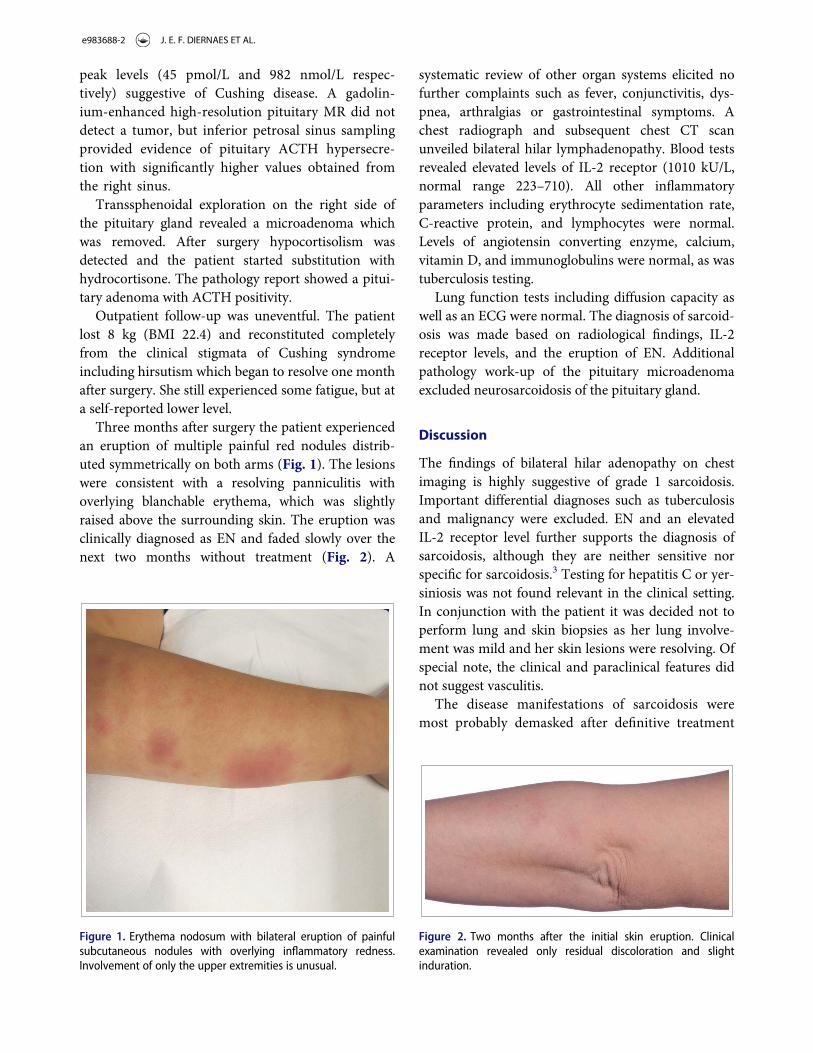
Three months after surgery the patient experiencedan eruption of multiple painful red nodules distrib-uted symmetrically on both arms (Fig. 1). The lesionswere consistent with a resolving panniculitis withoverlying blanchable erythema, which was slightlyraised above the surrounding skin. The eruption wasclinically diagnosed as EN and faded slowly over thenext two months without treatment (Fig. 2). A
systematic review of other organ systems elicited nofurther complaints such as fever, conjunctivitis, dys-pnea, arthralgias or gastrointestinal symptoms. Achest radiograph and subsequent chest CT scanunveiled bilateral hilar lymphadenopathy. Blood testsrevealed elevated levels of IL-2 receptor (1010 kU/L,normal range 223–710). All other inflammatoryparameters including erythrocyte sedimentation rate,C-reactive protein, and lymphocytes were normal.Levels of angiotensin converting enzyme, calcium,vitamin D, and immunoglobulins were normal, as wastuberculosis testing.
Lung function tests including diffusion capacity aswell as an ECG were normal. The diagnosis of sarcoid-osis was made based on radiological findings, IL-2receptor levels, and the eruption of EN. Additionalpathology work-up of the pituitary microadenomaexcluded neurosarcoidosis of the pituitary gland.
Discussion
The findings of bilateral hilar adenopathy on chestimaging is highly suggestive of grade 1 sarcoidosis.Important differential diagnoses such as tuberculosisand malignancy were excluded. EN and an elevatedIL-2 receptor level further supports the diagnosis ofsarcoidosis, although they are neither sensitive norspecific for sarcoidosis.3 Testing for hepatitis C or yer-siniosis was not found relevant in the clinical setting.In conjunction with the patient it was decided not toperform lung and skin biopsies as her lung involve-ment was mild and her skin lesions were resolving. Ofspecial note, the clinical and paraclinical features didnot suggest vasculitis.
The disease manifestations of sarcoidosis weremost probably demasked after definitive treatment
Figure 1. Erythema nodosum with bilateral eruption of painfulsubcutaneous nodules with overlying inflammatory redness.Involvement of only the upper extremities is unusual.
Figure 2. Two months after the initial skin eruption. Clinicalexamination revealed only residual discoloration and slightinduration.
e983688-2 J. E. F. DIERNAES ET AL.

for Cushing disease by transsphenoidal adenec-tomy. The eruption of EN in an atypical locationbecame a clue to the diagnosis. Alternatively thesarcoidosis could be part of a rebound phenome-non of autoimmunity presenting after cessation ofa prolonged period of hypercortisolism as recentlydescribed by da Mota.4 Finally it could also be acoincidence.
A review of the literature (MeSH-terms “Cushingsyndrome” AND “Sarcoidosis”; limits: humans, alllanguages, 1970–2014) revealed only five similarcase reports.1,2,5-9 Only one of these describes asimilar case of EN as the presenting sign of under-lying sarcoidosis.3,6
Taking a broader view of hypercortisolic statesthe study by da Mota et al.4 pooled 11 case reportsof overt immune dysfunction following remissionof Cushing syndrome. Another study even coinedthe term “Cushing cure syndrome” to highlight theimmunologic aftermath of supposed cure.10 In thestudy by da Mota overt autoimmune and allergicdiseases such as psoriasis, sarcoidosis, Graves’ dis-ease, autoimmune thyroiditis, eczema and asthmawere diagnosed in 11 of 66 patients (16.7%) whoachieved remission of Cushing syndrome. In eightpatients (73%) symptoms were noted for the firsttime, and in three patients (27%) symptoms wereexacerbated after remission. Of note, the female tomale ratio was 6:1.
It is interesting to speculate that enhanced ACTH(and other melanocortin peptides like proopiomelano-cortin or a-melanocyte–stimulating hormone) inaddition to increased cortisol levels may have contrib-uted to mask the skin inflammation by affecting theimmune system possibly mediated by melanocortinreceptors in skin adipocytes.11,12
Glucocorticoids inhibits a broad range of T cell andB cell responses and exhibits potent suppressive effectson the effector functions of phagocytes. This makesthem effective in controlling a wide range of inflam-matory diseases but can also lead to adverse events aspreviously mentioned.1,13
In this case the inherent hypercortisolic state ofCushing disease apparently suppressed the underlyingsarcoidosis.
In conclusion we would like to highlight the risk ofcorticosteroid-responsive diseases following successfultreatment of Cushing syndrome. We propose a pro-spective follow-up study to examine the incidence of
autoimmune and corticosteroid-responsive diseasesafter surgery.
Disclosure of Potential Conflicts of InterestNo potential conflicts of interest were disclosed.
ConsentInformed consent was obtained from the patient for publica-tion of this manuscript and any accompanying images.
References
[1] Ross EJ, Linch DC. Cushing’s syndrome–killing disease:discriminatory value of signs and symptoms aiding earlydiagnosis. Lancet 1982; 2:646-9; PMID:6125785; http://dx.doi.org/10.1016/S0140-6736(82)92749-0
[2] Nieman LK, Biller BMK, Findling JW, Newell-Price J, Sav-age MO, Stewart PM, Montori VM. The diagnosis ofCushing’s syndrome: an Endocrine Society Clinical Prac-tice Guideline. J Clin Endocrinol Metab 2008; 93:1526-40;PMID:18334580; http://dx.doi.org/10.1210/jc.2008-0125
[3] Garc�ıa-Porr�ua C, Gonz�alez-Gay MA, V�azquez-CarunchoM, L�opez-Lazaro L, Lueiro M, Fern�andez ML, Alvarez-Ferreira J, Pujol RM. Erythema nodosum: etiologic andpredictive factors in a defined population. ArthritisRheum 2000; 43:584-92; PMID:10728752; http://dx.doi.org/10.1002/1529-0131(200003)43:3<584::AID-ANR15>3.0.CO;2-6
[4] da Mota F, Murray C, Ezzat S. Overt immune dysfunc-tion after Cushing’s syndrome remission: a consecutivecase series and review of the literature. J Clin EndocrinolMetab 2011; 96:E1670-4; PMID:21816785; http://dx.doi.org/10.1210/jc.2011-1317
[5] McDaniel WE, Bard JW, Blakey LW, Hamilton RD. Sar-coid nodules in Cushing’s disease. Arch Dermatol 1970;101:356-8; PMID:5414894; http://dx.doi.org/10.1001/archderm.1970.04000030100017
[6] Takenaka K, Yanase T, Takayanagi R, Haji M, Ikuyama S,Nawata H. Cushing’s disease preceding sarcoidosis.Intern Med 1995; 34:580-3; PMID:7549148; http://dx.doi.org/10.2169/internalmedicine.34.580
[7] Marzano AV, Gasparini G, Caputo R, Alessi E. Subcuta-neous sarcoidosis following hypophysectomy for pitui-tary microadenoma inducing Cushing’s disease. Int JDermatol 1998; 37:798; PMID:9802695
[8] Maldonado M, Orlander P, Roshan SY, McCutcheon IE,Cleary KR, Friend KE. Sarcoidosis after surgicallyinduced remission of Cushing’s disease. Endocr Pract1999; 5:43-5; PMID:15251702; http://dx.doi.org/10.4158/EP.5.1.43
[9] Fichtel JC, Duckworth AKW, Soares T, Lesher JL Jr.Subcutaneous sarcoidosis presenting after treatmentof Cushing’s disease. J Am Acad Dermatol 2006;54:360-1; PMID:16443078; http://dx.doi.org/10.1016/j.jaad.2005.03.042
DERMATO-ENDOCRINOLOGY e983688-3

[10] Pivonello R, De Martino MC, De Leo M, Tauchmanov�aL, Faggiano A, Lombardi G, Colao A. Cushing’s syn-drome: aftermath of the cure. Arq Bras EndocrinolMetabol 2007; 51:1381-91; PMID:18209877; http://dx.doi.org/10.1590/S0004-27302007000800025
[11] B€ohm M, Luger TA, Tobin DJ, Garc�ıa-Borr�on JC.Melanocortin receptor ligands: new horizons for skinbiology and clinical dermatology. J Invest Dermatol2006; 126:1966-75; PMID:16912693; http://dx.doi.org/10.1038/sj.jid.5700421
[12] Brzoska T, Luger TA, Maaser C, Abels C, B€ohm M.Alpha-melanocyte-stimulating hormone and related tri-peptides: biochemistry, antiinflammatory and protectiveeffects in vitro and in vivo, and future perspectives forthe treatment of immune-mediated inflammatory dis-eases. Endocr Rev 2008; 29:581-602; PMID:18612139;http://dx.doi.org/10.1210/er.2007-0027
[13] Saag KG, Furst DE. Major side effects of systemic gluco-corticoids. In: Basow DS, editor. UpToDate. UpToDate;2013.
e983688-4 J. E. F. DIERNAES ET AL.