UNIVERSITY OF NOVA GORICA GRADUATE SCHOOL …library/magisterij/okolje/30Jancar.pdf · UNIVERSITY...
80
UNIVERSITY OF NOVA GORICA GRADUATE SCHOOL SEPARATION OF SMALL MOLECULES ON MONOLITHIC CHROMATOGRAPHIC MATERIALS MASTER'S THESIS Janez Jančar Mentor: Prof. dr. Aleš Podgornik Nova Gorica, 2013
Transcript of UNIVERSITY OF NOVA GORICA GRADUATE SCHOOL …library/magisterij/okolje/30Jancar.pdf · UNIVERSITY...

UNIVERSITY OF NOVA GORICA
GRADUATE SCHOOL
SEPARATION OF SMALL MOLECULES ON
MONOLITHIC CHROMATOGRAPHIC MATERIALS
MASTER'S THESIS
Janez Jančar
Mentor: Prof. dr. Aleš Podgornik
Nova Gorica, 2013


Janez Jančar, Separation of small molecules on monolithic chromatographic materials
- 1 -
Abstract
In this work, the isocratic separation of oligonucleotides in the ion-exchange mode
on thin layered glycidyl methacrylate-ethylene glycol dimethacrylate (GMA-
EDMA) polymeric material in the form of commercially available CIM®
(Convective Interaction Media) disks and in the form of custom-made GMA-
EDMA disks is presented. It was found that isocratic separation occurs even on
monoliths with a thickness of only 0.75 mm. Peak broadening of the components
retained on the monolith is proportional to the retention time, which in turn is
proportional to the thickness of the monolith. Peak height is inversely proportional
to the retention time. From these results it can be concluded that the mechanism of
the separation on such monolithic chromatographic material is similar to that in
HPLC columns filled with conventional porous particles. The HETP value of
GMA-EDMA monoliths is calculated to be 18.0 m. It was also found that the
difference between peak retention volumes slightly increases with the flow rate
when the experiments are performed in the range from 0.5 to 7 mL/min. From the
similarities between the isocratic separations on conventional columns and on thin
GMA-EDMA monoliths it is reasonable to believe that the separation based on a
multiple adsorption/desorption process also occurs in thin monoliths.
Furthermore, GMA-EDMA monolith for preparative scale in a shape of hollow
cylinder was prepared. To run this monolith in a radial flow mode, a special
housing was also constructed. When both parts were combined, a chromatographic
column capable of extremely high throughput was obtained and tested. The
column exhibits constant efficiency in the range of flow rate from 5 up to 70
CV/min. Pressure drop at 50 CV/min was below 15 bar. The effect of column
packing was tested, however no effect on column performance was found. The
packing procedure was completed within 2 minutes. Afterwards protein sample
BSA was applied on the column and the separation of BSA aggregates from
monomer at the flow rate of 70 CV/min in less than 1 minute was obtained.

Janez Jančar, Separation of small molecules on monolithic chromatographic materials
- 2 -
Keywords: GMA-EDMA monoliths; CIM®
(Convective Interaction Media) disks;
isocratic separation; oligonucleotides; radial flow chromatography; column
packing

Janez Jančar, Separation of small molecules on monolithic chromatographic materials
- 3 -
Statement
I am the original author of this work.
Janez Jančar
Date: April 2013

Janez Jančar, Separation of small molecules on monolithic chromatographic materials
- 4 -
Table of Contents:
I. LIST OF FIGURES............................................................................................... 7
II. LIST OF TABLES................................................................................................. 9
III. LIST OF SCHEMES.............................................................................................. 9
IV. LIST OF ABBREVIATIONS, ACRONYMES AND SYMBOLS..................... 11
V. LIST OF CHEMICALS........................................................................................ 13
VI. ACKNOWLEDGEMENTS................................................................................... 14
VII. PREGLED MAGISTRSKE NALOGE
(SUMMARY IN SLOVENIAN LANGUAGE) ..................................................
15
1. INTRODUCTION.................................................................................. 19
2. THEORETICAL BACKGROUND........................................ 23
2.1. High performance liquid chromatography – HPLC............................... 23
2.1.1. Isocratic elution chromatography – multiple adsorption desorption
process.............................................................................................................
24
2.2. Monolithic chromatographic materials......................................................... 26
2.2.1. GMA-EDMA monolithic chromatographic materials.................................. 26
2.2.2. GMA-EDMA chromatographic materials and column preparation
process.............................................................................................................
27
2.3. Radial flow geometry......................................................................................... 31
3. EXPERIMENTAL WORK............................................................ 35
3.1. Equipment............................................................................................................ 35
Analytical gradient HPLC system............................................................................ 35
Preparative gradient HPLC system.......................................................................... 35
Flow monitoring....................................................................................................... 36
Pressure drop monitoring......................................................................................... 36

Janez Jančar, Separation of small molecules on monolithic chromatographic materials
- 5 -
Electrophoresis......................................................................................................... 36
3.2. GMA-EDMA monolithic chromatographic columns................................. 36
GMA-EDMA monolithic chromatographic material............................................... 36
CIM® Disks – axial flow mode monolithic chromatographic column..................... 39
CIM® 8 mL Tube – radial flow mode monolithic chromatographic column........... 41
3.3. Oligonucleotide samples.................................................................................... 43
3.4. Mobile phases used for oligonucleotide separations................................... 43
3.5. Methods used for characterization of CIM®
monolithic
chromatographic columns................................................................................
43
Oligonucleotide separations..................................................................................... 43
Pulse response experiments...................................................................................... 44
Protein separations................................................................................................... 44
Dynamic binding capacity........................................................................................ 44
Pressure drop measurement...................................................................................... 45
3.6. Instrumentation methods.................................................................................. 45
Sodium dodecylsulfate–polyacrylamide gel electrophoresis (SDS–PAGE)............ 45
3.7. Chemicals............................................................................................................. 45
4. EXPERIMENTAL WORK............................................................ 47
4.1. Isocratic separation of oligonucleotides on CIM®
disks axial flow
mode monolithic chromatographic column..................................................
47
4.1.1. Peak width of oligo 8 is proportional to its retention time........................... 48
4.1.2. Peak height of oligo 8 is inversely proportional to its retention time........... 51
4.1.3. Effect of the monolith thickness (column length) on a resolution............... 52
4.1.4. Effect of the flow rate on a resolution.......................................................... 55
4.2. Design and characterization of high throughput CIM®
8 mL tube
radial flow mode monolithic chromatographic column..........................
59
4.3. Properties of high throughput CIM®
8 mL tube radial flow mode
monolithic chromatographic column.............................................................
63
4.3.1. Mobile phase distribution as a function of flow rate.................................... 63
4.3.2. Chromatographic properties......................................................................... 68

Janez Jančar, Separation of small molecules on monolithic chromatographic materials
- 6 -
4.3.3. Packing and refill.......................................................................................... 71
4.3.4. Separation of real samples............................................................................ 73
5. CONCLUSIONS...................................................................................... 75
6. REFERENCES......................................................................................... 77

Janez Jančar, Separation of small molecules on monolithic chromatographic materials
- 7 -
List of Figures:
Figure 1: Structural formula (left) and SEM picture (right) of common GMA-
EDMA monolithic chromatographic support……………………………...
28
Figure 2: Temperature increase during GMA-EDMA polymerization in cylindrical
mold with diameter of 50 mm……………………………………………..
29
Figure 3: Influence of polymerization temperature on a pore size………………..… 30
Figure 4: Picture of complete (left) and dismounted CIM® housing…………….….. 41
Figure 5: Picture of CIM® 8 mL tube monolithic chromatographic
column………………………………………………………………..........
42
Figure 6: Isocratic separation of oligonucleotides on a CIM DEAE disk with the
thickness of 3 mm………………………………………………………….
48
Figure 7: Effect of mobile phase composition on the retention time and peak shape
of oligo 8 on a 3 mm CIM DEAE disk…………………………………….
49
Figure 8: Peak width (t) as a function of the retention time. Data was derived from
chromatograms presented in figure 7……………………………….……..
50
Figure 9: The effect of the monolith thickness (column length) on the resolution of
the oligonucleotide isocratic separation……………………………………
53
Figure 10: The retention time dependency on the monolith thickness……………….. 54
Figure 11: Isocratic separation of oligonucleotides on a CIM DEAE disk with the
thickness of 0.75 mm…………………………………………………..…..
55
Figure 12: Effect of the flow rate on the isocratic separation of oligonucleotides on a
CIM DEAE disk with the thickness of 3 mm……………………………...
56
Figure 13: Pulse response experiment as a function of flow rate……….…………..… 63
Figure 14: Pulse response experiment at 80 mL/min as a function of
injections……………………………………………………………..….....
65
Figure 15: Pulse response experiment at 240 mL/min as a function of
injections…..………………………………………………………………
66
Figure 16: Pulse response experiment without column as a function of flow
rate.………………...............................................................................……
67
Figure 17: Effect of flow rate on protein separations…………………………….…… 69
Figure 18: Effect of flow rate in dynamic binding capacity……………………..…… 70
Figure 19: Pressure drop as a functions of flow rate………………………………….. 71
Figure 20: Effect of column refill on column efficiency…………….…………….…. 72
Figure 21: Separation of BSA monomer from BSA aggregates……………….…....... 73

Janez Jančar, Separation of small molecules on monolithic chromatographic materials
- 8 -
Figure 22: SDS–PAGE analysis: M: molecular weight standard, L: load, E1: peak
E1 , E2: peak E2…………………………………………………………...
74

Janez Jančar, Separation of small molecules on monolithic chromatographic materials
- 9 -
List of Tables:
Table 1: Composition of common polymerization mixture for preparation of
GMA-EDMA monolithic chromatographic supports……………….……
27
Table 2: Caracteristics of initial epoxy GMA-EDMA monolithic chromatographic
material........................................................................................................
37
Table 3: Scattering of N-plate values in pulse response experiment as a function
of flow rate..................................................................................................
63
Table 4: Scattering of N-plate values in pulse response experiment as a function
of injection at 80 and 240 mL/min..............................................................
66
Table 5: Scattering of N-plate values in pulse response experiment as a function
of flow rate..................................................................................................
68
Table 6: Scattering of N-plate values in pulse response experiment as a function
of flow rate.……………………………...………………………………..
72
List of Schemes:
Schemes 1: Three different alternatives for preparation of scale-up GMA-EDMA
chromatographic polymers…………..…………………………………...
31
Schemes 2: Axial (monolithic disk) and radial (tube monolith) flow mode; two
alternative patterns of flow through the stationary phase at the
chromatographic columns………………………………………………..
33
Schemes 3: Structural formula of Poly(glycidyl methacrylate-co-ethylene
dimethacrylate) polymer (above in scheme 3). Modification reaction of
epoxy group to obtain DEAE and QA chromatographic materials
(below in scheme 3)……………………………………………………...
38
Schemes 4: CIM® disk (left) and schematic presentation of CIM
® disk unit packed
in CIM® housing (right)…………………………………...……………..
40
Schemes 5: Schematic presentation of 4 CIM® disks units packed in a CIM
®
housing……………………………………...……………………...…….
40
Schemes 6: CIM® 8 mL tubular monolith (left) and schematic presentation of CIM
®
8 mL chromatographic column (right) ……………….…………...……..
42

Janez Jančar, Separation of small molecules on monolithic chromatographic materials
- 10 -
Schemes 7: Sliced design of 8 mL radial monolithic column with indicated
parts……………………………………………………………………....
61

Janez Jančar, Separation of small molecules on monolithic chromatographic materials
- 11 -
List of Abbreviations, Acronyms and, Symbols:
BSA bovine serum albumin
ºC Celsius
CA California
cm centimeter
cm/h centimeter/hour
CIM® Convective Interaction Media
CV/min column volume/minute
DEAE diethylamine active groups
DNA deoxyribonucleic acid
EMG exponentially modified Gaussian
Eq equation
GMA-EDMA glycidyl methacrylate-ethylene glycol dimethacrylate
GMBH gesellschaft mit beschränkter Haftung
hmax maximum peak height
Hz Hertz
HETP height equivalent to a theoretical plate
HPLC high performance liquid chromatography
I.D. internal diameter
inj injection
kDa kilodalton
L separation unit length
M molar
mg/mL milligram/milliliter
mL milliliter
mL/min milliliter/minute
mM millimolar
mm millimeter
mmol/g millimol/gram
N number of theoretical plate
nm nanometer
PEEK polyetheretherketon
pH measure of the activity of the hydrogen ion
QA trimethylamine active groups

Janez Jančar, Separation of small molecules on monolithic chromatographic materials
- 12 -
R Probability of the molecule to be in the mobile phase
R2 R-square
RNA ribonucleic acid
RSD relative standard deviation
s second
SDS–PAGE sodium dodecyl sulphate polyacrylamide gel electrophoresis
SS stainless steel
STI soybean trypsin inhibitor
TC Tri-clamp
tr retention time
UK United Kingdom
USA United States of America
UV ultraviolet
v linear velocity of the mobile phase
V/V volume/volume
g/mL microgram/milliliter
µL microliter
µm micrometer
π pi
t peak width
% percent

Janez Jančar, Separation of small molecules on monolithic chromatographic materials
- 13 -
List of Chemicals:
trade name IUPAC name Molecular
formula
Glycidyl methacrylate (GMA) oxiran-2-ylmethyl 2-methylprop-2-enoate C7H10O3
Ethylene glycol dimethacrylate (EDMA) 2-(2-Methyl-acryloyloxy)ethyl 2-methyl-
acrylate
C10H14O4
Cyclohexanol Cyclohexanol C6H12O
Dodecanol Dodecan-1-ol C12H26O
Benzoyl peroxide dibenzoyl peroxide C14H10O4
Acetone 2-Propanone C3H6O
Tris 2-Amino-2-hydroxymethyl-propane-1,3-
diol
C4H11NO3
sodium chloride sodium chloride NaCl
hydrochloric acid hydrochloric acid HCl
myoglobin
conalbumin
soybean trypsin inhibitor
bovine serum albumin
10–200 kDa standard
Commasie blue
oligodeoxynucleotide 8 (oligo 8): C CAT GTC T3’
oligodeoxynucleotide 10 (oligo 10): GTC CAT GTC T3’
oligodeoxynucleotide 12 (oligo 12): AG GTC CAT GTC T3’
oligodeoxynucleotide 14 (oligo 14): C GAG GTC CAT GTC T3’

Janez Jančar, Separation of small molecules on monolithic chromatographic materials
- 14 -
Acknowledgements
Aleš (Prof. dr. Aleš Podgornik), thank you, it is a privilege knowing you and
working with you!
I would like to thank BIA Separation d.o.o. Company for comprehensive support
during my master’s thesis work.
Martina and Ajda (my family) thank you!
I would like to thank Frenk (Dr. Franci Smrekar) for help with SDS–PAGE
electrophoresis.
I would like to thank Alenka (Professor of English Alenka Jančar) for proofreading
I would like to thank the University of Nova Gorica for all their assistance in
bringing my work to the end.

Janez Jančar, Separation of small molecules on monolithic chromatographic materials
- 15 -
Pregled Magistrske Naloge
(SUMMARY IN SLOVENIAN LANGUAGE)
SEPARACIJA MAJHNIH MOLEKUL NA MONOLITNIH
KROMATOGRAFSKIH MATERIALIH
Tekočinska kromatografija visoke ločljivosti (HPLC) je ena izmed
najučinkovitejših separacijskih tehnik za čiščenje velikih bioloških molekul. Ker
so potrebe po učinkovitih postopkih čiščenja velikih bioloških molekul predvsem v
farmacevtski industriji vedno večje, so bili v zadnjem času razviti tudi povsem
novi kromatografski materiali, namenjeni prav tem aplikacijam. Monolitni
kromatografski materiali CIM® (Convective Interaction Media) predstavljajo
enega od teh novih pristopov v tekočinski kromatografiji velikih biomolekul.
V nasprotju s tradicioalnimi kromatografskimi materiali, ki imajo obliko zelo
majhnih poroznih delcev, je CIM®
“monolit” visoko porozen enovit kos
polimernega materiala. Pri monolitnih kromatografskih nosilcih se tako aktivne
skupine ne nahajajo več v slepih porah, kar je značilno za delčne nosilce, ampak na
površini odprtih visokopretočnih kanalov. Izmenjava snovi med mobilno in
stacionarno fazo pri monolitnih ne poteka več s pomočjo difuzije kot pri delčnih
nosilcih, ampak s pomočjo konvekcije. Konvektivni prenos snovi je v primerjavi z
difuzijo bistveno hitrejši proces, kar še posebej pride do izraza v primeru velikih
biomolekul, katerih difuzivnost je zelo nizka. Posledica konvektivnega prenosa
snovi je tako bistveno hitrejša izmenjava molekul med mobilno in stacionarno
fazo, kar daje monolitnim kromatografskim materialom kar nekaj prednosti pred
tradicionalnimi nosilci. Za monolitne nosilce je značilno, da tako ločljivost kakor
tudi dinamična vezna kapaciteta nista več odvisni od linearne hitrosti mobilne faze,
kar je značilnost delčnih nosilcev.

Janez Jančar, Separation of small molecules on monolithic chromatographic materials
- 16 -
Kot že rečeno, so bili monolitni kromatografski nosilci razviti z namenom
izboljšati kromatografski proces ločevanja velikih bioloških molekul.
Kromatografski procesi ločevanja velikih biomolekul ne potekajo na izokratski
način, ki je značilen za ločevanje majnih molekul. Za monolitne kromatografske
nosilce tako praktično ni bilo nikakršnih podatkov, ki bi podrobneje obravnavali
ločevanje majhnih molekul, zaslediti pa je bilo celo mnenja, da na monolitnih
kromatografskih nosilcih ne poteka kromatografski proces.
V prvem sklopu te naloge so tako predstavljeni podatki o ločevanju štirih
oligonukleotidov (oligo 8, oligo 10, oligo 12 in oligo 14) pri različnih pogojih.
Ločevanje oligonukleotidov je potekalo na CIM®
QA ionsko izmenjevalnih
kromatografskih materialih, ki so imeli obliko diska. Pri različnih eksperimentih
ločevanja štirih oligonukleotidov je bil uporabjljen en ali več diskov, ki so bili
vstavljeni v ustrezno kromatografsko ohišje. CIM®
disk, vstavljen v
kromatografsko ohišje, tvori tako imenovano kratko monolitno kromatografsko
kolono. CIM®
disk je kratek valj premera 16 mm, njegova dolžina pa se po potrebi
lahko spreminja (Scheme 4).
V prvem eksperimentu je prikazano ločevanje vseh štirih oligonukleotidov na
CIM® QA disku dolžine 3 mm. Med ločevanjem je bila sestava mobilne faze ves
čas enaka, kar je značilno za izokratski način ločevanja.Vsi štirje oligonukleotidi
se lepo ločijo do osnovne črte in to v relativno kratkem času 2,5 minut. Iz
kromatograma je razvidno, da z naraščajočim zadrževalnim časom posameznega
oligonukleotida narašča tudi širina vrha (Figure 6).
V drugem eksperimentu so prikazani zadrževalni časi in oblika vrhov
oligonukleotida 8 na CIM® QA disku dolžine 3 mm v odvisnosti od sestave
mobilne faze. Iz kromatograma je razvidno, da z nižanjem koncentracije soli v
mobilni fazi zadrževalni čas oligonukleotida 8 narašča. Z naraščanjem
zadrževalnega časa narašča tudi širina posameznega vrha, medtem ko njegova
višina pada. Obe odvisnosti sta premosorazmerni, kar predpostavlja tudi model
teoretičnih prekatov, ki opisuje dogajanje pri izokratskem kromatografskem načinu
ločevanj (Figure 7, 8).

Janez Jančar, Separation of small molecules on monolithic chromatographic materials
- 17 -
V tretjem eksperimentu je prikazana ločljivost zgoraj omenjenih štirih
oligonukleotidov v odvisosti od dolžine separacijske poti (dolžine diska).
Uporabljenih je bilo 6 različnih dolžin CIM®
QA diskov; 0.75, 1.5, 3, 6, 9 in 12
mm. Iz kromatogramov je razvidno, da se ločljivost z daljšanjem separacijske poti
(dolžine diska) izboljšuje. Z daljšanjem separacijske poti zadrževalni časi
posameznega oligonukleotida naraščajo, odvisnost pa je premosorazmerna. Tudi ta
odvisnost je v skladu z modelom teoretičnih prekatov.
Zaradi že naštetih prednosti so CIM® monoliti zelo zanimivi za uporabo v
industrijskih aplikacijah čiščenja velikih bioloških molekul. V industriji pa
kromatografske kolon dosegajo velikosti tudi do več 10 litrov. Ker je izdelava
monolitnih kromatografskih kolone velikega volumna v obliki diska zaradi težav z
enakomerno distribucijo mobilne faze in mehanskih lastnosti samih diskov zelo
zapletena, v industriji srečujemo samo tako imenovane radialne monolitne
kromatografske kolone (Scheme 2). Zaradi specifičnega načina delovanja, radialna
kromatografska kolona potrebuje tudi posebno kromatografsko ohišje .
V drugem sklopu te naloge je predstavljena nova oblika radialnega
kromatografskega ohišja. Novo kromatrografsko ohišje je zasnovano tako, da
omogoča ustrezno distribucijo mobilne faze tudi pri kolonah večjega volumna
(Scheme 7). Izdelan in testiran je bil tudi prototip kolone, ki že vključuje novo
obliko ohišja. Kolona ima volumen 8 mL, in je napolnjena s CIM®
QA monolitom.
Prvi ekperiment drugega sklopa naloge prikazuje teste stopenjskih motenj,
opravljenih pri različnih pretokih. Namen tega testa je bil preveriti ustreznost
distribucije ohišja v odvisnosti od pretoka (Figure 13). Stopenjske motnje so bile
narejene pri šestih pretokih (20, 30, 40, 50 60, 70 CV/min), kot sledilec pa je bila
uporabljena 16,6 % raztopina aceton. Iz kromatograma je razvidno, da se z
večanjem pretoka število teoretičnih prekatov vrhov zmanjšuje, relativni
standardni odmik znaša 20,7 %. Pregled kromatogramov je pokazal, da se z
večanjem pretoka višine vrhov znižujejo, kar je tudi vzrok za nižanje števila
teoretičnih prekatov.

Janez Jančar, Separation of small molecules on monolithic chromatographic materials
- 18 -
Vzrok za zniževanje števila teoretičnih prekatov z višanjem pretoka je bil
pojasnjen z četrtim eksperimentom drugega sklopa (Figure 16). Izkazalo se je, da
za nižanje števila teoretičnih prekatov ni kriva slaba distribucija kromatografskega
ohišja, ampak neustreznost kromatografskeka sistema. Največje število podatkov,
ki jih lahko kromatografski sistem zabeleži, je 10 Hz, kar pa pri tako velikih
pretokih ne zadostuje. Maksimalen odziv detektorja se izgubi med dvema
sosednjima podatkoma in višina vrha je tako navidezno nižja, število teoretičnih
prekatov pa manjše.
Kromatografske lastnosti kolone so prikazane v eksperimentih petem in šestem
eksperimentu drugega skolpa. Kolona omogoča separacijo treh standardnih
proteinov tudi pri pretoku 50 CV/min (Figure 17). Ločljivost in dinamična
kapaciteta pa sta neodvisni od pretoka še pri pretokih 40 CV/min.
Pakiranje kolone je zelo enostavno in hitro, sam proces pa je zelo robusten, saj je
relativni standardni odmik stopenjskih motenj, ki so bile opravljene po vsakem
polnenju 4,3 % (Figure 20).
Na koloni je bil opravljen tudi eksperiment realnega čiščenja, v katerem je bilo
prikazano ločevanje agregatov proteina BSA od osnovnega monomera. Separacija
je bila opravljena pri pretoku 70 CV/min v času 2 minut (Figure 21).

Janez Jančar, Separation of small molecules on monolithic chromatographic materials
- 19 -
1. INTRODUCTION
In the recent years, a demand for separation of different substances has increased
dramatically. With the development of so called second-generation therapeutics
particularly strong interest for fast and efficient separation and purification of
proteins, polynucleotides like DNA and RNA and other large biological molecules
in general has been observed. Separation or purification steps are now a bottleneck
of the entire process of biopharmaceutical therapeutics production. Therefore, it is
not surprising that the development of the abovementioned areas was, and still is,
accompanied with the development of different separation techniques, among
which High Performance Liquid Chromatography (HPLC) plays a very important
role. Since one of the key components of HLPC technic is a HPLC column, many
new stationary phases were introduced. The main target was to increase the
stationary phase chemical stability as well as to enable fast and efficient
separations. To achieve the first goal, stationary phases based on polymers,
zirconia or combination of silica and polymer, just to mention some of them were
introduced [1]. To accomplish the second goal, besides optimizing the chemical
composition, an optimization of the matrix structure itself is a key feature [2].
One of these new stationary phases is also glycidyl methacrylate-ethylene glycol
dimethacrylate (GMA-EDMA) polymeric material, so called CIM®
(Convective
Interaction Media) monolith. Monoliths are a type of convective chromatographic
supports which exhibit flow unaffected chromatographic properties like resolution
and dynamic binding capacity [3]. Since their introduction, monoliths have been
successfully applied in various chromatographic separations of large biomolecules
using gradient elution in extremely short analysis times [4,5]. The main difference
between monoliths and conventional HPLC columns lies in the structure of the
support. Traditional columns are packed with highly porous particles with a
diameter in the range of 3 - 10 µm, up to 40 µm in industrial application. Most of
the active groups are located within the pores which represent more than 90 % of

Janez Jančar, Separation of small molecules on monolithic chromatographic materials
- 20 -
the total accessible surface area and provide a high specific surface area for
interactions between molecules in the mobile and stationary phases.
For separations under isocratic flow conditions multiple steps of the
adsorption/desorption process should take place. Conventional HPLC columns are,
therefore, normally fairly narrow (2 - 4.6 mm I.D. for most analytical purposes)
and rather long (10 - 25 cm), thus providing a long enough path across the
separation layer necessary for the high resolution separations of different
molecules.
In the case of monolithic column, the length of the separation layer is much shorter
(only up to few mm). For glycidyl methacrylate - ethylene glycol dimethacrylate
(GMA-EDMA) monolithic supports the maximum thickness was defined to be 15
mm [6]. Because of the short separation layer lengths and the resulting short
residence times of the molecules within the separation layer, the multiple steps of
the adsorption/desorption process were usually not considered as a possible
mechanism for the separation. It was even suggested that GMA-EDMA monoliths
do not fall into the category of real chromatography, since this basic
chromatographic feature is missing [7]. In fact, the separations of large
biomolecules on short columns are achieved by selective gradient elution based on
the so-called “on-off” mechanism [8,9]. Only in a few cases of some difficult
separations of proteins a combination of both “on-off” and multiple step
(differential migration) adsorption/desorption was applied [8].
However, isocratic separation of plasmid DNA conformers under isocratic flow
conditions on a 3 mm thick CIM QA (CIM - Convective Interaction Media, QA -
quaternary ammonium active groups) monolithic disk was presented [10], but no
clear explanation of the phenomena governing the separation mechanism was
provided.

Janez Jančar, Separation of small molecules on monolithic chromatographic materials
- 21 -
In this work, isocratic separation of oligonucleotides in the ion-exchange mode is
presented. The effects of the thickness of the separation layer and the mobile phase
composition, as well as the effects of the flow rate on the separation and peak
spreading are discussed in terms of the theory of isocratic separations on
conventional HPLC columns.
Due to the short separation layer and high porosity, disk - shaped monolithic
columns also exhibit very high throughput possibilities and thus short residence
times of adsorbed molecules. In case of industrial purification processes relating to
production of therapeutics the residence time plays a very important role. Since the
residence time of the molecules should be as low as possible because it prevents
their degradation, monolithic columns with its high throughput possibility can be a
very useful tool. However, if we want to use short monolithic columns in the shape
of a disk in industrial scale we can easily face serious problems with a uniform
distribution of the liqui.
In order to elucidate the possibility of using thin layered monolithic materials also
in preparative and industrial applications, a new approach to chromatographic
housing design enabling high throughputs is also presented in this work.
Furthermore, a preparative column combining a thin-layered cylindrical GMA-
EDMA monolith and new housing design was prepared. The behavior of the new
column in terms of resolution and dynamic binding capacity as well as real sample
separation at extremely high flow rates is presented.
An additional advantage of monolith technology is that it enables in-situ
preparation of the chromatographic resin – monolith within the column housing.
Because of that, packing can be completely omitted [11]. This is especially
advantageous in the case of microchips, capillaries and, to some extent, analytical
columns, where the cost of housing is significantly lower than the matrix itself. On
the other hand, for larger, semi-preparative and preparative columns, the cost of
column hardware is significant and chromatographic media should therefore be

Janez Jančar, Separation of small molecules on monolithic chromatographic materials
- 22 -
refilled. In this case it would be advantageous also for monolithic matrix to be
exchanged after its performance declines.
Column packing is one of the key factors to get high efficiency chromatographic
column since heterogeneity of packed bed layer results in poorer resolution [12-
13]. In recent years significant progress in developing procedures which are related
to chromatographic column packing has been made. However, several factors such
as column loading apparatus design, loading pressure and way of slurry
preparation, just to mention a few of them, are still to be considered [14]. When all
the parameters are optimized excellent reproducibility can be achieved even on a
preparative scale [15]. Since monolith matrix is composed of a single piece of
material one can speculate that packing would have no effect. However, monolith
has to be properly tightened into the housing to avoid any by-pass of the mobile
phase; therefore some shrinking of the monolith might occur, which can influence
its performance.
With this in view the effect of refilling a new preparative column in terms of
efficiency, reproducibility and speed of packing is also presented.

Janez Jančar, Separation of small molecules on monolithic chromatographic materials
- 23 -
2. THEORETICAL BACKGROUND
2.1. High performance liquid chromatography - HPLC
Chromatography is a general term which gathers a variety of separation
techniques. In chromatographic process a mixture of molecules to be separated is
distributed between the mobile and the stationary phase. Due to different affinity
to the stationary phase, one type of mixture molecules will be almost entirely
adsorbed onto the surface of the stationary phase whilst the other type of
molecules will mostly stay unadsorbed in the mobile phase. The mobile phase
flows along the stationary phase and elutes the adsorbed sample molecules.
Consequently molecules held preferentially in the stationary phase are retained
longer in the distribution system than those that are mainly distributed in the
mobile phase, and separation takes place.
One of the most recognizable chromatographic techniques is High Performance
Liquid Chromatography or HPLC. In HPLC the distribution system is in a form of
chromatographic column - cylinder packed with a stationary phase, where liquid
mobile phase under pressure passes along the stationary phase and elutes the
sample molecules. Chromatographic column design must fulfill two main
functions to produce effective separation. The first function is separation itself, and
is a result of different affinity forces between each molecular type and a stationary
phase. Each solute is retained to a different extent, thus the one more weakly held
will elute first and the more strongly bounded will elute last. Consequently, each
solute will be eluted from the column in the reverse order of the magnitude of the
interacting force between each solute and the stationary phase. This function is
mainly dependent of appropriate distribution system (stationary phase / mobile
phase) for selected molecule mixture.
The second function is minimum solute band spreading, and is achieved by
selecting the optimal column physical properties like column diameter, column

Janez Jančar, Separation of small molecules on monolithic chromatographic materials
- 24 -
length, mobile phase distribution system, stationary phase shape, linear flow
velocity of the mobile phase etc [16].
2.1.1. Isocratic elution chromatography – multiple adsorption
desorption process
The Theoretical plate concept is rather obsolete; however it introduces parameters
(the plate height) which can be used to characterize chromatographic zone
spreading and the resolution. In practice the plate height is used to describe the all
zone spreading phenomenon [17].
According to theoretical plate theory, zone development in the first several plates
gives high discontinuous concentration profile following the Poisson distribution.
After further migration of the zone to the next plates (up to 50 plates), zone
concentration profile loses its discontinuance, however concentration profile can
still be followed by Poisson distribution. At the following plates (after 50)
concentration profile becomes smooth and follows the Gaussian (normal)
distribution [17]. The standard Gaussian deviation is:
√ 1
Where ( ) is the plate height and the distance migrated by the center of
the zone. Even the plate theory does not describe the basic chromatographic
process; the plate height is a useful and widely accepted parameter to describe
zone spreading and resolution including also nonequilibrium and longitudinal
diffusion effect [17]. The plate height can be defined as:
2

Janez Jančar, Separation of small molecules on monolithic chromatographic materials
- 25 -
In elution chromatography the zone profile evolves at the column end with time as
a variable. The concentration-time profile has a standard deviation
analogous to except having the dimension of time. Both deviations are related:
3
Where is retention ratio characterizing migration rates and is regional velocity
of the mobile phase. Combining equations 2 and 3, the equation for plate height
gets form:
( )
4
Plate length over zone velocity is simply the elution (retention) time . Retention
time is proportional to separation lyre length[17].
5
Introducing plate length over zone velocity as a retention time and combining
equation 4 and 5, the standard deviation or zone width gets form:
√
6

Janez Jančar, Separation of small molecules on monolithic chromatographic materials
- 26 -
2.2. Monolithic chromatographic materials
In HPLC distribution system stationary phases are mainly found in the shape of
very small porous spherical particles to achieve their large surface area and
consequently incised adsorption possibilities.
Monolithic chromatographic supports represent a novel advanced type of
stationary phases used in chromatography. Monolithic stationary phase appears as
a homogenous continuous support and is an alternative to the conventional
stationary phases having the shape of very small porous particles the size of few
micrometers up to 100 micrometers. Their unique continuous bed structure
provides a high rate of mass transfer, which is driven by convection at relatively
low back pressure drop as well as high efficiency even at very high flow rates. As
such, monoliths are especially useful for separation of large biological molecules
whose mobility is limited due to their size, and convective mass transfer
particularly turns out to be important. Their appearance as a single piece of highly
porous structure also gives rise to the formation of the name - monolithic
chromatographic stationary phases.
2.2.1. GMA-EDMA monolithic chromatographic materials
One of the most significant representatives of monolithic chromatographic
stationary phases are also GMA-EDMA porous polymeric materials.
Glycidyl methacrylate-co-ethylene dimethacrylate (GMA-EDMA) monolithic
supports are prepared by free radical polymerization of two monomers; a glycidyl
methacrylate containing reactive epoxy group and ethylene glycol dimethacrylate
as a cross-linking agent. Reaction mixture also contains benzoyl peroxide as an
initiator and porogenic solvents cyclohexanol and 1-dodecanol as pore forming
agents.

Janez Jančar, Separation of small molecules on monolithic chromatographic materials
- 27 -
Table 1: List of chemical that make up common polymerization mixture for
preparation of GMA-EDMA monolithic chromatographic supports.
2.2.2. GMA-EDMA chromatographic materials and column
preparation process
Polymerization of GMA-EDMA material takes place in polymerization molds with
different shapes and sizes. Polymerization molds, after they are filled with
polymerization mixture, are exposed to an elevated temperature. Due to a heat,
decomposition of initiator starts and results formation of polymeric structure. After
polymerization is completed, the formed rigid block of polymer is moved from the
mold and precisely cut to get the appropriate shape. Cut monolith is further
mounted into a special housing and pore forming agents 1-dodenanol and
cyclohexanol are washed out from the matrix. After washing, rigid highly cross-
linked highly porous monolithic polyglycidyl methacrylate-co-ethylene glycol
dimethacrylate polymeric material is obtained (Figure 1) [18].
GMA
EDMA
Cyclohexanol
1-Dodecanol
benzoyl peroxide

Janez Jančar, Separation of small molecules on monolithic chromatographic materials
- 28 -
Figure 1: Structural formula (left) and SEM picture (right) of common GMA-EDMA
monolithic chromatographic support.
Further, the monolithic polymeric material containing epoxy groups is ready for
derivatization or immobilization to desired chemistry. GMA-EDMA polymeric
skeleton contains active epoxide groups, which can be easily modified using
various chemicals e.g. diethylamine (DEAE) and trimethylamine hydrochloride
(QA) to obtain ion exchange chromatographic stationary phases. The DEAE
groups (weak anion exchange groups) are introduced to the epoxy material by
placing it into diethylamine for 24 hours at a room temperature. QA (strong anion
exchange groups) groups are introduced in the epoxy matrix in a similar way as in
the case of DEAE. Epoxy material is immersed in reaction mixture of quaternary
amine solution for 4 hours. After the modification procedure is completed GMA-
EDMA monolith can be packed into appropriate chromatographic housing to get
the chromatographic column.
The preparation of monolithic columns is mainly considered an easy and
straightforward process, especially when compared to the sensitive and time-
consuming preparation of spherical particles and subsequent packing of these
particles into appropriate chromatographic housing. In principle, this is true while

Janez Jančar, Separation of small molecules on monolithic chromatographic materials
- 29 -
we are dealing with small size units where volume of monolithic polymeric block
is relatively small (up to a few mL in volume). However, the preparation of large
volume monolithic columns (which consist of large blocks of polymer) with a
well-defined and homogeneous structure still represents a big challenge. In
contrast to the scale up of particle columns, which is obtained by packing these
very small particles in larger chromatographic housing, large-scale monolithic
columns are obtained by producing a large block of polymer cast in a large size
mold. The main problems that occur during this process are connected to the heat
release (Figure 2) and heat dissipation during polymerization [19].
Figure 2: Temperature increase during GMA-EDMA polymerization in cylindrical
mold with diameter of 50 mm.
Together with the progress of polymerization reaction a significant amount of heat
is released (polymerization reaction is strongly exothermic) and consequently the
occurrence of large temperature gradient inside the polymeric mixture can be
observed. Since the pore size distribution is extremely sensitive to any temperature
fluctuation during polymerization the temperature gradient results in a non-
Janez Jančar, Separation of small molecules on monolithic chromatographic materials
- 30 -
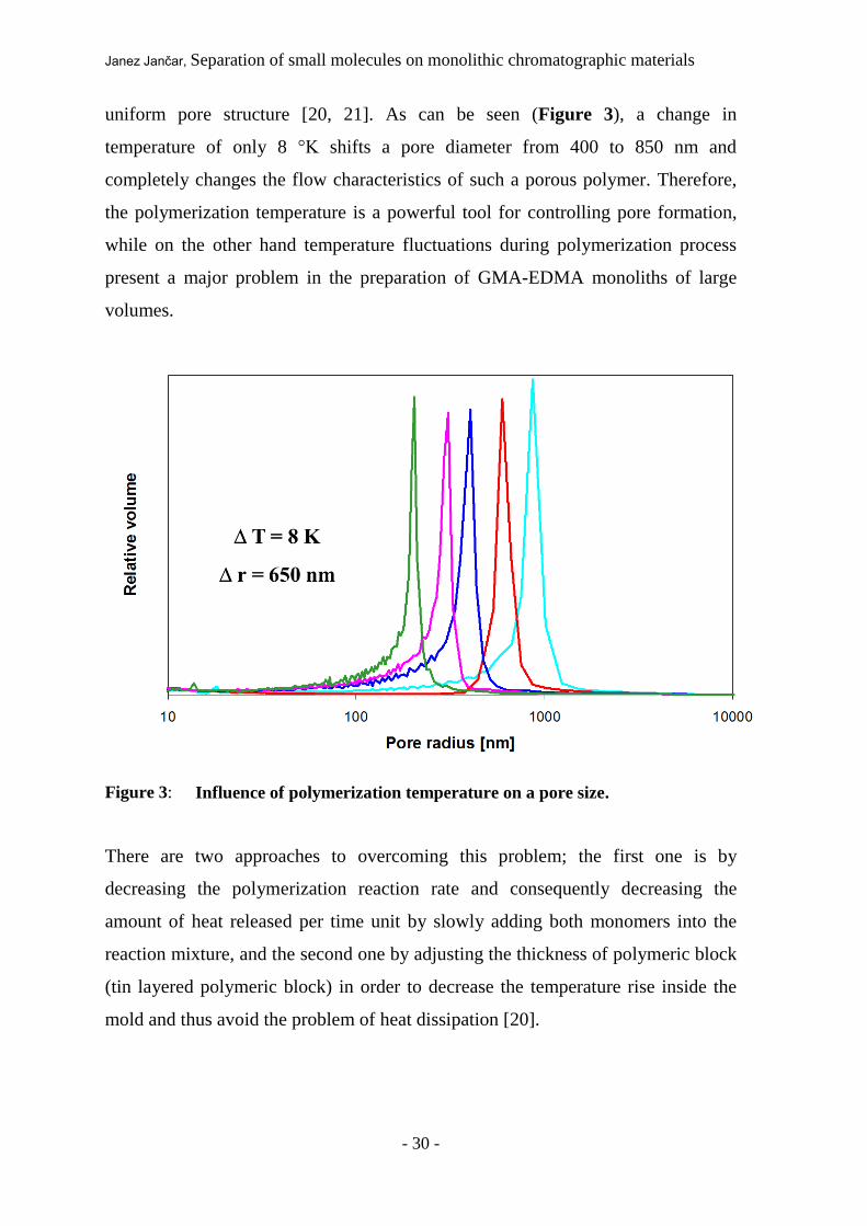
uniform pore structure [20, 21]. As can be seen (Figure 3), a change in
temperature of only 8 °K shifts a pore diameter from 400 to 850 nm and
completely changes the flow characteristics of such a porous polymer. Therefore,
the polymerization temperature is a powerful tool for controlling pore formation,
while on the other hand temperature fluctuations during polymerization process
present a major problem in the preparation of GMA-EDMA monoliths of large
volumes.
Figure 3: Influence of polymerization temperature on a pore size.
There are two approaches to overcoming this problem; the first one is by
decreasing the polymerization reaction rate and consequently decreasing the
amount of heat released per time unit by slowly adding both monomers into the
reaction mixture, and the second one by adjusting the thickness of polymeric block
(tin layered polymeric block) in order to decrease the temperature rise inside the
mold and thus avoid the problem of heat dissipation [20].

Janez Jančar, Separation of small molecules on monolithic chromatographic materials
- 31 -
If we adopt the second scenario, we can quickly find three appropriate geometries;
a relatively long rod with a small diameter, a disk shape geometry of small
thickness and a relatively large diameter and finally to thin walled hollow tube
(Scheme 1).
Scheme 1: Three different alternatives for preparation of scale-up GMA-EDMA
chromatographic polymers.
2.3. Radial flow geometry
GMA-EDMA monolithic chromatographic material with its convective mass
transferee feature was developed and optimized for fast and efficient separation of
large biological molecules. Large biological molecules usually exhibit very steep
adsorption isotherm, which means that they almost irreversibly bind to the matrix
under the bounding conditions. Therefore a change of the mobile phase
composition is normally required for the sufficient elution, which is carried out
through linear or step gradient (gradient elution mode chromatography).

Janez Jančar, Separation of small molecules on monolithic chromatographic materials
- 32 -
Separation of large biological molecules is thus almost exclusively based on a
gradient elution and column length has no significant effect on resolution of
separation. In fact longer columns might even result in additional band spreading
and lowering resolution. Additionally, increased length results in increased
backpressure.
Consequently, tin layered monolithic block strategy to overcome heat relishing
problems during polymerization procedure perfectly fits into gradient elution mode
concept. Following the idea of thin layered polymeric block, the smaller units of
GMA-EDMA monolithic columns are produced in the form of disks and the larger
units in the form of tubes (Scheme 2), whilst rods due to their long length (lower
resolution and higher backpressure) do not fit into the concept [22].

Janez Jančar, Separation of small molecules on monolithic chromatographic materials
- 33 -
Scheme 2: Axial (monolithic disk) and radial (tube monolith) flow mode; two
alternative patterns of flow through the stationary phase at the
chromatographic columns.
At the disk shape, mobile phase is entering in the direction of the cylinder (disk)
axis. The liquid is led into the column through the small diameter hole and then
equally spread across the entire disk surface. If we want to scale up the disk whilst
considering the requirement to maintain a relatively small disk thickness, only an
increase of the diameter is possible. Due to the problems with mechanical stability
of large diameter disks and uniform liquid distribution across the entire disk
surface, they are exclusively used in small scale.
On the other hand, tubular shape approach offers a higher degree of freedom since
inner and outer diameter as well as cylinder height can be varied. Tubular shape
also exhibits greater mechanical stability, regardless of the size. It was also shown
that certain diameters can give the lowest pressure drop for a given flow rate. In
tubular-shaped columns, the mobile phase is uniformly spread over the entire outer
monolith surface, passing through the monolith, and is collected in the central hole
Janez Jančar, Separation of small molecules on monolithic chromatographic materials
- 34 -
from where it exits the column. According to the outlined path of the mobile phase
they enter into the monolith perpendicular to the tube axis. Since the development
of the distribution system and further chromatographic housing design for radial
flow columns is part of this work, more data will be presented in Results and
Discussion chapter.

Janez Jančar, Separation of small molecules on monolithic chromatographic materials
- 35 -
3. EXPERIMENTAL WORK
3.1. Equipment
Analytical gradient HPLC system
A gradient HPLC system built with two Pumps 64 enabling maximum flow rate of
10 mL/min at a maximum pressure of 400 bar, an injection valve with a 20 l
stainless steel sample loop, a variable wavelength monitor set to 260 nm and
response time of 0.15 s, a flow-cell with a 10-mm optical path and volume of 10
L, connected by means of 0.25 mm I.D. PEEK (polyetheretherketon) capillary
tubes and HPLC hardware/software (data acquisition and control station), all from
Knauer (Berlin, Germany) were used when GMA-EDMA monolithic
chromatographic material in shape of CIM®
disks were tested. Knauer mixing
chamber with its relatively large dead volume was replaced by the PEEK Mixing
Tee with an extra low-dead volume (Jour Research, Uppsala, Sweden).
Preparative gradient HPLC system
All characterization of GMA-EDMA polymeric chromatographic material in shape
of CIM® 8 mL tube monolithic column was performed with preparative HPLC
system made by Knauer (Berlin, Germany). The system was built of two K-1800
preparative pumps (each pump contains SS pump head enabling maximum flow
rate of up to 1000 mL/min at a maximum pressure of 50 bar), T shaped static
mixing chamber, manually driven injection valve with 1 mL sample loop,
UV/Visible spectrophotometer model K-2500 with time constant set to 0,1 and
wavelength set to 280 nm, a flow cell with optical path of 2 mm, interface box and
Eurochrom 2000 software installed on a personal computer for real time data
acquisition. Data collecting was set to maximum possible value of 10 Hz.

Janez Jančar, Separation of small molecules on monolithic chromatographic materials
- 36 -
Flow monitoring
During the experiments with CIM® monolithic columns, where the influence of the
flow rate on the separation was studied, a validated digital flow meter (K-3773,
Phase Separations Limited, UK) was additionally introduced to monitor possible
discrepancies between set and real flow rates.
Pressure drop monitoring
Pressure drop measurements were performed with PE300 digital manometer from
Hittinger Baldwin Messtechnik GMBH (Darmstadt, Germany).
Electrophoresis
SDS–PAGE electrophoresis was performed using a Mini-Protean II
electrophoresis Cell made by Bio-Rad (CA, USA) on 20% separation gel with 4%
stacking gel.
3.2. GMA-EDMA monolithic chromatographic columns
GMA-EDMA monolithic chromatographic material
All CIM®
monolithic chromatographic columns used in this work were produced
on platform of glycidyl methacrylate - ethylene glycol dimethacrylate polymeric
material. The initial epoxy GMA-EDMA chromatographic material was prepared
by the radical copolymerization of glycidyl methacrylate (Aldrich, Steinheim,
Germany) and ethylene glycol dimethacrylate (Aldrich, Steinheim, Germany) in
the presence of pore producing solvents. A detailed polymerization procedure is
described at theoretical background in chapter 2.2.2. [18]. The resulting polymeric
material contains reactive epoxy groups which can be easily modified into
alternative chemical groups to obtain materials with desirable chromatographic
properties. Concentration of epoxy groups of initial material was always 4,22
mmol/g of dry support. Porosity of epoxy monolithic material was 60% whilst

Janez Jančar, Separation of small molecules on monolithic chromatographic materials
- 37 -
average pore radius was find to be 700 nm. The surface area of prepared epoxy
GMA-EDMA chromatographic material was 7,48 m2 of dry support.
Table 2: Characteristics of initial epoxy GMA-EDMA monolithic chromatographic
material.
Epoxy GMA-EDMA material
Porosity (%) 60
Average pore radius (nm) 700
Specific Surface area (m2/g dry support) 7,48
Epoxy group concentration (mmol/g dry support) 4,22

Janez Jančar, Separation of small molecules on monolithic chromatographic materials
- 38 -
Scheme 3: Structural formula of Poly(glycidyl methacrylate-co-ethylene
dimethacrylate) polymer (above in scheme 3). Modification reaction of
epoxy group to obtain DEAE and QA chromatographic materials
(below in scheme 3).
GMA Glycidyl methacrylate
EDMA Ethylene glycol dimethacrylate
Poly(glycidyl methacrylate-co-ethylene dimethacrylate)
O
O O
O O
O
O O
O O
O
O
O
O
O
O
O
O
O
O
O
O
O
O O
O O
O
O
O O
O
O
O
O
O
O O
O O
O
O
O O
O
O
O
O O
O
O O
O O
O
O
O O
O
O
O O O
O O
O
O
O O
O
O
O
O
O
O
O O
O
O
O + O
O
O
O

Janez Jančar, Separation of small molecules on monolithic chromatographic materials
- 39 -
The DEAE groups (weak anion exchange groups) were introduced to the CIM®
epoxy disk by placing the disk into pure diethylamin (Fluka, Buchs, Switzerland)
for 24 hours at room temperature. The DEAE group density, obtained by a mass
difference of the initial and modified disk, taking into account the reaction
stoichiometry, was 2.3 mmol/g dry support which correspond to 55% conversion
of epoxy group. Introduction of QA (strong anion exchange groups) groups in the
matrix of 8 mL tubular shaped epoxy monolith was performed in similar way as in
the case of CIM®
epoxy disk modification procedure. Tubular monolith was
immersed in reaction mixture of quaternary amine (Merck, Darmstadt, Germany)
solution for 4 hours whilst temperature was 35 ºC (Scheme 3). The QA group
density, obtained by a mass difference of the initial and modified tube was 2.0
mmol/g of dry support corresponding of 47% conversion of epoxy group.
CIM®
Disks – axial flow mode monolithic chromatographic column
Separation of oligonucleotides was mainly performed on commercially available
CIM®
DEAE (DEAE - diethylaminoethyl groups) disk columns (BIA Separations
d.o.o., Slovenia). CIM® disk column consists of one or more CIM
® disks packed
into commercially available CIM® chromatographic housing (BIA Separations,
Ljubljana, Slovenia) (Figure 4). Up to 4 disks can be packed into a single
chromatographic housing. The bed volume of commercially available CIM®
disk
is 0.34 mL, outer diameter of the disk bed is 12 mm and the length of the
separations layer (thickness) is 3 mm (Scheme 4).

Janez Jančar, Separation of small molecules on monolithic chromatographic materials
- 40 -
Scheme 4: CIM® disk (left) and schematic presentation of CIM
® disk unit packed in
CIM® housing (right).
Additionally, when the effect of the length of the separation layer on a separation
of oligonucleotides was studied, custom made CIM® DEAE disks with a thickness
of 0,75 mm and 1,5 mm were also prepared. The length of the separation layer was
also altered by placing a different number of commercially available CIM®
disks
units in the same housing [8]. In this way the length of the separation layer was
easily prolonged (Scheme 5).
Scheme 5: Schematic presentation of 4 CIM
® disks units packed in a CIM
®
housing.

Janez Jančar, Separation of small molecules on monolithic chromatographic materials
- 41 -
Figure 4: Picture of complete (left) and dismounted CIM
® housing.
CIM®
8 mL Tube – radial flow mode monolithic chromatographic column
All experiments concerning the possible use of thin layered GMA-EDMA
monolithic chromatographic materials as high-throughput columns were
performed on a novel 8 mL CIM QA ion-exchange radial flow monolithic column.
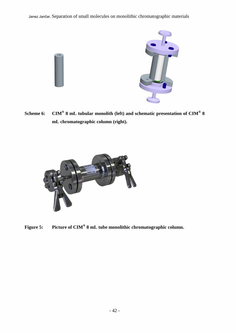
The bed volume of CIM®
8 mL column is 8 mL. Outer diameter of the tubular
monolith is 14,4 mm, inner diameter is 6,5 mm and height is 55 mm. Length of the
separations layer (thickness) is 3,95 mm (Scheme 6). Tubular monolith was
packed in a specially designed chromatographic housing with extra low dead
volume. Housing was made of high grade stainless still (Figure 5).
Janez Jančar, Separation of small molecules on monolithic chromatographic materials
- 42 -
Scheme 6: CIM
® 8 mL tubular monolith (left) and schematic presentation of CIM
® 8
mL chromatographic column (right).
Figure 5: Picture of CIM® 8 mL tube monolithic chromatographic column.

Janez Jančar, Separation of small molecules on monolithic chromatographic materials
- 43 -
3.3. Oligonucleotide samples
The oligonucleotides, synthetized at the National Institute of Chemistry, Ljubljana,
Slovenia, were of the following lengths and structure:
oligodeoxynucleotide 8 (oligo 8): C CAT GTC T3’
oligodeoxynucleotide 10 (oligo 10): GTC CAT GTC T3’
oligodeoxynucleotide 12 (oligo 12): AG GTC CAT GTC T3’
oligodeoxynucleotide 14 (oligo 14): C GAG GTC CAT GTC T3’
3.4. Mobile phases used for oligonucleotide separations
Mobile phase was a 20 mM Tris-HCl buffer, pH 8.5 with different concentrations
of NaCl.
Purified water and all buffer solutions for chromatographic experiments were
filtered through a 0.45 µm pore size filter composed of Sartolon polyamide
(Sartorius, Goettingen, Germany).
3.5. Methods used for characterization of CIM®
monolithic
chromatographic columns
Oligonucleotide separations
The sample of each substance was dissolved in the appropriate mobile phase used
for particular experiment and injected into the disk monolithic column under
isocratic conditions. The mobile phase composition (ionic strength) was then
changed in order to vary the retention time of the particular substance.

Janez Jančar, Separation of small molecules on monolithic chromatographic materials
- 44 -
Pulse response experiments
1 mL of 16.6 % (V/V) solution of acetone in deionized water was injected as a
tracer. Mobile phase was deionized water. Wavelength on detector was set to 280
nm, data acquisition frequency was 10 Hz and detector time constant was set to 0.1
to obtain the highest data density and the fastest detector response. Number of
theoretical plate (N) was calculated with PeakFit software, SeaSolve Software, Inc.
(Framingham, USA) Relative standard deviation (RSD) was calculated on the
target set of data using Microsoft Office Excel software. (Microsoft)
Protein separations
Effect of the flow-rate on protein separation was performed by injecting 1 mL of
protein mixture containing myoglobin (0.7 mg/mL), conalbumin (2.7 mg/mL) and
soybean trypsin inhibitor (2,7 mg/mL) dissolved in 20 mM Tris-HCl, pH 7.4.
Separation was performed at 7 different flow rates (16, 40, 80, 160, 240, 320, 400
mL/min being equal to 2, 5, 10, 20, 30, 40 and 50 CV/min) by applying the linear
gradient (from 0 M NaCl to 1 M NaCl in 20 mM Tris-HCl, pH 7.4), which was
calculated for each flow-rate to be constant and equal to 8 CV.
Separation of BSA aggregates from BSA monomer was performed by injecting 1
mL of loading buffer (20 mM Tris-HCl+ 0,2 M NaCl, pH 7.4) containing BSA
with concentration of 10 mg/mL on the 8 mL GMA-EDMA column. Linear
gradient from 0,2 M to 0,8 M NaCl was performed in 46 sec. Flow-rate was 560
mL/min (70 CV/min). Absorbance at 280 nm was monitored.
Dynamic binding capacity
Effect of the flow-rate on protein dynamic capacity was determined by frontal
analysis experiments. The column was first equilibrated with loading buffer
(20 mM Tris-HCl, pH 7.4) and then loaded with protein solution (1 mg/mL BSA in
20 mM Tris-HCl buffer, pH 7.4) at flow rates from 7.5 (approximately 1 CV) to
358 (approximately 45 CV) mL/min. The absorbance at 280 nm was measured and
the protein capacity at 50 % breakthrough was calculated.

Janez Jančar, Separation of small molecules on monolithic chromatographic materials
- 45 -
Pressure drop measurement
Pressure drop was measured at different flow rates (50, 100, 150, 200, 250, 300,
350, 400 mL/min) using purified water as a mobile phase. Pressure sensor was
connected at the column outlet. Experiment was repeated after disconnecting
monolithic column from the HPLC system to determine pressure of the HPLC
system alone. Both values were subtracted to get pressure drop on the monolithic
column.
3.6. Instrumentation methods
Sodium dodecylsulfate–polyacrylamide gel electrophoresis (SDS–PAGE)
Efficiency of protein separation was analyzed by loading collected fractions on 20
% separation gel with 4 % stacking gel. Electrophoresis was run at 200 V for 50
min. Band detection was performed with gel staining for 2 hours in Commassie
brilliant blue and washing in distillate water over the night.
3.7. Chemicals
All solutions were prepared using deionized water purified by Watek IWA-80 roi
purification system (Ledeč nad Sázavon, the Czeck Republic)
Acetone was purchased from Rathburn Chemicals (Walkerburn, Scotland).
Tris(hydroxymethyl)aminomethane (Tris), sodium chloride, hydrochloric acid
(37 %) were obtained from Merck (Darmstadt, Germany). Proteins myoglobin,
conalbumin and soybean trypsin inhibitor (STI) were purchased from Sigma-
Aldrich Inc. (St. Louis, USA) while bovine serum albumin (BSA) was purchased
from Fluka (Buchs, Switzerland). Buffer solutions were made by adding a known
mass of buffering species to 80% of the desired final volume of purified water.
Tris-HCl buffers were then titrated with HCl water solution (1:1). Molecular

Janez Jančar, Separation of small molecules on monolithic chromatographic materials
- 46 -
weight standards LowRange Biorad (CA, USA) and 10–200 kDa MBI Fermentas
(Vilnus, Lithuania) were used for electrophoreses. Commasie blue was used for
protein bands detection according to the manufacturer’s instructions for the
PlusOne Amersham Bioscience (Uppsala, Sweden).

Janez Jančar, Separation of small molecules on monolithic chromatographic materials
- 47 -
4. EXPERIMENTAL WORK
4.1. Isocratic separation of oligonucleotides on CIM®
disks axial flow mode
monolithic chromatographic column
To verify the possibility of performing isocratic separations of oligonucleotides on
GMA-EDMA monolithic chromatographic material, commercially available CIM®
DEAE disk was applied. The sample was a mixture of four oligonucleotides with
different chain lengths: oligo 8, oligo 10, oligo 12 and oligo 14. The results are
shown in Figure 6 where all four oligonucleotides are well separated within 3
minutes.

Janez Jančar, Separation of small molecules on monolithic chromatographic materials
- 48 -
Figure 6: Isocratic separation of oligonucleotides on a CIM
® DEAE disk with the
thickness of 3 mm.
Conditions: Mobile phase: 0,46 M NaCl in 20 mM Tris-HCl buffer, pH 8.5; Flow rate: 9
mL/min; Sample: 50 g/mL of oligo 8, 150 g/mL of oligo 10, 450 g/mL of oligo 12
and 750 g/mL of oligo 14 in buffer A; Injection volume: 20 L; Detection: UV at 260
nm.
4.1.1. Peak width of oligo 8 is proportional to its retention time
From Figure 6 we can deduce that retention time of oligonucleotides increases
with molecular weight. We could also observe a pronounced broadening of peaks
with increased retention time. To investigate the correlation between peak width
and the retention time isocratic runs of a single oligonucleotide oligo 8 as a
sample, using mobile phase with different NaCl concentrations ranging from 1 M
0
5
10
15
20
25
30
0 25 50 75 100 125 150 175
rela
tive
ab
so
rba
nce
at 2
60
nm
(m
AU
)
time (s)
oligo 8
oligo 10
oligo 12
oligo 14

Janez Jančar, Separation of small molecules on monolithic chromatographic materials
- 49 -
to 0,38 M, were performed. The resulting chromatograms are presented in Figure
7.
Figure 7: Effect of mobile phase composition on the retention time and peak shape of
oligo 8 on a 3 mm CIM® DEAE disk.
Conditions: Mobile phase: 20 mM Tris-HCl buffer, pH 8.5 + different concentrations of
NaCl (actual mobile phase composition is shown in the Figure); Flow rate: 3 mL/min;
Sample: 50 g/mL of oligo 8 in buffer A; Injection volume: 20 l; Detection: UV at 260
nm.
The increase of salt concentration in the mobile phase decreases the retention time.
When 1 M NaCl in buffer is used oligo 8 does not bind to the matrix and is eluted
with the front. At very low salt concentration (only buffer A) oligo 8 binds
irreversibly to the support and is not eluted at all.
0
20
40
60
80
100
120
140
160
180
200
0 20 40 60 80 100 120 140
rela
tive
ab
so
rba
nce
at 2
60
nm
(m
AU
)
time (s)
1 M
0.62 M
0.59 M
0.56 M
0.53 M
0.50 M
0.47 M
0.44 M
0.41 M
0.38 M

Janez Jančar, Separation of small molecules on monolithic chromatographic materials
- 50 -
According to the theory of isocratic multiple adsorption/desorption separation
process, the peak width (t) is proportional to the retention time (tr) via equation
[17]:
The experimentally determined peak width, t, was measured from the oligo 8
peaks at different molar concentrations of sodium chloride. The resulting values of
t are plotted against the retention time, tr, in the Figure 8.
Figure 8: Standar deviation (t) of peak as a function of the retention time (tt). Data
was derived from chromatograms presented in figure 7.
0
1
2
3
4
5
6
7
0 20 40 60 80 100
σt (
s)
tr (s)
t = (HETP/L)1/2 * tr
t = 0.0774 * tr + 0.1374 R2 = 0.999
HETP = 18 m
rt tL
HETP (6)

Janez Jančar, Separation of small molecules on monolithic chromatographic materials
- 51 -
The linear relationship between t and tr as predicted by Eq. (6) is clearly
demonstrated (R2 = 0.999). The slope of the line is equal to 0.0774, which
translates to the HETP (height equivalent to the theoretical plate) value
corresponding to 18.0 m. This value approaches the values of HETP for
conventional HPLC columns filled with 5-7 m porous particles.
4.1.2. Peak height of oligo 8 is inversely proportional to its retention
time
Furthermore, for the peaks that can be described by a Gaussian function,
characteristic for chromatographic processes based on multiple
adsorption/desorption steps, the maximum peak height (hmax) is inversely
proportional to the retention time [17]:
Experimental values of peak heights of oligo 8 correlated to reciprocal values of
the retention times can be successfully fitted with equation (7) giving the
correlation index R2 = 0.988.
From these results a close similarity between the isocratic separation mechanism
on conventional HPLC columns based on multiple adsorption/desorption steps and
isocratic separation on the thin GMA-EDMA monolithic chromatographic material
is indicated.
h
R vHETP
L
t rmax
1
2
1
(7)

Janez Jančar, Separation of small molecules on monolithic chromatographic materials
- 52 -
4.1.3. Effect of the monolith thickness (column length) on a resolution
If multiple adsorption/desorption steps occur in thin GMA-EDMA monoliths, the
retention time should be proportional to the monolith thickness [17]. To investigate
this possibility, monoliths of different lengths were prepared. Firstly, one disk was
placed in the appropriate housing and then the separation of oligonucleotides was
carried out. In the next experiment, an additional disk was placed in the same
housing. This progression was continued by placing 3 and 4 disks in the same
housing. In this way monoliths with different lengths were prepared, with a
thickness ranging from 3 mm (one disk in the housing) to 12 mm (4 disks in the
housing), which should be equivalent to a single monolith of the same thickness
assuming that the interface effects can be neglected. This assumption is justified
by the similarity existing between isocratic separation on a single 3 mm disk and a
combination of two disks with the thickness of 1.5 mm in the same housing. In
addition, specially prepared thin monolithic disks with a thickness of only 1.5 and
0.75 mm were also applied.

Janez Jančar, Separation of small molecules on monolithic chromatographic materials
- 53 -
Figure 9: The effect of the monolith thickness (column length) on the resolution of
the oligonucleotide isocratic separation.
Conditions: Mobile phase: 0,5 M NaCl in 20 mM Tris-HCl buffer, pH 8.5; Separation
unit: one or more CIM DEAE disks in the same housing (one 0,75 mm disk or one 1,5mm
disk or one 3 mm disk or two 3 mm disks to form 6 mm column length or three 3 mm
disks to form 9 mm column length or four 3 mm disks to form 12 mm column length);
Flow rate: 3mL/min; Sample: 50 g/mL of oligo 8 (1st peak; in the insert), oligo 10 (2
nd
peak; in the insert), oligo 12 (3rd
peak; in the insert) and oligo 14 (4th
peak; in the insert)
in buffer A; Injection volume: 20 L; Detection: UV at 260 nm.
Figure 9 shows that the separation improves significantly with the increase of the
column length. In fact, fitting the relationship between the retention times of
certain oligonucleotide and a particular column length, the straight line obtained
provides a correlation index higher than 0.99 for all four oligonucleotides. The
dependency of the retention times and the length of the column for all four
oligonucleotides is presented in Figure 10.

Janez Jančar, Separation of small molecules on monolithic chromatographic materials
- 54 -
Figure 10: The retention time dependency on the monolith thickness.
Under this salt concentration, separation on the 0.75 mm GMA-EDMA monolith
was not achieved. However, after the optimization of the mobile phase
composition, taking into account the special requirements of the disk related to its
thickness, the separation of all oligonucleotides was successfully carried out also
on the ultra-thin 0.75 mm GMA-EDMA monolith (Figure 11).
0
100
200
300
400
500
600
700
0 2 4 6 8 10 12 14
monolith length (mm)
rete
nti
on
tim
e (
s)
y = 4.4 * x -0.55 R2 =1.000
y = 17.65 * x - 10.02 R2 = 0.997
y = 31.06 * x - 20.08 R2 = 0.997
y = 57.77 * x - 40.34 R2 = 0.997

Janez Jančar, Separation of small molecules on monolithic chromatographic materials
- 55 -
Figure 11: Isocratic separation of oligonucleotides on a CIM DEAE disk with the
thickness of 0.75 mm.
Conditions: Mobile phase: 0,37 M NaCl in 20 mM Tris-HCl buffer, pH 8.5; Flow rate: 3
mL/min; Sample: 50 g/mL of oligo 8, oligo 10, oligo 12 and oligo 14 in buffer A;
Injection volume: 20 L; Detection: UV at 260 nm.
These data demonstrate that also very thin GMA-EDMA monoliths enable
isocratic separation. This rather surprising result could be ascribed to the particular
matrix structure.
4.1.4. Effect of the flow rate on a resolution
Since monoliths have low diffusion resistance due to convective transport inside
the flow-through pores it was shown that the flow rate has no pronounced effect on
0
20
40
60
80
100
120
140
0 20 40 60 80
rela
tive
ab
so
rba
nce
at 2
60
nm
[m
AU
]
time (s)
oligo 8
oligo 10
oligo 12
oligo 14

Janez Jančar, Separation of small molecules on monolithic chromatographic materials
- 56 -
the resolution [3]. In contrast to this fact, in the case of isocratic separation of
plasmid DNA conformers it was found that there is an optimum flow rate around 1
mL/min [10]. Therefore, the effect of the flow rate on the isocratic separation of 4
oligonucleotides was also investigated. The flow rate was measured by a validated
flow meter to provide accurate data for further analysis, where the results are
normalized to the volume of mobile phase run through the GMA-EDMA monolith.
The effect of the flow rate was investigated on CIM DEAE disk of 3 mm
thickness.
Figure 12: Effect of the flow rate on the isocratic separation of oligonucleotides on a
CIM DEAE disk with the thickness of 3 mm.
Conditions: Mobile phase: 0,46 M NaCl in 20 mM Tris-HCl buffer, pH 8.5; Sample: 1:
50 g/mL of oligo 8, 2: 150 g/mL of oligo10, 3: 450 g/mL of oligo12 and 4: 750
g/mL of oligo 14 in buffer A; Injection volume: 20 L; Detection: UV at 260 nm.
0
20
40
60
80
100
120
0 2 4 6 8 10 12 14 16 18 20 22 24 26 28 30
rela
tive
ab
so
rba
nce
at 2
60
nm
(m
AU
)
volume (ml)
flow rate: 0.5 mL/min
flow rate: 3 mL/min
flow rate: 7 mL/min
1
1
1 2
2
2
3
3
3 4
4
4

Janez Jančar, Separation of small molecules on monolithic chromatographic materials
- 57 -
Surprisingly, it was found that an increase in flow rate has a beneficial effect in
terms of the difference between peak retention volumes. This phenomenon is
presented in Figure 12 and can possibly be explained from the hydrodynamic
point of view. Higher flow rate causes higher back pressure drop on the monolith.
As a consequence, mobile phase containing the molecules to be separated can
penetrate into pores of smaller diameter [9,10]. In this way, the active surface
available for the adsorption/desorption process is larger, possibly resulting in
longer retention times.
From these results, another confirmation of the separation process based on
multiple adsorption/desorption mechanisms can be derived. Some speculated the
possibility that the separation under isocratic condition occurs due to shear forces.
In contrast to separations carried out in conventional HPLC columns, where fluid
in the pores is stagnant, in monoliths there is a laminar convective flow throughout
the pores. This flow could cause shear forces relative to the surface where the
molecules are adsorbed. It is possible that these forces could cause some kind of
erosion of adsorbed molecules from the surface. Since the molecules are bound to
the surface to a different degree because of a varying number of binding sites, they
can be selectively desorbed. This mechanism would lead to two effects: since the
shear forces increase with increased flow rate, it is reasonable to speculate that the
resolution would decrease. On the other hand, at very low flow rates shear forces
would be probably too low to desorb some strongly bound molecules. However,
these two effects were not observed either at a very low flow rate, where also all
molecules desorb from the matrix, or at higher flow rates, where the separation
even improves and no reduction in retention volume is observed (Figure 12).
Since both effects are different from the expected one in the case of the shear rate
elution, the multiple adsorption/desorption process is the most probable
phenomenon occurring.
All data show that equations derived for isocratic separations based on multiple
adsorption/desorption steps can also be efficiently applied for isocratic separations

Janez Jančar, Separation of small molecules on monolithic chromatographic materials
- 58 -
on thin monoliths. Therefore, it is reasonable to assume that similar mechanisms
also occur in monoliths. It is surprising, however, that the 0.75 mm thick monolith
can provide sufficient travel length for this phenomenon to occur. There are two
characteristics of the monoliths that might help to explain these results. The flow-
through pores in the GMA-EDMA monoliths have a broad size distribution [18].
In addition, they are highly interconnected. Therefore, the molecules travelling
through the monolith have to pass many constrictions and tortuosities. As a
consequence, the real path through the monolith is longer than its physical
thickness. Thus, the molecules have more time to interact with the surface. The
second characteristic of the monoliths is the reduced diffusion resistance. In the
case of particle supports the diffusion through the stagnant liquid inside the pores
is a major bottleneck of the mass transfer exchange between the mobile phase and
the surface of the support where the active groups are located. In the case of
monoliths, this stagnant layer is extremely reduced and the mass exchange is much
faster. The main result is that on the same length of the separation layer, many
more adsorption/desorption steps can take place in monoliths than in the
conventional porous supports. Thus, a much thinner monolith is sufficient for
achieving the same number of adsorption/desorption steps.
In addition, by comparing the separation of plasmid DNA conformers [10] and
oligonucleotides, better separation in the case of the latter was achieved. This
might be due to the smaller molecular size of the oligonucleotides and,
consequently, faster diffusion. It can be concluded that, in contrast to the selective
gradient elution where the separation resolution commonly improves with
increased molecular weight [9], in the case of isocratic separations smaller
molecules can be separated more easily.

Janez Jančar, Separation of small molecules on monolithic chromatographic materials
- 59 -
4.2. Design and characterization of high throughput CIM®
8 mL tube
radial flow mode monolithic chromatographic column
In the first part of the wok it was shown that GMA-EDMA monoliths material can
be prepared in extremely thin forms and then used as a stationary phase for
preparation of extremely short HPLC columns. In spite of very short separation
path due to their unique structure GMA-EDMA monolithic columns still enable
very good separation of oligonucleotides in isocratic mode.
Additionally, monolithic chromatographic columns, when used in a gradient
separation mode, also provide an excellent tool for fast and efficient separations of
very large biological molecules. If it were possible to design very thin layered
monoliths also on preparative or even industrial scale, significant benefits can be
expected. However, to develop such chromatographic columns appropriate
chromatographic housing must be designed.
On the market there are different designs of chromatographic columns, which can
be divided into two main groups. Most of today's HPLC column belongs to the
group of so-called axial mode operating columns, while the radial one with a radial
flow pattern are more rear. Which type performs better depends on the particular
case [23] but it seems that radial operating columns attracting interest since they
exhibit some beneficial features [24].
One of the main problems of radial operating chromatographic columns is the
changing of a mobile phase linear velocity over the chromatographic bed. Because
of that, matrix efficiency for porous particulate supports varies by its position
within the bed, and overall performance is more difficult to predict. This problem
is not present when the monolithic supports are used, since it was demonstrated
that their chromatographic properties are flow unaffected even at the extreme
linear velocity of 50000 cm/h [25] which was confirmed also for radial operating
mode [26]. It was demonstrated that even in the range of applicable flow-rates, bed

Janez Jančar, Separation of small molecules on monolithic chromatographic materials
- 60 -
utilization for monolithic column operating in a radial mode is still close to 100 %
[27]. Therefore, one can hardly distinguish between axial and radial operating
monolithic chromatographic column of the same bed length. This, together with
lower pressure drop of a radial design [28], was the motivation for designing a new
monolithic column in a radial operating mode.
The monolith and housing were designed for extremely high flow rates, up to 70
CV/min which is the range of the flow-rates applied on membrane columns. This
was achieved by proper monolith dimensions with the height of 55 mm, inner
diameter of 6.0 mm and thickness of just 4.5 mm. The construction of radial flow
monolithic column is shown in Scheme 7.

Janez Jančar, Separation of small molecules on monolithic chromatographic materials
- 61 -
Scheme 7: Sliced design of 8 mL radial monolithic column with indicated parts.
The monolith is fixed between two stainless steel (SS) covers with TC connectors
that prevent bypassing. Two covers close the column on both sides and are fixed to
the SS column body with SS bolts. Inside the monolith, there is a SS reinforcement
frit. The monolith is surrounded by the SS body cylinder and has a flow distributor
Outlet cover
Body
GMA-EDMA monolith
SS reinforcement frit
Inlet cover
Distributor plate
SS distribution frit

Janez Jančar, Separation of small molecules on monolithic chromatographic materials
- 62 -
that uniformly spreads the mobile phase over the entire outer surface of the
monolith.

Janez Jančar, Separation of small molecules on monolithic chromatographic materials
- 63 -
4.3. Properties of high throughput CIM®
8 mL tube radial flow mode
monolithic chromatographic column
4.3.1. Mobile phase distribution as a function of flow rate
To investigate the properties of the described design several pulse response tests at
different flow rates, from 20 up to 70 CV/min, were performed. Such a broad
range of flow rates could affect distribution of the mobile phase over the outer
surface of the monolith and consequently influence column efficiency. To
investigate all these data, pulse response experiments were normalized to the
elution volume and plotted in the same chromatogram (Figure 13).
Figure 13: Pulse response experiment as a function of flow rate.
Conditions: Mobile phase: deionized water; Flow rate: 160, 240, 320, 400, 480, 560
mL/min; Sample: 1 mL of 16,6 % acetone solution; Injection volume: 1 mL; Detection:
UV at 280 nm; Data acquisition frequency was 10 Hz and detector time constant was set
to 0,1
0
20
40
60
80
100
120
140
0 10 20 30 40
rela
tive
ab
so
rba
nce
at 2
80
nm
(m
AU
)
mL
160 mL/min
240 mL/min
320 mL/min
400 mL/min
480 mL/min
560 mL/min

Janez Jančar, Separation of small molecules on monolithic chromatographic materials
- 64 -
At Figure 13 we can see from the peak overlapping that there was a slight trend of
efficiency change over the entire range, also confirmed by calculation on a number
of theoretical plates (N-plate). Values were scattered within 20,7 % RSD (Table
2).
Table 3: Scattering of N-plate values in pulse response experiment as a function of
flow rate.
Flow rate
(mL/min)
N-plate (EMG)
160 93,7
240 81,4
320 77,5
400 73,0
480 68,8
560 47,9
RSD 20,7

Janez Jančar, Separation of small molecules on monolithic chromatographic materials
- 65 -
To investigate whether this was due to changes in column performance or due to
small variations in HPLC system performance consecutive pulse response
injections were performed at fixed flow-rate of 10 and 30 CV/min (Figures 14
and 15).
Figure 14: Pulse response experiment at 80 mL/min as a function of injections.
Conditions: Mobile phase: deionized water; Flow rate: 80 mL/min; Sample: 1 mL of 16,6
% acetone solution; Injection volume: 1 mL; Detection: UV at 280 nm; Data acquisition
frequency was 10 Hz and detector time constant was set to 0,1
0,0
20,0
40,0
60,0
80,0
100,0
120,0
140,0
0,0 0,1 0,2 0,3 0,4 0,5
rela
tive
ab
so
rba
nce
at 2
80
nm
(m
AU
)
time (min)
inj1
inj2
inj3
inj4

Janez Jančar, Separation of small molecules on monolithic chromatographic materials
- 66 -
Figure 15: Pulse response experiment at 240 mL/min as a function of injections.
Conditions: Mobile phase: deionized water; Flow rate: 240 mL/min; Sample: 1 mL of
16,6 % acetone solution; Injection volume: 1 mL; Detection: UV at 280 nm; Data
acquisition frequency was 10 Hz and detector time constant was set to 0,1
Table 4: Scattering of N-plate values in pulse response experiment as a function of
injection at 80 and 240 mL/min.
80 mL/min
Serial injection N-plate (EMG)
Inj 1 114,8
Inj 2 108,2
Inj 3 115,4
Inj 4 110,8
RSD 3,8
240 mL/min
Serial injection N-plate (EMG)
Inj 1 176,8
Inj 2 188,4
Inj 3 178,0
Inj 4 188,2
RSD 3,5
The number of theoretical plates together with RSD was found to be 3,8 % at 10
CV/min and 3,5 % at 30 CV/min. This indicates that injection to injection
reproducibility indeed causes constant variation of performance in a range of about
4 % regardless of applied flow-rate, on the other hand it is still not so high as we
0,0
20,0
40,0
60,0
80,0
100,0
120,0
0,0 0,1 0,2
rela
tive
ab
so
rba
nce
at 2
80
nm
(m
AU
time (min)
inj1
inj2
inj3
inj4

Janez Jančar, Separation of small molecules on monolithic chromatographic materials
- 67 -
can observe in Figure 13, where pulse response experiment as a function of flow
rate was performed.
A close look back to Figure 13 reveals a constant decrease of the peak height and
an increase of the flow rate. To investigate the cause of such behavior the column
was replaced with a union fitting and pulse response tests at different flow rates
were repeated.
Figure 16: Pulse response experiment without column as a function of flow rate.
Conditions: Column: union was used instead of column; Mobile phase: deionized water;
Flow rate: 160, 240, 320, 400, 480, 560 mL/min; Sample: 1.mL of 16,6 % acetone
solution; Injection volume: 1 mL; Detection: UV at 280 nm; Data acquisition frequency
was 10 Hz and detector time constant was set to 0,1
0
20
40
60
80
100
120
140
160
180
0 5 10 15 20 25 30
rela
tive
ab
so
rba
nce
at 2
80
nm
(m
AU
)
mL
160 mL/min
240 mL/min
320 mL/min
400 mL/min
480 mL/min
560 mL/min

Janez Jančar, Separation of small molecules on monolithic chromatographic materials
- 68 -
Table 5: Scattering of N-plate values in pulse response experiment as a function of
flow rate.
Flow rate
(mL/min)
N-plate (EMG)
160 95,4
240 73,0
320 69,4
400 53,0
480 55,6
560 53,2
RSD 23,9
A very similar trend of peak height decrease in case of higher flow rates was
observed in experiment when the chromatographic column was replaced with the
union fitting (Figure 16 and Table 4). This deviation probably needs to be
ascribed to the limitation of data collecting capability of detector since peak width
at higher flow rates was in range of second. The peaks are actually so sharp that
the maximum of the peak can be lost between two neighboring data points, as
already described in literature [29]. These data demonstrate that variations are in
fact caused by HPLC system itself while column performance is preserved over
the entire tested flow rate.
4.3.2. Chromatographic properties
The performance of developed column was further tested by investigating its
chromatographic properties. In Figure 17 a separation of standard protein mixture
at various flow rates (up to 50 CV/min) normalized to elution volume is presented.

Janez Jančar, Separation of small molecules on monolithic chromatographic materials
- 69 -
Figure 17: Effect of flow rate on protein separations.
Conditions: Mobile phase: mobile phase A, 20mM Tris-HCl buffer pH 7.4; mobile phase
B, 1M NaCl in buffer A, pH 7.4; Flow rate: 16, 40, 80, 160, 240, 320, 400 mL/min;
Sample: myoglobin (0,7 mg/mL), conalbumin (2,7 mg/mL) and STI (2,7 mg/mL)
dissolved in mobile phase A; Injection volume: 1 mL; Linear gradient from 0-100%
buffer B in 8 CV; Detection: UV at 280 nm; Data acquisition frequency was 10 Hz and
detector time constant was set to 0,1
Again it can be noted that no significant change in resolution is brought about by
changing the flow rate. A similar conclusion can be arrived at based on the
dynamic binding capacity measurements performed by loading BSA on the
monolithic column at different flow rates (Figure 18).
0
10
20
30
40
50
60
70
0 20 40 60 80 100
Ab
so
rba
nce
at 2
80
nm
(m
AU
)
Volume (mL)
8 mL/min
16 mL/min
40 mL/min
80 mL/min
160 mL/min
240 mL/min
320 mL/min
400 mL/min
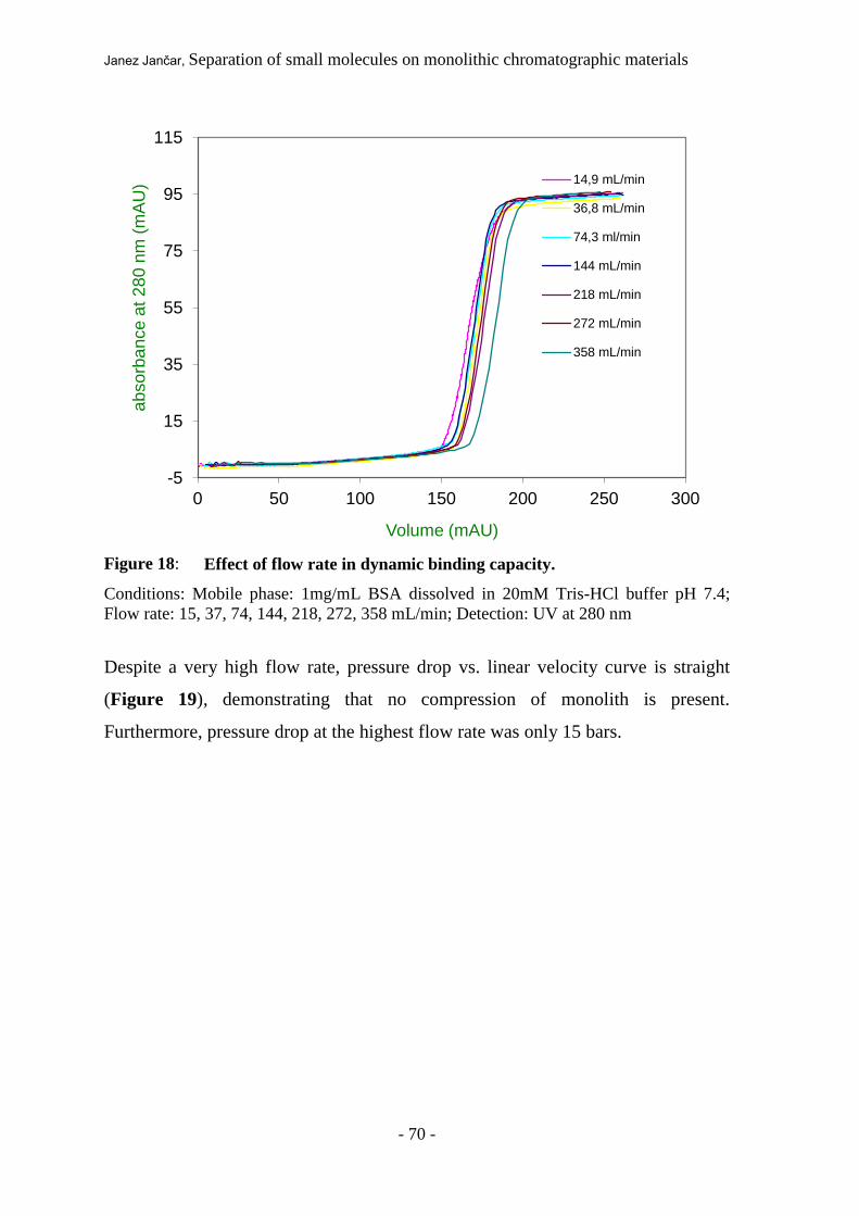
Janez Jančar, Separation of small molecules on monolithic chromatographic materials
- 70 -
Figure 18: Effect of flow rate in dynamic binding capacity.
Conditions: Mobile phase: 1mg/mL BSA dissolved in 20mM Tris-HCl buffer pH 7.4;
Flow rate: 15, 37, 74, 144, 218, 272, 358 mL/min; Detection: UV at 280 nm
Despite a very high flow rate, pressure drop vs. linear velocity curve is straight
(Figure 19), demonstrating that no compression of monolith is present.
Furthermore, pressure drop at the highest flow rate was only 15 bars.
-5
15
35
55
75
95
115
0 50 100 150 200 250 300
ab
so
rba
nce
at
28
0 n
m (
mA
U)
Volume (mAU)
14,9 mL/min
36,8 mL/min
74,3 ml/min
144 mL/min
218 mL/min
272 mL/min
358 mL/min

Janez Jančar, Separation of small molecules on monolithic chromatographic materials
- 71 -
Figure 19: Pressure drop as a functions of flow rate.
Conditions: Deionized water was used as a mobile phase
4.3.3. Packing and refill
Such column design also allows a refill of the monolith by easily taking out an old
monolith and exchanging it with a new one. To investigate how reproducible such
a procedure can be the column was disconnected from the HPLC system, the outlet
cover was unscrewed from the column (Scheme 7), the monolith, was taken out
than placed back into the same housing, the outlet cover was again tightened and
the column was reconnected to the HPLC system. The entire procedure was
completed within 2 minutes. After each refill, pulse response test procedure at the
flow rate of 30 CV/min was performed and the number of theoretical plates (N)
was calculated (Figure 20 and Table 5).
y = 0,0388x - 0,7714 R² = 0,9981
0
2
4
6
8
10
12
14
16
0 100 200 300 400 500
Pre
ssu
re (
ba
r)
Flow rate (mL/min)

Janez Jančar, Separation of small molecules on monolithic chromatographic materials
- 72 -
Figure 20: Effect of column refill on column efficiency.
Conditions: Mobile phase: deionized water; Flow rate: 240 mL/min; Sample: 1 mL of
16,6 % acetone solution; Injection volume: 1 mL; Detection: UV at 280 nm; Data
acquisition frequency was 10 Hz and detector time constant was set to 0,1
Table 6: Scattering of N-plate values in pulse response experiment as a
function of flow rate.
Flow rate
(mL/min)
N-plate (EMG)
packing 1 195,9
packing 2 222,7
packing 3 212,6
packing 4 216,6
packing 5 217,0
RSD 4,3
RSD of N was calculated to be 4.3 %, which is a value comparable to the one
obtained by the consecutive injections performed at constant flow rate described
above. Obviously, column filling procedure has no detectable influence on column
performance, therefore refilling of monolithic columns can be considered as
extremely reproducible, easy and fast.
0,0
20,0
40,0
60,0
80,0
100,0
120,0
140,0
0,0 0,1 0,2
ab
so
rba
nce
at
28
0 n
m
time (min)

Janez Jančar, Separation of small molecules on monolithic chromatographic materials
- 73 -
4.3.4. Separation of real sample
Finally on developed monolithic column separation of protein aggregates from a
monomer protein as example of macromolecule removal using high throughput
monolithic radial column was tested as well. For this purpose lyophilized protein
BSA was used, which is known to contain aggregates [30], and dissolved it in a
loading buffer. BSA dimers and aggregates interact slightly stronger with the anion
exchange matrix therefore they can be separated by implementation of salt
gradient. A typical chromatogram is shown in Figure 21.
Figure 21: Separation of BSA monomer from BSA aggrgates.
Conditions: Mobile phase: mobile phase A, 20 mM Tris-HCl buffer + 0,2 M NaCl pH
7.4; mobile phase B, 1M NaCl in buffer A, pH 7.4; Flow rate: 560 mL/min; Sample BSA
(10 mg/mL) dissolved in buffer A Injection volume: 1 mL; Linear gradient from 0-100 %
buffer B in 46 s; Detection: UV at 280 nm; Data acquisition frequency was 10 Hz and
detector time constant was set to 0,1
Separation was performed at extremely high flow rate of 70 CV/min and
completed within 46 seconds. Such short time implies the possibility of
implementing this type of column for in-process control during preparative
0
10
20
30
40
50
60
70
80
90
100
-0,2
1,8
3,8
5,8
7,8
9,8
11,8
13,8
0 0,5 1 1,5 2 2,5
gra
die
nt (%
)
ab
so
rba
nce
at
28
0 n
m (
mA
U)
time (min)

Janez Jančar, Separation of small molecules on monolithic chromatographic materials
- 74 -
aggregate removal but it can also be used for removal itself since the separation
was efficient as indicated in Figure 22.
Figure 22: SDS–PAGE analysis: M: molecular weight standard, L: load, E1: peak 1 ,
E2: peak 2.
M L E1 E2
Janez Jančar, Separation of small molecules on monolithic chromatographic materials
- 75 -
5. CONCLUSIONS
It was shown that it is possible to isocratically separate oligonucleotides on anion-
exchange CIM® DEAE disks and that the performance of the monolith stationary
phase is similar to those of 5 - 7 m porous particles. From the behaviour of the
isocratic separations it was concluded that the mechanism of the separation on thin
glycidyl methacrylate-ethylene glycol dimethacrylate (GMA-EDMA) monoliths is
very similar to the one on conventional columns.
All these observations indicate that isocratic separation on thin GMA-EDMA
monoliths is probably the result of the multistep adsorption/desorption process.
The fact that isocratic separation on the monolith with a thickness of less than 1
mm occurs, may be ascribed to convective mass transport through the flow-
through pores, which causes an increase in the number of adsorption/desorption
steps on shorter monolith lengths. This phenomenon opens many new areas of fast
separations or purification on this type of supports. For example, when comparing
the separation of plasmid DNA conformers with that of oligonucleotides much
better separation was achieved in the latter case. This is probably due to the lower
molecular weight of oligonucleotides and, consequently, higher diffusion
coefficients. According to this analogy one can speculate also the possibility of
efficient isocratic separation of peptides. Furthermore, it is reasonable to assume,
that also other smaller molecules having even higher diffusivities,might also be
separated isocratically.
It was also shown that thin layered GMA-EDMA monoliths can also be applied in
preparative scale format. Combining radial flow pattern together with appropriate
housing design, columns capable of very high throughput can be prepared, making
them comparable to the membrane one. From the experiments it can be concluded
that resolution of columns is unaffected in a broad range of flow rates, making
them the material of choice for various applications, especially for purification of
macromolecules and nanoparticles on an industrial scale. The column packing and

Janez Jančar, Separation of small molecules on monolithic chromatographic materials
- 76 -
refilling process is very fast and relatively unsophisticated, making thin layered
GMA-EDMA monoliths even more attractive for industrial application.

Janez Jančar, Separation of small molecules on monolithic chromatographic materials
- 77 -
6. REFERENCES
[1] M.R. Buchmaiser, J. Chromatogr. A, 918 (2001) 233.
[2] A.E. Rodrigues, J. Chromatogr. B, 699 (1997) 47.
[3] A. Štrancar, M. Barut, A. Podgornik, A. Buchaher, Dj. Josić, LC-GC, 11 (1998)
660.
[4] A. Štrancar, P. Koselj, H. Schwinn, Dj. Josić, Anal. Chem., 68 (1996) 3483.
[5] O.-W. Reif and R. Freitag, J. Chromatogr A, 654 (1993) 29.
[6] F. Švec, T. Bleha, T.B. Tennikova, B.G. Belenki, U.S. Patent 4,889,632, (1989).
[7] J.M.J. Frechet, F. Švec, U.S. Patent 5,334,310, (1994).
[8] J.L. Coffman, D.K. Roper, E.N. Lightfoot, Bioseparation, 4 (1994) 183.
[9] T.B. Tennikova and R. Freitag, Analytical and Preparative separation methods of
biomolecules, H. Y. Aboul-Enein (Editor), Marcel Dekker, New York (1999).225
[10] R. Giovannini, R. Freitag, T.B. Tennikova, Anal. Chem., 70 (1998) 3349.
[11] F. Švec, J.M.J. Fréchet, Anal. Chem. 54 (1992) 820.
[12] K. Miyabe, G. Guiochon, J. Chromatogr. A, 857 (1999) 69.
[13] J. Billen, P. Gzil, N. Vervoort, G.V. Baron, G. Desmet, J. Chromatogr. A 1073
(2005) 53.
[14] J.J. Kirkland, J.J. DeStefano, J. Chromatogr. A 1126 (2006) 50.
[15] C. Laub, J. Chromatogr. A 992 (2003) 41.
[16] L. R. Snyder, J.J. Kirkland, Introduction to Modern Liquid Chromatography,
Second Edition, John Wiley & Sons, Inc, New York, (1979).16
[17] J.C. Giddings, Dynamics of Chromatography, Part I, Marcel Dekker, New York,
(1965).13
[18] A. Podgornik, PhD Thesis, University of Ljubljana, Ljubljana, (1998).
[19] M. Merhar, PhD Thesis, University of Ljubljana, Ljubljana, (2005).
[20] E.C. Peters, F. Svec, J.M.J. Frechet, Chem. Mater. 9 (8), pp 1898–1902 (1997)
[21] I. Mihelic, M. Krajnc, T. Koloini, A. Podgornik, Ind Eng Chem Res, (2001)
[22] A. Štrancar, M. Barut, A. Podgornik, P. Koselj, H. Schwinn, P. Raspor, D. Josic, J
Chromatogr A, 760 (1997) 117
[23] P. R. Levison, J. Chromatogr. B 790 (2003) 17.
[24] C. Cabanne, M. Raedts, E. Zavadzky, X. Santarelli, J. Chromatogr. B 845 (2007)
191.

Janez Jančar, Separation of small molecules on monolithic chromatographic materials
- 78 -
[25] A. Jungbauer, R. Hahn, J. Sep. Sci. 27 (2004) 767.
[26] A. Podgornik, M. Barut, A. Štrancar, D. Josić, T. Koloini. Anal. Chem. 72 (2000)
5693.
[27] A. Jungbauer, R. Hahn, In: Švec F, Tennikova TB and Deyl Z (eds) Monolithic
materials: preparation, properties, and applications, Elsevier, Amsterdam, 2003, p.
561
[28] A. Podgornik, M. Barut, I. Mihelič, A. Štrancar, In: Švec F, Tennikova TB and
Deyl Z (eds) Monolithic materials: preparation, properties, and applications,
Elsevier, Amsterdam, 2003, p. 77.
[29] R. Hahn, A. Jungbauer, Anal. Chem. 72 (2000) 4853.
[30] A.K. Hunter, G. Carta, J. Chromatogr. A 937 (2001) 13.