University of Groningen The enterohepatic circulation of ... · Enterohepatic circulation of bile...
23
University of Groningen The enterohepatic circulation of bile salts in health and disease Hulzebos, Christian Victor IMPORTANT NOTE: You are advised to consult the publisher's version (publisher's PDF) if you wish to cite from it. Please check the document version below. Document Version Publisher's PDF, also known as Version of record Publication date: 2004 Link to publication in University of Groningen/UMCG research database Citation for published version (APA): Hulzebos, C. V. (2004). The enterohepatic circulation of bile salts in health and disease: a kinetic approach Groningen: s.n. Copyright Other than for strictly personal use, it is not permitted to download or to forward/distribute the text or part of it without the consent of the author(s) and/or copyright holder(s), unless the work is under an open content license (like Creative Commons). Take-down policy If you believe that this document breaches copyright please contact us providing details, and we will remove access to the work immediately and investigate your claim. Downloaded from the University of Groningen/UMCG research database (Pure): http://www.rug.nl/research/portal. For technical reasons the number of authors shown on this cover page is limited to 10 maximum. Download date: 01-06-2018
Transcript of University of Groningen The enterohepatic circulation of ... · Enterohepatic circulation of bile...

University of Groningen
The enterohepatic circulation of bile salts in health and diseaseHulzebos, Christian Victor
IMPORTANT NOTE: You are advised to consult the publisher's version (publisher's PDF) if you wish to cite fromit. Please check the document version below.
Document VersionPublisher's PDF, also known as Version of record
Publication date:2004
Link to publication in University of Groningen/UMCG research database
Citation for published version (APA):Hulzebos, C. V. (2004). The enterohepatic circulation of bile salts in health and disease: a kinetic approachGroningen: s.n.
CopyrightOther than for strictly personal use, it is not permitted to download or to forward/distribute the text or part of it without the consent of theauthor(s) and/or copyright holder(s), unless the work is under an open content license (like Creative Commons).
Take-down policyIf you believe that this document breaches copyright please contact us providing details, and we will remove access to the work immediatelyand investigate your claim.
Downloaded from the University of Groningen/UMCG research database (Pure): http://www.rug.nl/research/portal. For technical reasons thenumber of authors shown on this cover page is limited to 10 maximum.
Download date: 01-06-2018

Chapter 5
Enterohepatic circulation of bile salts in farnesoid X receptor-defi cient mice:
effi cient intestinal bile salt absorption in the absence of ileal bile acid-binding protein
Tineke Kok*1, Christian V. Hulzebos*1, Henk Wolters1,Rick Havinga1, Luis B. Agellon2, Frans Stellaard1, Bei Shan3,
Margrit Schwarz4, Folkert Kuipers1
*equally contributed to this study
1 Department of Pediatrics, Center for Liver, Digestive and Metabolic Diseases, University Hospital Groningen, Groningen, The Netherlands.
2 Department of Biochemistry, University of Alberta, Edmonton, Canada.3 Tularik Inc., South San Francisco, California, USA.
4 F.Hoffmann-La Roche Ltd., Pharmaceuticals Division Vascular and Metabolic Diseases, Basel, Switzerland.
The Journal of Biological Chemistry 2003;278:41930-41937© 2003 by the American Society for Biochemistry and Molecular Biology,
Inc. (http://www.jbc.org)

84
Chapter 5
ABSTRACT
Background/Aims: The bile salt-activated farnesoid X receptor (FXR; NR1H4)
controls expression of several genes considered crucial in maintenance of bile salt
homeostasis. We evaluated the physiological consequences of FXR defi ciency on
bile formation and on the kinetics of the enterohepatic circulation of cholate, the
major bile salt species in mice.
Methods: The pool size, fractional turnover rate, synthesis rate, and intestinal
absorption of cholate were determined by stable isotope dilution and were related
to expression of relevant transporters in the liver and intestines of FXR-defi cient
(Fxr (-/-)) mice.
Results: Fxr (-/-) mice showed only mildly elevated plasma bile salt concentrations
associated with a 2.4-fold higher biliary bile salt output, whereas hepatic mRNA
levels of the bile salt export pump (Bsep) were decreased. Cholate pool size
and total bile salt pool size were increased by 67% and 39%, respectively,
in Fxr (-/-) mice compared to wild-type mice. The cholate synthesis rate was
increased by 85% in Fxr (-/-) mice, coinciding with a 2.5-fold increase in cholesterol
7α-hydroxylase (Cyp7a1) and unchanged sterol 12α-hydroxylase (Cyp8b1)
expression in the liver. Despite a complete absence of ileal bile acid-binding
protein (Ibabp) mRNA and protein, the fractional turnover rate and cycling time of
the cholate pool were not affected. The calculated amount of cholate reabsorbed
from the intestine per day was ~2-fold higher in Fxr (-/-) than in wild-type mice.
Conclusions: Absence of FXR in mice is associated with defective feedback
inhibition of hepatic cholate synthesis, which leads to enlargement of the
circulating cholate pool with an unaltered fractional turnover rate. The absence of
Ibabp does not negatively interfere with the enterohepatic circulation of cholate
in mice.

85
Enterohepatic circulation of bile salts in farnesoid X receptor-defi cient mice
INTRODUCTION
Bile salts, synthesized from cholesterol in the liver, have a number of important
physiological functions in the body. Bile salts are essential for generation of bile
fl ow and for biliary excretion of cholesterol. In the intestine, they are required
for effi cient absorption of dietary fat and fat-soluble vitamins1. Finally, there is
a growing body of evidence that bile salts are involved in the control of high
density lipoprotein (HDL)2 and very low density lipoprotein (VLDL)3-5 metabolism.
To ensure the presence of adequate concentrations of these biologically active
compounds at the sites of their actions, i.e., liver, biliary tract and intestine,
bile salts are maintained within the enterohepatic circulation by the combined
actions of transporter systems in the liver and intestine. The bile salt export pump
(Bsep; Abcb11) has been identifi ed as the major canalicular bile salt-transporting
protein6. Intestinal absorption of bile salts is mediated, to a large extent, by the
apical sodium-dependent bile salt transporter (Asbt; Slc10a2) localized in the
terminal ileum7. The Asbt splice variant t-Asbt is localized basolaterally and may
be involved in effl ux of bile salts from enterocytes toward portal blood7. Ileal
bile acid-binding protein (Ibabp) is a small soluble protein of which expression
is restricted to the terminal ileum. Ibabp is thought to be involved in facilitating
uptake of bile salts and their intracellular traffi cking in the small intestine8,9.
After their reabsorption, bile salts are taken up by hepatocytes from portal
blood via the Na+-taurocholate co-transporting polypeptide (Ntcp; Slc10a1)
and Na+-independent organic anion-transporting polypeptides, including Oatp1
(or Slc21a1)10. Only a relatively small fraction of bile salts escapes intestinal
absorption and is lost into the feces, which is compensated for by de novo bile salt
biosynthesis in the liver. Therefore, under steady state conditions, bile salt pool
size remains constant.
Recently, it has been become clear that bile salts exert regulatory actions
on expression of specifi c genes via the nuclear farnesoid X receptor (FXR;
NR1H4)8. Bile salts such as chenodeoxycholate, deoxycholate, cholate, and their
conjugates are natural ligands for FXR. Activated FXR inhibits expression of the
gene encoding cholesterol 7α-hydroxylase (Cyp7a1)11, which catalyzes the fi rst
and rate-controlling step of bile salt synthesis12. This repression is achieved
indirectly via a coordinated regulatory cascade involving FXR-mediated induction
of the small heterodimer partner (SHP; NROB2), which, in turn, inhibits the
activity of the tissue-specifi c factor liver receptor homologue-1 (LRH-1; NR5A2),
which controls expression of Cyp7a111. In this way, bile salts exert negative
feedback control on their own synthesis. Sterol 12α-hydroxylase (Cyp8b1), the
enzyme that controls the ratio in which the primary bile salt species cholate
and chenodeoxycholate are formed, seems to be under the negative control of
bile salts in an FXR-dependent manner as well13. Activated FXR also controls
expression of hepatic bile salt transporters, i.e., it induces the expression of
Bsep14,15 and down-regulates the expression of Ntcp via SHP16. A recent study17
showed that mouse (but not rat) Asbt is also subjected to negative feedback
regulation mediated by FXR via SHP-dependent repression of LRH-1 activation.
Finally, bile salts strongly induce the expression of intestinal Ibabp in an FXR-
dependent manner8,18.
86
Chapter 5
Therefore, in general terms, FXR appears to control various crucial steps in
bile salt metabolism, which may provide possibilities for therapeutic interventions,
e.g., aimed at treatment of hypercholesterolemia or of cholestatic liver diseases.
However, in view of the wide variety of genes that are controlled by FXR, it is
essential to understand the impact of induced alterations in FXR activity, not only
on expression of individual genes, but also on metabolism at the whole body level.
Recent studies in chow-fed FXR-defi cient mice by Sinal et al.13 showed increased
hepatic Cyp7a1 mRNA levels, but, very surprisingly, reductions in total bile salt
pool size and fecal bile salt loss. Counterintuitively, these data imply that, in
the absence of functional FXR, bile salt synthesis would actually be suppressed.
Other mouse models with increased Cyp7a1 expression19,20 showed, as expected,
increased bile salt synthesis rates and expansion of bile salt pool sizes. This may
imply that FXR defi ciency has an impact on maintenance of bile salt pool size at
other levels, for instance in the intestine. To address this issue further, we have
evaluated parameters of the enterohepatic circulation of cholate, quantitatively
the major bile salt species in the mouse, in relation to bile formation and
the expression of transport proteins in a new mouse model of FXR defi ciency
generated by the ‘classical’ homologous recombination approach. For quantitation
of cholate kinetic parameters, a recently developed stable isotope dilution
technique was used21.
This study demonstrates that, in accordance with derepressed transcription
of Cyp7a1, Fxr (-/-) mice do show an increased cholate synthesis rate. Interestingly,
the calculated intestinal cholate reabsorption was markedly increased despite a
complete absence of Ibabp mRNA and protein, leading to an enlarged bile salt
pool size. This fi nding may imply that Ibabp functions as a negative regulator
rather than as a positive regulator of intestinal bile salt reabsorption in the
mouse.
EXPERIMENTAL PROCEDURES
Animals
Fxr (-/-) mice were generated by Deltagen, Inc. (Redwood City, CA) using
standard gene-targeting methods. To disrupt the Fxr locus, a 292 bp fragment
corresponding to a segment of exon 2 was replaced by a phosphoglycerate
kinase promoter-driven neomycin resistance cassette in a targeting vector. The
construct was linearized and electroporated into embryonic stem cells derived
from the 129/OlaHsd strain. Cells harboring the desired mutation were identifi ed
by positive selection and injected into recipient C57BL/6J blastocysts to produce
chimeras, which were used for the generation of F1 heterozygotes. F
2 wild-type,
heterozygous, and homozygous mice were produced from F1 intercross in the
expected mendelian ratios. Genotyping was accomplished by PCR using primer
pairs specifi c for the wild-type Fxr allele (5’-GTT GTA GTG GTA CCC AGA GGC CCT
G-3’ and 5’-TAT GCT AAC AGA ACA CGC GGC AGG C-3’) or the mutant allele (5’-
GTT GTA GTG GTA CCC AGA GGC CCT G-3’ and 5’-GGG TGG GAT TAG ATA AAT
GCC TGC TCT-3’).
Male homozygous (Fxr (-/-)), heterozygous (Fxr
(+/-)) and wild-type (Fxr (+/+)) mice
(C57BL/6Jx129/OlaHsd) of 25-30 g were bred at the animal facility of the

87
Enterohepatic circulation of bile salts in farnesoid X receptor-defi cient mice
University of Groningen and used for these studies. Mice were housed in a light-
and temperature-controlled facility. Food and water were available ad libitum,
and mice were maintained on standard laboratory chow (RMH-B; Hope Farms
BV, Woerden, The Netherlands). All experiments were approved by the Ethical
Committee on animal testing of the University of Groningen.
Materials
[2,2,4,4-2H4]-cholate ([2H
4]-cholate, isotopic purity 98%) was obtained from Isotec
(Miamisburg, OH). Cholylglycine hydrolase from Clostridium perfringens (welchii)
was purchased from Sigma Chemicals (St. Louis, MO). Pentafl uorobenzylbromide
(PFB) was purchased from Fluka Chemie (Buchs, Neu-Ulm, Switzerland). All other
chemicals and solvents used were of the highest purity commercially available.
Methods
Mice were anesthesized with a mixture of Hypnorm (1ml/kg) and Diazepam (10
mg/kg) (Janssen Pharmaceutica, Beerse, Belgium). Six Fxr (-/-), six Fxr
(+/-), and
six wild-type mice were subjected to bile duct cannulation for collection of bile22.
During the 30 min bile collection period, animals were placed in a humidifi ed
incubator to ensure maintenance of body temperature. Bile fl ow was determined
gravimetrically, assuming a density of 1 g/ml for bile. Bile was stored at -200C
until analysis. Blood was obtained by cardiac puncture and collected in EDTA-
containing tubes. Plasma was obtained by centrifugation at 9000 rpm for 10 min
and stored at -800C until analyzed. The livers were excised, weighed, cut into
small pieces, snap-frozen in liquid nitrogen, and stored at -800C until used for
isolation of RNA and for biochemical analyses. Samples for microscopic evaluation
were frozen in isopentane and stored at -800C or fi xed in paraformaldehyde
for hematoxylin/eosin and oil red O staining. The small intestine was rinsed
with phosphate-buffered saline (PBS) containing phenyl-methyl-sulfonylfl uoride
(PMSF) (Roche Applied Science) to prevent protein degradation and divided in
proximal, mid and distal parts. Tissue samples were immediately frozen in liquid
nitrogen and stored at -800C for membrane preparation and for RNA isolation.
Feces were lyophilized, weighed, and homogenized. To collect urine, four wild-type
and four Fxr (-/-) mice were placed in metabolic cages that allowed for separate
collection of feces and urine for a period of 24 h.
In a second experiment, 240 µg of [2H4]-cholate in a solution of 0.5% NaHCO
3 in
PBS (pH = 7.4) was intravenously administered to male Fxr (+/+) and Fxr
(-/-) mice.
Subsequently, blood samples (100 µL) were obtained at 24, 36, 48, and 60 hours
after administration of [2H4]-cholate. Plasma was obtained by centrifugation at
9000 rpm for 10 min and stored at –20ºC until analyzed. After 60 h, the mice
were anesthesized with Hypnorm and Diazepam (see above) and subjected to bile
duct cannulation22. To ensure that hepatic production was accurately measured,
bile produced during the initial 5 min after cannulation was discarded, and bile
was sampled for 30 min thereafter.
Steady-state mRNA levels determined by real-time quantitative PCR
Total RNA was isolated from frozen mouse liver and intestinal tissue using
TRIzol Reagent (Invitrogen) according to the manufacturer’s instructions. RNA

88
Chapter 5
was checked on an agarose gel for integrity, and RNA concentration was
measured using the Ribogreen RNA quantitation kit (Molecular Probes, Leiden,
The Netherlands). Reverse transcription was performed on 2.5 mg of total
RNA using random primers in a fi nal volume of 38 ml (Reverse Transcription
System, Promega, Madison, WI) for 10 min at 250C, followed by one hour at
450C. Samples were subsequently heated for 5 min at 950C to terminate the
reverse transcription reaction. Real-time quantitative PCR was performed on cDNA
samples as described by Heid et al.23 to quantify mRNA levels. Primer and probe
sequences for β-actin, Fxr (Nr1h4), Asbt (Slc10a2), truncated Asbt (t-Asbt), and
Ibabp have been described by Hulzebos et al.24. Primer and probe sequences for
Abcg5, Abcg8, Bsep (Abcb11), Cyp7a1, Cyp27, Mdr2 (Abcb4), Ntcp (Slc10a1),
and Oatp1(Slc21a1) have been described by Plösch et al.25. The following primer
sequences were used for Mrp2 (Abcc2); sense primer GGA TGG TGA CTG TGG
GCT GAT, anti-sense primer GGC TGT TCT CCC TTC TCA TGG and probe AGC TGC
ATC GTC AGG AAT TTC CTC CAC A (Accession number NM_013806). For Cyp8b1;
sense primer AAG GCT GGC TTC CTG AGC TT, anti-sense primer AAC AGC TCA
TCG GCC TCA TC and probe CGG CTA CAC CAA GGA CAA GCA GCA AG (Accession
number NM_010012). For Shp (Nr0b2); sense primer AAG GGC ACG ATC CTC TTC
AA, anti-sense primer CTG TTG CAG GTG TGC GAT GT and probe ATG TGC CAG
GCC TCC GTG CC (Accession number L76567). For Fic1 (Atp8b1); sense primer
CAC ACC AGG ATG GAG AAT CAG A, anti-sense primer GCC AGG AGC CAG TGA
TGA TTA and probe TCT CTG CGA AAT TTG CAC CTC CTG TG (Accession
number AF395823). And for Mrp3 (Abcc3); sense primer TCC CAC TTT TCG
GAG ACA GTA AC, anti-sense primer ACT GAG GAC CTT GAA GTC TTG GA
and probe CAC CAG TGT CAT TCG GGC CTA TGG C (Accession number
BF584533). Primers and detection probes for the gene of interest, labeled with
a fl uorescent reporter dye (6-carboxyfl uorescein) and a fl uorescent quenching
dye (6-carboxytetramethylrhodamine), were added. Fluorescence was measured
by an ABI Prism 7700 Sequence Detector version 1.6 software (Perkin Elmer
Life Sciences, Foster City, CA). For every PCR reaction, β-actin was used as
the internal control. The cycle number at the treshold (CT), after which the
intensity of reporter fl uorescent emission increases, was used to quantitate the
PCR product.
Preparation of intestinal membranes for protein analysis
Intestinal brush border membranes were isolated as described by Schmitz et
al.26. Total protein concentration of intestinal homogenates and brush-border
membrane fractions was determined using the method described by Lowry et
al.27.
Western blotting
Approximately 2.5 µg of protein of homogenates and of intestinal brush-border
membrane fraction of each group was separated using 4-15% Tris-HCL ready
gradient gels (Bio-Rad Laboratories, Hercules, CA) and transferred to nitrocellulose
(Amersham Biosciences, Buckinghamshire, UK) using a tankblotting system
(Bio-Rad laboratories). The Ibabp protein content of intestinal homogenates
and the Asbt protein content of brush-border membranes were determined
89
Enterohepatic circulation of bile salts in farnesoid X receptor-defi cient mice
using recombinant anti-murine Ibabp antibody28 and polyclonal anti-hamster
Asbt antibody29, respectively. The blots were incubated with the fi rst antibody
diluted in Tris-buffered saline (TBS) containing 5% dried milk powder and 0.1%
polyoxyethylene sorbitan monolaurate (Tween 20; Sigma), washed in TBS / 0.1%
Tween 20 and incubated with horseradish peroxidase-labeled donkey anti-rabbit
IgG (dilution 1:1000; Amersham Biosciences). Detection was done using the ECL
Western blot kit (Amersham Biosciences).
Analyses
Bile salt concentrations in plasma, bile, feces and urine were determined by an
enzymatic fl uorimetic assay30. Levels of biliary cholesterol and phospholipids were
measured as described by Kuipers et al.31. Aspartate transaminase (ASAT) and
alanine transaminase (ALAT) activities and total bilirubin concentrations in plasma
were determined by routine clinical chemical procedures.
Gas chromatography
Bile salt composition of bile samples was determined by capillary gas
chromatography on a Hewlett-Packard gas chromatograph (HP 5880A) equipped
with a 50 m x 0.32 mm CP-Sil-19 fused silica column (Chrompack BV, Middelburg,
The Netherlands). For this purpose, bile salts were converted to their methyl
ester/trimethylsilyl derivatives21.
Thin-layer chromatography
Conjugation patterns of biliary bile salts were analyzed by thin-layer
chromatography (TLC) on precoated silica gels (60F254, Merck, Darmstadt,
Germany) using n-butanol-acetic acid-water (10:2:1) as solvent system32.
Gas-liquid chromatography/electron capture negative chemical ionization
mass spectrometry
Plasma samples were prepared for isotopic analysis of bile salts by gas
chromatography mass spectrometry (GC-MS) as described21. All analyses
were performed on a Finnigan SSQ7000 Quadrupole GC-MS instrument. Gas
chromatographic separation was performed on a 15m x 0.25 mm column,
0.25-mm fi lm thickness (AT-5MS, Alltech Associates Inc., Deerfi eld, IL).
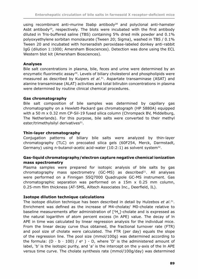
Isotope dilution technique calculations
The isotope dilution technique has been described in detail by Hulzebos et al.21.
Enrichment was defi ned as the increase of M4-cholate/ M0-cholate relative to
baseline measurements after administration of [2H4]-cholate and is expressed as
the natural logarithm of atom percent excess (ln APE) value. The decay of ln
APE in time was calculated by linear regression analysis for the individual mice.
From the linear decay curve thus obtained, the fractional turnover rate (FTR)
and pool size of cholate were calculated. The FTR (per day) equals the slope
of the regression line. The pool size (mmol/100g) was determined according to
the formula: (D . b . 100) / ea ) - D, where ‘D’ is the administered amount of
label, ‘b’ is the isotopic purity, and ‘a’ is the intercept on the y-axis of the ln APE
versus time curve. The cholate synthesis rate (mmol/100g/day) was determined

90
Chapter 5
by multiplying pool size and FTR.
Enterohepatic cycling time and intestinal reabsorbtion of cholate
The cholate cycling time, i.e., the time it takes the cholate pool to circulate a
single time in the enterohepatic circulation, was calculated by dividing the cholate
pool size (mmol/100g) by the biliary secretion rate of cholate (mmol/100g/h).
The cholate biliary secretion rate was calculated by multiplying the bile fl ow
(mL/100g/h) by the cholate concentration (mM) in a single 30 min fraction,
obtained from 5 to 35 min after cannulation of the gallbladder. The amount of
cholate reabsorbed per day was calculated by multiplying cholate pool size by
cycling frequency and subsequent subtraction of the daily cholate synthesis rate.
Statistical analyses
All results are presented as means ± SD. Differences between the two or three
groups were determined by t test or one-way analysis of variance (ANOVA),
with post hoc comparison by Newman-Keuls t test, respectively. The level of
signifi cance for all statistical analyses was set at p < 0.05. Analyses were
performed using SPSS for Windows software (SPSS, Chicago, IL).
Figure 1. Targeted disruption of the murine Fxr gene. (A) Genomic organization of the
wild-type allele and of the disrupted allele arising after homologous recombination. Neor,
neomycin resistance cassette; pA, polyadenylation signal. (B) PCR genotyping of wild-type
(Fxr (+/+)), Fxr
(+/-) and Fxr (-/-) mice. Genomic DNA was isolated from animals of the indicated
genotype and amplifi ed via PCR using allele-specifi c primers described under Experimental
Procedures. Products were separated by agarose gel electrophoresis. (C) Northern blot
analysis of hepatic RNA from wild-type(+/+) and Fxr (-/-) mice. Poly (A+) RNA was isolated
from livers and pooled within genotype groups (n=5). Aliquots (3 µg) were separated by
gel electrophoresis, transferred to nylon membranes, and hybridized to a radiolabeled FXR
probe representing a 837-bp BamHI fragment containing the ligand-binding domain (upper
panel). The fi lters were stripped and reprobed with a cDNA encoding rat cyclophilin (lower
panel).

91
Enterohepatic circulation of bile salts in farnesoid X receptor-defi cient mice
RESULTS
Deletion of the Fxr gene
Mutation of the Fxr gene was accomplished by replacement of 292 bp from exon
2 with a neomycin resistance cassette conferring antibiotic resistance (Figure 1A).
Homologous recombination in embryonic stem cells, injection into blastocysts,
and transmission of the mutation through the mouse germ line were carried
out by standard methods. The loss of ~97 amino acids encoded by exon 2 was
anticipated to remove a large part of the DNA-binding domain of the FXR protein.
PCR analysis (Figure 1B) of genomic DNA confi rmed the predicted recombinations.
RNA blotting revealed a Fxr transcript of ~2.0 kb in the livers of wild-type mice,
whereas no transcript was detectable in Fxr (-/-) mice (Figure 1C).
Animal characteristics
The body and liver weights of Fxr (-/-) and Fxr
(+/-) mice at three months of age
were slightly higher than those of wild-type mice, but liver weight/body weight
ratios were not affected (Table 1). There were no differences in plasma alanine
transaminase (ASAT), aspartate transaminase (ALAT) and bilirubin concentrations
between Fxr (-/-), Fxr
(+/-) and Fxr (+/+) mice. Plasma bile salt concentrations were
only slightly increased in Fxr (-/-) mice, in contrast to previously reported data in
another strain of Fxr (-/-) mice described by Sinal et al.13. Accordingly, urinary bile
salt loss was not signifi cantly different between wild-type and Fxr (-/-) mice, i.e.,
67.4 ± 14.6 nmol/day vs. 43.4 ± 11.0 nmol/day, respectively. Examination of
hematoxylin- and eosin-stained liver sections of wild-type, Fxr (+/-) and Fxr
(-/-) mice
did not reveal any overt abnormalities in FXR-defi cient mice (data not shown).
Table 1. Body and liver weights and plasma liver function parameters in wild-type, Fxr
(+/-) and Fxr (-/-) mice
Strain wild-type Fxr(+/-) Fxr(-/-)
Body weight (g) 23.0 ± 1.1 26.5 ± 1.7* 29.2 ± 3.1* Liver weight (g) 1.1 ± 0.1 1.4 ± 0.1* 1.5 ± 0.2* Ratio LW/BW 0.049 ± 0.004 0.051 ± 0.006 0.053 ± 0.006 ASAT (U/L) 98 ± 18 83 ± 14 120 ± 64 ALAT (U/L) 40 ± 8 36 ± 12 63 ± 32Bilirubin (mmol/L) 13.6 ± 2.6 13.0 ± 1.9 12.5 ± 1.0 Bile salts (mmol/L) 18.1 ± 3.6 21.6 ± 3.9 27.2 ± 7.4*
Values are expressed as means ± SD (n = 6 per group). *Signifi cant difference between wild-type and Fxr
(+/-) or Fxr (-/-) mice. BW, body weight; LW, liver weight; ASAT, aspartate
transaminase; ALAT, alanine transaminase.
Effects of FXR defi ciency on bile formation and bile composition
FXR defi ciency in mice was associated with an increase in bile fl ow (Table 2). The
concentrations of phospholipids and cholesterol in bile did not differ among the
three groups, but the biliary bile salt concentration was signifi cantly increased in
Fxr (-/-) mice. As a consequence, biliary output rates of bile salts were signifi cantly
increased in these animals. Output rates of phospholipid and cholesterol tended to

92
Chapter 5
be increased in Fxr (-/-) mice, but differences did not reach statistical signifi cance.
Because biliary secretion of phospholipids and cholesterol is tightly coupled to that
of bile salts32, the output rates of these biliary lipids were also expressed relative
to those of bile salts (Table 2). It is evident that, both for phospholipids and
cholesterol, these ratios were lower in Fxr (-/-) mice than in wild-type mice. When
Figure 2. Relationship between bile fl ow and biliary bile salt secretion in wild-type,
Fxr (+/-) and Fxr
(-/-) mice. Bile was collected for 30 min, and production rates were
determined gravimetrically. Biliary bile salt concentrations were determined as described
under Experimental Procedures. The combined data from wild-type (white circles), Fxr (+/-)
(grey circles) and Fxr (-/-) (black circles) mice reveal the characteristic linear relationship
between biliary bile salt output and bile fl ow that has been reported in several species.
Bile fl ow at the hypothetical zero value of bile salt output represents the magnitude of the
bile salt-independent fraction of bile fl ow (3 µl/min/100g BW), whereas the slope of the
relationship (8 µl/µmol) represents the choleretic activity of biliary bile salts.
Table 2. Concentrations of organic solutes in bile and biliary output rates in wild-type, Fxr (+/-) and Fxr (-/-) mice
Strain wild-type Fxr(+/-) Fxr(-/-)
Bile (mmol/l) Bile salts (BS) 51.3 ± 17.6 52.6 ± 9.5 82.6 ± 13.5* Phospholipids (PL) 6.65 ± 1.68 5.72 ± 1.36 6.74 ± 1.26 Cholesterol (CH) 0.40 ± 0.07 0.42 ± 0.06 0.43 ± 0.10
Bile fl ow (ml/min/100g body wt) 4.92 ± 1.70 6.40 ± 1.99 7.73 ± 1.5 Biliary output (nmol/min/100g body wt) Bile salts 272 ± 183 347 ± 158 640 ± 157* Phospholipids 32.8 ± 14.4 36.0 ± 13.3 51.3 ± 11.0 Cholesterol 1.99 ± 0.76 2.69 ± 0.98 3.32 ± 0.95PL/BS output ratio 0.14 ± 0.04 0.11 ± 0.04 0.08 ± 0.02* CH/BS output ratio 0.009 ± 0.003 0.008 ± 0.001 0.005 ± 0.002 Values are expressed as means ± SD ( n = 6 per group). *Signifi cant difference between wild-type and Fxr (+/-) or Fxr (-/-) mice.

93
Enterohepatic circulation of bile salts in farnesoid X receptor-defi cient mice
Table 3. Biliary bile salt composition (% total) in wild-type, Fxr(+/-) and Fxr(-/-) Mice
Strain wild-type Fxr(+/-) Fxr(-/-)
Deoxycholate 2.7 ± 1.1 3.2 ± 1.3 3.3 ± 1.8 α-Muricholate 3.8 ± 0.4 3.0 ± 0.6* 1.7 ± 0.3* β-Muricholate 16.0 ± 2.5 16.3 ± 2.5 11.8 ± 4.5 ω-Muricholate 9.8 ± 2.2 9.1 ± 3.2 6.3 ± 3.4 HDC 3.4 ± 3.0 1.9 ± 1.9 0.6 ± 0.2 Chenodeoxycholate 2.7 ± 1.0 2.0 ± 0.3 1.4 ± 0.4* Cholate 61.6 ± 4.7 64.6 ± 4.1 74.5 ± 7.3*
Values are expressed as means ± SD (n = 6 mice per group). *Signifi cant difference
between wild-type and Fxr (+/-) or Fxr
(-/-) mice.
biliary bile salt output rates were plotted against bile fl ow for the individual mice of
the three groups, the classical linear relationship between these parameters was
observed (Figure 2). This strongly indicates that the bile formation process itself
is not affected by FXR defi ciency and that the higher bile fl ow rate in Fxr (-/-) mice
is caused by the higher bile salt output.
Analysis of biliary bile salt composition (Table 3) revealed that, in all three groups,
cholate constituted the major fraction of biliary bile salts and that this fraction
was higher in Fxr (-/-) mice than in the other two groups. Accordingly, the relative
contents of α-muricholate and chenodeoxycholate were signifi cantly decreased
in Fxr (-/-) mice. Thin-layer chromatography revealed that essentially all biliary
cholate was conjugated to taurine in wild-type as well as in Fxr (-/-) mice (data not
shown). Despite the fact that bile salt-conjugated enzymes have recently been
identifi ed as FXR target genes34, unconjugated bile salts were undetectable by this
procedure in bile of wild-type and Fxr (-/-) mice.
Steady-state mRNA levels of genes involved in bile salt synthesis and bile
formation
Real-time quantitative PCR was used to evaluate hepatic expression of specifi c
genes as infl uenced by FXR defi ciency (Figure 3). As expected, expression of
Shp tended to be lower in Fxr (-/-) mice. Cyp7a1 was clearly increased in Fxr
(-/-)
mice, whereas the expression levels of Cyp27 and Cyp8b1 were not signifi cantly
affected (Figure 3A). The mRNA levels of the gene encoding the canalicular
bile salt transporter Bsep, a well-known FXR target gene14,15, were signifi cantly
decreased in Fxr (-/-) mice (Figure 3B). Expression of other transporters genes
relevant to bile formation, such as the phospholipid translocator Mdr2, Ntcp,
and the putative cholesterol transporters Abcg5/g8, was not changed by FXR
defi ciency. Likewise, no effects on expression of Oatp1, Mrp2, and Mrp3 were
observed.
Effects of FXR defi ciency on kinetic parameters of cholate metabolism
To evaluate the physiological consequences of the observed changes in expression
of bile salt synthesis and transporter genes, kinetic parameters of the enterohepatic
circulation of cholate were determined by stable isotope dilution21. Because of the
small differences between the heterozygotes and wild-type mice with respect to

94
Chapter 5
Figure 4. Decay of intravenously administered [2H4]-cholate in wild-type and Fxr
(-/-) mice.
A dose of 240 µg of [2H4]-cholate was intravenously injected into wild-type (open symbols)
and Fxr (-/-) (closed symbols) mice, and blood samples were collected at 24, 36, 48 and 60
h after injection for determination of plasma cholate enrichments by GC-MS as described
under Experimental Procedures. Values are expressed in a logarithmic fashion, and the pool
size (y-intercept), fractional turnover rate (slope of the curve), and synthesis rate (pool size
x fractional turnover rate) were calculated for individual mice. Data are means ± standard
deviation of n = 5 mice per group.
Figure 3. Steady-state mRNA levels of genes involved in bile salt synthesis and transport in
livers of wild-type, Fxr (+/-) and Fxr
(-/-) mice. Total mRNA was isolated form the livers of wild-
type (white bars), Fxr (+/-) (grey bars) and Fxr
(-/-) (black bars) mice, transcribed into cDNA,
and subjected to real-time PCR as described in Experimental Procedures. (A) Hepatic mRNA
levels of Fxr, Shp, Cyp7a1, Cyp27 and Cyp8b1. (B) Hepatic mRNA levels of Bsep, Mdr2,
Abcg5, Abcg8, Ntcp, Oatp1, Mrp2 and Mrp3. n = 5 for all groups. *Signifi cant difference
between wild-type and Fxr (+/-) or Fxr
(-/-) mice.

95
Enterohepatic circulation of bile salts in farnesoid X receptor-defi cient mice
bile formation and gene expression patterns, kinetic studies were conducted in
wild-type and Fxr (-/-) mice only. Analysis of plasma cholate enrichments over time
(Figure 4) demonstrated that the cholate pool size, calculated from the y-intercept
of the linear regression line shown in Figure 4, was larger in Fxr (-/-) mice than
in wild-type mice (Figure 5A: 42 ± 8 µmol/100g vs. 23 ± 3 µmol/100g, Fxr (-/-)
mice vs. wild-type; p < 0.0001). The percentage of cholate in hepatic bile, as
determined by gas chromatographic analysis in individual mice (Table 3), was
used to calculate total bile salt pool sizes. Under the assumption that all bile salt
species displayed a similar cycling frequency, the calculated total pool sizes of
non-cholate bile salts were similar (22 ± 4 mmol/100g vs. 23 ± 9 mmol/100g,
Fxr (-/-) vs. wild-type mice, respectively, NS), leading to calculated total bile salt
pool sizes of 64 ± 12 and 46 ± 13 mmol/100g in Fxr (-/-) and wild-type mice,
respectively (p < 0.05). Deuterated cholate disappeared from plasma at the same
rate in Fxr (-/-) and wild-type mice (Figure 4). The fractional turnover rate of
cholate, calculated from the slope of the linear regression curve, was similar in
Figure 5. Effects of FXR defi ciency on pool size (A), fractional turnover rate (B), synthesis
rate (C), cycling time (D), and daily intestinal reabsorption (E) of cholate as derived from
[2H4]-cholate isotope enrichment measurements in plasma of wild-type and Fxr (-/-) mice.
The pool size, fractional turnover rate (FTR), synthesis rate, cycling time and daily intesti-
nal reabsorption were calculated in wild-type (white bars) and Fxr (-/-) (black bars) mice as
described under Experimental Procedures. Data are means ± standard deviation of n = 5
mice per group. *Signifi cant difference between wild-type and Fxr (-/-) mice.

96
Chapter 5
both groups of mice. (Figure 5B: 0.5 ± 0.1 per day vs. 0.5 ± 0.2 per day, Fxr (-/-)
vs. wild-type, respectively, NS).
In the Fxr (-/-) mice, the calculated cholate synthesis rate (Figure 5C) was two
times increased compared to the wild-type mice (22 ± 2 mmol/100g/day vs.
11 ± 3 mmol/100g/day, Fxr (-/-) vs. wild-type mice; p < 0.001). In accordance with
the increased cholate synthesis rate determined by stable isotope dilution, fecal
loss of bile salts was increased by ~ 70 % (4.1 ± 1.1 mmol/day vs. 2.3 ± 0.7
mmol/day, Fxr (-/-) vs. wild-type mice; p < 0.05). The calculated cholate cycling
time (Figure 5D) was not affected by FXR defi ciency (4.4 ± 1.3 h vs. 4.3 ± 0.7 h,
Fxr (-/-) mice vs. wild-type mice; NS). The calculated absolute amount of cholate
reabsorbed in the intestines of Fxr (-/-) mice (Figure 5E) was ~ 2 fold larger than
that in the intestines of wild-type mice (227 ± 81 mmol/100g/day vs. 121 ± 11
mmol/100g/day, Fxr (-/-) mice vs. wild-type mice; p < 0.05).
Figure 6. mRNA and protein levels of intestinal bile salt transporters in wild-type and Fxr (-/-)
mice. (A) Steady-state mRNA levels of Asbt, Ibabp, t-Asbt, and Fic-1 in the ilea of wild-
type (white bars) and Fxr (-/-) (black bars) mice. Total mRNA was isolated from the ileum,
transcribed into cDNA, and subjected to real-time PCR as described under Experimental Pro-
cedures. *Signifi cant difference between wild-type and Fxr (-/-) mice. (B) Western blot analy-
sis of Asbt and Ibabp on brush-border membranes and liver homogenates, respectively,
from the ilea of wild-type and Fxr (-/-) mice. Apparent molecular masses are indicated to the
right.

97
Enterohepatic circulation of bile salts in farnesoid X receptor-defi cient mice
Intestinal expression of genes involved in bile salt transport
To provide an explanation for the high bile salt absorption rate in Fxr (-/-) mice,
the mRNA levels of several genes considered to be involved in intestinal bile salt
absorption were determined in the terminal ileum. These studies showed that
expression of Ibabp, a well known FXR target gene8,18 thought to be involved
in intracellular bile salt traffi cking and active bile salt reabsorption8,9, was very
strongly decreased at the mRNA level in Fxr (-/-) mice (Figure 6A). FXR defi ciency
did not affect expression of transporter protein-encoding genes Asbt, responsible
for the major part of active ileal bile salt reabsorption, and of t-Asbt, putatively
involved in basolateral bile salt effl ux. Expression of Fic1 (Atp8b1), a P-type
ATPase proposed to function as an aminophospholipid translocator and essential
for normal bile salt metabolism35, also did not differ at the mRNA level between
wild-type and Fxr (-/-) mice. Western blot experiments on brush-border membrane
fractions and homogenates of the terminal part of the ileum (Figure 6B) showed
that Asbt protein levels were similar in wild-type and Fxr (-/-) mice, whereas the
protein levels of Ibabp were essentially non-detectable in the Fxr (-/-) mice.
DISCUSSION
This study has established the physiological consequences of FXR defi ciency on
bile formation and on the kinetics of enterohepatic bile salt circulation employing
an FXR-null mouse model generated by homologous recombination. A microscale
stable isotope dilution technique21 was used to quantify important parameters
of bile salt metabolism. Data show that the bile formation process per se was
not affected by FXR defi ciency and that effects on bile fl ow seen in Fxr (-/-) mice
were secondary to alterations in bile salt metabolism. In accordance with current
concepts of the role of FXR in control of bile salt synthesis36,37, hepatic Cyp7a1
mRNA levels were signifi cantly increased in Fxr (-/-) mice and were associated with
an increased cholate synthesis rate. Enhanced bile salt synthesis was confi rmed
by increased fecal bile salt loss in Fxr (-/-) mice, although the absolute difference
was somewhat less pronounced among the strains using this methodology. We
attribute the discrepancy between outcome of fecal excretion and the isotope
dilution method to the fact that no stool marker has been applied, which is
required to correct for fecal balance measurements. Furthermore, the intrinsic
diffi culties of quantitative fecal bile salt analysis have been extensively reviewed
by Setchell et al.38. As a consequence of defective feedback inhibition of hepatic
bile salt synthesis, Fxr (-/-) mice developed an increased bile salt pool size, which
implies that potential adaptive responses of intestinal bile salt reabsorption were
not effective in maintenance of the bile salt pool size. No change in intestinal Asbt
mRNA and protein levels was found in FXR-defi cient mice. By contrast, the well
known FXR target gene Ibabp was not expressed at all in the terminal ileum of
Fxr (-/-) mice. Despite the absence of Ibabp, our kinetic study revealed that the
absolute amount of bile salts reabsorbed from the intestine was not reduced, but
was actually enhanced by 2-fold in Fxr (-/-) mice. These fi ndings suggest that Ibabp
may not function as a ‘facilitator’9, but rather as a negative regulator of intestinal
bile salt absorption under physiological conditions in the mouse.
FXR has been shown to be involved in control of various steps of bile

98
Chapter 5
salt metabolism, i.e., synthesis and transport13,36,37,39, as well as in regulation
of plasma lipoprotein metabolism2,3,13,40. FXR-defi cient mice have been very
informative in elucidation of the various functions of this nuclear bile salt-activated
receptor. Studies by Sinal et al.13 and Lambert et al.40 were performed with
FXR-defi cient mice (C57BL/6J-SV129 background) that were generated by Cre-
mediated deletion of a fragment containing the last exon of the Fxr gene, encoding
the ligand-binding/dimerization domain, and the 3’-untranslated region of the
Fxr mRNA. In theory, a truncated protein containing the DNA-binding domain
could be formed that might affect expression of FXR target genes. In this study,
we used an FXR knockout model (C57BL/6J-129/OlaHsd background) generated
by homologous recombination, in which 292 bp of exon 2, encoding a part of
the DNA-binding domain, were deleted. These mice showed plasma HDL and
triglyceride levels (Elzinga et al., unpublished) that were elevated to a similar
extent as reported earlier13. In our study, liver function parameters were found
to be unaffected in Fxr (-/-) mice. Plasma bile salt concentrations were only slightly
increased in Fxr (-/-) mice compared with wild-type mice, in marked contrast to the
8-fold increase in plasma bile salt concentration in Fxr (-/-) mice reported by Sinal et
al.13. These strongly elevated plasma bile salt concentrations have been attributed
to defective hepatobiliary bile salt transport due to down-regulation of the major
canalicular bile salt export pump (Bsep)13. However, we have shown that a similar
or even more pronounced down-regulation of Bsep expression in mice was not
associated with impaired biliary bile salt secretion41. Furthermore, the more than
4-fold increase in biliary bile salt secretion during bile salt feeding in mice42 is
accommodated by a very modest increase in hepatic Bsep expression. These
data have been interpreted to indicate that Bsep at normal expression levels has
a marked overcapacity in mice. In fact, in this study, biliary bile salt secretion
was more than 2-fold increased despite a 40% reduction of Bsep expression.
Therefore, it appears that the livers of Fxr (-/-) mice are well able to handle the
(increased) bile salt load. The discrepancy between both strains with respect to
control of plasma bile salt concentrations remains unexplained at the moment.
FXR defi ciency was associated with an enhanced bile fl ow, as determined
during a 30 min period of bile collection. Biliary bile salt concentrations and
secretion rates were clearly enhanced in Fxr (-/-) mice, reinforcing the issue that
decreased Bsep expression levels do not necessarily correlate with defective bile
salt transport. The increase in bile fl ow in Fxr (-/-) mice appeared to be exclusively
due to the higher bile salt output, as is evident from the linear relationship
between bile fl ow and biliary bile salt output that was obtained when data from
wild-type, Fxr (+/-) mice and Fxr
(-/-) mice were combined (Figure 2). The fact that
values of individual mice from the three groups fi tted well to this relationship
indicates that the actual bile formation process was not affected by FXR defi ciency.
The value found for the choleretic activity (8 µl/µmol) of biliary bile salts is similar
to that reported earlier in rodents43. The bile salt-independent fraction of bile
fl ow (3 µl/min/100g body weight) was unaffected by FXR defi ciency, which is
in accordance with unchanged expression of Mrp2. Mrp2 is crucially involved in
hepatobiliary transport of glutathione44, which represents the major driving force
for the generation of bile salt-independent fl ow in rodents43,45. Although Mrp2 has
been identifi ed as an FXR-target gene46, Fxr (-/-) mice did not show reduced Mrp2

99
Enterohepatic circulation of bile salts in farnesoid X receptor-defi cient mice
mRNA levels in this study or in a study by Schuetz et al.47. Biliary secretion rates
of both cholesterol and phospholipids were slightly enhanced in Fxr (-/-) mice as
compared to wild-type mice. Secretion of cholesterol and phospholipids into bile
is coupled to that of bile salts33. Cholesterol secretion appears to involve the
activity of Abcg5/Abcg8 dimers48,49, although the exact role of this twin transporter
remains to be defi ned50, whereas phospholipid secretion critically depends on the
activity of the Mdr2 P-glycoprotein51,52. Because expression of Abcg5/Abcg8 as
well as of Mdr2 was unaffected in Fxr (-/-) mice (Figure 3B), it is plausible to ascribe
the slight stimulation of biliary lipid secretion entirely to the enhanced bile salt
secretion. It should be noted that Lambert et al.40 did report reduced hepatic
Abcg5/Abcg8 expression in their strain of Fxr (-/-) mice, but this was found to
be associated with increased biliary cholesterol output rates. The reason for the
discrepancy in hepatic Abcg5/Abcg8 expression between both strains of mice is
not clear.
We have focused on the effects of FXR defi ciency on the kinetics of cholate
metabolism. For this purpose, we used a novel microscale isotope dilution
technique, applicable in unanesthetized animals21. FXR defi ciency was associated
with an increased cholate synthesis rate, in accordance with increased hepatic
Cyp7a1 mRNA levels in Fxr (-/-) mice. Although FXR has been advocated as the
major regulator of hepatic bile salt synthesis36,39, the effects of FXR defi ciency
on the basal expression of Cyp7a1 (~ +150%) and cholate synthesis (~ +67%)
were relatively modest. This fi nding underscores the importance of the recently
described FXR/SHP-independent mechanisms of regulation. Recent studies in bile
salt-fed SHP knockout mice53,54 have clearly demonstrated the existence of FXR/
SHP-independent repression of Cyp7a1 expression. Cyp27 and Cyp8b1 expression
levels were not signifi cantly affected in Fxr (-/-) mice, although the latter showed
tendency to increase, in accordance with an increase in the fractional contribution
of cholate in the bile salt pool of Fxr (-/-) mice. The fecal loss of bile salts was
increased by ~70 % in Fxr (-/-) mice. Because the mass of bile salts excreted
into feces is, by defi nition, directly proportional to the amount synthesized in
the liver36, the data on fecal loss confi rm a generalized derepression of bile salt
synthesis in FXR-defi cient mice. This again is at variance with the study of Sinal et
al.13, who reported increased expression of Cyp7a1, but decreased fecal bile salt
loss in Fxr (-/-) mice.
The fractional turnover rate of cholate was similar in wild-type and Fxr (-/-)
mice, whereas the cholate pool size was increased 2-fold in Fxr (-/-) mice, implying
enhanced intestinal cholate reabsorption in Fxr (-/-) mice. The calculated total bile
salt pool size was increased by ~40% in Fxr (-/-) mice. This is in contrast to the
situation reported by Sinal et al.13, i.e., a reduction of the total bile salt pool
size by ~50% in Fxr (-/-) mice fed a chow diet. In this case, bile salt pool size
was measured in homogenates of gallbladder, the liver immediately surrounding
the gall bladder, and the entire small intestine harvested after termination of
the animals. The bile salt contents of the homogenates, which were extracted
into ethanol, were determined colorimetrically. Whether methodological or strain-
differences underlie the deviating results between both studies in not clear: the
stable isotope dilution method is a well established procedure to quantify bile salt
kinetics in humans55 and in laboratory animals21.

100
Chapter 5
Maintenance of bile salt pool size can theoretically be regulated at the level
of the intestine by controlled reabsorption in the terminal ileum. Asbt had been
identifi ed as the major transporter involved in this process. However, expression
of Asbt was not different between wild-type and Fxr (-/-) mice. In contrast, Chen et
al.17 reported an increase in Asbt protein levels in Fxr (-/-) mice, in accordance with
the presence of LRH-1 sites in the promoter of the murine Asbt gene identifi ed by
these authors. t-Abst and Fic1 expression was also not changed at the mRNA level
between wild-type and Fxr (-/-) mice and therefore does not seem to play a role
in enhanced bile salt reabsorption effi ciency. Ibabp, a well known FXR gene, was
drastically down-regulated at mRNA and protein level in the ilea of Fxr (-/-) mice, in
accordance with earlier studies13,17. Ibabp is thought to be involved in intracellular
bile salt traffi cking and to facilitate reuptake of bile salts in the small intestine8,9.
Yet despite the complete absence of Ibabp protein in the ilea of the Fxr (-/-) mice,
daily intestinal cholate reabsorption was much higher than in wild-type mice. This
suggests that, under physiological conditions, Ibabp functions as a negative rather
than as a positive regulator of intestinal bile salt reabsorption in the mouse.
In conclusion, this work shows that the absence of FXR in vivo in mice is
associated with defective feedback inhibition of hepatic cholate synthesis, which
leads to an enlarged circulating cholate pool with an unaltered fractional turnover
rate. The absence of Ibabp does not negatively interfere with the enterohepatic
circulation of cholate in mice.
ACKNOWLEDGMENTS
We thank Sara Berdy, Theo Boer, Renze Boverhof, Anke ter Harmsel, Bert Hellinga,
Laura Hoffmann, Karen Siegler and Fjodor van der Sluijs for excellent technical
assistance. This work was supported by grant 902-23-191 from The Netherlands
Organization for Scientifi c Research (NWO).

101
Enterohepatic circulation of bile salts in farnesoid X receptor-defi cient mice
REFERENCES
1. Hofmann AF. Bile Acids: The Good, the Bad, and the Ugly. News Physiol Sci 1999; 14:24-9.
2. Claudel T, Sturm E, Duez H, Torra IP, Sirvent A, Kosykh V, Fruchart JC, Dallongeville J, Hum DW, Kuipers F, Staels B. Bile acid-activated nuclear receptor FXR suppresses apolipoprotein A-I transcription via a negative FXR response element. J Clin Invest 2002; 109:961-71.
3. Kast HR, Nguyen CM, Sinal CJ, Jones SA, Laffi tte BA, Reue K, Gonzalez FJ, Willson TM, Edwards PA. Farnesoid X-activated receptor induces apolipoprotein C-II transcription: a molecular mechanism linking plasma triglyceride levels to bile acids. Mol Endocrinol 2001; 15:1720-8.
4. Lin Y, Havinga R, Verkade HJ, Moshage H, Slooff MJ, Vonk RJ, Kuipers F. Bile acids suppress the secretion of very-low-density lipoprotein by human hepatocytes in primary culture. Hepatology 1996; 23:218-28.
5. Duane WC. Abnormal bile acid absorption in familial hypertriglyceridemia. J Lipid Res 1995; 36:96-107.
6. Gerloff T, Stieger B, Hagenbuch B, Madon J, Landmann L, Roth J, Hofmann AF, Meier PJ. The sister of P-glycoprotein represents the canalicular bile salt export pump of mammalian liver. J Biol Chem 1998; 273:10046-50.
7. Meier PJ, Stieger B. Bile salt transporters. Annu Rev Physiol 2002; 64:635-61.8. Makishima M, Okamoto AY, Repa JJ, Tu H, Learned RM, Luk A, Hull MV, Lustig KD,
Mangelsdorf DJ, Shan B. Identifi cation of a nuclear receptor for bile acids. Science 1999; 284:1362-5.
9. Landrier JF, Grober J, Zaghini I, Besnard P. Regulation of the ileal bile acid-binding protein gene: an approach to determine its physiological function(s). Mol Cell Biochem 2002; 239:149-55.
10. Kullak-Ublick GA, Stieger B, Hagenbuch B, Meier PJ. Hepatic transport of bile salts. Semin Liver Dis 2000; 20:273-92.
11. Goodwin B, Jones SA, Price RR, Watson MA, McKee DD, Moore LB, Galardi C, Wilson JG, Lewis MC, Roth ME, Maloney PR, Willson TM, Kliewer SA. A regulatory cascade of the nuclear receptors FXR, SHP-1, and LRH-1 represses bile acid biosynthesis. Mol Cell 2000; 6:517-26.
12. Russell DW, Setchell KD. Bile acid biosynthesis. Biochemistry 1992; 31:4737-49.13. Sinal CJ, Tohkin M, Miyata M, Ward JM, Lambert G, Gonzalez FJ. Targeted disruption
of the nuclear receptor FXR/BAR impairs bile acid and lipid homeostasis. Cell 2000; 102:731-44.
14. Ananthanarayanan M, Balasubramanian N, Makishima M, Mangelsdorf DJ, Suchy FJ. Human bile salt export pump promoter is transactivated by the farnesoid X receptor/bile acid receptor. J Biol Chem 2001; 276:28857-65.
15. Plass JR, Mol O, Heegsma J, Geuken M, Faber KN, Jansen PLM, Müller M. Farnesoid X receptor and bile salts are involved in transcriptional regulation of the gene encoding the human bile salt export pump. Hepatology 2002; 35:589-96.
16. Denson LA, Sturm E, Echevarria W, Zimmerman TL, Makishima M, Mangelsdorf DJ, Karpen SJ. The orphan nuclear receptor, shp, mediates bile acid-induced inhibition of the rat bile acid transporter, ntcp. Gastroenterology 2001; 121:140-7.
17. Chen F, Ma L, Dawson PA, Sinal CJ, Sehayek E, Gonzalez FJ, Breslow J, Ananthanarayanan M, Shneider BL. Liver receptor homologue-1 mediates species- and cell line-specifi c bile acid-dependent negative feedback regulation of the apical sodium-dependent bile acid transporter. J Biol Chem 2003; 278:19909-16.
18. Grober J, Zaghini I, Fujii H, Jones SA, Kliewer SA, Willson TM, Ono T, Besnard P. Identifi cation of a bile acid-responsive element in the human ileal bile acid-binding protein gene. Involvement of the farnesoid X receptor/9-cis-retinoic acid receptor heterodimer. J Biol Chem 1999; 274:29749-54.
19. Li-Hawkins J, Gafvels M, Olin M, Lund EG, Andersson U, Schuster G, Bjorkhem I, Russell DW, Eggertsen G. Cholic acid mediates negative feedback regulation of bile acid synthesis in mice. J Clin Invest 2002; 110:1191-200.
20. Miyake JH, Doung XD, Strauss W, Moore GL, Castellani LW, Curtiss LK, Taylor JM, Davis RA. Increased production of apolipoprotein B-containing lipoproteins in the absence

102
Chapter 5
of hyperlipidemia in transgenic mice expressing cholesterol 7alpha-hydroxylase. J Biol Chem 2001; 276:23304-11.
21. Hulzebos CV, Renfurm L, Bandsma RHJ, Verkade HJ, Boer T, Boverhof R, Tanaka H, Mierau I, Sauer PJJ, Kuipers F, Stellaard F. Measurement of parameters of cholic acid kinetics in plasma using a microscale stable isotope dilution technique: application to rodents and humans. J Lipid Res 2001; 42:1923-9.
22. Kuipers F, van Ree JM, Hofker MH, Wolters H, In ‘t Veld G, Havinga R, Vonk RJ, Princen HM, Havekes LM. Altered lipid metabolism in apolipoprotein E-defi cient mice does not affect cholesterol balance across the liver. Hepatology 1996; 24:241-7.
23. Heid CA, Stevens J, Livak KJ, Williams PM. Real time quantitative PCR. Genome Res 1996; 6:986-94.
24. Hulzebos CV, Wolters H, Plösch T, Kramer W, Stengelin S, Stellaard F, Sauer PJJ, Verkade HJ, Kuipers F. Cyclosporin A and enterohepatic circulation of bile salts in rats: decreased cholate synthesis but increased intestinal reabsorption. J Pharmacol Exp Ther 2003; 304:356-63.
25. Plösch T, Kok T, Bloks VW, Smit MJ, Havinga R, Chimini G, Groen AK, Kuipers F. Increased hepatobiliary and fecal cholesterol excretion upon activation of the liver X receptor is independent of ABCA1. J Biol Chem 2002; 277:33870-7.
26. Schmitz J, Preiser H, Maestracci D, Ghosh BK, Cerda JJ, Crane RK. Purifi cation of the human intestinal brush border membrane. Biochim Biophys Acta 1973; 323:98-112.
27. Lowry OH, Rosebrough AL, Farr AL, Randal RJ. Protein measurement with the Folin phenol reagent. J Biol Chem 1951; 193:265-75.
28. Labonte ED, Li Q, Kay CM, Agellon LB. The relative ligand binding preference of the murine ileal lipid binding protein. Protein Expr Purif 2003; 28:25-33.
29. Torchia EC, Stolz A, Agellon LB. Differential modulation of cellular death and survival pathways by conjugated bile acids. BMC Biochem 2001; 2:11.
30. Mashige F, Imai K, Osuga T. A simple and sensitive assay of total serum bile acids. Clin Chim Acta 1976; 70:79-86.
31. Kuipers F, Havinga R, Bosschieter H, Toorop GP, Hindriks FR, Vonk RJ. Enterohepatic circulation in the rat. Gastroenterology 1985; 88:403-11.
32. Parmentier G, Eyssen H. Thin-layer chromatography of bile salt sulphates. Journal of Chromatography 1978; 152:285-9.
33. Verkade HJ, Vonk RJ, Kuipers F. New insights into the mechanism of bile acid-induced biliary lipid secretion. Hepatology 1995; 21:1174-89.
34. Pircher PC, Kitto JL, Petrowski ML, Tangirala RK, Bischoff ED, Schulman IG, Westin SK. Farnesoid X receptor regulates bile acid-amino acid conjugation. J Biol Chem 2003; 278:27703-11.
35. Bull LN, van Eijk MJ, Pawlikowska L, DeYoung JA, Juijn JA, Liao M, Klomp LW, Lomri N, Berger R, Scharschmidt BF, Knisely AS, Houwen RH, Freimer NB. A gene encoding a P-type ATPase mutated in two forms of hereditary cholestasis. Nat Genet 1998; 18:219-24.
36. Chiang JY. Bile Acid regulation of gene expression: roles of nuclear hormone receptors. Endocr Rev 2002; 23:443-63.
37. Russell DW. The Enzymes, Regulation, and Genetics of Bile Acid Synthesis. Annu Rev Biochem 2003; 72:137-74.
38. Setchell KD, Street JM, Sjövall J. The bile acids: Chemistry, Physiology,and Metabolism. Plenum Publishing Corp, New York. 1988: 457-62.
39. Lu TT, Repa JJ, Mangelsdorf DJ. Orphan nuclear receptors as eLiXiRs and FiXeRs of sterol metabolism. J Biol Chem 2001; 276:37735-8.
40. Lambert G, Amar MJ, Guo G, Brewer HB, Gonzalez FJ, Sinal CJ. The farnesoid X-receptor is an essential regulator of cholesterol homeostasis. J Biol Chem 2003; 278:2563-70.
41. Kok T, Bloks VW, Wolters H, Havinga R, Jansen PLM, Staels B, Kuipers F. Peroxisome proliferator-activated receptor alpha (PPARalpha)-mediated regulation of multidrug resistance 2 (Mdr2) expression and function in mice. Biochem J 2003; 369:539-47.
42. Wolters H, Elzinga BM, Baller JF, Boverhof R, Schwarz M, Stieger B, Verkade HJ, Kuipers F. Effects of bile salt fl ux variations on the expression of hepatic bile salt transporters in vivo in mice. J Hepatol 2002; 37:556-63.
43. Kuipers F, Enserink M, Havinga R, van der Steen AB, Hardonk MJ, Fevery J, Vonk RJ. Separate transport systems for biliary secretion of sulfated and unsulfated bile acids in
103
Enterohepatic circulation of bile salts in farnesoid X receptor-defi cient mice
the rat. J Clin Invest 1988; 81:1593-9.44. Paulusma CC, Bosma PJ, Zaman GJ, Bakker CT, Otter M, Scheffer GL, Scheper RJ,
Borst P, Oude Elferink RPJ. Congenital jaundice in rats with a mutation in a multidrug resistance-associated protein gene. Science 1996; 271:1126-8.
45. Ballatori N, Truong AT. Glutathione as a primary osmotic driving force in hepatic bile formation. Am J Physiol 1992; 263:G617-G624.
46. Kast HR, Goodwin B, Tarr PT, Jones SA, Anisfeld AM, Stoltz CM, Tontonoz P, Kliewer S, Willson TM, Edwards PA. Regulation of multidrug resistance-associated protein 2 (ABCC2) by the nuclear receptors pregnane X receptor, farnesoid X-activated receptor, and constitutive androstane receptor. J Biol Chem 2002; 277:2908-15.
47. Schuetz EG, Strom S, Yasuda K, Lecureur V, Assem M, Brimer C, Lamba J, Kim RB, Ramachandran V, Komoroski BJ, Venkataramanan R, Cai H, Sinal CJ, Gonzalez FJ, Schuetz JD. Disrupted bile acid homeostasis reveals an unexpected interaction among nuclear hormone receptors, transporters, and cytochrome P450. J Biol Chem 2001; 276:39411-8.
48. Yu L, Li-Hawkins J, Hammer RE, Berge KE, Horton JD, Cohen JC, Hobbs HH. Overexpression of ABCG5 and ABCG8 promotes biliary cholesterol secretion and reduces fractional absorption of dietary cholesterol. J Clin Invest 2002; 110:671-80.
49. Yu L, Hammer RE, Li-Hawkins J, Von Bergmann K, Lutjohann D, Cohen JC, Hobbs HH. Disruption of Abcg5 and Abcg8 in mice reveals their crucial role in biliary cholesterol secretion. Proc Natl Acad Sci U S A 2002; 99:16237-42.
50. Kosters A, Frijters RJ, Schaap FG, Vink E, Plösch T, Ottenhoff R, Jirsa M, De Cuyper IM, Kuipers F, Groen AK. Relation between hepatic expression of ATP-binding cassette transporters G5 and G8 and biliary cholesterol secretion in mice. J Hepatol 2003; 38:710-6.
51. Smit JJ, Schinkel AH, Oude Elferink RPJ, Groen AK, Wagenaar E, van Deemter L, Mol CA, Ottenhoff R, van der Lugt NM, van Roon MA. Homozygous disruption of the murine mdr2 P-glycoprotein gene leads to a complete absence of phospholipid from bile and to liver disease. Cell 1993; 75:451-62.
52. Oude Elferink RPJ, Ottenhoff R, van Wijland M, Smit JJ, Schinkel AH, Groen AK. Regulation of biliary lipid secretion by mdr2 P-glycoprotein in the mouse. J Clin Invest 1995; 95:31-8.
53. Kerr TA, Saeki S, Schneider M, Schaefer K, Berdy S, Redder T, Shan B, Russell DW, Schwarz M. Loss of nuclear receptor SHP impairs but does not eliminate negative feedback regulation of bile acid synthesis. Dev Cell 2002; 2:713-20.
54. Wang L, Lee YK, Bundman D, Han Y, Thevananther S, Kim CS, Chua SS, Wei P, Heyman RA, Karin M, Moore DD. Redundant pathways for negative feedback regulation of bile acid production. Dev Cell 2002; 2:721-31.
55. Stellaard F, Sackmann M, Sauerbruch T, Paumgartner G. Simultaneous determination of cholic acid and chenodeoxycholic acid pool sizes and fractional turnover rates in human serum using 13C-labeled bile acids. J Lipid Res 1984; 25:1313-9.

104