University of Groningen Testicular germ cell tumors Gels ... fileRIJKSUNIVERSITEIT GRONINGEN...
138
University of Groningen Testicular germ cell tumors Gels, Maria Elisabeth IMPORTANT NOTE: You are advised to consult the publisher's version (publisher's PDF) if you wish to cite from it. Please check the document version below. Document Version Publisher's PDF, also known as Version of record Publication date: 1997 Link to publication in University of Groningen/UMCG research database Citation for published version (APA): Gels, M. E. (1997). Testicular germ cell tumors: developments in surgery and follow-up. s.n. Copyright Other than for strictly personal use, it is not permitted to download or to forward/distribute the text or part of it without the consent of the author(s) and/or copyright holder(s), unless the work is under an open content license (like Creative Commons). Take-down policy If you believe that this document breaches copyright please contact us providing details, and we will remove access to the work immediately and investigate your claim. Downloaded from the University of Groningen/UMCG research database (Pure): http://www.rug.nl/research/portal. For technical reasons the number of authors shown on this cover page is limited to 10 maximum. Download date: 08-06-2019
Transcript of University of Groningen Testicular germ cell tumors Gels ... fileRIJKSUNIVERSITEIT GRONINGEN...

University of Groningen
Testicular germ cell tumorsGels, Maria Elisabeth
IMPORTANT NOTE: You are advised to consult the publisher's version (publisher's PDF) if you wish to cite fromit. Please check the document version below.
Document VersionPublisher's PDF, also known as Version of record
Publication date:1997
Link to publication in University of Groningen/UMCG research database
Citation for published version (APA):Gels, M. E. (1997). Testicular germ cell tumors: developments in surgery and follow-up. s.n.
CopyrightOther than for strictly personal use, it is not permitted to download or to forward/distribute the text or part of it without the consent of theauthor(s) and/or copyright holder(s), unless the work is under an open content license (like Creative Commons).
Take-down policyIf you believe that this document breaches copyright please contact us providing details, and we will remove access to the work immediatelyand investigate your claim.
Downloaded from the University of Groningen/UMCG research database (Pure): http://www.rug.nl/research/portal. For technical reasons thenumber of authors shown on this cover page is limited to 10 maximum.
Download date: 08-06-2019

TESTICULAR GERM CELL TUMORSDevelopments in Surgery and Follow-up

ISBN 90 367 0727 7
Cover + lay-out: M.E. GelsPrinting: PrintPartners Ipskamp B.V.

RIJKSUNIVERSITEIT GRONINGEN
TESTICULAR GERM CELL TUMORSDevelopments in Surgery and Follow-up
Proefschrift
ter verkrijging van het doctoraat in de Medische Wetenschappenaan de Rijksuniversiteit Groningen
op gezag van deRector Magnificus, dr F. van der Woude,
in het openbaar te verdedigen opwoensdag 28 mei 1997
des namiddags te 2.45 uur precies
door
Maria Elisabeth Gels
geboren op 24 juni 1967te Wierden

Promotores Prof. Dr. H. Schraffordt KoopsProf. Dr. D.Th. Sleijfer
Co-promotor Dr. H.J. Hoekstra

Beoordelingscommissie Prof. Dr. H.J.A. MensinkProf. Dr. J. OldhoffProf. Dr. Th. Wobbes
Paranimfen J.P. van BastenW.F. Terpstra
All studies of this thesis were designed and carried out at the department of SurgicalOncology, University Hospital Groningen, in cooperation with the departments ofMedical Oncology, Pathology, Radiology, Immunochemistry, Gynaecology andObstetrics, Health Sciences, section of Epidemiology and Statistics, Thoracic Surgery,University Hospital Groningen, the Netherlands; and the department of BiomedicalScience, University of Sheffield, United Kingdom.
The research presented in this thesis was financially supported by the ’StichtingChirurgische Oncologie Groningen’ and the ’J.K. de Kock Stichting’.
Financial support for the publication of this thesis was kindly provided by the ’StichtingWerkgroep Interne Oncologie Groningen’, the ’Johan Vermeij Stichting’ andGlaxoWellcome.


aan mijn oudersvoor Peter

CONTENTS
Abbreviations 10
Chapter I Introduction 11
I.1 General outline 12I.2 Treatment and follow-up 17I.3 Research questions 22
Chapter II Detection of recurrence in patients with clinicalstage I nonseminomatous testicular germ celltumors and consequences for further follow-up:a single-center 10-year experience.Journal of Clinical Oncology 1995; 13: 1188-1194. 29
Chapter III The importance of a new tumor marker TRA-1-60in the follow-up of patients with clinical stage Inonseminomatous testicular germ cell tumors.Annals of Surgical Oncology 1997; in press. 49
Chapter IV Complications of venous access ports in 132 patientswith disseminated testicular cancer treated withpolychemotherapy.Journal of Clinical Oncology 1996; 14: 2916-2922. 67
Chapter V Complications of the postchemotherapy resection ofretroperitoneal residual tumor mass in patients withnonseminomatous testicular germ cell tumors.British Journal of Urology 1997; 79: 263-268. 83

Chapter VI Thoracotomy for postchemotherapy resection ofpulmonary residual tumor mass in patients withnonseminomatous testicular germ cell tumors:aggressive surgical resection is justified.Chest 1997; in press. 97
Chapter VII Summary. 115
Samenvatting 123
Dankwoord 131
Curriculum Vitae 135

ABBREVIATIONS
AFP Alpha-fetoproteinBEP Bleomycin, Etoposide and CisplatinC ChoriocarcinomaCEA Carcinoembryonic antigenCT Computed TomographyCXR Chest X-rayE Embryonal carcinomaEBRT External beam radiotherapyF FibrosishCG Human chorionic gonadotrophinHIV Human Immunodeficiency VirusI Immature teratomaLDH Lactate dehydrogenaseM Mature teratomaMRI Magnetic Resonance ImagingN NecrosisNSTGCT Nonseminomatous testicular germ cell tumorsNSTT Non-seminoma testistumorenPEG Polyethylene-glycolPET Positron Emission TomographyPLAP Placenta-like alkaline phosphatasePRTM Pulmonary Residual Tumor MassPVB Cisplatin, Vinblastine and BleomycinRPLND Retroperitoneal lymph node dissectionRRTM Retroperitoneal Residual Tumor MassRV Upper normal reference valueS SeminomaT½ Serum half-life timeTRA Terato Related AntigenUICC Union Internationale Contre le CancerVAP Venous Access PortWHO World Health OrganisationY Yolk sac tumor
10

CHAPTER I
INTRODUCTION
11
Introduction
I.1 GENERAL OUTLINE
Epidemiology
In the Netherlands, about 350 new patients per year are diagnosed with amalignant testicular germ cell tumor.1 This corresponds with an annual incidenceof about 3 per 100.000. Germ cell tumors are the most prevalent type ofmalignancy in men aged between 20 and 35 years. Two to three per cent of thepatients have bilateral tumors.2 Bilateral tumors usually occur metachronally, andonly very rarely synchronously.3 Malignant germ cell tumors occasionally run infamilies, but it is not clear whether heredity plays a role.4
Etiology
The only proven risk factor for the development of a testicular tumor iscryptorchidism. Twelve per cent of the testicular tumors arise in patients with anundescended testicle. The risk that a tumor will develop in an undescendedtesticle is 20 to 40 times higher than in a testicle that descends normally.5,6 Thereis also a higher incidence of testicular tumors in men infected with the humanimmunodeficiency virus (HIV).7,8 In addition, the presence of testicular atrophymay influence the development of testicular tumors. It has also been found thatthe Klinefelter syndrome is associated with mediastinal germ cell tumors and thatorchitis forms a possible risk factor.9
Dissemination
In principal, the dissemination route of testicular germ cell tumors is no differentfrom that of other malignant tumors. Tumor cells can spread via the lymphaticand circulatory systems. The first lymph node stations of the testes are the lumbarlymph nodes, which are located close to the vertebral column at the level of L2 toL4. From there, tumor cells can spread lymphogenously, via the thoracic duct tothe mediastinum and supraclavicular lymph nodes. Haematogenic disseminationcan take place in two ways: directly to the lungs via vascular invasion in thetesticle, or indirectly to the lungs via the lumbar lymph nodes, cisterna chyli andthe thoracic duct to the subclavian vein. Direct invasion of
12

Chapter I
adjacent structures, such as the rete testis, epididymis and the spermatic cord, canalso occur. However, malignant germ cell tumors chiefly disseminate via thelymphogenous route.10
Symptomatology
A testicular tumor usually presents as a painless swelling in the testicle. Slighttrauma often draws a patient’s attention to the swelling. Other possible symptomsresemble orchitis, epididymitis or testicular torsion. Some patients present withoutany testicular complaints or abnormalities, but with symptoms of distantmetastases. Retroperitoneal lymph node metastases can cause back pain or renalcolic by compressing or blocking the ureter. Lung metastases can give rise todyspnoea, haemoptysis and pleural irritation. In addition, gynaecomastia can formthe first clinical symptom of a tumor that is producing human chorionicgonadotrophin (hCG).11
Diagnosis and staging
Differential diagnoses in the case of testicular complaints comprise a malignancy,varicocele, hydrocele, spermatocele, epididymitis, orchitis, testicular torsion andlateral inguinal hernia. It is important to establish whether the scrotal swelling isintratesticular or extratesticular. Intratesticular swellings should be consideredmalignant until proven otherwise, whereas extratesticular swellings are generallybenign. Physical examination includes thorough palpation of the scrotal contents,palpation of the abdomen, supraclavicular lymph nodes and the mammae in casegynaecomastia is present. Ultrasonography of the scrotal contents is theexamination of choice to distinguish between an intratesticular and an extra-testicular swelling. Laboratory investigations play varying roles. In patients with aseminoma, the serum hCG may be slightly increased and there is usually a higherthan normal level of placenta-like alkaline phosphatase (PLAP). In patients withnonseminomatous germ cell tumors, specific serum tumor markers may bepresent, namely hCG and alpha-fetoprotein (AFP). In all patients with a malignantgerm cell tumor, the serum lactate dehydrogenase (LDH) level may be increased.Diagnosis is made on the basis of histological examination of tissue obtained afterorchidectomy via an inguinal incision. Orchidectomy is performed if the
13
Introduction
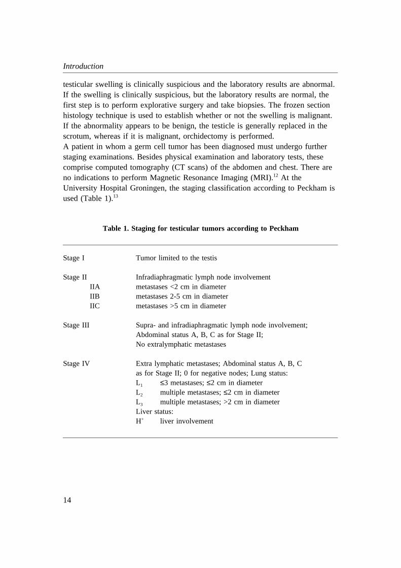
testicular swelling is clinically suspicious and the laboratory results are abnormal.If the swelling is clinically suspicious, but the laboratory results are normal, thefirst step is to perform explorative surgery and take biopsies. The frozen sectionhistology technique is used to establish whether or not the swelling is malignant.If the abnormality appears to be benign, the testicle is generally replaced in thescrotum, whereas if it is malignant, orchidectomy is performed.A patient in whom a germ cell tumor has been diagnosed must undergo furtherstaging examinations. Besides physical examination and laboratory tests, thesecomprise computed tomography (CT scans) of the abdomen and chest. There areno indications to perform Magnetic Resonance Imaging (MRI).12 At theUniversity Hospital Groningen, the staging classification according to Peckham isused (Table 1).13
Table 1. Staging for testicular tumors according to Peckham
Stage I Tumor limited to the testis
Stage II Infradiaphragmatic lymph node involvementIIA metastases <2 cm in diameterIIB metastases 2-5 cm in diameterIIC metastases >5 cm in diameter
Stage III Supra- and infradiaphragmatic lymph node involvement;Abdominal status A, B, C as for Stage II;No extralymphatic metastases
Stage IV Extra lymphatic metastases; Abdominal status A, B, Cas for Stage II; 0 for negative nodes; Lung status:L1 ≤3 metastases;≤2 cm in diameterL2 multiple metastases;≤2 cm in diameterL3 multiple metastases; >2 cm in diameterLiver status:H+ liver involvement
14

Chapter I
Histology
Testicular germ cell tumors can be divided into seminomas (± 55%) andnonseminomas (± 45%). Nonseminomas can be further divided into a number ofsubgroups. Various classifications have been developed over the years. At theUniversity Hospital Groningen, the classification of the World HealthOrganisation (WHO) is used (Table 2).14
Table 2. Histological classification of testicular tumors,World Health Organisation
A. Tumors of one histological type1. Seminoma2. Spermatocytic seminoma3. Embryonal carcinoma4. Yolk sac tumor5. Polyembryoma6. Choriocarcinoma7. Teratoma
a. Mature teratomab. Immature teratomac. With malignant transformation
B. Tumors of more than one histological type1. Embryonal carcinoma and teratoma (teratocarcinoma)2. Choriocarcinoma and any other types3. Other combinations
A nonseminoma can consist of one component, but there is usually a combinationof components, whether or not including seminoma. Embryonal carcinoma is themost poorly differentiated tumor component; no specific differentiation directionis recognisable. Yolk sac tumor and choriocarcinoma resemble fetal membranesand placental tissue, respectively. Teratoma can contain a mixture of tissues, suchas epithelium, cartilage, muscle or nerve tissue. When these tissues have a moreor less normal structure, with good recognition of the differentiation
15

Introduction
direction, the tumor is referred to as mature teratoma. When these tissues are notfully evolved, the tumor is referred to as immature teratoma. As testicular tumorsoften contain different components, it is very important to examine various areasof the tumor histologically. Staging of the primary tumor is performed accordingto the guidelines of the Union Internationale Contre le Cancer (UICC). In Table 3clinical-macroscopic staging and histological staging are described.15
Table 3. Tumor staging according to the UICC
Macroscopic Histologicalstaging staging
T0 no primary tumor pT0
T1 limited to the body of testis pT1
T2 invading beyond the tunica albuginea pT2
T3 invading rete testis or epididymis pT3
T4 infiltrating spermatic cord and/or pT4
scrotal wallT4a infiltrating spermatic cord pT4a
T4b infiltrating scrotal wall pT4b
TX minimum requirements to assess stage pTXcan not be met
16

Chapter I
I.2 TREATMENT AND FOLLOW-UP
Historical developments
The role of surgery in the treatment of patients with testicular tumors has changedconsiderably over the years. Until the end of the previous century, treatment forthese patients comprised orchidectomy alone.16,17 However, physicians had beenaware for some time that patients might have metastases in the retroperitoneum.In 1887, Kocher was the first to describe the resection of a retroperitoneal massfrom a patient with a testicular tumor.18 He removed a tumor mass that was ’aslarge as a man’s head’ from the retroperitoneum. However, the patient developedlocal recurrence five months later. The first transabdominal retroperitoneal lymphnode dissection was performed by Roberts in 1902; however, the patient died ofperitonitis.19 Lumbar retroperitoneal lymph node dissection was described for thefirst time by Chevassu20 and Howard21 in 1910. Coley was opposed to performingretroperitoneal lymph node dissection, because at that time (1915) the surgicalmortality rate was 10% to 15%, which he considered too high to be acceptable.An additional argument was that in 50% of the patients with testicular tumors, notumor-bearing lymphatic tissue was found during surgery. Coley therefore injectedpatients who had a malignant testicular tumor with ’Coley’s fluid’.17 The fluidcomprised a mixture of Bacillus prodigiosus toxin and a filtrate of haemolyticstreptococcus. The encouraging results of this therapy were very likely due tohyperthermia caused by the toxins.22 In this period, radiotherapy was also appliedfor the first time. In 1916, Béclère was probably the first to use radium on aretroperitoneal metastasis from a testicular tumor.23 Orbaan, a Dutch physician,was the first to publish the results of treating metastases from ovarian andtesticular tumors with radiotherapy.24
In 1923, Hinman described the results of patients with a testicular tumor who hehad treated with a so-called ’radical operation’, by this he meant retroperitoneallymph node dissection (RPLND).25 He concluded that his results signified animprovement of 100% in comparison with treatment comprising orchidectomyalone. At that time, orchidectomy alone cured only 15% of the patients, while the’radical operation’ was said to have cured 30%. Until the end of the 1930s,RPLND was the standard treatment after orchidectomy, irrespective of whether apatient had a seminoma or a nonseminomatous tumor. In the subsequent period,physicians somewhat lost interest in RPLND because of the good results of
17

Introduction
radiotherapy, particularly in patients with a seminoma.26 It was not until 1948 thatLewis propagated treating seminomas differently from nonseminomas.27 Heconcluded that patients with a seminoma should be treated with orchidectomy andradiotherapy, while patients with a nonseminomatous testicular tumor should betreated with orchidectomy, RPLND and radiotherapy.A little later, the discussion in the literature became chiefly focused on thesurgical approach to RPLND. Cooper et al. introduced a thoraco-abdominalapproach to ipsilateral RPLND.28 Variations of the technique were then describedby Vallet29 and Lewis.30 In 1953, Leadbetter published his results, on the basis ofwhich he concluded that besides ipsilateral lymph node dissection, contralaterallymph node dissection should also be performed.31 Following the work of Robertsin 1902,19 Mallis et al.32 and Stehlin et al.33 described a transabdominal approachto bilateral retroperitoneal lymph node dissection in 1958 and 1959, respectively.
Recent developments
At the University Hospital Groningen, bilateral RPLNDs were introduced in 1963.The transabdominal approach was used and dissection was limited to theretroperitoneal lymph nodes caudal to the renal vessels. In patients withnonseminomatous testicular germ cell tumors (NSTGCT), Wobbes described theresults of treatment over the period 1963 to 1977.10 During that period,retroperitoneal lymph node dissection was considered to be the standarddiagnostic and therapeutic intervention in patients with clinical stage I NSTGCT.Metastases were found in the retroperitoneal lymph nodes in 25% to 35% of thepatients. The majority of these patients were subsequently treated withactinomycin-D chemotherapy, because of its cytostatic effect.34 If, during surgery,it was found that RPLND was not possible because the metastases were tooextensive, the patient received actinomycin-D and a second attempt was made at alater date. In patients with distant metastases, chemotherapy has been playing amajor role since the beginning of the 1950s. At first, treatment usually compriseda single drug, sometimes in combination with actinomycin-D. In 1960, Lidescribed the results obtained with a combination of chlorambucil, methotrexateand actinomycin-D.35 The advantage of combination therapy is the synergisticeffect, because different drugs are active against different parts of the cellularprocess. In addition, a nonseminomatous tumor can contain one or morecomponents, with different levels of chemo-sensitivity.
18

Chapter I
At the beginning of the 1970s, the first results were described of treatment withvinblastine and bleomycin.36-38 Response rates varied from 32% withmonotherapy, to 90% with combination therapy. The first real breakthrough in thetreatment of patients with disseminated NSTGCT occurred in 1977. In that yearthe results were published of treatment with cisplatin in combination withvinblastine and bleomycin (PVB).39 This combination appeared to be so effectivethat it was decided to change the treatment policy of these patients into PVBfollowed by the possible resection of residual retroperitoneal and/or pulmonarymetastases. PVB polychemotherapy was introduced at the University HospitalGroningen in 1977.40 The combination was applied as the standard treatment formany years. In view of the high level of toxicity, various modifications wereinvestigated, which led to a new standard: the combination of bleomycin,etoposide and cisplatin (BEP).41
In the 1970s, CT scanning was introduced which enabled more accurate clinicalstaging than had been possible previously with lymphangiography and lungtomography.42,43 In the early stages, CT scanning of the retroperitoneal region wasnot considered to be totally reliable. Therefore, explorative laparotomy wasperformed to be completely certain about the presence or absence of retro-peritoneal lymph node metastases. However, it soon became apparent thatexplorative laparotomy did not have any consequences on the clinical stageestablished on the basis of the CT findings. During an interim analysis it wasdecided to perform laparotomy only in cases in whom CT scanning could notgive a definite answer about retroperitoneal lymph node metastases. The role ofMRI in determining the effect of the treatment for patients with disseminatedNSTGCT was also examined. Hogeboom found that MRI did not have anyadvantages over CT scanning.44 Since then, newer MRI equipment has becomeavailable (with breath holding techniques), so the value of MRI compared to CTscanning of the retroperitoneum will have to be re-evaluated.
Another important development in relation with diagnosis and staging is thepossibility of measuring tumor marker levels (hCG and AFP) in a patient’s serum.The response to treatment can be monitored by means of hCG and AFP, but it isimportant to measure the serum levels of these markers prior to orchidectomy torecord the original levels.45,46 Owing to the fact that at least 20% of NSTGCT donot produce tumor markers, attempts have been made to find new tumor markersover the past few years. In 1991, a serum immuno-enzymometric assay wasdeveloped at the Immunochemistry Laboratory of the
19
Introduction
University Hospital Groningen for the detection of TRA-1-60 reactive antigen.47
This antigen is found in patients whose primary tumor contains embryonalcarcinoma. TRA-1-60 is a very promising potential tumor marker, becauseembryonal carcinoma is the most common histological component of NSTGCT.
In patients with clinical stage I NSTGCT, the cure rate after RPLND was about80% to 90%.48 However, nearly all of the patients developed retrogradeejaculation, because the sympathetic ganglia and the hypogastric plexus had beendamaged. As this group of patients chiefly comprises young men, this is a veryserious complication.49,50 In addition, in 65% to 75% of the cases, RPLND onlyhad diagnostic value, without any therapeutic consequences, because no metastaticviable cancer was found during histological examination.Owing to the effectiveness of polychemotherapy, the availability of the tumormarkers hCG and AFP (which can also be used to detect recurrent disease), theincrease in reliability of clinical staging by CT scanning and the disadvantages ofRPLND, it was decided at the Groningen clinic to adopt Peckham’s ’wait-and-seepolicy’ in patients with stage I NSTGCT in 1982.51 This meant that afterorchidectomy, these patients entered an intensive outpatient follow-up programme.Besides physical examination, serum was obtained regularly to monitor tumormarkers and at specific intervals, a chest X-ray was taken and CT scanning ofabdomen and chest was performed. In this way, if any metastases developed, theycould be detected at a early stage. Although the recurrence rate in patients with astage I primary tumor was 25%, they could all be treated effectively withcisplatin-based polychemotherapy.
In order to be able to identify patients with an increased risk of diseaserecurrence, a search was made for unfavourable prognostic factors. The literaturementions various unfavourable prognostic factors, such as the presence ofvascular invasion or lymphatic invasion, the histological tumor (pT) stage, thepresence of embryonal carcinoma or teratoma in the primary tumor and increasedtumor marker levels prior to orchidectomy. Some clinics have suggested adjuvanttreatment with chemotherapy or radiotherapy for the subgroup of patients withclinical stage I NSTGCT and one or more of these risk factors. At present, theprimary treatment for patients with stage II or more advanced stage disease inEurope, is orchidectomy and combination chemotherapy. Occasionally, patientswith stage IIA or IIB NSTGCT undergo orchidectomy followed by nerve-sparingRPLND, if necessary supplemented by chemotherapy. This approach is mainlyapplied in the USA.52
20

Chapter I
The cytotoxic drugs used to treat patients with disseminated NSTGCT areadministered intravenously. To limit the renal toxicity of cisplatin, intravenousprehydration and posthydration are necessary. In practise this means that thepatient retains an intravenous needle for 24 hours per day for seven consecutivedays. An alternative approach was introduced at the end of the 1970s: the patientswere provided with an arteriovenous shunt (A-V shunt) in the wrist to facilitatethe administration of the chemotherapy and infusion fluids.53,54 This producedwidening of the veins because of arterialisation and made them easier to puncture.An additional advantage was that the rapid flow of blood through the vein quicklydiluted the cytotoxic drugs and reduced the irritation to the vessel walls.Unfortunately, the life span of the A-V shunts proved to be very short in practise,namely 2-12 months. Moreover, complications, such as thrombosis and infection,occurred fairly frequently.54 At the beginning of the 1980s, the Venous AccessPort (VAP) became available. This subcutaneous access system could remain insitu longer and was associated with far less morbidity than an A-V shunt.Therefore, the VAP formed a considerable advance for patients with an NSTGCTthat had to be treated with cisplatin-based polychemotherapy.
After completion of chemotherapy, the staging examinations are repeated. Fairlyoften residual tumor is detected. The treatment policy for such residual diseaseafter chemotherapy depends on whether the primary tumor was a seminoma or anonseminoma. Residual disease in patients with a disseminated seminoma can betreated with surgery,55 radiotherapy56 or they can enter a wait-and-seeprogramme.57-59 At the University Hospital Groningen, the wait-and-see policy isgenerally employed for these patients.If the primary tumor was an NSTGCT and the tumor markers have normalised,but residual disease is present or suspected, the patients usually undergoevaluative surgery. Surgery plays a major role in judging the outcome ofchemotherapy. The histology of residual lesions is of importance to determinewhether any further treatment is necessary. If the residual lesions only containnecrosis or fibrosis, resection does not have any therapeutic consequences;however, if the lesions contain viable cancer, the patient receives additionalpolychemotherapy. There is a great risk that if the primary tumor contained ateratoma component, the residual lesions will contain mature teratoma.Histologically, mature teratoma gives the impression of being benign, butcytogenetically it is malignant.60,61 In addition, it has been found that residualmature teratoma can develop into large, usually cystic tumors that eventuallycompress the adjacent organs and give rise to serious complications. This
21

Introduction
phenomenon is referred to as the "Growing Teratoma Syndrome".62 A matureresidual lesion can also develop into a second non-germ cell malignancy.63 Withthe aim of minimizing the risk of the growing teratoma syndrome or a secondmalignancy, relaparotomy was performed at the University Hospital Groningen,irrespective of the radiological findings, with resection of any palpableabnormalities, between 1978 and 1984.64 Thoracotomy was only carried out ifthere was radiological evidence of lung metastases. Partly based on reports in theliterature, the policy of re-laparotomy after chemotherapy was discontinued at theend of 1986 in patients without signs of residual lesions on their CT scans andwhose primary tumor had not contained a teratoma component.64 On the basis ofprognostic models, attempts have recently been made in the literature to sharpenthe indications for RPLND after polychemotherapy.65 Additionally, trials areunderway to investigate whether new diagnostic methods, such as MRI andPositron Emission Tomography (PET scanning) can be used to predict thehistology of metastases.12,66
I.3 RESEARCH QUESTIONS
The above shows that over the years, the role of surgery has been influencedconsiderably by the many new developments in diagnosis and treatment ofpatients with testicular tumors. Since the publication of Wobbes’ thesis in 1981,RPLND as the only curative option for these patients has evolved into adjuvantsurgery after chemotherapy.67 This thesis describes the present role of surgery inthe treatment of patients with malignant testicular germ cell tumors. The studiesfocus chiefly on the surgical treatment and follow-up of patients with NSTGCT.
At present, there is a more than 15 years of experience with the ’wait-and-seepolicy’ in patients with clinical stage I NSTGCT. A study was performed toinvestigate whether or not this is a reliable policy; the results are described inChapter II . Details are given about the recurrence rate of patients who enteredthe wait-and-see programme, the interval between orchidectomy and thedevelopment of metastases and the way in which the metastases were detected.A search was then made for unfavourable prognostic factors that could predict thedevelopment of metastases. Also was examined whether it is necessary tocontinue follow-up for ten years after orchidectomy.
22

Chapter I
The results of a study on the normal values and half-life of a new serum tumormarker, TRA-1-60, are described inChapter III . AFP and hCG are the standardtumor markers, so it was of interest to determine whether TRA-1-60 has anyadditional value in the follow-up of patients with clinical stage I NSTGCT. Anincreased serum TRA-1-60 value at the time of orchidectomy was examined as apotential unfavourable prognostic factor for the development of metastases.
Chapter IV describes the perioperative and late complications of the use of aVAP as an access system for chemotherapy in patients with disseminatedtesticular cancer. On the basis of a homogeneous group of patients, attempts weremade to identify factors that could predict the development of complications.
The importance of determining the histology of metastases after treatment withorchidectomy and polychemotherapy must be weighed against the complicationsand risks of surgical intervention. InChapter V the advantages and disadvantagesof resecting residual lesions from the retroperitoneum are discussed. Factors thatmight have predictive value regarding complications were evaluated.
On analogy with Chapter V,Chapter VI examines the advantages anddisadvantages of resecting residual lesions from the lungs. An additional questionis whether the indication for resecting residual lesions from the lungs should alsodepend on the histological results of the retroperitoneal lesions.
In summary, this thesis addresses the following research questions:
1. Is the current policy for all patients with a clinical stage I NSTGCTjustified, or should it be modified, possibly only for certain subgroups?
2. What is the significance and value of the new serum tumor markerTRA-1-60 in patients with clinical stage I NSTGCT?
3. What perioperative and late complications are associated with placing asubcutaneous access system for polychemotherapy in patients withdisseminated testicular germ cell tumors?
4. What are the advantages and disadvantages of surgical resection of
23

Introduction
residual retroperitoneal lesions after polychemotherapy in patients withdisseminated NSTGCT?
5. What are the advantages and disadvantages of surgical resection ofresidual pulmonary lesions after polychemotherapy in patients withdisseminated NSTGCT?
REFERENCES
1. Visser O, Coebergh JWW, Schouten LJ, Eds. Coordinating council ofComprehensive Cancer Centers. Incidence of cancer in the Netherlands 1993. 5threport of the Netherlands Cancer Registry 1996.
2. Parker SL, Tong T, Bolden S, et al. Cancer statistics, 1996. CA Cancer J Clin1996; 46: 5-27.
3. Hoekstra HJ, Wobbes Th, Sleijfer DTh, et al. Bilateral primary germ cell tumorsof testis. Urology 1982; 19: 152-154.
4. Wobbes Th, Hoekstra HJ, Sleijfer DTh, et al. Tumors of the testis in twobrothers: a case report. J Surg Oncol 1981; 17: 135-137.
5. Wobbes Th, Schraffordt Koops H, Oldhoff J. The relation between testiculartumours, undescended testes, and inguinal hernias. J Surg Oncol 1980; 14:45-51.
6. Batata MA, Chu FCH, Hilaris BS, et al. Testicular cancer in cryptorchids. CA1982; 49: 1023-1030.
7. Timmerman JM, Northfelt DW, Small EJ. Malignant germ cell tumors in meninfected with the human immunodeficiency virus: natural history and results oftherapy. J Clin Oncol 1995; 13: 1391-1397.
8. Bernardi D, Salvioni R, Vaccher E, et al. Testicular germ cell tumors and humanimmunodeficiency virus infection: a report of 26 cases. J Clin Oncol 1995; 13:2705-2711.
9. Rubin Ph, Ed. Clinical Oncology: a multidisciplinary approach for physicians andstudents. Rochester, NY: W.B. Saunders Company 1993: 442-453.
10. Wobbes Th. Non-seminomatous germ cell tumours of the testis. Staging andtreatment. Thesis, State University Groningen, 1981.
11. Einhorn LH, Richie JP, Shipley WU. Cancer of the testis. In: DeVita VT,Hellman S, Rosenberg SA, Eds. Cancer: principles & practice of oncology.Philadelphia: J.B. Lippincott Company 1989: 1126-1151.
24

Chapter I
12. Hogeboom WR, Hoekstra HJ, Mooyaart EL, et al. Magnetic resonance imagingof retroperitoneal lymph node metastases of non-seminomatous germ cell tumoursof the testis. Eur J Surg Oncol 1993; 19: 429-437.
13. Peckham MJ, McElwain TJ, Barrett A, et al. Combined management of malignantteratoma of the testis. Lancet 1979; 2: 267-270.
14. Mostofi FK, Sobin LH: Histological typing of testis tumours. In: Internationalhistological classification of tumors no. 16. Geneva, Switzerland: World HealthOrganisation 1977: 1-39.
15. Harmer MH: TNM classification of malignant tumours. Geneva, Switzerland:Union Internationale Contre le Cancer 1978: 122-125.
16. Chevassu M. Tumeurs du testicle. Thesis, Steinheil Paris, 1906.17. Coley WB. Cancer of the testis: 64 cases, 12 cases of the undescended testis. Ann
Surg 1915; 26: 40-73.18. Kocher T. Die Krankheiten der männlichen Geslechtsorganen. Verlag von
Ferdinand Enke, Stuttgart 1887; 491.19. Roberts JB. Excision of the lumbar lymphatic nodes and spermatic vein in
malignant disease of the testicle. Ann Surg 1902; 36: 539-549.20. Chevassu M. Le traitement chirurgical de cancers du testicule. Rev de Chir 1910;
61: 628-660.21. Howard R. A radical operation for malignant disease of the testis. Lancet 1910;
2: 1406-1408.22. Dickson JA. Hyperthermia in the treatment of cancer. Lancet 1979; 1: 202-205.23. Béclère M. La radiothérapie des neoplasmes intra abdomineaux d’origine
testiculaire. Bull Acad Med Paris 1916; 76: 72-81.24. Orbaan C. Behandeling der uitgebreide kwaadaardige gezwellen van de
mannelijke en vrouwelijke geslachtsklieren met röntgenstralen. Ned TijdschrGeneeskd 1920; 2: 695-702.
25. Hinman F, Gibson TE, Kutzmann AA. The radical operation for teratoma testis.Surg Gynec Obstet 1923; 37: 429-451.
26. Hinman F. The prognosis and treatment of tumors of the testis. J Urol 1935;34: 72-84.
27. Lewis LC. Testis tumors: Report on 250 cases. J Urol 1948; 59:763-772.28. Cooper JF, Leadbetter WF, Chute R. The thoracoabdominal approach for
retroperitoneal gland dissection: its application to testis tumors. Surg GynecObstet 1950; 90: 486-496.
29. Vallet BS. The Nagamatsu technique applied to retroperitoneal dissection in testistumor. J Urol 1952; 68: 337-339.
30. Lewis EL, Johnston RE, Rowe RB, et al. Retroperitoneal lymph node resection:the intercosto-inguinal approach. J Urol 1952; 67: 338-341.
31. Leadbetter WF. Treatment of testis tumors based on their pathological behaviour.JAMA 1953; 151: 275-280.
25

Introduction
32. Mallis N, Patton JF. Transperitoneal bilateral lymphadenectomy in testis tumor. JUrol 1958; 80: 501-503.
33. Stehlin JS, Jones JS, Crigler DM, et al. Lymphadenectomy via the trans-peritoneal (anterior abdominal) approach for cancer of the testis. Am J Surg1959; 97: 756-765.
34. Wobbes Th, Eibergen R, Oldhoff J, et al. Results of retroperitoneal lymph nodedissection and postoperative adjuvant chemotherapy with dactinomycin in thetreatment of retroperitoneal metastases of nonseminomatous testicular germ celltumors. Cancer 1983; 51: 1076-1079.
35. Li MC, Whitmore WF, Golbey R, et al. Effects of combined drug therapy onmetastatic cancer of the testis. JAMA 1960; 174: 145-153.
36. Samuels ML, Howe CD. Vinblastine in the management of testicular cancer.Cancer 1970; 25: 1009-1017.
37. Blum RH, Carter SK, Agre K. A clinical review of bleomycin - a newantineoplastic agent. Cancer 1973; 31: 903-914.
38. Samuels ML, Johnson DE, Holoye PY. Continuous intravenous bleomycin(NSC-125066) therapy with vinblastine (NSC-49842) in stage III testicularneoplasia. Cancer Chemother Rep 1975; 59: 563-570.
39. Einhorn LH, Donohue J. Cis-diamminedichloroplatinum, vinblastine, andbleomycin combination chemotherapy in disseminated testicular cancer. AnnIntern Med 1977; 87: 293-298.
40. Stoter G, Sleijfer DTh, Vendrik CP, et al. Combination chemotherapy withcis-diamminedichloroplatinum, vinblastine, and bleomycin in advanced testicularnon-seminoma. Lancet 1979; 1: 941-945.
41. Williams SD, Birch R, Einhorn LH, et al. Treatment of disseminated germ-celltumors with cisplatin, bleomycin, and either vinblastine or etoposide. N Engl JMed 1987; 316: 1435-1440.
42. Wobbes Th, Blom JM, Oldhoff J, et al. Lymphography in the diagnosis ofnon-seminoma tumours of the testis. J Surg Oncol 1982; 19: 1-4.
43. Gelderman WAH, van der Laan JG, Schraffordt Koops H, et al. Evaluation ofstaging methods for retroperitoneal metastases in patients with nonseminomatoustesticular tumours: CT scan plus lymphography versus exploratory laparotomy.J Med Imag 1989; 3: 288-296.
44. Hogeboom WR, Hoekstra HJ, Mooyaart EL, et al. The role of MagneticResonance Imaging and Computed Tomography in the treatment evaluation ofretroperitoneal lymph-node metastases of non-seminomatous testicular tumors.Eur J Radiol 1991; 13: 31-36.
45. Javadpour N, McIntire KR, Waldmann TA. Immunochemical determination ofhuman chorionic gonadotropin and alpha-fetoprotein in sera and tumors ofpatients with testicular cancer. Natl Cancer Inst Monogr 1978: 209-213.
26

Chapter I
46. Vugrin D, Friedman A, Whitmore WF, Jr. Correlation of serum tumor markers inadvanced germ cell tumors with responses to chemotherapy and surgery. Cancer1984; 53: 1440-1405.
47. Marrink J, Andrews PW, van Brummen PJ, et al. TRA-1-60: a new serum markerin patients with germ-cell tumors. Int J Cancer 1991; 49: 368-372.
48. Wobbes Th, Schraffordt Koops H, Oldhoff J, et al. Results of treatment ofnon-seminomatous tumours of the testis in pathological stage I. Neth J Surg1983; 35: 89-93.
49. Nijman JM, Schraffordt Koops H, Oldhoff J, et al. Sexual function after surgeryand combination chemotherapy in men with disseminated nonseminomatoustesticular cancer. J Surg Oncol 1988; 38: 182-186.
50. Van Basten JP, Jonker Pool G, van Driel MF, et al. The sexual sequelae oftesticular cancer. Cancer Treat Rev 1995; 21: 479-495.
51. Peckham MJ, Barrett A, Husband JE, et al. Orchidectomy alone in testicular stageI non-seminomatous germ-cell tumours. Lancet 1982; 2: 678-680.
52. Motzer RJ, Sheinfeld J, Mazumdar M, et al. Etoposide and cisplatin adjuvanttherapy for patients with pathologic stage II germ cell tumors. J Clin Oncol 1995;13: 2700-2704.
53. Wobbes Th, Slooff MJ, Sleijfer DTh, et al. Five years’ experience in accesssurgery for polychemotherapy. An analysis of results in 100 consecutive patients.Cancer 1983; 52: 978-982.
54. Slooff MJH. Secundaire toegangschirurgie voor hemodialyse en chemotherapie.Thesis, State University Groningen, 1981.
55. Motzer R, Bosl G, Heelan R, et al. Residual mass: an indication for furthertherapy in patients with advanced seminoma following systemic chemotherapy.J Clin Oncol 1987; 5: 1064-1070.
56. Fossa SD, Borge L, Aass N, et al. The treatment of advanced metastaticseminoma: experience in 55 cases. J Clin Oncol 1987; 5: 1071-1077.
57. Schultz SM, Einhorn LH, Conces DJ, Jr., et al. Management of postchemotherapyresidual mass in patients with advanced seminoma: Indiana University experience.J Clin Oncol 1989; 7: 1497-1503.
58. Fossa SD, Droz JP, Stoter G, et al. Cisplatin, vincristine and ifosphamidecombination chemotherapy of metastatic seminoma: results of EORTC trial30874. EORTC GU Group. Br J Cancer 1995; 71: 619-624.
59. Sleijfer S, Willemse PHB, de Vries EGE, et al. Treatment of advanced seminomawith cyclophosphamide, vincristine and carboplatin on an outpatient basis. Br JCancer 1996; 74: 947-950.
60. De Graaff WE. Tumor biological studies on adult testicular germ cell tumors.Thesis, State University Groningen, 1992.
61. Van Echten-Arends J. A cytogenetic study of male germ cell tumors. Thesis,State University Groningen, 1996.
27

Introduction
62. Logothetis CJ, Samuels ML, Trindade A, et al. The growing teratoma syndrome.Cancer 1982; 50: 1629-1635.
63. Molenaar WM, Oosterhuis JW, Meiring A, et al. Histology and DNA contents ofa secondary malignancy arising in a mature residual lesion six years afterchemotherapy for a disseminated nonseminomatous testicular tumor. Cancer1986; 58: 264-268.
64. Gelderman WAH. Surgery in the treatment of nonseminomatous testiculartumors. Thesis, State University Gronigen, 1987.
65. Steyerberg EW, Keizer HJ, Fossa SD, et al. Prediction of residual retro-peritonealmass histology after chemotherapy for metastatic nonseminomatous germ celltumor: multivariate analysis of individual patient data from six study groups. JClin Oncol 1995; 13: 1177-1187.
66. Stephens AW, Gonin R, Hutchins GD, et al. Positron Emission Tomographyevaluation of residual radiographic abnormalities in postchemotherapy germ celltumor patients. J Clin Oncol 1996; 14: 1637-1641.
67. Oldhoff J. Adjuvante chirurgie. In: Oncologisch Kabinet. Veranderde inzichten inde behandeling van tumoren. Amsterdam: Antoni van Leeuwenhoekhuis (NKI),1984: 1-7.
28

CHAPTER II
DETECTION OF RECURRENCE IN PATIENTS WITH CLINICALSTAGE I NONSEMINOMATOUS TESTICULAR GERM CELL TUMORS
AND CONSEQUENCES FOR FURTHER FOLLOW-UP:A SINGLE CENTER 10-YEAR EXPERIENCE
M.E. Gels,1 H.J. Hoekstra,1 D.Th. Sleijfer,2 J. Marrink,3 H.W.A de Bruijn,4
W.M. Molenaar,5 N.J.M. Freling,6 J.H.J. Droste,7 H. Schraffordt Koops.1
From the departments of Surgical Oncology1, Medical Oncology2,Immunochemistry3, Gynaecology and Obstetrics4, Pathology5, Radiology6
and Health Sciences, section Epidemiology and Statistics7,University Hospital Groningen, the Netherlands
Journal of Clinical Oncology 1995; 13: 1188-1194.
29

Follow-up in Stage I NSTGCT
ABSTRACT
A wait-and-see policy for patients with stage I nonseminomatous testicular germcell tumors (NSTGCT) was evaluated in a prospective study. The frequency andtime of recurrence, detection of recurrence and presence of unfavourableprognostic factors were investigated. During the period 1982 to 1992, 154patients with stage I NSTGCT (median age 29 years) underwent orchidectomyand were monitored at follow-up evaluation with physical examinations, alpha-fetoprotein (AFP) and human chorionic gonadotrophin (hCG) levels, chest X-rays(CXR) and computed tomography (CT) scans of the abdomen and chest.Multivariate logistic regression analyses were performed to identify prognosticfactors. During a median follow-up period of 7 years (range 2-12), recurrencewas found in 42 patients (27.3%). All cases of recurrence were detected withintwo years, 90% in the first year after orchidectomy. In 29 patients (69.0%),recurrence was detected in the abdominal lymph nodes. Nine patients (21.4%)had metastases in the retroperitoneum and the mediastinum and/or the lungs andfour patients (9.6%) had metastases only in the mediastinum or lungs. Themajority of recurrences (97.6%) were detected by tumor markers and CT scans.Recurrence was related to the presence of vascular invasion, embryonalcarcinoma (E), an elevated preoperative hCG level and the absence of matureteratoma (M). Only vascular invasion was an independent risk factor. Afterpolychemotherapy treatment for recurrence, the survival rate in the total groupwas 98.7%. The wait-and-see policy is a reliable method for follow-up monitoringof patients with stage I NSTGCT. Even in patients with unfavourable prognosticfactors, it is justified to await the possible appearance of metastases. For thefuture it is recommended that CXR be omitted from the schedule, and it might befeasible to discontinue the follow-up evaluations after five years.
INTRODUCTION
In the 1970s, cisplatin became available in various chemotherapy combinations.This considerably improved the prognosis of patients with disseminatednonseminomatous testicular germ cell tumors (NSTGCT), especially in the lowerstages.1,2 In addition, the introduction of computed tomography (CT) scanning of
30

Chapter II
the abdomen and chest enabled more accurate clinical staging than could beachieved with the former diagnostic procedures: lymphangiography and lungtomography.3,4 Before the introduction of cisplatin patients with stage I NSTGCTwere treated with radical retroperitoneal lymph node dissection (RPLND)5,6 orexternal beam radiotherapy (EBRT) of the para-aortic and ipsilateral pelvic lymphnodes.7 A major complication from RPLND had been retrograde ejaculation andEBRT may induce a radiation enteritis. The high cure rates obtainable withcisplatin-based chemotherapy, especially in patients with low stage disease, theavailability of serum tumor markers for NSTGCT and the increased accuracy ofthe CT scanning, as well as the adverse effects associated with RPLND andEBRT, led to the initiation of a number of studies to assess the utility ofsurveillance for stage I disease. Peckham and collegues introduced in 1979 thismanagement policy.8 They found a recurrence rate of 17% in 53 patients after amedian follow-up of 15 months. They argued that the proportion of patientscurable with orchidectomy alone should be 60-80%, and that if recurrenceoccurred, effective chemotherapy was available.
After orchidectomy for patients with a clinical stage I NSTGCT without anyevidence of metastases the wait-and-see policy was commenced. At the regularoutpatient check-ups, the patients underwent a physical examination, evaluation ofthe serum tumor markers alpha-fetoprotein (AFP) and human chorionicgonadotrophin (hCG) and a chest X-ray. At various intervals, CT scans weretaken of the abdomen and chest. In this way, any retroperitoneal, mediastinaland/or lung metastases could be detected early. Also several other groups havereported their preliminary or mature data. Recurrences occured at a rate of 16% to35.2%9-15 after a median follow-up varying between 10 and 64 months. Mostrecurrences, however, occurred within 12 months. Among the patients whorecurred, disease-free survival after treatment is 92% to 100%. Later on, effortshave been directed toward the identification of prognostic factors that may predictthe likelihood of recurrence. The unfavourable significance of vascular orlymphatic invasion,12,13,15-18histology13,16,18and local tumor extension13,19 has beengenerally accepted.20 This had led to the institution of trials using adjuvantchemotherapy in patients with high risk stage I disease.12,21
In the Netherlands, the University Hospital Groningen has been employing thewait-and-see policy since 1982.22,23 This prospective study describes the ten yearsof experience with this policy in patients with stage I NSTGCT. The number ofpatients with recurrence was evaluated, how this recurrence was detected was
31

Follow-up in Stage I NSTGCT
described and the number of months until recurrence was determined. Also, thepresence of any unfavourable prognostic factors which might be related torecurrence were investigated. The aim of the study was to examine the adequacyof a wait-and-see policy for patients with a stage I NSTGCT. Advice andrecommendations regarding follow-up evaluation are given.
PATIENTS AND METHODS
In the period 1982 to 1992, 154 patients with clinical stage I NSTGCT (medianage 29 years; range 15-66) were treated at the University Hospital Groningen, theNetherlands. All of the patients underwent orchidectomy and at staging there wasno evidence of regional and/or distant metastases. Clinical staging was performedas described by Peckham et al24 with the aid of physical examination, chest X-ray,serum tumor markers AFP and hCG, and CT scanning of the chest and abdomen.Values for AFP of >10 µg/l and for hCG of≥2.0 µg/l were considered to beelevated. If the serum tumor markers were elevated before the orchidectomy, theywere expected to normalize postoperatively in accordance with the half-life times.The half-life times for AFP and hCG are six days and two days, respectively. Inthe initial years of the study period, bilateral lymphangiography also formed partof the staging procedure. Since 1986, only CT scanning has been performedowing to the limited reliability of lymphangiography and its complicatedinterpretation.3,25
In the period 1982-1983, an explorative laparotomy was carried out inconjunction with the orchidectomy to make a definite diagnosis regarding thepresence or absence of retroperitoneal lymph node metastases. On the basis of aninterim analysis, it was decided from 1984 on to perform this laparotomy only ifradiologic findings were inconclusive. If there were no palpable pathologicretroperitoneal lymph nodes these patients were classified as stage I and stillentered the study.23
The tumors were classified in accordance with the nomenclature of the WorldHealth Organisation (WHO),26 which distinguishes the following histologicalcomponents in nonseminomatous testicular tumors: embryonal carcinoma (E),choriocarcinoma (C), yolk sac tumor (Y) and teratoma. Teratomas can contain
32

Chapter II
mature or immature components: mature teratoma (M) and immature teratoma (I).The nonseminomas are usually composed of more than one histological subgroup,sometimes with a seminoma (S) component.27 Data on the diameter of theprimary tumor and the presence or absence of vascular invasion were obtainedfrom the surgical specimens. pT stage was determined according to theInternational Union Against Cancer (UICC) 1978 edition.28
Outpatient check-ups were performed according to a strict schedule (Table 1).Physical examination and serum AFP and hCG determinations were performed ateach visit. A chest X-ray and CT scanning of the chest and abdomen were carriedout at regular intervals. After ten years, the follow-up was considered to becomplete and the patients were discharged from further evaluations. The patientswith recurrence were treated with cisplatin-based combination chemotherapy.
Table 1. Follow-up schedule of the wait-and-see policy for patients withstage I NSTGCT (1982-1992)
Year 1 Every 4 weeks: PE, TMEvery 8 weeks: PE, TM, CXREvery 3 months: PE, TM, CTa, CTc
Year 2 Every 8 weeks: PE, TMEvery 3 months: PE, TM, CXREvery 6 months: PE, TM, CTa, CTc
Year 3 Every 3 months: PE, TM, CXREvery 12 months: PE, TM, CTa, CTc
Years 4 and 5 Every 6 months: PE, TM, CXRYears 6, 7, 8, 9 and 10 Every 12 months: PE, TM, CXR
Abbreviations: PE, physical examination; TM, tumor markers AFP and hCG; CXR, chestX-ray; CTa, computed tomogram of the abdomen; CTc, computed tomogram of the chest.
33

Follow-up in Stage I NSTGCT
Statistical analysis
Univariate analyses were performed on differences in clinical characteristicsbetween the two subgroups ’recurrence’ and ’no-recurrence’ with Student’st testfor continuous variables and by means of the Chi-square test for categoricalvariables. Fisher’s exact test was used in case of small numbers. Multivariatelogistic regression analysis was used to examine correlations between variableswith possible prognostic significance and the appearance of recurrence;29 thedependent variable was recurrence within two years. Independent variables wereage, tumor location (left or right), maximum diameter of the primary tumor,preoperative elevation of AFP and/or hCG, histological components of theprimary tumor, the presence of vascular invasion in the primary tumor and pTstage. Age and maximum diameter of the primary tumor were continuousvariables, while the remaining variables were dichotomous (0=absent, 1=present).
By including all of the independent variables simultaneously in the regressionmodel, we could establish the relationship between each separate variable and theappearance of recurrence. No-recurrence acted as a reference for the dependentvariables, while the absence of a particular characteristic acted as a reference forthe independent variables. To obtain the best fitting regression model, the numberof independent variables was reduced by means of the step-wise elimination ofcovariables, with the likelihood-ratio statistic as selection criterion.Owing to the fact that the maximum diameter of the primary tumor was unknownin 19 patients, the regression analysis was also carried out without this variable.The statistical analyses were conducted using the SPSS-PC+ (V4.0) softwarepackage (SPSS inc., Chicago, Ill., USA.). Test statistics and odds ratios wereconsidered to be statistically significant at P≤.05.
RESULTS
Between January 1982 and December 1991, 154 patients with clinical stage INSTGCT were treated at the University Hospital Groningen. Before 1984 allpatients who entered the study had undergone an explorative laparotomy. Since1984, four patients have undergone laparotomy because the findings on CT-scanswere inconclusive: i.e. no palpable abnormalities were found during laparotomy.The median follow-up was seven years (range 2-12 years). The frequencies of the
34
Chapter II
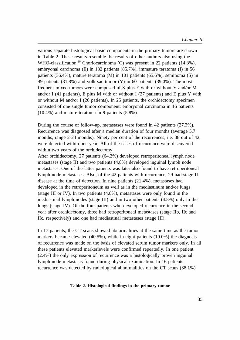
various separate histological basic components in the primary tumors are shownin Table 2. These results resemble the results of other authors also using theWHO-classification.30 Choriocarcinoma (C) was present in 22 patients (14.3%),embryonal carcinoma (E) in 132 patients (85.7%), immature teratoma (I) in 56patients (36.4%), mature teratoma (M) in 101 patients (65.6%), seminoma (S) in49 patients (31.8%) and yolk sac tumor (Y) in 60 patients (39.0%). The mostfrequent mixed tumors were composed of S plus E with or without Y and/or Mand/or I (41 patients), E plus M with or without I (27 patients) and E plus Y withor without M and/or I (26 patients). In 25 patients, the orchidectomy specimenconsisted of one single tumor component: embryonal carcinoma in 16 patients(10.4%) and mature teratoma in 9 patients (5.8%).
During the course of follow-up, metastases were found in 42 patients (27.3%).Recurrence was diagnosed after a median duration of four months (average 5.7months, range 2-24 months). Ninety per cent of the recurrences, i.e. 38 out of 42,were detected within one year. All of the cases of recurrence were discoveredwithin two years of the orchidectomy.After orchidectomy, 27 patients (64.2%) developed retroperitoneal lymph nodemetastases (stage II) and two patients (4.8%) developed inguinal lymph nodemetastases. One of the latter patients was later also found to have retroperitoneallymph node metastases. Also, of the 42 patients with recurrence, 29 had stage IIdisease at the time of detection. In nine patients (21.4%), metastases haddeveloped in the retroperitoneum as well as in the mediastinum and/or lungs(stage III or IV). In two patients (4.8%), metastases were only found in themediastinal lymph nodes (stage III) and in two other patients (4.8%) only in thelungs (stage IV). Of the four patients who developed recurrence in the secondyear after orchidectomy, three had retroperitoneal metastases (stage IIb, IIc andIIc, respectively) and one had mediastinal metastases (stage III).
In 17 patients, the CT scans showed abnormalities at the same time as the tumormarkers became elevated (40.5%), while in eight patients (19.0%) the diagnosisof recurrence was made on the basis of elevated serum tumor markers only. In allthese patients elevated markerlevels were confirmed repeatedly. In one patient(2.4%) the only expression of recurrence was a histologically proven inguinallymph node metastasis found during physical examination. In 16 patientsrecurrence was detected by radiological abnormalities on the CT scans (38.1%).
Table 2. Histological findings in the primary tumor
35

Follow-up in Stage I NSTGCT
Components Total (n=154) No Rec.(n=112) Rec.(n=42)
S+E 16 (10%) 10 6S+E+C+Y+M 1 (0.6%) 0 1S+E+C+M+I 1 (0.6%) 1 0S+E+Y 4 (2.6%) 2 2S+E+Y+M 4 (2.6%) 4 0S+E+Y+M+I 7 (4.5%) 5 2S+E+M 1 (0.6%) 1 0S+E+M+I 7 (4.5%) 5 2S+E+Y+I 1 (0.6%) 0 1S+E+I 1 (0.6%) 1 0S+C 1 (0.6%) 1 0S+Y 1 (0.6%) 1 0S+Y+M+I 1 (0.6%) 1 0S+M 1 (0.6%) 1 0S+M+I 2 (1.3%) 2 0E 16 (10%) 11 5E+C 1 (0.6%) 1 0E+C+Y 1 (0.6%) 1 0E+C+Y+M 4 (2.6%) 4 0E+C+Y+I 2 (1.3%) 2 0E+C+Y+M+I 5 (3.2%) 3 2E+C+M 4 (2.6%) 3 1E+C+M+I 2 (1.3%) 0 2E+Y 8 (5.2%) 3 5E+Y+M 8 (5.2%) 6 2E+Y+M+I 10 (6.5%) 8 2E+M 16 (10%) 13 3E+M+I 11 (7.1%) 7 4E+I 1 (0.6%) 1 0Y+M+I 1 (0.6%) 1 0Y+M 2 (1.3%) 1 1M 9 (5.8%) 8 1M+I 4 (2.6%) 4 0
Abbreviations: Rec., recurrence; C, choriocarcinoma; E, embryonal carcinoma;I, immature teratoma; M, mature teratoma; S, seminoma; Y, yolk sac tumor
36

Chapter II
In one patient lung metastases were histologically proven. None of the cases ofrecurrence were found by means of the chest X-ray. Fourteen patients whodeveloped radiological changes on CT-scans of abdomen, but without markerelevation, underwent an exploratory laparotomy before remission inductionchemotherapy was started. Three of these patients also had signs of metastases onCT-scan of the chest. All fourteen patients had palpable pathologic para-aorticlymph nodes. In one patient retroperitoneal lymph node metastases causedhydonephrosis of the left kidney, in this patient no exploratory laparotomy wasdone because the clinical appearance and CT-scan of the abdomen were thoughtto be conclusive for metastases. This patient and seven out of the fourteenpatients with retroperitoneal recurrence detected only on CT scan, wererelaparotomized after four remission-induction courses with cisplatin basedpolychemotherapy. In one case no palpable abnormalities were found and inseven cases residual tumor was found, and resected. Histologic examination of theresected specimen revealed necrosis and fibrosis in three cases and matureteratoma in the other four. Because in the other seven patients radiologicallydocumented recurrences disappeared completely, surgical evaluation was notperformed.
Table 3 shows a comparison of the characteristics present in the recurrence groupand the no-recurrence group. Univariate comparison of the clinical characteristicsbetween the group with and the group without recurrence did not reveal anysignificant differences in the average age, tumor location, average maximumdiameter of the primary tumor and pT stage≥2. Of the tumor markers,particularly the serum hCG level was elevated more frequently in the patientswith recurrence than in those without recurrence; this difference was notstatistically significant (P=.085). Of the histological characteristics of the primarytumor, embryonal carcinoma was present significantly more often in the patientswith recurrence, whereas mature teratoma was present more often (not significant)in the patients without recurrence (P=.083). Vascular invasion was presentsignificantly more often in the recurrence group than in the no-recurrence group:45.2% versus 15.2%. Multivariate logistic regression analysis was carried out onall the possible risk factors (n=135; Table 4) and repeated after the only variablewith missing values, i.e. the maximum diameter of the primary tumor, had beenomitted. The exclusion of this variable which was not found to be associated withthe appearance of recurrence using univariate or multivariate analysis, meant thatall the patients could be included in the analyses (n=154; Table 5).
37

Follow-up in Stage I NSTGCT
Table 3. Population characteristics by recurrence status
No Recurrence Recurrence P(n = 112) (n = 42)
Age, in years (mean±SD) 30±10 29±7 .648
Primary tumorLocation: Left 50 (44.6%) 17 (40.5%) |
Right 62 (55.4%) 25 (59.5%) | .642Max. diameter, mm (mean±SD) 39.0±19 41.4±18 .505Preoperative elevated AFP 53 (47.3%) 23 (54.8%) .411Preoperative elevated hCG 39 (34.8%) 21 (50.0%) .085Histological findings:
Seminoma 35 (31.3%) 14 (33.3%) .805Embryonal carcinoma 92 (82.1%) 40 (95.2%) .039Choriocarcinoma 16 (14.3%) 6 (14.3%) 1.000Yolk sac tumor 42 (37.5%) 18 (42.9%) .544Mature teratoma 78 (69.6%) 23 (54.8%) .083Immature teratoma 41 (36.6%) 15 (35.7%) .918
Vascular invasion present 17 (15.2%) 19 (45.2%) .000Histologic staging pT≥2 6 (5.4%) 4 (9.5%) .462
In both cases, vascular invasion was the only significant independent risk factorfor the manifestation of recurrence (Odds Ratios 4.28 and 4.06, respectively). Inthe patients who were included in the second regression analysis (and not thefirst), mature teratoma occurred more frequently, while embryonal carcinoma, anelevated AFP and an elevated hCG occurred less frequently. In this way, the step-wise elimination of covariables led to two slightly different models. Logisticregression analysis with step-wise elimination of covariables in the first case(n=135) gave rise to the following regression model: y = -1.275 + 0.808 x hCG -0.793 x mature teratoma + 1.534 x vascular invasion. In the second case (n=154),the regression model was: y = -2.519 + 1.250 x embryonal carcinoma + 1.445 xvascular invasion. The two models indicate that recurrence is related to thepresence of vascular invasion (Odds Ratios 4.63 and 4.24, respectively), apreoperative elevated hCG value (Odds Ratio 2.24), the absence
38

Chapter II
of mature teratoma (Odds Ratio 0.45) and the presence of embryonal carcinoma(Odds Ratio 3.49). The only independent prognostic factor was the presence ofvascular invasion.
Table 4. Multivariate logistic regression analysis of risk factors forrecurrence (n=135)
Independent variable Odds Ratio P
All potential risk factors simultaneously in the modelAge 1.00 .940Tumor location (Left vs Right) 0.82 .657Maximum diameter primary tumor 1.01 .517Preoperative elevated AFP 1.44 .514Preoperative elevated hCG 2.23 .105Seminoma present 0.70 .471Embryonal carcinoma present 2.10 .386Choriocarcinoma present 0.58 .393Yolk sac tumor present 1.10 .841Mature teratoma present 0.38 .082Immature teratoma present 0.96 .940Vascular invasion present 4.28 .001Histologic staging pT≥2 2.34 .321
Model after backward elimination of covariatesVascular invasion present 4.63 .001Preoperative elevated hCG 2.24 .062Mature teratoma present 0.45 .069
Owing to the fact that we found relationships between an elevated AFP and thepresence of choriocarcinoma, yolk sac tumor and mature teratoma, and betweenan elevated hCG and the presence of choriocarcinoma, the regression analyseswere also carried out with interaction terms for these variables. However, theaddition of these interaction terms did not produce any significant improvement inthe regression models.
39

Follow-up in Stage I NSTGCT
Table 5. Multivariate logistic regression analysis of risk factors forrecurrence (n=154)*
Independent variable Odds Ratio P
All potential risk factors simultaneously in the modelAge 1.00 .932Tumor location (Left vs Right) 0.89 .767Preoperative elevated AFP 1.54 .395Preoperative elevated hCG 1.84 .178Seminoma present 0.98 .965Embryonal carcinoma present 2.67 .228Choriocarcinoma present 0.60 .403Yolk sac tumor present 0.97 .949Mature teratoma present 0.46 .136Immature teratoma present 1.20 .701Vascular invasion present 4.06 .001Histologic staging pT≥2 1.54 .576
Model after backward elimination of covariatesVascular invasion present 4.24 .000Embryonal carcinoma present 3.49 .110
* Maximum diameter of primary tumor not included because of missing values
Of the 42 patients with metastases, 40 went into complete remission aftertreatment with cisplatin-based polychemotherapy, with or without explorativesurgery for resection and histological examination. Those patients in which noviable cancer was found in resected specimens, were classified as completeresponders. One patient refused to be treated with chemotherapy. The otherpatient only had a local inguinal lymph node metastasis on the left side. Afterexcision of this small metastasis, no further evidence could be found of distantmetastases, so there was no longer any indication for chemotherapy.
Two of the patients with recurrence died. One of these patients was the one whohad refused chemotherapy; he died 41 months after the orchidectomy. The other
40

Chapter II
patient went into complete remission after cisplatin-based polychemotherapy foringuinal, retroperitoneal and lung metastases, but he developed a secondrecurrence eleven months later. The second recurrence was also accompanied bylung metastases, for which he received second line polychemotherapy. He diedtwenty months after the orchidectomy. The ultimate disease-free survival of the154 patients with stage I NSTGCT who were monitored with the wait-and-seefollow-up policy was 98.7%; two out of the 154 patients (1.3%) died.
DISCUSSION
Over a 10-year period (median follow-up time 7 years), this study groupcomprised 154 patients with a clinical stage I nonseminomatous testicular germcell tumor. A total of 42 patients (27.3%) developed recurrence after a medianfollow-up of 4 months (range 2-24 months). At the time of detection ofrecurrence, the majority of patients (69.0%) had stage II disease, i.e. themetastases were located in the abdominal lymph nodes: in the retroperitonealand/or inguinal lymph nodes. In this series, all the cases of recurrence were foundwithin two years after the orchidectomy. Other authors have reported cases ofdissemination after two years,14,31 with rates of up to 7%. After five years, theappearance of recurrence is extremely rare. In such cases, if the histology of therecurrence does not exactly match that of the primary tumor, it is worthconsidering whether the second tumor is a new primary extragonadal germ cellcarcinoma.32 Forty patients had to be treated with cisplatin-basedpolychemotherapy during the course of the follow-up. With a median follow-upduration of seven years, a disease-free survival of 98.7% was achieved.
Serum tumor markers as well as CT scanning of the abdomen and chest played amajor role in the detection of recurrence. Both were indispensable for the earlydetection of metastases, so that treatment with polychemotherapy could be startedat an early stage. In eight patients, the diagnosis of recurrence was made purelyon the basis of elevation of one or both serum tumor markers. In one of thesepatients, palpable inguinal lymph nodes were found shortly afterwards and in theother seven, the CT scans became abnormal after 2, 2, 3, 3, 4, 5 and 20 months,respectively. These patients received polychemotherapy after the metastases hadbeen confirmed radiologically using CT. Prior to the orchidectomy, elevated
41

Follow-up in Stage I NSTGCT
values of one or both serum tumor markers were found in 93 patients (60.4%).Nine of these patients developed recurrence (21.4%), however the tumor markerswere not elevated when the recurrence was found. The opposite was observed insix patients (14.3%) who had normal serum tumor marker values beforeorchidectomy but elevated values at the time of recurrence. This discordance inthe behaviour of tumor markers has been described by other authors14,15,31,33,34andhas been reviewed.35 In cases where the primary tumor had exclusively producedAFP (6 patients), the metastases caused either an elevation in the AFP values (3patients) or no increase in tumor marker values. The same was valid for thetumors which had exclusively produced hCG (4 patients): if the metastases hadproduced a tumor marker (3 patients) it was always exclusively hCG.
CT scanning was also very helpful in the detection of recurrence. In only fourpatients CT scans were inconclusive during follow-up. Because a recurrence couldnot be excluded completely, these four patients underwent diagnostic laparotomy.However, no palpable abnormalities were found at laparotomy and none of thesepatients developed recurrence during follow-up.The chest X-ray was not found to have much value in this series. Abnormalitieson the chest X-ray never formed the first indication of recurrence. Of the 13patients with lung metastases, abnormalities were only visible on the relevantchest X-ray in two cases. Therefore, it seems to be not worthwhile to take regularchest X-rays in the follow-up of patients with stage I NSTGCT.
This study and others have shown that the presence of vascular invasion is anunfavourable prognostic factor.12,13,15-18,21,36 pT stage has also been identified as aprognostic factor by several authors, especially tumor invading into the tunicaalbuginea, rete epididymis or spermatic cord: pT≥2.13,35,37However this prognosticfactor was found in univariate analyses. In this study pT-stage was not found tobe predictive of subsequent recurrence, in multivariate nor in univariatecomparison. An embryonal carcinoma component in the primary tumor has alsooften been mentioned as an independent risk factor for the development ofrecurrence.18,30,36 In this study, only univariate comparison of the clinicalcharacteristics between the recurrence group and the no-recurrence group showedthat embryonal carcinoma occurred significantly more often in the recurrencegroup.In the literature, other unfavourable prognostic factors include: the absenceof yolk sac tumor,21 lymphatic invasion12,21,30,38,39and the absence of teratomatouselements.16 In this study we found that the development of recurrence was relatedto the presence of vascular invasion, embryonal carcinoma, a pre-orchidectomy
42
Chapter II
elevated serum hCG value, and the absence of mature teratoma. On the basis ofthe list of unfavourable prognostic factors, it is possible to select a group ofpatients from those with stage I NSTGCT who are at increased risk forrecurrence. Some authors have recommended adjuvant treatment for thesepatients.12,21 However, retroperitoneal lymph node dissection,40 adjuvantchemotherapy41 or radiotherapy14 cannot prevent the development of recurrence inall cases. In this series of patients, the administration of adjuvant polychemo-therapy to for example all the patients with vascular invasion in the primarytumor would have meant the ’overtreatment’ of 17 out of the 36 patients.
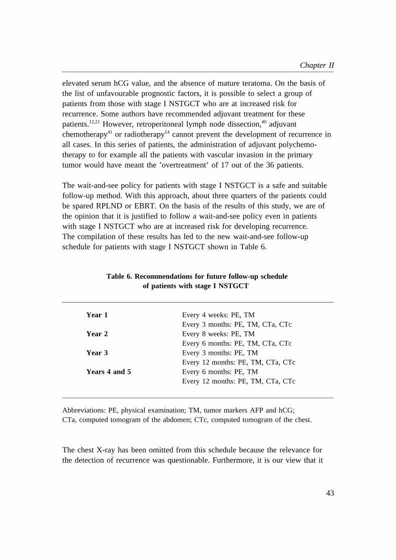
The wait-and-see policy for patients with stage I NSTGCT is a safe and suitablefollow-up method. With this approach, about three quarters of the patients couldbe spared RPLND or EBRT. On the basis of the results of this study, we are ofthe opinion that it is justified to follow a wait-and-see policy even in patientswith stage I NSTGCT who are at increased risk for developing recurrence.The compilation of these results has led to the new wait-and-see follow-upschedule for patients with stage I NSTGCT shown in Table 6.
Table 6. Recommendations for future follow-up scheduleof patients with stage I NSTGCT
Year 1 Every 4 weeks: PE, TMEvery 3 months: PE, TM, CTa, CTc
Year 2 Every 8 weeks: PE, TMEvery 6 months: PE, TM, CTa, CTc
Year 3 Every 3 months: PE, TMEvery 12 months: PE, TM, CTa, CTc
Years 4 and 5 Every 6 months: PE, TMEvery 12 months: PE, TM, CTa, CTc
Abbreviations: PE, physical examination; TM, tumor markers AFP and hCG;CTa, computed tomogram of the abdomen; CTc, computed tomogram of the chest.
The chest X-ray has been omitted from this schedule because the relevance forthe detection of recurrence was questionable. Furthermore, it is our view that it
43

Follow-up in Stage I NSTGCT
is accountable to discontinue the follow-up evaluations after five years because nocases of recurrence were found after two years. Other changes to the schedule, forexample, lengthening the interval between two follow-up visits, would not bewise, particularly in the first two years, because in our experience, disseminationcan be rapidly progressive. This would involve the risk of detecting recurrence ata later stage, with a decreasing chance of being able to cure the patient withpolychemotherapy.42
REFERENCES
1. Einhorn LH, Donohoe J: Cis-diamminedichloroplatinum, vinblastine and bleomy-cin combination chemotherapy in disseminated testicular cancer. Ann Intern Med1977; 87: 293-298.
2. Stoter G, Vendrik CPJ, Struyvenberg A, et al: Combination chemotherapy withcis-diamminedichloroplatinum, vinblastine and bleomycin in advanced testicularnon-seminoma. Lancet 1979; 1: 941-945.
3. Wobbes Th, Blom JMH, Oldhoff J, et al: Lymphography in the diagnosis of non-seminoma tumours of the testis. J Surg Oncol 1982; 19: 1-4.
4. Gelderman WAH, van der Laan JG, Schraffordt Koops H, et al: Evaluation ofstaging methods for retroperitoneal metastases in patients with nonseminomatoustesticular tumours: CT, lymphography and exploratory laparotomy. J MedImaging 1989; 3: 288-296.
5. Johnson DE, Bracken RB, Blight EM: Prognosis for pathologic stage Inonseminomatous germ cell tumors of the testic managed by retroperitoneallymphadenectomy. J Urol 1976; 116: 63-65.
6. Bredael JJ, Vugrin D, Whitmore WF Jr: Recurrences in surgical stage Inonseminomatous germ cell tumors of the testis. J Urol 1983; 130: 476-478.
7. Maier JG, Lee SN: Radiation therapy for nonseminomatous germ cell testicularcancer in adults. Urol Clin North Am 1977; 4: 477-493.
8. Peckham MJ, Barrett A, Husband JE, et al: Orchidectomy alone in testicular stageI non-seminomatous germ-cell tumours. Lancet 1982; 2: 678-680.
9. Read G, Johnson RJ, Wilkinson PM, et al: Prospective study of follow up alonein stage I teratoma of the testis. Br Med J 1983; 287, 1503-1505.
10. Sogani PC, Whitmore WF Jr, Herr HW, et al: Orchiectomy alone in the treatmentof clinical stage I nonseminomatous germ cell tumor of the testis.J Clin Oncol 1984; 2: 267-270.
44

Chapter II
11. Johnson DE, Lo RK, von Eschenbach AC, et al: Surveillance alone for patientswith clinical stage I nonseminomatous germ cell tumors of the testis: preliminaryresults. J Urol 1984; 131: 491-493.
12. Hoskin P, Dilly S, Easton D, et al: Prognostic factors in stage I non-seminoma-tous germ cell testicular tumors managed by orchiectomy and surveillance:Implications for adjuvant chemotherapy. J Clin Oncol 1986; 4: 1031-1036.
13. Pizzocaro G, Zanoni F, Milani A, et al: Orchiectomy alone in clinical stage Inonseminomatous testis cancer: a critical appraisal. J Clin Oncol 1986; 4: 35-40.
14. Rorth M, Jacobsen GK, vd Maase H, et al: Surveillance alone versus radio-thera-py after orchiectomy for clinical stage I nonseminomatous testicular cancer. JClin Oncol 1991; 9: 1543-1548.
15. Sturgeon JFG, Jewett MAS, Alison RE, et al: Surveillance after orchidectomy forpatients with clinical stage I nonseminomatous testis tumors. J Clin Oncol 1992;10: 564-568.
16. Klepp O, Olsson AM, Henrikson H, et al: Prognostic factors in clinical stage Inonseminomatous germ cell tumors of the testis: multivariate analysis of aprospective multicenter study. J Clin Oncol 1990; 8: 509-518.
17. Mc Leod DG, Weiss RB, Stablein DM, et al: Staging relationships and outcomein early stage testicular cancer: a report from the Testicular Cancer IntergroupStudy. J Urol 1991; 145: 1178-1183.
18. Sesterhenn IA, Weiss RB, Mostofi FK, et al: Prognosis and other clinical correla-tes of pathologic review in stage I and II testicular carcinoma: a report from theTesticular Cancer Intergroup Study. J Clin Oncol 1992; 10: 69-78.
19. Fung CY, Kalish LA, Brodsky GL, et al: Stage I nonseminomatous germ celltesticular tumor: prediction of metastatic potential by primary histopathology.J Clin Oncol 1988; 6: 1467-1473.
20. Fung CY, Garnick MB: Clinical stage I carcinoma of the testis: a review. J ClinOncol 1988; 6: 734-750.
21. Cullen MH, Stenning SP, Fossa SD, et al: Short course adjuvant chemotherapy inhigh risk stage I non-seminomatous germ cell tumours of the testis (NSGCTT):Preliminary report of an MRC study. Br J Cancer 1992; 65 (suppl XVI): 8.(Abstract)
22. Schraffordt Koops H, Sleijfer DTh, Oosterhuis JW, et al: Wait-and-see policy inclinical stage I non-seminomatous germ cell tumors of the testis. Eur J SurgOncology 1986; 12: 283-287.
23. Gelderman WAH, Schraffordt Koops H, Sleijfer DTh, et al: Orchidectomy alonein stage I nonseminomatous testicular germ cell tumors. Cancer 1987; 59:578-580.
24. Peckham MJ, Barret A, McElwain TJ, et al: Combined management of malignantteratoma of the testis. Lancet 1979; 2: 267-270.
45

Follow-up in Stage I NSTGCT
25. Lien HH, Fossa SD, Ous S, at al: Lymphography in retroperitoneal metastases innon-seminoma testicular tumor patients with a normal CT scan. Acta RadiolDiagnosis 1983; 24: 319-322.
26. Mostofi FK, Sobin LH: Histological typing of testis tumours. In: Internationalhistological classification of tumors no. 16. Geneva, Switzerland: World HealthOrganisation 1977: 1-39.
27. Ulbright TM: Germ cell neoplasms of the testis. Am J Surg Pathology 1993; 17:1075-1091.
28. Harmer MH: TNM classification of malignant tumours. Geneva, Switzerland:Union Internationale Contre le Cancer 1978: 122-125.
29. Rothmann KJ: Modern Epidemiology. Boston/Toronto: Little, Braun and co,1986.
30. Dunphy CH, Ayala AG, Swanson DA, et al: Clinical stage I nonseminomatousand mixed germ cell tumors of the testis. A clinicopathologic study of 93 patientson a surveillance protocol after orchiectomy alone. Cancer 1988; 62: 1202-1206.
31. Raghavan D, Colls B, Levi J, et al: Surveillance for stage I non-seminomatousgerm cell tumours of the testis: the optimal protocol has not yet been defined.Br J Urol 1988; 61: 522-526.
32. Myamoto H, Moriyama M, Fukushima S, et al: Retroperitoneal tumor elevenyears after initial treatment of testicular cancer. Urol 1994; 43: 116-117.
33. Peckham MJ, Barrett A, Horwich A, et al: Orchiectomy alone for Stage ITesticular Non-Seminoma. Br J Urol 1983; 55: 754-759.
34. Braunstein GD, McIntire KR, Waldmann TA: Discordance of human chorionicgonadotropin and alpha-fetoprotein in testicular teratocarcinomas. Cancer 1973;31: 1065-1068.
35. Bates SE, Longo DL: Use of serum tumor markers in cancer diagnosis andmanagement. Sem Oncol 1987; 14: 102-138.
36. Javadpour N, Canning DA, O’Connel KJ, et al: Predictors of recurrent clinicalstage I non-seminomatous testicular cancer. A prospective clinico-pathologicstudy. Urol 1986; 27: 508-511.
37. Raghavan D, Peckham MJ, Heyderman E, et al: Prognostic factors in clinicalstage I non-seminomatous germ-cell tumours of the testis. Br J Cancer 1982; 45:167-173.
38. Peckham MJ, Brada M: Surveillance following orchidectomy for stage I testicularcancer. Int J of Andrology 1987; 10: 247-254.
39. Thompson PI, Nixon J, Harvey VJ: Disease relapse in patients with stage Inonseminomatous germ cell tumor of the testis on active surveillance. J ClinOncol 1988; 6: 1579-1603.
46
Chapter II
40. Sujka SK, Huben, RP: Clinical stage I nonseminomatous germ cell tumors oftestis. Observation vs retroperitoneal lymph node dissection. Urol 1991; 38:29-31.
41. Pont J, Holtl W, Kosak D, et al: Risk-adapted treatment choice in stage Inonseminomatous testicular germ cell cancer by regarding vascular invasion inthe primary tumor: a prospective trial. J Clin Oncol 1990; 8: 16-20.
42. Vugrin D, Whitmore WF Jr, Golby RB: VAB-6 combination chemotherapywithout maintenance in treatment of disseminated cancer of the testis. Cancer1983; 51: 211-215.
47


CHAPTER III
THE IMPORTANCE OF A NEW TUMOR MARKER TRA-1-60 IN THEFOLLOW-UP OF PATIENTS WITH CLINICAL STAGE I
NONSEMINOMATOUS TESTICULAR GERM CELL TUMORS
M.E. Gels,1 J. Marrink,2 P. Visser,2 D.Th. Sleijfer,3 J.H.J. Droste,4
H.J. Hoekstra,1 P.W. Andrews,5 H. Schraffordt Koops.1
From the departments of Surgical Oncology,1 Immunochemistry,2
Medical Oncology3 and Health Sciences, Section of Epidemiology and Statistics,4
University Hospital Groningen, the Netherlands; Department of BiomedicalScience,5 University of Sheffield, United Kingdom
Annals of Surgical Oncology 1997; in press.
49

TRA-1-60 in Stage I NSTGCT
ABSTRACT
TRA-1-60 is a new tumor marker for embryonal carcinoma-positive non-seminomatous testicular germ cell tumors (NSTGCT). Upper normal referencevalue (RV) and serum half-life time (T½) were determined. The value wasdetermined in the follow-up of 154 patients with stage I NSTGCT. TRA-1-60 wasmeasured in normal controls (n=100) to determine RV and in patients withoutrecurrence for T½. In all patients TRA-1-60 was determined at the time oforchidectomy. In 42 patients with recurrence, values were also evaluated onemonth before and at the time of CT-confirmed recurrence. Predictive values andsurvival probability were examined and compared to AFP and hCG. RV was 230U/ml and T½ 9.5 days. Elevated TRA-1-60 at the time of orchidectomy was notassociated with recurrence. One month before recurrence, 21 of 42 patients hadelevated TRA-1-60 levels (50%); 10 were negative for both AFP and hCG. At thetime of recurrence, 24 patients had elevated TRA-1-60 levels (57.1%); nine werenegative for AFP/hCG. Patients with TRA-1-60 levels of >500 U/ml had a poorerrecurrence-free survival probability (P=.015). TRA-1-60 is useful in the follow-upof stage I NSTGCT. The combination of AFP, hCG and TRA-1-60 may improvethe early detection of recurrence.
INTRODUCTION
For many years, it has been known that nonseminomatous testicular germ celltumors (NSTGCT) may secrete human chorionic gonadotrophin (hCG) and alpha-fetoprotein (AFP), which are now the principal testicular serum tumor markers inwidespread clinical use. Diagnosis, staging and follow-up of patients with thesetumors have been aided considerably by assaying these markers.1-5 In the past fewyears, the patterns of secretion before, during and after treatment have beenextensively studied and described.6-9 Some understanding of the underlyingbiological implications of marker production has been reached. Despite this, theuse of AFP and hCG is not ideal, because at least 20% of the patients with anNSTGCT do not produce these markers.10 Therefore, in recent years, much efforthas been devoted to searching for new tumor markers.In 1984, Andrews et al. found that the Terato Related Antigen monoclonal
50

Chapter III
antibody (TRA-1-60), raised against embryonal carcinoma cells, recognizes ahigh-molecular-weight glycoprotein that is expressed on the surface of embryonalcarcinoma progenitor cells.11,12 In 1991, Marrink et al. developed a serumimmunoenzymometric assay for the detection of a TRA-1-60 reactive antigen.13
This antigen is released into the serum of patients with embryonal carcinoma-positive NSTGCT.The purpose of this study was to determine whether the TRA-1-60 antigen hasany additional value as a tumor marker to hCG and AFP in the follow-up ofpatients with clinical stage I NSTGCT. The upper normal reference value and theserum half-life of TRA-1-60 were determined. It was also investigated whether anelevated level of TRA-1-60 at the time of unilateral orchidectomy was anunfavourable prognostic factor that might be related to recurrence.
PATIENTS AND METHODS
At the University Hospital Groningen, the Netherlands, serum samples wereobtained from all consecutive patients with clinical stage I NSTGCT treatedaccording to the wait-and-see policy between 1982 and 1992.14 All the patientsunderwent orchidectomy and did not have any evidence of regional and/or distantmetastases at clinical staging. Clinical staging was performed as described byPeckham et al,15 with the aid of physical examination, chest X-rays, tumor markerassays of AFP and hCG and CT scans of the chest and abdomen. Tumors wereclassified in accordance with the nomenclature of the World HealthOrganisation;16 the pT stage was determined according to 1978 InternationalUnion Against Cancer criteria.17 Outpatient follow-up evaluations were performedaccording to a strict schedule and serum AFP and hCG determinations wereperformed at each visit. After ten years, the follow-up period was considered tobe complete and patients were discharged from any further evaluations. Duringthis period, 42 out of the 154 patients had developed a recurrence, all within twoyears of the orchidectomy.14 These patients were therefore treated with cisplatin-based combination chemotherapy.18,19
Serum samples were obtained before and/or as soon as possible afterorchidectomy from all 154 patients for the determination of tumor markerconcentrations. TRA-1-60 levels were included in the statistical analyses if they
51

TRA-1-60 in Stage I NSTGCT
had been determined from serum samples obtained before or≤ 10 days afterorchidectomy. From the 42 patients who developed a recurrence, serum sampleswere also obtained at the time that recurrence was confirmed by CT and onemonth before recurrence. In the patients who had elevated levels of TRA-1-60 atthe time of recurrence, TRA-1-60 levels were also determined during and afterpolychemotherapy treatment. TRA-1-60 values were compared to those for AFPand hCG determined in the same serum samples.
In the TRA-1-60 assay, the antigen was incubated with excess TRA-1-60antibody, after which the antigen/antibody complex was precipitated by 2.5%polyethylene-glycol (PEG). The non-bound antibodies in the supernatant weresubsequently transferred to antigen-coated wells in a microtiter plate. The amountof unbound antibody was then quantified by its ability to bind to the coatedantigen as detected by a peroxidase-coupled second antibody.13,20 Absorbance wasmeasured at 492 nm using a Titertek Multiscan (Flow, Irvine, UK). Thus, in thisindirect assay, the extinction measured was inversely related to the amount ofantigen present in the original sample. Results were computed according toRodbard et al21 and quantified in arbitrary units per millilitre (U/ml). Todetermine the upper normal reference value, TRA-1-60 was measured in a healthymale population of normal controls (n=100), age-matched to the tumorpopulation. Logarithmic transformation (natural logarithm) of TRA-1-60 levelswas performed to approximate a normal distribution. The upper normal referencevalue was calculated by taking the antilog of the geometrical mean, i.e. antilog ofmean ln TRA-1-60, plus twice its standard deviation.22,23 Serum AFP and serumhCG were determined by immunoassay.24 AFP and hCG values were consideredto be elevated at >10 µg/l and≥2 µg/l, respectively.To determine the serum half-life (T½) of TRA-1-60, serum samples wereobtained from patients with a clinical stage I NSTGCT without recurrence.Only the serum samples of patients who had elevated levels of TRA-1-60 of>850 U/ml and elevated levels of AFP and/or hCG were used. From the patientswith elevated levels of AFP and/or hCG, samples for tumor marker assays wereobtained two or three times a week after orchidectomy until normalisation. Duringfollow-up, AFP and hCG determinations were performed every four weeks. If theAFP and/or hCG levels in these sera normalized postoperatively in exactaccordance with the half-life for AFP of 6 days and hCG of 2 days, it wasassumed that the TRA-1-60 levels had also normalized in accordance with itshalf-life time. The TRA-1-60 half-life time was calculated using the methoddescribed by Lange et al.25
52

Chapter III
Statistical analysis
Log transformation (natural logarithm) was performed on tumor marker levels, toapproximate a normal distribution. Geometric means were calculated by takingthe antilog of the means of these log-transformed tumor marker levels.Differences in mean tumor marker levels between the two subgroups of patientswith recurrence and those without recurrence were tested by means of the Studentt-test in the case of continuous variables. Differences in dichotomous variablesbetween the two subgroups of patients with recurrence and those withoutrecurrence were tested by means of the Chi-square test. Fisher’s exact test wasused in the case of small numbers.23
Recurrence-free survival probabilities of the subjects with normal and those withelevated tumor marker levels were determined with Kaplan-Meier curves.Differences in survival probabilities were analysed by the logrank test. Follow-upstarted at the time of orchidectomy and ended at the time of CT-confirmedrecurrence in the patients with recurrence. In the patients without recurrence,follow-up ended ten years after orchidectomy or on 1 January 1995. Coxregression analysis was used to evaluate the independent associations betweenelevated tumor markers and recurrence, adjusted for the influence of other,previously published covariates: age, tumor location (left or right), histologicalcomponents of the primary tumor, presence of vascular invasion in the primarytumor, and the pT stage.9 Age and tumor markers were used as continuousvariables, while the remaining variables were dichotomized.Differences were considered to be statistically significant at P<.05. Hazard ratioswere considered to be statistically significant if the 95% confidence interval didnot contain the value 1. The statistical analyses were conducted using the SPSS-PC+ (version 4.0) software package (SPSS Inc, Chicago, IL, USA).
RESULTS
The mean TRA-1-60 antigen level in the healthy controls was 77.0 U/ml, median61.5 U/ml (range 20-268 U/ml, n=100). The geometric mean was 61.9 U/ml,standard deviation 1.9 U/ml. The upper normal reference value for TRA-1-60 wascalculated and after back-transformation, established at 230 U/ml (antilog of ln61.9 + 2 x ln1.9).
53

TRA-1-60 in Stage I NSTGCT
In 65 out of the 154 patients, serum samples were available from before and/or≤10 days after orchidectomy. A total of 35 of these 65 patients had an elevatedlevel of AFP and/or hCG. Ten of these patients developed a recurrence, whichleft 25 patients with elevated levels of AFP and/or hCG without recurrence. Fromthese 25 patients, five also had TRA-1-60 levels of > 850 U/ml in the first serumsample. The serum samples of these five patients were used to determine the T½of TRA-1-60; the median follow-up duration was 10 years, range 7 to 11 years.The mean serum half-life time was 9.5 days, median 9.5 days (range 8.5 - 10.5days).
In the 65 patients whose serum samples were available at the time oforchidectomy, 17 patients developed a recurrence. In the remaining 48 patientswithout recurrence, one patient with a very high level of AFP (970 µg/l) and onepatient with a very high level of hCG (1217 µg/l) were considered as outliers andwere excluded from the analyses. Statistical significance was not influenced bythis. Thus, our analyses were performed on a population of 63 patients: 17 withrecurrence and 46 without recurrence.
Tables 1a and 1b list comparisons of the tumor markers AFP, hCG andTRA-1-60 in the study population, stratified by the recurrence status. For all thetumor markers, the geometric mean was higher in the patients with recurrencethan in those without. However, these differences were not statistically significant(Table 1a).
Table 1a. Tumor markers at the time of orchidectomy inrelation to the recurrence status (continuous)
No Recurrence RecurrenceTumor marker n=46 n=17 P
Geometric meanAFP (µg/l) 10.8 18.3 .243hCG (µg/l) 1.0 1.5 .309TRA-1-60 (U/ml) 223.4 328.4 .089
54

Chapter III
Dichotomous comparison showed that the proportion of elevated tumor markerlevels was higher in the patients with recurrence than in those without. Again,none of these differences were statistically significant (Table 1b).
Table 1b. Tumor markers at the time of orchidectomy inrelation to the recurrence status (dichotomous)
No Recurrence RecurrenceTumor marker n=46 n=17 P
Tumormarker elevatedAFP (%) 21 (45.7) 10 (58.8) .353hCG (%) 7 (15.2) 5 (29.4) .279TRA-1-60 (%) 23 (50.0) 10 (58.8) .534
Predictive values of the tumor markers AFP, hCG and TRA-1-60 in these 63patients are compared in Table 2.
Table 2. Predictive values of the tumor markers
Characteristic AFP hCG TRA-1-60
Sensitivity 0.59 0.29 0.59Specificity 0.54 0.85 0.50Positive Predictive Value 0.32 0.42 0.30Negative Predictive Value 0.78 0.76 0.77
55

TRA-1-60 in Stage I NSTGCT
Serum TRA-1-60 and AFP had a similar sensitivity (0.59) and specificity (0.50;0.54). Serum hCG had a low sensitivity (0.29) with a high specificity (0.85).Positive and Negative Predictive Values for these three tumor markers werebasically the same. Thus, AFP and TRA-1-60 best detected disease and hCG bestdetected the absence of disease in this group of patients with clinical stage INSTGCT. However, overall AFP, hCG and TRA-1-60 were of comparable value.
The influences of AFP, hCG and TRA-1-60 on the recurrence-free survivalprobability were determined by means of Kaplan-Meier survival analyses. Patientswith elevated levels of TRA-1-60 of >230 U/ml in the first serum sample did nothave a significantly poorer recurrence-free survival probability. The same wastrue for elevated levels of AFP and hCG. Only the patients with elevated levels ofTRA-1-60 of >500 U/ml proved to have a significantly poorer recurrence-freesurvival probability (P=.015 in logrank test). Six out of the 17 patients withrecurrence (35.3%) and five out of the 46 patients (10.9%) without recurrence hadelevated levels of TRA-1-60 of >500 U/ml.
Figure 1. Recurrence-free survival probability in 52 patientswith a TRA-1-60 of <500 U/ml compared to 11 patients witha TRA-1-60 of >500 U/ml (X2 5.9; P=.015)
56

Chapter III
Figure 1 shows the Kaplan-Meier curves for the 52 patients with a TRA-1-60 of<500 U/ml, compared to the 11 patients with a TRA-1-60 of >500 U/ml.However, in the multivariate comparison by means of Cox regression analysis, noindependent association was found between a TRA-1-60 of >500 U/ml andrecurrence (Hazard Ratio 2.5, 95% Confidence Interval 0.5-13.1).
Table 3 shows the proportion of elevated tumor markers in the sera of the 42patients who developed a recurrence. Tumor marker levels were determined in thefirst available serum sample, which was obtained before or less than ten daysafter orchidectomy (I; n=17) and in the samples obtained one month beforeCT-confirmed recurrence (II; n=42) and at the time of CT-confirmed recurrence(III; n=42).
Table 3. Elevated tumor markers in patients with recurrence
I II III(n=17) (n=42) (n=42)
Tumor marker positive No. % No. % No. %
TRA-1-60 10 58.8 21 50.0 24 57.1AFP 10 58.8 8 19.0 14 33.3hCG 5 29.4 11 26.2 19 45.2AFP and/or hCG 10 58.8 18 42.9 26 61.9AFP a/o hCG a/o TRA-1-60 13 76.5 28 66.7 35 83.3
I, at the time of unilateral orchidectomy; II, one month before recurrence; III, at the timeof recurrence.
In the first serum sample, ten out of the 17 patients had an elevated level ofTRA-1-60 (58.8%), ten patients had an elevated level of AFP (58.8%) and fivepatients had an elevated level of hCG (29.4%). Ten patients had an elevated levelof AFP and/or hCG (58.8%), while 13 patients had elevated levels of AFP and/orhCG and/or TRA-1-60 (76.5%). Three of the TRA-1-60 positive patients werenegative for the conventional tumor markers.
57

TRA-1-60 in Stage I NSTGCT
One month before recurrence, 21 patients already had elevated levels ofTRA-1-60 (50.0%), whereas only 8 patients had elevated levels of AFP (19.0%)and 11 patients had elevated levels of hCG (26.2%). Ten of the 21 TRA-1-60positive patients were negative for both AFP and hCG. At the time ofCT-confirmed recurrence, 24 out of the 42 patients had an elevated level of TRA-1-60 (57.1%), while 26 patients (61.9%) had elevated levels of AFP and/or hCG.Nine out of the 24 patients with elevated levels of TRA-1-60 were negative forboth AFP and hCG. TRA-1-60 levels were elevated more often one month beforerecurrence and at the time of recurrence than the conventional tumor markersAFP and hCG. During polychemotherapy treatment all the elevated tumormarkers normalised.
DISCUSSION
AFP and hCG are currently in routine use for the diagnosis, staging and follow-up of patients with NSTGCT. AFP is produced by yolk sac tumor componentsand undifferentiated embryonal carcinoma cells,26 as well as by immature ormature teratoma.27 HCG staining is present in choriocarcinoma and syncytio-trophoblastic giant cells,26 but also in undifferentiated mononuclear embryonalcarcinoma cells.9 Lactate dehydrogenase (LDH) has been suggested as a potentialmarker, but it only appears to be reliable in bulky disease. LDH rarely providesadditional information to that of serum hCG or AFP, either during therapy ormonitoring.28 Carcinoembryonic antigen (CEA) offers some advantage in that it iselevated in the undifferentiated NSTGCT consisting of mature teratoma.29 In 80%of the NSTGCTs, at least one of these serum tumor markers is increased.9
Nevertheless, conventional tumor markers are liable to fail in a substantialnumber of patients because NSTGCTs generally contain various histologicalcomponents and cell types that differ in their ability to synthesize one or moremarkers. Embryonal carcinoma is the most prevalent histological component inNSTGCT; about 70% contain this component.30 Because the TRA-1-60 antigen isreleased into the serum of patients with embryonal carcinoma-positive NSTGCT,it is an important potential tumor marker.TRA-1-60 was measured in 100 healthy male controls, age-matched to the tumorpopulation. The upper normal reference value for TRA-1-60 was calculated andestablished at 230 U/ml.
58

Chapter III
In a former publication, we described the histology of the primary tumors in thisgroup of patients with clinical stage I NSTGCT: 132 out of the 154 primarytumors (85.7%) were embryonal carcinoma-positive.14 In the present study, serumsamples were available at the time of orchidectomy in 65 out of these 154patients; 56 (86.1%) of the primary tumors in these patients contained anembryonal carcinoma component, while 30 out of these 56 patients (53.6%) hadelevated levels of TRA-1-60 in the initial serum sample. However, four of thepatients with elevated levels of TRA-1-60 did not have an embryonal carcinomacomponent in the primary tumor. It is possible that an embryonal carcinomaelement in the primary tumor was overlooked during histological examination.
The metabolic clearance rate is expressed by the half-life time of a serum tumormarker. Several authors have proposed that the rate at which the serum tumormarker levels decrease during or after treatment could have prognosticsignificance.2,6,7,31,32If any marker-producing tissue remains in situ, the levels canbe expected to decrease more slowly because the markers would continue to bereleased into the circulation.8,25,33 For the calculation of the T½ of TRA-1-60, theserum samples of five patients without recurrence who had TRA-1-60 levels of>850 U/ml in the initial serum sample were used. Mean T½ was 9.5 days (range8.5-10.5 days). The T½ of TRA-1-60 is longer than the half-life time of six daysfor AFP and of two days for hCG. TRA-1-60 may be at a slight disadvantagebecause of this. However, the interval between tumor marker determination andorchidectomy was usually more than one week. Therefore it can be expected thatany increase or decline in TRA-1-60 levels would have been noted.
Marrink et al. found that TRA-1-60 was elevated more often (76%) than AFP(57%) or hCG (38%) in patients with disseminated NSTGCT and embryonalcarcinoma in the primary tumor.13 They described that in 10 out of their 15patients in whom the AFP and hCG levels were both within the normal range, theTRA-1-60 level was elevated. Therefore, it was the only detectable serum tumormarker released by the embryonal carcinoma cells. Our study population consistedof patients with clinical stage I NSTGCT only. In the patients with recurrence,TRA-1-60 was the only detectable serum tumor marker in three out of the 17patients at the time of orchidectomy (17.6%). One month before recurrence, itwas the only elevated marker in ten out of the 42 patients (23.8%); at the time ofrecurrence, it was the only elevated serum tumor marker in nine out of the 42patients (21.4%). Therefore, TRA-1-60 proved to be of additional
59

TRA-1-60 in Stage I NSTGCT
value to AFP and hCG in the follow-up of patients with clinical stage I NSTGCT.
Discordance in the behaviour of AFP and hCG has been described by otherinvestigators5,34-36 and has also been reviewed.37 This discordance presumablyreflects irradication of the marker-producing portion of the tumor cell populationwith survival and recurrence of another portion.38 TRA-1-60 also seems todisplay discordant behaviour: in this study, elevated values of TRA-1-60 werefound in the initial serum samples of ten out of the 17 patients who developedrecurrence. However, at the time of recurrence only seven of these patients hadelevated TRA-1-60 levels. The opposite was observed in four patients who hadnormal serum TRA-1-60 levels in the first serum sample but elevated levels at thetime of recurrence. Although the TRA-1-60 levels generally follow the AFP/hCGcurves, the three markers sometimes diverge during follow-up, which providessynergistic information for the management of patients with clinical stage INSTGCT.13
Other studies have shown that the incidence and perhaps the level of tumormarkers have prognostic significance.37,39,40 In our population, no significantdifferences in recurrence-free survival were found in the initial tumormarkerlevels of AFP, hCG and TRA-1-60 at cut-off point of 230 U/ml between thepatients with and those without recurrence. Only the patients who had elevatedlevels of TRA-1-60 of more than 500 U/ml had a significantly poorer recurrence-free survival probability (P=.015). Sensitivity, specificity and predictive values ofTRA-1-60 in the initial serum samples were similar to those of AFP and hCG.If, on the basis of clinical, biochemical and pathological criteria, patients at highrisk of developing metastases could be identified prospectively at the time oforchidectomy, then consideration could be given to administering immediateadjuvant treatment with chemotherapy.3,41,42 We have reported previously on thewait-and-see policy for patients with clinical stage I NSTGCT.14 In multivariatelogistic regression analyses, recurrence was found to be related to the presence ofvascular invasion, embryonal carcinoma, an elevated preoperative hCG level andthe absence of mature teratoma. Only vascular invasion was an independentunfavourable prognostic risk factor for developing recurrence. On the basis ofthese data, the question arises as to whether a combination of an elevated level ofTRA-1-60 of >500 U/ml and vascular invasion of the primary tumor arepredictive of recurrence. In our 17 patients with recurrence, five (29.4%) hadvascular invasion and an elevated level of TRA-1-60 of >500 U/ml. In the 46
60

Chapter III
patients without recurrence, there were two patients who had both characteristics(4.3%). Thus, 5 (71.4%) out of the seven patients with both characteristicsdeveloped recurrence. Compared to the patients without elevated tumor markersand without vascular invasion, all the patients with a combination of an elevatedlevel of one of the tumor markers at the time of orchidectomy and vascularinvasion, had a significantly poorer recurrence-free survival probability(Table 4). This was valid for univariate as well as for multivariate comparison.However, these results were obtained from a group of 63 patients with arelatively small number of recurrences (n=17) and should therefore beinterpretated with caution. If additional studies of a larger number of patientsconfirm these results, it may be justified to offer adjuvant treatment to patientswith a clinical stage I NSTGCT who have a combination of vascular invasion ofthe primary tumor and an elevated level of one of the tumor markers AFP, hCGor TRA-1-60. However, the administration of adjuvant polychemotherapy to all ofour patients with these characteristics would have meant the overtreatment of 2out of the 7 patients (28.6%).
Table 4. Differences in recurrence-free survival probabilityof patients with an elevated tumor marker and vascular invasion
(vasc), compared to patients without these characteristics
Univariate MultivariateP P
AFP+ and vasc+ .002 .016hCG+ and vasc+ .002 .010TRA-1-60+ and vasc+ .007 .039TRA-1-60 >500 U/ml and vasc+ .001 .006
These data indicate that the TRA-1-60 antigen, as a serum tumor marker in thefollow-up of patients with clinical stage I NSTGCT, provides useful additionalinformation. With the aid of TRA-1-60 we may be able to detect recurrencesearlier, especially in patients who are negative for both AFP and hCG. The TRA-1-60 level in the initial serum samples taken at the time of orchidectomy is
61

TRA-1-60 in Stage I NSTGCT
not an independent risk factor for recurrence. However, univariate comparisonshowed that TRA-1-60 elevation of more than 500 U/ml was an unfavourableprognostic factor. If confirmed in other studies, it might be feasible to addTRA-1-60 assays to the follow-up evaluation schedules of patients with NSTGCT.
REFERENCES
1. Read G, Johnson RJ, Wilkinson PM, et al: Prospective study of follow-up alonein stage I teratoma of the testis. Br Med J 1983; 287: 1503-1505.
2. Price P, Hogan SJ, Bliss JM, et al: The growth rate of metastatic non-seminomatous germ cell testicular tumours measured by marker productiondoubling time - II. Prognostic significance in patients treated by chemotherapy.Eur J Cancer 1990; 26: 453-457.
3. Hoskin P, Dilly S, Easton D, et al: Prognostic factors in stage I non-seminoma-tous germ cell testicular tumors managed by orchiectomy and surveillance:Implications for adjuvant chemotherapy. J Clin Oncol 1986; 4: 1031-1036.
4. Sesterhenn IA, Weiss RB, Mostofi FK, et al: Prognosis and other clinical correla-tes of pathologic review in stage I and II testicular carcinoma: a report from theTesticular Cancer Intergroup Study. J Clin Oncol 1992; 10: 69-78.
5. Sturgeon JFG, Jewett MAS, Alison RE, et al: Surveillance after orchidectomy forpatients with clinical stage I nonseminomatous testis tumors. J Clin Oncol 1992;10: 564-568.
6. Toner GC, Geller NL, Tan C, et al: Serum tumor marker half-life duringchemotherapy allows early prediction of complete response and survival innonseminomatous germ cell tumors. Cancer Res 1990; 50: 5904-5910.
7. Stevens MJ, Norman AR, Dearnaley DP, et al: Prognostic significance of earlyserum tumor marker half-life in metastatic testicular teratoma. J Clin Oncol 1995;13: 87-92.
8. Murphy BA, Motzer RJ, Mazumdar M, et al: Serum tumor marker decline is anearly predictor of treatment outcome in germ cell tumor patients treated withcisplatin and ifosfamide salvage chemotherapy. Cancer 1994; 73: 2520-2526.
9. Suurmeijer AJH, Oosterhuis JW, Marrink J, et al: Non-seminomatous germ celltumors of the testis. Analysis of AFP and HCG production by primary tumorsand retroperitoneal lymph node metastases after PVB combination chemotherapy.Oncodev Biol Med 1983; 4: 289-308.
62

Chapter III
10. Horwich A: Testicular Cancer. Investigation and management. London, UK:Chapman & Hall Medical 1991.
11. Andrews PW, Banting GS, Damjanov I, et al: Three monoclonal antibodiesdefining distinct differentiation antigens associated with different high molecularweight polypeptides on the surface of human embryonal carcinoma cells.Hybridoma 1984a; 3: 347-361.
12. Andrews PW, Damjanov I, Simon D, et al: Pluripotent embryonal carcinomaclones derived from the human teratocarcinoma cell line Tera-2. Lab Invest1984b; 50: 147-162.
13. Marrink J, Andrews PW, van Brummen PJ, et al: TRA-1-60: a new serum markerin patients with germ-cell tumors. Int J Cancer 1991; 49: 368-372.
14. Gels ME, Hoekstra HJ, Sleijfer DTh, et al: Detection of recurrence in patientswith clinical stage I nonseminomatous testicular germ cell tumors andconsequences for further follow-up: a single-center 10-year experience. J ClinOncol 1995; 13: 1188-1194.
15. Peckham MJ, Barret A, McElwain TJ, et al: Combined management of malignantteratoma of the testis. Lancet 1979; 2: 267-270.
16. Mostofi FK, Sobin LH: Histological typing of testis tumours. In: Internationalhistological classification of tumours no. 16. Geneva, Switzerland: World HealthOrganisation 1977: 1-39.
17. Harmer MH: TNM classification of malignant tumours. Genevava, Switzerland:Union Internationale Contre le Cancer 1978: 122-125.
18. Einhorn LH, Donohue JP: Cis-diamminedichloroplatinum, vinblastine andbleomycin combination chemotherapy in disseminated testicular cancer. AnnIntern Med 1977; 87: 293-298.
19. Stoter G, Vendrik CPJ, Struyvenberg A, et al: Combination chemotherapy withcis-diamminedichloroplatinum, vinblastine and bleomycin in advanced testicularnon-seminoma. Lancet 1979; 1: 941-945.
20. Andrews PW, Marrink J, Hirka G, et al: The surface antigen phenotype of humanembryonal carcinoma cells: modulation upon differentiation and viral infection.Springer-Verlag Berlin: Rec Res Cancer Res, 1991: 123.
21. Rodbard D, Bridson W, Rayford PL: Rapid calculation of radioimmunoassayresults. J Lab clin Med 1969; 74: 770-781.
22. Hirata S, Odajima T, Kohama G, et al: Significance of GlutathioneS-Transferase-π as a tumor marker in patients with oral cancer. Cancer 1992; 70:2381-2387.
23. Altman DG: Practical statistics for medical research. London: Chapman & Hall1991.
24. Willemse PHB, Sleijfer DTh, Schraffordt Koops H, et al: Tumor markers inpatients with non-seminomatous germ cell tumors of the testis. Oncodev BiolMed 1981; 2: 117-128.
63

TRA-1-60 in Stage I NSTGCT
25. Lange PH, Vogelzang NJ, Goldman A, et al: Marker half-life analysis as aprognostic tool in testicular cancer. J Urol 1982; 128: 708-711.
26. Kurman RJ, Scardino PT, McIntire KR, et al: Cellular localization of alpha-fetoprotein and human chorionic gonadotropin in germ cell tumors of the testisusing an indirect immunoperoxidase technique. Cancer 1977; 40: 2136-2151.
27. Wagener C, Menzel B, Breuer H, et al: Immunohistochemical localisation ofalpha-fetoprotein (AFP) in germ cell tumours: evidence for AFP production bytissues different from endodermal sinus tumour. Oncology 1981; 38: 236-239.
28. Bartlett NL, Freiha FS, Torti FM: Serum markers in germ cell neoplasms.Hematology/Onc Clin North Am 1991; 5: 1245-1260.
29. Milford Ward A: Serum markers in the diagnosis and management of testicularcancer. Advanc Biosci 1986; 55: 123-125.
30. Mostofi FK, Sesterhenn IA, Davis CJ: Immunopathology of germ cell tumors ofthe testis. Semin Diagnost Pathol 1987; 4: 320-341.
31. Vugrin D, Friedman A, Whitmore WF: Correlation of serum tumor markers withresponses to chemotherapy and surgery. Cancer 1984; 53: 1440-1445.
32. Bosl GJ, Geller NL, Cirrincione C, et al: Serum tumor markers in patients withmetastatic germ cell tumors of the testis. A 10-year experience. Am J Med 1983;75: 29-35.
33. Newlands ES, Rustin GJS, Begent RJH, et al: Further advances in themanagement of malignant teratomas of the testis and other sites. Lancet 1983;1: 948-951.
34. Rorth M, Jacobsen GK, Maase H vd, et al: Surveillance alone versus radio-thera-py after orchiectomy for clinical stage I nonseminomatous testicular cancer. JClin Oncol 1991; 9: 1543-1548.
35. Peckham MJ, Barrett A, Horwich A, et al: Orchiectomy alone for Stage ITesticular Non-Seminoma. Br J Urol 1983; 55: 754-759.
36. Raghavan D, Colls B, Levi J, et al: Surveillance for stage I non-seminomatousgerm cell tumours of the testis: the optimal protocol has not yet been defined.Br J Urol 1988; 61: 522-526.
37. Bates SE, Longo DL: Use of serum tumor markers in cancer diagnosis andmanagement. Sem Oncol 1987; 14: 102-138.
38. Oosterhuis JW, Suurmeijer AJH, Sleijfer DTh, et al: Effects of multiple-drugchemotherapy (cis-diammine-dichloro-platinum, bleomycin and vinblastine) onthe maturation of retroperitoneal lymph node metastases of non-seminomatousgerm cell tumors of the testis. No evidence for de novo induction ofdifferentiation. Cancer 1983; 51: 408-416.
39. Murphy P, Johnson DH: Staging and prognostic factors in nonseminomatoustesticular cancer. Hematology/Onc Clin North Am 1991; 5: 1233-1243.
64

Chapter III
40. Klepp O, Olsson AM, Henrikson H, et al: Prognostic factors in clinical stage Inonseminomatous germ cell tumors of the testis: multivariate analysis of aprospective multicenter study. J Clin Oncol 1990; 8: 509-518.
41. Dunphy CH, Ayala AG, Swanson DA, et al: Clinical stage I nonseminomatousand mixed germ cell tumors of the testis. A clinicopathologic study of 93 patientson a surveillance protocol after orchiectomy alone. Cancer 1988; 62: 1202-1206.
42. Pont J, Höltl W, Kosak D, et al: Risk-adapted treatment choice in stage Inonseminomatous testicular germ cell cancer by regarding vascular invasion inthe primary tumor: a prospective trial. J Clin Oncol 1990; 8: 16-20.
65


CHAPTER IV
COMPLICATIONS OF VENOUS ACCESS PORTS IN 132 PATIENTSWITH DISSEMINATED TESTICULAR CANCER
TREATED WITH POLYCHEMOTHERAPY
N.W.M. Lemmers,1 M.E. Gels,1 D.Th. Sleijfer,2 J.Th. Plukker,1
W.T.A. van der Graaf,2 Z.J. de Langen,1 J.H.J. Droste,3
H. Schraffordt Koops,1 H.J Hoekstra.1
From the departments of Surgical Oncology,1 Medical Oncology,2 andHealth Sciences, section Epidemiology and Statistics,3
University Hospital Groningen, the Netherlands
Journal of Clinical Oncology 1996; 14: 2916-2922.
67

VAPs in Testicular Cancer
ABSTRACT
Venous access ports (VAPs) can be used to administer polychemotherapy topatients with malignancies. The purpose of this study was to evaluateperioperative and late complications related to VAP implantations and to analysefactors that may predict the development of complications. During the period1983 to 1994, 135 VAPs were implanted in 132 patients with disseminatedtesticular tumors. In a retrospective study, the perioperative and latecomplications were recorded in this homogeneous patient group. Multivariateanalysis was performed to detect factors that may predict the development ofcomplications. The median age of the patients was 28 (range 16-55) years.Perioperative complications were recorded in five patients (3.7%): pneumothoraxin two (1.5%), blood loss in two (1.5%) and mediastinal bleeding in one (0.7%).The ports remained in situ for a total of 55247 days, median 413 (range 7-1607)days. In 31 patients (23%), 42 late complications developed (31%): systemobstruction in 13 (9.6%), thrombosis in 11 (8.1%), infection in six (4.4%),catheter defect in six (4.4%), extravasation in four (3.0%) and local skin necrosisin two (1.5%). Late complications were significantly more common in the patientswho had received chemotherapy prior to VAP implantation (P<.001). Univariateanalysis showed that there were significantly more complications after VAPimplantation under local anesthesia than under general anesthesia (P<.05).Polychemotherapy could be administered in an adequate manner using a VAP.Complications occurred in 26.7% of a homogeneous group of patients whoreceived a VAP implantation for polychemotherapy for disseminated testicularcancer. Chemotherapy treatment prior to VAP implantation was the onlyindependent risk factor for late complications.
INTRODUCTION
At the end of the 1970s, cisplatin became available for use in polychemotherapycombinations to treat patients with disseminated testicular cancer. Thisconsiderably improved the prognosis of these patients.1 In the most frequentlyused chemotherapy combinations, PVB (Platinum, Vinblastine and Bleomycin)1
and BEP (Bleomycin, Etoposide and Platinum),2 cisplatin is administered for five
68

Chapter IV
consecutive days. To prevent nephrotoxicity induction by cisplatin, it is necessaryto perform pre- and post-hydration. In addition, blood samples have to beobtained frequently to monitor the efficacy and toxicity of the polychemotherapy.Vena puncture and long-term peripheral infusions may damage the venous intima.Sclerosing effects of cytostatics often cause chemical thrombophlebitis, whileintima damage and thrombophlebitis lead to secondary thrombosis.3 Consequently,it is more difficult to insert a peripheral infusion and the risk of extravasationincreases.4
Since the introduction of chemotherapy, a great deal of attention has been paid toachieve an adequate means of venous access that is suitable for long-term use.5
Based on the poor experience with peripheral access infusions, at the UniversityHospital Groningen, the Netherlands, initially arteriovenous (AV) shunts wereused for this purpose. Although these shunts were used widely for haemodialysis,they proved to be unsuitable for the long-term administration of chemotherapy topatients with disseminated testicular tumors. Even though this is a youngotherwise healthy patient population a high incidence of phlebosclerosis andthrombosis was noted.3 In 1982, the venous access port (VAP) was introduced foradministering chemotherapy. A reservoir was implanted subcutaneously and atta-ched to a catheter that provided access to the central blood vessels.6 This system,which was fully implanted under the skin, carried a low risk of infection andimposed little or no restriction to the patient in the daily activities.6-8
Because of the disappointing experiences with peripheral access infusions and AVshunts, at the University Hospital Groningen, from 1983 the VAP was used forthe administration of cisplatin-based polychemotherapy to patients withdisseminated testicular cancer.9 This report describes more than ten years of expe-rience with the VAP in this homogeneous patient group. The occurrence ofperioperative and late complications related to the VAP were studied and specialattention was paid to factors that may predict the development of latecomplications.
PATIENTS AND METHODS
In the period from January 1983 to January 1994, 135 VAPs (Strato/Infusaid Inc.Norwood, MA, USA) were implanted in 132 patients with disseminated testicularcancer at the University Hospital Groningen. The VAPs consisted of a
69
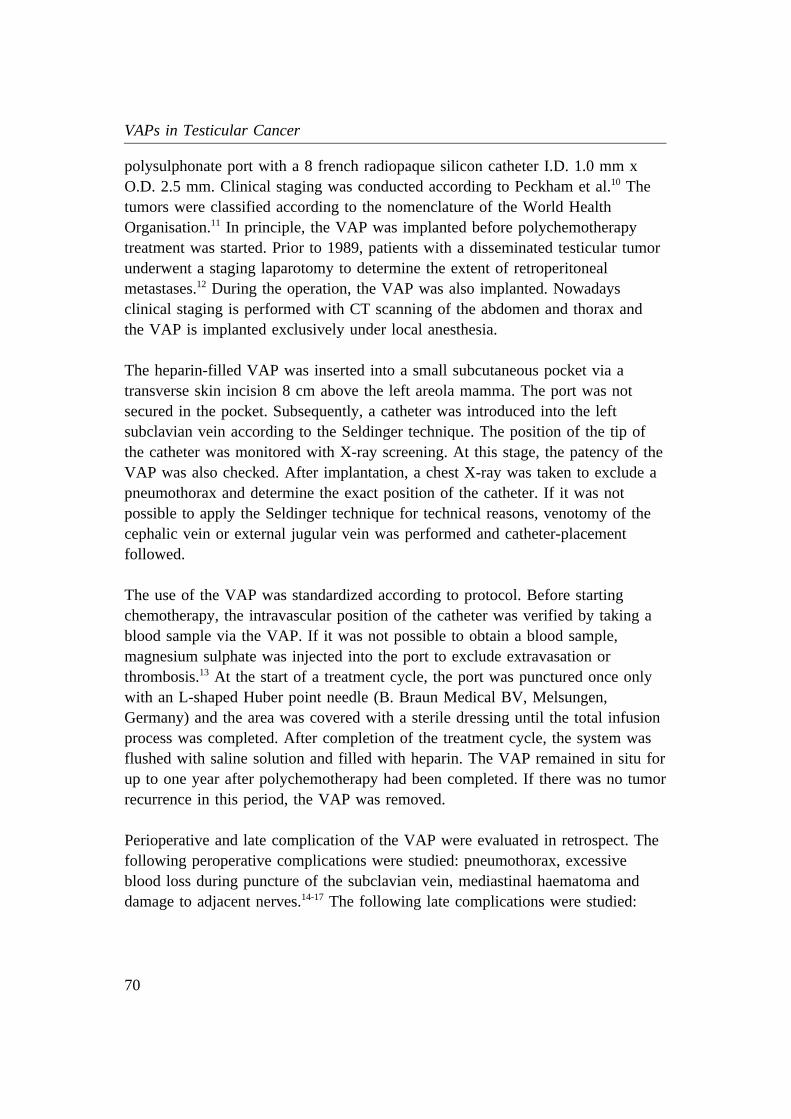
VAPs in Testicular Cancer
polysulphonate port with a 8 french radiopaque silicon catheter I.D. 1.0 mm xO.D. 2.5 mm. Clinical staging was conducted according to Peckham et al.10 Thetumors were classified according to the nomenclature of the World HealthOrganisation.11 In principle, the VAP was implanted before polychemotherapytreatment was started. Prior to 1989, patients with a disseminated testicular tumorunderwent a staging laparotomy to determine the extent of retroperitonealmetastases.12 During the operation, the VAP was also implanted. Nowadaysclinical staging is performed with CT scanning of the abdomen and thorax andthe VAP is implanted exclusively under local anesthesia.
The heparin-filled VAP was inserted into a small subcutaneous pocket via atransverse skin incision 8 cm above the left areola mamma. The port was notsecured in the pocket. Subsequently, a catheter was introduced into the leftsubclavian vein according to the Seldinger technique. The position of the tip ofthe catheter was monitored with X-ray screening. At this stage, the patency of theVAP was also checked. After implantation, a chest X-ray was taken to exclude apneumothorax and determine the exact position of the catheter. If it was notpossible to apply the Seldinger technique for technical reasons, venotomy of thecephalic vein or external jugular vein was performed and catheter-placementfollowed.
The use of the VAP was standardized according to protocol. Before startingchemotherapy, the intravascular position of the catheter was verified by taking ablood sample via the VAP. If it was not possible to obtain a blood sample,magnesium sulphate was injected into the port to exclude extravasation orthrombosis.13 At the start of a treatment cycle, the port was punctured once onlywith an L-shaped Huber point needle (B. Braun Medical BV, Melsungen,Germany) and the area was covered with a sterile dressing until the total infusionprocess was completed. After completion of the treatment cycle, the system wasflushed with saline solution and filled with heparin. The VAP remained in situ forup to one year after polychemotherapy had been completed. If there was no tumorrecurrence in this period, the VAP was removed.
Perioperative and late complication of the VAP were evaluated in retrospect. Thefollowing peroperative complications were studied: pneumothorax, excessiveblood loss during puncture of the subclavian vein, mediastinal haematoma anddamage to adjacent nerves.14-17 The following late complications were studied:
70
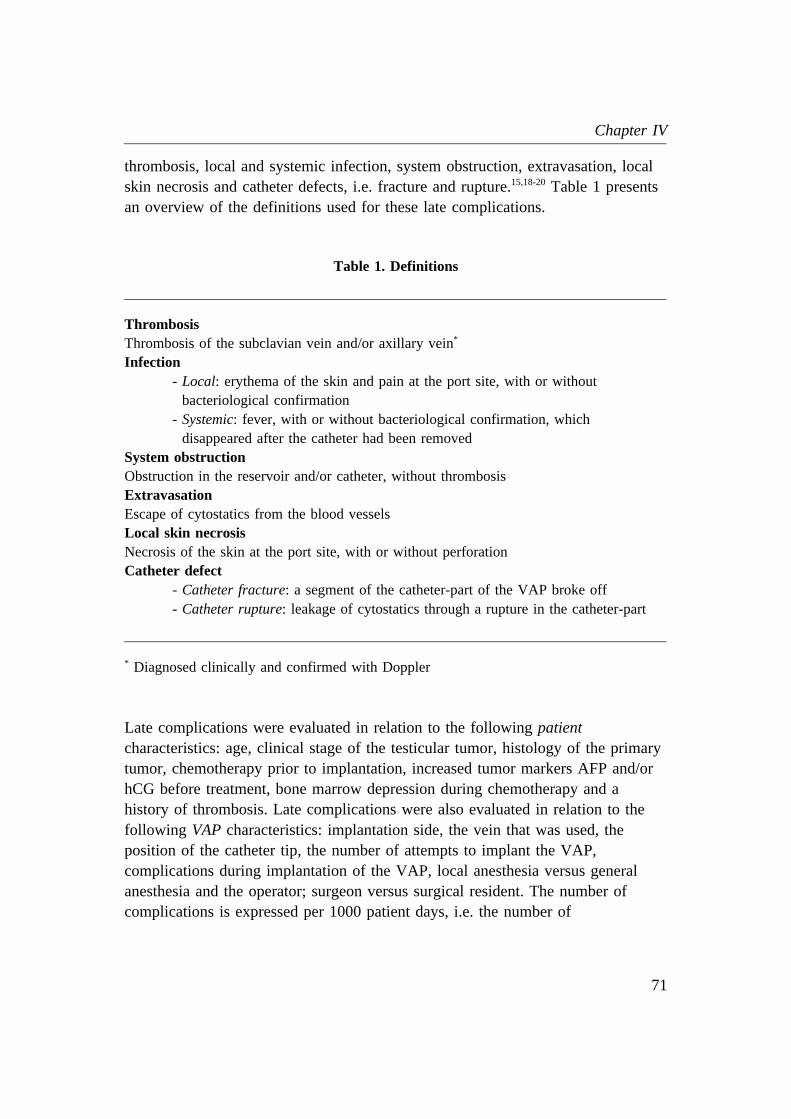
Chapter IV
thrombosis, local and systemic infection, system obstruction, extravasation, localskin necrosis and catheter defects, i.e. fracture and rupture.15,18-20 Table 1 presentsan overview of the definitions used for these late complications.
Table 1. Definitions
ThrombosisThrombosis of the subclavian vein and/or axillary vein*
Infection- Local: erythema of the skin and pain at the port site, with or without
bacteriological confirmation- Systemic: fever, with or without bacteriological confirmation, which
disappeared after the catheter had been removedSystem obstructionObstruction in the reservoir and/or catheter, without thrombosisExtravasationEscape of cytostatics from the blood vesselsLocal skin necrosisNecrosis of the skin at the port site, with or without perforationCatheter defect
- Catheter fracture: a segment of the catheter-part of the VAP broke off- Catheter rupture: leakage of cytostatics through a rupture in the catheter-part
* Diagnosed clinically and confirmed with Doppler
Late complications were evaluated in relation to the followingpatientcharacteristics: age, clinical stage of the testicular tumor, histology of the primarytumor, chemotherapy prior to implantation, increased tumor markers AFP and/orhCG before treatment, bone marrow depression during chemotherapy and ahistory of thrombosis. Late complications were also evaluated in relation to thefollowing VAP characteristics: implantation side, the vein that was used, theposition of the catheter tip, the number of attempts to implant the VAP,complications during implantation of the VAP, local anesthesia versus generalanesthesia and the operator; surgeon versus surgical resident. The number ofcomplications is expressed per 1000 patient days, i.e. the number of
71

VAPs in Testicular Cancer
days that the VAP was in situ.21 Follow-up continued until the VAP was removedor the patient died.
Statistical analysis
Differences in patient characteristics and VAP characteristics between the twosubgroups of patients with and without complications were analysed as continuousvariables by means of Student’st test (all complication together) or the Mann-Whitney test (each complication separately). The Chi-square test was used forcategorical variables and Fisher’s exact test for small numbers. Multivariatelogistic regression analysis was performed to evaluate independent associationsbetween the various prognostic factors and the occurrence of complications.Possible prognostic factors were chosen on a theoretical basis and on the basis ofthe univariate analyses.22 The following prognostic factors were included in themultivariate analyses: age, clinical stage IV (yes versus no),pre-VAP chemotherapy (yes versus no), number of implantation attempts(>1 versus 1) and the type of anesthesia (local versus general). The presence of alate complication was the dependent variable with the absence of a complicationas reference category. In the multivariate analyses, patients with multiplecomplications were considered as separate cases for each complication. The levelof statistical significance was set at P<.05. Odds ratios were considered to besignificant if the 95% confidence interval did not contain the value 1. Statisticalanalyses were conducted using the SPSS-PC+ statistical software package (SPSSInc. Chicago, III., USA).
RESULTS
A total of 135 VAPs were implanted in 132 patients who underwent cisplatin-based chemotherapy for disseminated testicular cancer. The median age was 28(range 16-55 years, SD 7.8) years. In 125 patients the primary tumor was anonseminomatous testicular tumor (94.7%); the seven remaining patients had aseminoma (5.3%). At the time of VAP implantation, 59 patients had stage IIdisease (44.7%), 14 had stage III (10.6%) and 58 had stage IV (43.9%). In onepatient the stage was unknown (0.8%). It was necessary to start chemotherapy
72

Chapter IV
immediately in 13 patients (9.8%) due to poor clinical condition and/or extensivemetastatic disease. In these 13 cases, the VAP was implanted at the firstopportunity, usually directly after the first treatment cycle. The median number ofpolychemotherapy cycles was four (range 1-14).
The techniques used to implant the 135 VAPs were the Seldinger method, i.e.subclavian vein puncture (n=126, 93.3%), venotomy of the cephalic vein (n=8,5.9%) and venotomy of the right jugular vein (n=1, 0.8%). Implantation wasconducted under general anesthesia during a staging laparotomy12 in 103 cases(76.3%) and under local anesthesia in 32 cases (23.7%); 121 VAPs wereimplanted on the left and 14 on the right (10.4%). In 19 patients, more than oneattempt was made to position the catheter correctly. The tip of the catheter waslocated in the superior vena cava in 126 cases (93.3%), in the right atrium in fivecases (3.7%) and in the internal jugular vein in four cases (3.0%).
Perioperative complications
Perioperative complications occurred in five of the 135 VAPs (3.7%). Thesecomprised a pneumothorax in two patients (1.5%) which necessitated thoraxdrainage; excessive blood loss in two patients (1.5%) which could be treatedconservatively and mediastinal bleeding in one patient (0.8%) in whom the VAPimplantation procedure had to be discontinued.
Late complications
The 135 VAPs were in situ for a total of 55247 days and there were 42 (31.1%)complications in 31 patients. Eight patients had two complications and one patienthad four complications. The total number of complications corresponds with 0.76complication episodes per 1000 patient days. The median implantation time of theVAPs was 413 (range 7-1607) days. In four VAPs, all without complications, theprecise number of days in situ was unknown. The incidence of late complicationswas therefore calculated for 131 VAPs. In the subgroup of VAPs with one com-plication, the median number of days in situ was 308 (range 55-1055) days, whilein the subgroup with more than one complication, it was 265 (range 78-894) days.There were no complications in 101 patients (76.5%). During follow-up, 13patients died (10.2%); mortality was not related to VAP
73

VAPs in Testicular Cancer
complications. Table 2 lists the late complications.
Table 2. Incidence and interval until late complicationsoccurred in 135 VAPs
Complication No. of patients Incidence1 No. of days (range)2
System obstrucion 13 (9.6%) 0.24 40 (5-601)Thrombosis 11 (8.1%) 0.20 33 (2-518)Infection 6 (4.4%) 0.11 75 (39-430)Extravasation 4 (3.0%) 0.06 72 (41-85)Local skin necrosis 2 (1.5%) 0.04 197 (60-333)Catheter defect 6 (4.4%) 0.11 363 (55-1241)
1 Per 1000 patient days;2 Median number of days in situ until complication occurred.
System obstructionwas the most frequent complication. The obstruction could bealleviated by injecting the VAP with saline solution, heparin or urokinase.Thrombosiswas diagnosed clinically (redness, swelling and pain) and wasconfirmed with Doppler scanning. It occurred in 11 cases: six in the subclavianvein, four in the axillary vein and in one in the brachiocephalic vein. The portwas removed for this reason in one patient, four patients received a thrombolyticagent and six patients were treated expectatively. All patients recovered fullyfrom this thrombosis, which was confirmed with Doppler scanning.Infection: six cases of infection occurred in five patients. Infection was local intwo patients. There were four cases of systemic infection in three patients. Allthese patients received intravenously antibiotic therapy. Two of the three VAPshad to be removed because of continued systemic infection.Extravasationoccurred in four patients and comprised subcutaneous leakage ofthe cytostatics at the port site. Three of these patients were treated conservatively;the VAP was removed in one patient.Local skin necrosisdeveloped in two patients at the port site. In both cases, theVAP had to be removed.Catheter defects: these mechanical complications occurred in six cases (five
74

Chapter IV
fractures and one rupture). Two of the five fractures were detected on routinechest X-ray 256 and 454 days after implantation, respectively. The remainingthree fractures were detected during VAP removal after 350, 376 and 1241 days,respectively. The broken catheter segments migrated to the pulmonary artery infour of the five cases and to the right atrium in the remaining patient. In all casesthey could be removed by catheterisation under local anesthesia via the femoralvein.19 The catheter ruptured in one patient after 55 days, replacement of the VAPwas performed.
The relationship between patient characteristics and late complications wasanalysed. Of the 13 patients who had received chemotherapy prior to VAPimplantation, 9 (69.2%) developed at least one late complication. This wassignificantly different from the 33 complications (27.7%) that occurred in the 119patients who started chemotherapy after VAP implantation (P<.001). Thedifference was mainly due to thrombosis (n=4) and system obstruction (n=4) inthe pre-chemotherapy implantation group versus seven thromboses and ninesystem obstructions in the post-chemotherapy implantation group (P<.05). Nosignificant relationships were found between late complications and: age, clinicalstage, histology of the primary tumor, increased tumor markers, bone marrowdepression and a history of thrombosis (data not shown).Analysis was also made of the relationship between the implantation procedure ofthe VAP and late complications. In the 32 VAPs that were implanted under localanesthesia, 12 patients had one or more late complications (37.5%). This differedsignificantly from the 19 (18.4%) complications that occurred in the 103implanted VAPs under general anesthesia (P<.05). This difference was mainly dueto thrombosis: four cases in the 32 VAP implants conducted under localanesthesia versus seven cases in the 103 implants conducted under generalanesthesia. No significant relationships were found between late complicationsand: implantation side, implantation vein, position of the catheter tip, number ofattempts to position the catheter, complications during implantation and theoperator (data not shown).
Multiple logistic regression analysis was performed to examine the relationshipbetween each patient characteristic or VAP characteristic and the occurrence oflate complications, adjusted for the remaining potential characteristics (Table 3).Chemotherapy treatment prior to VAP implantation was found to be the onlyindependent risk factor for late complications (Odds Ratio 7.4; 95% ConfidenceInterval 1.8-30.4).
75

VAPs in Testicular Cancer
Table 3. Multiple logistic regression analysis: Odds Ratiosfor the occurrence of late complications (n=135)*
Independent variable Odds Ratio 95% CI
Age 1.0 0.9 - 1.1Stage IV (yes versus no) 1.2 0.5 - 2.9Pre-VAP chemotherapy (yes versus no) 7.4 1.8 - 30.4Number of attempts (>1 versus 1) 2.1 0.7 - 6.4Anesthesia (local versus general) 1.5 0.5 - 4.5
* No complication was the reference category
DISCUSSION
Aubaniac was the first to describe the use of a central catheter in 1952.23 It wasmainly used for parenteral feeding.24 In 1973, Broviac introduced the tunnelledsilicone rubber catheter,25 which was adapted later by Hickman and Ivey.26,27
These central venous catheters carried the disadvantages of an increased risk ofinfection and inconvenience for the patient.28-30 Since the introduction of the VAPin the eighties, various authors have evaluated its advantages over traditionalexternal catheters.7,31 In a prospective randomized study, Mueller et al. did notfind any significant difference in the incidence of infection, thrombosis ormechanical complications between the VAP and the external catheter.31 Cardealso performed a prospective randomized study and found that in patients with asolid tumor who had been receiving treatment with intravenous chemotherapy forlonger than six months, the totally implantable systems were more reliable, saferand better tolerated than the traditional external catheters.7 The totally implantablesystems are cosmetically superior and more easily accepted by the patients thanthe external catheters.6 Therefore, the VAP usually takes preference as accesssystem for chemotherapy.This is the first study to describe the use of the VAP on a large homogeneouspatient group and to use it exclusively for administering cisplatin-based
76

Chapter IV
chemotherapy. Other authors described complications that occurred with the VAPin patients with diverse malignancies and non-malignancies.15,18,32,33In a greatnumber of these studies, the VAP was not only used for administeringchemotherapy, but also for other purposes, such as parenteral feeding and/orblood transfusions.
In this study, two types of complications were distinguished: perioperative andlate complications. The number of complications in this homogeneous patientgroup did not differ essentially from the percentages reported in the literature forvarious patient groups. The number of perioperative complications consisting ofpneumothorax and blood loos was described in percentages of 1.7 to 8.0%.14,15,34,35Late complications consisting of system obstruction, thrombosis,infections, extravasation and local skin necrosis was described in percentages of0.0 to 55.5%.14,15,21,31-43Only the percentage of catheter defects (4.4%) was largerthan in the other studies (0.4%-1.4%).31,32,33,39Possible explanations for this are:too medial positioning which caused friction of the catheter between the clavicleand the first rib (’pinch off sign’) and/or the long duration in situ time.44,45
The incidence of complications per 1000 patient days was also in agreement withthe incidences reported in other studies (Table 4). The VAPs in this studyremained in situ for much longer than they did in other studies and wereconsequently at risk for a longer period, because at the University HospitalGroningen, it was decided to leave the VAPs in situ for up to one year after thecompletion of treatment so that if recurrent disease was diagnosed, chemotherapytreatment could be continued via the VAP. However, leaving the VAP in situ fora longer period increased the risk of catheter defects. Therefore, it may be worthconsidering earlier removal.
In 13 patients, chemotherapy had to be started immediately because of extensivemetastasic disease and/or poor clinical condition. Both univariate and multivariateanalyses showed that a significantly greater proportion of these patients developedlate complications (P<.001; Odds Ratio 7.4). These were mainly venousthrombosis and system obstruction. Donohue and Rowland reported thatmorbidity, unfortunately not further specified, was higher in the patients whounderwent laparotomy after chemotherapy for a testicular tumor, than in thosewho had not received chemotherapy.46 Their explanation was that the ensuingdecrease in pulmonary, renal and nutritive reserves may have caused this increasein morbidity.
77
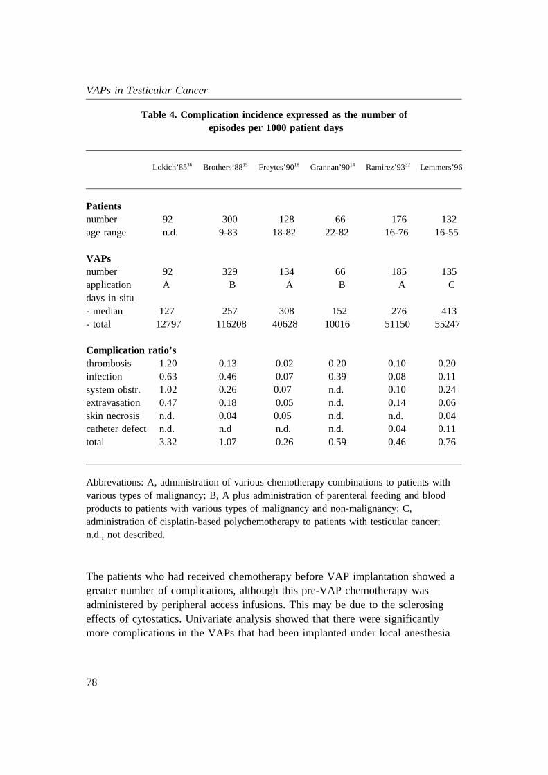
VAPs in Testicular Cancer
Table 4. Complication incidence expressed as the number ofepisodes per 1000 patient days
Lokich’8536 Brothers’8815 Freytes’9018 Grannan’9014 Ramirez’9332 Lemmers’96
Patientsnumber 92 300 128 66 176 132age range n.d. 9-83 18-82 22-82 16-76 16-55
VAPsnumber 92 329 134 66 185 135application A B A B A Cdays in situ- median 127 257 308 152 276 413- total 12797 116208 40628 10016 51150 55247
Complication ratio’sthrombosis 1.20 0.13 0.02 0.20 0.10 0.20infection 0.63 0.46 0.07 0.39 0.08 0.11system obstr. 1.02 0.26 0.07 n.d. 0.10 0.24extravasation 0.47 0.18 0.05 n.d. 0.14 0.06skin necrosis n.d. 0.04 0.05 n.d. n.d. 0.04catheter defect n.d. n.d n.d. n.d. 0.04 0.11total 3.32 1.07 0.26 0.59 0.46 0.76
Abbrevations: A, administration of various chemotherapy combinations to patients withvarious types of malignancy; B, A plus administration of parenteral feeding and bloodproducts to patients with various types of malignancy and non-malignancy; C,administration of cisplatin-based polychemotherapy to patients with testicular cancer;n.d., not described.
The patients who had received chemotherapy before VAP implantation showed agreater number of complications, although this pre-VAP chemotherapy wasadministered by peripheral access infusions. This may be due to the sclerosingeffects of cytostatics. Univariate analysis showed that there were significantlymore complications in the VAPs that had been implanted under local anesthesia
78

Chapter IV
than under general anesthesia (P<.05); these were also mainly thrombosis. Apossible explanation for this is that it was easier to puncture the subclavian veinunder general anaesthesia with positive pressure mechanical ventilation causingvasodilatation, than under local anesthesia. However, this did not prove to be anindependent risk factor in the multivariate analysis.Mansfield found a lower percentage of complications in catheterisations of thesubclavian vein performed by experienced physicians (10.2%) than in thoseperformed by inexperienced physicians (13.5%); however, this difference was notstatistically significant.35 In this study, the number of complications was notrelated to the experience, surgeon versus surgical resident.In conclusion, implantation and use of a VAP to administer polychemotherapy topatients with disseminated testicular cancer was accompanied by a complicationrate of 26.7%. The only independent risk factor for occurrence of latecomplications in this study was the administration of chemotherapy prior to VAPimplantation. Therefore, in patients with a diagnosis of disseminated testicularcancer who are scheduled to undergo long-term polychemotherapy, the VAP mustbe implanted before chemotherapy treatment is initiated.
REFERENCES
1. Einhorn LH, Donohue JP: Cis-diamminedichloroplatinum, vinblastine andbleomycin combination chemotherapy in disseminated testicular cancer. Ann IntMed 1977; 87: 293-298.
2. Peckham JM, Barret A, Lieuw KM, et al: The treatment of metastatic germ celltesticular tumors with bleomycin, etoposide and cisplatin (BEP). Br J Cancer1983; 47: 613-619.
3. Wobbes Th, Slooff MJH, Sleijfer DTh, et al: Five years experience in accesssurgery for polychemotherapy. Cancer 1983; 52: 978-982.
4. Rudolph R, Larson DL: Etiology and treatment of chemotherapeutic agentextravasation injuries: A review. J Clin Oncol 1987; 5: 1116-1126.
5. Daly J, Lawson M, Speir A, et al: Angioaccess in cancer patients. Curr ProblCancer 1981; 5: 1-37.
6. Niederhuber JE, Ensminger W, Gyves JW, et al: Totally implanted venous andarterial access system to replace external catheters in cancer treatment. Surgery1982; 92: 706-712.
79

VAPs in Testicular Cancer
7. Carde P, Cosset-Delaigue MF, Laplanche A, et al: Classical external indwellingcentral venous catheter versus totally implanted venous access systems forchemotherapy administration: A randomized trial in 100 patients with solidtumors. Eur J Cancer Clin Oncol 1989; 25: 939-944.
8. Bothe A, Piccione W, Ambrosino JJ, et al: Implantable central venous accesssystem. Am J Surg 1984; 147: 565-569.
9. Gels ME, Hoekstra HJ, Sleijfer DTh, et al: Detection of recurrence in patientswith clinical stage I Nonseminomatous Testicular Germ Cell Tumors andconsequences for further follow-up: A single-center 10-year experience. J ClinOncol 1995; 13: 1188-1194.
10. Peckham MJ, Barret A, McElwain TJ, et al: Combined management of malignantteratoma of the testis. Lancet 1979; 2: 267-270.
11. Mostofi FK, Sobin LH: Histological typing of testis tumours. in Internationalhistological classification of tumors No. 16. Geneva, Switzerland: World HealthOrganisation 1977: 1-39.
12. Gelderman WAH, van der Laan JG, Schraffordt Koops H, et al: Evaluation ofstaging methods for retroperitoneal metastases in patients with nonseminomatoustesticular tumours: CT, lymphography and exploratory laparotomy. J MedImaging 1989; 3: 288-296.
13. Sleijfer DTh, Mulder NH, de Vries EGE: Confirming position of venous port(letter). Ann Int Med 1990; 113: 86.
14. Grannan KJ, Taylor PH: Early and late complications of totally implantablevenous access devices. J Surg Oncol 1990; 44: 52-54.
15. Brothers TE, von Moll LK, Niederhuber JE, et al: Experience with subcutaneousinfusion ports in three hundred patients. Surg Gynecol Obstet 1988; 166:295-301.
16. Defalgue RJ, Fletcher MV: Neurological complications of central venous cannula-tion. J Parent Ent Nutr 1988; 11: 406-409.
17. Conces DJ Jr, Holden RW: Aberrant locations and complications in initialplacement of subclavian vein catheters. Arch Surg 1984; 119: 293-295.
18. Freytes C, Reid P, Smith KL: Long-term experience with a totally implantedcatheter system in cancer patients. J Surg Oncol 1990; 45: 99-102.
19. Noyen J, Hoorntje J, de Langen ZJ, et al: Spontaneous fracture of the catheter ofa totally implantable venous access port: Case report of a rare complication.J Clin Oncol 1987; 5: 1295-1299.
20. Bach F, Videbaek C, Holst-Christensen J, et al: Cytostatic extravasation. Aserious complication of long-term venous access. Cancer 1991; 68: 538-539.
21. Howell PB, Walters PE, Donowitz GR, et al: Risk factors for infection of adultpatients with cancer who have tunnelled central venous catheters. Cancer 1995;75: 1367-1375.
80

Chapter IV
22. Rothmann KJ: Modern Epidemiology. Boston, MA: Little Brown 1986.23. Aubaniac R: L‘injection intraveineuse sous-claviculaire: Avantages et technique.
Press Med 1952; 60: 1456.24. Dudrick SJ, Wilmore DW: Long-term parenteral feeding. Hosp Pract 1968; 3: 65-
78.25. Broviac J, Cole J, Scribner, B: A silicone rubber atrial catheter for prolonged
parenteral alimentation. Surg Gynecol Obstet 1973; 136: 602-606.26. Hickman RO, Buckner CD, Clift RA, et al: A modified right atrial catheter for
access to the venous system in marrow transplant recipients. Surg Gynecol Obstet1979; 148: 871-875.
27. Ivey MF, Adam SM, Hickman RO, et al: Right atrial indwelling catheterfor patients requiring long-term intravenous therapy. Am J Hosp Pharm 1978; 35:1525-1527.
28. Shapiro ED, Wald ER, Nelson KA, et al: Broviac catheter-related bacteremia inoncology patients. Am J Dis Child 1982; 136: 679-681.
29. Press OW, Ramsey PG, Larson EB, et al: Hickman catheter infections in patientswith malignancies. Medicine 1984; 63: 189-200.
30. Johnson PR, Decker MD, Edwards KM, et al: Frequency of Broviac catheterinfections in pediatric oncology patients. J Infect Dis 1986; 154: 570-578.
31. Mueller BA, Skelton J, Callender DPE, et al: A prospective randomized trialcomparing the infectious and noninfectious complications of an externalizedcatheter versus a subcutaneously implanted device in cancer patients.J Clin Onc 1992; 10: 1943-1948.
32. Ramirez JM, Miguelena JM, Guemes A, et al: Fully implantable venous accesssystems. Br J Surg 1993; 80: 347-348.
33. Brincker H, Saeter G: Fifty-five patient years experience with a totally implantedsystem for intravenous chemotherapy. Cancer 1986; 57: 1124-1129.
34. Eastridge BJ, Lefor AT: Complications of indwelling venous access devices incancer patients. J Clin Oncol 1995; 13: 233-238.
35. Mansfield PF, Hohn DC, Fornage BD, et al: Complications and failures ofsubclavian vein catheterization. N Engl J Med 1994; 331: 1735-1738.
36. Lokich J, Bothe A Jr, Benotti P, et al. Complications and management of implan-ted venous access catheters. J Clin Oncol 1985; 3: 710-717.
37. Greidanus J, de Vries EGE, Nieweg MB, et al: Evaluation of a totally implantedvenous access port and portable pump in a continuous chemotherapy infusionschedule on an outpatient basis. Eur J Cancer Clin Oncol 1987; 23: 1653-1657
38. Treiman GS, Silberman H: Chronic venous access in patients with cancer. Cancer1993; 72: 760-765.
39. Franceschi D, Specht MA, Farrell CF: Implantable venous access device.J Cardiovasc Surg 1989; 30: 124-129.
81

VAPs in Testicular Cancer
40. Dionigi P, Cebrelli T, Jemos V, et al: Use of subcutaneous implantable infusionsystems in neoplastic and AIDS patients requiring long-term venous access. Eur JSurg 1995; 161: 137-142.
41. Stanislav GV, Fitzgibbons RJ, Bailey RT, et al: Reliability of implantable centralvenous access devices in patients with cancer. Arch Surg 1987; 122: 1280-1283.
42. Lokich JJ, Becker B: Subclavian vein trombosis in patients treated with infusionchemotherapy for advanced malignancy. Cancer 1983; 52: 1586-1589.
43. Vlasveld LT, Rodenhuis S, Rutgers EJH, et al: Catheter-related complications in52 patients treated with continuous infusion of low dose recombinant inter-leukin-2 via an implanted central venous catheter. Eur J Surg Oncol 1994; 20: 122-129.
44. Lafreniere R: Indwelling subclavian catheters and a visit with the "Pinched offsign". J Surg Oncol 1991; 47: 261-264.
45. Aitken DA, Minton JP: The "pinch-off" sign. A warning of impending problemswith permanent subclavian catheters. Am J surg 1984; 148: 633-636.
46. Donohue JP and Rowland RG: Complications of retroperitoneal lymph nodedissection. J Urol 1981; 125: 338-340.
82

CHAPTER V
COMPLICATIONS OF THE POSTCHEMOTHERAPY RESECTION OFRETROPERITONEAL RESIDUAL TUMOR MASS IN PATIENTS WITH
NONSEMINOMATOUS TESTICULAR GERM CELL TUMORS
M.E. Gels,1 A.P. Nijboer,1 H.J. Hoekstra,1 D.Th. Sleijfer,2
W.M. Molenaar,3 J.Th. Plukker,1 J.H.J. Droste,4 H. Schraffordt Koops.1
From the departments of Surgical Oncology,1 Medical Oncology,2
Pathology,3 and Health Sciences, section Epidemiology and Statistics.4
University Hospital Groningen, the Netherlands
British Journal of Urology 1997; 79: 263-268.
83

Resection of RRTM in NSTGCT
ABSTRACT
The objective of this study was to evaluate the resection of the retroperitonealresidual tumor mass (RRTM) for histological examination after chemotherapy inpatients with disseminated nonseminomatous testicular germ cell tumors(NSTGCT), with particular attention to surgical morbidity. From 1979 to 1995,112 patients (mean age 28 years, range 16-53) with disseminated NSTGCT hadresidual disease after chemotherapy for which surgical evaluation was indicated;the histology of the residual tumor and the surgical complications were assessed.Possible associations between the occurrence of surgical complications and theage of the patient, size the residual tumor, operative duration, previouslaparotomy and pathological findings were evaluated. The median size of theresidual tumor was four cm (range 0-18); histological examination revealedviable cancer in 9.0%, mature teratoma in 44.1% and necrosis/fibrosis in 44.1%of the patients. In 3 patients (2.8%) no residual tumor mass was found atlaparotomy. There were 26 complications in 20 patients (18.0%); urinary tractinfection was the most common, occurring in nine patients (8.1%). One patientdied during the induction of anesthesia. There were no significant relationshipsbetween the occurrence of complications and age, size of the residual tumor,operative duration, previous laparotomy or pathological findings. The resection ofRRTM after polychemotherapy treatment for disseminated NSTGCT is a safesurgical procedure, with low treatment morbidity, mainly consisting of urinarytract infection. Knowledge of the potential complications may help to preventmorbidity. However, the surgical evaluation of the ultimate effect of poly-chemotherapy remains the gold standard.
INTRODUCTION
The initial treatment of patients with disseminated nonseminomatous testiculargerm cell tumors (NSTGCT) is cisplatin-based polychemotherapy. The previouslydismal prognosis of these patients has been improved dramatically with thedevelopment of this effective treatment.1 However, on completion of theremission-inducing polychemotherapy, retroperitoneal residual tumor mass(RRTM) is still found regularly. Shrinkage of metastases may cease due to
84

Chapter V
necrosis and fibrosis or they may persist as mature teratoma.2,3 Sometimes viablecancer is present. All mature teratoma tissue has to be removed to prevent theoccurrence of the growing teratoma syndrome4 or the development of a secondarymalignancy in residual mature teratoma.5-7 If malignant tumor tissue is still foundto be present in the postchemotherapy resected specimen, salvage chemotherapy isindicated. Nevertheless postchemotherapy resection of RRTM has an importantrole in patients with retroperitoneal residual disease following normalisation oftumor markers with polychemotherapy. Resection of all the residual lesions forhistopathology is necessary to evaluate the ultimate response. If no pathologicalmass is visible on the postchemotherapy computed tomogram (CT scan), someauthors still advocate resection in the following situations: if a teratomatouscomponent was present in the primary tumor;3 if prechemotherapy retroperitoneallymph node metastases were larger than 30 mm;8 or if laparotomy is performedroutinely as part of the treatment policy.9
At the University Hospital Groningen, the Netherlands, laparotomy is performedon all patients with a disseminated NSTGCT who become serum tumor marker-negative following remission-inducing polychemotherapy and who show evidenceof retroperitoneal residual disease, or had mature teratoma in the primary tumor.The current paper describes the experience with postchemotherapy resection ofRRTM and attention is focused on postoperative morbidity.
PATIENTS AND METHODS
In the period July 1979 to March 1995, 112 consecutive resections of RRTMwere performed at the University Hospital Groningen, the Netherlands, in patientswith disseminated NSTGCT. All patients were treated with cisplatin-basedpolychemotherapy; in all cases it was the first operation after chemotherapy treat-ment. Initial clinical staging was performed by physical examination, chest X-ray,serum tumor markers alpha-fetoprotein (AFP) and human chorionic gonadotrophin(hCG) levels and CT scans of the chest and abdomen.10 Until 1989, staginglaparotomy was performed in patients with disseminated NSTGCT in conjunctionwith orchidectomy, to make a definite diagnosis regarding the size of theretroperitoneal lymph node metastases. On the basis of previous analyses, it wasdecided from that time onwards to perform this staging laparotomy only if radio-logical findings were inconclusive.11 Since then
85

Resection of RRTM in NSTGCT
patients have been clinically staged with CT scans of the chest and abdomen.Patients with Stage I disease were treated according to the wait-and-see policy.12
Patients with Stage II or higher were treated with cisplatin-basedpolychemotherapy: cisplatin, vinblastine and bleomycin (PVB) or bleomycin,etoposide and cisplatin (BEP). A total of four remission-inducing courses wereadministered; each course had a duration of three weeks. Up to 1982, patientswere given maintenance treatment, as was usual at that time. This policy waschanged on the basis of data that became available in the early eighties.13 Aftercompletion of the polychemotherapy, the patients were clinically restaged withCT scans of the chest and abdomen and monitored for serum tumor markers. Ifserum tumor markers were still high or failed to decrease according to the knownserum half-life times, supplementary salvage chemotherapy was administered.Otherwise any residual tumor was removed surgically and examinedpathologically. This treatment strategy has been described previously by Donohueand Rowland.14
Over eighty percent of the resections of RRTM were performed by, or in thepresence of the same surgeon. In total, four surgeons were involved in thetreatment. The technique of trans-abdominal and thoraco-abdominal resection ofRRTM used at the University Hospital Groningen had been described byWobbes.15 Generally, partial retroperitoneal lymph node dissection was performed.Standard preoperative procedures included subcutaneous heparin injection toprevent thrombosis, introduction of a gastric tube to prevent stomach dilatationand aspiration, and the introduction of a urinary catheter to monitor urineproduction during the operation. The urinary catheter also ensuredcollapse of the bladder and thus facilitated surgery in the pelvic area. Anestheticmanagement included continuous monitoring of oxygen saturation and intermittentarterial blood gas analysis. After induction, the FiO2 was maintained at the lowestlevel possible to still allow adequate oxygenation (21-25%).16 The patients did notreceive any perioperative antibiotics. Postoperative pain medication was notstandardised. The histology of the resected residual tumor mass was compared tothat of the primary testicular tumor, using the nomenclature of the World HealthOrganisation.17
Complications of postchemotherapy resection of RRTM were studied. A possibleassociation between the occurrence of complications and the age of the patient,size of the residual tumor, operative duration, previous laparotomy andpathological findings was evaluated. Differences between subgroups of patients
86

Chapter V
with and without complications were analysed by means of the Mann-WhitneyU-test for continuous data; for categorical variables, the Chi-square test was usedor Fisher’s exact test in case of small numbers.
RESULTS
The mean age of the 112 patients was 28 (range 16-53) years; 77 of them hadundergone staging laparotomy before remission-inducing polychemotherapy wasinitiated. Clinical staging at the time of diagnosis was as follows: Stage IIa, eightpatients (7.1%); Stage IIb, 38 patients (33.9%), Stage IIc, 16 patients (14.3%);Stage III, 23 patients (20.6%); and Stage IV, 27 patients (24.1%). The medianinterval between the last dose of combination chemotherapy and resection of theRRTM was 38 (range 6-467) days. Of the 112 patients, 111 eventually underwentsurgery. One patient (0.9%) died during induction of anesthesia. He experiencedasphyxia, bradycardia and despite resuscitation efforts cardiac arrest. This patientwas excluded from the analyses. The trans-abdominal technique was used in 109patients. In two patients who also had residual pulmonary disease, the thoraco-abdominal approach was used, i.e. laparotomy with median sternotomy in onepatient and laparotomy with left posterolateral thoracotomy in the other patient.The median operative duration, defined as time from incision to skin closure, was150 (range 30-550) minutes. Median size of the residual tumor mass was four(range 0-18) centimetres. To enable the removal of all residual tumor tissue, parti-al resection and reconstruction of the aorta had to be performed in one patientand nephrectomy in four other patients.
Evaluation of the response to polychemotherapy
Of the 111 patients operated on, 67 (60.4%) had a mature teratoma component inthe primary tumor. This mature teratoma component was also present in theretroperitoneal residual tumor mass in 42 out of these 67 patients (63%), as wellas in the residual tumor in 11 out of the 44 patients (25%) who did not havemature teratoma in the primary tumor. Thus, a total of 53 patients (47.7%) hadresidual mature teratoma. Despite normalisation of the serum tumor markers,histological examination still revealed viable cancer in 10 (9.0%) patients
87

Resection of RRTM in NSTGCT
(Table 1), among whom four also had a mature teratoma component in theresidual tumor. Forty-nine patients had only residual mature teratoma in theresected tumor tissue (44.1%). In 49 other patients (44.1%) only necrosis and/orfibrosis was found. Although CT scans of the abdomen had identified a smallretroperitoneal residual tumor mass, in three patients (2.8%) no residual retroperi-toneal tumor mass was found at laparotomy, therefore resection was notperformed.
Table 1. Histological composition of the retroperitoneal massin the ten patients with residual viable cancer (n=111)
Components No. %
E 2 1.8Y 2 1.8E+C 1 0.9E+M 1 0.9E+Y 1 0.9M+I 1 0.9E+M+I 1 0.9E+Y+M+I 1 0.9
Total 10 9.0
Abbreviations: C, choriocarcinoma; E, embryonal carcinoma; I, immature teratoma;M, mature teratoma; Y, yolk sac tumor
Complications of resection of RRTM
There were 26 complications in 20 patients (18.0%). Three patients had twocomplications and one patient had four complications. Median hospitalization foruncomplicated resection of RRTM was seven (range 4-14) days, compared to 11(range 5-33) days in patients with complications (P=.005). Table 2 shows a list ofthese complications, compared to the current literature.
88

Chapter V
Table 2. Complications of retroperitoneal surgery
Complication Skinner’8218 Moul’8919 Jewett’9120 Wahle’9421 Baniel’9522 Gels’96
No. patients 149* 148* 100* 40** 603** 112***
Wound infection 3 4 7 1 29 1Wound dehiscence 2 2 2 1 - 1Paralytic ileus < 6 days - 0 2 - 1 3Paralytic ileus > 6 days - 3 1 - 13 0Ileus; laparotomy 3 8 6 2 0 0Chylous ascites - 1 2 - 12 1Lymphocele 1 2 3 2 10 2Pancreatitis - 1 - - 6 0Gastro-intest. bleeding - - 3 - 3 0RP bleeding; laparotomy - - 1 - 1 1RP/abdominal abscess 1 1 - - - 0RP/abdominal hematoma - 1 1 - 1 1Colon necrosis - - - - 1 0Ureteral injury - 1 - - 6 0Ureteral stricture/obstruction - - 5 - - 0Urinary tract infection - - 1 - 5 9Ischemic damage kidney - 2 4 - 1 0Renovasc. hypertension 1 2 1 - 2 0Atelectasis/Pneumonia 2 2 4 - 34 0ARDS - 1 - - 6 0Prolonged ventilation - - - - 5 2Pulmonary embolism - - - - 1 0Femoral neuropathy - - - 1 3 2Other neural injury - - - - 4 0Other complications 7 19 6 - - 3Total complications 20 50 49 7 144 26in number of patients 19 45 35 6 125 20
(13.0%) (30.4%) (35.0%) (15.0%) (20.7%) (18.0%)Mortality post-operative 2 0 0 0 5 0
(1.3%) (0%) (0%) (0%) (0.8%) (0%)Mortality anesthesia - - - - - 1
(0.9%)
* pre- and postchemotherapy retroperitoneal lymph node dissection (RPLND)/resection ofresidual retroperitoneal tumor mass (rRRTM);** postchemotherapy RPLND/rRRTM;*** postchemotherapy rRRTM; - not described; RP, retroperitoneal; ARDS, AdultRespiratory Distress Syndrome
89

Resection of RRTM in NSTGCT
Wound infection.This complication occurred in only one patient, who alsodeveloped wound dehiscence four days after the infection.Gastrointestinal complications.Ileus of less than six days duration was noted inthree patients and was of a paralytic nature. All recovered after conservativetreatment. Significant ileus for six days or longer postoperatively was notobserved.Lymphatic complications.Complications related to the lymphatic system wereobserved in 3 patients. Chylous ascites appeared in one patient after nine days.This patient responded well to conservative treatment with a low fat diet anddiuretics. In two patients lymphocele occurred; both were diagnosed within onemonth after surgery. In one patient the lymphocele was aspirated underultrasonographic guidance and did not recur. In the other patient, the lymphoceleappeared to be infected. This necessitated puncture under ultrasonographicguidance and drainage for 9 days.Retroperitoneal bleeding or hematoma.A significant decrease in the hemoglobinlevel was noted in one patient, with the clinical suspicion of retroperitonealbleeding. This required relaparotomy. However, except for some oozing, nosignificant source of bleeding was found. One patient on anticoagulation therapyfor pulmonary embolism (before surgery) complained about abdominal pain after12 days. Ultrasonographic imaging showed a solid mass in the retroperitoneum,which after puncture turned out to be a hematoma. This patient responded well toconservative treatment.Urinary tract complications.Urinary tract infection was the most commoncomplication in this study: nine patients (8.1%). These patients were treatedsuccessfully with antibiotics.Pulmonary complications.Two patients needed prolonged postoperativeventilation because of inappropriate oxygenation for two and five days,respectively. Respiratory failure due to pneumonia, the adult respiratory distresssyndrome or pulmonary embolism was not observed.Neurological complications.Femoral nerve neuropathy occurred in two patients.Both patients recovered early in the convalescent phase.Other complications.Two patients developed moderately severe postoperativediarrhoea of undetermined etiology. One patient developed a bacterial infection inboth eyes, due to insufficient care during anesthesia. No significant differenceswere found in the age of the patient, size of residual tumor, operative duration,previous laparotomy or pathological findings between the patients with andwithout postoperative complications.
90

Chapter V
DISCUSSION
Surgical resection is widely accepted as the treatment of choice for retroperitonealresidual masses after polychemotherapy for metastatic NSTGCT.1 Resectionenables histological diagnosis of the residual mass, which may bepurely benign with necrotic and/or fibrotic remnants only, or may contain matureteratoma elements or viable cancer. Resection of masses containing necrosis orfibrosis only is assumed to be of no therapeutic benefit and is usually notfollowed by additional treatment. Resection of mature teratoma is considered tobe beneficial as it prevents growth of potentially malignant cells.8 If resection isnot performed, masses that contain mature teratoma may start to grow duringfollow-up (growing teratoma syndrome).4 Resection may then be morecomplicated than it would have been shortly after the completion ofchemotherapy. Finally, leaving masses with residual viable cancer unresectedinvolves a serious risk. The presence of viable cancer in the residual mass directsthe decision to administer additional chemotherapy.14
In this series, histological examination revealed viable cancer in only ten out ofthe 111 patients (9.0%); four of these patients also had a mature teratomacomponent in the residual tumor. Mature teratoma without viable cancer wasfound in 49 patients (44.1%). In 49 other patients only necrosis and/or fibrosiswas found (44.1%) and in three patients resection was not performed because noresidual tumor mass was found at laparotomy. The incidence of necrotic debris,mature teratoma, or viable cancer varies in different studies. In general, pathologyreports revealed necrosis and/or fibrosis in about 40%, teratoma in another 40%and viable cancer in the remaining 20%.2
As histology determines the need for of resection, attempts have been made topredict the postchemotherapy histology.23-25 Steyerberg found that predictors ofnecrosis were the absence of teratoma elements in the primary tumor,prechemotherapy normal AFP, normal hCG and elevated lactate dehydrogenase(LDH) levels, a small prechemotherapy or postchemotherapy mass and largeshrinkage of the mass during chemotherapy. However, mature teratoma cannoteasily be distinguished from viable cancer. The value of Magnetic ResonanceImaging (MRI) in the treatment evaluation of retroperitoneal lymph nodemetastases from NSTGCT was studied by Hogeboom et al.26 They found thatMRI was unable to predict the outcome of chemotherapy treatment.
91

Resection of RRTM in NSTGCT
Recently, Stephens et al have described a study to assess the ability of PositronEmission Tomography (PET-scanning) to differentiate between residualradiographic abnormalities in postchemotherapy NSTGCT patients. They foundthat PET could not differentiate necrosis/fibrosis from teratoma, but it coulddifferentiate viable cancer from residual necrosis/fibrosis or teratoma.27 Until now,the only procedure which gives optimal and accurate information regarding thehistology of a residual retroperitoneal mass is pathological investigation ofresected specimens obtained by surgery. However, the great advantage of surgerymust be weighed against the disadvantages, i.e. complications.
Postoperative complications after resection of RRTM can be divided into twogroups: general complications of the type that can develop after any laparotomyand specific complications of retroperitoneal lymph node dissection (RPLND).General complications include: postoperative haemorrhage, pneumonia,thromboembolism, urinary infection and disturbed wound healing. Althoughchylous ascites and lymphocele cannot be regarded as specific complications ofRPLND, they are nevertheless conditions caused by damage to the retro-peritoneal lymph node plexus. Infertility due to the loss of emission as a result ofresection of the sympathetic nerves that traverse the area of resection, is a specificand common complication in patients who undergo full bilateralpostchemotherapy RPLND. Less frequently it occurs in patients who undergoresection of RRTM after polychemotherapy. This complication has recently beenassessed specificially by Van Basten et al.28 They found that after resection ofRRTM the prevalence rate of absence of ejaculation was 25.9%. The volume ofthe retroperitoneal residual masses was higher (95 cm3) in the patients with absen-ce of ejaculation than in those whithout ejaculatory dysfunction (40 cm3). Theabsence of ejaculation was reported significantly more often in patients with rightparacaval/inter aorta-caval located tumor (34.5%) than in those with left para-aortal located tumor (16.7%).
Overall, the number of complications did not differ essentially from the numbersreported in the recent literature (Table 2). It should be realized that the acute andlong-term toxicity of chemotherapy can give rise to complications during surgicalprocedures. For instance bleomycin produces acute interstitial pneumonia andchronic fibrotic changes in the lung. Its use in standard testis cancer protocolsplaces all such patients at risk for pulmonary insufficiency after long periods ofanesthesia.16 However, we did not observe an increased number of pulmonarycomplications, which may have been due to the low level of FiO2 during
92

Chapter V
anesthesia. Although most authors did not report urinary tract infections, thiscomplication occurred fairly frequently in this study. This may have been due tothe fact that a urinary catheter was always introduced prior to the operation and asample of urine was taken for culture directly after removal. Wheneverpathogenic micro-organisms were found in the culture, antibiotics were given onthe basis of the antibiogram and the urinary infection was documented as acomplication.
It should be taken into account that resection of RRTM was well-tolerated by allof these patients, despite prior chemotherapy. The relatively young age and thevigorous premorbid condition enabled the patients to endure the major surgicalprocedure relatively well and with only a short postoperative period ofhospitalisation. Depending on the volume as well as on the location of theresidual tumor, adjacent structures or organs may have to be resected. In thisstudy, nephrectomy had to be performed in four patients, while partial resectionof the aorta followed by reconstruction had to be performed in one patient.Hendry et al described a series of 231 postchemotherapy RPLNDs. The ipsilateralkidney was removed with the lymph node mass in 12.5%; the aorta or iliac arterywas resected or grafted in 4% and the vena cava was resected or tied off in 3%.29
Operative mortality rates of between 0% and 1.3% have been reported in theliterature (Table 2). In our study none of the postoperative complications resultedin mortality. However, one patient died during theinduction of anesthesia. Obduction did not reveal any explanation for the suddendeath of this patient.
The prognosis after the resection of residual disease is generally favourable, witha 5-year recurrence-free survival of over 85% after resection of necrosis ormature teratoma and of between 50% and 80% after the resection of viable cancerfollowed by additional chemotherapy.8,30 Increased awareness of potential compli-cations may help to prevent morbidity in patients with disseminated NSTGCT.However, surgical resection of RRTM remains the only procedure that givesoptimal and accurate information about the histology of the retroperitonealresidual mass. The long-term remission rate obtained with surgery in adjunct tochemotherapy still justifies complete resection whenever it is indicated and pos-sible.
93

Resection of RRTM in NSTGCT
REFERENCES
1. Einhorn LH: Treatment of testicular cancer: a new and improved model. J ClinOncol 1990; 8: 1777-1781.
2. Bajorin DF, Herr H, Motzer RJ, et al: Current perspectives on the role ofadjunctive surgery in combined modality treatment for patients with germ celltumors. Semin Oncol 1992; 19: 148-158.
3. Gelderman WAH, Schraffordt Koops H, Sleijfer DTh, et al: Results of adjuvantsurgery in patients with stage III and IV nonseminomatous testicular tumors aftercisplatin-vinblastine-bleomycin chemotherapy. J Surg Oncol 1988; 38: 227-232.
4. Logothetis CJ, Samuels ML, Trindade A, et al: The growing teratoma syndrome.Cancer 1982; 50: 1629-1635.
5. Molenaar WM, Oosterhuis JW, Meiring A, et al: Histology and DNA contents ofa secondary malignancy arising in a mature residual lesion six years afterchemotherapy for a disseminated nonseminomatous testicular tumor. Cancer1986; 58: 264-268.
6. Cromheecke M, Mehta DM, Sleijfer DTh, et al: The ultimate effect ofintraoperative radiotherapy (IORT) on an irresectable retroperitoneal recurrence ofa non-seminomatous testicular tumour [letter]. Radiother Oncol 1993; 29:352-354.
7. Ulbright TM, Loehrer PJ, Roth LM, et al: The development of non-germ cellmalignancies within germ cell tumors. A clinicopathologic study of 11 cases.Cancer 1984; 54: 1824-1833.
8. Toner GC, Panicek DM, Heelan RT, et al: Adjunctive surgery after chemotherapyfor nonseminomatous germ cell tumors: recommendations for patient selection. JClin Oncol 1990; 8: 1683-1694.
9. Aass N, Klepp O, Cavallin Stahl E, et al: Prognostic factors in unselectedpatients with nonseminomatous metastatic testicular cancer: a multicenterexperience. J Clin Oncol 1991; 9: 818-826.
10. Peckham MJ, Barret A, McElwain TJ, et al: Combined management of malignantteratoma of the testis. Lancet 1979; 2: 267-270.
11. Gelderman WAH, van der Laan JG, Schraffordt Koops H, et al: Evaluation ofstaging methods for retroperitoneal metastases in patients with nonseminomatoustesticular tumours: CT, lymphography and exploratory laparotomy. J MedImaging 1989; 3: 288-296.
12. Gels ME, Hoekstra HJ, Sleijfer DTh, et al: Detection of recurrence in patientswith clinical stage I nonseminomatous testicular germ cell tumors and con-sequences for further follow-up: a single-center 10-year experience. J Clin Oncol1995; 13: 1188-1194.
94

Chapter V
13. Einhorn LH, Williams SD, Toner M, et al: The role of maintenance therapy indisseminated testicular cancer. N Engl J Med 1981; 305: 727-731.
14. Donohue JP, Rowland RG: The role of surgery in advanced testicular cancer.Cancer 1984; 54: 2716-2721.
15. Wobbes Th: Non-seminomatous germ cell tumours of the testis. Staging andtreatment. Thesis, State University Groningen, the Netherlands, 1981.
16. Waid-Jones MI, Coursin DB: Perioperative considerations for patients treatedwith Bleomycin. Chest 1991; 99: 993-999.
17. Mostofi FK, Sobin LH: Histological typing of testis tumours. In: InternationalHistological Classification of Tumours No. 16. Geneva, Switzerland: WorldHealth Organisation 1977: 1-39.
18. Skinner DG, Melamud A, Lieskovsky G: Complications of thoracoabdominalretroperitoneal lymph node dissection. J Urol 1982; 127: 1107-1110.
19. Moul JW, Robertson JE, George SL, et al: Complications of therapy for testicularcancer. J Urol 1989; 142: 1491-1496.
20. Jewett MAS, Wesley-James T: Early and late complications of retroperitoneallymphadenectomy in testis cancer. JCC 1991; 34: 368-373.
21. Wahle GR, Foster RS, Bihrle R, et al: Nerve sparing retroperitoneallymphadenectomy after primary chemotherapy for metastatic testicular carcinoma.J Urol 1994; 152: 428-430.
22. Baniel J, Foster RS, Rowland RG, et al: Complications of post-chemotherapyretroperitoneal lymph node dissection. J Urol 1995; 153: 976-980.
23. Donohue JP, Rowland RG, Kopecky K, et al: Correlation of computerizedtomographic changes and histological findings in 80 patients having radicalretroperitoneal lymph node dissection after chemotherapy for testis cancer.J Urol 1987; 137: 1176-1179.
24. Fossa SD, Aass N, Ous S, et al: Histology of tumor residuals followingchemotherapy in patients with advanced nonseminomatous testicular cancer.J Urol 1989; 142: 1239-1242.
25. Steyerberg EW, Keizer HJ, Fossa SD, et al: Prediction of residual retroperitonealmass histology after chemotherapy for metastatic nonseminomatous germ celltumor: multivariate analysis of individual patient data from six study groups. JClin Oncol 1995; 13: 1177-1187.
26. Hogeboom WR, Hoekstra HJ, Mooyaart EL, et al: Magnetic resonance imagingof retroperitoneal lymph node metastases of non-seminomatous germ cell tumoursof the testis. Eur J Surg Onc 1993; 19: 429-437.
27. Stephens AW, Gonin R, Hutchins GD, et al: Positron Emission Tomographyevaluation of residual radiographic abnormalities in postchemotherapy germ celltumor patients. J Clin Oncol 1996; 14: 1637-1641.
95

Resection of RRTM in NSTGCT
28. Van Basten JP, Jonker-Pool G, van Driel MF, et al: Sexual functioning afterchemotherapy for disseminated nonseminomatous testicular germ cell tumors.Submitted.
29. Hendry WF: Decision making in abdominal surgery following chemotherapy fortesticular cancer. Eur J Cancer 1995; 31A: 649-650.
30. Steyerberg EW, Keizer HJ, Zwartendijk J, et al: Prognosis after resection ofresidual masses following chemotherapy for metastatic nonseminomatoustesticular cancer: a multivariate analysis. Br J Cancer 1993; 68: 195-200.
96

CHAPTER VI
THORACOTOMY FOR POSTCHEMOTHERAPY RESECTION OFPULMONARY RESIDUAL TUMOR MASS IN PATIENTS WITHNONSEMINOMATOUS TESTICULAR GERM CELL TUMORS:
AGGRESSIVE SURGICAL RESECTION IS JUSTIFIED
M.E. Gels,1 H.J. Hoekstra,1 D.Th. Sleijfer,2 A.P. Nijboer,1
W.M. Molenaar,3 T. Ebels,4 H. Schraffordt Koops.1
From the departments of Surgical Oncology,1 Medical Oncology,2
Pathology3 and Thoracic Surgery.4
University Hospital Groningen, the Netherlands
Chest 1997; in press.
97

Resection of PRTM in NSTGCT
ABSTRACT
In patients with disseminated nonseminomatous testicular germ cell tumors(NSTGCT) a retroperitoneal residual tumor mass (RRTM) and/or a pulmonaryresidual tumor mass (PRTM) are often present after successful treatment withcisplatin-based polychemotherapy. Results and complications of postchemotherapyresection of PRTM were studied and survival was calculated.In the period 1979 to 1996, 31 patients with a median age of 28 (range 17-44)years underwent 32 thoracotomies for the resection of an PRTM. A solitary lesionwas encountered nine times (28.1%) and multiple lesions were encountered 23times (71.9%). The median size was 15 (range 2-60) mm. There were only threemajor postoperative complications (9.6%): prolonged ventilation, pneumothoraxand pneumonia. In 16 patients (51.6%), the resected PRTM showed matureteratoma, while in four patients (12.9%) it showed viable cancer. In 11 patientsonly necrosis and/or fibrosis was found (35.5%). Resection of an RRTM had beenperformed prior to thoracotomy in 20 patients. There was dissimilarity betweenthe histology of the resected RRTM and PRTM in 10 out of the 20 patients (50%).During a median follow-up of 80 (range 2.5-203) months, five patients died frommetastatic disease (16.1%). The 5-year survival rate was 86.8% and the 10-yearsurvival rate was 82.2%. Owing to the dissimilarity between the histology of thepostchemotherapy resected RRTM and PRTM in 50% of the patients, all sites ofpulmonary residual disease must be resected in patients with disseminatedNSTGCT, irrespective of the histology of previously resected retroperitonealresidual disease. This approach offers minimal morbidity and a high 10-yearsurvival rate.
INTRODUCTION
The main selection criteria for pulmonary metastasectomy in patients withdisseminated cancer are: no detectable metastatic site other than the lungs; thepossibility of complete resection of all nodules; and adequate pulmonary reservesto tolerate resection.1,2 Other variables that may be of prognostic value includetumor histology, the number of metastatic lesions present and the disease-freeinterval between primary treatment and pulmonary recurrence.3 Patients with
98

Chapter VI
chemoresistant tumors, such as renal cell carcinoma, soft tissue sarcoma,colorectal carcinoma and melanoma, benefit from early surgical intervention,because pulmonary metastasectomy is the only curative treatment option.3-5 Incontrast, for patients with disseminated chemotherapy-sensitive tumors, such astesticular germ cell tumors, the primary treatment consists of chemotherapy. Anyresidual disease, including pulmonary metastases, can be removed surgicallyafterwards.6
Testicular cancer is the most common solid tumor that affects men aged between20 and 35 years.7,8 Lymphatic drainage of the testes occurs via the lumbar lymphnodes, adjacent to the second to the fourth lumbar vertebrae. Haematogenicspread of testicular cancer may follow two routes: a direct route from the testis tothe lungs, or an indirect route via the lumbar lymph nodes through the thoracicduct to the subclavian vein and the lungs. Nowadays patients who present withdisseminated nonseminomatous testicular germ cell tumors (NSTGCT) are treatedwith cisplatin-based polychemotherapy after initial orchidectomy.9 Theintroduction of cisplatin has considerably improved the prognosis of thesepatients.10,11 However, after a complete biochemical response topolychemotherapy, metastatic disease often remains in the retroperitoneum and/orthe lungs. In this particular group of patients, surgical resection of these residualtumor masses is necessary to distinguish pathologically between necrosis, fibrosis,mature teratoma or viable cancer.12 Leaving masses with residual viable cancer insitu involves a serious risk. The presence of viable cancer directs the decision toadminister additional second-line chemotherapy.13 Similarly, masses that containmature teratoma may start to grow during follow-up: the so-called growingteratoma syndrome.14 Resection may then be more complicated than it would havebeen shortly after the completion of chemotherapy.
At the University Hospital Groningen, the Netherlands, resection of pulmonaryresidual tumor masses (PRTM) is performed after primary chemotherapy inpatients with PRTM who have become serum tumor marker-negative or who havea mature teratoma component in their primary tumor. The current paper describesthe results and the complications of postchemotherapy resection of an PRTM inpatients with disseminated NSTGCT, treated with orchidectomy and cisplatin-based polychemotherapy.
99

Resection of PRTM in NSTGCT
PATIENTS AND METHODS
Between July 1979 and March 1996, 474 consecutive patients with disseminatedNSTGCT were treated at the University Hospital Groningen, the Netherlands.Clinical staging was performed by physical examination, chest X-ray, serumtumor markers alpha-fetoprotein (AFP) and human chorionic gonadotrophin(hCG) and computed tomography (CT) scans of the abdomen and chest.15 In theearly years, bilateral lymphangiography and tomography of the lungs also formedpart of the staging procedure. From 1986 onwards, only CT scanning has beenperformed, because it enables more accurate clinical staging than can be achievedwith the former diagnostic procedures.16,17
Out of these 474 patients, 73 had pulmonary metastases (Stage IV according tothe Peckham classification15). All these patients were treated with cisplatin-basedpolychemotherapy. A total of four remission-induction courses were administered;each course had a duration of three weeks. Chemotherapy was administered withthe use of a venous access port (Strato/Infusaid Inc. Norwood, MA, USA18). Aftercompletion of the polychemotherapy, the patients were clinically restaged withCT scans of the abdomen and chest and monitored for serum tumor markers. If apatient had become serum tumor marker negative, but still had residualretroperitoneal and/or pulmonary disease, resection of the residual tumor masswas performed. Indications for the resection of a retroperitoneal residual tumormass (RRTM) have been described previously.19 Indications for the resection ofan PRTM partly depended on the histology of resected RRTM.20 If the resectedRRTM contained necrosis and/or fibrosis only, as a rule, pulmonarymetastasectomy was not performed and the patients were followed-up asoutpatients, unless no further regression occurred. Resection of the PRTM wasperformed if the resected RRTM contained mature teratoma or if no surgicalevaluation of the retroperitoneum has been performed. If the resected RRTMcontained viable cancer, second-line chemotherapy was administered. It wasnecessary for all the patients who underwent resection of an PRTM to haveadequate pulmonary reserves to tolerate resection.
The standard surgical approach to resect an PRTM was posterolateralthoracotomy. In individual cases, median sternotomy, anterolateral thoracotomy ortransverse incision was performed. If possible, metastasectomy was performed byminimal resection or wedge resection. In order to achieve complete resection insome patients, lobectomy was necessary.
100

Chapter VI
Perioperative prophylactic antibiotic therapy was given. Anesthetic managementincluded continuous monitoring of oxygen saturation and intermittent arterialblood gas analysis. After induction, the FiO2 was maintained at the lowest levelpossible to still allow adequate oxygenation (21-25%).21 Generally, epiduralcatheters were inserted at the operating theatre prior to thoracotomy forpostoperative pain control. The first attempts at extubation were made within 24hours of surgery. Intravenous, intramuscular and oral analgesics were used asrequired.
Pulmonary complications were divided into two categories based on their severity.Major complications included the need for mechanical ventilatory support formore than four days, pneumothorax requiring secondary closed chest tubedrainage and pneumonia documented by positive sputum culture. Minorcomplications included the need for mechanical ventilatory support for two tofour days, air leakage for less than one week, minor pneumothorax requiring nofurther treatment and atelectasis in the absence of infection. Other complicationswere defined as minor if they did not lead to more than two days prolongation ofhospitalisation.
Tumors were classified in accordance with the nomenclature of the World HealthOrganisation.22 The histology of the resected PRTM was compared to that of theprimary testicular tumor and to the histology of the resected RRTM if resectionof an RRTM had been performed and if the patient had not been treated withchemotherapy in the period between resection of the RRTM and PRTM. Survivalwas calculated from the date of the first PRTM resection to 1 July 1996, or to thedate of death of the patient. Survival probability was estimated by the Kaplan-Meier method. The statistical analyses were conducted using the EGRET softwarepackage (Statistics and Epidemiology Corporation, Seattle, WA., USA).
RESULTS
In the group of 73 patients with Stage IV disease, 31 patients (42.5%) underwentpostchemotherapy PRTM resection. The operation was not indicated in the other42 patients (57.5%) because they had a radiologically complete pulmonaryresponse, or their serum tumor markers had failed to normalise, or RRTM
101

Resection of PRTM in NSTGCT
resection had revealed viable cancer. A total of 32 thoracotomies were performed.One patient underwent two operations. The second thoracotomy was an electiveoperation for the management of metastases in the contralateral lung, after theother lung had been operated on four weeks earlier.
The median age of the 31 patients was 28 (range 17-44) years. The medianinterval between the last dose of combination chemotherapy and PRTM resectionwas 109 days (range 31 days to 9 years). In two of these patients the interval wasvery long, namely six and nine years, respectively. In both patients only anPRTM was detected, without any retroperitoneal disease. In 20 patients (64.5%)RRTM resection was performed between polychemotherapy and PRTM resection.The median interval between RRTM resection and PRTM resection was 81 days(range 0 days to 8.9 years). In two patients, PRTM resection was performed inthe same session as the RRTM resection. The surgical approach in these twopatients was laparotomy with median sternotomy (3.1%) and laparotomy and leftposterolateral thoracotomy (3.1%), respectively. In the other 30 thoracotomies, thesurgical approach was 13 left posterolateral thoracotomies (40.6%), nine rightposterolateral thoracotomies (28.2%), six median sternotomies (18.8%), oneanterolateral thoracotomy on the left and right (3.1%), and one transverse incisionwith transverse sternotomy (3.1%). Minimal resection was performed in sixthoracotomies (18.8%), wedge resection in 24 thoracotomies (75.0%), lobectomyin one thoracotomy (3.1%) and wedge resection together with lobectomy in theremaining thoracotomy (3.1%). In nine cases a solitary lesion was removed(28.1%) and in 23 cases multiple lesions (71.9%) were removed. The mediannumber of metastases removed was two (range 1-15) and the median size was 15(range 2-60) mm.
Perioperative complications.There were five complications in four patients(12.9%). In two patients a small air leak was accepted at the time of chestclosure. In one of these patients and in one other patient, an accidental arteriallesion caused major bleeding. In the remaining patient, separate ventilation ofboth lungs was not possible because of retention of secretion. In this patient,suction by bronchoscopy was performed.Major postoperative complications.Three major complications occurred (9.6%).Prolonged mechanical ventilation for five days was necessary in one patient whounderwent resection of bilateral PRTM by median sternotomy. A second patientneeded closed chest tube drainage for a pneumothorax one day after rightposterolateral thoracotomy. The third patient developed pneumonia three days
102

Chapter VI
after left posterolateral thoracotomy. This patient was treated successfully withantibiotics. There was no procedure-related mortality.
Table 1. Complications associated with 32 thoracotomies forpulmonary metastasectomy in 31 patients
PerioperativeAir leakage 2Accidental arterial lesion 2Hypoventilation 1
Total peroperative complications 5in number of patients 4 (12.9%)
Postoperative, majorProlonged ventilation 1Pneumothorax 1Pneumonia 1
Total major postoperative complications 3in number of patients 3 (9.6%)
Postoperative, minorProlonged ventilation 1Air leakage 2Pneumothorax 2Atelectasis 1Allergic reaction to antibiotics 1Frozen shoulder 1Neurological complications 3
Total minor postoperative complications 11in number of patients 11 (35.5%)
Minor postoperative complications.There were 11 minor postoperativecomplications (35.5%). One patient required prolongation of postoperativeventilation for one day, because of insufficient oxygenation. Two patientsexperienced air leakage for two and three days, respectively. In two patients asmall postoperative pneumothorax occurred for which no further treatment was
103

Resection of PRTM in NSTGCT
necessary. Atelectasis without infection was diagnosed in one patient and anotherpatient developed an allergic reaction to the prophylactic antibiotics; neither ofthem needed any further treatment. One patient had a frozen shoulder on the rightside after right posterolateral thoracotomy. This was treated successfully withphysiotherapy. Postoperative neurological complications consisted of transientparesthesia in two patients and of unilateral miosis in one patient.In this group of 31 patients, the median postoperative period of hospitalisationwas eight (range 4-14) days. Perioperative and postoperative complications areshown in Table 1.
In all 31 patients, complete resection of the PRTM was achieved; 20 of them hadundergone also a laparotomy. However, although CT scanning of the abdomenhad revealed a small RRTM, in three patients no abnormalities were found atlaparotomy and therefore resection was not performed. So, a total of 17 patientseventually underwent a resection of RRTM. There was a complete radiologicalresponse in the retroperitoneum in eleven patients. Therefore, laparotomy was notperformed and they underwent thoracotomy alone.The histologies of the primary tumor, the resected RRTM and the resected PRTMare shown in Table 2. Mature teratoma was found in the PRTM in 16 patients(51.6%). Four out of the 20 patients (20.0%) who had undergone both RRTM andPRTM resection, had mature teratoma in the RRTM, but not in the PRTM, whilethree patients (15.0%) had mature teratoma in the PRTM, but not in de RRTM. Infour patients (12.9%) histological examination of the resected PRTM revealedviable cancer, despite normalisation of the serum tumor markers. In three of them,the malignant components were choriocarcinoma, embryonal carcinoma and yolksac tumor, respectively; no viable cancer was found in the RRTM of any of them.In the remaining patient, the resected PRTM revealed a second primarymalignancy, consisting of chondrosarcoma and rhabdomyo-sarcoma. Thesecomponents were also found in the resected RRTM. In this particular patient theindication to perform thoracotomy was therefore slightly different; surgery wasalso aimed to be curative.Five patients had necrosis and/or fibrosis only in the resected RRTM. In three outof these five patients also necrosis was found in the PRTM. In the other twopatients mature teratoma and embryonal carcinoma, respectively, was found in thePRTM. In the latter two patients the decision to resect the PRTM was made atthe same time as the decision to resect the RRTM. In one patient both resectionswere performed in the same session, in the other patient the interval betweenresection of RRTM and PRTM was 21 days.
104

Chapter VI
Table 2. Histological findings in the primary tumor,abdomen and chest
Patient Orchidectomy rRRTM rPRTM
1 E+M M M2 E+C+M - N+F3 E+M - M4 E+M N+F+M N+F5 E+M+Y 0 M6 E+M+I+S M+ sec.mal sec.mal7* E+C+M+Y - N8 C - N9 E+M+I - F+M
10 C 0 N+F+C11 E+M+I+S - M12 E+M+Y F N13* E+Y N+F N14 E+C+M N N15 E+M+I F+M M16* E - 1.F+M / 2.N17 E+M+I+Y - F+M18 E N+M F+M19* E+C+M+I+Y M M20 E F+M M21 C+M N+F+M N22 C+M M N23 E+S - N24 E+M+S 0 M25 E+M+S - M26 E+C+M+I+Y - F+M27 E+M+Y F M28* E N+F N+E29 E+C+M N+F+M M30 E+C+I+Y F+M N+F31 E+M+I+Y M M+Y
rRRTM, resection of retroperitoneal residual tumor mass; rPRTM, resection of pulmonaryresidual tumor mass; C, choriocarcinoma; E, embryonal carcinoma;F, fibrosis; I, immature teratoma; M, mature teratoma; N, necrosis; S, seminoma;Y, yolk sac tumor; sec. mal., second primary malignancy; 0, no palpable abnormalities; *,deceased; -, resection not performed because of complete radiological response.
105

Resection of PRTM in NSTGCT
There was dissimilarity between the histological findings at the two sites in 10out of the 20 patients (50.0%) who had undergone both RRTM and PRTMresection.
The median follow-up duration was 80 months (6.7 years), range 2.5 to 203months. During the course of follow-up, five patients died (16.1%) frommetastatic disease after 2.5, 9, 25, 26 and 65 months, respectively. One of thesepatients suffered massive gastro-intestinal bleeding of unknown origin duringsecond-line polychemotherapy treatment. At the time of this study the remaining26 patients (83.9%) were alive with no evidence of disease.Figure 1 shows the Kaplan-Meier survival curve of the 31 patients after their firstpulmonary metastasectomy. Five-year survival rate was 86.8% (95% CI: 68.5-94.8%) and the 10-year survival rate was 82.2% (95% CI: 62.0-92.3%).
Figure 1. Survival curve (Kaplan-Meier) of 31 patients afterthoracotomy for the resection of an PRTM
106

Chapter VI
DISCUSSION
The indication for PRTM resection in patients with NSTGCT is different fromthat of patients with pulmonary metastases from other malignancies. In general,surgery is the only curative option for patients with chemoresistant tumors. Forexample, after curative resection of pulmonary metastases in patients withpulmonary metastases from soft tissue sarcomas, a 5-year survival rate of 25%can be achieved.23 Testicular cancer reacts very well to cisplatin-basedpolychemotherapy. In patients with NSTGCT, postchemotherapy resection ofresidual radiographically detectable abnormalities is important, because it enablesa histological diagnosis. Resection of metastases with mature teratoma isnecessary to exclude the risk of the growing teratoma syndrome;14 similarly, it isnot desirable to leave masses with residual viable cancer in situ. Therefore,resection of RRTM and PRTM are well-established and effective adjunctiveprocedures in patients with NSTGCT.
It is generally recommended that after chemotherapy, all patients with resectedviable cancer should undergo second-line polychemotherapy, with the aim ofimproving recurrence-free survival.12,24-27 Nevertheless, despite this salvagepolychemotherapy treatment, the recurrence-free survival of these patients issubstantially poorer than that of patients whose metastases contain necrotic debrisor fibrosis.9,12 Moreover, Steyerberg et al. found that the incomplete resection ofresidual tumor was associated with a significantly poorer prognosis than completeresection.28 The current study describes 31 patients who underwent completeresection of an PRTM. At present, twenty-six of them are alive without anyevidence of disease, despite the fact that viable cancer was found in themetastases of three of them (11.5%). In two patients, late pulmonary metastaseswere detected after six and nine years, respectively. Both patients are still alive,without any evidence of disease. Baniel et al. reported on 47 patients withtesticular germ cell cancer who recurred more than five years after the successfulmanagement of their initial disease.29 They stated that surgery is the preferredmode of therapy for late recurrence, because chemotherapy is only moderatelysuccessful in these cases.
107

Resection of PRTM in NSTGCT
Overall 10-year survival rates of 23% and 30% have been reported amongpatients with pulmonary metastases from cancer at various primary sites.2,4,23 Thehigh recurrence-free 10-year survival rate of our patients (82.2%) indicates that
in patients with NSTGCT, complete resection of an PRTM can form an extremelyeffective curative approach. Our findings also confirm those reported by otherauthors, although the long-term recurrence-free survival rates vary from 50% to87% after surgery in adjunct to chemotherapy.28,30,31Besides the advantages of theprocedure, thoracotomy also has disadvantages. The perioperative complicationrate of 12.9%, the postoperative major complication rate of 9.6%, and thepostoperative minor complication rate of 35.5% are comparable with thosementioned in the literature, in which the complication rates range from 6.3% to39%.2,6,32-35These complications include pneumonia 3.8% to 22%; prolongedthoracic drainage for more than one week 3.2% to 17.3%; wound infection 3% to11.3%; postoperative hemorrhage leading to reoperation 1% to 3%; secondaryclosed chest tube drainage up to 5%; and prolonged mechanical ventilation 3.2%to 7.5%. In our study, no procedure-related mortality occurred. However, in the literature, mortality rates of up to5.3% have been reported.6,36,37
At the University Hospital Groningen, the Netherlands, the decision to resect anPRTM in patients with both residual retroperitoneal and pulmonary disease, ispartly made on the basis of the histological diagnosis of the resected RRTM, e.g.necrosis/fibrosis or mature teratoma. The results of this study indicate that thismay not be a sound policy, because in 10 out of the 20 patients (50.0%) thehistological findings in the resected RRTM were different from those in thePRTM. In four of these patients (20.0%) mature teratoma was found in theresected RRTM, but not in the resected PRTM; in three patients (15.0%) matureteratoma was found in the resected PRTM, but not in the resected RRTM; inthree patients (15.0%) viable cancer was found in the resected PRTM but not inthe resected RRTM.Other authors have also indicated that the histology at one resected site does notnecessarily predict the histology at other sites in patients with multiple distantmetastases. Recently, Brenner et al. have described 24 patients who underwentsimultaneous resection of residual postchemotherapy masses in theretroperitoneum and chest.35 They reported that in six patients (25%) thepathology in the chest differed from that in the abdomen. Tiffany et al. described23 patients with stage III NSTGCT who underwent both retroperitoneal lymph
108

Chapter VI
node dissection (RPLND) and thoracotomy.38 The histological findings at the twosites were dissimilar in 35%. In the separate series described by Donohue et al.,13
Qvist et al.39 and Mandelbaum et al.40 there was dissimilarity between thethoracotomy and laparotomy histologies in 29%, 47% and 29%, respectively.
Table 3 shows an overview of recent experience with dissimilarities in thehistologies of resected RRTM and RPTM.
Table 3. Dissimilarities in the histologies of the resectedresidual retroperitoneal and pulmonary tumor masses
Number of Number ofpatients Dissimilarities
Mandelbaum et al. ’8340 24 7 (29%)Donohue et al. ’8413 24 7 (29%)Tiffany et al. ’8638 23 8 (35%)Qvist et al. ’9139 15 7 (47%)Brenner et al. ’9635 24 6 (25%)Gels et al. ’97 20 10 (50%)
Toner et al. described 39 patients who underwent 47 separate procedures forpulmonary parenchymal nodules. Three out of the 8 patients who underwentbilateral thoracotomy (37.5%) showed different histologies in the masses resectedfrom each lung.30 Recently, Steyerberg et al. have demonstrated that necrosis wasfound at thoracotomy in 89% of those patients with necrosis at RPLND.41 In thepresent study only three out of the five patients (60%) with necrosis and/orfibrosis in the resected RRTM had also necrosis in the PRTM. The resectedPRTM of the other two patients showed mature teratoma and embryonalcarcinoma, respectively. If there was only necrotic and/or fibrotic tissue in theretroperitoneum, the percentage of necrotic tissue in the chest differs from 50% to100% in the literature.13,35,38-43Because the histology of the PRTM can not bepredicted in all patients based on the histology of the resected RRTM, it isjustified to perform a pulmonary resection, even in patients with necrosis in the
109

Resection of PRTM in NSTGCT
retroperitoneum.
The current retrospective study demonstrates that an aggressive surgical approachto residual pulmonary disease in disseminated NSTGCT after cisplatin-basedpolychemotherapy is highly justified. All the sites of residual disease must be
resected, irrespective of the histological results of the initial RRTM resection. Anaggressive approach to testicular cancer using combined treatment modalitiesoffers the best recurrence-free and overall survival, with minimal treatment-relatedmorbidity and no mortality.
ACKNOWLEDGEMENTThe authors wish to thank Jos H.J. Droste for his contributions to the statistical analysis.
REFERENCES
1. Masters GA, Golomb HM. Management of pulmonary metastases. Lancet 1995;346: 68.
2. Girard P, Baldeyrou P, Le Chevalier T, et al. Surgery for pulmonary metastases.Who are the 10-year survivors? Cancer 1994; 74: 2791-2797.
3. Todd TR. Pulmonary metastasectomy. Current indications for removing lungmetastases. Chest 1993; 103: 401S-403S.
4. McCormack PM, Burt ME, Bains MS, et al. Lung resection for colorectalmetastases. 10-year results. Arch Surg 1992; 127: 1403-1406.
5. Roth JA, Putnam JB, Jr., Wesley MN, et al. Differing determinants of prognosisfollowing resection of pulmonary metastases from osteogenic and soft tissuesarcoma patients. Cancer 1985; 55: 1361-1366.
6. Anyanwu E, Krysa S, Buelzebruck H, et al. Pulmonary metastasectomy assecondary treatment for testicular tumors. Ann Thorac Surg 1994; 57: 1222-1228.
7. Parker SL, Tong T, Bolden S, et al. Cancer statistics, 1996. CA Cancer J Clin1996; 46: 5-27.
8. Visser O, Coebergh JWW, Schouten LJ, Eds. Coordinating council ofComprehensive Cancer Centers. Incidence of cancer in the Netherlands 1993. 5threport of the Netherlands Cancer Registry 1996.
110

Chapter VI
9. Dearnaley DP, Horwich A, A’Hern R, et al. Combination chemotherapy withbleomycin, etoposide and cisplatin (BEP) for metastatic testicular teratoma:long-term follow-up. Eur J Cancer 1991; 27: 684-691.
10. Einhorn LH, Donohue J. Cis-diamminedichloroplatinum, vinblastine, andbleomycin combination chemotherapy in disseminated testicular cancer. AnnIntern Med 1977; 87: 293-298.
11. Stoter G, Sleijfer DTh, Vendrik CP, et al. Combination chemotherapy withcis-diamminedichloroplatinum, vinblastine, and bleomycin in advanced testicularnon-seminoma. Lancet 1979; 1: 941-945.
12. Jansen RL, Sylvester R, Sleijfer DTh, et al. Long-term follow-up ofnon-seminomatous testicular cancer patients with mature teratoma or carcinoma atpostchemotherapy surgery. EORTC Genitourinary Tract Cancer CooperativeGroup (EORTC GU Group). Eur J Cancer 1991; 27: 695-698.
13. Donohue JP, Rowland RG. The role of surgery in advanced testicular cancer.Cancer 1984; 54: 2716-2721.
14. Logothetis CJ, Samuels ML, Trindade A, et al. The growing teratoma syndrome.Cancer 1982; 50: 1629-1635.
15. Peckham MJ, McElwain TJ, Barrett A, et al. Combined management of malignantteratoma of the testis. Lancet 1979; 2: 267-270.
16. Wobbes T, Blom JM, Oldhoff J, et al. Lymphography in the diagnosis ofnon-seminoma tumours of the testis. J Surg Oncol 1982; 19: 1-4.
17. Lien HH, Fossa SD, Ous S, et al. Lymphography in retroperitoneal metastases innon-seminoma testicular tumor patients with a normal CT scan. Acta RadiolDiagn Stockh 1983; 24: 319-322.
18. Lemmers NWM, Gels ME, Sleijfer DTh, et al. Complications of venous accessports in 132 patients with disseminated testicular cancer treated withpolychemotherapy. J Clin Oncol 1996; 14: 2916-2922.
19. Gels ME, Nijboer AP, Hoekstra HJ, et al. Complications of postchemotherapyresection of retroperitoneal residual tumor mass in patients with nonseminomatoustesticular germ cell tumors. Br J Urol 1997; 79: 263-268.
20. Gelderman WA, Schraffordt Koops H, Sleijfer DTh, et al. Results of adjuvantsurgery in patients with stage III and IV nonseminomatous testicular tumors aftercisplatin-vinblastine-bleomycin chemotherapy. J Surg Oncol 1988; 38: 227-232.
21. Waid Jones MI, Coursin DB. Perioperative considerations for patients treatedwith bleomycin. Chest 1991; 99: 993-999.
22. Mostofi FK, Sobin LH. Histological typing of testis tumours. In: InternationalHistological Classification of Tumors No. 16. Geneva, Switzerland: World HealthOrganisation 1977: 1-39.
23. Van Geel AN, Pastorino U, Jauch KW, et al. Surgical treatment of lungmetastases: The European Organisation for Research and Treatment of Cancer -
111

Resection of PRTM in NSTGCT
Soft Tissue and Bone Sarcoma Group study of 255 patients. Cancer 1996; 77:675-682.
24. Bajorin DF, Herr H, Motzer RJ, et al. Current perspectives on the role ofadjunctive surgery in combined modality treatment for patients with germ celltumors. Semin Oncol 1992; 19: 148-158.
25. Mead GM, Stenning SP, Parkinson MC, et al. The Second Medical ResearchCouncil study of prognostic factors in nonseminomatous germ cell tumors.
Medical Research Council Testicular Tumour Working Party [published erratumin J Clin Oncol 1992; 10: 867]. J Clin Oncol 1992; 10: 85-94.
26. Tait D, Peckham MJ, Hendry WF, et al. Post-chemotherapy surgery in advancednon-seminomatous germ-cell testicular tumours: the significance of histology withparticular reference to differentiated (mature) teratoma. Br J Cancer 1984; 50:601-609.
27. Geller NL, Bosl GJ, Chan EY. Prognostic factors for relapse after completeresponse in patients with metastatic germ cell tumors. Cancer 1989; 63: 440-445.
28. Steyerberg EW, Keizer HJ, Zwartendijk J, et al. Prognosis after resection ofresidual masses following chemotherapy for metastatic nonseminomatoustesticular cancer: a multivariate analysis. Br J Cancer 1993; 68: 195-200.
29. Baniel J, Foster RS, Gonin R, et al. Late relapse of testicular cancer. J Clin Oncol1995; 13: 1170-1176.
30. Toner GC, Panicek DM, Heelan RT, et al. Adjunctive surgery after chemotherapyfor nonseminomatous germ cell tumors: recommendations for patient selection. JClin Oncol 1990; 8: 1683-1694.
31. Toth L, Bodrogi I, Baki M, et al. Thoracic surgery of testicular cancer patients.Eur J Surg Oncol 1993; 19: 609-613.
32. Berry M, Van Schil P, Van Oosterom A, et al. Pulmonary metastasectomy. ActaChir Belg 1995; 95: 278-280.
33. Busch E, Verazin G, Antkowiak JG, et al. Pulmonary complications in patientsundergoing thoracotomy for lung carcinoma. Chest 1994; 105: 760-766.
34. Johnston MR. Median sternotomy for resection of pulmonary metastases.J Thorac Cardiovasc Surg 1983; 85: 516-522.
35. Brenner PC, Herr HW, Morse MJ, et al. Simultaneous retroperitoneal, thoracic,and cervical resection of postchemotherapy residual masses in patients withmetastatic nonseminomatous germ cell tumors of the testis. J Clin Oncol 1996;14: 1765-1769.
36. Putnam JB, Jr., Suell DM, Natarajan G, et al. Extended resection of pulmonarymetastases: is the risk justified? Ann Thorac Surg 1993; 55: 1440-1446.
37. Gerl A, Clemm C, Schmeller NT, et al. Sequential resection of residualabdominal and thoracic masses after chemotherapy for metastaticnon-seminomatous germ cell tumours. Br J Cancer 1994; 70: 960-965.
112

Chapter VI
38. Tiffany P, Morse MJ, Bosl G, et al. Sequential excision of residual thoracic andretroperitoneal masses after chemotherapy for stage III germ cell tumors. Cancer1986; 57: 978-983.
39. Qvist HL, Fossa SD, Ous S, et al. Post-chemotherapy tumor residuals in patientswith advanced nonseminomatous testicular cancer. Is it necessary to resect allresidual masses? J Urol 1991; 145: 300-302.
40. Mandelbaum I, Yaw PB, Einhorn LH, et al. The importance of one-stage mediansternotomy and retroperitoneal node dissection in disseminated testicular
cancer. Ann Thorac Surg 1983; 36: 524-528.41. Steyerberg EW, Keizer HJ, Messemer JE, et al. Residual pulmonary masses after
chemotherapy for metastatic nonseminomatous germ cell tumor. Cancer 1997; 79:345-355.
42. Einhorn LH: Do all germ cell tumor patients with residual masses in multiplesites require postchemotherapy resection? (Letter to the Editor). J Clin Oncol1997; 15: 409.
43. Burt M, Motzer R, Sheinfeld J. Do all germ cell tumor patients with residualmasses in multiple sites require postchemotherapy resection? (In Reply: Do allgerm cell tumor patients with residual masses in multiple sites requirepostchemotherapy resection? Letter to the Editor). J Clin Oncol 1997; 15:409-410.
113


CHAPTER VII
SUMMARY
115

Summary
Over the past twenty years, the role of surgery has been influenced considerablyby many new developments in the diagnosis and treatment of patients withmalignant testicular germ cell tumors. This thesis describes a number of aspectsof the present role of surgery in the treatment of patients with malignant testiculargerm cell tumors in the light of five studies. The majority of patients were treatedfor a nonseminomatous testicular germ cell tumor (NSTGCT).
In the general part of the Introduction, brief descriptions are given of theepidemiology and etiology of malignant testicular tumors. Dissemination,symptomatology and diagnosis are discussed. Overviews are presented of thestaging classification according to Peckham and the histological classification ofthe World Health Organisation; a description is also given of primary tumorstaging according to the guidelines of the Union Internationale Contre le Cancer.The second part of the Introduction deals extensively with historical and recentdevelopments in the diagnosis and treatment of patients with malignant testiculargerm cell tumors. Particular attention is paid to the changing role of surgery.Over the years, the value of surgery has shifted from being primarily therapeuticto being more diagnostic in an adjuvant setting; developments such as thediscovery and application of very effective cisplatin-based polychemotherapy andserum tumor markers, and improvements in clinical staging through computedtomography (CT scanning) are responsible. In addition, surgery plays a facilitatingrole in monitoring the effect of treatment with polychemotherapy.The research questions which formed the basis of this thesis are formulated at theend of the Introduction and addressed in the five chapters that follow.
Chapter II describes 154 patients with clinical stage I NSTGCT who were treatedaccording to the wait-and-see policy between 1982 and 1992. These patients werefollowed-up prospectively over a ten-year period by means of solely awaiting theprogress of events. This means that if there were no demonstrable metastases afterorchidectomy, further management over the subsequent years was confined tofrequent outpatient check-ups. At each follow-up visit, a patient underwentphysical examination and the serum tumor markers human chorionicgonadotrophin (hCG) and alpha-fetoprotein (AFP) were determined. In addition,chest X-rays and CT scanning of the abdomen and chest were conductedaccording to a fixed schedule.Records were kept of the number of patients who developed metastases, theinterval between orchidectomy and the diagnosis of metastases and how themetastases were detected. Using multivariate logistic regression analyses, a
116

Chapter VII
search was made for unfavourable prognostic factors for the development ofmetastases. During a median follow-up of seven (range 2-12) years, 42 patientsdeveloped metastases (27.3%). All the cases of disseminated disease weredetected within two years of orchidectomy, while more than 90% of them wereeven detected within the first year. The vast majority were detected by increasedlevels of serum hCG and/or AFP and by CT scanning. None of them weredetected via the chest X-ray. Development of metastases appeared to be related tothe presence of vascular invasion of the primary tumor, an embryonal carcinomacomponent in the primary tumor, an increased hCG level prior to orchidectomyand the absence of mature teratoma in the primary tumor. Vascular invasionproved to be the only independent risk factor for the development of metastases(P=.001). After treatment with cisplatin-based polychemotherapy, the survival ratein the total group of 154 patients was 98.7%. Two patients died of progressivedisease, but it should be mentioned that one of them refused to be treated withpolychemotherapy and opted instead for homeopathic treatment.It can be concluded that it is justified to apply a wait-and-see policy to patientswith clinical stage I NSTGCT. This also applies to the possible development ofmetastases in patients with unfavourable prognostic factors. Owing to the fact thatall metastatic disease developed within two years of orchidectomy, it does notappear to be necessary to continue follow-up for 10 years. In the light of reportsin the literature on patients who developed metastases after two years, it isconsidered advisable to continue follow-up for five years. Furthermore, it does notseem to be worthwhile to take chest X-rays, because none of the metastaticdisease was detected in this manner. These findings have led to the formulation ofa new wait-and-see follow-up schedule for patients with clinical stage I NSTGCT.
Chapter III evaluates the additional value of a new serum tumor marker,TRA-1-60, for the follow-up of patients in the wait-and-see group. TRA is theabbreviation for Terato Related Antigen.In 1991, Marrink developed a serum immunoenzymometric assay for thedetection of TRA-1-60 reactive antigen. This antigen is released into the serum ofpatients who have an embryonal carcinoma component in the primary tumor. Asembryonal carcinoma, with a frequency of 70%, is by far the most commonhistological component of NSTGCT, TRA-1-60 formed a promising potentialtumor marker. The upper normal reference value and the serum half-life ofTRA-1-60 were determined and a possible relationship was investigated betweenan increased serum TRA-1-60 level at the time of orchidectomy and the
117

Summary
development of metastases. To establish the normal serum level, TRA-1-60 wasmeasured in 100 healthy men in the same age group as the patients with anNSTGCT. The upper normal reference value was found to be 230 U/ml. Theserum half-life time of TRA-1-60 was determined from the sera of five patientswithout metastases who had an initial value of >850 U/ml in combination withstrongly increased hCG and/or AFP levels prior to orchidectomy. Afterorchidectomy, their serum tumor marker levels were determined two or threetimes per week until the hCG and/or AFP levels had normalised. As the tumormarkers hCG and AFP in these patients normalised according to their known half-lives: two days for hCG and six days for AFP, it could be assumed that TRA-1-60 would also normalise according to its own half-life. The half-life of TRA-1-60was found to be 9.5 days. Sensitivity, specificity and positive and negativepredictive values of the three tumor markers, hCG, AFP and TRA-1-60, werecalculated and proved to be comparable. In the 42 patients with clinical stage INSTGCT who developed metastases during follow-up, the TRA-1-60 levels wereevaluated one month prior to the detection of metastases by CT scanning and atthe time that metastases were confirmed by CT scanning. One month prior todetection, 21 out of the 42 patients (50.0%) were found to have elevated TRA-1-60 levels. In 10 of them, it was the only tumor marker that was elevated. At thetime that metastases were confirmed by CT scanning, 24 patients had increasedTRA-1-60 levels, while in nine of them it was the only elevated tumor marker.Disease free survival was significantly poorer in patients with a TRA-1-60 levelof >500 U/ml at the time of orchidectomy (P=.015).It can be concluded that TRA-1-60 has definite additional value to hCG and AFP.Monitoring a combination of these three tumor markers can be expected toimprove the early detection of metastases.
Chapter IV describes 132 patients with disseminated testicular cancer who weretreated with polychemotherapy via a Venous Access Port (VAP) between 1983and 1994. Perioperative and late complications related to VAP implantation wererecorded and multivariate analyses were performed to detect any factors thatcould predict the occurrence of complications. In the literature, variouscomplications of VAP implantation have been described, but contrary to ourinvestigation, none of the studies were performed on a homogeneous group ofpatients.The median age of the patients was 28 (range 16-55) years. Perioperativecomplications occurred in five patients (3.7%); two patients had a pneumothorax,two had severe blood loss and one had a mediastinal bleeding. The VAPs
118

Chapter VII
remained in situ for a median of 413 (range 7-1607) days. In 31 patients(23.0%), 42 late complications were recorded (31.0%). The total number of latecomplications corresponded with 0.76 complication episodes per 1000 patientdays. These late complications comprised: system obstruction in 13 patients(9.6%), thrombosis in 11 patients (8.1%), infection in six patients (4.4%), catheterdefect in six patients (4.4%), extravasation in four patients (3.0%) and local skinnecrosis in two patients (1.5%). Significantly more complications occurred inpatients who had received intravenous chemotherapy prior to VAP implantation(P<.001). This difference was demonstrated with both multivariate and univariateanalysis. In addition, univariate analysis showed that more complications occurredwith VAP implantation under local anesthesia than under general anesthesia. Inorder to make a valid comparison of the results of this study with those in theliterature, the complication incidence of various other authors was calculated andexpressed as the number of episodes per 1000 patient days. In this way it wasfound that broadly speaking, our results agreed with those reported in theliterature, but it appeared that the VAPs implanted into the University HospitalGroningen patients remained in situ for a relatively longer period than those atother clinics. This was because it was standard policy at the University HospitalGroningen to leave the VAP in situ for up to one year after the completion ofchemotherapy, so that if further chemotherapy was required to treat additionalresidual disease it could be administered via the same VAP. However, as the riskof late catheter defects appeared to increase, it is worthwhile to consider removalat an earlier stage.It can be concluded that the implantation and use of a VAP forms a safe meansof administering polychemotherapy. The only independent risk factor for thedevelopment of late complications was a history of polychemotherapyadministration prior to VAP implantation.
Chapter V presents a study on 112 patients who underwent resection ofretroperitoneal residual tumor mass (RRTM) after the completion of cisplatin-based polychemotherapy. Attention is focused on establishing the histology of theresidual tumor, in order to be able to evaluate the ultimate effect of treatment.RRTM was found to comprise necrosis and/or fibrosis, mature teratoma or viablecancer. The resection of necrosis or fibrosis is not actually necessary and does nothave any therapeutic consequences, whereas the resection of mature teratoma isworthwhile to prevent the growth of potentially malignant cells. If a residualtumor mass contains viable cancer, chemotherapy should be continued.The histology of the RRTM and surgical morbidity were examined. In addition, asearch was made for factors that might explain why complications arose. The
119

Summary
median size of the resected RRTM was 4 cm, ranging from 0-18 cm. In tenpatients (9.0%) the RRTM contained viable cancer; in 49 patients (44.1%) onlynecrosis and/or fibrosis were found; in 49 other patients (44.1%) the RRTMcontained mature teratoma and in three patients (2.8%) no residual tumor masswas found at laparotomy. A total of 26 complications occurred in 20 patients(18.0%). The most frequent complication was urinary tract infection, whichoccurred in nine patients (8.1%). One patient died during the induction ofanesthesia. Postmortem examination did not reveal any clear indications about thecause of death. There were no significant relationships between the occurrence ofcomplications and the age of the patient, the size of the residual tumor, theoperative duration, previous laparotomy or the histological findings. In the lightof recent literature data, the discussion centres on the complications that occurredin these patients and compares them to those reported by authors from otherclinics; attention is also paid to techniques for predicting the histology of residualtumor.It can be concluded that surgical resection of RRTM should be considered as thegolden standard for evaluating the effect of polychemotherapy treatment. Thesurgical technique is relatively safe, with a morbidity of 18.0%, which mainlycomprises urinary tract infection (8.1%). Knowledge of the potentialcomplications associated with this intervention will help to limit their occurrence.
Chapter VI reveals that after radical treatment with polychemotherapy, residuallesions may not only be found in the retroperitoneum, but also in the lungs. Theindication for thoracotomy in patients with NSTGCT is completely different fromthat in other disseminated malignancies. In general, surgery for the latter group ofpatients is the only remaining curative option, whereas in patients with NSTGCTthe indication for thoracotomy chiefly entails evaluation of the effect ofchemotherapy, or prevention of the growing teratoma syndrome. At the UniversityHospital Groningen, the decision to perform thoracotomy partly depended on thehistology of the retroperitoneal residual tumor mass. It was expected that apulmonary residual tumor mass (PRTM) would have the same histology as theRRTM. If the RRTM contained only necrosis or fibrosis, thoracotomy was notgenerally considered to be necessary; if it contained mature teratoma, thoracotomywas performed to prevent the growing teratoma syndrome; and if it containedviable cancer, the patient firstly received further polychemotherapy.The results and complications of resection of residual tumor mass from the lungs
were evaluated in 31 patients with a stage IV NSTGCT; survival was calculated.
120

Chapter VII
In the patients who had previously undergone laparotomy, the histology of thePRTM was compared to that of the RRTM. Between 1979 to 1996, 32thoracotomies were performed on 31 patients. In nine cases (28.2%) a solitarymetastasis was removed and in 23 cases (71.8%) multiple metastases wereremoved. The median size was 15 mm, ranging from 2-60 mm. There were fiveperioperative complications in four patients (12.5%): minimal air leakage in twocases, accidental arterial bleeding in two cases and mucous retention in one casethat interfered with artificial ventilation. Three serious postoperative complicationsoccurred (9.4%): one patient needed artificial ventilation for five days, one patienthad a pneumothorax which required drainage and the third patient developedpneumonia a few days after surgery. There were also 11 less serious postoperativecomplications (34.4%), which did not lead to prolongation of the period ofhospitalisation by more than two days.Histological examination of the resected PRTM revealed mature teratoma in 16patients (51.6%), necrosis and/or fibrosis in 11 patients (35.5%) and viable cancerin four patients (12.9%). In 20 out of the 31 patients, laparotomy had beenperformed prior to thoracotomy to resect RRTM. In 10 out of these 20 patients(50.0%) the histology of the retroperitoneal tumor mass did not match that of thepulmonary tumor mass. During a median follow-up of seven (range 0.2-16.9)years, five patients died from the disease. The five-year survival rate was 86.6%,while the 10-year survival rate was 82.2%.It can be concluded that all residual tumor masses should be surgically resectedfrom patients who have received chemotherapy for a NSTGCT, irrespective of thehistology of the lesion(s) removed first. This policy, with minimal morbidity, canlead to a 10-year survival rate of 82.2%.
121


SAMENVATTING
Onder invloed van vele nieuwe ontwikkelingen in de diagnostiek en behandelingvan patiënten met maligne kiemceltumoren van de testis, is de rol van dechirurgie de laatste twintig jaar aanzienlijk gewijzigd. In dit proefschrift wordtaan de hand van vijf artikelen een aantal aspecten van de huidige rol van dechirurgie bij de behandeling van patiënten met maligne kiemceltumorenbeschreven. Het merendeel van de beschreven patiënten werd behandeld wegenseen non-seminoma testis tumor (NSTT).
In de inleiding wordt in het algemene deel kort ingegaan op de epidemiologie ende etiologie van maligne testistumoren. Metastaseringspatroon, symptomatologieen diagnostiek worden behandeld. De in gebruik zijnde stadiumindeling volgensPeckham en de histologische classificatie volgens de nomenclatuur van de WorldHealth Organisation worden weergegeven. Ook het vaststellen van het stadiumvan de primaire tumor volgens de richtlijnen van de Union Internationale Contrele Cancer wordt beschreven.In het tweede deel van de inleiding wordt uitgebreid ingegaan op de historischeen recente ontwikkelingen in de diagnostiek en behandeling van patiënten metmaligne kiemceltumoren van de testis. Hierbij komt vooral de veranderende rolvan de chirurgie aan de orde. Mede door het beschikbaar komen van zeereffectieve, op cisplatine gebaseerde polychemotherapie en de mogelijkheid omtumormerkstoffen in het serum van de patiënt aan te tonen, en daarnaast eenverbeterde klinische stagering door de computertomografie (CT-scan), is dewaarde van de chirurgie verschoven van een voornamelijk therapeutische naar eenmeer diagnostische, onder andere door de adjuvante setting. Bovendien heeft dechirurgie een faciliterende rol voor de beoordeling van de effectiviteit van debehandeling met polychemotherapie. Aan het einde van de inleiding worden devraagstellingen geformuleerd die de basis vormden voor dit proefschrift. In dedaarop volgende vijf hoofdstukken komt de beantwoording van deze vragen aande orde.
In Hoofdstuk II worden 154 patiënten met klinisch stadium I NSTT besproken diein de periode van 1982 tot 1992 zijn behandeld volgens de ’wait-and-see policy’.Deze patiënten zijn gedurende tien jaar prospectief vervolgd met een afwachtendbeleid. Dit beleid houdt in dat, wanneer er na de orchidectomie geenmetastasering is aangetoond, volstaan wordt met zeer frequente controle in de
123

Samenvatting
eerstvolgende jaren. Tijdens de poliklinische controles wordt steeds eenlichamelijk onderzoek uitgevoerd en zijn de serum tumormerkstoffen humaanchoriogonadotropine (hCG) en alpha-foetoproteïne (AFP) bepaald. Daarnaast zijn,volgens een vaststaand schema, röntgenfoto’s van de thorax en CT-scans van hetabdomen en de thorax gemaakt. Het aantal opgetreden recidieven, het tijdsintervaltussen orchidectomie en recidief en de manier waarop het recidief is ontdekt zijnbestudeerd. Verder is onderzocht of er prognostisch ongunstige factoren zijn voorhet ontstaan van een recidief. Hiertoe zijn multivariate logistische regressieanalyses uitgevoerd. Gedurende een mediane follow-up van zeven jaar (spreiding2-12 jaar) werd bij 42 patiënten een recidief gevonden (27,3%). Al dezerecidieven zijn binnen twee jaar na de orchidectomie gedetecteerd, meer dan 90%zelfs in het eerste jaar. Veruit de meeste (97,6%) zijn opgespoord door eenverhoging van het serum hCG en/of AFP en door afwijkingen op de CT-scans.Bij geen enkele patiënt werd het recidief ontdekt op de röntgenfoto van de thorax.Het optreden van een recidief bleek gerelateerd te zijn aan de aanwezigheid vanvasculaire invasie door de primaire tumor, aan een embryonaalcel carcinoomcomponent in de primaire tumor, aan een verhoogd hCG voor de orchidectomieen aan de afwezigheid van matuur teratoom in de primaire tumor. Alleen deaanwezigheid van vasculaire invasie bleek een onafhankelijke risicofactor voor hetontstaan van een recidief te zijn (P=.001). Na behandeling met op cisplatinegebaseerde polychemotherapie van de patiënten die een recidief kregen, was deoverleving voor de hele groep van 154 patiënten 98,7%; twee patiënten zijnoverleden door progressie van de ziekte. Hierbij moet worden opgemerkt dat éénvan deze twee patiënten heeft geweigerd zich te laten behandelen metpolychemotherapie, en er de voorkeur aan gaf homeopathisch behandeld teworden. Geconcludeerd wordt dat de ’wait-and-see policy’ voor patiënten metklinisch stadium I NSTT een verantwoord beleid is. Zelfs bij patiënten metprognostisch ongunstige factoren kan het ontstaan van eventuele metastaseringworden afgewacht. Het lijkt echter niet nodig patiënten tien jaar lang te vervolgenwanneer alle recidieven binnen twee jaar worden opgespoord. Omdat in deliteratuur wel patiënten zijn beschreven die na twee jaar nog een recidief kregen,is er toch voor gekozen om de duur van de follow-up op vijf jaar te brengen.Verder lijkt het niet zinvol röntgenfoto’s van de thorax te maken, omdat eenrecidief bij deze patiënten hierdoor nooit werd opgespoord. Met deze tweebelangrijke veranderingen is er een nieuw ’wait-and-see’ follow-up schemageformuleerd voor patiënten met klinisch stadium I NSTT.
124
Samenvatting
In Hoofdstuk III wordt de additionele waarde van de nieuwe serumtumormerkstof TRA-1-60 in de follow-up van patiënten uit de ’wait-and-see’groep bepaald. De afkorting TRA staat voor Terato Related Antigen. In 1991ontwikkelde Marrink een serum immunoenzymometrische test voor de detectievan TRA-1-60 reactief antigeen. Dit antigeen komt vrij in het serum van patiëntendie een embryonaalcel carcinoom component in de primaire tumor hebben. Omdatembryonaalcel carcinoom met 70% de meest voorkomende histologischecomponent van NSTT is, zou TRA-1-60 een veelbelovende potentiëletumormerkstof kunnen zijn. Normaalwaarde en halfwaardetijd zijn berekend, eneen mogelijke relatie tussen een verhoogde TRA-1-60 ten tijde van deorchidectomie en het ontstaan van een recidief is onderzocht. Om denormaalwaarde te kunnen bepalen is bij 100 gezonde mannen in dezelfdeleeftijdsgroep als de patiënten met NSTT, het serum TRA-1-60 bepaald. Denormaalwaarde is vastgesteld op 230 U/ml. De halfwaardetijd is berekend doorTRA-1-60 te bepalen uit de sera van vijf patiënten zonder recidief met een initiëlewaarde van >850 U/ml én een sterk verhoogde waarde van hCG en/of AFP tentijde van de orchidectomie. Bij deze patiënten werden na de orchidectomie tweetot drie maal per week de tumormerkstoffen in het serum bepaald totdatnormalisatie van hCG en/of AFP ontstond. Omdat de tumormerkstoffen bij dezepatiënten volgens de bekende halfwaardetijden van twee dagen voor hCG en vanzes dagen voor AFP normaliseerden, mocht worden aangenomen dat ook de TRA-1-60 volgens haar eigen halfwaardetijd zou normaliseren. De halfwaardetijd voorTRA-1-60 blijkt 9,5 dagen te zijn. Sensitiviteit, specificiteit en positieve ennegatieve voorspellende waarden van de drie tumormerkstoffen hCG, AFP enTRA-1-60 zijn berekend en bleken in dit onderzoek vergelijkbaar te zijn. Bij de42 patiënten met een klinisch stadium I, die in de follow-up een recidiefontwikkelden, is het TRA-1-60 bepaald een maand voor het CT-bevestigderecidief, en ten tijde van het CT-bevestigde recidief. Een maand voor het recidiefbleken 21 van de 42 patiënten (50,0%) al verhoogde waarden van TRA-1-60 tehebben. TRA-1-60 was hierbij de enige verhoogde tumormerkstof in tienpatiënten. Ten tijde van het recidief hadden 24 patiënten een verhoogd TRA-1-60,terwijl in negen hiervan TRA-1-60 de enige verhoogde tumormerkstof was.Patiënten met een TRA-1-60 van >500 U/ml ten tijde van de orchidectomiebleken een significant slechtere recidief-vrije overleving te hebben (P=.015). Uitdit onderzoek is geconcludeerd datTRA-1-60 zeker een additionele waarde heeft naast het hCG en AFP. Decombinatie van deze drie tumormerkstoffen zou de vroegtijdige detectie van eenrecidief kunnen verbeteren.
125

Samenvatting
In Hoofdstuk IV worden 132 patiënten met gedissemineerde testistumorenbesproken die in de periode van 1983 tot 1994 via een Venous Access Port(VAP) polychemotherapie kregen toegediend. Perioperatieve en late complicatiesgerelateerd aan de VAP implantatie zijn beschreven en een multivariate analyse isverricht om eventuele factoren aan te tonen die een complicatie zouden kunnenvoorspellen. In de literatuur zijn wel reeds complicaties van de VAP beschreven,maar nog niet eerder bij een homogene patiëntengroep, zoals in dit onderzoek. Demediane leeftijd van de patiënten was 28 jaar (spreiding 16-55 jaar).Perioperatieve complicaties ontstonden bij vijf patiënten (3,7%) en bestonden bijtwee patiënten uit een pneumothorax, bij twee andere patiënten uit hevigbloedverlies en uit een mediastinale bloeding bij de laatste patiënt. De VAPsbleven gedurende een mediane tijd van 413 dagen in situ (spreiding 7 tot 1607dagen). Bij 31 patiënten (23,0%) ontstonden 42 late complicaties (31,0%). Hettotale aantal late complicaties correspondeerde met 0,76 complicatie episodes per1000 patiënt dagen. Deze late complicaties bestonden uit: systeemobstructie bij 13patiënten (9,6%), trombose bij 11 patiënten (8,1%), infectie bij zes patiënten(4,4%), catheterdefect bij zes patiënten (4,4%), extravasatie bij vier patiënten(3,0%) en lokale huidnecrose bij twee patiënten (1,5%). Late complicaties tradensignificant vaker op bij patiënten die voorafgaand aan de VAP implantatie alintraveneus chemotherapie toegediend hadden gekregen (P<.001). Dit verschilwerd zowel bij multivariate als univariate analyse aangetoond. Daarnaast werd bijunivariate analyse gevonden dat er meer complicaties optraden bij VAPimplantaties onder lokaal anesthesie, dan bij VAP implantaties onder narcose. Omde resultaten van dit onderzoek goed te kunnen vergelijken met de resultaten inde literatuur is van verschillende andere auteurs ook de complicatie incidentieberekend en uitgedrukt in het aantal episodes per 1000 patiënt dagen. Zo bleekdat onze resultaten in grote lijnen overeenkwamen met de literatuurgegevens. Welbleven de VAPs bij de patiënten van het Academisch Ziekenhuis Groningenrelatief lang in situ, vergeleken met andere klinieken. Dit komt doordat de VAPbij deze patiënten standaard tot een jaar na afloop van de cytostatischebehandeling in situ blijft, zodat bij een eventueel recidief, de polychemotherapieopnieuw via de VAP toegediend kan worden. Het risico op late catheterdefectenlijkt hierdoor echter toe te nemen, zodat een eerdere verwijdering van de VAP teoverwegen is. Uit dit onderzoek is geconcludeerd dat het plaatsen en gebruikenvan de VAP een veilige manier is om polychemotherapie toe te dienen. De enigeonafhankelijke risicofactor voor het ontstaan van late complicaties is detoediening van polychemotherapie voorafgaand aan de VAP implantatie.
126

Samenvatting
In Hoofdstuk V worden 112 patiënten beschreven bij wie na de behandeling metop cisplatine gebaseerde polychemotherapie chirurgisch resttumor uit hetretroperitoneum is verwijderd. De aandacht wordt gevestigd op het belang van hetverkrijgen van histologie van de resttumor om de uiteindelijke effectiviteit van debehandeling te kunnen beoordelen. De resttumor kan necrose en/of fibrose,matuur teratoom of vitaal tumorweefsel bevatten. Resectie van necrose of fibroseis eigenlijk niet nodig en heeft ook geen therapeutische consequenties. Resectievan matuur teratoom is wel zinvol omdat het de groei van deze in potentiemaligne cellen voorkomt. Indien er nog vitaal tumorweefsel wordt gevonden,dient de chemotherapeutische behandeling te worden voortgezet. De histologievan de resttumor en de chirurgische morbiditeit van de ingreep zijn bestudeerd.Daarnaast is gezocht naar mogelijke factoren die het ontstaan van complicatieskunnen verklaren. De mediane grootte van de verwijderde resttumor was vier cm,met een spreiding van 0-18 cm. Bij tien patiënten (9,0%) bevatte de resttumornog vitaal tumorweefsel. Bij 49 patiënten (44,1%) werd alleen necrose en/offibrose gevonden en bij 49 andere patiënten (44,1%) bevatte de resttumor matuurteratoom. Bij drie patiënten (2,8%) werd geen resttumor gevonden tijdens delaparotomie. Er zijn 26 complicaties bij 20 patiënten opgetreden (18,0%). Demeest voorkomende complicatie was een urineweginfectie, dit trad bij negenpatiënten op (8,1%). Er is een patiënt overleden tijdens de inductie van deanesthesie. Obductie heeft bij deze patiënt geen duidelijke verklaring opgeleverdvoor de doodsoorzaak. Er kon geen significante relatie worden aangetoond tussenhet optreden van een complicatie en de leeftijd van de patiënt, de grootte van deresttumor, de operatieduur, het ondergaan hebben van een eerdere laparotomie ofde histologische bevindingen. In de discussie worden de opgetreden complicatiesbij deze patiënten aan de hand van de beschikbare recente literatuur vergelekenmet de beschreven complicaties door auteurs uit andere klinieken, en er wordtaandacht besteed aan technieken om de histologie van de resttumor tevoorspellen. Uit dit onderzoek is geconcludeerd dat de chirurgische verwijderingvan resttumor nog steeds moet worden gezien als de gouden standaard voor debeoordeling van de effectiviteit van polychemotherapeutische behandeling. Het iseen relatief veilige chirurgische procedure met een morbiditeit van 18,0%, dievooral bestaat uit urineweginfecties (8,1%). Kennis van de potentiële complicatiesdie bij deze ingrepen kunnen ontstaan, zou het optreden hiervan kunnen beperken.
127

Samenvatting
In Hoofdstuk VI wordt beschreven dat na succesvolle behandeling metpolychemotherapie er niet alleen resttumor kan achterblijven in hetretroperitoneum, maar ook in de longen. Er wordt aandacht besteedt aan het feitdat de indicatie voor thoracotomie bij patiënten met NSTT heel anders is dan bijandere gedissemineerde maligniteiten. In het algemeen is chirurgie bij deze laatstegroep namelijk de enige curatieve optie, terwijl bij patiënten met NSTT deindicatie voor thoracotomie eerder een controle op de effectiviteit vanchemotherapie is, of het voorkomen van een ’growing teratoma’. In hetAcademisch Ziekenhuis Groningen was de beslissing om een thoracotomie teverrichten mede afhankelijk van de histologie van de resttumor in hetretroperitoneum. De verwachting was namelijk dat de resttumor in de longendezelfde componenten zou bevatten als de retroperitoneale resttumor. Indien deresttumor uit het retroperitoneum alleen necrose of fibrose liet zien, werd er in hetalgemeen geen thoracotomie uitgevoerd. Als de retroperitoneale resttumor matuurteratoom bevatte, werd een thoracotomie verricht om een ’growing teratoma’ tevoorkomen. Als er in het retroperitoneum vitaal tumorweefsel was gevondenkreeg de patiënt eerst tweedelijns polychemotherapie. De resultaten encomplicaties van resectie van resttumor uit de longen bij 31 patiënten metstadium IV NSTT zijn onderzocht, daarnaast is de overleving van deze patiëntenberekend. De verkregen histologie van de pulmonale resttumor is vergeleken metde retroperitoneale resttumor, als bij dezelfde patiënt voorafgaand aan dethoracotomie een laparotomie was uitgevoerd. In de periode van 1979 tot 1996zijn er 32 thoracotomieën uitgevoerd bij 31 patiënten. Bij negen thoracotomieënwerd er een solitaire metastase verwijderd (28,2%) en bij 23 thoracotomieën(71,8%) multipele metastasen. De mediane grootte was 15 mm, met een spreidingvan 2-60 mm. Er zijn vijf peroperatieve complicaties opgetreden bij vier patiënten(12,5%), bestaande uit respectievelijk: twee maal minimale luchtlekkage, tweemaal een accidentele arteriële bloeding en eenmaal slijmretentie, waardoor debeademing werd bemoeilijkt. Er deden zich drie ernstige postoperatievecomplicaties voor (9,4%): een patiënt moest gedurende vijf dagen nabeademdworden, bij een andere patiënt trad een pneumothorax op waarvoor drainagenoodzakelijk was en de derde patiënt ontwikkelde een pneumonie enkele dagen nade ingreep. Daarnaast zijn er 11 minder ernstige postoperatieve complicatiesopgetreden (34,4%), die niet hebben geleid tot een verlenging van deziekenhuisopname van langer dan twee dagen. Histologisch onderzoek van deresttumor liet bij 16 patiënten matuur teratoom zien (51,6%), bij 11 patiëntenalleen necrose en/of fibrose (35,5%) en bij vier patiënten vitaal tumorweefsel(12,9%). Bij 20 van de 31 patiënten was er voorafgaand aan de
128

Samenvatting
thoracotomie ook een laparotomie verricht waarbij resectie van resttumor uit hetretroperitoneum plaatsvond. Bij 10 van deze 20 patiënten (50,0%) kwam dehistologie van de verwijderde resttumor uit retroperitoneum en longen nietovereen. Gedurende een mediane follow-up van zeven (spreiding 0,2-16,9) jaar,overleden er vijf patiënten aan de gevolgen van hun ziekte. De vijfjaarsoverleving is 86,8% en de tienjaars overleving is 82,2%. Geconcludeerd wordt datalle resttumor na behandeling met chemotherapie bij patiënten met NSTTchirurgisch verwijderd dient te worden, onafhankelijk van de histologie van eerderverwijderde lesies. Met dit beleid kan, met een minimale morbiditeit, een tienjaarsoverleving van 82,2% worden bereikt.
129


DANKWOORD
Na het behalen van mijn artsenbul in december 1993 werd bekend dat ik vijfmaanden later in het Academisch Ziekenhuis Groningen kon komen werken alsAGNIO (assistent-geneeskundige-niet-in-opleiding) Chirurgie. Om deze wachttijdte overbruggen ben ik bij de Chirurgische Oncologie begonnen met het onderzoekdat heeft geleid tot dit proefschrift. Hoewel ik deze wachttijd destijds erg langvond, ben ik achteraf blij dat ik de tijd heb gehad om een aantal zaken goed opde rails te zetten. Later bleek namelijk maar al te duidelijk, dat het doen vanonderzoek naast een volledige baan als AGNIO en later als AGNIO/docent nietaltijd meevalt. Op de momenten dat de stress en de frustraties hoog opliepen hebik vaak tegen mezelf gezegd dat het ook wel een ontzettend stom idee was omonderzoek te doen in je vrije tijd en dan ook nog te willen promoveren. Gelukkighebben de afgelopen drie jaar de positieve periodes de boventoon gevoerd. Hethouden van voordrachten op nationale en internationale congressen en deacceptatie van mijn manuscripten in vooraanstaande tijdschriften maakten dat ikaf en toe oprecht trots was op de behaalde resultaten. Ook prees ik mezelf vaakgelukkig met zoveel mensen om me heen die mij op allerlei manieren hebbengeholpen:
Allereerst wil ik mijn promotores Prof. Dr. H. Schraffordt Koops en Prof. Dr.D.Th. Sleijfer en mijn co-promotor Dr. H.J. Hoekstra bedanken. Mijn collega’shielden vaak klaagzangen over hun respectievelijke begeleiders omdat het altijdzo lang duurde voordat ze hun manuscripten terug kregen. Van mijn driewetenschappers kreeg ik de manuscripten echter altijd binnen twee dagen, zonietdezelfde dag nog, gecorrigeerd en wel terug. Dat heb ik steeds enormgewaardeerd, omdat ik weet hoeveel tijd en energie dit kost. Professor SchraffordtKoops dank ik in het bijzonder voor het in mij gestelde vertrouwen, voor deimmateriële en materiële steun die ik tijdens het onderzoek heb gekregen, voor devele aan mij gewijde uren en voor de zorgvuldig door hem bijgehoudenpatiëntenregistratie. Professor Dirk Sleijfer wil ik graag bedanken voor zijn groteinzicht, voor zijn gestructureerde werkwijze, opbouwende kritiek en voor alles (!)wat ik van hem geleerd heb, maar ook voor de bemoedigende woorden die af entoe nodig waren. Doctor Harald Hoekstra bedank ik vooral voor zijn roemruchtedirecte benaderingswijze, het ongeëvenaarde vermogen om vaart in de wetenschapte houden en voor de vele waardevolle adviezen.
131

Dankwoord
De leden van de beoordelingscommissie, Prof. Dr. H.J.A. Mensink, Prof. Dr.J. Oldhoff en Prof. Dr. Th. Wobbes, bedank ik hartelijk voor hun bereidheid omdit proefschrift te beoordelen.
Hiernaast zijn er verschillende personen die ik speciaal wil noemen, omdat zijzich op verschillende fronten hebben ingezet voor de in dit proefschriftbeschreven onderzoeken.In de eerste plaats is dit Jos Droste, die voor alle onderzoeken de statistiek heeftverzorgd. Door zijn hulp en inzicht is de kwaliteit van de manuscripten enormgestegen. Ook Jos heeft het meeste van dit werk naast zijn normale baan verricht,waarvoor ik hem heel hartelijk wil bedanken. Dr. Jan Marrink en Petra Visserhebben een groot aandeel gehad in het artikel over de tumormerkstof TRA-1-60.Jan Marrink bedank ik voor de prettige samenwerking en voor het bieden van demogelijkheden om dit onderzoek te kunnen uitvoeren. Petra heeft alle (honderden)bepalingen verricht in de onderzoeks- en de controlegroep: heel hartelijk bedankthiervoor! Twee studenten, Nicole Lemmers en Arjen Nijboer hebben elkafzonderlijk hard gewerkt aan de onderzoeken over respectievelijk de VAPs en deresecties van resttumoren. Beide wens ik veel succes in de toekomst toe! CeesKoppe bedank ik voor al haar hulp als secretaresse. Vooral in het begin, bij hetontcijferen van de handschriften van Schraffordt Koops en Hoekstra heb ik dezehard nodig gehad. Dhr. J. Bosker, zonder twijfel de vriendelijkste man op hetAZG-terrein, heeft keer op keer alle statussen aangevraagd, opgezocht, bewaarden zo nodig achtergehouden. Mw. Judith Abma heeft op voortreffelijke wijze deEngelse teksten gecorrigeerd of uit het Nederlands vertaald. Prof. Dr. W.M.Molenaar, Dr. H.W.A. de Bruijn, Dr. N.J.M. Freling, Dr. J.Th. Plukker, Dr.W.T.A. van der Graaf, Dr. Z.J. de Langen, Prof. Dr. T. Ebels en P.W. Andrews,MD, PhD, bedank ik voor hun waardevolle bijdragen als medeauteurs.
Nu ik mijn opleiding tot algemeen chirurg zal volgen in de regio Nijmegen wil ikop deze plaats ook graag Prof. Dr. R. van Schilfgaarde, hoofd van de afdelingChirurgie, alle (vaste en tijdelijke) stafleden Heelkunde, het personeel van deCentrale Spoedopvang, alle verpleegkundigen en mijn collega assistenten inGroningen bedanken voor de plezierige tijd die ik op de afdeling gehad heb.Vooral de tijd die we met de assistentengroep hebben gewerkt aan het cabaretvoor de chirurgendagen was onvergetelijk!
132

Dankwoord
Hoogst waarschijnlijk zal ik heimwee krijgen naar mijn driemaandelijkseonderwijsperiodes. Ik heb altijd met veel plezier onderwijs gegeven aan destudenten in de Introductiecursus en ook de ’onderwijsgroep’ van de afdelingChirurgie zal ik missen. De enthousiaste inzet van deze groep mensen voor hetonderwijs aan studenten en co-assistenten moet binnen de faculteit der MedischeWetenschappen als een voorbeeld gezien worden. Met name noem ik hierbij Prof.Dr. Rein Zwierstra en Ruud Venekamp van wie ik onder anderepresentatievaardigheden en verschillende onderwijsmethoden heb geleerd. Beidehebben een talent voor het geven van onderwijs met daarnaast een soort vrolijkedeskundigheid die moeilijk is uit te leggen. Bovendien hebben zij veel waarderingvoor mij getoond en ik ben er zeker van dat ik alles wat ik van hen geleerd hebin de toekomst nog vaak goed kan gebruiken! Marjo Keijer, Atie van Onselen enMarjan van der Kolk bedank ik voor alle steun en de gezelligheid op ’onze’kamer.
Dan zijn er nog enkele personen die er, elk op hun eigen manier, aan hebbenbijgedragen dat ik met dit promotie onderzoek begonnen ben. Wim de Graaff, diezelf promoveerde in 1992, heeft mij enthousiast gemaakt voor de (ik citeer)’wonderlijke wereld van de kiemceltumoren van de testis’ (teratoma betekentwondergezwel). Hij introduceerde mij bij de Chirurgische Oncologie.Samen met Willem Terpstra heb ik in mijn studententijd langdurig onderzoekgedaan bij de toenmalige vakgroep Fysiologie. In die periode heeft Dr. JaapMuntinga ons laten zien dat het doen van onderzoek heel leuk kan zijn, en dat hetbelangrijk is om nieuwsgierig te blijven. Ik ben er nog steeds van overtuigd dathet niet toevallig is dat zowel Willem als ik ervoor gekozen hebben om door tegaan met het doen van onderzoek.Jean Paul van Basten heeft in nagenoeg dezelfde periode als ik bij de chirurgischeoncologie onderzoek gedaan naar de gevolgen van het hebben van eenkwaadaardige testistumor. Een onderzoekslijn die parallel loopt, maar natuurlijkook veel raakvlakken heeft met de in dit proefschrift beschreven onderwerpen. Ikben zeer vereerd dat mijn twee ’wetenschapsmaatjes’ Jean Paul en Willem bereidzijn mij als paranimfen tijdens de promotie bij te staan.
Dit proefschrift is opgedragen aan mijn ouders, Annie en Wim Gels. Zij hebbenervoor gezorgd dat ik samen met mijn zus(je) Trudy en broer(tje) Hans eenonbezorgde jeugd heb gehad. Daarnaast hebben zij mij gestimuleerd en in degelegenheid gesteld om te studeren. Hen wil ik in het bijzonder bedanken voorhun onvoorwaardelijke steun en voor het feit dat ikaltijd op ze kan rekenen.
133

Dankwoord
Ook mijn schoonouders, Riet en Ben Keemers, ben ik veel dank verschuldigd.Naast de mentale steun die ik ontvang word ik ’schandalig’ verwend door dezetwee mensen die een jaar geleden officieel mijn tweede vader en moeder werden.
Het meest, tot besluit, bedank ik mijn levenspartner Peter Keemers.Lieve Peter, je aanmoediging, ondersteuning, geduld, financiële deskundigheid enliefde waren onmisbaar. Samen met onze je-weet-wel-kater Gijs heb je heel watNederlandse en Engelse proefvoordrachten moeten aanhoren. Vooral mijn eerstebuitenlandse voordracht in Boston kende je uiteindelijk bijna woordelijk uit jehoofd. Er zijn momenten geweest dat je meer in mij geloofde dan ikzelf, maartenslotte had jij gelijk (’Natuurlijk!’ zou je als rechtgeaard accountant zeggen).Het is moeilijk in woorden uit te drukken hoeveel je voor mij betekent. In iedergeval is je vermogen om een warme en solide thuisbasis te creëren mij heeldierbaar. En gelukkig voor ons beiden is dit proefschrift nu dan eindelijk echtgewoon helemaal af.
134

CURRICULUM VITAE
De schrijfster van dit proefschrift werd geboren op 24 juni 1967 te Wierden.Zij bezocht het Pius X-College te Almelo en behaalde in 1985 haar VWOdiploma. Aansluitend studeerde zij Geneeskunde aan de RijksuniversiteitGroningen. Tijdens haar studie was zij actief als bestuurslid van de studententennisvereniging ORIOL en in het studiejaar ’89/’90 was zij secretaris van hetdagelijks bestuur van de Overkoepelende Studenten Sport Organisatie ACLO(Academische Centrale voor Lichamelijke Opvoeding). Haar wetenschappelijkestage verrichtte zij bij de vakgroep Fysiologie. In de tweede fase was zij student-assistent bij de vakgroep Gynaecologie & Obstetrie in het AZG. Zij doorliep haarco-schappen in het Martiniziekenhuis te Groningen met als afsluiting een keuzeco-schap Heelkunde.
In december 1993 behaalde zij haar artsexamen en aansluitend begon zij metonderzoek bij de Chirurgische Oncologie in het AZG. In juni 1994 verkreeg zijeen aanstelling als AGNIO (assistent geneeskundige niet in opleiding) bij devakgroep Chirurgie. Van maart 1995 tot maart 1997 werkte zij deels als AGNIOChirurgie en deels als universitair docent waarbij zij het vaardigheidsonderwijsaan medisch studenten verzorgde. Daarnaast werkte zij aan de voltooiing van ditproefschrift. In april 1997 begon zij met haar opleiding tot algemeen chirurg in deregio Nijmegen.
135

STELLINGENbehorende bij het proefschrift
Testicular Germ Cell Tumors. Developments in Surgery and Follow-up.
1. De ’wait-and-see policy’ bij patiënten met een klinisch stadium I non-seminoma testistumor is een verantwoord beleid; hierbij is een follow-upperiode van vijf jaar na de orchidectomie voldoende.Dit proefschrift
2. Voor de vroege opsporing van eventuele longmetastasen in de follow-upvan patiënten met een klinisch stadium I non-seminoma testistumor kanbeter een CT-scan dan een röntgenfoto van de thorax worden gemaakt.Dit proefschrift
3. De nieuwe tumormerkstof TRA-1-60 is veelbelovend in de vroegediagnostiek van een recidief bij patiënten met een non-seminomatestistumor en heeft een additionele waarde naast de bestaandetumormerkstoffen AFP en hCG.Dit proefschrift
4. Na behandeling met polychemotherapie van patiënten met eengedissemineerde non-seminoma testistumor dienen alle eventueelaanwezige retroperitoneale en/of pulmonale resttumoren chirurgischverwijderd te worden, onafhankelijk van de histologie van eerderverwijderde lesies.Dit proefschrift
5. De ervaring van het team dat zich bezighoudt met de behandeling vanpatiënten met maligne kiemceltumoren van de testis, is een belangrijkeprognostische factor voor de overleving.
6. Ruim 10% van alle patiënten met een zwelling in het hoofd-hals gebieddie wordt verwezen naar een perifeer ziekenhuis heeft een maligniteit,ongeacht of deze zwelling initieel als verdacht is aangemerkt.Niet gepubliceerd
7. Het is op zijn minst merkwaardig dat de universiteit de enige sector in hetonderwijs is waar van docenten geen bewijs van didactische bekwaamheidwordt verlangd.

8. Het hebben van een goede gezondheid is niet zonder meer een gegevengoed. De verantwoordelijkheid hiervoor zou in het algemeen betergedragen kunnen worden.
9. Wanneer vrouwelijke artsen naast een carrière als specialist ook eengezinsleven nastreven getuigt dit niet van een gebrek aan ambitie, maarjuist van veel ambitie.
10. Wat het leven ook de moeite waard maakt, zonder vriendschap stelt hetniets voor.Epicurus 341-270 v.C.
11. Wanneer iemand van plan is een motor(fiets) te kopen, zou hij of zijverplicht moeten worden voorafgaand aan deze koop een week lang meete lopen op de spoedopvang van een traumacentrum.
12. De schoonheid van een aquarel is omgekeerd evenredig aan de afstandwaarop het bekeken wordt.
13. Alle overkoepelende studenten sport organisaties in Nederland dieneneen voorbeeld te nemen aan de ACLO (Academische Centrale voorLichamelijke Opvoeding) in Groningen.
14. Een huwelijk wordt niet gesloten, maar geopend.
Mariël Gels Groningen, 28 mei 1997.