University of Groningen Amperometric enzyme-based ... · Abstract: Amperometric enzyme-based...
31
University of Groningen Amperometric enzyme-based biosensors: refined bioanalytical tools for in vivo biomonitoring De Lima Braga Lopes Cordeiro, Carlos IMPORTANT NOTE: You are advised to consult the publisher's version (publisher's PDF) if you wish to cite from it. Please check the document version below. Document Version Publisher's PDF, also known as Version of record Publication date: 2018 Link to publication in University of Groningen/UMCG research database Citation for published version (APA): De Lima Braga Lopes Cordeiro, C. (2018). Amperometric enzyme-based biosensors: refined bioanalytical tools for in vivo biomonitoring. [Groningen]: University of Groningen. Copyright Other than for strictly personal use, it is not permitted to download or to forward/distribute the text or part of it without the consent of the author(s) and/or copyright holder(s), unless the work is under an open content license (like Creative Commons). Take-down policy If you believe that this document breaches copyright please contact us providing details, and we will remove access to the work immediately and investigate your claim. Downloaded from the University of Groningen/UMCG research database (Pure): http://www.rug.nl/research/portal. For technical reasons the number of authors shown on this cover page is limited to 10 maximum. Download date: 17-08-2019
Transcript of University of Groningen Amperometric enzyme-based ... · Abstract: Amperometric enzyme-based...

University of Groningen
Amperometric enzyme-based biosensors: refined bioanalytical tools for in vivo biomonitoringDe Lima Braga Lopes Cordeiro, Carlos
IMPORTANT NOTE: You are advised to consult the publisher's version (publisher's PDF) if you wish to cite fromit. Please check the document version below.
Document VersionPublisher's PDF, also known as Version of record
Publication date:2018
Link to publication in University of Groningen/UMCG research database
Citation for published version (APA):De Lima Braga Lopes Cordeiro, C. (2018). Amperometric enzyme-based biosensors: refined bioanalyticaltools for in vivo biomonitoring. [Groningen]: University of Groningen.
CopyrightOther than for strictly personal use, it is not permitted to download or to forward/distribute the text or part of it without the consent of theauthor(s) and/or copyright holder(s), unless the work is under an open content license (like Creative Commons).
Take-down policyIf you believe that this document breaches copyright please contact us providing details, and we will remove access to the work immediatelyand investigate your claim.
Downloaded from the University of Groningen/UMCG research database (Pure): http://www.rug.nl/research/portal. For technical reasons thenumber of authors shown on this cover page is limited to 10 maximum.
Download date: 17-08-2019

Cordeiro, CA1,2*; de Vries, MG1; Cremers, TIFH1,2 and Westerink, BHC1,2
1Brains On-Line BV, Groningen, the Netherlands
2University of Groningen, Institute of Pharmacy, Groningen, the Netherlands
Published as original article in the journal “Sensors and Actuators B: Chemical”
CHAPTER 2The role of surface availability in membrane-induced
selectivity for amperometric enzyme-based biosensors.

Abstract:
Amperometric enzyme-based biosensors, increasingly used for in vivo brain biomonitoring, typically suffer from electrochemical interference. At the potential (≥ 500 mV) necessary to oxidize the target analyte, often hydrogen peroxide (H2O2), non-specific electroactive species are easily oxidizable, resulting in poor biosensor selectivity. The use of permselective membranes, alone or in combination, is an efficient method to improve biosensor selectivity. These membranes are thin-film polymers (nm to µm thick), able to reduce electrochemical interference. However, the exact mechanism by which these membranes reduce interference is not entirely understood. As membrane assembly is a surface dependent process, we explored the putative role of surface availability in membrane-induced selectivity. We modified the surface of microelectrodes with the most effective permselective membrane configurations available. Microelectrodes surface was characterized by electrochemical methods and scanning electron microscopy. All membranes reduced non-specific oxidation for all non-specific electroactive species. However, only PmPD (poly-m-phenyenediamine) (alone and combined with Nafion) and OPPy (overoxidized polypyrrole) were selective against both cations and anions. Besides reducing electrochemical interference, all membranes also reduced the sensitivity for the target analyte, H2O2. The use of membrane combinations resulted in an additional decrease in non-specific oxidation without an increase in selectivity. This additional decrease was highly correlated with a loss in H2O2 sensitivity, suggesting a reduction of active electrode surface. Additionally, microscopic evaluation indicated an intriguing “inner polymerization” process in microelectrodes coated with membrane combinations. Our results point to a significant role of surface availability in the mechanisms underlying membrane-induced selectivity, crucial for the design and performance of enzyme-based amperometric biosensors.
Keywords: amperometry, permselective membranes, surface availability, selectivity, “inner”polymerization, Scanning Electron Microscopy
CHAPTER 2
56

The role of surface availability in membrane-induced selectivity for amperometricenzyme-based biosensors
2
57
2.1- Introduction
Amperometric enzyme-based biosensors are powerful bioanalytical tools increasingly employed in several fields, ranging from food technology and environmental biomonitoring to biomedical applications (Arruda et al. 2009; Castillo et al. 2004; Wilson and Gifford 2005). Within the biomedical field, these biosensors are used for biomonitoring in a wide variety of physiological matrices (Wang 2000). In recent years, they have been successfully employed in in vivo monitoring of neurotransmitters in the living brain (Burmeister et al. 2002; Fillenz 2005; Kulagina et al. 1999; Lowry et al. 1998a; McMahon et al. 2007; Palmisano et al. 2000; Schuvailo et al. 2006; Vasylieva et al. 2011). However, as stated by John Lowry in the 90’s measuring analytes in the living brain with biosensors remains a supreme technical challenge. Amperometric enzyme-based biosensors typically convert the target analyte into an electroactive product, often H2O2, by an enzymatic reaction (Campàs et al. 2009; Fogel and Limson 2011), and can be classified into 1st, 2nd or 3rd generation (Castillo, 2004). First generation biosensors rely on direct electron transfer at the electrode surface. Changes in current at the electrode surface are related to changes in concentration of the target analyte. Unfortunately, at high applied potentials, necessary to oxidize H2O2 (≥ 500 mV), these biosensors are prone to suffer from electrochemical interference from the oxidation of non-specific electroactive species present within physiological matrices. Electrochemical interference results in low biosensor selectivity (O’Neill et al. 2008). In the case of experimental neuroscience the major interferants are dopamine (DA), ascorbic acid (AA), uric acid (UA) and 3-4-dihydroxyphenylacetic acid (DOPAC) (Wahono et al. 2012). While AA exhibits the highest absolute oxidation currents, DA is the most difficult to eliminate and has the higher oxidation current per µM (Burmeister et al. 2002; Gerhardt et al. 1984; Oldenziel et al. 2006; Wahono et al. 2012). An elegant alternative to reduce interference and increase selectivity in 1st generation biosensors is the application of permselective membranes. These membranes are able to effectively reduce the current generated by oxidation/reduction of the non-specific electroactive species. Permselective membranes are typically polymeric thin films (nm to µM thick) assembled by self- assembly (as self-assembled monolayers, SAM) and/or by electropolymerization (Burmeister et al. 2002; Lowry et al. 1998b; McMahon et al. 2007; Moatti-Sirat et al. 1994; O’Neill et al. 2008; O’Neill and Craig 2003; Wahono et al. 2012; Walker et al. 2007). These membranes are thought to reduce interference by either size exclusion and/or by charge exclusion. However, the exact mechanism of action is not entirely understood (Burmeister et al. 2002; Wahono et al. 2012). The most effective and commonly used permselective membranes in in vivo biomonitoring are Nafion (Burmeister et al. 2002; Maalouf et al. 2007; Moatti-Sirat et al. 1994), poly(phenylenediamine) (PPD) (Dixon et al. 2002; O’Neill and Craig 2003; Vasylieva et al. 2011; Yang et al. 2002), and overoxidized polypyrrole (OPPy) (Moon et al. 2013; Palmisano et al. 2000; Walker et al. 2007).

CHAPTER 2
58
Nafion is often employed in the construction of modified 1st generation amperometric enzyme- based biosensors, in particular for in vivo applications. It is a negatively charged perfluorosulfonated derivative of Teflon, an ion-exchange polymer that is presumed to selectively block anions, but not cations (e.g. monoamines) (Gerhardt et al. 1984). Nafion thin films (nm to µm) thick are self-assembled and highly biocompatible. However, recent studies show that Nafion is less selective than other membranes such as PPD and OPPy (Wahono et al. 2012). Unlike Nafion, both PPD and OPPy are assembled by electropolymerization and thought to reduce interference by a mechanism of size exclusion. Electropolymerization is a self-limiting process, dependent on the active electrode surface area. PPD membranes can be assembled based on each of its 3 arene substitution patterns (o-, m-, and p-). However, PoPD and PmPD membranes are more selective than PpPD (O’Neill and Craig 2003). Electropolymerization of PPy results in a conductive polymer that requires an additional overoxidation step to produce non-conductive OPPy. It has been described that OPPy membranes are able to prevent electrochemical interference from most non-specific electroactive species, including DA (Wahono et al. 2012; Walker et al. 2007). Although the use of a single membrane is the most common application, a few in vivo studies report the successful use of Nafion combined with electropolymerized membranes, based on a layer-by- layer assembly (Santos et al. 2008; Wahono et al. 2012). Whilst the use of membrane combinations resulted in less electrochemical interference, it did not significantly improve selectivity. Analyte sensitivity has been suggested by Lowry et al. as a reliable approximation of the available surface in thin films (O’Neill et al. 2008). Several studies indicate that the assembly of permselective membranes may result in a decrease the analyte sensitivity of H2O2 of membrane coated electrodes. (Lowry et al. 1998b; O’Neill et al. 2008). However, it is still unclear if and how the assembly of permselective membranes influences analyte sensitivity and therefore electrode surface availability. Since effective in vivo application of amperometric enzyme-based require the use of a permselective membrane (Rothwell et al. 2010), it is our belief that a better understanding of the mechanisms of permselective membranes and its impact on surface availability is fundamental to develop highly sensitive biosensors with superior spatial resolution for in vivo application. The goal of this study is to understand the role of surface availability on membrane-induced electivity and it implications for future amperometric enzyme-based biosensor design for in vivo applications. Therefore, we explored the putative role of surface availability in membrane selectivity. We modified the surface of platinum needle type microelectrodes by coating them with a series of permselective membranes configurations. We have tested microelectrodes coated with Nafion, PmPD, PoPD and OPPy (alone or in combination) and compared them with bare microelectrodes. These microelectrodes were evaluated both electrochemically (by in vitro calibration and cyclic voltammetry) and visually by scanning electron microscopy.

59
2.2- Materials and methods
2.2.1- Materials
Platinum, silver, and stainless steel wires were obtained from Advent Research Materials. Silica tube (275 µm ID, 350 µm OD), was purchased from Avantes (Appeldoorn, The Netherlands). Nafion (5% wt in aliphatic alcohols), glutaraldehyde, o-phenylenediamine (oPD) m-phenylenediamine (mPD), pyrrole, DA, AA, UA, DOPAC, H2O2 (35% wt) and K3Fe(CN)6
- were purchased from Sigma (St. Louis, Missouri, USA). A phosphate buffer solution (PBS) was used containing 145 mM Na+, 1.2 mM Ca2+, 2.7 mm K+, 1.0 mM Mg2+, 152 mm Cl-, and 2.0 mM PO4- in ultrapurified water, brought to pH 7.4 with sodium hydroxide and degassed before use.
2.2.2- Biosensor manufacturing
Needle type platinum microelectrodes (200 µm Ø x 1 mm long) were prepared in a similar fashion as described in Wahono et al, 2012 and Cordeiro et al, 2015. The surface of all microelectrodes (excluding the bare ones) was modified by membrane assembly and allowed to cure for 48 hours prior to electrochemical evaluation or microscopy evaluation (Scanning Electron Microscopy).
2.2.3- Membrane assembly
Microelectrodes were coated with either Nafion, PoPD, PmPD, OPPy, or combinations of Nafion-PoPD, Nafion- PmPD or Nafion/OPPy. Assembly procedures used for each of the membrane can be found in the Supplementary Information (section 6.1):
2.2.4- Microelectrode evaluation
To evaluate the role of surface availability on membrane selectivity, we have characterized the surface of bare and modified platinum microelectrodes. Evaluation was performed electrochemically using amperometric methods, and visually by scanning electron microscopy.
2.2.4.1- Electrochemical evaluation
Microelectrode calibrations were carried out in PBS of pH 7.4 at 700 mV vs. Ag/AgCl using a potentiostat (Pinnacle, model 3104 Pinnacle Tech. Inc., USA). Sensors were placed in PBS and steady state parameters (noise and baseline) were assessed after an initial equilibration period (approximately 45 min) when a stable current was reached. All interfering compounds
2
The role of surface availability in membrane-induced selectivity for amperometricenzyme-based biosensors

CHAPTER 2
60
(DA 2 µM; DOPAC 20 µM; UA 50 µM; and AA 200 µM) were added sequentially to a constantly stirred solution, prior to consecutive additions of H2O2 (50, 100 and 200 µM) (Burmeister et al. 2002; Wahono et al. 2012; Walker et al. 2007). We monitored changes in oxidation currents and calculated limit of detection (LOD) and Linear Range Sensitivity (LRS) based upon linear regression analysis. Selectivity Coefficient (SC) (Equation 1) and Rejection Coefficients (RC) (Equation 2) were calculated using previously described models (M. and Buck 1996; O’Neill et al. 2008):
Equation 2
Cyclic voltammetry experiments were performed in presence of either Fe(CN)63-(50 mM in 1M KCl) or H2O2 (100 µM in PBS, pH 7.4) and carried out at different scan rates (10 to 300 mV/s). Microelectrode active surfaces were estimated according to the Randles-Sevcik equation for reversible (Equation 3) and irreversible (Equation 4) redox systems.
2.2.4.2- Electron microscopy evaluation
All of the different microelectrode membrane configurations were visually inspected by high definition scanning electron microscopy. Biosensor tips were fixed with double sided adhesive carbon tape onto metal stubs. Observation and imaging was performed using a cold filed emission scanning electron microscope (JEOL FE-SEM 6301F) at 3 kV and a secondary electron detector (JEOL LTD. 1-2 Mushasino 3-chome, Akishama Tokyo 196). A section of the electrode surface was cut and the thicknesses of the various layers were estimated from the photographs by pixel analysis using the ImageJ freeware package (Version 1.47).

2.2.5- Data processing and statistical analysis
Analytical parameters were calculated by linear regression using GraphPad Prism 5.0. Data are presented as mean+SEM (standard error of the mean). All parameters were statistically evaluated amongst different biosensor designs and against bare electrodes either with one-way or two-way ANOVA, according to the evaluated parameters. When necessary, additional Bonferroni post-hoc tests were performed. p < 0.05 and p< 0.001 was considered, statistically significant and highly significant, respectively. All statistical analyses were performed using SigmaStat 12.0.
2.3- Results and Discussion
2.3.1- Electrochemical evaluation
2.3.1.1 - Evaluation of the electrochemical interference
Following equilibration, we have evaluated microelectrode oxidation currents (Fig 1) in presence of the main interfering electroactive species (DA, DOPAC, UA, AA), within its physiologically relevant levels (Burmeister et al. 2002; Wahono et al. 2012). In general, microelectrodes exhibited low noise levels (≤ 1 nA) and low baseline currents (≤ 10 nA) (see Supplementary Material S1). However, microelectrodes coated with OPPy exhibited higher noise and baseline levels when compared with any other configuration (0.67+0.23 nA and 54.50+5.67 respectively vs. all; p ˂ 0.001, see Supplementary data, Table 1).
2
61
The role of surface availability in membrane-induced selectivity for amperometricenzyme-based biosensors

CHAPTER 2
62
Figure 1- Oxidation currents of microelectrodes coated with several membrane configurations exposed to non-specific electroactive compounds. * and ** indicate a significant difference compared to bare electrodes (p ˂ 0.05 and p ˂ 0.001); # and ## denotes a significant difference between Nafion coated microelectrodes and electrodes coated with Nafion-combined designs (p˂ 0.05 and p ˂ 0.001). Data are means+SEM.
Bare microelectrodes had the highest oxidation currents regardless of the electroactive species, especially for AA. Oxidation currents of coated microelectrodes exposed to any of the tested interfering compounds were lower than those observed for bare microelectrodes (p ˂ 0.001). Our results confirm that all membranes effectively reduce electrochemical interference. We have found no differences in the oxidation currents of coated microelectrodes when exposed to either UA or DOPAC. However, we have found significant differences in the response of microelectrodes coated with different membrane configurations for DA and AA All of the tested membrane configurations, except Nafion, were reported to be selective for all of the tested interfering compounds (Gerhardt et al. 1984; O’Neill and Craig 2003; Wahono et al. 2012; Walker et al. 2007). Nafion is known to effectively reject negatively charged molecules, such as AA, UA and DOPAC, but not DA. However, it was found that DA oxidation current of Nafion coated microelectrodes was lower (14.27+1.75 nA vs. 9.46 +0.73 nA p ˂ 0.001) than observed for bare electrodes. Additionally, we found that microelectrodes coated with Nafion in combination with PmPD and OPPy produced lower DA and AA oxidation currents when compared with microelectrodes coated with Nafion alone (Fig 1-A; p < 0.05).

Although the use of a membrane combination resulted in lower oxidation for all compounds currents when compared with electrodes coated with any single membrane, it does not necessarily imply additional selectivity. Membrane selectivity has been described as a ratio between the oxidation current of the interfering and analyte sensitivity (M. and Buck 1996; O’Neill et al. 2008). Therefore, we have analyzed the performance of each microelectrode configuration for H2O2.
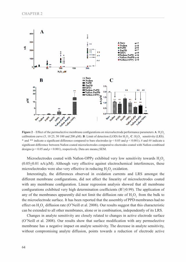
2.3.1.2 - H2O2 sensitivity performance
Following IUPAC guidelines (Thevenot et al. 1999), we have monitored changes in oxidation currents by consecutive additions of H2O2 (0 to 200 µM) in presence of all interfering species. We have found a reduction of the oxidation current of coated microelectrodes, when compared with bare microelectrodes. That reduction reached statistical significance at H2O2 levels above 50 µM for microelectrodes coated with any membrane configuration except PmPD alone (Fig 2-A). The oxidation currents of microelectrodes coated with PmPD were only significantly different for 200 µM of H2O2 (p ˂ 0.001). In contrast, microelectrodes coated with Nafion-OPPy had lower oxidation currents than any other membrane configuration, when exposed to any concentration of H2O2 (p < 0.05). Microelectrodes coated with a combination of Nafion and any electropolymerized membrane displayed lower oxidation currents than Nafion coated microelectrodes when exposed to 200 µM of H2O2 (p ˂ 0.001). The differences observed in oxidation currents resulted in significant differences in other analytical parameters, such as limit of detection (LOD) and linear range slope (LRS) (Fig 2-B and C). All microelectrodes, except those coated with Nafion-OPPy, had low LOD (≤ 3 µM) for H2O2. The LOD of Nafion-OPPy coated microelectrodes was more than 20-fold higher when compared to bare microelectrodes (0.11+0.02 vs 24.33+7.48 µM; p ˂ 0.001), and can be attributed to the combination of high noise levels and low LRS. The application of any type of membrane on microelectrode surface had a major effect on H2O2 LRS. Bare microelectrodes had higher LRS than microelectrodes coated with any membrane configuration (1.95+0.08 nA/µM vs. all; p ˂ 0.05 and p ˂ 0.001). Microelectrodes coated with PmPD displayed the highest LRS of coated microelectrodes (1.37+0.12 nA/µM). Additionally, we have found that the LRS of microelectrodes coated with Nafion in combination with any of the other membranes was lower than microelectrodes coated with Nafion alone (1.08+0.07 nA/µM vs. all p ˂ 0.001). The combination of Nafion with any electropolymerized membrane resulted in a decrease in oxidation currents for both DA and AA but also for H2O2.
2
63
The role of surface availability in membrane-induced selectivity for amperometricenzyme-based biosensors
CHAPTER 2
64
Figure 2 – Effect of the permselective membrane configurations on microelectrode performance parameters A: H2O2 calibration curve (5, 10 25, 50 100 and 200 µM). B: Limit of detection (LOD) for H2O2. C: H2O2 sensitivity (LRS). * and ** indicate a significant difference compared to bare electrodes (p ˂ 0.05 and p ˂ 0.001); # and ## indicate a significant difference between Nafion coated microelectrodes compared to electrodes coated with Nafion-combined designs (p ˂ 0.05 and p ˂ 0.001), respectively. Data are means+SEM.
Microelectrodes coated with Nafion-OPPy exhibited very low sensitivity towards H2O2 (0.05+0.01 nA/µM). Although very effective against electrochemical interferences, these microelectrodes were also very effective in reducing H2O2 oxidation. Interestingly, the differences observed in oxidation currents and LRS amongst the different membrane configurations, did not affect the linearity of microelectrodes coated with any membrane configuration. Linear regression analysis showed that all membrane configurations exhibited very high determination coefficients (R2≥0.99). The application of any of the membranes apparently did not limit the diffusion rate of H2O2 from the bulk to the microelectrode surface. It has been reported that the assembly of PPD membranes had no effect on H2O2 diffusion rate (O’Neill et al. 2008). Our results suggest that this characteristic can be extended to all other membranes, alone or in combination, independently of its LRS. Changes in analyte sensitivity are closely related to changes in active electrode surface (O’Neill et al. 2008). Our results show that surface modification with any permselective membrane has a negative impact on analyte sensitivity. The decrease in analyte sensitivity, without compromising analyte diffusion, points towards a reduction of electrode active

surface due to membrane assembly. The use of combined membranes results in even lower analyte sensitivities, probably due to an additional decrease in active electrode surface. The assembly of electropolymerized membranes depends on the access of the monomers to reach the electrode surface. The oxidation of the PPD and pyrrole at the electrode surface triggers the chain reaction responsible for the polymer formation. Microelectrodes coated with Nafion in combination with either PPD or OPPy produced lower oxidation currents for both major interfering species (AA and DA) and target analyte (H2O2) than Nafion coated microelectrodes. These results suggest that PPD and OPPy polymerize on the surface left available after the application of Nafion, thus reducing electrode active surface. The PPy polymerization on Nafion-coated microelectrodes drastically reduces the electrode active surface. Electropolymerization of PPD is self-limiting, in contrast to PPy. The polymer PPy is conductive and therefore keeps “growing” as long as current is applied to the electrode surface (Ramanavicius et al. 2005). Our data suggests that, instead of assembled in a classical LBL manner (Qin et al. 2008; Wahono et al. 2012), the electropolymerised membranes grow underneath the existing Nafion membrane. They may even displace Nafion from the electrode surface. Evaluation by scanning electron microscopy supported these findings (see section 3.2). Based on the presented results, we propose that the effectiveness of permselective membranes in reducing electrochemical interference is partially due to a significant and membrane dependent decrease in active surface of the electrode.
2.3.1.3 - Selectivity and Rejection Coefficients
To further investigate the putative role of surface availability on membrane selectivity we have calculated both selectivity (SC) and rejection coefficients (RC) for all membrane configurations and all electroactive species. SC specifies how much a given membrane-modified sensor responds to an interfering analyte relatively to the target analyte, and RC specifies how much a membrane is able to reduce the interference as compared to bare electrodes. Effective permselective membranes should have a low SC and high RC (O’Neill et al. 2008; O’Neill and Craig 2003).
2
65
The role of surface availability in membrane-induced selectivity for amperometricenzyme-based biosensors

CHAPTER 2
66
Figure 3-Selectivity Coefficient (SC) for the microelectrodes coated with various membrane configurations for the major non-specific electroactive species in the brain (Dopamine, 2 µM, DOPAC 20 µM; Uric acid 50 µM and Ascorbic acid, 250 µM). * and ** indicate a significant difference or difference compared to bare electrodes (p ˂ 0.05 and p ˂ 0.001). Data are mean+SEM.
Bare electrodes were slightly selective towards DOPAC and UA (SC ˂ 80%), but not selective towards AA (SC ~ 100%) or DA (SC ≥ 350%). The use any of the membrane configurations resulted in an decrease in SC, hence increase in selectivity, of all of the non-specific electroactive species, with the exception of DA (Fig 3). Despite the fact that all membranes effectively reduced DA oxidation currents, not all were selective for DA. Only microelectrodes coated with PmPD, OPPy and Nafion-PmPD displayed a significantly lower SC for DA when compared with bare electrodes (P< 0.001). The most selective membrane for DA was PmPD (SC ≤ 5%). These results are in line with a recent comparative study performed by Wahono et al. (Wahono et al. 2012) and confirm that PmPD is the most efficient membrane to eliminate electrochemical interference in the brain environment. The high selectivity of PPD has been assigned to a close and orderly packing of the macromolecular chain in PD films. While some authors favor PoPD (O’Neill and Craig 2003), our results support the conclusions from Wahono et al., favoring PmPD. Selectivity of OPPy has been attributed to the second step of the polymerization procedure, where a carbonyl group is introduced into the polymer backbone. Its high electron density seems to act as a barrier and repels both anions and cations (Wahono et al. 2012). Instead, the selective properties of

Nafion have been attributed to the negative charge of the self-assembled polymer (Gerhardt et al. 1984). Although effective in avoiding interference of negative species (AA, UA and DOPAC), negatively charged Nafion membranes may attract cations like DA (Wahono et al. 2012). Interestingly, we have found that combination of Nafion, with either PPD or PPy, resulted in microelectrodes less selective for DA (higher SC) when compared to electrodes coated with either P (m- or o-)PD or OPPy alone. These results are in line with earlier findings (Wahono et al. 2012), and suggest the existence of an different assembly, rather than a clasical”layer-by-layer” (LBL) assembly of combined membranes.
Figure 4 - Rejection Coefficients (RC) for the tested microelectrodes coated with permselective layers for major non -specific electroactive species in the brain (Dopamine, 2 µM, DOPAC 20 µM; Uric acid 50 µM and Ascorbic acid, 250 µM) and target analyte (H2O2, 50 µM). * and ** denotes a significant difference or highly significant difference between Nafion coated microelectrodes compared to electrodes coated with Nafion-combined designs (p ˂ 0.05 and p ˂ 0.001, respectively). Data are mean+SEM.
The calculated RCs (close to 100%) show a nearly complete rejection of DOPAC, UA and AA for microelectrodes coated with any membrane configuration (Figure 4 B, C and D). We haven’t found any differences in the RC for those non-specific electroactive species, for microelectrodes coated with any of the membrane configurations. However, we have found significant differences in the RC of dopamine. (Fig 4-A). As expected, microelectrodes coated with membranes selective against DA (PmPD, OPPy, Nafion-PmPD) displayed high RC, very close to 100%. Nonetheless, we have also observed significant RC (≥ 30%) for microelectrodes coated with membrane configurations
2
67
The role of surface availability in membrane-induced selectivity for amperometricenzyme-based biosensors

CHAPTER 2
68
non-selective against DA (Nafion, PoPD, Nafion- PoPD and Nafion-OPPy). Microelectrodes coated with Nafion-OPPy even had a nearly complete DA rejection (RC ~ 100%). Interestingly, microelectrodes coated with Nafion in combination with either PPD or OPPy had higher RC for DA (P˂ 0.001) when compared with microelectrodes coated only with Nafion. Membranes non-selective against DA (SC > 100%) were also effective in reducing oxidation current and had a rather high RC for DA. Although all tested membranes were reported as permselective (i.e. rejecting interference but not target analyte) we found that all coated microelectrodes had significant rejection of H2O2. All membranes configurations displayed an H2O2 RC between 40% and 80%, with the exception of microelectrodes coated with Nafion-OPPy, which had a nearly complete rejection of H2O2. Additionally, we have found that microelectrodes coated with Nafion in combination with either P(m- or o-)PD or OPPy had significantly higher RC of H2O2, than microelectrodes coated only with Nafion. These results might be explained by an additional coverage of the microelectrode surface, due to the polymerization of either P(m- or o-)PD or OPPy. These polymers would occupy the surface left available after Nafion assembly, reducing the access, but not diffusion of H2O2 to the electrode surface. This would explain the low H2O2 LRS displayed by microelectrodes coated with membrane combinations. Even though all membranes effectively reduced electrochemical interference, none was truly selective. Besides reducing oxidation current form interfering species, all membranes also reduced target analyte (H2O2) sensitivity. These results show that membrane assembly causes a reduction of available surface with implications on membrane selectivity. The role of surface availability in selectivity is membrane dependent, and more pronounced in membrane combinations. Membrane selectivity is, at some extent, defined by surface availability. Our results challenge the generally assumed LBL membrane assembly. An additional decrease in oxidation currents observed in membrane combinations of both target analyte and major interfering species points to a further reduction of the microelectrode active surface. This might be caused by an “inner polymerization” of either P(m- or o-)PD or OPPy following Nafion assembly.
2.3.1.4- Voltammetry evaluation
To further evaluate the impact of membrane assembly in surface availability we performed additional electrochemical characterization. Cyclic voltammetry has been widely used for reliable estimation of electrode active surface (Jarzabek and Borlowska 1997; Trasatti and Petriii 1991). Therefore, all microelectrodes configurations were submitted to cyclic voltammetry analysis in presence of a redox probe (Fe(CN)63-, at different scan rates. Our data revealed that the assembly of any membrane configuration (except OPPy) resulted in the shift from a classical reversible redox system to an irreversible system (∆Ep ≥ 0.059/n.V) (Fig S1 and S2). It seems that membrane assembly resulted in sluggish electron transfer kinetics(Wang 1994). Although the assembly of OPPy also resulted in a small shift

of both the anodic and cathodic peak potential (Ep,a and Ep,c), it was not as dramatic as for the other membrane configurations. The residual charge of these electrodes (supported by the high noise and background current observed) may explain the behavior of OPPy coated microelectrodes. Based on the data obtained throughout voltammetry characterizations, we have plotted the peak current (ip) vs square root of the scan rate (v1/2) (Fig S3). The use of the Randles-Sevcik model allowed us to estimate electrode active surface for each microelectrode configuration (Table 1)
Table 1- Analytical Parameters estimated based on an Ip (A) vs v1/2 (V/s) plot, using the Randles-Sevcik models (Eq. 3 and Eq. 4).
Bare Nafion PoPD PmPD OPPy Nafion-PoPDNafion-PmPD
Nafion-OPPy
Fe(C
N) 63-
Slope 1.05x10⁻⁴ 3.19x10⁻⁶ 2.71x10⁻⁵ 4.77x10⁻⁵ 1.99x10⁻⁵ 6.3x10⁻⁷ 4.7x10⁻⁷ 5.0x10⁻⁸
R2 0.999 0.997 0.999 0.998 0.968 0.999 0.936 0.975
Active Surface (cm2) 6.49x10⁻³ 1.73x10⁻⁴ 1.47x10⁻³ 2.87x10⁻³ 7.22x10⁻⁴ 3.82x10⁻⁵ 2.89x10⁻⁵ 3.45x10⁻⁶
H2O
2
Slope 2.88x10⁻⁶ 1.83x10⁻⁶ 1.74x10⁻⁶ 2.60x10⁻⁶ 2.10x10⁻⁶ 1.10x10⁻⁶ 1.48x10⁻⁶ 8.57x10⁻⁷
R2 0.998 0.988 0.992 0.995 0.994 0.991 0.990 0.996
Active Surface (cm2) 6.54x10⁻³ 4.18x10⁻³ 3.97x10⁻³ 5.89x10⁻³ 4.69x10⁻³ 2.50x10⁻³ 3.08x10⁻³ 1.95x10⁻³
Roughness Factor* -- 0.63 0.60 0.90 0.72 0.38 0.46 0.29
H2O2LRS per Active Surface- µA/cm2
Mean SEM Mean SEM Mean SEM Mean SEM Mean SEM Mean SEM Mean SEM Mean SEM
0.30 0.01 0.25 0.02 0.22 0.04 0.23 0.02 0.21 0.03 0.24 0.03 0.25 0.02 0.02 0.01
*- Geometrical area of the microelectrodes is 0.006459 cm2.
While the surface of bare electrodes was similar to its geometrical area (0.0064 cm2), microelectrodes coated with any permselective membranes displayed an unexpected, much lower active surface. The larger molecular weight of ferricyaninde, when compared to H2O2, thus higher steric hindrance, may be lead to a decrease in analyte diffusion leading to an underestimation of the real active surface. This underestimations seems to be largely enhanced in Nafion coated microelectrodes. The extremely low active surfaces for Nafion coated electrodes maybe attributed to the charged nature of Nation. The negative charge of the membrane is likely to function as an additional diffusion barrier, in mechanism similar to the reduction of electrochemical interference. It has been described that, in defined conditions, Nafion can act as an ion diffusion barrier. It seems that for Nafion coated microelectrodes, the diffusion coefficient of the ferricyanide is probably much lower than expected. Nevertheless microelectrodes coated
2
69
The role of surface availability in membrane-induced selectivity for amperometricenzyme-based biosensors

with membrane combinations have a lower electrode active surface than any membrane configuration assemble alone. This observation suggests, once again, that the decrease in oxidation current for both target analyte and electrochemical interference observed in membrane combinations may be due to an additional reduction in surface availability. Due to the unexpected behavior of membrane coated microelectrodes in presence of ferricyanide, we also performed cyclic voltammetry in presence of H2O2 (Miao et al. 2014; Song et al. 2010). Our data (Fig S4 and S5) show that unlike observed for ferrycianinde, there was no significant difference in the Ep,a between bare and coated microelectrodes, in presence of H2O2. Once again we have used the Randle-Sevcik model to estimate the active surfaces for each microelectrode configurations, based on the Ip vs v1/2 plot (Fig S6). We did not observe differences in the estimated active surface of bare electrodes estimated in presence of either ferricyanide or H2O2. However, we found differences in the estimated areas of coated microelectrodes, using the two redox probes Active surfaces for coated microelectrodes estimated in presence of H2O2 were higher when compared with estimations made in presence of ferricianide. These differences were largely enhanced (up to 103 folds) in the case of Nafion coated membranes. All membrane coated microelectrodes displayed lower active surfaces when compared to bare microelectrodes. Microelectrodes coated with PmPD displayed the highest active surfaces. Interestingly, microelectrodes coated with membrane combinations had lower active surfaces when compared with microelectrodes coated with a single membrane. Finally we calculated the LRS for H2O2, normalized for microelectrode active surface. Our results show that all coated microelectrodes display lower sensitivity than bare electrodes. This suggests a decrease in analyte diffusion for coated microelectrodes. However we didn’t observe any significant differences in the normalized LRS amongst coated microelectrodes, except for Nafion-OPPy coated microelectrodes. These data clearly shows that the membrane-dependent decrease in sensitivity is closely related to an effective decrease in electrode active surface. In all, the additional electrochemical evaluation supports the hypothesis that membrane assembly results in a membrane-dependent decrease in surface availability, enhanced in the case of membrane combinations. Additionally, our results suggests that the use of classical redox probes for estimation of electrode active surfaces in coated microelectrodes may not be trivial. Since all implantable biosensors are prone to biofouling (Wisniewski and Reichert 2000), we pretend to evaluate in the near future the effects of biofouling on surface dependent membrane-induce selectivity.
2.3.2- Evaluation of the surface by scanning electron microscopy
To study membrane morphology and its influence on microelectrode surface availability,
CHAPTER 2
70

we have evaluated microelectrode surfaces using scanning electron microscopy. Coated microelectrodes were evaluated intact and in cross-section in order to examine layer thickness. The electron microscopy images revealed a complete coverage of the electrode by all membrane configurations (Fig 5). However, there were significant membrane-dependent differences in surface morphology. Microelectrodes coated with Nafion (Fig 5A) had a smooth surface, with a layer thickness of 3.1 µm. The thickness of Nafion membranes assembled on electrode surfaces is reported to be in μm range (Hashemi et al. 2011; Jusoh et al. 2012; Sotomayor et al. 2002). However, these dimensions may vary, depending on polymerization and curing procedures. Our Nafion membranes were much thicker than any of other electropolymerized layers (PmPD, PoPD and OPPy) (3.1 vs 0.06; 0.08 and 1.8 µm, respectively).
2
71
The role of surface availability in membrane-induced selectivity for amperometricenzyme-based biosensors

CHAPTER 2
72
Figure 5 – Scanning electron microscopy of a cross section of microelectrodes coated with various permselective membrane configurations at magnified 250 x. The thickness of each membrane configuration is displayed in the top right of each figure. Insert- Detail of the cross-section using higher magnification (10000 x)

Both PPD membranes( m- and o-) were thinner than OPPy (0.06 and 0.08 vs. 1.8 µM) but comparable between each other and in line with values (˂ 0.1 µm) reported (Osborne and Hashimoto, 2004)(O’Neill and Craig 2003). We observed morphological differences in the surface of microelectrodes coated with either PoPD or PmPD. Electrodes coated with PoPD (Fig 5B) membranes had a darker contrast than PmPD (Fig 5C) coated microelectrodes. This darker coloration might be due to a more compact and more ordered polymer, allowing less penetration and less reflection of the emitted electrons. PoPD coated electrodes exhibit lower selectivity and rejection coefficients than PmPD membranes. Our results suggest that PmPD membranes are more structured than PoPD membranes. These features might explain the better selectivity, with less impact on the active electrode surface. Microelectrodes coated with OPPy (Fig 5D) exhibited a very rough, relatively thick (1.8 µm) and irregular surface. The roughness of OPPy membranes has been previously reported and could be observed at naked eye (Wahono et al. 2012; Walker et al. 2007). However, a more detailed observation (10.000 x) revealed a well-organized “fishnet-like” structure (Figure 5D, insert). According to the literature the selectivity of OPPy is due to size exclusion caused by the formation of closed channels implicating that analytes can only pass/migrate by diffusion (Farrington and Slater 1997). Our observations do not support that hypothesis. The “fishnet-like” structure of the outer portion of OPPy is unlikely to act as an effective barrier. That distinctive structure lies on top of a much thicker and denser inner layer that covers almost the entire electrode surface. We assume that the inner layer, rather than the “fishnet-like” structure, is the one responsible for the selective properties of OPPy. Unexpectedly, Nafion-OPPy coated electrodes did not have the typical “fishnet-like “structure observed in OPPy coated microelectrodes. Instead, Nafion-OPPy coated electrodes exhibited a generally homogeneous and smooth surface, interrupted by “irregular holes” (Fig 5G). A closer inspection revealed that those “holes” are actually covered with another material (presumably OPPy), creating “irregular depressions” rather than “holes”. Cross-section evaluation exposed two different layers, embedded on each other. Although the Nafion membrane has been assembled prior to OPPy polymerization, the later has apparently polymerized underneath the already existing membrane. We hypothesize that OPPy membranes occupy the surface left available after Nafion assembly. The polymerization of OPPy underneath Nafion reduced drastically the electrode active surface. This phenomenon explains the low oxidation currents for all tested analytes (including H2O2) of microelectrodes coated with Nafion-OPPy. Similar to OPPy, PPD membranes also polymerized underneath the early assembled Nafion membrane. The surface of microelectrodes coated with either Nafion-PoPD or Nafion-PmPD was smooth and mostly covered with Nafion. However, on some parts of the surface either PoPD or PmPD was visible, suggesting a possible displacement of Nafion by PPD polymerization. The thickness of combined membranes of Nafion with either PoPD (2.8 µm), PmPD (2.0 µm) or OPPy (3.5 µm) was not different from the thickness of Nafion membrane alone (3.1 µm), and is much thicker than any of the single PPD layers. The inner
2
73
The role of surface availability in membrane-induced selectivity for amperometricenzyme-based biosensors

CHAPTER 2
74
polymerization implies that Nafion is on the outside, even in combination with either PPD or OPPy. Nafion membranes are negatively charged, able to attract cationic species like DA. The inner polymerization phenomena, exposed by scanning electron microscopy, can explain the lower selectivity of combined membranes towards DA. Besides attracting DA from the bulk to the surface, Nafion can conceal the selective properties of PPD and OPPy, since most of the electrode surface is covered with the charged membrane. Moreover, inner polymerization most likely affects the conformational structure of the later assembled membrane, hence its performance. Additionally, our scanning electron microscope evaluation questions the classical layer-by-layer model that has been described for such membrane combinations, in which the second polymerized membrane is supposed to be localized “on top” of the first membrane (Wahono et al. 2012). The suggested “inner polymerization” phenomenon has not been previously reported. Electropolymerization reactions are self-limiting and largely dependent on the electrode surface. Inner polymerization implies a significant reduction in active electrode surface. Moreover, it reveals that surface availability plays an important role in membrane selectivity, especially in membrane combinations, whose active surface is much lower when compared with single membranes. Although membrane thickness seemed to be membrane dependent, we haven’t observed a correlation between thickness and H2O2 sensitivity (Fig S7).
2.4- Conclusion
All membrane configurations evaluated were very effective in reducing electrochemical interference. Assembly of any permselective membrane configuration reduced the oxidation currents of all tested non-specific electroactive species. Microelectrodes coated with membrane combinations were more effective in reducing the interference by the major non-specific electroactive species: AA and DA. However, only PmPD (alone or in combination with Nafion) and OPPy were selective against all non- specific electroactive species. Nevertheless, all membranes both reduced the sensitivity and effectively rejected H2O2. Interestingly, membrane combinations displayed both higher RC and lower sensitivity for H2O2 than single membrane configurations. Additional electrochemical evaluation revealed that the decrease in analyte sensitivity was related to a decrease in electrode active surface. The assembly of any membrane configuration has a negative impact on surface availability. This effect is membrane dependent, enhanced in membrane combinations, and has an active role in membrane-induced selectivity. Although more efficient in reducing electrochemical interference, membrane combinations do not increase selectivity. Instead, the additional reduction in electrochemical interference is due to an additional decrease in the active electrode surface, caused by a previously unknown “inner polymerization” phenomenon.

Taken together, the presented evidence point to an unprecedented role of surface availability in membrane-induced selectivity.
2
75
The role of surface availability in membrane-induced selectivity for amperometricenzyme-based biosensors

CHAPTER 2
76
2.5- BibliographyJ. Castillo, S. Gáspár, S. Leth, M. Niculescu, A. Mortari, I. Bontidean, et al., Biosensors for life quality: Design, development and applications, Sensors And Actuators B:Chemical, 102(2004) 179–94.
G.S. Wilson, R. Gifford, Biosensors for real-time in vivo measurements, Biosensors and Bioelectronics, 20(2005)2388–403.
D. Arruda, W.C. Wilson, C. Nguyen, Q. Yao, R.J. Caiazzo Jr, I. Talpasanu, et al., Microelectrical sensors as emerging platforms for protein biomarker detection in point-of-care diagnostics, Expert Rev Mol Diagn, 9(2009)749–55.
J. Wang, Glucose Biosensors: 40 Years of Advances and Challenges, Electroanalysis 13(2000) 983-8.
J. Burmeister, F. Pomerleau, M. Palmer, B.K. Day, P. Huettl, G. Gerhardt, Improved ceramic-based multisite microelectrode for rapid measurements of L-glutamate in the CNS, Journal of Neuroscience Methods, 119(2002) 163-71.
M. Fillenz, In vivo neurochemical monitoring and the study of behaviour, neubiorev, 29(2005) 949–62.
N.D. Kulagina, S. Latha , A.C. Michael, Monitoring Glutamate and Ascorbate in the Extracellular Space of BrainTissue with Electrochemical Microsensors, Analytical Chemistry, 71(1999) 5093-100.
J.P. Lowry, M. Demestre, M. Fillenz, Relation between Cerebral Blood Flow and Extracellular Glucose in Rat Striatum during Mild Hypoxia and Hyperoxia, Dev Neurosci, 20(1998) 52-8.
F. Palmisano, R. Rizzi, D. Centonze, P.G. Zambonin, Simultaneous monitoring of glucose and lactate by an interference and cross-talk free dual electrode amperometric biosensor based on electropolymerized thin film, Biosensors and Bioelectronics, 15(2000) 531–9.
N. Vasylieva, B. Barnych, A. Meiller, C. Maucler, L. Pollegioni, J.-S. Lina, et al., Covalent enzyme immobilization by poly(ethylene glycol) diglycidyl ether (PEGDE) for microelectrode biosensor preparation, Biosensors and Bioelectronics, 26(2011) 3993–4000.
C.P. McMahon, G. Rocchitta, S.M. Kirwan, S.G. Killoran, P.A. Serra, J.P. Lowry, et al., Oxygen tolerance of an implantable polymer/enzyme composite glutamate biosensor displaying polycation-enhanced substrate sensitivity, Biosensors and Bioelectronics, 22(2007) 1466–73.
O.M. Schuvailo, O.O. Soldatkin, A. Lefebvre, R. Cespuglioa, A.P. Soldatkin, Highly selective microbiosensors for in vivo measurement of glucose, lactate and glutamate, Analytica Chimica Acta 573–574(2006) 110-6.
M. Campàs, B. Prieto-Simónc, J.-L. Marty, A review of the use of genetically engineered enzymes in electrochemical biosensors, Seminars in Cell & Developmental Biology, 20(2009) 3–9.
R. Fogel, J.L. Limson, Probing fundamental film parameters of immobilized enzymes—Towards enhanced biosensor performance. Part I—QCM-D mass and rheological measurements, Enzyme and Microbial Technology, 49(2011) 146– 52.
[R.D. O’Neill, J.P. Lowry, G. Rocchitta, C.P. McMahon, P.A. Serra, Designing sensitive and selective polymer/enzyme composite biosensors for brain monitoring in vivo, Trends, 27(2008) 78-88.
N. Wahono, S. Qin, P. Oomen, T.I.F. Cremers, M.G. de Vries, B.H.C. Westerink, Evaluation of permselective membranes for optimization of intracerebral amperometric glutamate biosensors, Biosensors and Bioelectronics, 33 (2012) 260– 6.G.A. Gerhardt, A.F. Oke, G. Nagy, B. Moghadda, R.N. Adams, Nation-coated electrodes with high selectivity forCNS electrochemistry, Brain Research, 290(1984).
W.H. Oldenziel, G. Dijkstra, T.I.F.H. Cremers, B.H.C. Westerink, Evaluation of Hydrogel-Coated Glutamate

Microsensors, Analytical Chemistry, 78(2006) 3366-78.
J.P. Lowry, M. Miele, R.D. O’Neill, M.G. Boutelle, M. Fillenz, An amperometric glucose-oxidase/poly(o- phenylenediamine) biosensor for monitoring brain extracellular glucose: in vivo characterisation in the striatum of freely-moving rats., Journal Of Neuroscience Methods, 79(1998) 65-74.
D. Moatti-Sirat, V. Poitout, V. Thom, M.N. Gangnerau, Y. Zhang , Y. Hu, et al., Reduction of acetaminophen interference in glucose sensors by a composite Nafion membrane: demonstration in rats and man, Diabetologia,37(1994) 610-6.
R.D. O’Neill, J.D. Craig, Comparison of simple aromatic amines for electrosynthesis of permselective polymersin biosensor fabrication, The Analyst, 128(2003) 905–11.
E. Walker, J. Wang, N. Hamdi, H.G. . Monbouquetteb, N.T. Maidment, Selective detection of extracellular glutamate in brain tissue using microelectrode arrays coated with over-oxidized polypyrrole, The Analyst,132(2007) 1107-11.
R. Maalouf, H. Chebib, S. Y., O. Vittori, M. Sigaud, N. Jaffrezic-Renault, Amperometric and impedimetric characterization of a glutamate biosensor based on Nafion® and a methyl viologen modified glassy carbon electrode, Biosensors and Bioelectronics, 22(2007) 2682–8.
B.M. Dixon, J.P. Lowry, R.D. O’Neill, Characterization in vitro and in vivo of the oxygen dependence of an enzyme/polymer biosensor for monitoring brain glucose, Journal Of Neuroscience Methods, 119(2002) 135-42.
H. Yang, T.D. Chung, C.K. Choi, T.Y. Kim, C.H. Jun, H.C. Kim, Glucose sensor using a microfabricated electrode and electropolymerized bilayer films, Biosensors and Bioelectronics, 17(2002) 251-9.
B.U. Moon, M.G. De Vries, C.A. Cordeiro, B.H.C. Westerink, E. Verpoorte, Microdialysis-Coupled Enzymatic Microreactor for in Vivo Glucose Monitoring in Rats, Analytical Chemistry, 85(2013) 10949−55.
R.M. Santos, L. C.F., A.P. Piedade, R. Andrews, F. Pomerleau, P. Huettl, et al., A comparative study of carbon fiber-based microelectrodes for the measurement of nitric oxide in brain tissue, Biosensors and Bioelectronics, 24 (2008) 704–9.
S. Rothwell, S.J. Killoran, R.D. O’Neill, Enzyme immobilization strategies and electropolimerization conditions to control sensitivity and selectivity parameters of a polymer-enzyme composite glucose biosensor, Sensors,10(2010) 6439-62.
Madaras M., R.P. Buck, Miniaturized Biosensors Employing Electropolymerized Permselective Films and TheirUse for Creatinine Assays in Human Serum, Analytical Chemistry, 68(1996) 3832-9.
D. Thevenot, K. Toth, R. Durst, G.S. Wilson, Electrochemical Biosensors: Recomended definitions and classification, Pure and Applied Chemistry, 71(1999) 2333-48.
A. Ramanavicius, A. Ramanaviciene, A. Malinauskas, Electrochemical sensors based on conducting polymer-polypyrrole, Electrochimica Acta, 51(2005) 6025-37.
S. Qin, M. van de Zeyden, W.H. Oldenziel, T.I.F.H. Cremers, B.H.C. Westerink, Microsensors for in vivo Measurement of Glutamate in Brain Tissue, Sensors, 8(2008) 6860-84.
S. Trasatti, O.A. Petriii, Real Surface Area Measurements in Electrochemistry, Pure and Applied Chemistry, 63(1991) 711-34.
G. Jarzabek, Z. Borlowska, On the real surface area of smooth solid electrodes, Electrochimica Acta, 42(1997). J. Wang, Analytical Electrochemistry: VCH Publishers Inc. ; 1994.
M-J. Song, S.W. Hwang, D. Whang, Amperometric hydrogen peroxide biosensor based on a modified gold electrode with silver nanowires, Journal of Applied Electrochemistry, 40(2010) 2099-105.
2
77
The role of surface availability in membrane-induced selectivity for amperometricenzyme-based biosensors

CHAPTER 2
78
Z. Miao, D. Zhang, Q. Chen, Non-enzymatic hydrogen peroxide sensors based on multi-wall carbon nanotube/Pt nanoparticles nanohybrids, Materials, 7(2014) 2945-55.
N. Wisniewski, W.M. Reichert, Methods for reducing biosensor membrane biofouling, Colloids and Surfaces B- Biointerfaces, 18(2000) 197-219.
N. Jusoh, A. Abdul-Aziz, E. Supriyanto, Improvement of Glucose Biosensor Performances Using Poly(hydroxyethyl methacrylate) Outer Membrane, International Journal of Biology and Biomedical Engineering, 6(2012) 77-86.
M.P.T. Sotomayor, A. Tanaka, L. ´Kubota, Development of an amperometric sensor for phenol compounds using a Nafion membrane doped with copper dipyridyl complex as a biomimetic catalyst, Journal of Electroanalytical Chemistry, 536(2002) 71-81.
P. Hashemi, P.L. Walsh, T.S. Guillot, J. Gras-Najjar, P. Takmakov, F.T. Crews, et al., Chronically Implanted, Nafion-Coated Ag/AgCl Reference Electrodes for Neurochemical Applications, ACS Chemical Neuroscience,2(2011) 658–66.
A. Farrington, J. Slater, Prediction and Characterization of the Charge/size Exclusion Properties of Over- Oxidized
Poly(pyrro1e) Films, Electroanalysis, 9(1997) 843-7.

2.6- Supplementary Material
2.6.1-Membrane assembly
Nafion assembly: Nafion was applied manually by dipping the electrode tip for 10 s followed by drying at room temperature for 20 s. This procedure was repeated 10 times. The electrodes were annealed at 175 ºC for 4 min (Wahono et al. 2012c). PoPD and PmPD: PPD was applied by cyclic voltammetry (CV) scan technique. For the electroplymerization 5 mM of the monomer (o- or m-) in 100 mM PBS pH 7.4 was used. Electroploymerization was carried out by scanning the potential from +200 to +700 mV with a scan rate of 20 mV/s over 200 cycles (Cordeiro et al. 2014; Wahono et al. 2012c). Overoxidized polypyrrole (OPPy): Polymerization of PPy was carried out with 200 mM pyrrole in 100 mM PBS at pH 7.4 by holding the potential at 850 mV with reference to a Ag/AgCl electrode for 300 s. OPPy was overoxidized at a constant potential at 850 mV vs. Ag/AgCl in 100 mM PBS at pH 7.4 during 6 h, until the steady-state current was recorded (Wahono et al. 2012c; Walker et al. 2007). Combined membranes: We have coated Nafion modified electrodes with an additional electropolymerized layer, either PoPD, PmPD or OPPy. Electropolymerization procedures applied were similar to those described for bare electrodes.
2.6.2-Amperometry steady state parameters
Table 1- Steady state analytical parameters for bare and coated microelectrodes. * and ** Indicate significantly different when compared to bare electrodes (p˂0.001 and p˂0.05). Data are expressed as mean±SEM.
Noise levels (nA) Baseline (nA)
Design Mean SEM Mean SEM
Bare (n=7) 0.08 0.02 4.52 0.41
Nafion (n=8) 0.06 0.05 4.42 0.70
PoPD(n=9) 0.17 0.09 4.84 0.63
PmPD (n=7) 0.08 0.01 7.43 0.47
OPPy (n=9) 0.67 ** 0.23 54.50** 5.67
Nafion-PoPD (n=8) 0.08 0.06 3.14 0.57
Nafion-PmPD (n=8) 0.08 0.02 6.13 0.71
Nafion- OPPy (n=9) 0.57** 0.26 12.24** 1.36
2
79
The role of surface availability in membrane-induced selectivity for amperometricenzyme-based biosensors

CHAPTER 2
80
2.6.3-Voltammetry evaluation
Voltametry evaluation was performed using two different probes, the classical FeCn and the target analyte for amperometric enzyme based biosensors, H2O2.
2.6.3.1- Ferricynide
All microelectrodes were submitted to cyclic voltammetry in presence of 50 mM of redox probe (Fe(CN)6
3- ) at different scan rates (10 to 300 mV/s).
Fig S1- Typical voltamograms for bare and coated microelectrodes in presence of 50 mM of Fe(CN6)3- scanned at 50 mV/s.
Fig S2- Typical voltamograms for Nafion coated (alone or in combination) microelectrodes in presence of 50 mM of
Fe(CN6)3- scanned at 50 mV/s.
Based on the data obtained from the voltamograms, we plotted Ip (A) vs v1/2 (V/s).

Fig S3- Ip vs v1/2 plot for all microelectrode configurations, based on the voltamograms performed in presence of Fe(CN)6
3- (50 mM) at increasing scan rates (10 to 300 mV/s).
The slope of the Ip vs v1/2 plot was used to estimate the electrode active surface, based on the Randles- Sevcik model.
2.6.3.2- Hydrogen Peroxide
Besides the voltammetric evaluation in presence of the classical redox probe, ferricyanide, we also performed a series of cyclic voltammetry experiments in presence of the target electroactive specie in amperometric enzyme-based biosensors, H2O2.
2
81
The role of surface availability in membrane-induced selectivity for amperometricenzyme-based biosensors

Fig S4- Typical voltamograms for bare and coated microelectrodes in presence of 100 µM of H2O2 scanned at 50 mV/s.
Fig S5- Typical voltamograms for Nafion coated microelectrodes (alone or in combination) in presence of 100 µM of H2O2 scanned at 50 mV/s.
Based on the data obtained from the voltamograms, we plotted Ip (A) vs v1/2 (V/s).
CHAPTER 2
82

Fig S6- Ip vs v1/2 plot for all microelectrode configurations, based on the voltamograms performed in presence of H2O2 (100 µM) at increasing scan rates (10 to 300 mV/s).
The slope of the Ip vs v1/2 plot was used to estimate the electrode active surface, based on the Randles-Sevcik model.
2.6.4-Influence of membrane thickness on LRS
In order whether membrane thickness influenced the sensitivity of the different microelectrode configuration, we have plotted the sensitivity as a function of each membrane thickness (assuming a thickness of 0 for bare electrodes).
Fig S6- Sensitivity of each microelectrode (LRS to H202, nA/µM) configuration plotted as a function of its membrane
83
The role of surface availability in membrane-induced selectivity for amperometricenzyme-based biosensors
thickness (µM).
The low correlation coefficient (0.54) denotes an independency of the LRS from its membrane thickness.
2.6.5-Bibliography
N. Wahono, S. Qin, P. Oomen, T.I.F. Cremers, M.G. de Vries, B.H.C. Westerink, Evaluation of permselective membranes for optimization of intracerebral amperometric glutamate biosensors, Biosensor and Bioelectronics,33 (2012) 260– 6.
C.A. Cordeiro, M.G. de Vries, N. Wahono, P.E. Oomen, T.I.F.H. Cremers, B.H.C. Westerink, In vivo continuous and simultaneous monitoring of brain energy substrates with a multiplex amperometric enzyme-based biosensor device, Biosensors & Bioelectronics, (2014).
E. Walker, J. Wang, N. Hamdi, H.G. . Monbouquetteb, N.T. Maidment, Selective detection of extracellular glutamate in brain tissue using microelectrode arrays coated with over-oxidized polypyrrole, The Analyst, 132(2007) 1107-11.
84
CHAPTER 2