University of Cyprus Biomedical Imaging and … · University of Cyprus Biomedical Imaging and...
38
University of Cyprus Biomedical Imaging and Applied Optics Biomedical Imaging and Applied Optics Endoscopic Imaging Endoscopic Imaging
-
Upload
vuongthien -
Category
Documents
-
view
233 -
download
1
Transcript of University of Cyprus Biomedical Imaging and … · University of Cyprus Biomedical Imaging and...

University of CyprusBiomedical Imaging and Applied OpticsBiomedical Imaging and Applied Optics
Endoscopic ImagingEndoscopic Imaging
History of Endoscopy
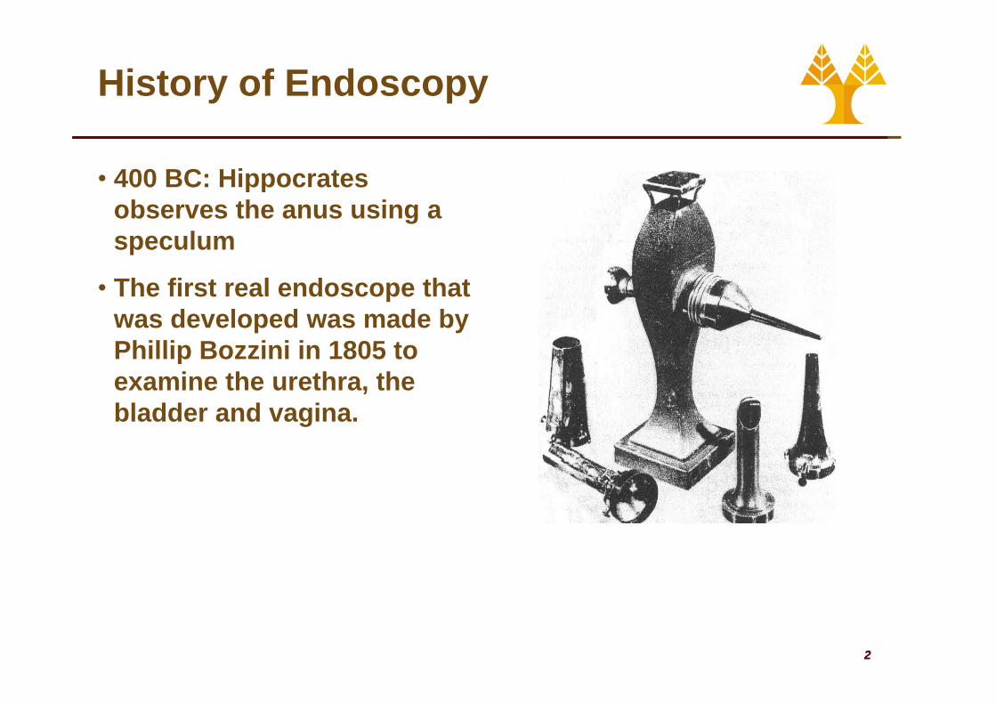
• 400 BC: Hippocrates observes the anus using a speculum
• The first real endoscope that was developed was made by Phillip Bozzini in 1805 toPhillip Bozzini in 1805 to examine the urethra, the bladder and vagina.
22
History of Endoscopy
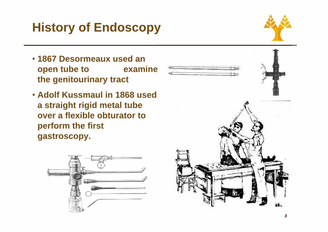
• 1867 Desormeaux used an open tube to examine the genitourinary tract
• Adolf Kussmaul in 1868 used a straight rigid metal tube over a flexible obturator toover a flexible obturator to perform the first gastroscopy.
33
History of Endoscopy
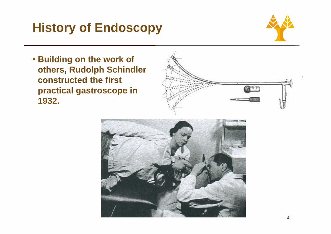
• Building on the work of gothers, Rudolph Schindler constructed the first practical gastroscope inpractical gastroscope in 1932.
44
History of Endoscopy
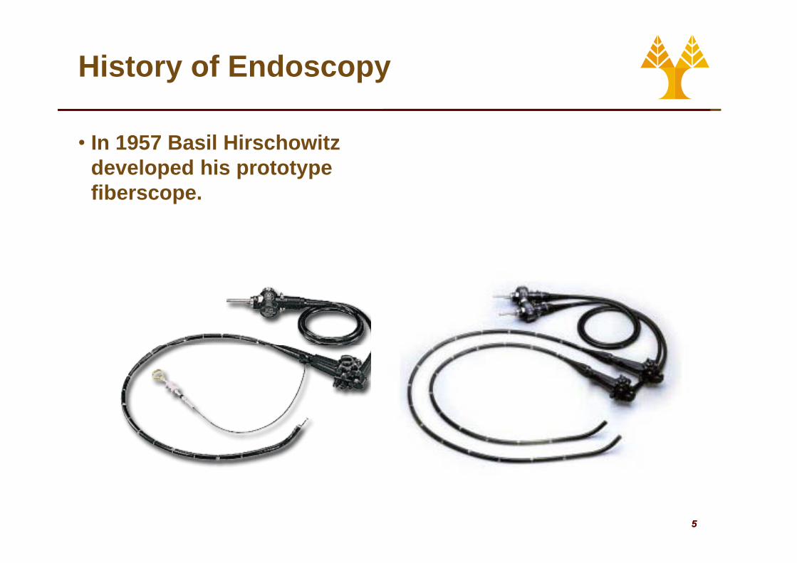
• In 1957 Basil Hirschowitz developed his prototype fiberscope.
55
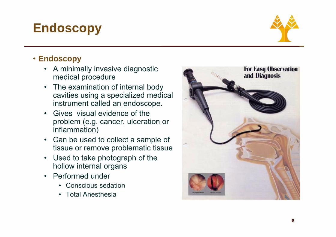
Endoscopy
• Endoscopy• A minimally invasive diagnostic
medical procedure• The examination of internal body
cavities using a specialized medical instrument called an endoscope.
• Gives visual evidence of the (problem (e.g. cancer, ulceration or
inflammation)• Can be used to collect a sample of
ti bl ti titissue or remove problematic tissue• Used to take photograph of the
hollow internal organs• Performed under
• Conscious sedation• Total Anesthesia
66

Endoscopy
• Physicians use endoscopy to diagnose, monitor, and surgically treat various medical problems
• A surgeon introduces theA surgeon introduces the endoscope into the body either through a body opening, such as the mouth or the anus or throughthe mouth or the anus, or through a small incision in the skin.
77
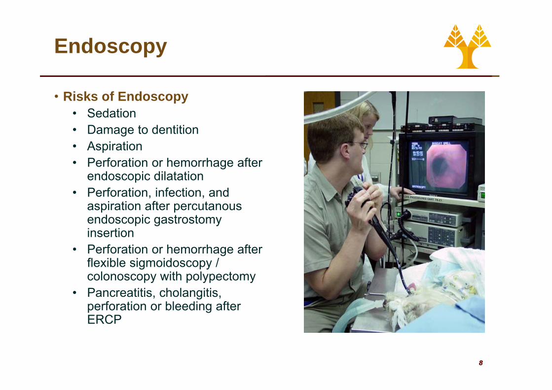
Endoscopy
• Risks of Endoscopy• Sedation• Damage to dentition• Aspirationp• Perforation or hemorrhage after
endoscopic dilatation• Perforation infection andPerforation, infection, and
aspiration after percutanousendoscopic gastrostomyinsertion
• Perforation or hemorrhage after flexible sigmoidoscopy / colonoscopy with polypectomy
• Pancreatitis, cholangitis, perforation or bleeding after ERCP
88
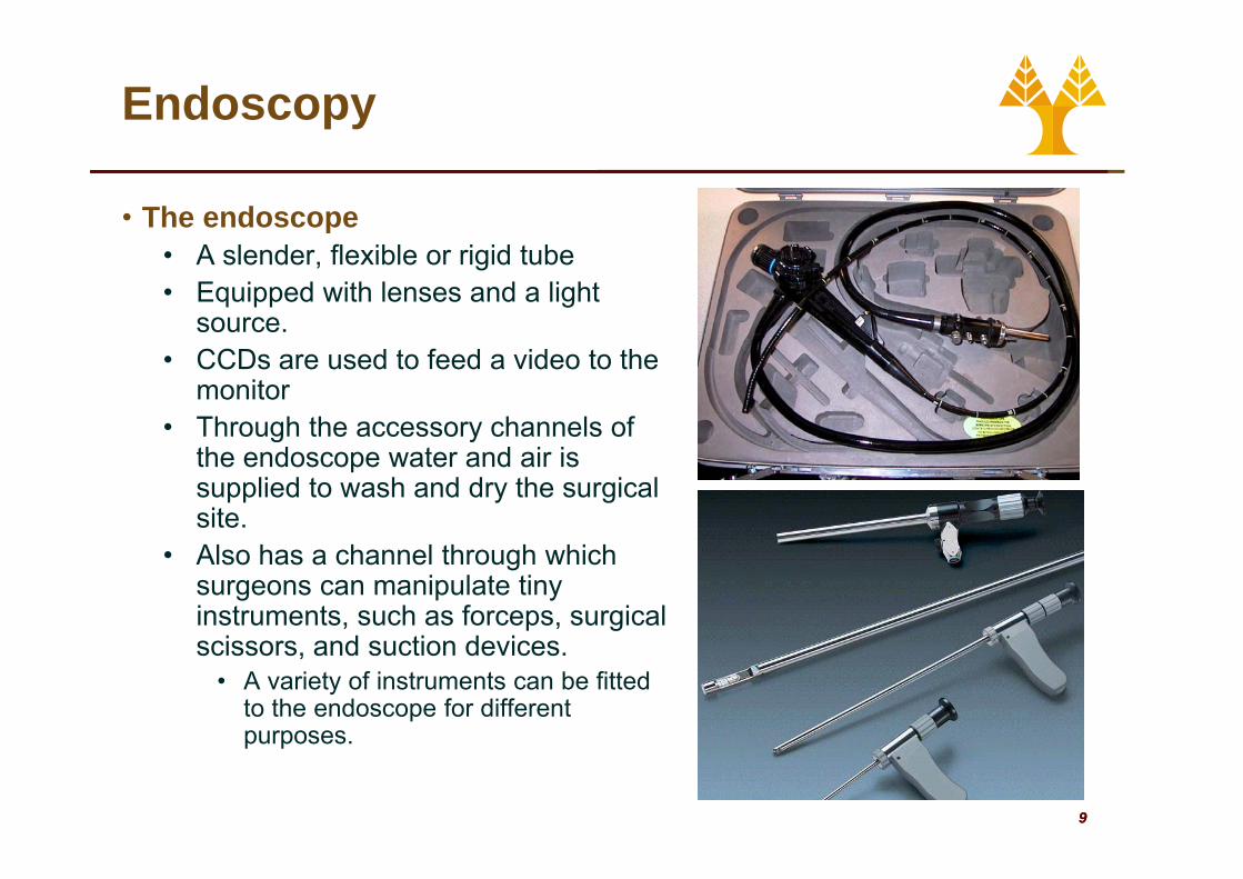
Endoscopy
• The endoscope • A slender, flexible or rigid tube• Equipped with lenses and a light
source. • CCDs are used to feed a video to the
monitor• Through the accessory channels of g y
the endoscope water and air is supplied to wash and dry the surgical site.
• Also has a channel through which surgeons can manipulate tiny instruments, such as forceps, surgical
i d ti d iscissors, and suction devices.• A variety of instruments can be fitted
to the endoscope for different purposes
99
purposes.

The Flexible Endoscope
• Fiberoptic instrumentsB d ti l i i b dl• Based on optical viewing bundles
• 2–3·mm in diameter and contains 20·000–40·000 fine glass fibers, each close to 10·μm in diameterclose to 10 μm in diameter
• Each individual glass fiber is coated with glass of a lower optical density to prevent leakage of light from within the fiber
• The space between the fibers causes a dark ‘packing fraction’ fine mesh frequently apparent in the fiberoptic image
• AdvantagesAdvantages• Fiberoptic bundles are extremely flexible,
and an image can be transmitted even when tied in a knot. S ll di t• Small diameter
• Direct view (monitor not necessary)• Limitations
• The image quality of a fibreoptic bundle
1010
• The image quality of a fibreoptic bundle, though excellent, can never equal that of a rigid lens system or a video-endoscope
• Limited number of “pixels”
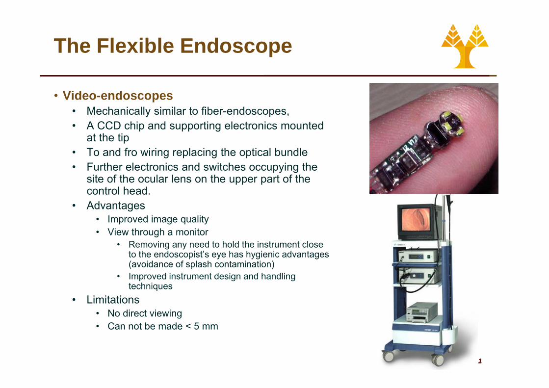
The Flexible Endoscope
• Video-endoscopes• Mechanically similar to fiber-endoscopes, • A CCD chip and supporting electronics mounted
at the tipT d f i i l i th ti l b dl• To and fro wiring replacing the optical bundle
• Further electronics and switches occupying the site of the ocular lens on the upper part of the control head.control head.
• Advantages• Improved image quality• View through a monitor
• Removing any need to hold the instrument close to the endoscopist’s eye has hygienic advantages (avoidance of splash contamination)
• Improved instrument design and handling techniques
• Limitations• No direct viewing• Can not be made < 5 mm
1111
• Can not be made < 5 mm
The Flexible Endoscope
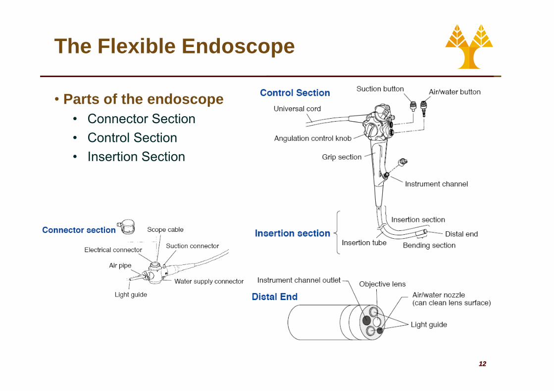
• Parts of the endoscope• Connector Section• Control Section• Insertion Section
1212
The Flexible Endoscope
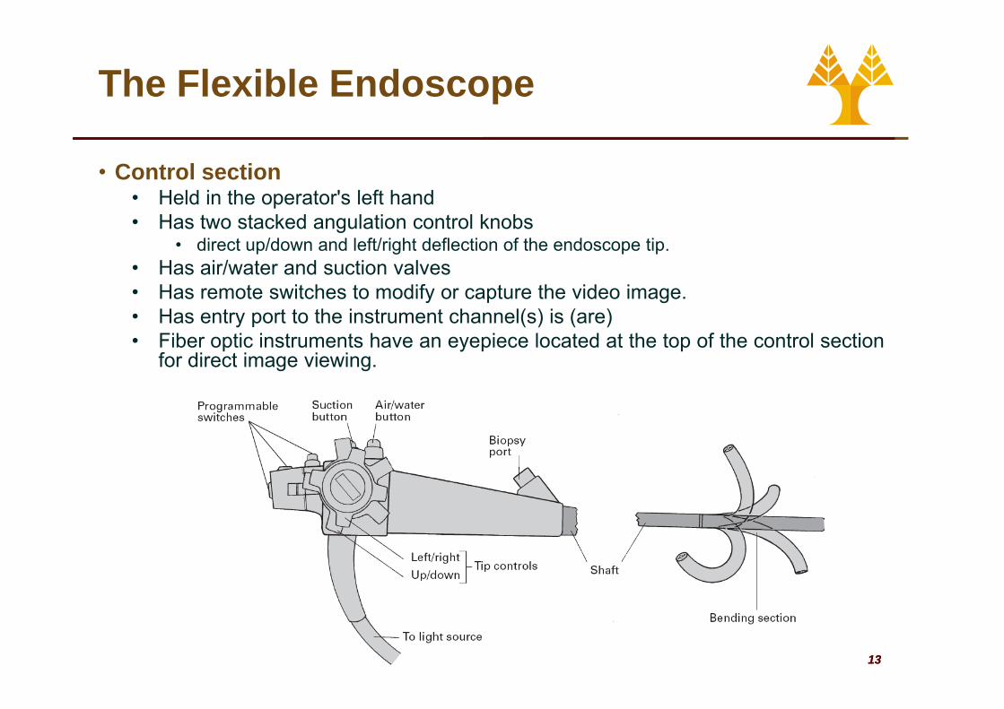
• Control sectionHeld in the operator's left hand• Held in the operator's left hand
• Has two stacked angulation control knobs • direct up/down and left/right deflection of the endoscope tip.
• Has air/water and suction valvesHas air/water and suction valves• Has remote switches to modify or capture the video image. • Has entry port to the instrument channel(s) is (are)• Fiber optic instruments have an eyepiece located at the top of the control section
f di t i i ifor direct image viewing.
1313
The Flexible Endoscope
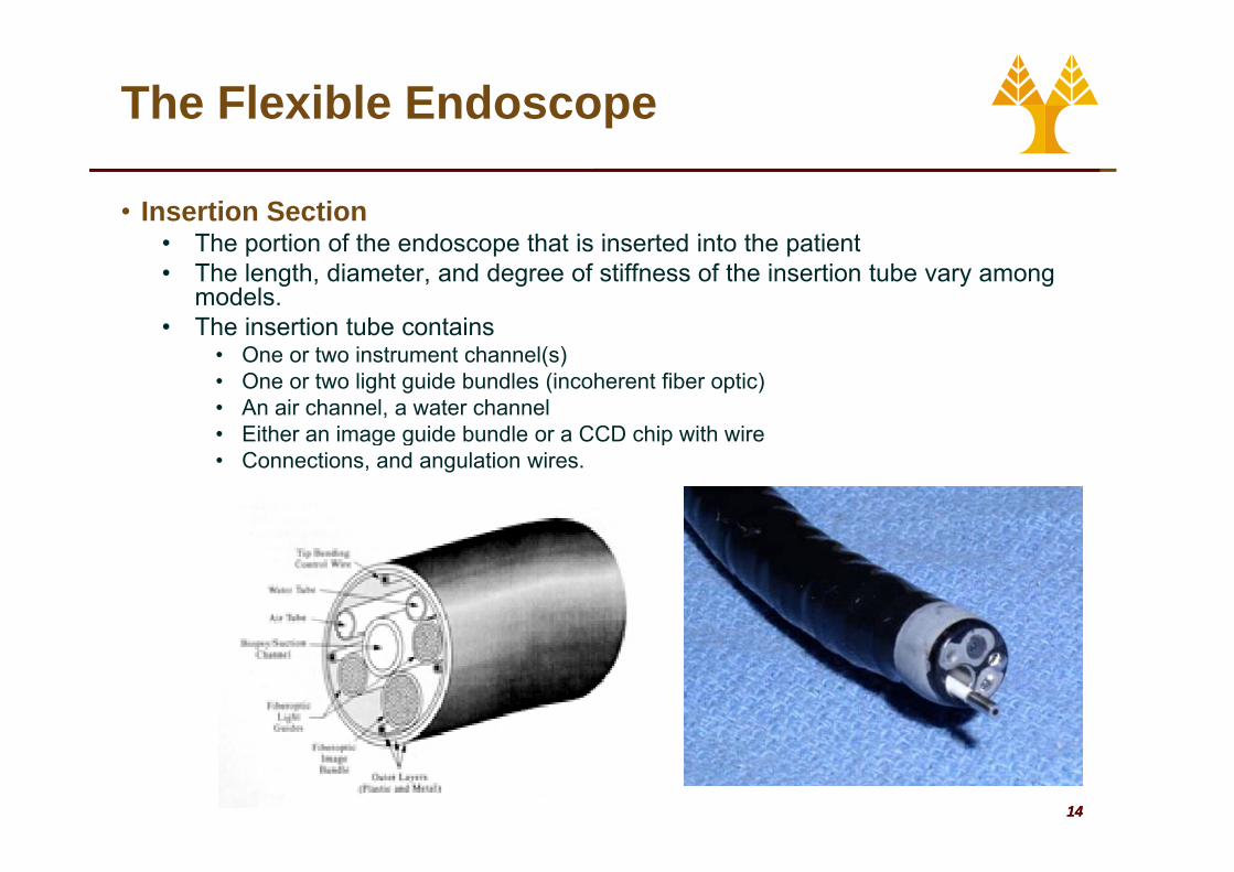
• Insertion SectionThe portion of the endoscope that is inserted into the patient• The portion of the endoscope that is inserted into the patient
• The length, diameter, and degree of stiffness of the insertion tube vary among models.
• The insertion tube contains • One or two instrument channel(s)• One or two light guide bundles (incoherent fiber optic)• An air channel, a water channel• Either an image guide bundle or a CCD chip with wire• Either an image guide bundle or a CCD chip with wire• Connections, and angulation wires.
1414

The Flexible Endoscope
• Endoscopic Accessories• Biopsy forceps• Graspers• Baskets• Injectors • Dilators• Dilators• Knives• HF endo-therapy
accessories• . . . too many types of
accessoriesaccessories.
1515
The Flexible Endoscope
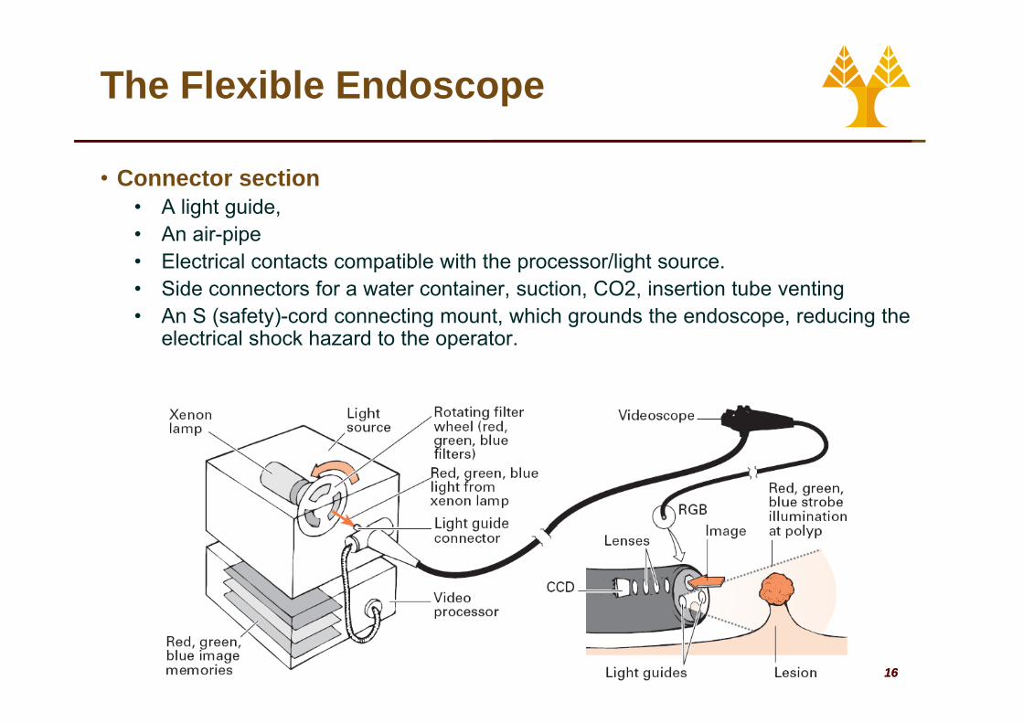
• Connector section• A light guide, • An air-pipe• Electrical contacts compatible with the processor/light source. • Side connectors for a water container, suction, CO2, insertion tube venting• An S (safety)-cord connecting mount, which grounds the endoscope, reducing the
electrical shock hazard to the operator.
1616
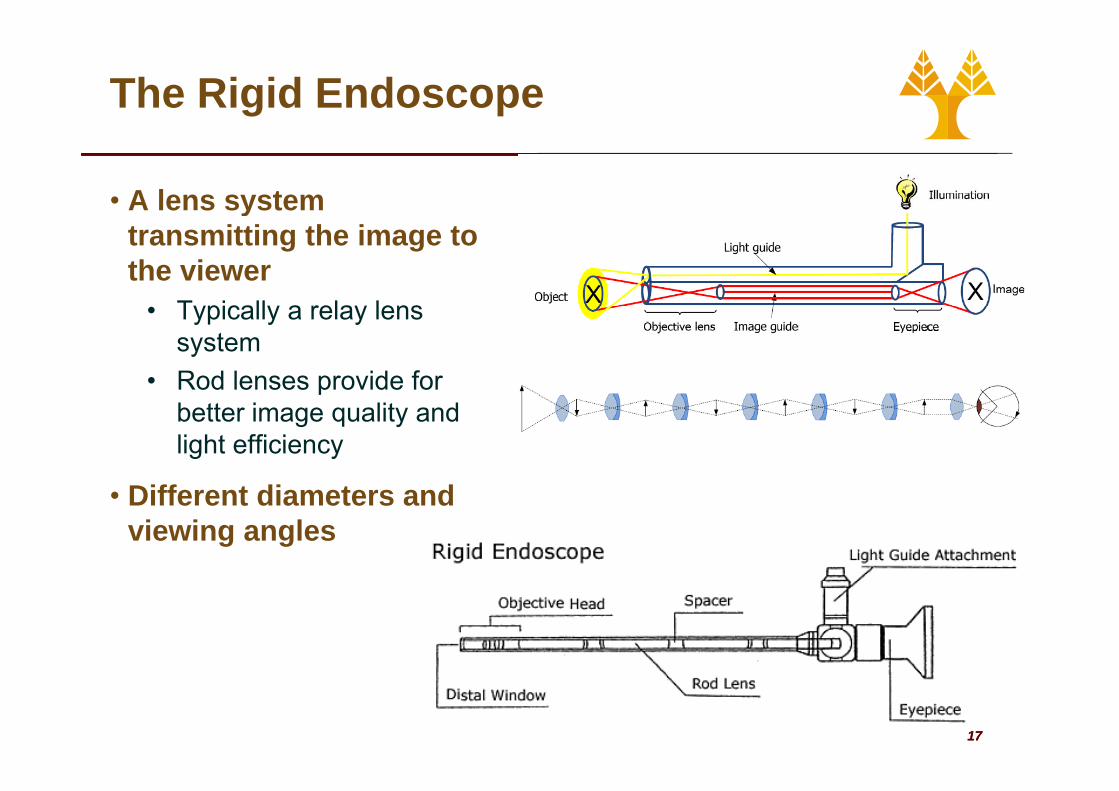
The Rigid Endoscope
• A lens system ytransmitting the image to the viewer
Typically a relay lens• Typically a relay lens system
• Rod lenses provide for better image quality and light efficiency
Diff t di t d• Different diameters and viewing angles
1717
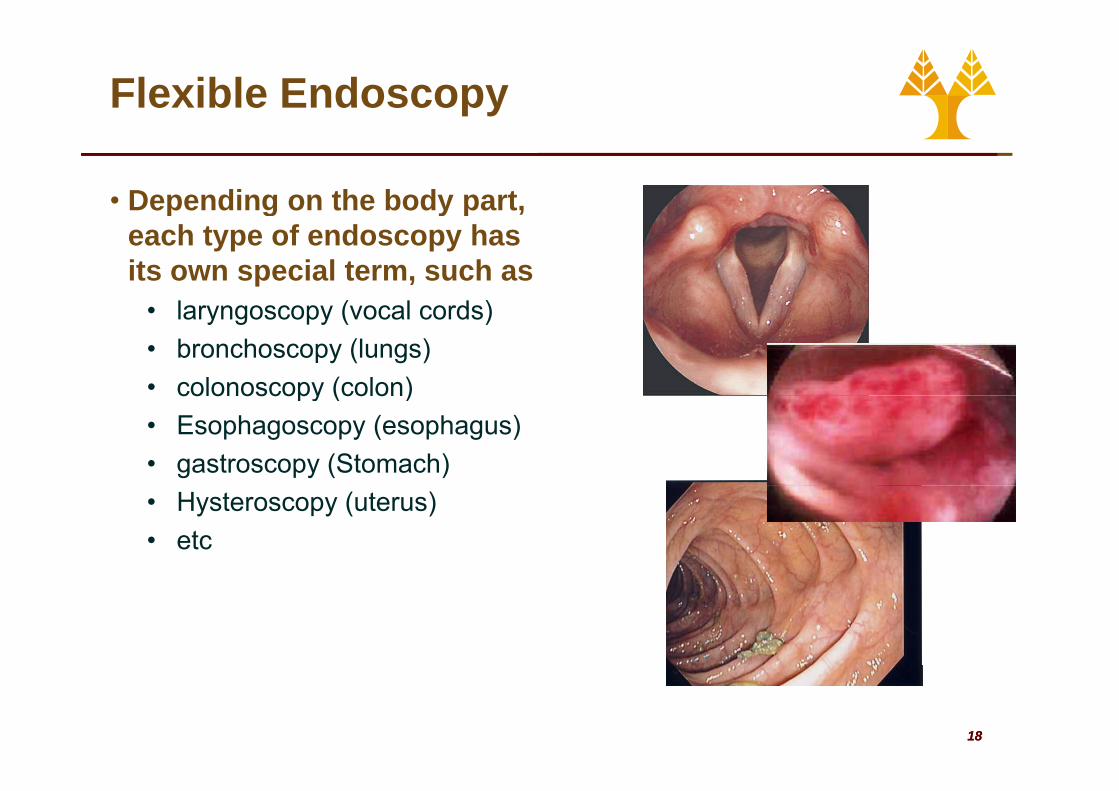
Flexible Endoscopy
• Depending on the body part, g yeach type of endoscopy has its own special term, such as
laryngoscopy (vocal cords)• laryngoscopy (vocal cords) • bronchoscopy (lungs) • colonoscopy (colon)colonoscopy (colon) • Esophagoscopy (esophagus)• gastroscopy (Stomach)• Hysteroscopy (uterus)• etc
1818
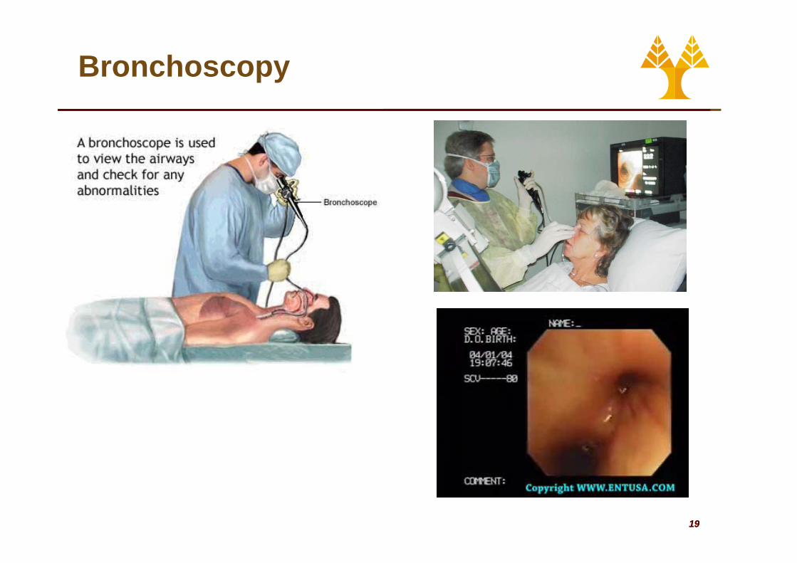
Bronchoscopy
1919
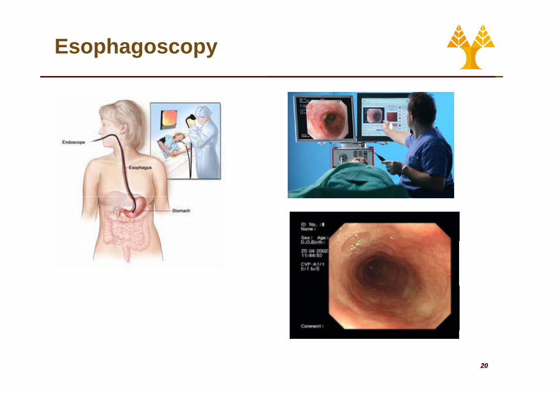
Esophagoscopy
2020
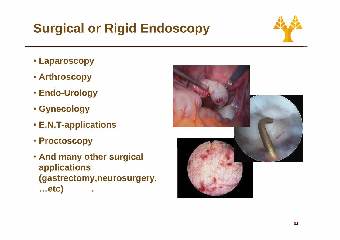
Surgical or Rigid Endoscopy
• Laparoscopyy
• Arthroscopy
• Endo Urology• Endo-Urology
• Gynecology
• E.N.T-applications
• Proctoscopy
• And many other surgical applications ((gastrectomy,neurosurgery,…etc) .
2121
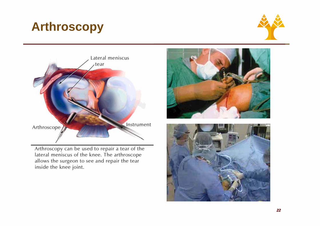
Arthroscopy
2222

Urethrocytoscopy
2323
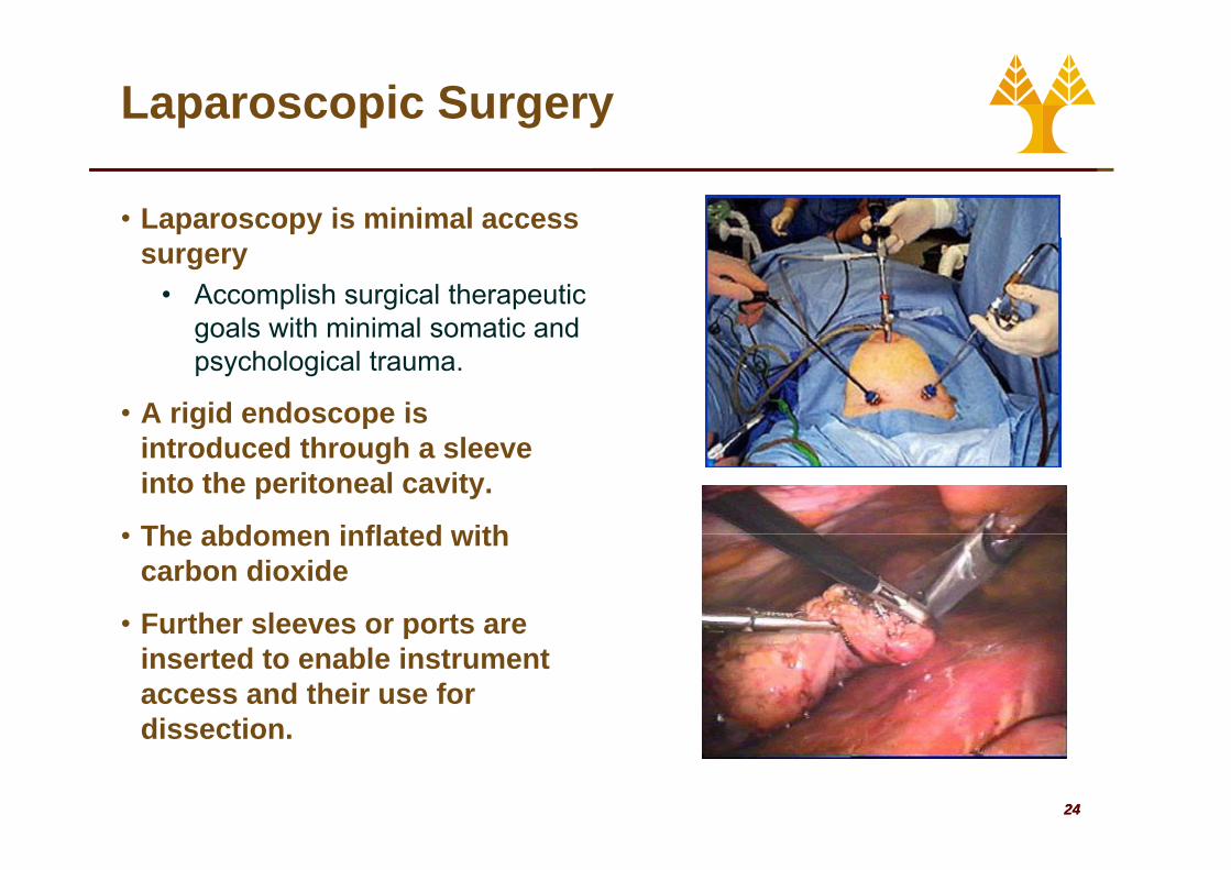
Laparoscopic Surgery
• Laparoscopy is minimal access surgery
• Accomplish surgical therapeutic goals with minimal somatic andgoals with minimal somatic and psychological trauma.
• A rigid endoscope is introduced through a sleeve into the peritoneal cavity.
• The abdomen inflated with• The abdomen inflated with carbon dioxide
• Further sleeves or ports areFurther sleeves or ports are inserted to enable instrument access and their use for dissection
2424
dissection.
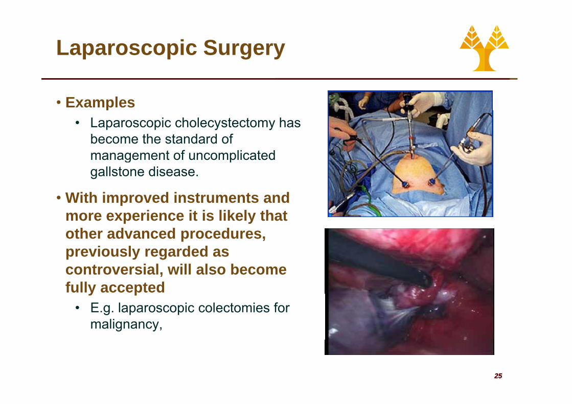
Laparoscopic Surgery
• Examples• Laparoscopic cholecystectomy has
become the standard of management of uncomplicatedmanagement of uncomplicated gallstone disease.
• With improved instruments andWith improved instruments and more experience it is likely that other advanced procedures,
i l d dpreviously regarded as controversial, will also become fully acceptedy p
• E.g. laparoscopic colectomies for malignancy,
2525
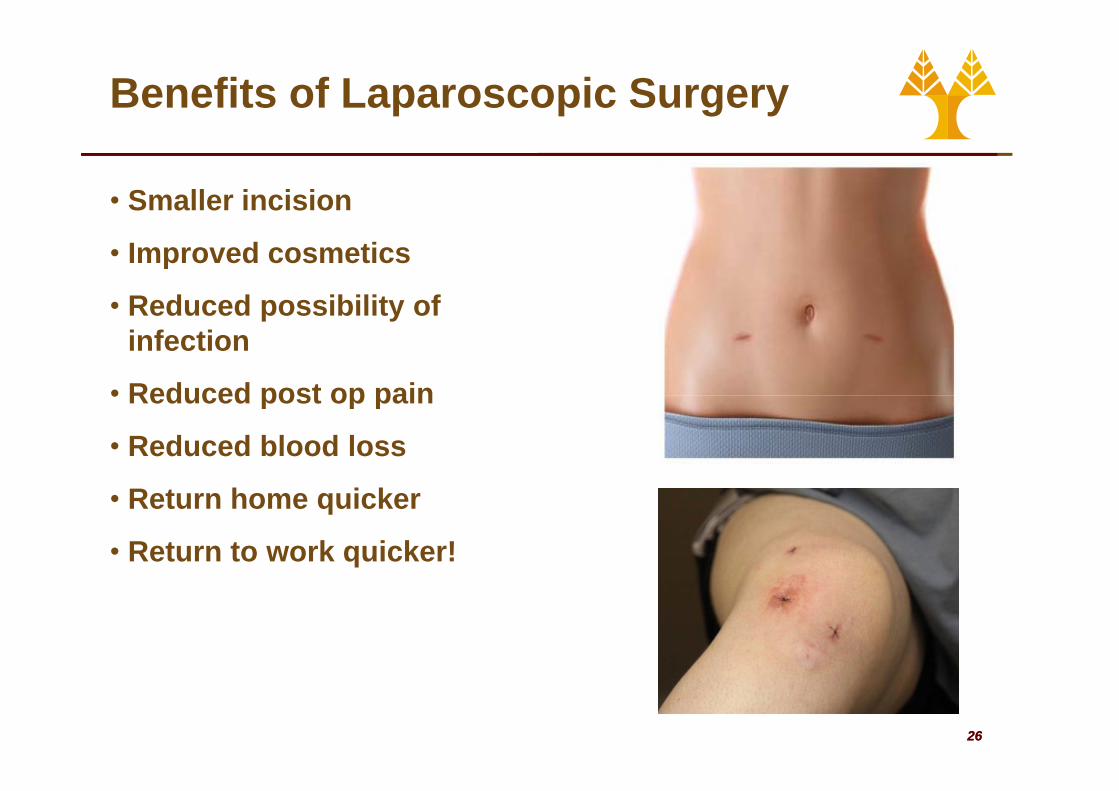
Benefits of Laparoscopic Surgery
• Smaller incision
• Improved cosmetics
• Reduced possibility of• Reduced possibility of infection
• Reduced post op pain• Reduced post op pain
• Reduced blood loss
• Return home quicker
• Return to work quicker!
2626
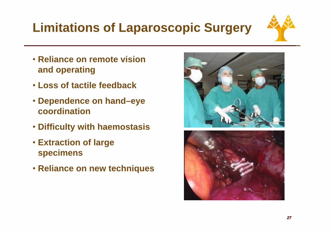
Limitations of Laparoscopic Surgery
• Reliance on remote vision and operating
• Loss of tactile feedback
• Dependence on hand–eye coordination
• Difficulty with haemostasis
• Extraction of largeExtraction of large specimens
• Reliance on new techniquesReliance on new techniques
2727
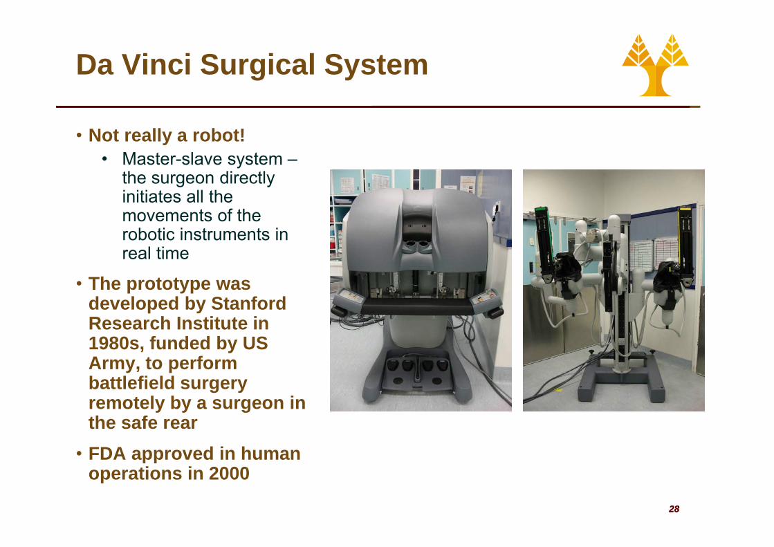
Da Vinci Surgical System
• Not really a robot!• Master-slave system –
the surgeon directly initiates all the
t f thmovements of the robotic instruments in real time
• The prototype was developed by Stanford Research Institute in 1980s, funded by US Army, to perform battlefield surgery
t l b iremotely by a surgeon in the safe rear
• FDA approved in human
2828
app o ed u aoperations in 2000
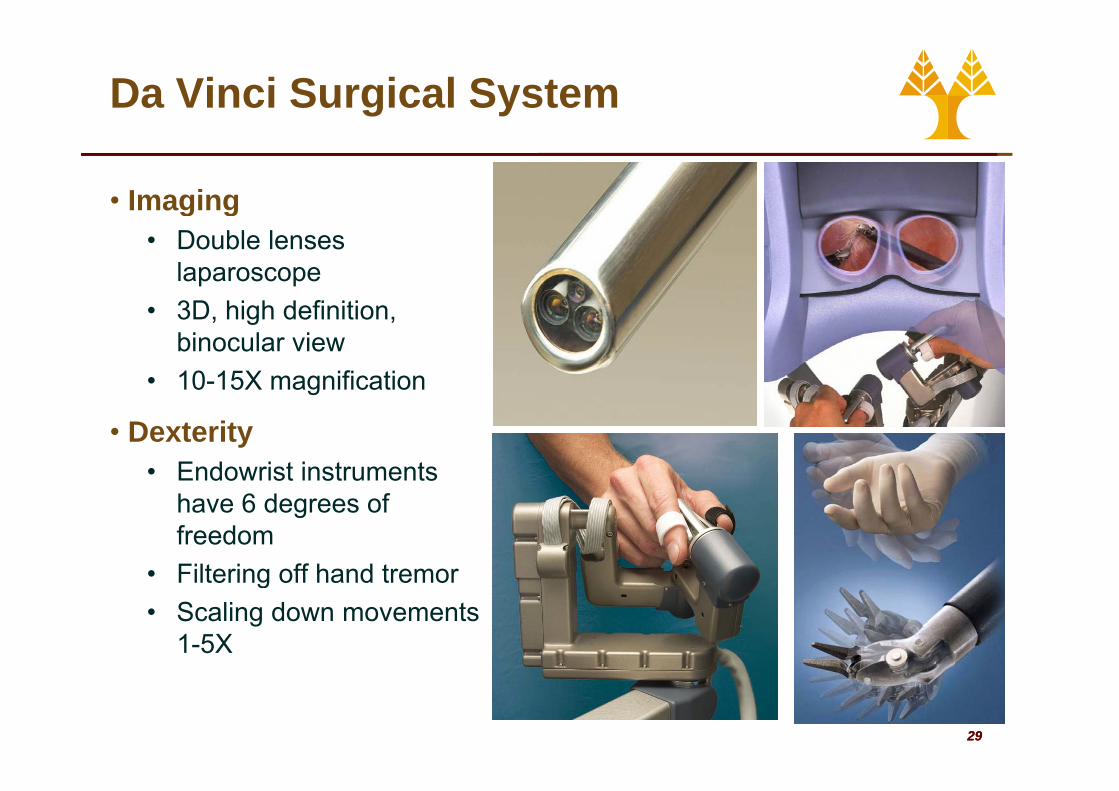
Da Vinci Surgical System
• Imagingg g• Double lenses
laparoscope3D high definition• 3D, high definition, binocular view
• 10-15X magnification
• Dexterity• Endowrist instruments
have 6 degrees of freedom
• Filtering off hand tremor• Filtering off hand tremor• Scaling down movements
1-5X
2929
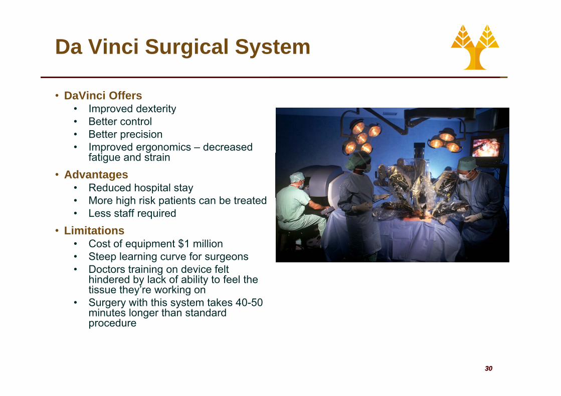
Da Vinci Surgical System
• DaVinci Offers• Improved dexterity• Improved dexterity• Better control• Better precision• Improved ergonomics – decreased
ffatigue and strain• Advantages
• Reduced hospital stay M hi h i k ti t b t t d• More high risk patients can be treated
• Less staff required• Limitations
• Cost of equipment $1 million• Cost of equipment $1 million• Steep learning curve for surgeons• Doctors training on device felt
hindered by lack of ability to feel the tissue they’re working ontissue they’re working on
• Surgery with this system takes 40-50 minutes longer than standard procedure
3030
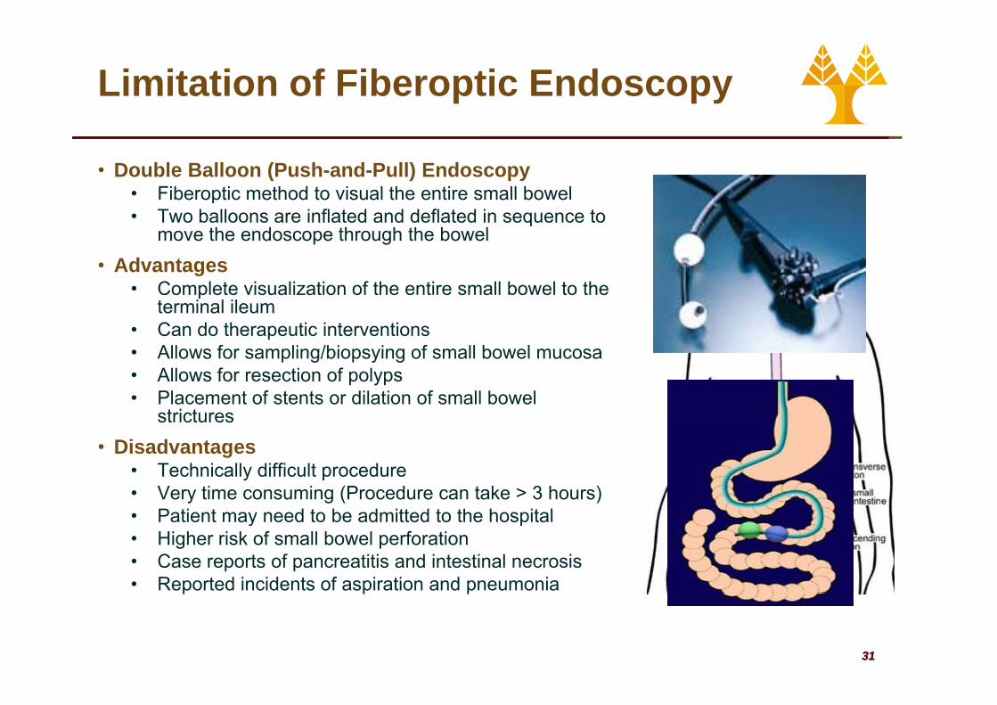
Limitation of Fiberoptic Endoscopy
• Double Balloon (Push-and-Pull) Endoscopy • Fiberoptic method to visual the entire small bowel• Fiberoptic method to visual the entire small bowel• Two balloons are inflated and deflated in sequence to
move the endoscope through the bowel• Advantagesg
• Complete visualization of the entire small bowel to the terminal ileum
• Can do therapeutic interventions• Allows for sampling/biopsying of small bowel mucosa• Allows for sampling/biopsying of small bowel mucosa• Allows for resection of polyps• Placement of stents or dilation of small bowel
strictures• Disadvantages
• Technically difficult procedure• Very time consuming (Procedure can take > 3 hours)
Patient may need to be admitted to the hospital• Patient may need to be admitted to the hospital• Higher risk of small bowel perforation• Case reports of pancreatitis and intestinal necrosis• Reported incidents of aspiration and pneumonia
3131
p p p
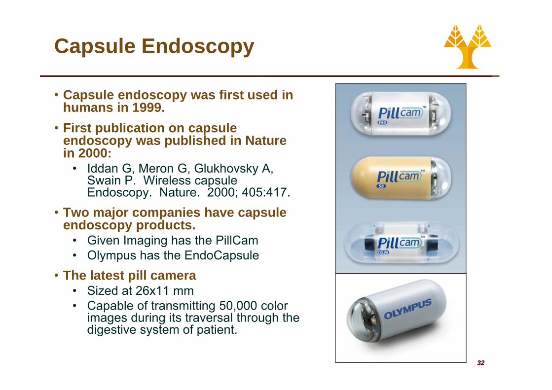
Capsule Endoscopy
• Capsule endoscopy was first used in humans in 1999humans in 1999.
• First publication on capsule endoscopy was published in Nature in 2000:in 2000:
• Iddan G, Meron G, Glukhovsky A, Swain P. Wireless capsule Endoscopy Nature 2000; 405:417Endoscopy. Nature. 2000; 405:417.
• Two major companies have capsule endoscopy products.
• Given Imaging has the PillCam• Given Imaging has the PillCam• Olympus has the EndoCapsule
• The latest pill camera• Sized at 26x11 mm • Capable of transmitting 50,000 color
images during its traversal through the digestive system of patient
3232
digestive system of patient.
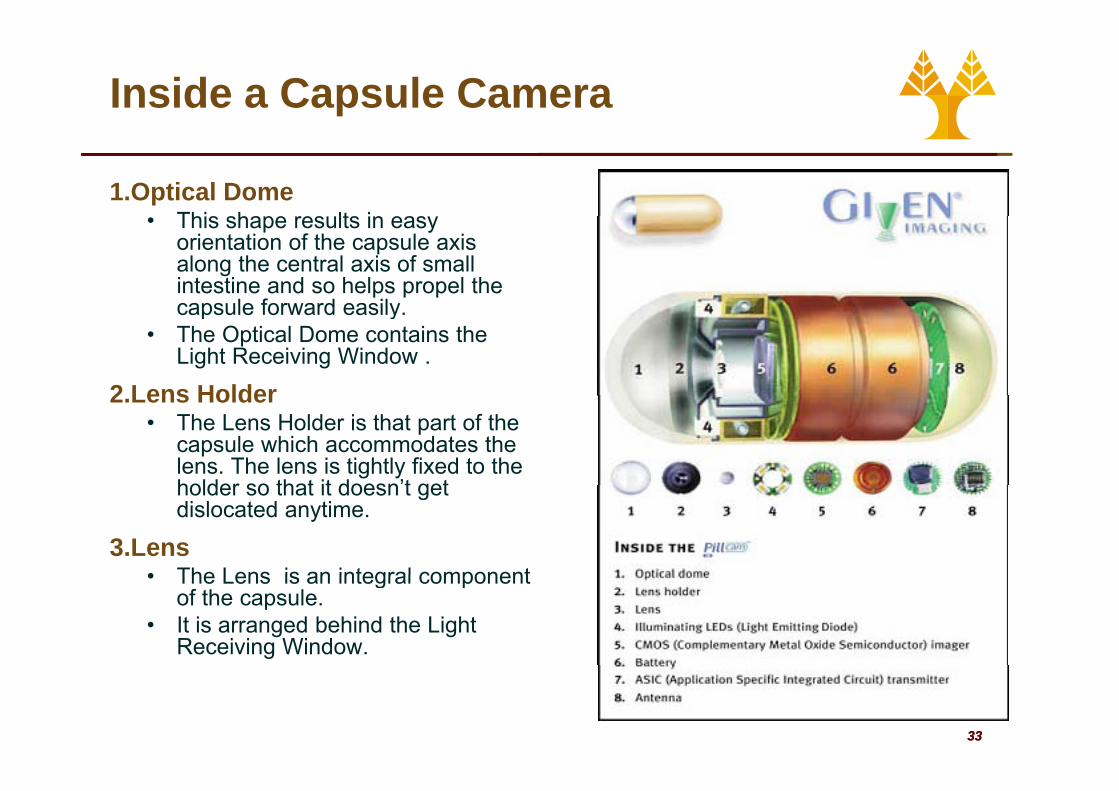
Inside a Capsule Camera
1.Optical DomeThis shape res lts in eas• This shape results in easy orientation of the capsule axis along the central axis of small intestine and so helps propel the capsule forward easilycapsule forward easily.
• The Optical Dome contains the Light Receiving Window .
2 Lens Holder2.Lens Holder• The Lens Holder is that part of the
capsule which accommodates the lens. The lens is tightly fixed to the holder so that it doesn’t getholder so that it doesn t get dislocated anytime.
3.Lens• The Lens is an integral component• The Lens is an integral component
of the capsule. • It is arranged behind the Light
Receiving Window.
3333

Inside a Capsule Camera
4.Illuminating LED’s• Around the Lens & CMOS
Image Sensor, four LED’s (Light Emitting Diodes) are present. Th l l li hti d iThese plural lighting devices are arranged in donut shape.
5.CMOS Image Sensorg• CMOS (Complementary Metal
Oxide Semiconductor) Image Sensor is the most important part of the capsule. It is highly sensitive and produces very high quality images.
• It has 140º field of view and can detect objects as small as possible.
3434
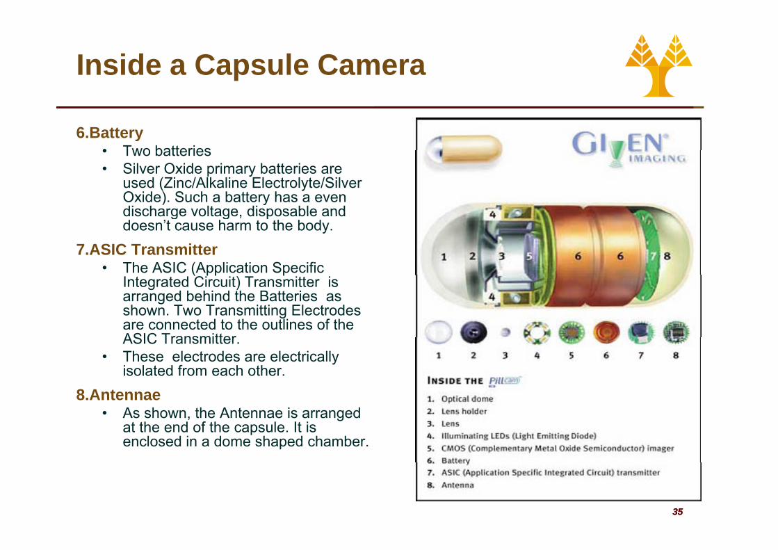
Inside a Capsule Camera
6.Battery• Two batteries• Two batteries• Silver Oxide primary batteries are
used (Zinc/Alkaline Electrolyte/Silver Oxide). Such a battery has a even discharge voltage disposable anddischarge voltage, disposable and doesn’t cause harm to the body.
7.ASIC Transmitter• The ASIC (Application Specific ( pp p
Integrated Circuit) Transmitter is arranged behind the Batteries as shown. Two Transmitting Electrodes are connected to the outlines of the ASIC TransmitterASIC Transmitter.
• These electrodes are electrically isolated from each other.
8.Antennae8.Antennae• As shown, the Antennae is arranged
at the end of the capsule. It is enclosed in a dome shaped chamber.
3535
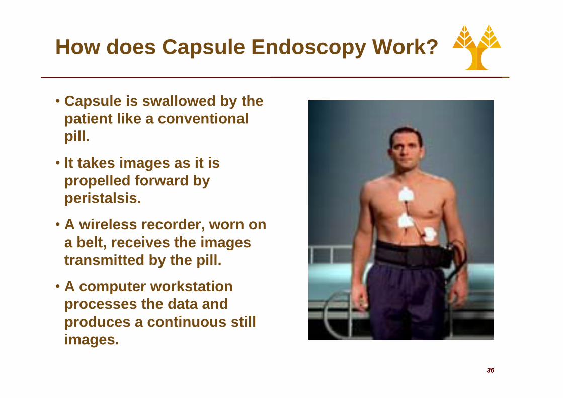
How does Capsule Endoscopy Work?
• Capsule is swallowed by the ypatient like a conventional pill.
• It takes images as it is propelled forward by peristalsisperistalsis.
• A wireless recorder, worn on a belt receives the imagesa belt, receives the images transmitted by the pill.
• A computer workstationA computer workstation processes the data and produces a continuous still i
3636
images.
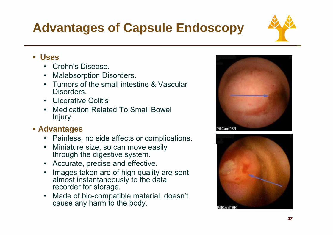
Advantages of Capsule Endoscopy
• UsesC h ' Di• Crohn's Disease.
• Malabsorption Disorders.• Tumors of the small intestine & Vascular
DisordersDisorders. • Ulcerative Colitis • Medication Related To Small Bowel
InjuryInjury. • Advantages
• Painless, no side affects or complications.• Miniature size, so can move easily
through the digestive system. • Accurate, precise and effective.
I t k f hi h lit t• Images taken are of high quality are sent almost instantaneously to the data recorder for storage.
• Made of bio-compatible material doesn’t
3737
Made of bio compatible material, doesn t cause any harm to the body.
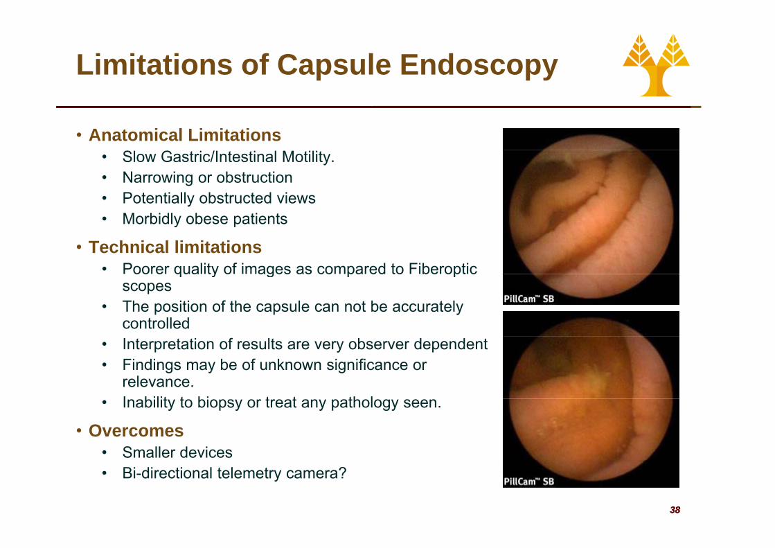
Limitations of Capsule Endoscopy
• Anatomical Limitations• Slow Gastric/Intestinal Motility. • Narrowing or obstruction• Potentially obstructed views• Morbidly obese patients
• Technical limitations• Poorer quality of images as compared to Fiberopticoo e qua ty o ages as co pa ed to be opt c
scopes• The position of the capsule can not be accurately
controlled• Interpretation of results are very observer dependent• Findings may be of unknown significance or
relevance.I bilit t bi t t th l• Inability to biopsy or treat any pathology seen.
• Overcomes• Smaller devices
3838
• Bi-directional telemetry camera?