UNIVERSITY OF BUCHAREST FACULTY OF CHEMISTRY PhD …
10
1 UNIVERSITY OF BUCHAREST FACULTY OF CHEMISTRY PhD DEGREE IN CHEMISTRY PhD THESIS OXIDATIVE REMOVAL OF CERTAIN INHIBITORS OF THE DNA AMPLIFICATION PROCESS SUMMARY Candidate, PÎRLEA SORINA PhD coordinator, Prof. Dr. DUMITRU OANCEA 2017
Transcript of UNIVERSITY OF BUCHAREST FACULTY OF CHEMISTRY PhD …

1
UNIVERSITY OF BUCHAREST
FACULTY OF CHEMISTRY
PhD DEGREE IN CHEMISTRY
PhD THESIS
OXIDATIVE REMOVAL OF CERTAIN INHIBITORS OF THE DNA AMPLIFICATION PROCESS
SUMMARY
Candidate,
PÎRLEA SORINA
PhD coordinator,
Prof. Dr. DUMITRU OANCEA
2017

2
PH D Thesis - TABLE OF CONTENTS I.THE CURRENT STATE OF KNOWLEDGE IN THE FIELD ................................................................ 3 I.1. IDENTIFICATION OF INDIVIDUALS THROUGH GENETIC FINGERPRINT ............................. 3
I.1.1. The importance of genetic fingerprint ................................................................................................. 3
I.1.2. PCR amplification process .................................................................................................................. 5
I.1.3. Capillary electrophoresis – PCR amplification mirror (efficient or failed) ........................................ 8 I.1.4. Inhibitors of PCR amplification process ............................................................................................. 9
I.2. DEGRADATION METHODS OF TEXTILE DYES, POSSIBLE INHIBITORS OF PCR REACTION .................................................................................................................................................................... 18 I.2.1. Physical methods ............................................................................................................................... 20
I.2.2. Chemical methods ............................................................................................................................. 21
I.2.3. Biological methods ............................................................................................................................ 24
I.2.4. Electrochemical methods .................................................................................................................. 25
I.2.5. Degradation of textile dyes with potassium permanganate ............................................................... 27 I.2.6. Oxidation with potassium permanganate – mechanisms .................................................................. 28 II. ORIGINAL PART ................................................................................................................................. 32
INRODUCTION ........................................................................................................................................ 32
MATERIALS AND METHODS ............................................................................................................... 33
II.1. OXIDATIVE DEGRADATION OF TEXTILE DYES ...................................................................... 36 Objectives ................................................................................................................................................... 36 II.1.1. Evaluation of methods for the oxidative degradation of textile dyes under conditions compatible with DNA identification ..................................................................................................................................... 36
II.1.2. Oxidative degradation study of βCuPcS4 with KMnO4 in basic environment ................................ 48 Conclusions ................................................................................................................................................ 71 II.2. TESTING THE INHIBITORY EFFECT OF POTASSIUM PERMANGANATE AND TEXTILE DYES ON THE DNA AMPLIFICATION PROCESS THROUGH REAL-TIME PCR QUANTIFICATION METHOD ................................................................................................................................................... 72 Objectives ................................................................................................................................................... 72 II.2.1. Testing the inhibitory effect of potassium permanganate and textile dyes studied in chapter ii.1, through Real-Time PCR method with Quantiplex Investigator kit ............................................................ 73 Conclusions ................................................................................................................................................ 81 II.2.2. Testing the inhibitory effect of the dyes studied after their oxidation with potassium permanganate in a basic environment at 56°C through Real-Time PCR method with the Quantiplex Investigator kit .......... 82
Conclusions ................................................................................................................................................ 87 II.3. DNA IDENTIFICATION FROM BIOLOGICAL SAMPLES WITH DENIM DYES ..................... 86 Objectives ................................................................................................................................................... 87 II.3.1 DNA identification from biological samples (saliva) treated with potassium permanganate in the presence of phthalocyanines βCuPcS4, Cl-αCuPc and βCuPc ................................................................... 87 II.3.2. DNA identification from biological samples fixed on denim (saliva, blood, epithelial cells) ....... 103 Conclusions .............................................................................................................................................. 113 GENERAL CONCLUSIONS ................................................................................................................. 114
ANNEXES ............................................................................................................................................... 117
BIBLIOGRAPHY .................................................................................................................................... 129
ABBREVIATIONS LIST………………………………………………………….……........................136

3
INTRODUCTION
Since DNA identification from biological samples taken from textile materials (especially denim) can be made difficult by the presence of the dyes in the environment of extraction, the discovery of new dye degradation methods is of great interest. Most of the advanced oxidation methods used in industry are carried out under acidic pH conditions, which also leads to DNA degradation by making impossible its further identification [1].
As a result, several enzymatic/chemical/electrochemical oxidation methods that take place in a neutral or basic environment have been tested in order not to damage the DNA structure. After finding the most efficient method, this was tested on several textile dyes and on several denim materials for checking its efficiency.
Quantification by the Real Time PCR method, which for several years offer the possibility to identify the inhibitors from biological samples, depending on the quantification kit used, has been of great help [2]. Thus, the dyes used could have been tested for their inhibitory effect through this method. Also, after their oxidation through the most efficient method, they could have been tested again to highlight whether they have or not an inhibitory effect on polymerase chain reaction (PCR).
Further on, depending on the obtained results, the method was applied on biological samples in the presence of inhibitory dyes and subsequently on biological samples deposited on denim, as usually arrived in genetic analysis laboratories for identification of individuals based on DNA. The dye used for the preliminary studies was a copper tetrasulphonate phthalocyanine: Heliogen blue A. This was chosen because it is part from a class of dyes that are very resistant to degradation and it also binds to DNA, thus being an inhibitor in the process of DNA identification [3].
Due to their physical and chemical stability, the phthalocyanines, being insoluble in water and in most solvents [4], are resistant dyes used in many fields such as inks and painting, textile staining, plastics, food packaging [5] and also as semiconducters, in computer industry [6] and so on. These substances are also used to color textiles for blue and green shades, especially denim for blue jeans, outfits almost indispensable among humans worldwide [7]. Lately it has been discovered that phthalocyanines are able to bound single- and double-stranded DNA molecules [8], thus being tested as photosensitizers in photodynamic therapy of cancer [9]. Recently it has been proposed their use as medicaments due to their antitumor effects [10].
I. THE CURRENT STATE OF KNOWLEDGE IN THE FIELD
The polymerase chain reaction (PCR) is an enzymatic process through which specific DNA sequences of interest are replicated (multiplied) 28-34 times, generating approximately one billion (109) copies [11]. This multiplication reaction of target DNA regions is extremely important in obtaining a sufficient amount of DNA for determining gene-based identification. Therefore, at this stage both the initial amount of DNA and its quality, respectively the absence of PCR inhibitors or degraded DNA, are very important in order to achieve efficient multiplication.
Inhibitors of the known PCR amplification process include the blood hem [12], collagen from tissues [13], humic acid from soil and plants [14], denim dyes[15],melanin from hair and skin [16],calcium ions from milk and bone [17] etc. Apart from indigo, unsubstituted phthalocyanines together with mono - copper (II) phthalocyanines and polysulfonated are commonly used as denim base pigments (Blue Reagent 15, Blue Reagent 21 [18], Reagent Blue 38[19] and C.I. Direct Blue 199 [20]).
These inhibitory substances can react with nucleic acids during extraction, degrade or modify target DNA sequences, interfere with PCR primers, degrade, alter or inhibit polymerase, and last but not least interfere with the sample or with its fluorophores. Inhibitors can be detected through the

4
Real Time PCR method which, in parallel, quantifies the DNA already present in the sample, a method used in this research, too [21].
DNA isolation is the first important step in forensic and medical analysis. The main extraction methods of DNA used by all laboratories in the field are the manual ones - the phenol-chloroform organic method, the chelex method and the FTA paper method - and the solid phase extraction techniques (on silica gel or particle membranes magnetic silicon) that can operate both manually and automatically [11, 22].
The degradation of textile dyes, which can be inhibitors of the PCR reaction, may be achieved through various methods: physical, chemical, biological and electrochemical [23].
II. THE ORIGINAL PART
Initially, the PCR inhibitory effect of several textile dyestuffs was tested: phthalocyanine, azo, indigo and triphenylmethane dyes. The inhibitory effect was verified during the DNA quantification step, by the Real-Time PCR method, using the Quantiplex Investigator kit and a Rotor-Gene Qapparatus [24]. The cycling threshold (CT) parameter for DNA / dye samples was determined and it was found, according to CT values obtained on the yellow channel, that tetrasulphonated copper beta phthalocyanine (βCuPcS4) had the greatest inhibitory effect, followed by beta-copper- phthalocyanine (βCuPc) and fuchsin. In fact, phthalocyanines are particularly resistant to degradation and are practically insoluble in water and in most solvents. Finally, phthalocyanine dyes were solubilized in an aqueous medium in the presence of a non-ionic alkyl polyglycoside surfactant (APG), with continuous stirring for two hours.
The next objective was to develop a method of removing phthalocyanines from solutions under conditions compatible with DNA extraction in the presence of chelex chelating resins (at basic pH and temperature 55-56 ° C), without modifying the structure of double stranded DNA. Initially enzyme oxidation was used with lacase and then with peroxidase, without a significant decrease in the concentration of phthalocyanine tested. Then, it was attempted to remove βCuPcS4 by electrolysis in acidic, basic and neutral media (with potassium sulphate). Oxidative degradation was monitored qualitatively by discoloration of the solution.
Electrolysis was followed for a minimum of 2 hours in a stainless steel electrode cell, determining the absorbance of the anode solution at different time intervals. In an acidic medium, in the presence of sulfuric acid, a degree of decay of 89% is obtained at 90 minutes. Visual, color change was observed from blue to green then orange, with dark orange precipitate formation and discoloration of the supernatant even after 55 minutes. In a basic environment, in the presence of NaOH, the reaction is much slower.
For a neutral environment, a solution of 0.05% K2SO4 was introduced into the electrolysis cell. Electrolysis was followed for 195 minutes, measuring the absorbance of the electrolysis product in the anode compartment every 15 minutes. The discoloration occurs in about 2 hours with a 90% discoloration degree. As the pH of the potassium sulphate electrolysis solution drops continuously from 6.5 to 1.5, it was attempted to adjust it during electrolysis with a 1M NaOH solution, gradually dropwise, to maintain a basic pH, appropriate for applications of interest (removal of phthalocyanine from biological samples in the DNA extraction step). The results are shown in Figure II.1. It is noted that the highs of the two peaks are recorded at 627 nm and 719 nm respectively. The formation of the dark orange precipitate and the visible discoloration of the solution in the anode compartment, together with the maximum decrease in phthalocyanine absorption, is recorded at 150 minutes.

5
Figure II.1. Electrolytic degradation of βCuPcS4 with potassium sulphate, with continuous pH adjustment (basic)
It can be observed that almost total bleaching occurs in 150 minutes. In the first part, the increase in absorbance can be attributed to the formation of an intermediate absorbing at the same wavelength as phthalocyanine.
The method has also been successfully applied to a dye solution obtained from 5 cm2 of dark blue dyed denim dissolved in APG. From the initial spectrum of the solution, a maximum absorbance at 660 nm corresponding to the indigo dye was obtained. The reaction was followed for 6.5 hours, with product absorbance determinations every 15 minutes.
To determine the constant speed and the degree of discoloration corresponding to βCuPcS4 by electrolysis, a wavelength of 718 nm was chosen, wavelength to which the other oxidation methods were also processed. It is noted that phthalocyanine discoloration takes place within two hours. From the time-absorbance data, the first order constant speed was estimated by fitting the equation obtained from the experimental data. The results show that both types of dyes can be degraded by electrolysis while maintaining a basic pH.
Another oxidative method was tested with KMnO4 in basic medium. This turned out to be the most efficient and most feasible in removing phthalocyanines and other dyes along with DNA isolation from biological samples. Thus, the oxidative degradation of βCuPcS4 with KMnO4 in the basic medium was studied. For this, phthalocyanine solutions (between 1.8 mg / L and 30 mg / L), excess potassium permanganate (between 0.4 mM and 12 mM) and sodium hydroxide (between 30 mM and 100 mM) were mixed and followed for at least 7 hours at temperatures between 40 and 70 ° C and pH 10-12. The mixtures were spectrophotometrically analyzed at wavelengths ranging from 200 to 900 nm at 15 minutes intervals. During the reaction, the color of the solution changes from blue to green, then a green precipitate appears and the solution turns pale yellow to colorless. The time required for complete discoloration was between 4 and 24 hours, depending on the reactant concentrations. Kinetic analyzes were performed at 714 nm because at 615 nm there could be some interference due to manganese ion (MnO4
2-) formation at 608 nm during the reaction [25]. Extended kinetic curves show an increase in absorbance, followed by an exponential
decrease. Color is also more intense during the time of the absorption increase. In our experiments, the pH value has varied between 10 and 12 so we can consider that the
permanganate is reduced by forming all the manganese oxidation states - Mn (VI), Mn (V) and Mn (IV) [26]..We can assume that the reaction takes place in at least two consecutive steps, with the formation of a stable intermediate having a similar color to βCuPcS4.
The reaction of βCuPcS4 with potassium permanganate in alkaline media is a complex reaction, being possible a variety of mechanisms in which the oxidation takes place via one or two
0
0.05
0.1
0.15
0.2
0.25
0 100 200 300
A
timp (min)
627nm K2SO4
cu ajustare pH
719nm K2SO4
cu ajustare pH

6
electrons. The reaction cannot be simplified in two consecutive steps but the form of the curves A = f (t) can be explained by the formation of an intermediate product that absorbs light at the same wavelength with βCuPcS4.
An important parameter for the degradation of dyes is the degree of discoloration (D %) calculated as:
��%� �������
���∙ 100, where Amax and Afin represent the values of maximum absorbance and
final absorbance (at a settled time).
The effect of potassium permanganate, natrium hydroxide, phthalocyanine concentrations and also the effect of the temperature on βCuPcS4 degradation were studied and the best parameters of the oxidative process were established: phthalocyanine concentration (18 mg/L), KMnO4 (7,6 mM) and NaOH (0,066 M), at 56 oC – the temperature at which occurs DNA isolation from samples (figure II.2). Under these conditions a degree of discoloration of over 90% is achieved in 4 hours [27].
Figure II.2 – Extended kinetic curve for βCuPcS4 degradation at ([KMnO4] = 7,6 mM, [βCuPcS4] = 18 mg/L and
[NaOH] = 0,066 M at 56 oC.
To explain the increase of absorbance on the initial portion, a kinetic model of two consecutive single reactions was used, from which it was obtained a kinetic equation of total absorption vs time that explains the shape of the experimental curves obtained:
���→�
��→ � (1)
Starting from the time evolution of reactant and intermediate concentrations (cAșicB) and assuming that both the reactant and intermediate have a significant absorbance (AA și AB) at the same wavelength, the total measured absorbance (A) is given by:
� � �� � �� � ����� � ����� (2) where �� is the molar absorptivity of j component and l is the path length. One finally obtains:
� � ��� ��! �
"#$%&'��
����� ��! ( ��!� (3)
Kinetic parameters were estimated according to this model, and there was good agreement between the experimental data and the fitted model, the values r2 (of the determination coefficient) being between 0.936 and 0.987.
Reaction products were analyzed by FTIR spectroscopy and X-ray fluorescence spectroscopy, and it was concluded that the precipitate could contain MnO2 and phthalocyanine residue and the aqueous phase (of interest to our applications) no longer contains Cu and Mn ions.

7
The method of KMnO4 degradation in basic media was also tested under NaOH replacement with a 5% chelex solution on pH 11 and extended for other colorants of different classes. The same reaction conditions previously established as optimal were used and the reaction was followed for 150 minutes. The method is efficient for all types of dyes, resulting in fading degrees ranging from 41 to 90% after 60 minutes of reaction.
Further, KMnO4-oxidized dyes in basic medium were again tested for the inhibitory effect on the PCR amplification reaction by the Real Time PCR method, following the CT values on the yellow chanel of Rotor Gene Qapparatus. It was found that none had a CT compatible value with a PCR inhibitor effect ( > 31). The effect of KMnO4 as a possible inhibitor under the same conditions was also investigated, demonstrating that it had no effect on the PCR reaction.
The next objective was to apply this method to biological samples with phthalocyanines and other denim dyes. Initially, saliva samples of known concentration were tested from persons with known DNA profiles, mixed with three phthalocyanines: βCuPcS4, βCuPc şi Cl-αCuPc. According to the results shown in Table II.1, the method was particularly successful for samples with phthalocyanines having an inhibitory effect and being oxidized with KMnO4 [28].
DNA sample DNA amount
(ng/µL) CT value
Amount of
extract used
(µL)
Full genetic
profil? /
(number of
genetic
markers)
Average
height of
peaks
Saliva + βCuPcS4
no KMnO4
0.161 32.4 6 No (5) 354
Saliva + βCuPcS4
+ KMnO4
0.087 30.2 9.5 Yes (16) 5345
Saliva + βCuPc
no KMnO4
0.032 32.0 9.5 No (12) 323
Saliva + βCuPc
+ KMnO4
0.145 29.9 7 Yes (16) 1076
Saliva + Cl-αCuPc
no KMnO4
0.170 30.6 6 Yes (16) 1223
Saliva + Cl-αCuPc
+ KMnO4
0.226 30.5 4.5 Yes (16) 1343
Table no. II.1. – Results obtained from genetic analyzes of biological samples mixed with phthalocyanines.
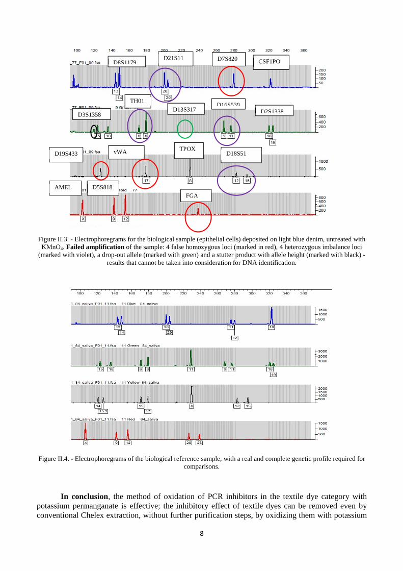
Then the method was successfully applied to biological samples (blood, saliva, epithelial cells) fixed on dark blue denim. Tests were also performed for the same types of samples fixed on light blue denim, but with no great differences between the results obtained from the permanganate-treated and non-permanganate-treated biological samples, probably due to the low amount of denim dye.
However, for epithelial cells fixed on light blue denim, it was noted that the failed amplification according to Figure II.3 was caused by two factors: insufficient amount of DNA matrix and the presence of inhibitors. In Figure II.4. the electropherograms corresponding to the biological reference sample (successful amplification) are presented for comparisons.
8
Figure II.3. - Electrophoregrams for the biological sample (epithelial cells) deposited on light blue denim, untreated with KMnO4. Failed amplification of the sample: 4 false homozygous loci (marked in red), 4 heterozygous imbalance loci
(marked with violet), a drop-out allele (marked with green) and a stutter product with allele height (marked with black) - results that cannot be taken into consideration for DNA identification.
Figure II.4. - Electrophoregrams of the biological reference sample, with a real and complete genetic profile required for comparisons.
In conclusion, the method of oxidation of PCR inhibitors in the textile dye category with potassium permanganate is effective; the inhibitory effect of textile dyes can be removed even by conventional Chelex extraction, without further purification steps, by oxidizing them with potassium
D8S1179 D21S11 D7S820 CSF1PO
D3S1358
TH01 D16S539
D13S317 D2S1338
D19S433 vWA TPOX D18S51
AMEL D5S818 FGA

9
permanganate simultaneously with DNA isolation. Potassium permanganate does not affect the integrity of DNA molecules. The reduction of textile dye amount can be observed with the naked eye through the disappearance of color in time. DNA does not degrade if it stays longer at 56° C. In addition, potassium permanganate can be added additionally, with prolonged incubation time, until the color of the supernatant containing the DNA molecules disappears. This chelex / permanganate system is more economically convenient and perhaps more efficient for biological samples fixed on textiles than other more expensive and longer-lasting procedures.
SELECTIVE BIBLIOGRAPHY
[1] P. Hermann, E. Frederick The role of the AT pairs in the acid denaturation of DNA, Nucleic Acids Research (1977), 4 (8): 2939-2947.
[2] S.B. Seo, H.Y. Lee, A.H. Zhang, H.Y. Kim, D.H. Shin, S.D. Lee, Effects of humic acid on DNA quantification with Quantifiler human DNA Quantification kit and short tandem repeat amplification efficiency, Int. J. Legal Medicine (2012), 126, 961-968.
[3] H. Yaku, T. Fujimoto, T. Murashima, D. Miyoshi and N. Sugimoto, Phthalocyanines: a new class of G-quadruplex-ligands with many potential applications, Chem. ComMun., (2012), 48, 6203–6216
[4] A. Heinfling, M. Bergbauer, U. Szewzyk, Biodegradation of azo and phthalocyanine dyes by Trametes versicolor and Bjerkandera adusta. Applied Microbiology and Biotechnology (1997), 48 (2), 261-266.
[5] E. Marechal, Polymeric dyes — synthesis, properties and uses. Progress in Organic Coatings, (1982), 10 (3), 251-287.
[6] H. Cao, W. He, Y. Mao, X. Lin, K. Ishikawa, J.H. Dickerson, W.P. Hess, Recent progress in degradation and stabilization of organic solar cells. Journal of Power Sources, (2014), 264, 168-183.
[7] M. Sanchez, Dyeing of denim yarns with non-indigo dyes. In: Paul R (edition) Denim. Woodhead Publishing, (2015), 5 , 107-157.
[8] C. Uslan, S.B. Şebnem, Synthesis of novel DNA-interacting phthalocyanines. Dyes and Pigments, (2012), 94 (1), 127-135.
[9] C-K Lim, J. Heo, S. Shin, K. Jeong K, Y.H. Seo, W.D. Jang, C.R. Park, S.Y. Park, S. Kim, I.C. Kwon IC, Nanophotosensitizers toward advanced photodynamic therapy of Cancer, Cancer Letters (2013), 334 (2), 176-187.
[10] C-K Lim, J. Heo, S. Shin, K. Jeong K, Y.H. Seo, W.D. Jang, C.R. Park, S.Y. Park, S. Kim, I.C. Kwon IC, Nanophotosensitizers toward advanced photodynamic therapy of Cancer, Cancer Letters (2013), 334 (2), 176-187.
[11] J. M. Butler, Fundamentals of Forensic DNA Typing, Academic Press, USA, (2009). [12] A. T. Hall, A. M. Zovanyi, D.R. Christensen, J.W. Koehler, T. Devins Minogue, Evaluation of
Inhibitor-Resistant Real-Time PCR Methods for Diagnostics in Clinical and Environmental Samples, PLoS ONE, (2013), 8 (9), e73845.
[13] C.H. Kim et al. Optimization of the PCR for detection of Staphylococcus aureus nucgene in bovine milk. J. Dairy Sci., (2001), 84, 74–83.
[14] S. Schmedes, P. Marshall, J. King, B. Budowle, Effective removal of co-purified inhibitors from extracted DNA samples using synchronous coefficient of drag alteration (SCODA) technology, Int J Legal Med, (2013), 127, 749-755.
[15] R. Alaeddini, Forensic implications of PCR inhibition—A review, Forensic Science International: Genetics, (2012), 6, 297-305.
[16] L. Eckhart et al. Melanin binds reversibly to thermostable DNA polymerase and inhibits its activity. Biochem. Biophys.Res. ComM. (2000) 271, 726–30.
[17] H. A. Powell et al. Proteinase inhibition of the detection of Listeria monocytogenes in milk using the polymerase chain reaction. Lett. Appl. Microbiol. (1994), 18, 59–61.
[18] M.C. Silva, A.D. Corrêa, M.T.S.P. Amorim, P. Parpot, J.A. Torres, P.M.B. Chagas, Decolorization of the phthalocyanine dye reactive blue 21 by turnip peroxidase and assessment of its oxidation products, Journal of Molecular Catalysis B: Enzymatic, (2012), 77, 9-14.
[19] A. Heinfling, M. Bergbauer, U. Szewzyk, Biodegradation of azo and phthalocyanine dyes by

10
Trametes versicolor and Bjerkandera adusta, Appl Microbiol Biotechnol, (1997), 48, 261-266. [20] H.-Y. Shu, M.-C. Chang, Decolorization and mineralization of a phthalocyanine dye C.I. Direct Blue
199 using UV/H2O2 process, Journal of Hazardous Materials, (2005), 125, 96-101. [21] S.B. Seo, H.Y. Lee, A.H. Zhang, H.Y. Kim, D.H. Shin, S.D. Lee, Effects of humic acid on DNA
quantification with Quantifiler human DNA Quantification kit and short tandem repeat amplification efficiency, Int. J. Legal Medicine (2012), 126, 961-968.
[22] F. Stanciu, V. Cuţăr, S. Pîrlea, V. Stoian, I.M. Stoian. Population data for Y-chromosome haplotypes defined by 17 STRs in South-East Romania, Legal Medicine (Tokyo), (2010), 12(5), 259-64
[23] W. Azmi, R.K. Sani, U.C. Banerjee, Biodegradation of triphenylmethane dyes, Microbial Technology, (1998), 22, 185-191.
[24] Investigator Quantiplex Handbook, QIAGEN Sample and Assay Technologies, March (2011). [25] R.M. Mulla, S.T. Nandibewoor, Mechanistic and spectral investigation of the oxidation of 4-
hydroxycoumarin by aqueous alkaline permanganate using the stopped flow technique, Polyhedron, (2004), 23 (16), 2507-2513.
[26] S. Dash, S. Patel, B.K. Mishra, Oxidation by permanganate: synthetic and mechanistic aspects, Tetrahedron (2009), 65 (4), 707-739.
[27] S. Pîrlea, A. Răducan M. Puiu and D. Oancea, Kinetic insights into mild oxidation of phtalocyanine dyes: potential application in forensic analysis, Revue Roumaine de Chimie (2017).
[28]S. Pîrlea, A. Răducan M. Puiu and D. Oancea, Permanganate assisted removal of PCR inhibitors during the DNA CHELEX extraction from stained denim samples, Int. J. Legal Med. (2017), 131, 323-331.