University of Alberta · reduction conditions were tried, and Birch reduction is underway to reach...
156
I University of Alberta Synthesis of Porphyrin-based Multimeric Fluorescent Compounds and Studies Towards the Formation of Cis-anti-cis Linear Triquinane by Linghui Yu A thesis submitted to the Faculty of Graduate Studies and Research in partial fulfillment of the requirements for the degree of Master of Science Department of Chemistry ©Linghui Yu Fall 2011 Edmonton, Alberta Permission is hereby granted to the University of Alberta Libraries to reproduce single copies of this thesis and to lend or sell such copies for private, scholarly or scientific research purposes only. Where the thesis is converted to, or otherwise made available in digital form, the University of Alberta will advise potential users of the thesis of these terms. The author reserves all other publication and other rights in association with the copyright in the thesis and, except as herein before provided, neither the thesis nor any substantial portion thereof may be printed or otherwise reproduced in any material form whatsoever without the author's prior written permission.
Transcript of University of Alberta · reduction conditions were tried, and Birch reduction is underway to reach...

I
University of Alberta
Synthesis of Porphyrin-based Multimeric Fluorescent Compounds and Studies Towards the Formation of Cis-anti-cis Linear Triquinane
by
Linghui Yu
A thesis submitted to the Faculty of Graduate Studies and Research in partial fulfillment of the requirements for the degree of
Master of Science
Department of Chemistry
©Linghui Yu Fall 2011
Edmonton, Alberta
Permission is hereby granted to the University of Alberta Libraries to reproduce single copies of this thesis and to lend
or sell such copies for private, scholarly or scientific research purposes only. Where the thesis is converted to, or otherwise made available in digital form, the University of Alberta will advise potential users of the thesis of these
terms.
The author reserves all other publication and other rights in association with the copyright in the thesis and, except as herein before provided, neither the thesis nor any substantial portion thereof may be printed or otherwise reproduced in
any material form whatsoever without the author's prior written permission.

II
ABSTRACT
Prion diseases are life-threatening diseases found in many mammals, including
humans, and there is still no effective vaccine, treatment or therapy. In Chapter 1, a
concise and effective synthetic route was reported to synthesize a library of porphyrin-
based multimeric fluorescent compounds with anticipated anti-prion activity, and nine
molecules have been synthesized, purified and characterized by using different binding
units. The anti-prion assay results of the porphyrin-based and arene-based (prepared by
other people) multimeric compounds were discussed as well.
In Chapter 2, the studies toward the formation of linear triquinanes with natural cis-
anti-cis configuration via pyran-2-one photochemistry were discussed. Linear triquinanes
with unnatural cis-syn configuration were readily available from substituted pyran-2-ones
in a concise route. Two steps of modification produced an enone, and reduction of the
enone presented an opportunity to obtain the natural cis-anti-cis configuration. Different
reduction conditions were tried, and Birch reduction is underway to reach the natural
configuration.

III
ACKNOWLEDGEMENTS
I would first like to give special thanks to professor Frederick G. West for his
outstanding mentorship, intellectual guidance and great patience.
Secondly, I would also like to thank all the group members for their help and the
friendly environment of the group during my stay. In particular, thanks to Dr. Graham
Murphy and Shaon Joy for their careful proofreading of my thesis.
Thirdly, many thanks to all the supporting staff in the department of chemistry for
their great job and help. In addition, I would like to thank the Westaway group at the
University of Alberta, especially Charles Mays III, for the work on the anti-prion assays
and toxicity tests of the anti-prion compounds.
Lastly, I would like to thank my family and friends for their love, support and
encouragement over the past years.

IV
TABLE OF CONTENTS
CHAPTER 1
SYNTHESIS OF PORPHYRIN-BASED MULTIMERIC FLUORESCENT
COMPOUNDS FOR SELECTIVE BINDING TO INFECTIOUS PRION PROTEIN
1.1 Introduction of the Prion Diseases …….………..…………………………...… 1
1.2 Challenges and Recent Progress in Prion Research ..………………………….. 7
1.3 Significance and Goal of Our Research ...……………….……………………. 10
1.4 Project Rationale …………………………………………..…………..……… 12
1.5 Synthesis of Porphyrin-based Multimeric Molecules
1.5.1 Using Quinolines as the Binding Units ...……………………..………… 17
1.5.2 Using 9-Amino-acridine as the Binding Units ..………...………….…… 28
1.6 Preliminary Biological Evaluation of Porphyrin-Based Multivalent Prion Binders
1.6.1 Introduction of Experimental Designs & Evaluation Results for Porphyrins
3-4 and Mono-Binding Units 12-17 ………………………………………...… 34
1.6.2 Evaluation Results for Porphyrin-Based Multimeric Compounds 18-23 &
Promising Results of Arene-Based Multimeric Compounds ……...………..… 39
1.7 Experimental
1.7.1 General Information …...………………………………………….……. 42
1.7.2 Substrate Synthesis ………...…………………….................................... 43
1.8 References and Notes …………………………………………………………. 56
CHAPTER 2
STUDIES TOWARDS THE FORMATION OF CIS-ANTI-CIS
LINEAR TRIQUINANES VIA PYRAN-2-ONE PHOTOCHEMISTRY
2.1 Introduction of Polyquinanes and Linear Triquinanes ………….…………….. 61
2.2 Reported Routes to Build Linear Triquinanes ………………………………… 65
2.3 Concise Route to Linear Triquinanes From Pyran-2-Ones …………………… 71

V
2.4 Research In The Modification of The Relative Configuration of Unnatural Linear
Triquinane
2.4.1 Preparation of the Advanced Intermediate Octatriene 64 ……….…...…. 74
2.4.2 Progress In The Modification of The Configuration of Unnatural Linear
Triquinane ……………………………………………….……...……..……… 77
2.5 Experimental Details
2.5.1 General Information …………………………………………….….…… 80
2.5.2 Substrate Synthesis ……………………………………………………… 81
2.6 References and Notes ………………………………………………………..... 84
CHAPTER 3
CONCLUSIONS AND FUTURE PLANS
3.1 Conclusions of the Prion and Triquinane Projects …..…………...…………… 87
3.2 Future Directions of The Research Projects
3.2.1 Future Plan of the Prion Project ………………………………………… 88
3.2.2 Future Plan of the Triquinane Project …………………………...……… 90
3.3 References .………….………………………………………………………… 90
APPENDIXES
A SELECTED NMR SPECTRA FROM CHAPTER 1………………………….. 92
B SELECTED NMR SPECTRA FROM CHAPTER 2 …………...…………… 116
C X-RAY CRYSTALLOGRAPHIC DATA FOR COMPOUND 83
(CHAPTER 2) ………………………………………………..……………… 122

VI
LIST OF FIGURES
Chapter 1
Figure 1.1 Microscopic view of spongy architecture in tissue section with prion
diseases …………………………….....…………………………………... 4
Figure 1.2 Heterodimer model of prion replication ...………………………………... 5
Figure 1.3 Fibril Model of Prion Replication ...……………………………………… 6
Figure 1.4 Generic structures of dimeric, trimeric or tetrameric compounds …….… 11
Figure 1.5 Structures of selected scaffolds ………………..……………..……....…. 13
Figure 1.6 Structures of linkers ………………………………………………..……. 14
Figure 1.7 Structures of binding units ……………………………………………… 14
Figure 1.8 Generalized structures of congo red-based dimeric products ……….….. 15
Figure 1.9 Generalized structures of 1,3,5-trisubstituted arene-based trimeric products
……………………………………………………………………...……. 15
Figure 1.10 Generalized structures of porphyrin-based tetrameric products ………… 16
Figure 1.11 Generic multimeric fluorescent products ……………..………………… 17
Figure 1.12 The target molecules with quinolines as binding units …………...…….. 18
Figure 1.13 Retrosynthetic analysis of the target molecules ………...………………. 19
Figure 1.14 Structures of four N-(6-Chloro-2-methoxy acridin-9-yl)-alkyl diamine
compounds ………………………………………………………………. 29
Figure 1.15 The structure of the desired compound 29 and undesired tri-substituted
product ……………………………………………………….………….. 32
Figure 1.16 Antiprion activity of N-(7-chloroquinolin-4-yl)-alkyl-diamines 12-17 … 36
Figure 1.17 Antiprion activity of simple porphyrins 3-4 …………………………….. 38
Figure 1.18 Antiprion activity of six porphyrin-based multimeric molecules 18-23 ... 40
Figure 1.19 Structures of arene-based trimeric compounds prepared by Shaon Joy … 41
Chapter 2

VII
Figure 2.1 Diquinane, tetraquinane and three classes of triquinanes ……………….. 61
Figure 2.2 Linear triquinanes with natural cis-anti-cis and unnatural cis-syn-cis
relative configurations …………………..……………………………… 62
Figure 2.3 Five types of natural linear triquinanes ………………………………… 63
Figure 2.4 Structures of coriolin, hirsutic acid, capnellene and one type of capnellanol
………………………...………………………………………………… 64
Figure 2.5 X-ray ORTEP representation of 83 at 20% probability level …...……... 78
Chapter 3
Figure 3.1 Structures of mono-binding units 1-5 …………………...……………… 88
Figure 3.2 Structures of 2-methylquinoline-diamines 1, 2-phenylquinozaline-diamine
2, acridine-diamines 3-4 ........................................................................... 89
VIII
LIST OF SCHEMES Chapter 1
Scheme 1.1 Synthesis of meso-tetrasubstituted phenyl porphyrins ………………... 20
Scheme 1.2 Saponification of porphyrin 3 to get its corresponding acid 4 ………... 21
Scheme 1.3 General method for preparation of N-(7-chloroquinolin-4-yl)-alkyl
diamines …………….…………………………………………...…….. 21
Scheme 1.4 Amide bond formation reaction to form one of the target molecules … 23
Scheme 1.5 The reaction to prepare N-(6-Chloro-2-methoxy acridin-9-yl)-alkyl
diamine compounds ………………………..…………………………. 29
Chapter 2
Scheme 2.1 The biosynthetic pathway to form (+)-hirsutene 6 ……………………. 64
Scheme 2.2 Intramolecular 1,3-diyl trapping reactions to synthesize hirsutene …… 65
Scheme 2.3 Intramolecular meta-cycloaddition/cyclopropane cleavage pathway .... 66
Scheme 2.4 Radical initiated tandem polyolefinic cyclization pathways ………….. 67
Scheme 2.5 Diels-Alder/Paterno-Buchi reaction/reductive fragmentation pathway
…………………………………………………………………………. 68
Scheme 2.6 Diels-Alder reaction/1,2-acyl shift/reductive cleavage pathway ……... 69
Scheme 2.7 Palladium catalyzed intramolecular cyclopentanation pathway …….... 70
Scheme 2.8 Tandem oxy-Cope-transannular ractions ……………………………... 70
Scheme 2.9 Concise route developed by a previous group member from pyran-2-ones
to linear triquinanes with cis-syn configuration through a sequence of
[4+4] cycloaddition/thermal decarboxylation/MeLi-mediated
rearrangement …………………………………………………………. 71
Scheme 2.10 Proposed mechanism for the formation of linear triquinane ……….…. 72
Scheme 2.11 Preparation of furan 74 from aldehyde 72 …………………………….. 75
Scheme 2.12 Preparation of furan 74 from acid 75 ……………………………...…. 75

IX
Scheme 2.13 Preparation of 63 from furan 74 and pyran 78 …………………….…. 76
Scheme 2.14 Preparation of cyclooctatriene 64 ……………………………….……. 76
Scheme 2.15 Attempts to reduce enone 65 by L-selectride …………………………. 77
Scheme 2.16 Hydrogenation of compound 65 ………………………………………. 78
Scheme 2.17 Dehydration of compound 83 …………………………………………. 79
Scheme 2.18 Hydrogenation of compound 84 ………………………………………. 80
Chapter 3
Scheme 3.1 Synthesis of arene-based trimesic compounds by using acridine or others
as the binding units ………………………...………………………….. 89
Scheme 3.2 Birch reduction and methylation at α-position …...…………………… 90

X
LIST OF TABLES
Chapter 1
Table 1.1 Classification of TSEs discovered in different hosts ……………………. 3
Table 1.2 A list of prepared N-(7-chloroquinolin-4-yl)-alkyl-diamine products 12-17
……………………………...…………………………………………… 22
Table 1.3 Structures and yields of six tetrameric porphyrin products ………..…… 25
Table 1.4 Reactions of amide bond formation to get to the second series of target
molecules ……………..………………………………………….……... 30
Chapter 2
Table 2.1 Scope of MeLi-mediated reaction to form triquinanes …………………. 73
Table 2.2 Triquinane formations from branched substrates ………………………. 74
Table 2.3 Attempts to reduce enone 84 with SmI2-mediated reaction conditions .... 79

XI
LIST OF CHARTS
Chapter 1
Chart 1.1 UV-Vis absorbance spectrum of compound 18 ………………………… 27
Chart 1.2 Fluorescence analysis of compound 18 ………………………………… 28
Chart 1.3 UV-Vis absorbance spectrum of compound 30 ………….…...………… 33
Chart 1.4 Fluorescence analysis of compound 30 ………………….………..……. 34
Chart 1.5 Densitometric analysis of Western blots from experiments of compounds
12-17 at various concentrations ………………………..….……………. 37
Chart 1.6 Densitometric analysis of Western blots from experiments of compounds
3, 4 at various concentrations ……………………………….………….. 38
Chart 1.7 Densitometric analysis of Western blots from experiments of porphyrin-
based multimeric compounds 18-23 at various concentrations …..…….. 40

XII
LIST OF ABBREVIATIONS
Ac acetyl
AIBN 2,2-azobisisobutyronitrile
Ar aryl
app apparent (spectral)
aq aqueous
BORSM based on recovered starting material
Bn benzyl
br Broad (spectral)
n-Bu n-butyl
t-Bu tert-butyl oC Degrees Celsius
calcd. calculated
cat. catalytic
COSY homonuclear correlation spectroscopy
Conc. concentrated
CSA camphor-10-sulfonic acid
d day(s); doublet (spectral)
DBU 1,8-diazabicyclo[5.4.0]undec-7-ene
DCC 1,3-dicyclohexylcarbodiimide
DCM dichloromethame
DDQ 2,3-dichloro-5,6-dicyanobenzoquinone
dd doublet of doublets (spectral)
ddd doublet of doublet of doublets (spectral)
dddd doublet of doublet of doublet of doublets (spectral)
Dibal-H Diisobutylaluminum hydride
DMAP 4-N,N-dimethylaminopyridine
DME 1,2-dimethoxyethane

XIII
DMF N,N-dimethylformamide
DMSO dimethyl sulfoxide
dr diastereomeric ratio
ee enantiomeric
EI electron impact (mass spectrometry)
equiv equivalents
ESI electronspray ionization (mass spectrometry)
EtOAc ethyl acetate
g gram(s)
h hour(s)
HMQC heteronuclear multiple bond coherence
HMBC heteronuclear multiple quantum coherence
hν photochemical irradiation
HRMS high resolution mass spectrum
Hz herz
J coupling constant (in NMR)
L liter(s)
M moles per liter
m multiplet (spectral)
Me methyl
MHz megahertz
min minute(s)
mL milliliter(s)
mmol milimole(s)
mol mole(s)
m.p. melting point
Ms methanesulfonyl
m/z mass to charge ratio (in mass spectrometry)
nm nanometers (light)

XIV
NMR nuclear magnetic resonance
Nu nucleophile
Ph phenyl
PK proteinase K
piv pivaloyl
ppm parts per million (in NMR)
pyr pyridine
Rf retention factor (in chromatography)
r.t. room temperature
s singlet (NMR); second(s)
SDS-PAGE sodium dodecyl sulfate polyacrylamide gel electrophoresis
t triplet (spectral)
TEOA triethyl orthoacetate
THF tetrahydrofuran
TLC thin layer chromatography
TMS trimethylsilyl

1
CHAPTER 1
SYNTHESIS OF PORPHYRIN-BASED MULTIMERIC
FLUORESCENT COMPOUNDS FOR SELECTIVE BINDING
TO INFECTIOUS PRION PROTEIN
1.1 Introduction of the Prion Diseases
The spread of transmissible spongiform encephalopathies (TSEs)1 is of great
concern, not only because during one major outbreak it can result in huge economic losses
in the cattle industry,2 but also because it is a life-threatening disease that can be
transmitted to humans.3 TSEs can cause degenerative tissue damage of brain and other
components of the central nervous system of animals and humans. Currently they are
untreatable and ultimately fatal, as the process of impairment of the central nervous system
is irreversible.1c, 3
According to the most widely accepted hypothesis, TSEs are transmitted by
prions4 and thus TSEs are also known as prion diseases. The name ‘prion’, derived from
both the words “proteinaceous” and “infectious”,5 was proposed by Dr. Stanley B. Prusiner
in 1982, who received the Nobel Prize in Physiology or Medicine in 1997 for his work in
prion research.6 Prion is the theoretical infectious unit that is mainly composed of
mis-folded protein, usually labeled as PrPSc.7 In the term PrPSc, ‘Sc’ signifies ‘scrapie’,
which is a type of prion disease found in sheep.8 PrPC is the normal endogenous form of
PrPSc and it has an extensive existence in healthy humans and animals. In PrPC, ‘C’ means
‘cellular’ or ‘common’.8 PrPC has mainly an α-helical structure, and its three dimentional
(3D) structure is well defined. In contrast, PrPSc has a high portion of β-sheets and its exact
3D structure is still unknown.9 With high percentage of β-sheets, PrPSc can aggregate to
form highly compact amyloid fibers, which can accumulate at its ends to form insoluble

2
plaques.9
A number of prion diseases have been found in many different types of
mammals. For example, mad cow disease, also known as bovine spongiform
encephalopathy, is the prion disorder most widely known to the public. This
neurodegenerative disorder was first observed in England in 1986, and it remains the worst
affected country. It was estimated that hundreds of thousands of cattle in the United
Kingdom have been infected and millions were destroyed to minimize the spread of this
disease.2 Many cases were discovered and reported in other nations as well, such as Ireland,
Portugal, France, Japan, Canada, United States and so on.10 The spread of mad cow disease
has become a big global health concern, which requires extensive international cooperation
to prevent the epidemic effectively. So far, prion diseases have been found in a variety of
mammals, and Table 1.1 provides detailed information for TSEs discovered in different
species.11 In general, the incubation time of the prion diseases is quite long. For example,
the period of incubation in cattle affected by mad cow diseases usually varies from three to
eight years. However, once the symptoms of amyloid plaque formation and spongiform
change appear, the disorders can develop very rapidly, resulting in death.

3
Host Diseases Abbr. Prions
Kuru - Kuru prion
Creutzfeldt-Jakob disease CJD CJD prion
Variant Creutzfeldt-Jakob disease vCJD vCJD prion
Fatal familial insomnia FFI FFI prion
Humans
Gerstmann-Straussler-Sheinker syndrome GSS GSS prion
Cattle Bovine spongiform encephalopathy BSE BSE prion
Cats Feline spongiform encephalopathy FSE FSE prion
Mink Transmissible mink encephalopathy TME TME prion
Sheep and goats Scrapie - Scrapie prion
Elk, deer Chronic wasting disease CDW CWD prion
Nyala, greater
kudu
Exotic ungulate encephalopathy EUE EUE prion
Table 1.1 Classification of TSEs discovered in different hosts.11
More worrisomely, the cross-species spread of the mad cow disease can occur
quite easily to humans through the food chain. Consumption of contaminated bovine
products can result in a fatal human TSE, called new variant Creutzfeldt-Jakob disease
(vCJD).12 By October 2009, 280 cases of vCJD have been diagnosed and reported across
the world.13 What is more, there is an unknown number with infection, as the incubation
period can span up to decades without any symptom. This has led to another concern of
secondary transmission of vCJD by blood transfusion.14 By October 2009, this disorder
had already killed 210 individuals worldwide, and most of the victims were located in the
United Kingdom.13 Besides vCJD, there are four other fatal prion diseases found in
humans, which are Kuru, Fatal familial insomnia, Gerstmann-Straussler-Sheinker
syndrome and Creutzfeldt-Jakob disease (CJD).15
Prions aggregate extracellularly within the central nervous system to form
amyloid plaque. The formation of amyloid plaque can cause cell death and great damage to
4
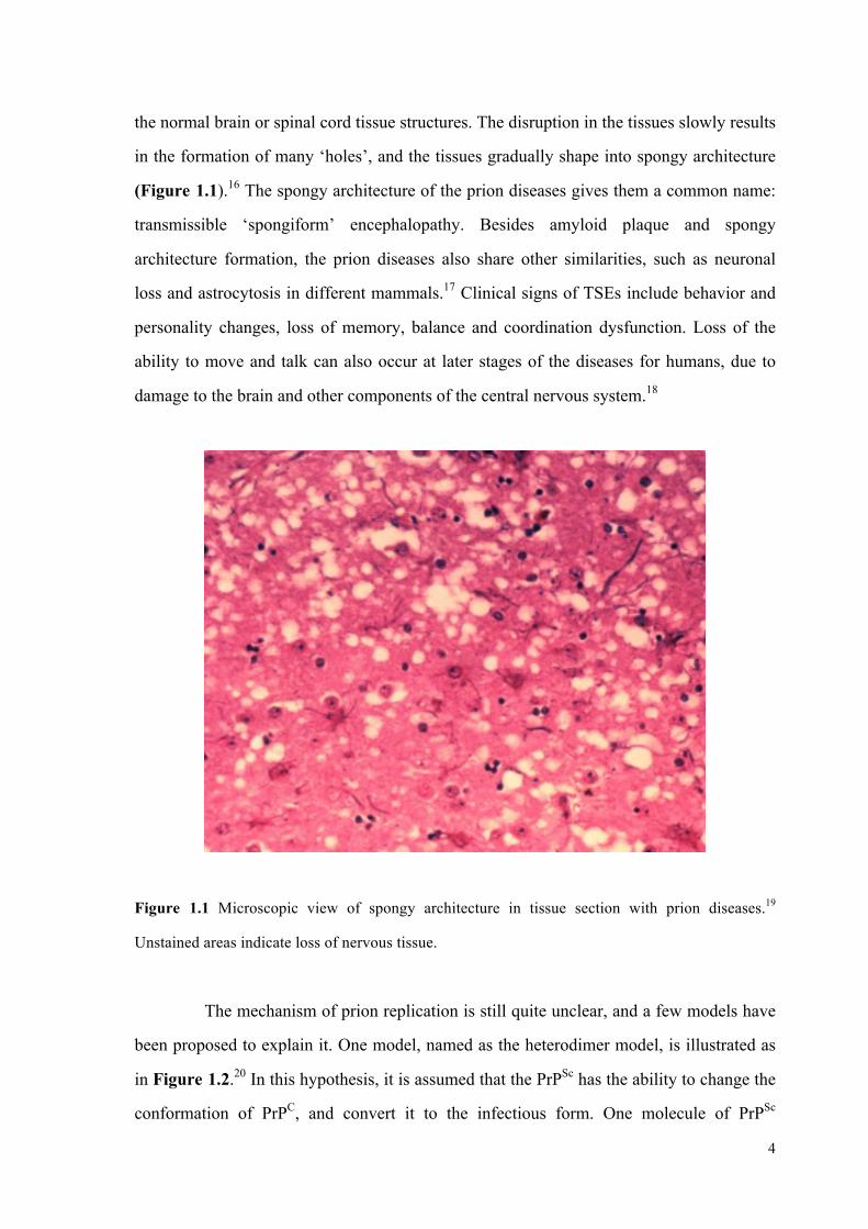
the normal brain or spinal cord tissue structures. The disruption in the tissues slowly results
in the formation of many ‘holes’, and the tissues gradually shape into spongy architecture
(Figure 1.1).16 The spongy architecture of the prion diseases gives them a common name:
transmissible ‘spongiform’ encephalopathy. Besides amyloid plaque and spongy
architecture formation, the prion diseases also share other similarities, such as neuronal
loss and astrocytosis in different mammals.17 Clinical signs of TSEs include behavior and
personality changes, loss of memory, balance and coordination dysfunction. Loss of the
ability to move and talk can also occur at later stages of the diseases for humans, due to
damage to the brain and other components of the central nervous system.18
Figure 1.1 Microscopic view of spongy architecture in tissue section with prion diseases.19
Unstained areas indicate loss of nervous tissue.
The mechanism of prion replication is still quite unclear, and a few models have
been proposed to explain it. One model, named as the heterodimer model, is illustrated as
in Figure 1.2.20 In this hypothesis, it is assumed that the PrPSc has the ability to change the
conformation of PrPC, and convert it to the infectious form. One molecule of PrPSc

5
interacts with one molecule of PrPC, and PrPSc works as the catalyst. After the conversion
is done, both PrPSc molecules separate to alter more PrPC to its infectious isoform. Thus,
according to this hypothesis, the rate of PrPC to PrPSc conversion increases exponentially as
the disease progresses, which consists with the finding that the disease progresses very
rapidly at the later phase. However, this model does not fully explain why the incubation
period is so long if the amount of PrPSc increase is also exponential during early stages. On
the other hand, it is possible that in spite of exponential increase, accumulated PrPSc might
have to reach a certain level (threshold) to cause disease symptoms.
Endogenous PrPCInteraction between PrPC and PrPSc
Conversion of PrPC to PrPSc
Accumulation of PrPSc
Spontaneous generationof PrPSc; Mutation of PrPC to PrPSc; Infection of PrPSc
Figure 1.2 Heterodimer model of prion replication.20 In the cycle, interaction of PrPSc (dark circle)
with PrPC (light circle) causes conformational changes in PrPC, resulting in the conversion of PrPC
to PrPSc.
An alternative prion replication model, known as the fibril model, is introduced
as in Figure 1.3. This model suggests that PrPSc only exists in the aggregate form of

6
‘fibrils’, rather than PrPSc monomers. The replication cycle is initiated by either infection
of fibrils from external sources or nucleation of PrPC. Since nucleation is rare, the cycles
are likely to start with infection of fibrils in most cases. In the replication cycle, the end of
fibril binds with PrPC and converts it to its misfolded form. As the fibrils grow, they can
later break into multiple new seeds to initiate more replication cycles, and thus the quantity
of fibrils also increases in a manner of exponential growth.21
Endogenous PrPC
Interaction between PrPC and fibrils
Conformational change of PrPC to PrPSc, and elongation of fibrils
Breakage into multiple new seeds for new cycles
Infection of fibrils; Nucleation of PrPC (rare)
Fibrils
Figure 1.3 Fibril Model of Prion Replication.21a, b In this model, PrPSc only exists in the form of
fibrils. Interaction of fibrils (dark squires) with PrPC (light circle) causes conformational changes in
PrPC, resulting in the conversion of PrPC to PrPSc and the growth of the fibrils.

7
The main cause of transmission of prion diseases is through infection, for
example, direct contact with affected particles, such as oral digestion of infected food,
blood transfusion, or by contact with other body fluids.22 Another cause of these diseases is
considered as familial or genetic, as a mutation of the prion protein PRNP gene is found in
all inherited cases.16 The spontaneous occurrence of TSEs in humans is also discovered
where no gene mutation or obvious exposure to external prion resources is identified, but it
is very rare.23
The tightly packed prion structure is extremely stable, and it is resistant to many
common biological, chemical or physical denaturizing methods, such as protease, boiling,
irradiating and so on.24 As a result, the established methods to combat microbial infection
are not effective towards prions at all. This makes disposal of the contaminated animal
particles very difficult, as prion protein can linger in the environment in the infective form
for a very long time.25
Unfortunately, there is still no effective vaccine, treatment or therapy for the
universally fatal disease. The only existing way that we can minimize the danger and cost
is to take precautions against the disease to prevent the spread. The common practice
includes isolation, killing, burning of the affected animals, and deep burying of the remains
of these animals beneath the ground. Also, feeding the cattle with cooked meat or bone
meal of slaughtered cattle is strictly banned now, which was once a very common but
unsafe practice in the European cattle industry.26
In conclusion, the prion diseases are life-threatening diseases across many
species, including humans, and there is still no effective vaccine, treatment or therapy. We
hope that our effort and work for this prion project can help making new progress toward
this great challenge.
1.2 Challenges and Recent Progresses in Prion Research
The prion diseases are a big mystery, and they have drawn a lot of attention from
scientists worldwide. However, not much progress has been made so far towards their

8
treatment, not only because the TSEs have only been identified in very recent decades, but
also because the research is highly challenging. A few challenges are as follows:
1. The precise mechanism of conversion of PrPC to PrPSc is poorly understood, and
the 3D structure of PrPSc is also unknown. No monomer of PrPSc has ever been
isolated, and the aggregation of PrPSc makes the effort to reveal its 3D structure
even more difficult;
2. The incubation period of TSEs in animals and humans is generally very long,
ranging from a few months to many years. Thus projects in prion research are often
costly and span long time frames;
3. Prion diseases are hard to diagnose in the early stages, due to the fact that no
immune response can be triggered by prion. However, when the symptoms appear,
the diseases develop very rapidly until death occurs;
4. The prion is primarily composed of protein that has highly compact and stable
structure. Therefore, the prion protein is resistant to most established denaturing
methods that are effective for common viruses or bacteria. In addition, the
development of new denaturing methods specifically for the prion protein is also
very difficult;
5. The understanding of transmission of the diseases is still very limited, even though
direct contact with prion particles is generally considered as the main pathway.
Mostly, if not entirely, these reasons make the treatment of the prion diseases still a great
global challenge.
Recent research into diagnosis, vaccination, or possible cures for prion diseases
suggests a few interesting and promising options towards the treatment of the illness.27 The
main strategies include, but are not limited to, the prevention of the conversion of PrPC to
PrPSc, clearance of the aggregation of misfolded proteins and so on, and these can be
achieved by targeting of PrPC, PrPSc or the intermediate in the process of conversion by
anti-prion compounds or antibodies. Discussion for research progress will focus on mainly
four areas, which are genetic techniques, immunomodulation, immunotheraputics and
discovery of anti-prion compounds
Experiments of transgenic techniques by several groups show interesting results.

9
One effective approach developed by the Collinge group to inhibit further development of
the prion diseases in infected mice is to knockout neuronal PrPC through transgenic
techniques at the early or mid stage of incubation period.28 The result of these experiments
also validates the hypothesis that the presence of PrPC is necessary for the propagation and
development of the prion diseases. However, the research is still at the experimental level,
and it is not directly applicable to humans. Aguzzi and collaborators reported another
experiment using transgenic techniques to produce dimeric protein PrP-Fc2, which is a
fusion protein of PrPC and immunoglobulin Fcγ.29 PrP-Fc2 cannot be converted to the
disease-related isoform, and they have higher affinity towards PrPSc than endogenous PrPC.
Consequently, PrP-Fc2 is competitive to block the interaction of PrPC and PrPSc in
transgenic mice of wild-type Prnp background, and thus prolong the incubation time.
Studies by different groups on immunomodulation of the prion diseases also led
to some interesting discoveries which suggested possible ways to develop effective
vaccines and theraputics for the diseases.27, 30 Methods of immunomodulation that are
found to be able to prolong incubation time slightly or modestly include:
1. Active immunization with prion peptide prior to infection;31
2. Passive immunization with monoclonal antibodies;32
3. Immunostimulations by cytidyl-guanyl oligodeoxynucleotide 1826 (CpG 1826)
alone or with complete Freund’s adjuvant (CFA);33
4. A combination of immunosuppression and anti-inflammatory agent treatment.34
Some progress has been made in immunotherapeutics as well. Anti-prion
antibodies with specificity and high affinity have been discovered through in vitro
screening and animal model testing, which are able to reduce infectivity and inhibit PrPSc
accumulation.27a These antibodies can mainly be divided into several sub-categories as
monoclonals, polyclonals, and single chain variable fragments, which include V5B2,
6H10, 6H4, D13/D18 (Fab), SAF34/SAF61, 110/31C6/44B1/72, Sha31, 3S9/2H9,
8B4/8H4, ICSM18/35, 6D11, 15B3.
In addition, a few small molecules were found to have certain promising affinity
towards the PrPSc or were able to prolong the incubation period after thousands of synthetic
or natural compounds were screened by different research groups in cell-based models or

10
animal models.27a These anti-prion compounds can mainly be divided into several types
based on their structures:
1. Congo red (CR) and its analogues,35 which is widely used as a dye for
histopathological stain of amyloid deposits;
2. Suramin and its derivatives and analogues,36 which share certain structural
similarity as congo red;
3. The antimalarial compound quinacrine37 and its related hetero-tricyclic
molecules,38 such as chlorpromazine and acridine;
4. Tetrapyrrole molecules, such as phthalocyanines and porphyrins;39
5. Dendritic polyamines and other cationic polyamines;40
6. Polysulphated polyanionic compounds, such as heparan sulphate and analogues,41
dextran sulfate;42
7. Nuclease-resistant 2’-amino(deoxy)pyrimidine-modified RNA aptamers;43
8. PrP-derived peptides,44 which are able to break β-sheets;
9. Other small molecules and divalent metal ions,45 such as curcumin, statins, zinc,
manganese, copper and copper chelator.
Based on the previous discussion, progress at genetic technique,
immunomodulation, immunotheraputics and discovery of anti-prion compounds provide
some promising results towards vaccines and treatment of prion diseases. However, most
of them are at preclinical levels, and the results are unpredictable from in vitro to in vivo
system or from animal models to humans.
1.3 Significance and Goal of Our Research
Although some small molecules have been discovered to have promising
anti-prion properties, and they are effective in some cases in either prolonging incubation
time or reducing severity of prion diseases in animal models, it remains unknown whether
they will also be effective in humans. Thus, it is desirable to have additional more effective
compounds available for future clinical trials to increase the probability of obtaining

11
therapeutically effective compounds in humans. In particular, compounds with higher
PrPSc binding affinity than those currently available ones should be developed, because
they can more effectively prevent the conversion of PrPC to its mis-folded isoform.
As a synthetic chemistry group, our strategy was to take advantage of these
existing small molecules, which are known to have promising features, to make multimeric
molecules with multi-binding units of improved binding affinity towards PrPSc.
Specifically, the main objective of this project was to synthesize a library of multimeric
(dimeric, trimeric or tetrameric) compounds with two, three or four effective binding units
(Figure 1.4). Based on the knowledge that infectious prion particles consist of small
oligomeric aggregates of PrPSc, the binding affinities of the newly synthesized compounds
are expected to increase significantly through multivalent interactions, and some of them
may be effective in halting the conversion of PrPC to PrPSc.39
LinkerScaffolds Binding unit
A B C
Figure 1.4 Generic structures of dimeric (A), trimeric (B) or tetrameric(C) compounds.
Besides their anticipated higher binding affinity and stronger anti-prion functions,
the multimeric compounds might offer two additional benefits, including determination of
the 3D structure of PrPSc and early detection and location of prion in vivo. The 3D structure
of PrPSc is still quite unclear, and because PrPSc tends to aggregate to form insoluble plaque,
the structure has been more difficult to be revealed. With the help of multimeric
compounds that have high affinity towards PrPSc, there is a possibility of obtaining the

12
crystal structure of PrPSc bound with one multimeric anti-prion compound. If the crystal
can indeed be obtained, the 3D structure of PrPSc may be revealed by X-ray
crystallography. Although such an event is improbable, the potential importance of the
outcome makes it a worthwhile effort. In addition, there is another probability to
characterize the anti-prion compound stabilized PrPSc oligomers by NMR methods.
Revealing the structure of PrPSc can not only help to improve the understanding of the
mechanism of PrPC to PrPSc conversion, but also help to design and develop new effective
drugs specifically by targeting the potential binding sites/cavities in the structure.
The second potential benefit is that the availability of multimeric fluorescent
compounds may facilitate early detection and location of the prion in vivo. Although some
monomeric anti-prion compounds, such as acridine, are fluorescent and can be activated to
give off fluorescent signals in vivo by binding to prion, their relatively weak binding
affinity results in poor resolution between the target protein and background signal due to
nonspecific binding. However, multimeric fluorescent compounds may provide much
better resolution and potentially stronger fluorescent signals due to their higher binding
affinity towards PrPSc. Our plan was to synthesize and find new multimeric fluorescent
compounds with high affinity towards PrPSc by utilizing these monomeric fluorescent
compounds as binding sites. If these compounds can concentrate in the area where there is
a high concentration of PrPSc, they will help early detection and location of the prion in
vivo. Early diagnosis of prion diseases is crucial for effective treatment before extensive
damage to the central nerve system has occurred.
1.4 Project Rationale
This initial pilot study was carried out to synthesize a library of multimeric
compounds that may have the capability to effectively bind to PrPSc. Multimeric
compounds would be generated by adding two or more monomeric anti-prion binding units
to a scaffold molecule, which is capable of accepting two to four monomeric binding units.
Disubstituted congo red, trisubstituted trimesic acid and tetrasubstituted

13
porphyrins were selected as scaffolds for the three categories of multimeric compounds for
mainly two reasons (Figure 1.5). One reason is that they are able to provide a rigid and flat
platform so that the binding units can be extended to different directions. The other reason
is that they are multimeric molecules and this allows them to link to several monomeric
binding units, and thus generating a structure with anticipated enhanced PrPSc binding
affinity. In addition, porphyrins are fluorescent compounds, and the target molecules may
be fluorescent as well if a porphyrin molecule is used as the scaffold.46
HN
N
NH
N
OOH
O
OHO
O
HO
OOO
OH
NNNN
SO2NaNaO2S
NH2 H2N
OH
OHHO
meso-Tetrakis(4-carboxyphenyl)porphyrin
Trimesic acidCongo red
Figure 1.5 Structures of selected scaffolds.
Diamines were selected as linkers to connect to the scaffold through amide bond
formation or palladium catalyzed coupling reactions (Figure 1.6). These diamines were
chosen as linkers, because they have different rigidity and length. By building target
molecules that have linkers with various lengths, it was hoped that some members of the
library would have the right length to simultaneously complex with several binding sites in
PrPSc oligomers, and thus show enhanced affinity.

14
NH
HN
H2N NH2
H2NNH2
H2N NH2
H2N NH2 H2NNH2
Piperazine 1,6-Hexanediamine
1,4-Butanediamine1,3-Propanediamine1,2-Ethanediamine
2,2-Dimethyl-1,3-propane diamine
Figure 1.6 Structures of linkers.
Chloroquinoline47 and methoxyacridine38 were selected as the binding units,
because they already show binding affinity towards PrPSc and are known to have anti-prion
activity. It was hoped that the binding affinity of target molecules would be significantly
enhanced through multivalent interaction between these known anti-prion binding units
and PrPSc aggregates (Figure 1.7). In addition, some acridines display fluorescence, and
the target molecules may be fluorescent if acridines are used as the binding units. With
these multimeric target molecules in hand, they were to be evaluated for anti-prion activity
by the Westaway group at the Centre for Prions and Protein Folding Diseases at the
University of Alberta.
N N
Cl
Cl
OCl
4,7-Dichloroquinoline 6,9-Dichloro-2-methoxyacridine
Cl
Figure 1.7 Structures of binding units.
Based on which scaffold is used, target molecules can be classified into three
types. The first type of anticipated products are azo dye-based dimeric compounds as in
Figure 1.8, mainly congo red and its derivatives. These targets were the focus of Dr. Lei
Li.

15
N N
ClCl
n = 0, 1, 2, 3 ...
NNNN
SO2NaNaO2S
N N
HN NHn n
Figure 1.8 Generalized structures of congo red-based dimeric products.
The second type of target multimeric products is a series of 1,3,5-trisubstituted
arene-based compounds, which are symmetrical molecules with three binding units
(Figure 1.9). Shaon Joy synthesized and purified this type of target molecules.
OOO
NH
HN
HN
HN
HN
NH
N N
N
Cl
Cl
Cl
n
n = 0, 1, 2, 3 ...
n n
Figure 1.9 Generalized structures of 1,3,5-trisubstituted arene-based trimeric products.
The third type of anticipated products are porphyrin-based multimeric
molecules as in Figure 1.10. As discussed earlier, their flat tetrameric structure makes
them suitable scaffold for organic synthesis. The synthesis, purification and
characterization of these compounds are the subject of this chapter.

16
HN
N
NH
N
ONH
O
ONHHN
O
NH
HN
HN
HNNH
N
N N
N
Cl
ClCl
Cln = 0, 1, 2, 3 ...
nn
n n
Figure 1.10 Generalized structures of porphyrin-based tetrameric products.
Besides exploring the efficient synthetic routes towards the three types of
multimeric compounds, there is another plan to make fluorescent compounds when the
routes of synthesis are well established. To achieve this, one pathway is to replace one of
the binding units with a fluorophore, as illustrated by icon A in Figure 1.11. One example
of the fluorophore is the cyanine dye Cy5, which can avoid quenching by azo dye,
porphyrins or the acridine binder, because it emits longer wavelength.48 Besides this, there
are two other pathways to prepare fluorescent compounds. Simple installation of
fluorescent binding units or usage of fluorescent scaffolds may achieve this goal as well, as
exampled by icon B and C. When both binding units and scaffold are fluorescent, the
target molecule as icon D might be fluorescent as well, although the potential for internal
quenching must be taken into account.

17
Binding sites
Fluorescent tag
Fluorescent binding sites
A B
Fluorescent scaffold
Scaffold
Linker
C D Figure 1.11 Generic multimeric fluorescent products.
The properties of ideal candidate molecules that may be clinically useful to treat
prion diseases are highlighted in the following box:
1. The compounds show strong anti-prion activity;
2. They are fluorescent. Although this property is not necessary for
compounds to be a drug candidate, fluorescent compounds provide
additional benefits;
3. They can go across cell membrane easily;
4. They are able to go across blood-brain barrier;
5. They are resistant to digestion and are stable in vivo;
6. They are not toxic towards normal cells in humans or animals.
1.5 Synthesis of Porphyrin-Based Multimeric Molecules
1.5.1 Using Quinolines as the Binding Units

18
The porphyrin-based multimeric molecules are the target molecules that may
have enhanced affinity towards PrPSc. An effective synthetic route needed to be explored
and established for us to readily access a library of these compounds. In the initial attempts,
the quinolines were employed as the binding units as in Figure 1.12, and other small
molecules would be used later as the binding units once the synthetic routes have been well
established.
HN
N
NH
N
HN
O
NH
O O
ONH
HN
N
Cl
linker
linker linkerNH
HN
linker
HN
N
Cl
N
Cl
N
Cl
NH
Target molecules Figure 1.12 The target molecules with quinolines as binding units.
In the retrosynthetic analysis, the key steps to build these molecules mainly
include assembling the porphyrin molecules and formation of the amide bond linkages. As
in Figure 1.13, two alternative synthetic routes A and B were proposed, differing only in
the order of steps. Since the synthesis of amide bond has already been well established and
widely applied,49 we envisioned using this step last. As a result of this decision, the
porphyrin would be assembled using a relatively simple carboxybenzaldehyde building
block. With the choice of route A, route B was held in reserve as a contingency plan in
case the first approach was not satisfactory.

19
HN
N
NH
N
O
HO OHO O
O
NClOHHO
HN
HN
4
4
O
O H
HO+
NH2
+
linker
NCl
HN
4
NH
linker
OO
H
NCl
HN
NH2
linker
Target molecules
+
Route A
Route B
Figure 1.13 Retrosynthetic analysis of the target molecules.
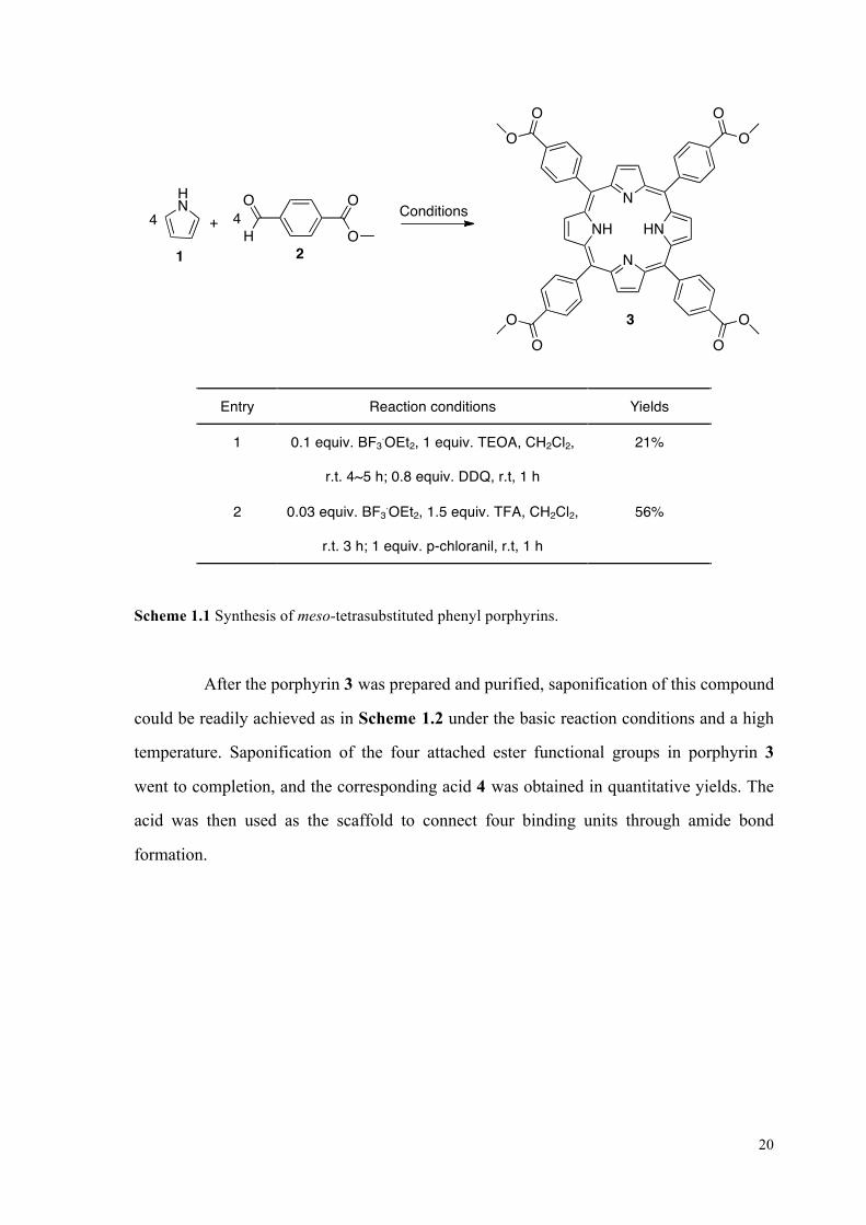
As in the synthetic route A, the first step is to make the central porphyrin
molecule from pyrrole 1 and simple benzaldehyde 2. Under the condition of Lewis acid
BF3.OEt2, the benzaldehyde 2 was consumed completely before oxidant DDQ or
p-chloranil was added.50 In a single pot reaction, porphyrin 3 is obtained in a low 21%
yield. The yield can be significantly improved to 56% by using BF3.OEt2 and TFA as the
cocatalysts, with subsequent addition of p-chloranil as the oxidant (Scheme 1.1).51
20
HN
N
NH
N
OO
O OO O
OO3
O
OH
O4
2
HN
4
1
+Conditions
Entry Reaction conditions Yields
1 0.1 equiv. BF3.OEt2, 1 equiv. TEOA, CH2Cl2,
r.t. 4~5 h; 0.8 equiv. DDQ, r.t, 1 h
21%
2 0.03 equiv. BF3.OEt2, 1.5 equiv. TFA, CH2Cl2,
r.t. 3 h; 1 equiv. p-chloranil, r.t, 1 h
56%
Scheme 1.1 Synthesis of meso-tetrasubstituted phenyl porphyrins.
After the porphyrin 3 was prepared and purified, saponification of this compound
could be readily achieved as in Scheme 1.2 under the basic reaction conditions and a high
temperature. Saponification of the four attached ester functional groups in porphyrin 3
went to completion, and the corresponding acid 4 was obtained in quantitative yields. The
acid was then used as the scaffold to connect four binding units through amide bond
formation.

21
HN
N
NH
N
OO
O OO O
OO
HN
N
NH
N
HOO
HO OHO O
OOH
quantitative
43
Basic conditions
Entry Conditions
1 4 wt% KOH solution, THF (v/v 3:4), 70-80 oC, 12h; 1N HCl solution
2 10 wt% NaOH solution, CH2Cl2, EtOH (v/v/v 1:1:1), 70-80 oC, 12h; 1N HCl solution
Scheme 1.2 Saponification of porphyrin 3 to get its corresponding acid 4.
Diamines were utilized as linkers to connect the quinoline binding units and the
porphyrin scaffold 4. Before the last step of amide bond formation, synthesis of
N-(7-chloroquinolin-4-yl)-alkyl-diamine compounds 12-17 was accomplished by heating
the mixture of excess diamine and quinoline for a few hours, in a 5:1 molar ratio without
any solvent as in Scheme 1.3.52 Six different products were obtained with a range of yields,
depending upon diamines used (Table 1.2).
NCl
Cl
80 oC, 1 h, no stirring;140 oC, 12 h, stirring1 +
5
5 H2N NH2HN
NH2
NCl
linkerlinker
Scheme 1.3 General method for preparation of N-(7-chloroquinolin-4-yl)-alkyl-diamines.

22
Entry Diamines Products Yields
1 H2N
NH26
N
HN
Cl
NH2
12
53%
2 7
H2N NH2
13N
HN
Cl
NH2
88%
3 H2N
NH2
8
14
N
HN
Cl
NH23
32%
4 H2N
NH2
9
N
HN
Cl
NH2
15
5
25%
5
10NH2H2N
N
HN
Cl
NH2
16
79%
6
NH
HN
11
17N
N
Cl
HN
52%1
1. Part of compound 17 is provided by Shaon Joy.
Table 1.2 A list of prepared N-(7-chloroquinolin-4-yl)-alkyl-diamine products 12-17.
With both the porphyrin 4 and the products 12-17 in hand, the target multimeric
molecules could be obtained through the amide bond formation as in Scheme 1.4 using a

23
5:1 ratio of N-(7-chloroquinolin-4-yl)alkyldiamine and porphyrin. The diamine partner was
used in excess to minimize the occurrence of incomplete conversion to the desired
tetraamide. The amide bond formation reaction worked smoothly in this case as well, and
the porphyrin-based tetra-substituted target molecule 19 was obtained in the yield of
70%.53
HN
N
NH
N
HN
O
NH
NH
O O
O
HN
19
NH
NH
HN
HN
N N
N N
Cl Cl
Cl Cl
HN
N
NH
N
O
HO OHO O
OOH4HO
HN
NCl
NH2
5+
12
EDC, HOBt. DMF, 40 h 70%
Scheme 1.4 Amide bond formation reaction to form one of the target molecules.
Therefore, a short and effective synthetic route has been established to make
porphyrin-based tetrameric compounds, which comprises of four steps in total from

24
commercially available starting materials. This synthetic route includes a sequence of
assembling prophyrin rings, saponification, synthesis of N-(7-chloroquinolin-4-yl)-alkyl
-diamine compounds, and amide bond formation.
Besides compound 19, five other tetrameric compounds 18, 20-23 as listed in
Table 1.3 were also prepared with this synthetic route by employing different diamines
and moderate to good yields have been obtained. They share linear or cyclic carbon chains
with different length and rigidity, as the length and rigidity of the chains may also play an
important role in enhancing the binding affinity towards PrPSc.

25
HN
N
NH
N
HN
O
NH
O O
ONH
HN
N
Cl
linker
linker linkerNH
HN
linker
HN
N
Cl
N
Cl
N
Cl
NH
Compounds 18 - 23
Entry Linkers Products Yields
1 H2NNH2
18 82%
2 H2N NH2 19 70%
3 H2NNH2
20 70%
4 H2NNH2
21 69%
5 H2N NH2
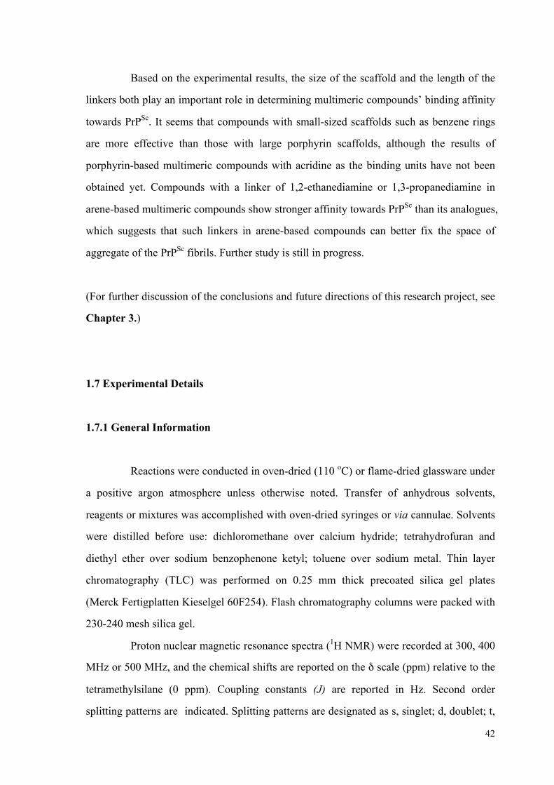
22 65%
6
NH
HN
23 79%
Table 1.3 Structures and yields of six tetrameric porphyrin products.
These newly synthesized multimeric compounds are very polar compounds and
all of them show poor solubility in most commonly used solvents in the lab, such as
hexane, EtOAc, acetone, DCM, toluene, Et2O, THF. In general, these compounds are
moderately soluble in DMF and DMSO at the room temperature, as the color change of

26
DMF or DMSO solution can be easily detected by eye when the brightly colored porphyrin
derivatives undergo dissolution. Most of them also show very slight solubility in methanol
as well. These properties of the compounds give rise to a few problems for purification and
characterization.
Many purification methods were tried to purify these porphyrin-based
multimeric compounds, including flash chromatography, preparative reverse-phase thin
layer chromatography, and recrystalization in different solvent systems. However, these
common purification methods were not successful for them.
Flash chromatography was not applicable to purify any of the six compounds,
because the compounds are very polar and their Rf values on TLC are all close to zero even
when a polar solvent system of 20% MeOH in DCM was used as the eluent.
The reverse-phase chromatography was also tried in small scale using
reverse-phase TLC plates. The sample of compound 18 was loaded on the reverse-phase
TLC, separation occurred when polar eluent solvent systems was used. The gel of each
band of separation was removed to recover the compounds on it. However, no expected
product was isolated and identified in this way. Furthermore, the efficiency is quite low,
due to the weak solubility of compounds in the eluent solvent as well as the solvent used to
load the compounds on the plates.
Recrystalization in two-solvent system, for example DMF and Et2O, was tried to
purify compounds 18, 19 and 22, but in all cases only precipitates rather than crystals of
the products were obtained.
Eventually, trituration was found to provide the best option for obtaining the
desired compounds free of soluble starting materials and impurities. As a result, the
impurities could be removed, but some products were lost as well. Also, solvents such as
water, MeOH and DMF are difficult to be removed completely from these purified polar
macromolecules, and trace amounts of toluene were also sometimes present when it was
used in the trituration, due to π-stacking. However, the presence of traces of solvents was
considered to be tolerable, since sample preparation for biological testing would entail
preparation of dilute DMSO solutions.
Similar problems also exist for the characterizations of these compounds due to

27
their poor solubility in most solvents. It is difficult to find the right solvent or solvent
system besides DMF and DMSO to dissolve these compounds for the analytical methods
of UV, fluorescence analysis and HRMS. Eventually, MeOH or a combination of MeCN,
H2O or MeOH provides a solution to the problems. However, only qualitative results of
fluorescence analysis have been obtained. All 1H NMR spectra (d-DMSO) of the
compounds have a shielded proton peak around -3 ppm (2H), which locates in the center of
the porphyrin. The purity of these compounds has also been confirmed by their 1H NMR
spectra, although solvent peaks (mostly water) do exist in most samples.
UV-Visible absorbance spectra of all the six molecules shows a significant Soret
band around 415-418 nm, which is a common feature of porphyrin molecules (Chart 1.1).
Broad Q bands around 550 nm were also observed in these samples, but the intensity was
found to be much weaker if compared with that of Soret bands.54 Besides the Soret and Q
bands, peaks around 220, 230 and 330 nm were also observed, and these absorbances were
attributed to the quinoline moieties.
Chart 1.1 UV-Vis absorbance spectrum of compound 18.
Fluorescence analysis was also carried out for all the products, as porphyrins are
expected to be fluorescent compounds. In general, after the excitation scans, wavelength of
425 nm was used for emission scan. Wavelengths of emission around 648 and 715 nm
were observed in all products, which is consistent with that of the starting porphyrin

28
material 3 and 4 (Chart 1.2). Therefore, all six of the products are fluorescent compounds.
This characteristic could be very useful, if these molecules were also found to have high
affinity towards PrPSc, because fluorescent compounds that bind effectively to PrPSc could
be used to detect and locate infectious prion particles in vivo.
Chart 1.2 Fluorescence analysis of compound 18. When excited by light of 425 nm, emission of
648 and 715 nm were observed.
Therefore, the porphyrin-based multimeric compounds by using quinolines as the
binding units have been synthesized and purified, and the results have been confirmed by
the characterization methods of IR, UV, fluorescence analysis, HRMS, 1H NMR and 13C
NMR.
1.5.2 Using 9-Amino-acridine as the Binding Units
Besides quinolines, 9-amino-acridines are also well known anti-prion
compounds that show promising affinity towards PrPSc.38 Therefore, it was considered
desirable to make a second series of porphyrine-based tetrameric compounds by using
9-amino-acridine as the binding units. These compounds were prepared by following the
synthetic route and purification methods described in the previous section.

29
The N-(6-chloro-2-methoxyacridin-9-yl)-alkyl-diamine compounds could be
prepared through the reaction of direct coupling of the corresponding 6-chloroacridine and
diamine with excellent yield as shown in Scheme 1.5.55
N NCl Cl
OOCl HN
NH2n
H2N(CH2)(n+1)NH2, 90 oC, 4h;
90-95%
Scheme 1.5 The reaction to prepare N-(6-chloro-2-methoxyacridin-9-yl)-alkyl diamine
compounds.
Four starting materials of N-(6-Chloro-2-methoxyacridin-9-yl)-alkyl diamine
compounds 25-28 listed in Figure 1.14 were prepared and provided to me by following
literature procedures by another group member, Dr. Kendre Dhananjay, as he has extra
amount of these materials besides his own usage.56
N
HN
Cl
O
N
HN
Cl
O
NH2 NH2
n=1, 25n=2, 26n=3, 27
28
n
Figure 1.14 Structures of four N-(6-Chloro-2-methoxyacridin-9-yl)-alkyl diamine compounds.
The last step of amide bond formation as in Table 1.4 also worked well in the
case of three N-(6-chloro-2-methoxyacridin-9-yl)-alkyl-diamine compounds 26-28 to
furnish the second series of target molecules, and different products have been obtained in
good or moderate yields.

30
HN
N
NH
N
O
HO OHO O
OOH
4HO
HN
NCl
NH2
5
+
26-27
EDC, HOBt. DMF, 48 h
On
n = 2,3
HN
N
NH
N
HN
O
NH
O O
ONH
HN
N
Cl
linker
linker linkerNH
HN
linker
HN
N
Cl
N
Cl
N
Cl
NH
Compounds 30-32
O
O O
O
HN
NCl5
28
ONH2
or
Entry Linkers Related products Yields
1 H2N NH2 30 75 %
2 H2NNH2
31 57 %
3 H2N NH2
32 54 %
Table 1.4 Reactions of amide bond formation to get to the second series of target molecules

31
In the case of compound 25, however, the reaction did not go to completion, and
a mixture of mono-, di- and tri-substituted compounds besides the desired tetra-substituted
product 29 were obtained in an overall yield of 76%. In another attempt, the reaction did
not go to completion either, although an excess amount of the starting material 25 (10
equiv.) was used and the reaction time was extended to 4 days. In both reactions, the
undesired mono-, di- and tri-substituted compounds were identified by both low- and
high-resolution mass spectrometry (LRMS & HRMS), and the peak of tri-substituted
product was always the most significant peak among the undesired products (Figure 1.15).
The products in the mixture cannot be separated from each other, because they are all very
polar compounds with weak solubility in most solvents. Compound 25 has a shorter
1,2-ethanediamine linker than those of compounds 26-28. The shorter linkers may increase
the effect of π-stacking, and thus decrease the solubility of the products of compound 25.
This may explain the lower reactivity of compound 25 in the amide bond formation
reaction, as once some of the undesired products precipitates, they will not react further
more.

32
HN
N
NH
N
NO
N NO O
ON
N N
N N
N N
N N
Cl Cl
Cl Cl
O O
O O
H H
H H
H H
H H
HN
N
NH
N
NO
N NO O
OOH
N
N N
N
N N
Cl
Cl Cl
O O
O
H
H H
H H
H
Compound 29
HRMS (MALDI) calculated for C112H87Cl4N16O8 ([MH]+) 1923.5641, found 1923.5641
HRMS (MALDI) calculated for C96H73Cl3N13O8 ([MH]+) 1640.4765, found 1640.4772
The undesired tri-substituted product
Figure 1.15 The structures of the desired compound 29 and undesired tri-substituted product.
The polarity of the products 30-32 was found to be very similar as the

33
quinoline-binded compounds in Section 1.5.1, as they are also very polar compounds that
cannot be purified by flash chromatography. The solubility of these
9-aminoacridine-linked compounds is even weaker than the quinoline-linked products.
They only show slight solubility in DMF and DMSO at the room temperature, and they can
be dissolved in methanol very slightly as well. Similarly, these compounds were purified
by trituration to remove soluble impurities. They are characterized by the analytical
methods of IR, UV, fluorescence analysis, HRMS and 1H NMR. However, the signals in 13C NMR spectra were very weak for the three compounds due to their weak solubility,
and many sp2 carbons were missing for compounds 30 and 31 in their 13C NMR spectra.
UV-Visible absorbance spectra of the three samples shows a significant Soret
band around 416-418 nm and a weak broad Q band between 500 and 650 nm (Chart 1.3).
In addition, strong peaks around 225, 270 nm were also observed, which may be the
characteristic peaks of 9-amino-acridine moieties.
Chart 1.3 UV-Visible absorbance spectrum of compound 30.
Furthermore, these compounds were found to be fluorescent as well. When they
were excited at 430 nm, emission at wavelengths of around 475, 650, 720 nm was
observed (Chart 1.4). Besides the porphyrin emission of 650 and 720 nm, emission of 475

34
nm is from the 9-amino-acridine moieties.
Chart 1.4 Fluorescence analysis of compound 30. When the compound is excited by light of 430
nm, emission of wavelength of 470 nm, 650 nm and 715 nm were observed.
1.6 Preliminary Biological Evaluation of Porphyrin-Based Multivalent Prion Binders
1.6.1 Introduction of Experimental Designs & Evaluation Results for Porphyrins 3-4
and Mono-Binding Units 12-17
After the compounds were synthesized and purified, the samples were prepared
in DMSO in the concentration of 1 mM or 0.5 mM. They were sent to our collaborator, the
Westaway group, for anti-prion assays. The starting materials 3-4 and 12-17 used to build
porphyrin-based multimeric compounds were also submitted for the assay for the purpose
of comparison and analysis.
The anti-prion assay is based on Scrapie-infected Mouse Brain (SMB) cells in
vitro.57 The SMB cells were treated with different concentrations of putative anti-prion
compounds for six days. Treated cells were digested by Proteinase K (PK) to remove the
proteins except the proteinase resistant PrPSc. Western blotting analysis was performed to
quantify PrPSc levels after treatment with compounds.58 A sample from untreated cells was

35
also included in the analysis to serve as a control. The more effective an anti-prion
compound is, the less amount of PrPSc will be left in the sample after the six-day treatment.
In the western blotting analysis, the three bands of un-, mono-, di-glycosylated PrPSc
proteins are detected between 16 and 36 kilodaltons (kDa). There is a reference band
where no treatment of putative anti-prion compounds but blank DMSO solution is applied
for the purpose of comparison. The band color intensity in the film indicates the amount of
PrPSc left. Densitometric analysis of the results of western blots gives a semi-quantitative
evaluation of the effectiveness of the anticipated anti-prion compounds.
The results of the mono-binding units 12-17 show that these compounds exhibit
anti-prion activity more or less at the concentration of 5 µM (Figure 1.16 and Chart 1.5).
At this concentration, compound 16 is very effective in completely clearing the amount of
PrPSc, and compounds 13 and 15 are also quite effective in reducing the amount of PrPSc by
more than 50%. Although the other three compounds are not as effective as compounds 13,
15 and 16, they still show positive effect in lowering the levels of PrPSc by from 20% to
40%.

36
NCl
HN NH2linker
Figure 1.16 Antiprion activity of N-(7-chloroquinolin-4-yl)-alkyl-diamines 12-17.59 Western
blotting analysis of SMB cells was utilized to measure the anti-prion activity of the
N-(7-chloroquinolin-4-yl)-alkyl-diamine compounds at concentrations of 0, 0.3, 0.6, 1.25, 2.5, or 5
µM after the 6-day treatment. Three bands in the film between 16 and 36 kDa are the un-, mono-,
di-glycosylated PrPSc proteins that resist to PK digestion. The band intensity indicates the amount
of PrPSc in the assay after the treatment of different compounds. The bands in the first column from
left are the reference bands from untreated cells. As an example, compound 16 is able to clear PrPSc
efficiently at the concentration of 5 µM.

37
Chart 1.5 Densitometric analysis of Western blots from experiments of compounds 12-17 at
various concentrations.59 This is used to estimate the response of PrPSc propagation to various
concentrations of N-(7-chloroquinolin-4-yl)-alkyl-diamines. When there is no treatment, the PrPSc
level will be at 1. With complete clearance of PrPSc, PrPSc level will drop to 0. Increasing of the
amount of PrPSc will result in the increase in PrPSc level in this chart. It shows that compound 16 is
the most effective one in inhibiting the propagation of PrPSc under different concentrations.
Based on the outcome of the assays, the porphyrins 3 and 4 basically exhibited
little anti-prion activity at all concentrations, with an exception of the compound 4 at 5 µM,
where it shows slight inhibition by reducing 10% of PrPSc amount (Figure 1.17 and Chart
1.6).

38
HN
N
NH
N
O
O O
O
R
R R
R
Figure 1.17 Antiprion activity of simple porphyrins 3-4.59 Western blotting analysis of SMB cells
was utilized to measure the anti-prion activity of the compounds at concentrations of 0, 0.3, 0.6,
1.25, 2.5, or 5 µM, and the results show that both compounds are ineffective in inhibiting the
propagation of PrPSc.
Chart 1.6 Densitometric analysis of Western blots from experiments of compounds 3, 4 at various
concentrations.59

39
1.6.2 Evaluation Results for Porphyrin-Based Multimeric Compounds 18-23 &
Promising Results of Arene-Based Multimeric Compounds
Anti-prion assays of the porphyrin-based multimeric compounds showed that
unfortunately no significant reduction in the levels of PrPSc was observed in most cases
(Figure 1.18 and Chart 1.7). Only compounds 22 and 23 show slight anti-prion activity by
reducing the amount of PrPSc by less than 40% or 50% at high concentrations. In some
cases, however, the amounts of PrPSc increase slightly or significantly when the SMB cells
were treated with different concentrations of the six porphyrin-based multimeric products.
This suggests that this type of macromolecules may play a role in accelerating the growth
of PrPSc sometimes rather than inhibiting it, which is exactly the opposite of our
expectation. Despite the presence of four units, these multimeric compounds were less
effective than the simple monomeric quinoline diamine fragments in suppressing the
formation of PrPSc. It may be that the extended porphyrin scaffold does not permit
effective binding by all of the individual quinolines. Observation of enhanced production
of PrPSc in one case also raises the possibility that two conflicting mechanisms may be
largely cancelling each other out.
HN
N
NH
N
HN
O
NH
O O
ONH
HN
N
Cl
linker
linker linker
NH
HN
linker
HN
N
Cl
N
Cl
N
NH

40
Figure 1.18 Antiprion activity of six porphyrin-based multimeric molecules 18-23.59 Western
blotting analysis was utilized to measure the anti-prion activity of the compounds as listed, and the
results show that none of them is very effective in inhibiting the propagation of PrPSc.
Chart 1.7 Densitometric analysis of Western blots from experiments of porphyrin-based
multimeric compounds 18-23 at various concentrations.59

41
The corresponding acridine-containing multimeric porphyrin compounds 30-32
and the acridine diamine building blocks 25-28 have not yet been subjected to the same
assays as the quinoline series. However, the results described above suggest that these
compounds will be unlikely to display favorable activity. Even though the porphyrin-based
mutimeric fluorescent compounds do not have significantly enhanced anti-prion activity,
they still may have the potential application in diagnosis if they can bind effectively to the
prion particles. Therefore, it is desirable to have the affinity test of these fluorescent
compounds.
Although results for the six porphyrin-based tetrameric molecules are unexpected,
the outcomes for smaller sized arene-based trimeric compounds are very interesting and
promising (Figure 1.19). These molecules, prepared by Shaon Joy, show strong anti-prion
activity in vitro and are very efficient in the clearance of PrPSc at concentrations of 1.25
µM or greater. They show stronger anti-prion activity than quinacrine in the anti-prion
assay, which is a drug under full clinical trial in the United Kingdom, the United States and
Japan for the treatment of the human-based Creutzfldt-Jakob disease.37, 60 Therefore, the
results of arene-based compounds are very exciting and promising. Toxicity tests of both
the porphyrin-based and arene-based multimeric compounds show that all these
compounds are not toxic towards normal mouse brain cells at the concentration of 5 µM or
below, which are the concentrations used in the treatment.61
OOO
NH
HN
HN
HN H
N
NH
N N
N
Cl
Cl
Cl
linker linker
linker
Figure 1.19 Structures of arene-based trimeric compounds prepared by Shaon Joy. Linker =
-CH2CH2-, -CH2CH2CH2-, or -CH2C(CH3)2CH2-.
42
Based on the experimental results, the size of the scaffold and the length of the
linkers both play an important role in determining multimeric compounds’ binding affinity
towards PrPSc. It seems that compounds with small-sized scaffolds such as benzene rings
are more effective than those with large porphyrin scaffolds, although the results of
porphyrin-based multimeric compounds with acridine as the binding units have not been
obtained yet. Compounds with a linker of 1,2-ethanediamine or 1,3-propanediamine in
arene-based multimeric compounds show stronger affinity towards PrPSc than its analogues,
which suggests that such linkers in arene-based compounds can better fix the space of
aggregate of the PrPSc fibrils. Further study is still in progress.
(For further discussion of the conclusions and future directions of this research project, see
Chapter 3.)
1.7 Experimental Details
1.7.1 General Information
Reactions were conducted in oven-dried (110 oC) or flame-dried glassware under
a positive argon atmosphere unless otherwise noted. Transfer of anhydrous solvents,
reagents or mixtures was accomplished with oven-dried syringes or via cannulae. Solvents
were distilled before use: dichloromethane over calcium hydride; tetrahydrofuran and
diethyl ether over sodium benzophenone ketyl; toluene over sodium metal. Thin layer
chromatography (TLC) was performed on 0.25 mm thick precoated silica gel plates
(Merck Fertigplatten Kieselgel 60F254). Flash chromatography columns were packed with
230-240 mesh silica gel.
Proton nuclear magnetic resonance spectra (1H NMR) were recorded at 300, 400
MHz or 500 MHz, and the chemical shifts are reported on the δ scale (ppm) relative to the
tetramethylsilane (0 ppm). Coupling constants (J) are reported in Hz. Second order
splitting patterns are indicated. Splitting patterns are designated as s, singlet; d, doublet; t,

43
triplet; q, quartet; m, multiplet; br, broad; dd, doublet of doublets, dt, doublet of triplets,
etc. Carbon nuclear magnetic resonance spectra (13C NMR) were recorded at 100 MHz or
125 MHz and are reported in ppm relative to the centerline of the triplet from
deuterochloroform at 77.23 ppm. Infrared (IR) spectra were measured with a Magna 750
FT-IR infrared spectrophotometer. Ultraviolet-visible (UV-Vis) absorption spectra were
measured with a Hewlett Packard 8453 UV-VIS Spectrophotometer between 200-900 nm.
Fluorescence analysis was carried on Photon Technology International (PTI) MP1
Fluorescence Syetem. Mass spectra were determined on a Agilent 6220 oaTOF
electrospray positive ion mode spectrometer (ESI) or a Bruker Daltonics Apex-Qe FTICR
MS high resolution MALDI spectrometer. All reagents and catalysts were purchased from Aldrich or Sigma or Strem and
were used without further purification unless otherwise stated.
1.7.2 Substrate Synthesis
Preparation of meso-tetrakis [4-(methoxycarbonyl)phenyl]porphyrin 3.
HN
N
NH
N
OO
O OO O
OO
3
Freshly distilled pyrrole (694 µL, 10.0 mmol) was added to a solution of
methyl-4-formyl-benzoate (1.64 g, 10.0 mmol) in 1.00 L dry dichloromethane in a 3 L
3-neck round-bottom flask, which had been purged with argon. The mixture was stirred for
5 minutes, and the reaction vessel was shielded with aluminum foil to avoid ambient

44
lighting. Trifluoroacetic acid (1.11 mL, 15.0 mmol) was added to the reaction mixture, and
then BF3.OEt2 (37 µL, 0.30 mmol) was added. The reaction was stirred under argon for 2.5
h before p-chloranil (2.46 g, 10.0 mmol) was added to the solution. The flask was
immersed into a preheated 45 o C water bath, and the solution was stirred at reflux for
another 1 h. After cooling to room temperature, solvent was removed with a rotary
evaporator. The crude product was purified via flash chromatography (silica gel,
Et2O/DCM 1:19) to yield 3 (1.19 g, 56%) as purple solid: m.p. > 300 oC; Rf = 0.53 (1:19
Et2O/DCM); IR (microscope) 3319, 3120, 2951, 2843, 1722 cm-1; UV-Vis absorbance
peaks 229, 420, 451 nm; fluorescence analysis: when excited at 425 nm, wavelength of
emission detected at 640, 715 nm; 1H NMR (400 MHz, CDCl3) δ 8.82 (s, 8H), 8.45 (d, J =
8.1 Hz, 8H), 8.30 (d, J = 8.1 Hz, 8H), 4.12 (s, 12H), -2.80 (s, 2H); 13 C NMR (125 MHz,
CDCl3) δ 167.2, 146.6, 134.5, 131.3, 129.8, 128.0, 119.4, 52.5 (1 sp2 carbon missing);
HRMS (ESI) calculated for C52H39O8N4 ([M•H]+) 847.2762, found 847.2759.
Preparation of meso-tetrakis[(4-carboxy)phenyl]porphyrin 4.
HN
N
NH
N
HOO
HO OHO O
OOH
4
Compound 3 (166 mg, 0.20 mmol) was dissolved in 10 mL DCM, and the solution was
added to a mixture of ethanol/water (1:1 v/v, 20 mL) containing NaOH (1.00 g, 25.0 mmol)
in a 100 mL round-bottom flask. The mixture was stirred at reflux under argon at the
temperature around 70 – 80 oC for 24 h. After cooling to room temperature, ethanol and
DCM were removed with a rotary evaporator. 1N HCl was added dropwise to the aqueous

45
solution until no more green precipitate formed (pH = 1 by pH paper). The precipitate was
collected by vacuum filtration and washed with DI water several times. The compound 4
was dried under high-pressure vacuum for 8 h to afford the desired compound (155 mg,
quantitative yield) as a green powder: m.p. > 300 oC; IR (microscope) 3170, 3036, 1720,
1682, 1606 cm-1; UV-Vis absorbance peaks 230, 415 nm; fluorescence analysis: when
excited at 450 nm, wavelength of emission detected at 648, 715 nm; 1H NMR (400 MHz,
CDCl3) δ 8.85 (s, 8H), 8.45 (d, J = 8.1 Hz, 8H), 8.30 (d, J = 8.1 Hz, 8H), -2.80 (s, 2H), (4
COOH protons missing); 13 C NMR (125 MHz, CDCl3) δ 167.4, 145.4, 134.5, 131.6, 130.5,
127.9, 119.3 (1 sp2 carbon missing); HRMS (ESI) calculated for C48H27O8N4 ([M-H]-)
789.1991, found 789.1990; C48H27O8N4 ([M-2H]-2) 394.0959, found 394.0966.
One example for the direct coupling of 4,7-dicholoroquinoline and diamines.
Preparation of N-(7-chloroquinolin-4-yl)-propane-2,2-dimethyl-1,3-diamine 16.
N
HN
Cl
NH2
16
4,7-Dichlororoquinoline (198 mg, 1.00 mmol) was mixed with
propane-2,2-dimethyl-1,3-diamine (610 mg, 5.00 mmol) in a 10 mL round-bottom flask,
and it was heated at 80 oC for 1 hour without stirring to make a paste. The temperature was
then raised to 140 oC with stirring. After 12 hours, the reaction mixture was cooled to the
room temperature, and a 1M NaOH solution (5 mL) was added to the solution. The organic
product was extracted by EtOAc (5×10 mL), washed with DI water (2×5 mL) and dried
over MgSO4. Compound 16 was obtained by flash chromatography (15% MeOH/DCM) as
a pale yellow solid (208 mg, 79%): m.p. 128-130 oC; Rf = 0.52 (20% MeOH/DCM); IR
(DCM, cast film) 3474, 3315, 3081, 2958 cm-1; 1H NMR (400 MHz, CDCl3) δ 8.48 (d, J =
5.6 Hz, 1H), 8.46 (br s, 1H), 7.91 (d, J = 2.1 Hz, 1H), 7.72 (d, J = 8.8 Hz, 1H), 7.29 (dd, J
= 8.8, 2.1 Hz, 1H), 6.27 (d, J = 5.6 Hz, 1H), 3.18 (s, 2H), 2.87 (s, 2H), 1.56 (br s, 2H), 1.08

46
(s, 6H); 13 C NMR (125 MHz, d-DMSO) δ 151.9, 150.8, 149.1, 133.3, 127.5, 124.0, 123.9,
117.4, 98.6, 37.5, 35.6, 24.6, 24.4; HRMS (ESI) calculated for C14H19ClN3 ([M•H]+)
264.1262, found 264.1259.
The preparation of other N-(7-chloroquinolin-4-yl)-alkyl diamine compounds 12-15 and 17
were carried out by following the literature procedures (the same as described above),
which are reported compounds in the supplementary information of the literature.52
Reactions for the amide bond formation.
Representative Example:
Preparation of
meso-tetrakis[(N-(7-chloroquinolin-4-yl)-ethane-1,2-diaminecarbonyl)phenyl]porphyr
in 18.
HN
N
NH
N
HN
O
NH
NH
O O
O
HN
18
HN
HN
NH NH
N N
NN
ClCl
Cl Cl
A mixture of compound 4 (29.5 mg, 0.0373 mmol), compound 12 (41.2 mg, 0.186 mmol),
HOBt (25.2 mg, 0.186 mmol), and EDC (35.8 mg, 0.186 mmol) in DMF (2 mL) in a 10 ml
round-bottom flask was stirred at room temperature under argon atmosphere for 40 h. After

47
the reaction was complete, the solution in the round-bottom flask was transferred to a 25
ml test tube by a pipet. Et2O (20 mL) was added to the test tube to dilute DMF solution,
and dark red precipitate formed. The mixed solvent was removed by pipet carefully, and
the residue in the test tube was washed successively with DI water, DCM, acetone and
MeOH several times. The solid was dried under high-pressure vacuum for 8 h to afford the
desired compound 18 (49.0 mg, 82%) as a dark red solid: m.p. > 300 oC; IR (microscope)
3308, 3059, 2940, 1930, 1815, 1628, 1606, 1577, 1539 cm-1; UV-Vis absorbance peaks
220, 253, 331, 416 nm; fluorescence analysis: when excited at 425 nm, wavelength of
emission detected at 648, 715 nm; 1H NMR (400 MHz, d-DMSO) δ 9.13 (t, J =5.6 Hz,
4H), 8.84 (s, 8H), 8.47 (d, J = 5.2 Hz, 4H), 8.31 (m, 16H), 8.29 (d, J = 8.8 Hz, 4H), 7.81 (d,
J = 2.2 Hz, 4H), 7.62 (t, J = 5.2 Hz, 4H), 7.49 (dd, J = 8.8, 2.2 Hz, 4H), 6.73 (d, J = 5.2
Hz, 4H), 3.72 (td, J = 9.2, 5.2 Hz, 8H), 3.61 (td, J = 9.2, 5.2 Hz, 8H), -2.93 (s, 2H); 13 C
NMR (125 MHz, d-DMSO) δ 166.7, 152.5, 148.3, 144.7, 143.9, 135.3, 134.1, 133.8, 131.6,
125.8, 125.3, 124.7, 124.1, 119.4, 116.7, 98.7, 42.5, 38.0 (1 sp2 carbon missing); HRMS
(MALDI) calculated for C92H71Cl4N16O4 ([M•H]+) 1603.4593, found 1603.4604.
Preparation of
meso-tetrakis[(N-(7-chloroquinolin-4-yl)-propane-1,3-diaminecarbonyl)phenyl]porph
yrin 19.

48
HN
N
NH
N
HN
O
NH
NH
O O
O
HN
19
NH
NH
HN
HN
N N
N N
Cl Cl
Cl Cl
The procedure described above in the synthesis and purification of compound 18 was
applied in this example to give 19 (92 mg, 70 %) as a dark red solid: m.p. > 300 oC; IR
(microscope) 3315, 3066, 2936, 1928, 1810, 1633, 1610, 1581, 1547, 1500 cm-1; UV-Vis
absorbance peaks 217, 253, 330, 416 nm; fluorescence analysis: when excited at 425 nm,
wavelength of emission detected at 648, 715 nm; 1H NMR (400 MHz, d-DMSO) δ 8.96 (t,
J =5.6 Hz, 4H), 8.84 (s, 8H), 8.48 (d, J = 5.8 Hz, 4H), 8.41 (d, J =9.0 Hz, 4H), 8.30 (m,
16H), 7.94 (t, J = 5.1 Hz, 4H), 7.84 (d, J = 1.5 Hz, 4H), 7.56 (dd, J = 9.0, 1.5 Hz, 4H),
6.68 (d, J = 5.8 Hz, 4H), 3.57 (td, J = 8.8, 5.6 Hz, 8H), 3.47 (td, J = 8.8, 5.6 Hz, 8H), 2.01
(m, 8H), -2.93 (s, 2H); 13 C NMR (125 MHz, d-DMSO) δ 166.3, 152.5, 147.8, 144.2, 143.8,
135.5, 134.1, 134.0, 131.3, 125.8, 125.3, 124.8, 123.6, 119.4, 116.6, 98.7, 40.6, 37.3, 27.7
(1 sp2 carbon missing); HRMS (MALDI) calculated for C96H79Cl4N16O4 ([M•H]+)
1659.5219, found 1659.5201.
Preparation of
meso-tetrakis[(N-(7-chloroquinolin-4-yl)-butane-1,4-diaminecarbonyl)phenyl]porphy
rin 20.

49
HN
N
NH
N
HN
O
NH
NH
O O
O
HN
20
NH
NH
HN
HN
N N
N N
Cl Cl
Cl Cl
2 2
22
The procedure described in the synthesis and purification of compound 18 was applied in
this example to give 20 (77 mg, 70 %) as a dark red solid: m.p. > 300 oC; IR (microscope)
3312, 3062, 2941, 2867, 1716, 1628, 1605, 1576, 1531 cm-1; UV-Vis absorbance peaks
219, 253, 331, 416 nm; fluorescence analysis: when excited at 400 nm, wavelength of
emission detected at 648, 715 nm; 1H NMR (400 MHz, d-DMSO) δ 8.85 (t, J =5.6 Hz, 4H),
8.83 (s, 8H), 8.41 (d, J = 5.6 Hz, 4H), 8.32 (d, J = 8.8 Hz, 4H), 8.28 (m, 16H), 7.77 (s, 4H),
7.45 (d, J = 8.8 Hz, 4H), 6.56 (d, J = 5.2 Hz, 4H), 3.50 (J = 9.2, 5.2 Hz, 8H), 1.40 (td, J
=9.2, 5.2 Hz, 8H), 1.80 (m, 16H), -2.94 (s, 2H); 13 C NMR (125 MHz, d-DMSO) δ 166.1,
152.6, 147.5, 144.0, 143.7, 135.5, 134.2, 134.1, 131.1, 125.8, 125.2, 124.9, 123.4, 119.4,
116.5, 98.6, 42.5, 26.8, 25.5, 25.2 (1 sp2 carbon missing); HRMS (MALDI) calculated for
C100H87Cl4N16O4 ([M•H]+) 1715.5845, found 1715.5860.
Preparation of
meso-tetrakis[(N-(7-chloroquinolin-4-yl)-hexane-1,6-diaminecarbonyl)phenyl]porphy
rin 21.

50
HN
N
NH
N
HN
O
NH
NH
O O
O
HN
21
NH
NH
HN
HN
N N
N N
Cl Cl
Cl Cl
4 4
44
The procedure described in the synthesis and purification of compound 18 was applied in
this example to give 21 (130 mg, 61 %) as a dark red solid: m.p. > 300 oC; IR (microscope)
3258, 3027, 2933, 2859, 1943, 1631, 1612, 1551 cm-1; UV-Vis absorbance peaks 254, 330,
416 nm; fluorescence analysis: when excited at 425 nm, wavelength of emission detected
at 648, 715 nm; 1H NMR (400 MHz, d-DMSO) δ 8.86 (t, J =5.6 Hz, 4H), 8.84 (s, 8H),
8.45 (d, J = 9.2 Hz, 4H), 8.43 (d, J = 6.4 Hz, 4H), 8.28 (m, 20H), 7.85 (d, J = 2.0 Hz, 4H),
7.55 (dd, J = 9.2, 2.0 Hz, 4H), 6.64 (d, J = 6.0 Hz, 4H), 3.41 (m, 16H), 1.73 (m, 8H), 1.67
(m, 8H), 1.47 (m, 16H), -2.95 (s, 2H); 13 C NMR (125 MHz, d-DMSO) δ 166.1, 153.9,
145.0, 143.7, 141.3, 136.6, 134.3, 134.1, 131.4, 125.8, 125.4, 121.1, 119.4, 115.9, 98.5,
42.9, 30.6, 29.1 27.6, 26.3, 26.2 (2 sp2 carbons missing); HRMS (MALDI) calculated for
C108H103Cl4N16O4 ([M•H]+) 1827.7097, found 1827.7116.
Preparation of
meso-tetrakis[(N-(7-chloroquinolin-4-yl)-propane-2,2-dimethyl-1,3-diaminecarbonyl)
phenyl]porphyrin 22.
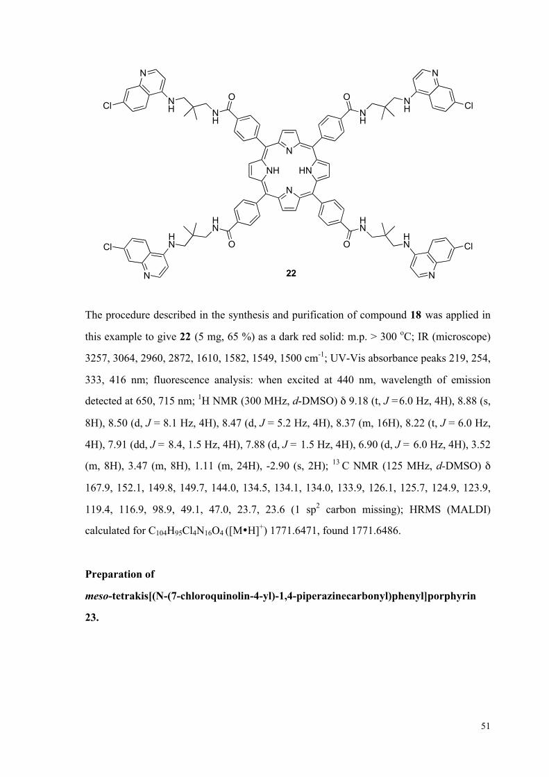
51
HN
N
NH
N
HN
O
NH
NH
O O
O
HN
22
NH
NH
HN
HN
N N
N N
Cl Cl
Cl Cl
The procedure described in the synthesis and purification of compound 18 was applied in
this example to give 22 (5 mg, 65 %) as a dark red solid: m.p. > 300 oC; IR (microscope)
3257, 3064, 2960, 2872, 1610, 1582, 1549, 1500 cm-1; UV-Vis absorbance peaks 219, 254,
333, 416 nm; fluorescence analysis: when excited at 440 nm, wavelength of emission
detected at 650, 715 nm; 1H NMR (300 MHz, d-DMSO) δ 9.18 (t, J =6.0 Hz, 4H), 8.88 (s,
8H), 8.50 (d, J = 8.1 Hz, 4H), 8.47 (d, J = 5.2 Hz, 4H), 8.37 (m, 16H), 8.22 (t, J = 6.0 Hz,
4H), 7.91 (dd, J = 8.4, 1.5 Hz, 4H), 7.88 (d, J = 1.5 Hz, 4H), 6.90 (d, J = 6.0 Hz, 4H), 3.52
(m, 8H), 3.47 (m, 8H), 1.11 (m, 24H), -2.90 (s, 2H); 13 C NMR (125 MHz, d-DMSO) δ
167.9, 152.1, 149.8, 149.7, 144.0, 134.5, 134.1, 134.0, 133.9, 126.1, 125.7, 124.9, 123.9,
119.4, 116.9, 98.9, 49.1, 47.0, 23.7, 23.6 (1 sp2 carbon missing); HRMS (MALDI)
calculated for C104H95Cl4N16O4 ([M•H]+) 1771.6471, found 1771.6486.
Preparation of
meso-tetrakis[(N-(7-chloroquinolin-4-yl)-1,4-piperazinecarbonyl)phenyl]porphyrin
23.

52
HN
N
NH
N
N
O
N N
O O
O
N23
N
NN
N
N N
N N
Cl
Cl Cl
Cl
The procedure described in the synthesis and purification of compound 18 was applied in
this example to give 23 (15 mg, 79 %) as a dark red solid: m.p. > 300 oC; IR (microscope)
3318, 3030, 2893, 2822, 2095, 1636, 1577 cm-1; UV-Vis absorbance peaks 226, 355, 415
nm; fluorescence analysis: when excited at 425 nm, wavelength of emission detected at
648, 715 nm; 1H NMR (300 MHz, d-DMSO) δ 8.93 (s, 8H), 8.78 (d, J = 6.6 Hz, 4H), 8.35
(d, J =7.8 Hz, 8H), 8.30 (d, J = 9.6 Hz, 4H), 8.07 (s, 4H), 7.96 (d, J = 7.8 Hz, 8H), 7.71 (d,
J = 9.6 Hz, 4H), 7.24 (d, J = 6.6 Hz, 4H), 4.08 (m, 16H), 3.86 (m, 16H), -2.89 (s, 2H); 13 C
NMR (125 MHz, d-DMSO) δ 169.1, 156.0, 152.2, 149.6, 142.3, 135.4, 134.3, 134.1, 133.7,
128.1, 126.1, 125.9, 125.8, 121.4, 119.4, 109.8, 51.9, 51.7 (1 sp2 carbon missing); HRMS
(MALDI) calculated for C100H79Cl4N16O4 ([M•H]+) 1707.5219, found 1707.5216.
Preparation of
meso-tetrakis[(N-(6-chloro-2-methoxyacridin-9-yl)-propane-1,3-diaminecarbonyl)phe
nyl]porphyrin 30.

53
HN
N
NH
N
HN
O
NH
NH
O O
O
HN
30
NH
NH
HN
HN
N N
N N
Cl Cl
Cl Cl
O
OO
O
The procedure described in the synthesis and purification of compound 18 was applied in
this example to give 30 (30 mg, 75 %) as a dark red solid: m.p. > 300 oC; IR (microscope)
3246, 3039, 2937, 2833, 1630, 1606, 1587, 1560 cm-1; UV-Vis absorbance peaks 268, 342,
416 nm; Fluorescence analysis: when excited at 430 nm, wavelength detected at 480, 650,
720 nm; 1H NMR (500 MHz, CDCl3) δ 9.01 (s, 4H), 8.84 (s, 8H), 8.56 (d, J = 8.1 Hz, 4H),
8.28 (d, J=8.1 Hz, 8H), 8.20 (d, J = 8.1 Hz, 8H), 8.12 (s, 4H), 7.98 (s, 4H), 7.90 (s, 4H),
7.85 (d, J = 8.1 Hz, 4H), 7.63 (d, J = 8.1 Hz, 4H), 7.47 (d, J = 8.1 Hz, 4H); 4.20 (br s, 8H),
4.01 (s, 12H), 3.57 (d, J = 5.5 Hz, 8H), 2.25 (br s, 8H), -2.93 (s, 2H); 13 C NMR (125 MHz,
d-DMSO) δ 166.2, 155.7, 143.8, 134.1, 133.7, 128.5, 126.6, 125.7, 123.0, 119.4, 56.2, 46.6,
36.8, 29.5 (11 sp2 carbons missing); HRMS (MALDI) calculated for C116H95Cl4N16O8
([M•H]+) 1979.6267, found 1979.6266. HRMS (ESI) calculated for C116H98Cl4N16O8
([M•4H]4+) 495.6621, found 495.6625; calculated for C116H97Cl4N16O8 ([M•3H]3+)
660.5471, found 660.5480; calculated for C116H96Cl4N16O8 ([M•2H]2+) 990.3170, found
990.3161.
Preparation of
meso-tetrakis[(N-(6-chloro-2-methoxyacridin-9-yl)-butane-1,4-diaminecarbonyl)phen
54
yl]porphyrin 31.
HN
N
NH
N
HN
O
NH
NH
O O
O
HN
31
NH
NH
HN
HN
N N
N N
Cl Cl
Cl Cl
2 2
22
OO
OO
The procedure described in the synthesis and purification of compound 18 was applied in
this example to give 31 (23 mg, 57 %) as a dark red solid: m.p. > 300 oC; IR (microscope)
3242, 3030, 2936, 2869, 1629, 1586, 1561 cm-1; UV-Vis absorbance peaks 223, 275, 344,
416 nm; Fluorescence analysis: when excited at 430 nm, wavelength detected at 480, 648,
725 nm; 1H NMR (500 MHz, CDCl3) δ 8.88 (s, 4H), 8.83 (s, 8H), 8.57 (d, J = 8.7 Hz, 4H),
8.29 (d, J = 8.0 Hz, 8H), 8.24 (d, J = 7.8 Hz, 8H), 7.98 (s, 4H), 7.90 (s, 4H), 7.85 (d, J =
9.1 Hz, 4H), 7.65 (d, J = 8.5 Hz, 4H), 7.53 (d, J = 8.5 Hz, 4H), 7.32 (s, 4H), 4.19 (br s, 8H),
3.99 (s, 12H), 3.48 (d, J = 5.5 Hz, 8H), 2.05 (d, J = 6.1 Hz, 8H), 1.79 (t, J = 6.9 Hz, 8H),
-2.93 (s, 2H); 13 C NMR (125 MHz, d-DMSO) δ 166.2, 155.8, 143.7, 138.7, 134.1, 127.0,
126.7, 125.8, 123.2, 120.8, 119.4, 117.5, 56.3, 48.4, 47.4, 26.6, 26.5 (9 sp2 carbons
missing); HRMS (ESI) calculated for C120H106Cl4N16O8 ([M•4H]4+) 509.6778, found
509.6783; calculated for C120H105Cl4N16O8 ([M•3H]3+) 679.2346, found 679.2348;
calculated for C120H104Cl4N16O8 ([M•2H]2+) 1018.3483, found 1018.3467.

55
Preparation of
meso-tetrakis[(N-(6-chloro-2-methoxyacridin-9-yl)-propane-2,2-dimethyl-1,3-diamine
carbonyl)phenyl]porphyrin 32.
HN
N
NH
N
HN
O
NH
NH
O O
O
HN
32
NH
NH
HN
HN
N N
N N
Cl Cl
Cl Cl
O
OO
O
The procedure described in the synthesis and purification of compound 18 was applied in
this example to give 32 (28 mg, 54 %) as a dark red solid: m.p. > 300 oC; IR (microscope)
3236, 3066, 2960, 2930, 2873, 1626, 1607, 1578, 1548 cm-1; UV-Vis absorbance peaks
227, 282, 346, 417 nm; Fluorescence analysis: when excited at 430 nm, wavelength
detected at 485, 652, 720 nm; 1H NMR (500 MHz, CDCl3) δ 9.42 (s, 4H), 8.88 (s, 8H),
8.65 (d, J = 6.6 Hz, 4H), 8.37 (br s, 16H), 8.23 (s, 4H), 8.06 (s, 4H), 7.95 (s, 4H), 7.92 (d, J
= 9.3 Hz, 4H), 7.69 (d, J = 7.6 Hz, 4H), 7.55 (d, J = 7.2 Hz, 4H), 4.09 (s, 12H), 4.01 (br s,
8H), 3.49 (br s, 8H), 1.04 (s, 24H), -2.91 (s, 2H); 13 C NMR (125 MHz, d-DMSO) δ 168.1,
160.0, 144.3, 140.9, 138.1, 135.7, 134.0, 133.1, 131.3, 128.1, 126.9, 123.3, 121.8, 119.3,
119.0, 118.5, 114.8, 110.5, 102.5, 56.1, 54.5, 48.6, 46.6, 23.8 (2 sp2 carbons missing);
HRMS (ESI) calculated for C124H115Cl4N16O8 ([M•5H]5+) 419.1562, found 419.1567;
C124H114Cl4N16O8 ([M•4H]4+) 523.6934, found 523.6935; C124H113Cl4N16O8 ([M•3H]3+)
697.9222, found 697.9212; C124H112Cl4N16O8 ([M•2H]2+) 1046.3796, found 1046.3791.
56
1.8 References and Notes
1. (a) Watts, J.; Westaway, D., Biochim. Biophys. Acta 2007, 1772, 654-672; (b) Ronga,
L.; Palladino, P.; Costantini, S.; Facchiano, A.; Ruvo, M.; Benedetti, E.; Ragone, R.; Rossi,
F., Curr. Protein and Pept. Sci. 2007, 8, 83-90; (c) Aguzzi, A.; Calella, A. M., Physiol. Rev.
2009, 89, 1105-1152; (d) Vilette, D., Vet. Res. 2008, 39, 10-28.
2. Brown, D., The 'recipe for disaster' that killed 80 and left a £5bn bill. The Daily
Telegraph, June 19, 2001.
3. Aguzzi, A., Proc. Natl. Acad. Sci. USA 2008, 105, 11-12.
4. (a) Simpson, J.; Weiner, E., In Oxford English Dictionary, 2nd ed.; Oxford University
Press: Oxford, 1989; (b) DeArmond, S. J.; Bouzamondo, E., Toxicology 2002, 2002, 9-16.
5. Prusiner, S. B., Proc. Natl. Acad. Sci. USA 1998, 95, 13363-13383.
6. "The Nobel Prize in Physiology or Medicine 1997". Nobelprize.org.
http://www.nobelprize.org/nobel_prizes/medicine/laureates/1997/NobelPrize. (last
accessed Sep. 29, 2011).
7. Ryan, K. J.; Ray, C. G., In Sherris Medical Microbiology 4th ed.; McGraw Hill: 2004,
pp. 624-628.
8. Priola, S. A.; Chesebro, B.; Caughey, B., Science 2003, 300, 917-919.
9. Pan, K.-M.; Baldwin, M.; Nguyen, J.; Gasset, M.; Serban, A.; Groth, D.; Mehlhorn, I.;
Huang, Z.; Fletterick, R. J.; Cohen, F. E.; Prusiner, S. B., Proc. Natl. Acad. Sci. USA 1993,
90, 10962-10966.
10. (a) Becker, G. S., "Bovine Spongiform Encephalopathy and Canadian Beef Imports."
CRS Report for Congress. March 11, 2005; (b) McNeil, D. G., Case of Mad Cow in Texas
Is First to Originate in U.S. New York Times, June 30, 2005.
11. Ramasamy, I.; Law, M.; Collins, S.; Brook, F., Lancet Infect. Dis. 2003, 3, 214-222.
12. Jones, M.; Peden, A.; Prowse, C., J. Pathol. 2007, 213, 21-26.
13. Variant Creutzfeld-Jakob Disease, Current Data (October 2009), The National
Creutzfeldt-Jakob Disease Surveillance Unit (NCJDSU), University of Edinburgh. 2009.
14. Ironside, J. W., Haemophillia 2006, 12, 8-15.
15. (a) Collins, S.; McLean, C.; Masters, C., J. Clin. Neurosci. 2001, 8, 387-397; (b) Niimi,

57
Y.; Iwasaki, Y.; Umemura, T.; , Neuropathology 2008, 28 645–651.
16. Goldmann, W., Vet. Res. 2008, 39, 30.
17. Belay, E. D., Annu. Rev. Microbiol. 1999, 53, 283-314.
18. Collinge, J., Annu. Rev. Neurosci. 2001, 24, 519-550.
19. Microscopic image of tissue section from Animal and Plant Health Inspection Service
(APHIS), United States, Department of Argriculture (USDA).
20. Cohen, F. E.; Pan, K.-M.; Huang, Z.; Baldwin, M.; Fletterick, R. J.; Prusiner, S. B.,
Science 1994, 264, 530-531.
21. (a) Bamborouph, P.; Wille, H.; Telling, G. C.; Yehiely, F.; Prusiner, S. B.; Cohen, F.
E., Cold Spring Harb. Symp. Quant. Biol., 1996; Vol. 61, pp 495-509; (b) Tanaka, M.;
Collins, S.; Toyama, B.; Weissman, J., Nature 2006, 442, 585-589; (c) Dobson, C., Philos.
Trans. R. Soc. Lond. B Biol. Sci. 2001, 356, 133-145.
22. Brown, P.; Preece, M.; Brandel, J.; Sato, T.; McShane, L.; Zerr, I.; Fletcher, A.; Will,
R.; Pocchiari, M.; Cashman, N.; d'Aignaux, J.; Cervenakova, L.; Fradkin, J.; Schonberger,
L.; Collins, S., Neurology 2000, 55, 1075-1081.
23. Geissen, M.; Krasemann, S.; Matschke, J.; Glatzel, M., Vaccine 2007, 25, 5631-5636.
24. Li, J.; Browning, S.; Mahal, S.; Oelschlegel, A.; Weissmann, C., Science 2010, 327,
869-872.
25. Johnson, C.; Pedersen, J.; Chappell, R.; McKenzie, D.; Aiken, J., PLoS Pathogens
2007, 3, e93.
26. BSE: Disease control & eradication - Causes of BSE, Department for Environment,
Food, and Rural Affairs, March 2007.
27. (a) Trevitt, C. R., Brain 2006, 129, 2241-2265; (b) He, J.; Zhang, Y.; Hong, T., Sci.
China Life Sci. 2010, 53, 959-963.
28. Mallucci, G.; Dickinson, A.; Linehan, J.; Klöhn, P.-C.; Brandner, S.; Collinge, J.,
Science 2003, 302, 871-874.
29. Meier, P.; Genoud, N.; Prinz, M.; Maissen, M.; Rulicke, T.; Zurbriggen, A.; Raeber, A.
J.; Aguzzi, A., Cell 2003, 113, 49-60.
30. (a) Sakaguchi, S.; Ishibashi, D.; Matsuda, H., Expert Opin. Ther. Pat. 2009, 19,
907-917; (b) Aguzzi, A.; Heppner, F. L.; Heikenwalder, M.; Prinz, M.; Mertz, K.; Seeger,

58
H.; Glatzel, M., Br. Med. Bull. 2003, 66, 141-159.
31. Sigurdsson, E.; Brown, D.; Daniels, M.; Kascsak, R.; Kascsak, R.; Carp, R., Am. J.
Pathol. 2002, 161, 7.
32. White, A.; Enever, P.; Tayebi, M.; Mushens, R.; Linehan, J.; Brandner, S., Nature
2003, 422, 3.
33. Sethi, S.; Lipford, G.; Wagner, H.; Kretzschmar, H., Lancet 2002, 360, 229-230.
34. (a) Outram, G.; Dickinson, A.; Fraser, H., Nature 1974, 249, 855-856; (b) Outram, G.;
Dickinson, A.; Fraser, H., Lancet 1975, 305, 198–200.
35. (a) Sellarajah, S.; Lekishvili, T.; Bowring, C.; Thompsett, A. R.; Rudyk, H.; Birkett, C.
R.; Brown, D. R.; Gilbert, I. H., J. Med. Chem. 2004, 2004, 5515-5534; (b) Frid, P.;
Anisimov, S. V.; Popovic, N., Brain Res. Rev. 2007, 53, 135-160.
36. Gilch, S.; Winklhofer, K.; Groschup, M.; Nunziante, M.; Lucassen, R.; Spielhaupter,
C., EMBO J. 2001, 20, 3957-3966.
37. May, B. C. H.; Witkop, J.; Sherrill, J.; Anderson, M. O.; Madrid, P. B.; Zorn, J. A.;
Prusiner, S. B.; Cohen, F. E.; Guy, R. K., Bioorg. Med. Chem. Lett. 2006, 16, 4913-4916.
38. Barret, A.; Tagliavini, F.; Forloni, G.; Bate, C.; Salmona, M.; Colombo, L.; De Luigi,
A.; Limido, L.; Suardi, S.; Rossi, G.; Auvre, F.; Adjou, K. T.; Sales, N.; Williams, A.;
Lasmezas, C.; Deslys, J. P., J. Virol. 2003, 77, 8462-8469.
39. Priola, S. A.; Raines, A.; Caughey, W. S., Science 2000, 287, 1503-1506.
40. Supattapone, S.; Nguyen, H.; Cohen, F.; Prusiner, S.; Scott, M., Proc. Natl. Acad. Sci.
USA 1999, 96, 14529-14534.
41. Adjou, K.; Simoneau, S.; Sales, N.; Lamoury, F.; Dormont, D.; Papy-Garcia, D., J
Gen Virol 2003 84, 2595–2603.
42. Caughey, B.; Raymond, G., J Virol 1993, 67, 643-650.
43. Rhie, A., J. Biol. Chem. 2003, 278, 39697-39705.
44. Lau, A. L.; Yam, A. Y.; Michelitsch, M. M. D.; Wang, X.; Gao, C.; Goodson, R. J.;
Shimizu, R.; Timoteo, G.; Hall, J.; Medina-Selby, A.; Coit, D.; McCoin, C.; Phelps, B.;
Wu, P.; Hu, C.; Chien, D.; Peretz, D., Proc. Natl. Acad. Sci. USA 2007, 104, 11551-11556.
45. Jackson, G. S., Proc. Natl. Acad. Sci. USA 2001, 98, 8531-8535.
46. (a) Milgrom, L. R., The Colours of Life: An Introduction to the Chemistry of

59
Porphyrins and Related Compounds. Oxford, 1997; (b) Yasuhiro, I.; Yuko, N.; Masahiko,
I.; Masaharu, N.; Shigenobu, F., Inorg. Chem. 2000, 39, 4793-4801.
47. Klingenstein, R.; Melnyk, P.; Leliveld, S.; Ryckebusch, A.; Korth, C., J. Med. Chem.
2006, 49, 5300-5308.
48. Southwick, P. L.; Ernst, L. A.; Tauriello, E. W.; Parker, S. R.; Mujumdar, R. B.;
Mujumdar, S. R.; Clever, H. A.; Waggoner, A. S., Cytometry 1990, 11, 418-430.
49. Valeur, E.; Bradley, M., Chem. Soc. Rev. 2009, 38, 606-631.
50. (a) Lindsey, J. S.; MacCrum, K. A.; Tyhonas, J. S.; Chuang, Y.-Y., J. Org. Chem.
1994, 59, 579-587; (b) Lindsey, J. S.; Schreiman, I. C.; Hsu, H. C.; Kearney, P. C.;
Marguerettaz, A. M., J. Org. Chem. 1987, 52, 827-836.
51. Geier III, G. R.; Riggs, J. A.; Lindsey, J. S., J. Porphyrins Phthalocyanines 2001, 5,
681-690.
52. Musonda, C. C.; Little, S.; Yardly, V.; Chibale, K., Bioorg. Med. Chem. Lett. 2007, 17,
4733-4736.
53. Nishida, Y.; Tsurumi, T.; Sasaki, K.; Watanabe, K.; Dohi, H.; Kobayashi, K., Org.
Lett. 2003, 5, 3775-3778.
54. (a) Smith, K. M., Porphyrins and Metalloporphyrins. Elsevier, Amsterdam, 1975 ed;
(b) Sagun, E. I., Chem. Phys. 2002, 275, 211.
55. Chibale, K.; Haupt, H.; Kendrick, H.; Yardley, V.; Saravanamuthu, A.; Fairlambc, A.
H.; Croftb, S. L., Bioorg. Med. Chem. Lett. 2001, 11, 2655-2657.
56. Thanks Dr. Kendre Dhananjay for providing the starting materials, four
N-(6-Chloro-2-methoxy-acridin-9-yl)-alkyldiamine compounds.
57. (a) Haig, D. A.; Clarke, M. C., Nature 1970, 234, 106-107; (b) Birkett, C. R.; Hennion,
R. M.; Bembridge, D. A.; Clarke, M. C.; Chree, A.; Bruce, M. E.; Bostock, C. J., EMBO J
2001, 20, 3351-3358; (c) Rubenstein, R.; Carp, R. I.; Callahan, S., J. gen. Virol. 1984,
2191-2198.
58. (a) Towbin, H.; Staehelin, T.; Gordon, J., Proc. Natl. Acad. Sci. USA 1979, 76,
4350-4354; (b) Renart, J.; Reiser, J.; Stark, G. R., Proc. Natl. Acad. Sci. USA 1979, 76,
3116-3120.
59. Results of antiprion activity and densitometric analysis of Western blots were provided

60
by Charles May III from the Westaway group in the University of Alberta's Centre for
Prions and Protein Folding Diseases.
60. (a) Doh-Ura, K.; Iwaki, T.; Caughey, B., J Virol 2000, 74, 4894-4897; (b) Kobayashi,
Y.; Hirata, K.; Tanaka, H.; Yamada, T., Rinsho Shinkeigaku 2003, 43, 403-408.
61. Results of toxicity test of multimeric compounds were provided by Charles May III
from the Westaway group in the University of Alberta's Centre for Prions and Protein
Folding Diseases.

61
CHAPTER 2
STUDIES TOWARDS THE FORMATION OF CIS-ANTI-CIS
LINEAR TRIQUINANES VIA PYRAN-2-ONE
PHOTOCHEMISTRY
2.1 Introduction of Polyquinanes and Linear Triquinanes
The polyquinanes make up an important class of sesquiterpenoids found in
nature with a saturated polycyclic hydrocarbon framework.1 The unique molecular
architecture and promising biological activities in some of the members have made them
frequent synthetic targets. According to the number of five-membered rings in the
structures, polyquinanes can be classified into diquinane, triquinane and tetraquinane
(Figure 2.1).1a Among them, triquinanes are the most commonly seen carbocyclic
framework in all isolated polyquinane natural products. Triquinanes can be further divided
into linear triquinanes, angular triquinanes, or triquinanes fused in propellane as in Figure
2.1, depending on the connection of the tricyclic skeletons.1b
Linearly fused triquinane Angularly fused triquinane Triquinane in propellane structure
TetraquinaneDiquinane
Figure 2.1 Diquinane, tetraquinane and three classes of triquinanes.

62
In particular, the linear triquinanes are the most abundant ones among various
triquinanes isolated from plant,2 marine3 and microbial resources.4 They are all found to
possess the cis-anti-cis relative configuration; that is, both ring-fusions are cis, but the
angular substituents of each ring-fusion are in an anti relationship (Figure 2.2). This
relative stereochemistry is generally considered to be thermodynamically more favored.1b
So far, five categories of linear triquinane products have been identified from the natural
resources. Each type has a unique fundamental carbocyclic skeleton, and the five types are
named as hirsutane type, isohirsutane type, pleurotellane type, ceratopicane type and
capnellane type (Figure 2.3).1b In contrast, the alternative cis-syn-cis relative
stereochemistry is not found in any of the naturally occurring triquinanes. They are found
to have a potential application to be used as basic building blocks in the synthesis of
polyhedral compounds, such as dodecahedrane and peristylane.5
H
H
H
HCis-anti-cis Cis-syn-cis
H
H H
H
Figure 2.2 Linear triquinanes with natural cis-anti-cis and unnatural cis-syn-cis relative
configuration.

63
HH
H O
O
H
H
O
O H
HOH
HO H
H H H
H
Hirsutane type Isohirsutane type Pleurotellane type
Ceratopicane type Capnellane type
Hirsutene 6 Cucumin E 7 Pleurotellol 8
Ceratopical 9 Capnellene 10
1 2 3
4 5
Figure 2.3 Five types of natural linear triquinanes. Their fundamental carbon skeletons 1-5 and the
selected examples of natural products 6-10 are listed.
A variety of polyquinane compounds, especially the linear triquinanes, have been
discovered to exhibit interesting biological properties. For example, coriolin 11 has both
antitumor and antibacterial properties,6 and hirsutic acid 12 shows antibiotic activities.4b, 7
More over, capnellene 13 and capnellanols have the capability to inhibit the growth of
microorganisms as well as to prevent larval settlement (Figure 2.4).8

64
H
H
O
OH
HO
OO
H H
H
H
H
O
HOOCOH
H OH
HHO
OH
Coriolin 11 Hirsutic acid 12
Capnellene 13 Capnellene-3,6,10-triol 14 Figure 2.4 Structures of coriolin, hirsutic acid, capnellene and one type of capnellanol.
Among all linear triquinane natural products, hirsutene 6 has been a popular
synthetic target molecule to illustrate newly developed methods or strategies in building
tricyclic ring systems, due to its comparatively simple structure. Hirsutene is a fungal
metabolite isolated from Basidomycete Coriolus consors with cis-anti-cis tricyclo
[6.3.0.02,6]-undecane core.4e In addition, hirsutene is presumed to be the biogenetic
precursor of other more complexed natural products as well, such as coriolin 11 and
hirsutic acid 12. In the biogenetic pathway, hirsutene is thought to be produced from its
precursor farnesyl pyrophosphate 15 via humulene-type 16 and other intermediates 17-19
by a sequence of cationic rearrangements as in Scheme 2.1.9
H
HH
H
H
HH
HH
H
H
H
H
OPP
H
(+)-Hirsutene 6
Farnesyl pyrophosphate 15 Humulene 16 17
1819 Scheme 2.1 The biosynthetic pathway to form (+)-hirsutene 6.

65
2.2 Reported Routes to Build Linear Triquinanes
The dense connectivity, contiguous stereogenic centers, and infrequent
occurrence of functional groups in the natural linear triquinane products make the synthesis
a challenge, and this has drawn extensive attention from the synthetic community to
develop more effective and rapid approaches. Many common methods of making
five-membered carbocyclic rings have been employed in the synthesis of linear triquinanes,
which include aldol condensation,10 Nazarov cyclization,11 [3+2] cycloaddition,12 as well
as Pauson-Khand reaction13 and others. In addition, versatile synthetic methods have been
specifically designed and developed from readily available precursors to build the
three-ring fused skeletons in a single step. A few approaches reported since 1980s are
discussed as below.
Intramolecular 1,3-diyl trapping reaction was first developed by Little and his
coworkers, and it was later applied in the synthesis of hirsutene and capnellene in 1981 by
the same group.14 For the synthesis of hirsutene, the precursor 22 was prepared through
condensation of cyclopentadiene and aldehyde 21 (Scheme 2.2). Subsequent Diels-Alder
reaction and selective reduction of the cyclic olefin gave rise to azo-compound 23. When
azo-compound 23 was heated in MeCN, it underwent intramolecular 1,3-diyl reaction to
form compound 25 with linear triquinane framework, and further modification led to
hirsutene 6.

66
O OO
O
O O
O
NN
O
O
OO
O
H
H
H
R
H H
H
H
H
H
H
20 21 22
23
25 26 6
CH3CN, reflux;
H
O
24
85%
Scheme 2.2 Intramolecular 1,3-diyl trapping reactions to synthesize hirsutene.
Intramolecular photochemical meta-cycloaddition/cyclopropane cleavage was
reported by Wender and Howbert in the total synthesis of hirsutene in 1982.15 Arene-olefin
29 was prepared through condensation of aldehyde 28 and a Grignard-reagent that was
derived from bromo-arene 27. Arene-olefin 29 underwent the key transformation through
intramolecular meta-cycloaddition to form strained tetracyclic compound 30 as the major
product. Under the acidic condition, the highly strained cyclopropane in 30 was cleaved to
form linear triquinane 31, which was further converted to natural product 6.
BrAcO
OH
H H H H
H
H
O27 28 29
30 31 6
10-Camphosulfonic acid
hν (Vycor),cyclohexane
12
345
6
1 23 4 5
6
+23%
71%
Scheme 2.3 Intramolecular meta-cycloaddition/cyclopropane cleavage pathway.

67
Curran and his coworkers applied radical initiated tandem polyolefinic
cyclization in the total synthesis of (+/-) hirsutene in 1985 and (+/-) hypnophilin in1988.16
The same starting material, lactone 32, was used to make two precursors 33 and 34 in a
few steps in both routes A and B as in Scheme 2.4. In the key step, Bu3SnH was used to
initiate the radical chain reaction for the formation of hirsutene 6 (Route A), while
samarium iodide was found to be most effective in triggering the tandem polyolefinic
cyclization in the synthesis of the related triquinane hypnophilin 36 (Route B).
OO
H
I
H H H
H
H
O
H HO
O
H
H
H
O
32 33 Hirsutene 6
34 35
O
H
H
O
OHOHHypnophilin 36
1. SmI2, THF,DMPL (20/1);
2. pTSA, acetone;
69%
SnBu3H, C6H6, cat. AIBN, reflux
80%
Route A
Route B
Scheme 2.4 Radical initiated tandem polyolefinic cyclization pathways.
A sequence of Diels-Alder/Paterno-Büchi reaction/reductive fragmentation was
reported by Rawal and his co-workers to build diquinanes and triquinanes efficiently,17 and
this pathway was applied in the synthesis of isocomene18 and endo-hirsutene.19 In the case
of endo-hirsutene, fulvene 37 was reduced by LAH to its related cyclopentadiene, and
subsequent oxidation and intra-molecular Diels-Alder reaction gave rise to norbornene 38.
With irradiation, a highly strained cage compound, oxetane 39, was obtained after
Paterno-Büchi reaction. Subsequent cleavage of oxetane 39 under basic conditions and
oxidation gave rise to ketone 40. Reductive fragmentation opened the caged ring system,

68
and further modifications led to the formation of endo-Hirsutene 43.
H
H
H
43
OH37
O O
O
H
H
HO
38 39
40
42
1. LAH, THF2. Al(O-iPr)3, acetone, heat
hν, C6H6 (Corex)
1. i-Pr2NMgI,THF, 50oC2. PDC, DMF
LDBB, THF,-78oC,
LiO
66% 90%
65% 66%
41
Scheme 2.5 Diels-Alder/Paterno-Büchi reaction/reductive fragmentation pathway.
The Diels-Alder reaction/1,2-acyl shift (oxa-di-π-methane
rearrangment)/reductive cleavage sequence was developed by Demuth in 1982,20 and it
was later applied in the total synthesis of hirsutene by the Singh group in 2002.21 Bridged
tetracyclic epoxide 46 was assembled from saligenin 44 and cyclopentadiene 45 in a single
pot reaction. Modification of 46 led to tetracyclic compound 47 in a few steps. With UV
irradiation of 47, 1,2-acyl shift occurred to give rise to strained compound 50. Reductive
cleavage of compound 50 and further modifications led to natural product hirsutene 6.

69
H
H
H
OH
OHO
OH
H
+
O
H
HO
H
HO O
H
HO OOH
H
H
H
O
OH
H
H
H
O
hν, acetone
Bu3SnH, AIBN, C6H6, heat
44 4546
47
50
51 6
NaIO4, CH3CN, H2O, 0oC
88%
70%
46%
48 49
Scheme 2.6 Diels-Alder reaction/1,2-acyl shift/reductive cleavage pathway.
A pathway of palladium catalyzed cyclization was applied by the Balme group in
1994 in the total synthesis of capnellene 13.22 Precursor 54 was prepared through the
addition of Grignard reagent 52 to unsaturated lactone 53 and other steps. With the
treatment of KH and palladium catalyst, compound 54 underwent Wacker-type process as
elegantly designed to generate linear triquinane 55 in a single step with the expected
stereochemistry. Further transformation of 55 gave rise to the natural product, capnellene
13.

70
H
H
H13
OO Si
MgBr
CO2MeMeO2CI
+
MeO2C CO2MeH
KH, Pd(OAc)2,10% dppe
52 53 54
55
65% H
H
Scheme 2.7 Palladium catalyzed intramolecular cyclopentanation pathway.
The last approach involved tandem oxy-Cope-transannular reactions, which was
developed by the Moore group to synthesize triquinanes as well as other polyquinanes.23 In
the example in Scheme 2.8, precursor 59 was prepared in a few steps from dimethyl
squarate 58, and it underwent oxy-Cope-transannular ring closure sequences to form
tricyclic product 62 efficiently with natural configuration.
5859 62
O
O
O
O
OTMSO
H
H
OHH
O
TMSO
H
O
O
O Si
1. 2. Warm to RT3. NaHCO3
Li
67%
60 61 Scheme 2.8 Tandem oxy-Cope-transannular reactions.

71
2.3 Concise Route to Linear Triquinanes From Pyran-2-ones
Our group has developed a concise route to build linear triquinanes with
unnatural cis, syn relative configuration from pyran-2-ones with pendent furan traps.24 As
the example in Scheme 2.9, Pyran-2-one 63 underwent a sequence of [4+4] cycloaddition
reaction and subsequent thermal decarboxylation to produce advanced bicyclic
intermediate 64, which contained a cyclooctatriene ring system.25 When compound 64 was
treated with excess methyllithium, linear triquinane 65 was obtained in a moderate yield.
However, the relative configuration of the triquinane 65 is cis-syn, which is the unnatural
form.
OO
O
OAc
O
OAc
O H
OH H
1. hν, aq. MeOH2. 55 oC
60%
2.1 equiv MeLi, -78 oC;Et2O, Reflux
63%
63 64
65
With unnatural cis-cyn geometry
Scheme 2.9 Concise route developed by a previous group member from pyran-2-ones to linear
triquinanes with cis-syn configuration through a sequence of [4+4] cycloaddition/thermal
decarboxylation/MeLi-mediated rearrangement.24a
A proposed mechanism is illustrated in Scheme 2.10 for the unusual formation of
the linear triquinane in the last step.25c With the treatment of MeLi, deacetylation occurred
to generate intermediate 66. Subsequent β-elimination cleaved the bridged ether to
generate intermediate 67, and a process of 1,5-hydrogen shift was believed to occur
immediately afterwards to get to the stage of intermediate 68, which underwent
transannular aldol addition to furnish the tricyclic ring system after aqueous workup. The

72
outcome of a deuterium labeling experiment at C-5 position was consistent with the
proposed mechanism. The transfer of the deuterium was both regio- and stereo-specific
and the yield of d-65 was comparable to that of the unlabelled case under identical reaction
conditions.
O
O
O H
O H
64
69
H
O
Deacetylation O
O66
HO
O67
H
O
68
H
OO H
OH H
65
Aquaous workup
D 5
D
OOH
D D
DD
D
Scheme 2.10 Proposed mechanism for the formation of linear triquinane.24a
The generality of this reaction was investigated, and it was found that the
substitutions at R1 and R2 on the cyclooctatriene ring have an important influence upon the
generation of the linear triquinanes as in Table 2.1. Entry 2 was the only case that has
similar yield of linear triquinane as the one in Scheme 2.9, while in other cases either
enones 70 or hemiketals 71 was isolated as the major product. That indicated that there are
competing pathways beside the β-elimination route, which are affected by the substitutions
on the cyclooctatriene ring.

73
OR1
O64 a-f
H
O
MeLi, Et2O, -78 oC;Reflux
OR1
O70 e-f
H
O
OH71 d
H
O
R1
H
OH H65 a-c
HR2R2
+
Entry Substrate R1 R2 Product Yield (%)
1 64 a Me H 65 a 63
2 64 b Et H 65 b 60
3 64 c Ph H 65 c Trace
4 64 d Et Me 71 d 67
5 64 e Me Me 70 e 90
6 64 f (CH2) 3 70 f 80
Table 2.1 Scope of MeLi-mediated reaction to form triquinanes.24a
However, the tolerance of substitutions at different positions on the fused
cyclopentane rings was quite good, as moderate yields of a single diastereomer of linear
triquinane products 65 i-k were obtained in the four cases in Table 2.2.

74
O
OAc64 h-i
H
MeLi, Et2O, -78 oC;Reflux
O H
OH H65 h-i
O
OAc64 j-k
H
R1R2
O H
OH H65 j-k
R1R2
R1R2
R1 R2
Entry Substrate R1 R2 Product Yield (%)
1 64 h Me H 65 h 56
2 64 i H Me 65 i 68
3 64 j Me H 65 j 52
4 64 k H Me 65 k 63
Table 2.2 Triquinane formations from branched substrates.24a
It is therefore desirable to develop a convenient route to invert the configuration
of the readily available cis-cyn products to cis-anti-cis natural form, and thus permit the
application of this method in the synthesis of natural products. Conversion of the
configuration is the main subject of this chapter.
2.4 Research In The Modification of The Relative Configuration of Unnatural Linear
Triquinane
2.4.1 Preparation of the Advanced Intermediate Octatriene 64
Furan 74 was prepared by reduction of its corresponding aldehyde 72 or acid 75
by using NaBH4 or LAH respectively, followed by hydroxyl group protection (Scheme

75
2.11 and 2.12). However, over-reduction of the conjugated olefin occurred when LAH was
used to reduce acid 75. Furan 76 was identified as a minor by-product by 1H NMR, and the
by-product is inseparable from desired compound 73. Various equivalent of LAH (0.75 – 2
equiv.) under different temperatures (-78 oC - room temperature) were tried to avoid over
reduction, but this could not be achieved under any circumstances. Nevertheless, the ratio
of the over-reduced product 76 could be optimized to as low as 1:19 compared with the
desired product, basing on 1H NMR interpretation (Scheme 2.12). Another reducing
reagent borane (BH3 in THF) was also tried to reduce acid 75, but only starting material
was recovered. Luckily, it turned out that the existence of the minor by-product 76 does
not affect the yield of subsequent Pd-catalyzed coupling reaction.
7372
O O
H
OOH
OOAc
74
NaBH4, MeOH Ac2O, cat. DMAP,DCM, NEt393% over 2 steps
Scheme 2.11 Preparation of furan 74 from aldehyde 72.
O O
OH
OOH
OOH
OOAc
OOAc
75 73 76
74 77
1 equiv. LAH, Et2O, 0 oC
Ac2O, cat. DMAP,DCM, NEt3
inseparable
inseparable
+
+
19:1 based on 1H NMR interpretion
19:1
75% B.R.S.M.
95%
Scheme 2.12 Preparation of furan 74 from acid 75.
Pyran-2-one 63 with pendent furan was prepared through the step of palladium
catalyzed coupling reaction as in Scheme 2.13.26 Coupling of furan 74 and pyranone 78
gave rise to pyran-2-one 79 in moderate yields, and subsequent hydrogenation and
protection of hydroxyl group as an acetate led to the key photo-substrate 63.

76
O
O
OH
OOAc
74 78 79
63
+
5% Pd(PPh3)4, DBU, toluene, 80 oC, 8h
40 - 50%
1.10% Pd/C, NEt3, DCM;2. Ac2O, cat. DMAP,DCM, NEt3
65% over two steps
OO
O
OH
OO
O
OAc
Scheme 2.13 Preparation of 63 from furan 74 and pyran 78.
The advanced intermediate 64 was obtained in a moderate yield in a one-pot
reaction of UV irradiation and subsequent overnight heating. An alternative effective
pathway to get to 64 was to isolate endo-octadiene 80 and exo-octadiene 81 by flash
chromatography after UV irradiation and then treat the mixture of two isomers with
microwave and a high temperature (Scheme 2.14).
O
O
OAcO
OAc
O
OAc
63 64
80 64
O
O
81AcO
1. hν, aq MeOH2. 55 oC, 12 h60% over 2 steps
over 90%
Microwave, MeOH, H2O, 110 oC, 1-2 h
+
OO
O
OAc
O O
Scheme 2.14 Preparation of cyclooctatriene 64.
With cyclooctatriene 64 in hand, unnatural linear triquinane 65 could be obtained
through MeLi-mediated anionic rearrangement and transannular aldolization in moderate
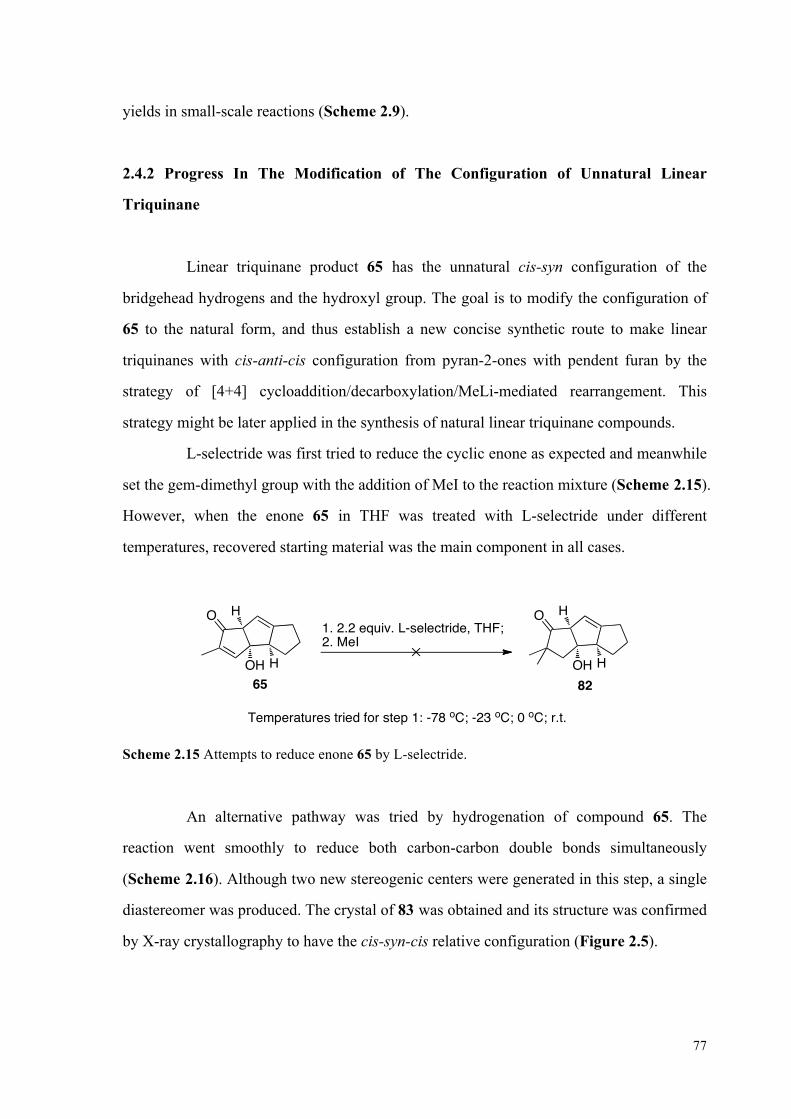
77
yields in small-scale reactions (Scheme 2.9).
2.4.2 Progress In The Modification of The Configuration of Unnatural Linear
Triquinane
Linear triquinane product 65 has the unnatural cis-syn configuration of the
bridgehead hydrogens and the hydroxyl group. The goal is to modify the configuration of
65 to the natural form, and thus establish a new concise synthetic route to make linear
triquinanes with cis-anti-cis configuration from pyran-2-ones with pendent furan by the
strategy of [4+4] cycloaddition/decarboxylation/MeLi-mediated rearrangement. This
strategy might be later applied in the synthesis of natural linear triquinane compounds.
L-selectride was first tried to reduce the cyclic enone as expected and meanwhile
set the gem-dimethyl group with the addition of MeI to the reaction mixture (Scheme 2.15).
However, when the enone 65 in THF was treated with L-selectride under different
temperatures, recovered starting material was the main component in all cases.
O H
OH H65
O H
OH H82
1. 2.2 equiv. L-selectride, THF;2. MeI
Temperatures tried for step 1: -78 oC; -23 oC; 0 oC; r.t.
Scheme 2.15 Attempts to reduce enone 65 by L-selectride.
An alternative pathway was tried by hydrogenation of compound 65. The
reaction went smoothly to reduce both carbon-carbon double bonds simultaneously
(Scheme 2.16). Although two new stereogenic centers were generated in this step, a single
diastereomer was produced. The crystal of 83 was obtained and its structure was confirmed
by X-ray crystallography to have the cis-syn-cis relative configuration (Figure 2.5).

78
O H
OH H65
O H
OH H83
H2, 10% Pd/C, EtOAc, r.t., 1 h
H
92%
Scheme 2.16 Hydrogenation of compound 65.
Figure 2.5 X-ray ORTEP representation of 83 at the 20% probability level.
Subsequent dehydration of 83 under basic conditions produced cyclic enone 84
in a decent yield as in Scheme 2.17. Two stereogenic centers were removed where a new
carbon-carbon double bond was formed. The loss of both bridgehead stereocenters of one
of the ring-fusions now presented an opportunity to obtain the alternative cis-anti-cis
configuration.

79
O H
OH H83
H1.7 equiv. MsCl, NEt3, DCM, r.t., 1 h
72%
O
H84
H
Scheme 2.17 Dehydration of compound 83.
With 84 in hand, it was anticipated that two new stereogenic centers would be set
by the reduction of the olefin. Under the right conditions, the product with
thermodynamically favored configuration of cis-anti-cis would be expected. The
SmI2-mediated reaction conditions were tried for selective reduction of carbon-carbon
double bond in the conjugated system.27 A mixture of H2O/amine in THF was used as
co-solvent because they are able to accelerate the rate of reduction of α,β-unsaturated
enone.27a, 27c However, this reaction condition was found to be ineffective in reducing the
conjugated double bond since the starting material were recovered in both cases as in
Table 2.3 after more than 1 hour.
O
H
H
85
O
H84
H H
H
SmI2, H2O, NEt3, THF, r.t.
Unit (equivalents) SmI2 H2O NEt3
Condition a 2.5 6 5
Condition b 6 60 12
Table 2.3 Attempts to reduce enone 84 with SmI2-mediated reaction conditions.
Hydrogenation of the enone 84 gave a single diastereomer 85 with a yield of
88% (Scheme 2.18). No correlation between the methyl group and bridgehead hydrogen
was detected in the NOE experiment, which might suggest that linear triquinane 85 has the
unnatural configuration of cis-syn-cis. In addition, the hydrogenation was more likely to

80
occur from the less hindered side to give the product 86 with the unnatural configuration.
O HH
H86
O
H
H
84H
H2, 10% Pd/C, EtOAc, r.t., 0.5 h
88%
Scheme 2.18 Hydrogenation of compound 84.
Birch reduction (Li, liq. NH3) under thermodynamic control is still underway to
get to the desired cis-anti-cis configuration.
(For further discussion of the conclusions and future directions of this research project, see
Chapter 3.)
2.5 Experimental Details
2.5.1 General Information
Reactions were conducted in oven-dried (110 oC) or flame-dried glassware under
a positive argon atmosphere unless otherwise noted. Transfer of anhydrous solvents,
reagents or mixtures was accomplished with oven-dried syringes or via cannulae. Solvents
were distilled before use: dichloromethane over calcium hydride; tetrahydrofuran and
diethyl ether over sodium benzophenone ketyl; toluene over calcium hydride. Thin layer
chromatography (TLC) was performed on 0.25 mm thick precoated silica gel plates
(Merck Fertigplatten Kieselgel 60F254). Flash chromatography columns were packed with
230-240 mesh silica gel.
Proton nuclear magnetic resonance spectra (1H NMR) were recorded at 400 MHz
or 500 MHz, and the chemical shifts are reported on the δ scale (ppm) relative to the
tetramethylsilane (0 ppm). Coupling constants (J) are reported in Hz. Second order

81
splitting patterns are indicated. Splitting patterns are designated as s, singlet; d, doublet; t,
triplet; q, quartet; m, multiplet; br, broad; dd, doublet of doublets, dt, doublet of triplets; b,
broad, etc. Carbon nuclear magnetic resonance spectra (13C NMR) were recorded at 100
MHz or 125 MHz and are reported in ppm relative to the centerline of the triplet from
deuterochloroform at 77.23 ppm. Infrared (IR) spectra were measured with a Magna 750
FT-IR infrared spectrophotometer. Mass spectra were determined on a Kratos Analytical
MS-50G spectrometer (EI). Elemental analyses were obtained on a Carlo Erba CHNS-0
EA1108 Elemental Analyzer. X-ray crystallographic experiments were carried out on a
Bruker PLATFORM diffractometer / SMART APEX II CCD area detector system with an
associated liquid nitrogen based crystal cooling system capable of temperatures ranging
from ambient to -150 °C.
UV irridiation were carried out by using an Ace-Hanovia 450W median pressure
quartz, mercury vapor lamp in a quartz water-cooled jacket. Reactions were carried out in a
Pyrex round bottom flask under argon after 10 mins of slow stream of argon for
deoxygenation before UV irridiation in a light proof enclosure. Microwave heating was
carried out in a Biotage Initiator microwave reactor.
All reagents and catalysts were purchased from Aldrich or Sigma or Strem and
were used without further purification unless otherwise stated.
2.5.2 Substrate Synthesis
O
OAc
64
79
63
OO
O
OAc
OOAc
74
O H
OH H
65
OO
O
OH

82
Preparation of the reported compounds 74, 79, 63, 64 and 65 were carried out by following
literature procedures or procedures described in Lei Li’s PhD Dissertation.
O H
OH H65
O H
OH H83
H2, 10% Pd/C, EtOAc, r.t., 1 h
H
92%
Preparation of 83. 10 mol% Pd/C (35 mg, 0.034 mmol) was added to a solution of 65 (65
mg, 0.34 mmol) in EtOAc (12 mL) in a 25 mL round bottom flask. The reaction was
monitored by TLC, and the mixture was stirred under H2 (balloon pressure) for 1 h until
the starting material was completely consumed. The mixture was then filtered over celite
and concentrated with rotatory evaporator. Flash chromatography (silica gel, 230-400 mesh,
50% EtOAc/Hexane) of the crude product afforded 60 mg (0.31 mmol, 92%) of the desired
83 as a white solid: m.p. 47-49 oC; Rf = 0.48 (70% EtOAc/Hexane); IR (DCM cast film)
3427, 2951, 2869, 1736 cm-1; 1H NMR (500 MHz, CDCl3) δ 2.90-2.80 (m, 1H), 2.79-2.65
(m, 2H), 2.61-2.54 (m, 1H), 2.19-2.08 (m, 2H), 1.73-1.48 (m, 6H), 1.39-1.29 (m, 1H), 1.14
(d, J = 4.0 Hz, 3H), 1.11-0.99 (m, 1H) (OH proton was not detected); 13 C NMR (125 MHz,
CDCl3) δ 219.8, 88.1, 63.1, 55.3, 45.0, 43.8, 40.3, 33.7, 32.2, 26.9, 26.8, 14.6; HRMS (EI)
calculated for C12H18O2 (M•+) 194.1307, found 194.1308.
O H
OH H83
H1.7 equiv. MsCl, NEt3, DCM, r.t., 1 h
72%
O
H84
H
Preparation of triquinane 84. MsCl (0.032 mL, 0.41 mmol) was added dropwise to a
solution of 83 (46 mg, 0.24 mmol) in 3 mL DCM at the temperature of 0 oC, and NEt3
(0.16 mL, 1.1 mmol) was added to the solution immediately afterwards. The reaction was

83
stirred under argon and was monitored by TLC until the complete consumption of the
starting material (typically < 1 h). The reaction was then quenched by the addition of 2 mL
10% HCl solution. The two layers were separated and the aqueous layer was extracted with
DCM (3x5 mL). The combined organic layers were washed with water (5 mL) and brine (5
mL), dried over MgSO4 and concentrated to give the crude product as pale yellow oil.
Flash chromatography (silica gel, 230-400 mesh, 5:10:1 Et2O/Hexane/DCM) of the crude
product gave 30 mg (0.17 mmol, 88%) of enone 84 as a colorless oil: Rf = 0.40 (5:5:1 Et2O:
Hexane: DCM); IR (CDCl3 cast film; microscope) 2946, 2864, 1698, 1638 cm-1; 1H NMR
(500 MHz, CDCl3) δ 3.23-3.10 (m, 2H), 2.76-2.56 (m, 3H), 2.02 (d, J = 12.0 Hz, 2H),
1.86-1.73 (m, 2H), 1.60-1.44 (m, 3H), 1.42-1.34 (m, 1H), 1.20 (d, J = 8.0 Hz, 3H); 13 C
NMR (125 MHz, CDCl3) δ 207.2, 186.5, 146.0, 49.1, 46.7, 46.6, 35.3, 33.1, 32.2, 30.0,
26.2, 17.0; HRMS (EI) calculated for C12H16O (M•+) 176.1201, found 176.1198.
O HH
H86
O
H
H
84H
H2, 10% Pd/C, EtOAc, r.t., 0.5 h
88%
Preparation of triquinane 86. 10 mol% Pd/C (2.0 mg, 0.0018 mmol) was added to a
solution of 84 (3.2 mg, 0.018 mmol) in EtOAc (0.5 mL) in a 10 mL round bottom flask.
The reaction was monitored by TLC, and the mixture was stirred under H2 (balloon
pressure) for 0.5 h until the starting material was completely consumed. The mixture was
then filtered over Celite and concentrated with rotatory evaporator. Flash chromatography
(silica gel, 230-400 mesh, 3% EtOAc/Hexane) of the crude product afforded 2.8 mg (0.016
mmol, 88%) of the reduced product 86 as a colorless oil: Rf = 0.31 (5% EtOAc/Hexane);
IR (CDCl3 cast film; microscope) 2960, 2870,1723 cm-1; 1H NMR (500 MHz, CDCl3) δ
2.80-2.60 (m, 3H), 2.38-2.22 (m, 1H), 2.20-2.10 (m, 1H), 2.09-2.00 (m, 1H), 1.73-1.52 (m,
6H), 1.40-1.21 (m, 3H), 1.15 (d, J = 8.0 Hz, 3H); 13 C NMR (125 MHz, CDCl3) δ 221.6,
55.6, 47.3, 46.2, 46.1, 43.1, 33.8, 32.7, 32.1, 27.9, 27.6, 14.5; HRMS (EI) calculated for
C12H18O (M•+) 178.1357, found 178.1356.

84
2.6 References and Notes
1. (a) Mehta, G.; Srikrishna, A., Chem. Rev. 1997, 97, 671-720; (b) Singh, V.; Thomas,
B., Tetrahedron 1998, 54, 3647-3692.
2. (a) Bohlman, F.; Jakupovic, J., Phytochem. 1980, 19 ( 259-265); (b) Dominguez, K. A.;
Cano, C.; France, R.; Villareal, A. M.; Watson, W. H.; Zabel, V., Phytochem. 1980, 19,
2478; (c) Corbett, R. E.; Lauren, D. R.; Weavers, R. T., J. Chem. Soc., Perkin. Trans. 1979,
1, 1774-1790; (d) Kaneda, M.; Takahashi, R.; litaka, Y.; Shibata, S., Tetrahedron Lett.
1972, 13, 4609-4611.
3. (a) Sheikh, Y. M.; Singy, G.; Kaisin, M.; Eggert, H.; Djerassi, C.; Tursch, B.; Daloze,
D.; Braekman, J. C., Tetrahedron 1976, 32, 1171-1178; (b) Kaisin, M.; Sheikh, Y. M.;
Durham, L. J.; C.;, D.; Tursch, B.; Daloze, D.; Braekman, J. C.; Losman, D.; R., K.,
Tetrahedron Lett. 1974, 75, 2239-2242; (c) Ayanoglu, E.; Gebreyesus, T.; Beechan, C. M.;
Djerassi, C.; Kaisin, M., Tetrahedron Lett. 1978, 19, 1671-1674.
4. (a) Martin, D. G.; Slomp, G.; Mizsak, S.; Duchamp, D. J.; Chidester, C. G.,
Tetrahedron Lett. 1970, 11, 4901-4904; (b) Comer, F. W.; McCapra, F.; Qureshi, I. H.;
Scott, A. I., Tetrahedron 1967, 23, 4761-4768; (c) Shuji, T.; Naganawa, H.; linuma, H.;
Takita, T.; Maeda, K.; Umezawa, H., Tetrahedron Lett. 1971, 12, 1955-1958; (d)
Nakamura, H.; Takita, T.; Umezawa, H.; Kunishima, M.; Nakayama, Y.; Itaka, Y., J.
Antibiot. 1974, 27, 301-302; (e) Nozoe, S.; Furukawa, J.; Sankawa, U.; Shibata, S.,
Tetrahedron Lett. 1976, 17, 195-198; (f) Ranieri, R. L.; Calton, G. J., Tetrahedron Lett.
1978, 19, 499-502.
5. (a) Eaton, P. E.; Mueller, R. H.; Carlson, G. R.; Cullison, D. A.; Cooper, G. F.; Chou,
T. C.; Krebs, E. P., J. Am. Chem. Soc. 1977, 99, 2751-2767; (b) Eaton, P. E.; Mueller, R.
H., J. Am. Chem. Soc. 1972, 94, 1014-1016; (c) Paquette, L. A.; Balogh, D. W.; Usha, R.;
Koontz, D.; Christopher, G. C., Science 1981, 211, 575-576.
6. (a) Takeuchi, T.; Iinuma, H.; Iwanaga, J.; Takahashi, S.; Takita, T.; Umezawa, H., J.
Antibiot. 1969, 22, 215-217; (b) Tatsuta, K.; Akimoto, K.; Kinoshita, M., J. Antibiot. 1980,
23, 100-102.
7. Comer, F. W.; Trotter, J., J. Chem. Soc. 1966, 6, 11-18.

85
8. (a) Burkholder, P.; Burkholder, L., Science 1958, 127, 1174-1175; (b) Ciereszko, L. S.,
Trans. N.Y. Acad. Sci. 1962, 24, 502-503; (c) Stille, J. R.; Grubbs, R. H., J. Am. Chem. Soc.
1986, 108, 855-856; (d) Mehta, G.; Murthy, N. A.; Reddy, S. D.; Reddy, V. A., J. Am.
Chem. Soc. 1986, 108, 3443-3452.
9. (a) Feline, T. C.; Mellows, G.; Jones, R. B.; Phillips, L., J. Chem. Soc., Chem.
Commun. 1974, 1974, 63-64; (b) Ohfune, Y.; Shirahama, H.; Matsumoto, T., Tetrahedron
Lett. 1976, 17, 2795-2796; (c) Misumi, S.; Matsushima, H.; Shirahama, H.; Matsumoto, T.,
Chem. Lett. 1982, 11, 855-858.
10. (a) Mehta, G.; Pallavi, K., Tetrahedron Lett. 2006, 47, 8355-8360; (b) Chandler, C. L.;
List, B., J. Am. Chem. Soc. 2008, 130, 6737-6739.
11. (a) Srikrishna, A.; Dethe, D. H., Org. Lett. 2003, 5, 2295-2298; (b) Srikrishna, A.;
Beeraish, B., Tetrahedron: Asymmetry 2008, 19, 884-886; (c) Rieder, C. J.; Fradette, R. J.;
West, F. G., Chem. Commun. 2008, 13, 1572-1574.
12. (a) Marino, J. P.; Laborde, E., J. Am. Chem. Soc. 1985, 107, 734-735; (b) Lee, H.-Y.;
Kim, Y., J. Am. Chem. Soc. 2003, 125, 10156-10157.
13. Veretenov, A. L.; Koltun, D. O.; Smit, W. A.; Strelenko, Y. A., Tetrahedron Lett.
1995, 36, 4651-4654.
14. (a) Little, R. D.; Muller, G. W., J. Am. Chem. Soc. 1981, 103, 2744-2749; (b) Little, R.
D.; Muller, G. W., J. Am. Chem. Soc. 1979, 101, 7129-7130; (c) Little, R. D.; Carroll, G.
L., Tetrahedron Lett. 1981, 22, 4389-4392.
15. Wender, P. A.; Howbert, J. J., Tetrahedron Lett. 1982, 23, 3983-3986.
16. (a) Curran, D. P.; Rakiewicz, D. M., J. Am. Chem. Soc. 1985, 107, 1448-1449; (b)
Fevig, T. L.; Elliott, R. L.; Curran, D. P., J. Am. Chem. Soc. 1988, 110, 5064-5067.
17. (a) Rawal, V. H.; Dufour, C., J. Am. Chem. Soc. 1994, 116, 2613-2614; (b) Rawal, V.
H.; Dufour, C.; Iwasa, S., Tetrahedron Lett. 1995, 36, 19-22.
18. Rawal, V. H.; Dufour, C.; Eschbach, A., J. Chem. Soc., Chem. Commun.
1994, 1994, 1797-1798.
19. Rawal, V. H.; Fabre, A.; lwasa, S., Tetrahedron Lett. 1995, 36, 6851-6854.
20. Demuth, M.; Schaffner, K., Angew. Chem. Int. Ed. Engl. 1982, 21, 820-836.
21. Singh, V.; Vedantham, P.; Sahu, P. K., Tetrahedron Lett. 2002, 43, 519-522.

86
22. Balme, G.; Bouyssi, D., Tetrahedron 1994, 50, 403-414.
23. Santora, V. J.; Moore, H. W., J. Am. Chem. Soc. 1995, 117, 8486-8487.
24. (a) Li, L.; McDonald, R.; West, F. G., Org. Lett. 2008, 10, 3733-3736; (b) Li, L. Ph.D.
Dissertation, University of Alberta, Edmonton, 2009.
25. (a) West, F. G.; Chase, C. E.; Arif, A. M., J. Org. Chem. 1993, 58, 3794-3795; (b)
Chase, C. E.; Bender, J. A.; West, F. G., Synlett. 1996, 1173-1175; (c) Li, L.; Chase, C. E.;
West, F. G., Chem. Commun. 2008, 13, 4025-4027.
26. Moreno-Manas, M.; Ribas, J.; Virgili, A., J. Org. Chem. 1988, 53, 5328-5335.
27. (a) Dahlen, A.; Hilmersson, G., Tetrahedron Lett. 2002, 43, 7197-7200; (b) Dahlen, A.;
Hilmersson, G., Tetrahedron Lett. 2003, 44, 2661-2664; (c) Dahlen, A.; Hilmersson, G.,
Chem. Eur. J. 2003, 9, 1123-1128; (d) Girard, P.; Namy, J. L.; Kagan, H. B., J. Am. Chem.
Soc. 1980, 102, 2693-2698.
87
CHAPTER 3
CONCLUSIONS AND FUTURE PLANS
3.1 Conclusions of the Prion and Triquinane Projects
In this thesis, we first reported a concise and effective synthetic route to
synthesize and purify a library of porphyrin-based multimeric fluorescent compounds with
anticipated anti-prion activity, and nine such molecules have been synthesized, purified
and characterized by using either quinoline or 9-amino-acridine as the binding units. The
anti-prion assay results of the six quinoline-linked target molecules have been obtained,
but they are unexpectedly ineffective in reducing the amount of PrPSc in vitro, which may
suggest that other porphyrin-based multimeric compounds are not likely to have strong
anti-prion activity as well. In contrast, three arene-based multimeric compounds prepared
by another group member show promising anti-prion activity.
In the second chapter, the studies toward the formation of linear triquinanes with
natural cis-anti-cis configuration via pyran-2-one photochemistry were discussed. Linear
triquinanes with unnatural cis-syn configuration were readily available from substituted
pyran-2-ones in a concise route of [4+4] photochemistry/thermal
decarboxylation/MeLi-mediated rearrangement.1 Hydrogenation and subsequent
dehydration produced an enone. Reduction of the enone presented an opportunity to obtain
the natural cis-anti-cis configuration. Different reduction conditions were tried, but they
were either ineffective in reduction or gave the product with cis-syn-cis configuration.
Reduction under thermodynamic control (Li, liq. NH3) is still underway to get to the
desired natural configuration.

88
3.2 Future Directions of The Research Projects
3.2.1 Future Plan of the Prion Project
Since three arene-based trimeric compounds have been identified by the
anti-prion assay to have strong capability in reducing the amount of PrPSc in vitro (Figure
1.19), it suggests that such compounds with a linker of 1,2-ethanediamine or
1,3-propanediamine can better fix the space of aggregate of PrPSc and thus bind to them
effectively. Therefore, new series of arene-based multimeric molecules can be envisioned
by employing different binding units. Some compounds that already show promising
anti-prion activity can be selected as the mono-binding units, which may include
2-methylquinoline,2 2-phenylquinazoline,2 acridines,2-3 and phenothiazine derivatives4
(Figure 3.1). In particular, fluorescent mono-binding units, 9-amino-acridines, will be of
great interests in building arene-based fluorescent multimeric compounds. A fluorescent
compound with high affinity towards PrPSc could represent a major breakthrough, since it
would have the potential to both inhibit the propagation of PrPSc and detect the presence of
infectious particles.
N NN
NCl ClCl
N
Cl
Cl
O
4-Chloro-2-methylquinoline 1 4-Chloro-2-phenylquinazoline 2 9-Chloroacridine 3
6,9-Dichloro-2-methoxyacridine 4S
N
NH2
RR = H, Cl,
O
Phenothiazine derivatives 5 Figure 3.1 Structures of mono-binding units 1-5.

89
Compounds 6-8 in Figure 3.2 can be prepared by heating the mixture of 1-4 and
diamines as described in Scheme 1.5.5 Subsequently, the target molecules can be obtained
through the amide bond formation of trimesic acid and compounds 5-8 (Scheme 3.1).
N N
NN NH H NH2NH2
N
NH NH2
R1
R2
n nn
n = 1 or 2
6 7 8
R1 = R2 = H;R1 = Cl, R2 = OMe
n = 1 or 2 n = 1 or 2
Figure 3.2 Structures of 2-methylquinoline-diamines 1, 2-phenylquinozaline-diamine 2,
acridine-diamines 3-4.
OOO H
NHO
HO OH
O
O
O
+EDC, HOBt,DMF, rt.
Trimesic acid
HN
HN
R R3 or 4 equiv. RNH2
Mono-binding unitsAnticipated products
R
N N
N
N NH H NH2NH2n n
n = 1 or 2
6 7
n = 1 or 2
N
NH NH2
R1
R2n
8
R1 = R2 = H;R1 = Cl, R2 = OMe
n = 1 or 2
S
N
NH2
RR = H, Cl,
O
Phenothiazine derivatives 5
Mono-binding units 5-8:
Scheme 3.1 Synthesis of arene-based trimesic compounds by using acridine or others as the
binding units.

90
3.2.2 Future Plan of the Triquinane Project
Birch reduction under thermodynamic control may give rise to the product with
natural cis-anti-cis configuration, and further methylation at α-position will set the gem
dimethyl group which is commonly found in natural triquinane compounds (Scheme 3.2).
O
H
H2 or 3 equiv. Li, liq. NH3, THF;Saturated NH4Cl solution workup
O
H
H
H
H
LDA, MeI O
H
H
H
H
Scheme 3.2 Birch reduction and methylation at α-position.
Once the inversion of configuration strategy is fully developed, this overall route
will be applied to one or more linearly fused triquinane natural products. Possible targets
include hirsutene, ceratopical and cucumins.
3.3 References
1. Li, L.; McDonald, R.; West, F. G., Org. Lett. 2008, 10, 3733-3736.
2. Cope, H.; Mutter, R.; Heal, W.; Pascoe, C.; Brown, P.; Pratt, S.; Chen, B., European
Journal of Medicinal Chemistry 2006, 41, 1124-1143.
3. Barret, A.; Tagliavini, F.; Forloni, G.; Bate, C.; Salmona, M.; Colombo, L.; De Luigi,
A.; Limido, L.; Suardi, S.; Rossi, G.; Auvre, F.; Adjou, K. T.; Sales, N.; Williams, A.;
Lasmezas, C.; Deslys, J. P., J. Virol. 2003, 77, 8462-8469.
4. Korth, C.; May, B. C. H.; Cohen, F. E.; Prusiner, S. B., Proc. Natl. Acad. Sci. USA
2001, 98, 9836-9841.

91
5. Chibale, K.; Haupt, H.; Kendrick, H.; Yardley, V.; Saravanamuthu, A.; Fairlambc, A.
H.; Croftb, S. L., Bioorg. Med. Chem. Lett. 2001, 11, 2655-2657.

92
APPENDIX A
SELECTED NMR SPECTRA FROM CHAPTER 1

93
HN
N
NH
N
OO
OO
OO O
O3

94
HN
N
NH
N
OO
OO
OO O
O3

95
HN
N
NH
N
O
HO
OH
OO O
OH
4HO

96
HN
N
NH
N
O
HO
OH
OO O
OH
4HO

97
HN N
Cl
NH2
16

98
HN N
Cl
NH2
16

99
HN
N
NH
N
O
O O
O
R R
RR
Generalized structure of compound 18-23 and 30-32

100
NHN
Cl
H N
R= 18

101
NHN
Cl
H N
R= 18

102
N
HN
Cl
R= 19
N H

103
N
HN
Cl
R= 19
N H

104
N
HN
Cl
R=
20
H N

105
N
HN
Cl
R=
20
H N

106
N
HN
Cl
H N
21
R=
3

107
N
HN
Cl
H N
21
R=
3

108
N
HN
Cl
R=
22
N H
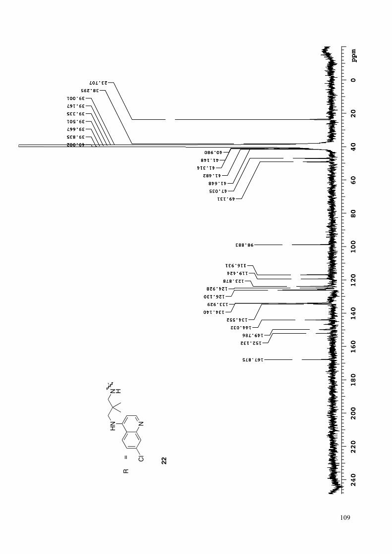
109
N
HN
Cl
R=
22
N H

110
NN
Cl 23
R=
N

111
NN
Cl 23
R=
N

112
NNH
Cl
R=
30
NH
O

113
NNH
Cl
R= 31
NH
O
2

114
NNH
Cl
R= 32
NH
O

115
NNH
Cl
R= 32
NH
O

116
APPENDIX B
SELECTED NMR SPECTRA FROM CHAPTER 2

117
O
HOH
H
83
H

118
O
HOH
H
83
H

119
O
H
84
H

120
O
H
HH
86
H

121
O
H
HH
86
H

122
APPENDIX C
X-RAY CRYSTALLOGRAPHIC DATA TABLES FOR COMPOUND 83
(CHAPTER 2)

123
STRUCTURE REPORT
XCL Code: FGW1104 Date: 13 July 2011
Compound: 3a-hydroxy-2-methyldecahydro-1H-cyclopenta[a]pentalen-1-one Formula: C12H18O2 Supervisor: F. G. West Crystallographer: R. McDonald
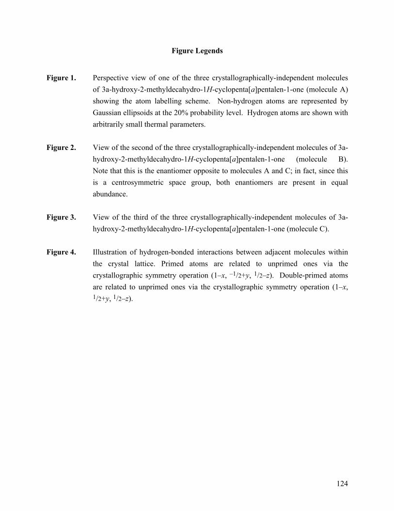
124
Figure Legends
Figure 1. Perspective view of one of the three crystallographically-independent molecules
of 3a-hydroxy-2-methyldecahydro-1H-cyclopenta[a]pentalen-1-one (molecule A) showing the atom labelling scheme. Non-hydrogen atoms are represented by Gaussian ellipsoids at the 20% probability level. Hydrogen atoms are shown with arbitrarily small thermal parameters.
Figure 2. View of the second of the three crystallographically-independent molecules of 3a-
hydroxy-2-methyldecahydro-1H-cyclopenta[a]pentalen-1-one (molecule B). Note that this is the enantiomer opposite to molecules A and C; in fact, since this is a centrosymmetric space group, both enantiomers are present in equal abundance.
Figure 3. View of the third of the three crystallographically-independent molecules of 3a-
hydroxy-2-methyldecahydro-1H-cyclopenta[a]pentalen-1-one (molecule C). Figure 4. Illustration of hydrogen-bonded interactions between adjacent molecules within
the crystal lattice. Primed atoms are related to unprimed ones via the crystallographic symmetry operation (1–x, –1/2+y, 1/2–z). Double-primed atoms are related to unprimed ones via the crystallographic symmetry operation (1–x, 1/2+y, 1/2–z).

125

126

127

128

129
List of Tables
Table 1. Crystallographic Experimental Details
Table 2. Atomic Coordinates and Equivalent Isotropic Displacement Parameters
Table 3. Selected Interatomic Distances
Table 4. Selected Interatomic Angles
Table 5. Hydrogen-Bonded Interactions
Table 6. Torsional Angles
Table 7. Anisotropic Displacement Parameters
Table 8. Derived Atomic Coordinates and Displacement Parameters for Hydrogen Atoms

130
Table 1. Crystallographic Experimental Details
A. Crystal Data formula C12H18O2 formula weight 194.26 crystal dimensions (mm) 0.37 ´ 0.16 ´ 0.06 crystal system monoclinic space group P21/c (No. 14) unit cell parametersa a (Å) 9.0349 (5) b (Å) 32.1439 (16) c (Å) 11.6359 (6) b (deg) 109.0391 (7) V (Å3) 3194.4 (3) Z 12 rcalcd (g cm-3) 1.212 µ (mm-1) 0.080
B. Data Collection and Refinement Conditions diffractometer Bruker PLATFORM/APEX II CCDb radiation (l [Å]) graphite-monochromated Mo Ka (0.71073) temperature (°C) –100 scan type w scans (0.3°) (20 s exposures) data collection 2q limit (deg) 50.80 total data collected 22892 (-10 ≤ h ≤ 10, -38 ≤ k ≤ 38, -14 ≤ l ≤ 14) independent reflections 5877 (Rint = 0.0724) number of observed reflections (NO) 3514 [Fo2 ≥ 2s(Fo2)] structure solution method direct methods (SHELXDc) refinement method full-matrix least-squares on F2 (SHELXL–97d) absorption correction method Gaussian integration (face-indexed) range of transmission factors 0.9954–0.9712 data/restraints/parameters 5877 / 0 / 382 goodness-of-fit (S)e [all data] 1.025 final R indicesf R1 [Fo2 ≥ 2s(Fo2)] 0.0516 wR2 [all data] 0.1315 largest difference peak and hole 0.156 and –0.185 e Å-3 aObtained from least-squares refinement of 3421 reflections with 4.76° < 2q < 39.20°. bPrograms for diffractometer operation, data collection, data reduction and absorption correction
were those supplied by Bruker. (continued)

131
Table 1. Crystallographic Experimental Details (continued) cSchneider, T. R.; Sheldrick, G. M. Acta Crystallogr. 2002, D58, 1772-1779. dSheldrick, G. M. Acta Crystallogr. 2008, A64, 112–122. eS = [Sw(Fo2 – Fc2)2/(n – p)]1/2 (n = number of data; p = number of parameters varied; w =
[s2(Fo2) + (0.0449P)2 + 1.3669P]-1 where P = [Max(Fo2, 0) + 2Fc2]/3). fR1 = S||Fo| – |Fc||/S|Fo|; wR2 = [Sw(Fo2 – Fc2)2/Sw(Fo4)]1/2.

132
Table 2. Atomic Coordinates and Equivalent Isotropic Displacement Parameters
(a) molecule A Atom x y z Ueq, Å2 O1A 0.4515(2) 0.30938(6) 0.14811(17) 0.0498(5)* O2A 0.1818(2) 0.22113(5) 0.22994(14) 0.0402(4)* C1A 0.3587(3) 0.28057(8) 0.1219(2) 0.0349(6)* C2A 0.3986(3) 0.23709(8) 0.0915(2) 0.0367(6)* C3A 0.2393(3) 0.21610(7) 0.0402(2) 0.0327(6)* C4A 0.1374(3) 0.23681(7) 0.1081(2) 0.0315(6)* C5A -0.0403(3) 0.23718(7) 0.0436(2) 0.0347(6)* C6A -0.1104(3) 0.20550(8) -0.0601(2) 0.0390(6)* C7A -0.1334(3) 0.22926(8) -0.1780(2) 0.0425(7)* C8A -0.1819(3) 0.27268(9) -0.1501(2) 0.0462(7)* C9A -0.0786(3) 0.28081(8) -0.0179(2) 0.0372(6)* C10A 0.0799(3) 0.30087(8) -0.0032(2) 0.0381(6)* C11A 0.1904(3) 0.28231(7) 0.1157(2) 0.0334(6)* C12A 0.5026(3) 0.23625(9) 0.0113(3) 0.0535(8)*
(b) molecule B Atom x y z Ueq, Å2 O1B 0.6753(2) 0.47296(6) 0.37366(17) 0.0479(5)* O2B 0.3519(2) 0.39188(5) 0.14847(15) 0.0452(5)* C1B 0.5751(3) 0.44586(8) 0.3523(2) 0.0352(6)* C2B 0.6036(3) 0.40160(8) 0.3980(2) 0.0386(6)* C3B 0.4378(3) 0.38346(8) 0.3691(2) 0.0372(6)* C4B 0.3347(3) 0.40787(7) 0.2591(2) 0.0317(6)* C5B 0.1610(3) 0.41313(8) 0.2481(2) 0.0356(6)* C6B 0.0911(3) 0.38304(9) 0.3205(2) 0.0446(7)* C7B 0.0929(3) 0.40734(9) 0.4342(2) 0.0509(8)* C8B 0.0572(3) 0.45170(10) 0.3887(3) 0.0540(8)* C9B 0.1460(3) 0.45725(8) 0.2981(2) 0.0414(7)* C10B 0.3147(3) 0.47279(8) 0.3547(2) 0.0364(6)* C11B 0.4046(3) 0.45175(7) 0.2802(2) 0.0311(6)* C12B 0.7074(3) 0.39864(9) 0.5302(2) 0.0517(8)*
(c) molecule C Atom x y z Ueq, Å2 O1C 0.9034(2) 0.63806(6) 0.26806(16) 0.0460(5)* O2C 0.5872(2) 0.55607(5) 0.33578(14) 0.0428(5)* C1C 0.8022(3) 0.61111(8) 0.2415(2) 0.0334(6)* C2C 0.8300(3) 0.56658(8) 0.2104(2) 0.0371(6)* C3C 0.6643(3) 0.54908(7) 0.1542(2) 0.0351(6)* Table 2. Atomic Coordinates and Displacement Parameters (continued)

133
Atom x y z Ueq, Å2 C4C 0.5641(3) 0.57273(7) 0.2166(2) 0.0323(6)* C5C 0.3882(3) 0.57810(8) 0.1449(2) 0.0374(6)* C6C 0.3150(3) 0.54894(9) 0.0355(2) 0.0448(7)* C7C 0.3076(3) 0.57474(9) -0.0770(2) 0.0467(7)* C8C 0.2709(3) 0.61845(9) -0.0434(2) 0.0513(8)* C9C 0.3693(3) 0.62271(8) 0.0909(2) 0.0404(7)* C10C 0.5374(3) 0.63838(8) 0.1130(2) 0.0375(6)* C11C 0.6329(3) 0.61671(7) 0.2310(2) 0.0312(6)* C12C 0.9380(3) 0.56338(9) 0.1342(3) 0.0515(8)* Anisotropically-refined atoms are marked with an asterisk (*). The form of the anisotropic displacement parameter is: exp[-2π2(h2a*2U11 + k2b*2U22 + l2c*2U33 + 2klb*c*U23 + 2hla*c*U13 + 2hka*b*U12)].

134
Table 3. Selected Interatomic Distances (Å)
(a) molecule A Atom1 Atom2 Distance Atom1 Atom2 Distance O1A C1A 1.219(3) O2A C4A 1.433(3) C1A C2A 1.514(3) C1A C11A 1.500(3) C2A C3A 1.524(3) C2A C12A 1.526(4) C3A C4A 1.546(3) C4A C5A 1.534(3)
C4A C11A 1.532(3) C5A C6A 1.548(3) C5A C9A 1.561(3) C6A C7A 1.524(3) C7A C8A 1.529(4) C8A C9A 1.540(3) C9A C10A 1.528(3) C10A C11A 1.538(3)
(b) molecule B Atom1 Atom2 Distance Atom1 Atom2 Distance O1B C1B 1.222(3) O2B C4B 1.440(3) C1B C2B 1.511(3) C1B C11B 1.507(3) C2B C3B 1.539(3) C2B C12B 1.520(3) C3B C4B 1.531(3) C4B C5B 1.542(3)
C4B C11B 1.532(3) C5B C6B 1.547(3) C5B C9B 1.555(3) C6B C7B 1.532(4) C7B C8B 1.519(4) C8B C9B 1.530(4) C9B C10B 1.533(3) C10B C11B 1.526(3)
(c) molecule C Atom1 Atom2 Distance Atom1 Atom2 Distance O1C C1C 1.224(3) O2C C4C 1.437(3) C1C C2C 1.517(3) C1C C11C 1.505(3) C2C C3C 1.532(3) C2C C12C 1.521(3) C3C C4C 1.534(3) C4C C5C 1.545(3)
C4C C11C 1.531(3) C5C C6C 1.545(3) C5C C9C 1.552(4) C6C C7C 1.533(4) C7C C8C 1.523(4) C8C C9C 1.532(3) C9C C10C 1.539(3) C10C C11C 1.530(3)

135
Table 4. Selected Interatomic Angles (deg)
(a) molecule A Atom1 Atom2 Atom3 Angle Atom1 Atom2 Atom3 Angle O1A C1A C2A 124.3(2) O1A C1A C11A 125.9(2) C2A C1A C11A 109.7(2) C1A C2A C3A 103.5(2) C1A C2A C12A 113.6(2) C3A C2A C12A 116.4(2) C2A C3A C4A 104.67(19) O2A C4A C3A 109.45(19) O2A C4A C5A 113.41(19) O2A C4A C11A 107.30(18) C3A C4A C5A 117.10(19) C3A C4A C11A 102.33(19) C5A C4A C11A 106.15(19)
C4A C5A C6A 118.2(2) C4A C5A C9A 105.74(19) C6A C5A C9A 105.11(19) C5A C6A C7A 105.9(2) C6A C7A C8A 103.3(2) C7A C8A C9A 104.2(2) C5A C9A C8A 106.1(2) C5A C9A C10A 105.57(19) C8A C9A C10A 115.1(2) C9A C10A C11A 104.2(2) C1A C11A C4A 105.1(2) C1A C11A C10A 114.0(2) C4A C11A C10A 102.67(19)
(b) molecule B Atom1 Atom2 Atom3 Angle Atom1 Atom2 Atom3 Angle O1B C1B C2B 124.8(2) O1B C1B C11B 125.3(2) C2B C1B C11B 109.9(2) C1B C2B C3B 103.8(2) C1B C2B C12B 113.1(2) C3B C2B C12B 115.7(2) C2B C3B C4B 105.8(2) O2B C4B C3B 110.52(19) O2B C4B C5B 111.63(19) O2B C4B C11B 108.25(19) C3B C4B C5B 116.8(2) C3B C4B C11B 103.36(19) C5B C4B C11B 105.52(19)
C4B C5B C6B 118.2(2) C4B C5B C9B 106.23(19) C6B C5B C9B 105.4(2) C5B C6B C7B 105.1(2) C6B C7B C8B 103.7(2) C7B C8B C9B 104.5(2) C5B C9B C8B 106.3(2) C5B C9B C10B 105.2(2) C8B C9B C10B 114.4(2) C9B C10B C11B 104.5(2) C1B C11B C4B 105.47(19) C1B C11B C10B 112.1(2) C4B C11B C10B 103.37(19)
(c) molecule C Atom1 Atom2 Atom3 Angle Atom1 Atom2 Atom3 Angle O1C C1C C2C 124.0(2) O1C C1C C11C 126.3(2) C2C C1C C11C 109.7(2) C1C C2C C3C 103.5(2) C1C C2C C12C 113.0(2) C3C C2C C12C 116.7(2) C2C C3C C4C 105.16(19)
O2C C4C C3C 110.44(19) O2C C4C C5C 111.3(2) O2C C4C C11C 108.19(18) C3C C4C C5C 117.50(19) C3C C4C C11C 103.19(19) C5C C4C C11C 105.41(19) C4C C5C C6C 117.7(2)

136
Table 4. Selected Interatomic Angles (continued)
(c) molecule C Atom1 Atom2 Atom3 Angle Atom1 Atom2 Atom3 Angle C4C C5C C9C 106.21(19) C6C C5C C9C 105.4(2) C5C C6C C7C 105.3(2) C6C C7C C8C 103.4(2) C7C C8C C9C 104.0(2) C5C C9C C8C 106.3(2)
C5C C9C C10C 105.2(2) C8C C9C C10C 114.3(2) C9C C10C C11C 103.8(2) C1C C11C C4C 105.32(19) C1C C11C C10C 112.8(2) C4C C11C C10C 103.40(19)
Table 5. Hydrogen-Bonded Interactions
D–H…A D–H (Å)
H…A (Å)
D…A (Å)
∠D–H…A (deg)
Note
O2A–H2OA…O1C a 0.84 1.94 2.781(2) 177.5 aAt 1–x, –1/2+y, 1/2–z. O2B–H2OB…O1A 0.84 1.99 2.801(3) 162.2 O2C–H2OC…O1B 0.84 1.95 2.781(3) 169.1

137
Table 6. Torsional Angles (deg)
(a) molecule A Atom1 Atom2 Atom3 Atom4 Angle Atom1 Atom2 Atom3 Atom4 Angle O1A C1A C2A C3A -168.4(2) O1A C1A C2A C12A -41.3(3) C11A C1A C2A C3A 12.1(3) C11A C1A C2A C12A 139.3(2) O1A C1A C11A C4A -167.5(2) O1A C1A C11A C10A 80.9(3) C2A C1A C11A C4A 12.0(2) C2A C1A C11A C10A -99.7(2) C1A C2A C3A C4A -31.4(2) C12A C2A C3A C4A -156.7(2) C2A C3A C4A O2A -74.8(2) C2A C3A C4A C5A 154.4(2) C2A C3A C4A C11A 38.8(2) O2A C4A C5A C6A -109.0(2) O2A C4A C5A C9A 133.7(2) C3A C4A C5A C6A 20.0(3) C3A C4A C5A C9A -97.3(2) C11A C4A C5A C6A 133.5(2) C11A C4A C5A C9A 16.1(2) O2A C4A C11A C1A 84.4(2)
O2A C4A C11A C10A -156.04(19) C3A C4A C11A C1A -30.7(2) C3A C4A C11A C10A 88.8(2) C5A C4A C11A C1A -154.02(18) C5A C4A C11A C10A -34.5(2) C4A C5A C6A C7A -98.7(2) C9A C5A C6A C7A 18.9(3) C4A C5A C9A C8A 131.2(2) C4A C5A C9A C10A 8.6(2) C6A C5A C9A C8A 5.4(3) C6A C5A C9A C10A -117.2(2) C5A C6A C7A C8A -36.1(3) C6A C7A C8A C9A 39.2(3) C7A C8A C9A C5A -27.5(3) C7A C8A C9A C10A 88.8(3) C5A C9A C10A C11A -30.0(2) C8A C9A C10A C11A -146.6(2) C9A C10A C11A C1A 152.9(2) C9A C10A C11A C4A 39.8(2)
(b) molecule B Atom1 Atom2 Atom3 Atom4 Angle Atom1 Atom2 Atom3 Atom4 Angle O1B C1B C2B C3B 168.6(2) O1B C1B C2B C12B 42.5(3) C11B C1B C2B C3B -9.5(3) C11B C1B C2B C12B -135.7(2) O1B C1B C11B C4B 169.9(2) O1B C1B C11B C10B -78.2(3) C2B C1B C11B C4B -11.9(3) C2B C1B C11B C10B 99.9(2) C1B C2B C3B C4B 27.5(2) C12B C2B C3B C4B 152.0(2) C2B C3B C4B O2B 80.7(2) C2B C3B C4B C5B -150.2(2) C2B C3B C4B C11B -34.9(2) O2B C4B C5B C6B 109.4(2) O2B C4B C5B C9B -132.6(2) C3B C4B C5B C6B -19.1(3)
C3B C4B C5B C9B 98.9(2) C11B C4B C5B C6B -133.3(2) C11B C4B C5B C9B -15.3(2) O2B C4B C11B C1B -88.8(2) O2B C4B C11B C10B 153.36(19) C3B C4B C11B C1B 28.5(2) C3B C4B C11B C10B -89.4(2) C5B C4B C11B C1B 151.62(19) C5B C4B C11B C10B 33.7(2) C4B C5B C6B C7B 99.3(3) C9B C5B C6B C7B -19.2(3) C4B C5B C9B C8B -130.7(2) C4B C5B C9B C10B -9.0(3) C6B C5B C9B C8B -4.5(3) C6B C5B C9B C10B 117.2(2) C5B C6B C7B C8B 35.8(3)

138
Table 6. Torsional Angles (continued)
(b) molecule B Atom1 Atom2 Atom3 Atom4 Angle Atom1 Atom2 Atom3 Atom4 Angle C6B C7B C8B C9B -38.7(3) C7B C8B C9B C5B 26.7(3) C7B C8B C9B C10B -89.0(3) C5B C9B C10B C11B 29.9(2)
C8B C9B C10B C11B 146.2(2) C9B C10B C11B C1B -152.6(2) C9B C10B C11B C4B -39.5(2)
(c) molecule C Atom1 Atom2 Atom3 Atom4 Angle Atom1 Atom2 Atom3 Atom4 Angle O1C C1C C2C C3C -166.2(2) O1C C1C C2C C12C -39.1(3) C11C C1C C2C C3C 12.2(2) C11C C1C C2C C12C 139.3(2) O1C C1C C11C C4C -171.1(2) O1C C1C C11C C10C 76.8(3) C2C C1C C11C C4C 10.6(2) C2C C1C C11C C10C -101.5(2) C1C C2C C3C C4C -30.3(2) C12C C2C C3C C4C -155.0(2) C2C C3C C4C O2C -78.5(2) C2C C3C C4C C5C 152.4(2) C2C C3C C4C C11C 37.0(2) O2C C4C C5C C6C -110.5(2) O2C C4C C5C C9C 131.7(2) C3C C4C C5C C6C 18.2(3) C3C C4C C5C C9C -99.6(2) C11C C4C C5C C6C 132.4(2) C11C C4C C5C C9C 14.7(2) O2C C4C C11C C1C 88.1(2)
O2C C4C C11C C10C -153.23(19) C3C C4C C11C C1C -28.9(2) C3C C4C C11C C10C 89.7(2) C5C C4C C11C C1C -152.72(18) C5C C4C C11C C10C -34.1(2) C4C C5C C6C C7C -100.1(3) C9C C5C C6C C7C 18.1(3) C4C C5C C9C C8C 131.9(2) C4C C5C C9C C10C 10.2(2) C6C C5C C9C C8C 6.2(3) C6C C5C C9C C10C -115.4(2) C5C C6C C7C C8C -35.7(3) C6C C7C C8C C9C 39.4(3) C7C C8C C9C C5C -28.2(3) C7C C8C C9C C10C 87.4(3) C5C C9C C10C C11C -31.3(2) C8C C9C C10C C11C -147.5(2) C9C C10C C11C C1C 153.8(2) C9C C10C C11C C4C 40.5(2)

139
Table 7. Anisotropic Displacement Parameters (Uij, Å2)
Atom U11 U22 U33 U23 U13 U12 O1A 0.0478(12) 0.0437(11) 0.0596(13) -0.0120(10) 0.0198(10) -0.0074(10) O2A 0.0503(12) 0.0428(10) 0.0263(9) 0.0059(8) 0.0109(8) 0.0024(9) C1A 0.0387(16) 0.0387(15) 0.0280(13) -0.0040(12) 0.0118(11) -0.0009(13) C2A 0.0324(15) 0.0391(15) 0.0362(14) -0.0048(12) 0.0081(12) 0.0056(12) C3A 0.0359(15) 0.0306(14) 0.0308(13) 0.0004(11) 0.0098(11) 0.0061(11) C4A 0.0408(16) 0.0325(14) 0.0232(12) 0.0024(10) 0.0131(11) 0.0026(11) C5A 0.0367(15) 0.0404(15) 0.0307(13) 0.0010(12) 0.0160(11) 0.0014(12) C6A 0.0370(16) 0.0449(16) 0.0387(15) -0.0027(12) 0.0173(12) -0.0032(12) C7A 0.0400(17) 0.0547(18) 0.0305(14) -0.0025(12) 0.0084(12) -0.0010(13) C8A 0.0423(17) 0.0566(18) 0.0371(15) 0.0058(13) 0.0093(13) 0.0105(14) C9A 0.0369(16) 0.0417(15) 0.0348(14) 0.0003(12) 0.0143(12) 0.0119(12) C10A 0.0448(17) 0.0325(14) 0.0384(15) 0.0032(12) 0.0153(12) 0.0086(12) C11A 0.0438(16) 0.0317(14) 0.0271(13) -0.0029(11) 0.0148(11) 0.0053(12) C12A 0.0418(18) 0.0587(19) 0.065(2) -0.0166(16) 0.0249(15) -0.0009(15) O1B 0.0379(11) 0.0496(12) 0.0599(13) -0.0036(10) 0.0209(10) -0.0055(10) O2B 0.0695(14) 0.0398(10) 0.0301(10) -0.0042(8) 0.0213(9) 0.0032(10) C1B 0.0376(16) 0.0407(15) 0.0328(14) -0.0036(12) 0.0191(12) 0.0005(13) C2B 0.0420(16) 0.0407(15) 0.0337(15) -0.0003(12) 0.0134(12) 0.0122(13) C3B 0.0480(17) 0.0323(14) 0.0308(14) 0.0022(11) 0.0123(12) 0.0022(12) C4B 0.0424(15) 0.0322(14) 0.0229(12) -0.0005(10) 0.0138(11) -0.0006(11) C5B 0.0364(15) 0.0433(15) 0.0253(13) 0.0018(11) 0.0077(11) -0.0078(12) C6B 0.0396(16) 0.0593(18) 0.0324(15) 0.0026(13) 0.0083(12) -0.0169(14) C7B 0.0388(17) 0.080(2) 0.0363(16) -0.0048(15) 0.0155(13) -0.0177(16) C8B 0.0287(16) 0.081(2) 0.0544(19) -0.0157(16) 0.0161(14) -0.0014(15) C9B 0.0330(16) 0.0479(17) 0.0404(15) 0.0036(13) 0.0080(12) 0.0061(12) C10B 0.0342(15) 0.0340(14) 0.0426(15) -0.0016(12) 0.0149(12) 0.0024(12) C11B 0.0340(15) 0.0304(13) 0.0311(13) 0.0036(11) 0.0135(11) -0.0011(11) C12B 0.0494(19) 0.063(2) 0.0397(16) 0.0015(14) 0.0106(14) 0.0149(15) O1C 0.0368(11) 0.0471(11) 0.0520(12) -0.0051(9) 0.0117(9) -0.0084(9) O2C 0.0623(13) 0.0405(10) 0.0288(10) -0.0020(8) 0.0191(9) -0.0055(10) C1C 0.0371(16) 0.0383(15) 0.0243(13) 0.0016(11) 0.0093(11) -0.0004(12) C2C 0.0396(16) 0.0372(15) 0.0335(14) 0.0057(12) 0.0107(12) 0.0083(12) C3C 0.0445(16) 0.0305(13) 0.0316(14) -0.0032(11) 0.0141(12) 0.0008(12) C4C 0.0402(16) 0.0342(14) 0.0239(13) -0.0033(11) 0.0121(11) -0.0044(11) C5C 0.0340(15) 0.0502(16) 0.0311(14) -0.0122(12) 0.0147(12) -0.0096(12) C6C 0.0417(17) 0.0552(17) 0.0373(15) -0.0155(13) 0.0126(13) -0.0128(14) C7C 0.0405(17) 0.0622(19) 0.0318(15) -0.0116(13) 0.0040(12) 0.0002(14) C8C 0.0363(17) 0.0614(19) 0.0458(17) -0.0078(15) -0.0006(13) 0.0075(14) C9C 0.0334(15) 0.0461(16) 0.0393(15) -0.0107(13) 0.0087(12) 0.0057(13) C10C 0.0411(16) 0.0330(14) 0.0352(14) -0.0012(11) 0.0079(12) 0.0041(12)

140
Table 7. Anisotropic Displacement Parameters (continued)
Atom U11 U22 U33 U23 U13 U12 C11C 0.0345(15) 0.0307(13) 0.0291(13) -0.0053(10) 0.0113(11) -0.0005(11) C12C 0.0446(18) 0.0592(19) 0.0566(18) 0.0056(15) 0.0245(15) 0.0158(15) The form of the anisotropic displacement parameter is: exp[-2π2(h2a*2U11 + k2b*2U22 + l2c*2U33 + 2klb*c*U23 + 2hla*c*U13 + 2hka*b*U12)]

141
Table 8. Derived Atomic Coordinates and Displacement Parameters for Hydrogen Atoms
Atom x y z Ueq, Å2 H2OA 0.1537 0.1962 0.2287 0.060 H2A 0.4567 0.2229 0.1697 0.044 H3AA 0.2480 0.1858 0.0558 0.039 H3BA 0.1941 0.2208 -0.0485 0.039 H5A -0.0946 0.2342 0.1056 0.042 H6AA -0.0380 0.1818 -0.0532 0.047 H6BA -0.2117 0.1947 -0.0571 0.047 H7AA -0.2164 0.2163 -0.2465 0.051 H7BA -0.0351 0.2303 -0.1981 0.051 H8AA -0.2943 0.2734 -0.1579 0.055 H8BA -0.1620 0.2936 -0.2057 0.055 H9A -0.1373 0.2979 0.0247 0.045 H10A 0.0739 0.3315 0.0028 0.046 H10B 0.1157 0.2938 -0.0727 0.046 H11A 0.1782 0.2965 0.1886 0.040 H12A 0.6017 0.2503 0.0531 0.080 H12B 0.4494 0.2505 -0.0655 0.080 H12C 0.5234 0.2073 -0.0053 0.080 H2OB 0.3661 0.3660 0.1547 0.068 H2B 0.6569 0.3865 0.3474 0.046 H3AB 0.4372 0.3535 0.3496 0.045 H3BB 0.3998 0.3869 0.4392 0.045 H5B 0.0959 0.4119 0.1603 0.043 H6AB -0.0172 0.3751 0.2720 0.054 H6BB 0.1554 0.3575 0.3430 0.054 H7AB 0.0122 0.3967 0.4673 0.061 H7BB 0.1967 0.4055 0.4981 0.061 H8AB 0.0943 0.4718 0.4565 0.065 H8BB -0.0567 0.4557 0.3483 0.065 H9B 0.0865 0.4760 0.2303 0.050 H10C 0.3197 0.5034 0.3487 0.044 H10D 0.3577 0.4646 0.4413 0.044 H11B 0.3904 0.4667 0.2019 0.037 H12D 0.8097 0.4110 0.5393 0.078 H12E 0.7213 0.3694 0.5548 0.078 H12F 0.6583 0.4136 0.5816 0.078 H2OC 0.6137 0.5310 0.3376 0.064 H2C 0.8809 0.5515 0.2885 0.044 H3AC 0.6626 0.5188 0.1698 0.042 H3BC 0.6253 0.5539 0.0653 0.042

142
Table 8. Derived Parameters for Hydrogen Atoms (continued)
Atom x y z Ueq, Å2 H5C 0.3270 0.5758 0.2024 0.045 H6AC 0.3808 0.5239 0.0410 0.054 H6BC 0.2088 0.5401 0.0324 0.054 H7AC 0.4089 0.5740 -0.0927 0.056 H7BC 0.2241 0.5644 -0.1499 0.056 H8AC 0.1580 0.6215 -0.0549 0.062 H8BC 0.3014 0.6396 -0.0934 0.062 H9C 0.3146 0.6410 0.1339 0.048 H10E 0.5750 0.6304 0.0451 0.045 H10F 0.5433 0.6690 0.1225 0.045 H11C 0.6217 0.6310 0.3040 0.037 H12G 1.0400 0.5755 0.1790 0.077 H12H 0.8919 0.5785 0.0577 0.077 H12I 0.9517 0.5341 0.1169 0.077