UNIVERSITI PUTRA MALAYSIA CYTOTOXIC EFFECTS …psasir.upm.edu.my/8425/1/FSMB_2000_15_A.pdf ·...
25
UNIVERSITI PUTRA MALAYSIA CYTOTOXIC EFFECTS OF METHYLGERAMBULLIN AND BIS (METHYLTHIOMETHYL)-DISULPHIDE (SB) ON T-LYMPHOBLASTIC LEUKEMIC CELL LINE (CEM-SS) SHAR MARIAM MOHAMED FSMB 2000 15
Transcript of UNIVERSITI PUTRA MALAYSIA CYTOTOXIC EFFECTS …psasir.upm.edu.my/8425/1/FSMB_2000_15_A.pdf ·...

UNIVERSITI PUTRA MALAYSIA
CYTOTOXIC EFFECTS OF METHYLGERAMBULLIN AND BIS (METHYLTHIOMETHYL)-DISULPHIDE (SB) ON T-LYMPHOBLASTIC
LEUKEMIC CELL LINE (CEM-SS)
SHAR MARIAM MOHAMED
FSMB 2000 15
CYTOTOXIC EFFECTS OF METHYLGERAMBULLIN AND BIS(METHYLTHIOMETHYL)-DISULPHIDE (SB) ON T -LYMPHOBLASTIC
LEUKEMIC CELL LINE (CEM-SS)
By
SHAR MARIAM MOHAMED
Thesis Submitted in Fulfilment of the Requirements for the Degree of Doctor of Philosophy in the Faculty of
Food Science and Biotechnology Universiti Putra Malaysia
August 2000

DEDICATION
Dedicated to my dear husband, I cannot thank: you enough for your love and
support. Having the blessings of being your wife is certainly my wildest dream
come true. Your incredibly unique love has touched me in ways I cannot describe
and I am deeply thankful (syukur) to the Almighty that I've been given the
chance to weave my love with yours. My deepest gratitude and love to my dear
parents Mohamed Hj. Ismail and Hasnah Midon, sister Ayu and brother Don for
their everlasting eternal support, courage and love, which keep me going as a
stronger person each day. I am always with you more than ever. To my in-laws, I
am so blessed with your warmth and support .. , ... thank: you.
11

Abstract of thesis presented to the Senate of Universiti Putra Malaysia in fulfilment of the requirements for the degree of Doctor of Philosophy
CYTOTOXIC EFFECTS OF METHYLGERAMBULLIN AND BIS(METHYLTHIOMETHYL)-DISULPHIDE (SB) ON T -L YMPHOBLASTIC
LEUKEMIC CELL LINE (CEM-SS)
By
SHAR MARIAM MOHAMED
August 2000
Chairman: Associate Professor Abdul Manaf Ali, Ph.D.
Faculty: Food Science and Biotechnology
The cytotoxic effects of 2 sulphur-containing compounds were studied on T-
lymphoblastic leukemic cell line. Methylgerambullin is believed to be a new
sulphone derived from a methylthiopropenoic acid isolated from Glycosmis
calcicola (family Rutaceae). Another sui phonic compound IS bis-
(methylthiomethyl)-disulphide, an extract from Scorodocarpus borneensis
(family Olacaceae) with irritating garlic-like odor. Cytotoxic activities of
methylgerambullin and bis-(methylthiomethyl)-disulphide were tested against
CEM-SS (T-Iymphoblastic leukaemia), KU812F (chronic myelogeneous
leukaemia), UACC-62 (melanoma) and HT29 (colon cancer) cell lines using
MTT, a colorimetric tetrazolium-based assay. Cytotoxic concentrations of the
compounds that killed cells by 50% (CD50) with respect to untreated cell
population, varied among the cell lines tested. CEM-SS was found to be the most
sensitive cell line to methylgerambullin and bis-(methylthiomethyl)-disulphide
with CD50 = 0.25 )lg/ml and 3.50 )lg/ml respectively. The cytotoxic effects
exerted by both compounds on this cell line was studied from both morphological
III

manner over 72 hours period. Microscopic observations, including inverted
microscopy of live cultures, fluorescent microscopy of acridine orange-propidium
iodide stained cultures, and scanning and transmission electron microscopy
showed that both necrotic and apoptotic death occurred in meiliylgerambullin
and bis-(methylthiomethyl)-disulphide-treated cell populations, based on
morphological criteria. From agarose gel electrophoresis and quantitative
analyses of intemucleosomal cleavage, treatments with these compounds at their
respective CD50 doses did not yield random or multiple of 180-200 bp DNA
fragmentation which often associated with necrotic and apoptotic deaths
respectively. Such observation may simply owe to the fact that the percentage of
apoptosis and necrosis events were fairly low as quantified after acridine orange
propidium iodide staining, or may also suggest the involvement of sulphur
residue in methylgerambullin and bis-(methylthiomethyl)-disulphide which act as
an antioxidant, thus protecting DNA degradation from occuring. Flow cytometric
analyses based on annexin V-FITC (fluorescein isothiocynate) binding to the
phosphatidylserines residue which was translocated from the inner to the outer
leaflet of the plasma membrane showed that the onset of apoptosis in both
methylgerambullin- and bis-(methylthiomethyl)-disulphide-treated population
was at 6 hours exposure. Both methylgerambullin and bis-(methylthiomethyl)
disulphide induced GO/G 1 arrest up to 48 hours and 24 hours respectively
followed by arrest in the subsequent S phase.
tv
Abstrak tesis yang dikemukakan kepada Senat Universiti Putra Malaysia sebagai memenuhi keperluan untuk ijazah Doktor Falsafah.
KESAN SITOTOKSIK OLEH METHYLGERAMBULLIN AND BIS(METHYLTHIOMETHYL)�DISULPHIDE (SB) KE ATAS SEL-SEL T
LIMFOBLASTIK LEUKEMIA (CEM-SS)
Oleh
SHAR MARIAM MOHAMED
Ogos 2000
Pengerusi: Profesor Madya Dr. Abdul Manaf Ali
Fakulti: Sains Makanan dan Bioteknologi
Kesan sitotoksik oleh 2 sebatian yang mengandungi sulfur telah dikaji ke atas sel-
sel T-limfoblastik leukemia. Metilgerambullin, dipercayai satu sebatian sulfon
yang terbaru ditemui adalah hasilan terbitan daripada asid metiltiopropenoik yang
diekstrak daripada Glycosmis calcicola (keluarga Rutaceae). Satu lagi sebatian
ialah bis-(metiltiometil)-disulfida yang diekstrak daripada Scorodocarpus
borneensis (keluarga Olacaceae) yang berbau seperti bawang putih. Aktiviti
sitotoksik metilgerambullin dan bis-(metiltiometil)-disulfida telah diuji ke atas
sel-sel CEM-SS (T-limfoblastik leukemia), KU812F (kronik myelogeneous
leukemia), UACC-62 (melanoma) dan HT29 (kanser kolon) menggunakan
kaedah MTT yang bergantung kepada perubahan wama tetrazolium. Hasil kajian
menunjukkan bahawa sel CEM-SS adalah sel yang paling sensitif terhadap
metilgerambullin dan bis-(metiltiometil)-disulfida dengan kepekatan yang
berupaya membunuh 50% daripada populasi sel (CD50) iaitu 0.25 Ilg!ml bagi
metilgerambullin dan 3.50 Ilg!ml bagi bis-(metiltiometil)-disulfida. Kesan
sitotoksik sebatian-sebatian ini dinilai melalui perubahan pada morfologi sel dan
v
perubahan di peringkat molekular selepas rawatan dengan sebatian-sebatian
tersebut selama 72 jam. Penilaian mikroskopik termasuklah menggunakan
mikroskop terbalikan ke atas kultura hidup, mikroskop floresen selepas
pewamaan sel dengan akridin oren- propidium iodida dan mikroskopi elektron
menunjukkan bahawa kematian apoptosis dan nekrosis berlaku di dalam populasi
sel yang dirawat dengan metilgerambullin dan bis-(metiltiometil)-disulfida.
Daripada keputusan yang diperolehi melalui elektroforesis pada jel agaros dan
analisis kuantitatif pada belahan nukleosom, rawatan dengan kedua-dua sebatian
pada dos CD50 masing-masing tidak menghasilkan belahan rawak atau belahan
spesifik 180-200 bp pada DNA yang masing-masing dikaitkan dengan kematian
nekrosis dan apoptosis. Pemerhatian tersebut mungkin disebabkan oleh peratusan
kematian apoptosis dan nekrosis yang terlalu sedikit sepertimana yang telah
dikenalpasti secara kuatitatif melalui kaedah pewamaan sel dengan akridin oren
dan propidium iodida, atau mungkin disebabkan oleh kesan perlindungan sulfur
yang terdapat pada methylgerambullin dan bis-(metiltiometil)-disulfida yang
bertindak sebagai antioksidan yang melindungi daripada berlakunya belahan
DNA. Analisis menggunakan flow saitometer berdasarkan pada ikatan annexin
V-FITC (fluoresen isotiosianida) pada fosfatidilserine yang mengalami
translokasi dari lapisan dalam ke lapisan luar membran plasma, menunjukkan
bahawa kejadian apoptosis dalam populasi sel yang dirawat dengan
methylgerambullin dan SB berlaku seawal 6 jam selepas rawatan.
Methylgerambullin dan SB juga didapati berupaya merencatkan sel pada fasa
GO/G 1 dalam kitaran sel hingga 48 jam dan 24 jam masing-masing selepas
rawatan, diikuti dengan rencatan pasa fasa S dalam kitaran sel.
VI

ACKNOWLEDGEMENTS
I would like to especially thank my supervisor, Associate Professor Dr. Abdul
Manaf Ali for his help, guidance, encouragement, critism and patience, without
whom I would not be able to go through my research years successfully. Above
all, thank you so much for being so generous with ideas. My utmost appreciation
to my supervisory committee members, Associate Professor Dr. Mawardi
Rahmani, Dr. Jasbir S. Dhaliwal and Dr. Raha Abdul Rahim for being very
helpful throughout my research years.
For kindly allowing me to use all the facilities within the Faculty of Food Science
and Biotechnology, I thank the Dean, Professor Dr. Gulam Rasul Rahmat Ali. To
all the staffs of the faculty, thank you so much for the technical assistance given.
Not forgetting the Universiti Putra Malaysia, I am truly grateful for the financial
support from which I was able to complete my research work successfully.
I gratefully acknowledge the following people for their time, advice and generous
contributions for making this piece of work possible: staffs of the Electron
Microscopy Unit, Faculty of Veterinary Medicine; staffs of the Immunology
Division, Institute for Medical Research Kuala Lumpur especially Mr. Ong, Mr.
Quek, Kak Zaitun and Kak Zuraidah; Dr. Leong Kah Ho from Becton Dickinson;
and last but not least the staffs of Makmal Penyelidikan Pusat such as Kak
Fadillah and Kak Liza. To my fellow Animal Cell Culture Lab colleagues; Tony,
Lim, Khor, Kak Niza, Kak Siti, Yih Yih, and all my juniors, thanks a zillion for
sharing the work load of keeping the lab tidy with me (you know I got a little
VII

upset once in a while when the contaminants were not discarded properly). To
have the wonderful opportunity to work with you guys and to experience the
enjoyable moments is an everlasting souvenir that I will always cherish.
V1l1
I certify that an Examination Committee met on 28 August 2000 to conduct the final examination of Shar Mariam Mohamed on her Doctor of Philosophy thesis entitled "Cytotoxic Effects of Methylgerambullin and Bis-(methylthiomethyl)disulphide (SB) on T-Lymphoblastic Leukemic Cell Line (CEM-SS)" in
. accordance with Universiti Pertanian Malaysia (Higher Degree) Act 1980 and Universiti Pertanian Malaysia (Higher Degree) Regulations 1981. The Committee recommends that the candidate be awarded the relevant degree. Members of the Examination Committee are as follows:
Professor Suhaila Mohamed, Ph.D, Faculty of Food Science and Biotechnology, Universiti Putra Malaysia ( Chairperson)
Associate Professor Abdul Manaf Ali, Ph.D, Faculty of Food Science and Biotechnology, Universiti Putra Malaysia (Member)
Associate Professor Mawardi Rahmani, Ph.D, Faculty of Environmental Science, Universiti Putra Malaysia (Member)
Raha Abdul Rahim, Ph.D, Faculty of Food Science and Biotechnology, Universiti Putra Malaysia (Member)
Jasbir S. Dhaliwal, Ph.D, Division of Immunology, Institute for Medical Research, Kuala Lumpur (Member)
Professor Peter 1. Houghton, Ph.D, King's College of London (Independent Examiner)
. GHAZALI MOHA YIDIN, Ph.D.
Pro essorlDeputy Dean of Graduate School, Universiti Putra Malaysia
Date: 0 1 NOV 2000 IX

This thesis submitted to the Senate of Universiti Putra Malaysia and was accepted
as fulfilment of the requirements for the degree of Doctor of Philosophy.
x
KAMIS A WANG, Ph.D, Associate Professor Dean of Gradute School, Universiti Putra Malaysia
Date: 14 DEC 2000
DE CLARA TION
I hereby declare that the thesis is based on my original work except for quotations and citations which have been duly acknowledged. I also declare that it has not been previously or concurrently submitted for any other degree at UPM or other institutions.
Candidate SHAR MARIAM MOHAMED
Date: 25 October 2000
Xl

TABLE OF CONTENTS
Page
DEDICATION ............................................................................................. ii ABSTRACT .................................................................................................. iii ABSTRAK .................................................................................................... v ACKN"OWLEDGEMENTS ........................................................................ vii APPROVAL SHEETS ................................................................................. ix DECLARATION ......................................................................................... xi LIST OF TABLES ....................................................................................... xv LIST OF FIGURES ..................................................................................... xvi LIST OF PLATES ....................................................................................... xviii LIST OF ABBREVIATIONS ..................................................................... xix
CHAPTER
I INTRODUCTION ................................... . ................................ . . ....... 1
II LITERATURE REVIEW .................................................................. 6 Sulphur-containing Compounds ........................................................ 6 Leukemia ..................... .................................................................. ..... 12 Cell Death
Nomenclature and Terminology .............. ........... .............. . ........... 15 Biochemical and Genetic Control of Cell Death ............... ........... 22 Mitochondria As Damage Sensor and Execution of Cell Death ... 23 Nuclear Events in Apoptosis ......................................................... 27 Cell Death and Its Relevance in Animal Development and
Disease .................. .. ...................................................................... 31 Apoptosis in Hematopoiesis and Hematological Malignancies .... 33
Cell Death Detection Screening for potential anticancer drugs ......... . ............................. 36 Microculture Tetrazolium Assay (MTA) ...................................... 36 Microscopic Analyses .. ............. , .................................................. .40
Basic Principle of Flow Cytometry ................................................... .44 The Cell Cycle ....................................................................... 45 Flow Cytometric Analyses of DNA Content in Human Malignancy 50 Cell Cycle and Its Significance in Cytotoxicity and Chemotherapy .. 52 Detection of Apoptosis Using Flow Cytometric Method .................. 55
III MATERIALS AND METHODS .................. .............................. .... ... 59 Cell Line ...... ......................................................................... ...... ... .... 59 Reviving Cells ... ................................................................................. 59 Storing Cells .............................................. ....................... ..... . ... ... ..... 60 Cell Line Maintenance
Suspension Cell Culture ............................................................... 60
Xll
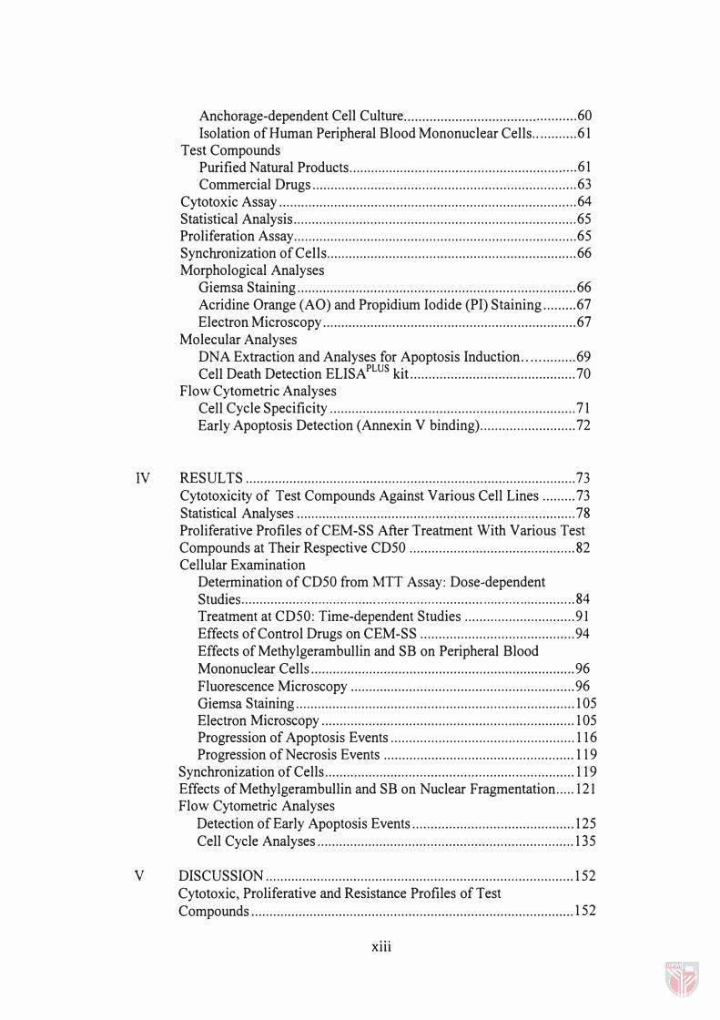
Anchorage-dependent Cell Culture ............................................... 60 Isolation of Human Peripheral Blood Mononuclear Cells ............ 61
Test Compounds Purified Natural Products .............................................................. 61 Commercial Drugs ........................................................................ 63
Cytotoxic Assay ................................................................................. 64 Statistical Analysis ............................................................................. 65 Proliferation Assay ............................................................................. 65 Synchronization of Cells .................................................................... 66 Morphological Analyses
Giemsa Staining ............................................................................ 66 Acridine Orange (AO) and Propidium Iodide (PI) Staining ......... 67 Electron Microscopy ..................................................................... 67
Molecular Analyses DNA Extraction and Analyses for Apoptosis Induction .. ............ 69 Cell Death Detection ELISA PLUS kit ............................................. 70
Flow Cytometric Analyses Cell Cycle Specificity ................................................................... 71 Early Apoptosis Detection (Annex in V binding) .......................... 72
IV RESULTS .......................................................................................... 73 Cytotoxicity of Test Compounds Against Various Cell Lines ......... 73 Statistical Analyses ............................................................................ 78 Proliferative Profiles of CEM-SS After Treatment With Various Test Compounds at Their Respective CD50 ............................................. 82 Cellular Examination
Determination of CD50 from MTT Assay: Dose-dependent Studies ................ . . . . . . . . . . . . . . . . ..... . . . . . . .. . . . . . . . . . . . .......... . . . . . . . . . . . . . . . .......... 84 Treatment at CD50: Time-dependent Studies .............................. 91 Effects of Control Drugs on CEM-SS .......................................... 94 Effects of Methylgerambullin and SB on Peripheral Blood Mononuclear Cells ........................................................................ 96 Fluorescence Microscopy ............................................................. 96 Giemsa Staining ............................................................................ 1 05 Electron Microscopy ..................................................................... 1 05 Progression of Apoptosis Events .................................................. 116 Progression of Necrosis Events .................................................... 119
Synchronization of Cells .................................................................... 119 Effects of Methylgerambullin and SB on Nuclear Fragmentation ..... 121 Flow Cytometric Analyses
Detection of Early Apoptosis Events ............................................ 125 Cell Cycle Analyses ...................................................................... 135
V DISCUSSION .................................................................................... 152 Cytotoxic, Proliferative and Resistance Profiles of Test
Compounds ........................................................................................ 152
Xlll
Proliferative Profiles of CEM-SS After Treatment With Test Compounds ........................................................................................ 156 Effects of Methylgerambullin and SB on Cell Morphology .............. 162 Effects of Methylgerambullin and SB on Nuclear Fragmentation ..... 173 Flow Cytometry: Detection of Apoptosis . . ........................................ 182 Flow Cytometry: Cell Cycle Analyses ....... .................... .................... 186
VI CONCLUSION ...................... ........................................ .................... 194
REFERENCES ............................................................................................. 200
APPENDICES .............................................................................................. 219 A Preparation of Media and Solutions ................................................... 220 B Calculation for Cell Viability and Cell Concentration ...................... 222 C The Means and Standard Error of Means (SEM) Corresponding to
Figures 12 and 13 ....... ........................................ ............................... 223 D Two-ways Analysis of Variance (ANOVA) ...................................... 224
VITA . ........................................................................................... . ............... 228
XIV

LIST OF TABLES
Table Page
1 ......... Biochemical determinants of apoptosis and necrosis ........................ 26
2 ......... Commercial drugs of various classes used as controls in this study. 63
3 ......... Sensitivity of various cell lines to methylgerambullin and SB ......... 73
4 ......... Sensitivity of CEM-SS to various commercial drugs . . . . . . .. . . .. . . . . . ..... . 78
5 ......... Sensitivity of CEM-SS to methylgerambullin, SB and various commercial drugs .............................................................................. 80
6 ......... The percentage of mitotic cells induced by colchicine at different concentrations ................................................................................... 121
7 . . . . . .... The summary of dose-dependent percentage of viable, apoptotic and necrotic CEM-SS cells .............................................................. . 129
8 ......... The summary of time-dependent percentage of viable, apoptotic and necrotic CEM-SS cells ............................................................... 134
9 ......... Cell cycle distribution in CEM-SS after24 hours incubation with methylgerambullin, SB, gonoiothalamin, cisplatin, etoposide and colchicine at various concentrations ........................................... 144
10 ....... Cell cycle distribution in CEM-SS after incubation with methylgerambullin, SB, gonoiothalamin, cisplatin, etoposide and colchicine at CD50 doses for various duration of time .............. 150
11 ....... Time-dependent cytotoxic profiles of control drugs, methylgerambullin and SB against CEM-SS cell line ...................................................... 223
12 ....... Proliferative profiles of CEM-SS cells after treatment with methylgerambullin, SB and control drugs at their respective CD50 dose . . . . . . . . . . . . . . . . . . . . . .. . . . .. . . . . . . . . . . . . . . . . . . . . . . . . . . .. . . . . . . . . . . . . . . . ... . . . ....... . . . . . 223
13 ....... The summary of means of CD50 for different compounds/drugs (Xi) and means of CD50 exhibited by different CEM-SS sub-populations at different time (Xj) ......................................................................... 224
14 ....... The summary of two-ways ANOV A. . .. . . . . . . ... . . . . . . ....... . . . . ...... . .. ......... . 225
xv

LIST OF FIGURES
Figure Page
1 ......... The experimental approach implemented in the current study ......... 5
2 ......... The structure of methylgerambullin and SB (bis-(methylthiomethyl)-disulphide) ......................................................................................... 11
3 ......... Morphological changes during apoptosis and necrosis ..................... 20
4 ......... Flow diagrams of apoptotic and necrotic major events ..................... 21
5 ......... Tetrazolium salts and its formazan .................................................... 37
6 ......... Evolution of tetrazolium salts used in measuring cell viability ........ 38
7 ......... The cell cycle .................................................................................... 48
8 ......... Cyclical changes in DNA content of a dividing cell ......................... 49
9 ......... The percentage of cell viability of various cell lines after treatment with different concentrations of methylgerambullin for 72 hours ............ 75
10 ....... The percentage of cell viability of various cell lines after treatment with different concentrations of SB for 72 hours ...................................... 76
11 ....... The percentage of cell viability of CEM-SS after treatment with different concentrations of control drugs, methylgerambullin and SB for 72 hours74 .............................................................................................. 77
12 ....... Time-dependent cytotoxic profiles of control drugs, methylgerambullin and SB against CEM-SS cell line ...................................................... 81
13 ....... Proliferative profiles of CEM-SS cell line ........................................ 83
14 ....... Time-dependent trends of apoptotic and necrotic incidence in CEM-SS cell population ................................................................................... 103
15 ....... Detection of mono- or oligonucleosomal DNA fragmentation in methylgerambullin- and SB-treated CEM-SS population by ELISAPLUS immunoassay ..................................................................................... 124
16 ....... Classification of viable, apoptosis and necrosis based on Annexin V -FITC and propidium iodide uptake ............................................................. 125
17 ....... Flow cytometric analyses on detection of apoptosis and necrosis at different doses for 24 hours ............................................................... 127
XVI

18 ....... Time course flow cytometric analyses on detection of apoptosis and necrosis at CD50 doses ..................................................................... 132
19 ....... Determination of cell cycle statistics from dot plot and histogram using CE LLFIT and LYSIS II softwares .................................................... 138
20 ....... Flow cytometric cell cycle analyses based on DNA content ............ 142
21 ....... Time-dependent flow cytometric cell cycle analyses based on DNA content ............................................................................................... 148
22 ....... Possible stages in the process of responding to cellular damage ...... 153
23 ....... Characterization of viable, apoptotic and necrotic cells based on the chromatin structure after dual staining with acridine orange and propidium iodide ............................................................................... 167
24 ....... The distribution of means between samples in the hypothesis testing ................................... ............................................................. 226
25 ....... The distribution of means within samples in the hypothesis testing ................................................................................................ 226
XVII

LIST OF PLATES
Plate Page
1 ......... Photomicrographs from inverted microscopy ....... . . .......................... 85
2 ......... Photomicrographs of cells after the addition ofMTT ....................... 89
3 ......... Photomicrograph of formazan crystals surrounding viable CE M-SS cells detected after 4 hours incubation with MTT ....................... .............. 90
4 . .. . ..... Time-dependent morphological changes of CE M-SS cells treated with methylgerambullin ............................. ........................................ . ...... 92
5 ......... Time-dependent morphological changes of CEM-SS cells treated with SB ......................... .......................................................... , ..... " .. , 93
6 ......... CE M-SS cells after 72 hours treatment with control drugs ............... 95
7 ......... Peripheral blood mononuclear cells .................... ............................. . 97
8 ......... Photomicrographs of acridine-orange-propidium iodide stained CEM-SS cells .. , ........ , ... , .......... , ........................ " ............. ,", .. , .... ,.,. , ........... , , ..... 100
9 ......... Photomicrographs of viable, apoptotic and necrotic cells ... , ............. 102
10 ....... Giemsa staining ......... . . . . ........... . . . . .................... ......................... . . . ..... 106
11 . . . ... , Scanning electron micrographs of methylgerambullin-treated CEM-SS cells ... " .... ,.,', .......... , ........ , ............. , ..... , .................................. , ........... 109
12 ....... Scanning electron micrographs of SB-treated CE M-SS cells .......... . 111
13 ....... Close up view of scanning electron micrographs .............................. 113
14 ....... Transmission electron micrographs of CEM-SS treated with methylgerambullin and SB ............... .................. . . . . . . . ........................ 115
15 ....... Ultrastructural changes in apoptotic cells ......................................... 117
16 ... . , .. Ultrastructural changes in necrotic cells ........................................... 120
17 ....... DNA profiles from agarose gel electrophoresis ........... , ............ , ....... 123
XVlll

LIST OF ABBREVIATIONS
2-CDA ........... chloro deoxyadenosine ABTS ............ 2,2' -Azino-di[3-ethylbenzthiazolin-sulfonat] ADP ... ........... adenosine diposphate AIF. .... ........... apoptosis indusing factor ALL ... ........... acute lymphoblastic leukemia AML .. . . . . . ... .. . acute myeloid leukemia AO ..... ........... acridine orange Ara-c ............. arabinofuranosylcytosine ATP .............. adenosine triposphate CA ..... ........... calyculin A CD50 . ........... cytotoxic dose resulting in 50% reduction of cell population CGM .. ........... complete growth medium CLL . ... ........... chronic lymphocytic leukemia CML ............. chronic myeloid leukemia C02
. . . • . • . . . • • . • . • carbon dioxide CV ..... ........... coefficient of variation Cyt-c ............. cytochrome c DCF ... ........... deoxycorfomycin DDS .............. diaminodiphenyl sulphone DMSO ........... dimethyl sulfoxide DNA .. ........... deoxyribonucleic acid ER ...... ........... endoplasmic reticulum
FDA .............. Food and Drug Administration FITC .. ........... fluorescein isothiocyanate FSC .... ........... forward scatter GSH .............. glutathione HCI .... ........... hydrochloric acid HIV .... ........... human immunodeficiency virus ICAD ............ inhibitor of caspase-activated DNAse ICE. ............... interleukin converting enzyme KCI .... ........... potassium chloride MTT .. ........... 3(4 ,5-dimethylthiazol-2-yl)-2,5-diphenyltetrazolium bromide MT A . ............ microculture tetrazolium assay MTS ... ........... 3-[ 4,5-dimethylthiazol-2-yl]-5-( carboxymethoxyphenyl),( 4-
sulphophenyl)-2H-tetrazolium MeOH ........... methanol MTP . .. .... . . .. . .. microtiter plate NaCI .. ........... sodium chloride NHAC ........... N4-hexadecyl-l-p-D-arabinofuranosylcytosine OD ..... ........... optical density OA ..... ........... okadaic acid PARP ............ poly(ADP-ribose) polymerase PBMC ........... peripheral blood mononuclear cell PBS .... ........... phosphate buffered saline Pl ....... ........... propidium iodide PMS ... ........... phenazine methosulphate
XIX
PMT ... .. ......... photo multiplier PS ...... . .. ......... phosphotidylserine PT ...... ........... permeability transition RNA. .. ........... ribonucleic acid RNP ... ........ ... ribonucleoprotein particles ROS ... ........ ... reactive oxygen species SB ...... ........... bis-(methylthiomethyl)-disulphide isolated from Scorodocarpus
borneensis Becc. SDS .. .. . .. .. ... ... sodium dodecyl sulphate SE M . . . ... ... ..... scanning electron microscope sse .... ... . . . . .... side scatter TE M . . ..... ...... transmission electron microscope TNF . .. .. ...... .. . tumor necrosis factor TUNEL ... ....... Tdt-mediated dUTP-biotin nick end labelling UV ..... ...... ..... ultraviolet XTT ... ........... sodium (2,3-bis[2-methoxy-4-nitro-5-sulphophenyl]-2H
tetrazolium-5-carboxanilide)
xx

CHAPTER I
INTRODUCTION
1
Screening for potential anticancer compounds has been one of the most
important aspects in cancer research. Many commercially available drugs are
synthetically derived, for example etoposide, a semi-synthetic
epipodophyllotoxin that is one of the most commonly used anti-cancer drugs
(Joel, 1996). Although these synthetic drugs are known to be very effective
against cancer cells, they also possess some undesirable side effects such as
neurotoxicity, fibrinolysis and ototoxicity (hearing loss) (Casciato and Lowitz,
1995). Thus, choices for improvement are confined to introduce natural products
as potential anticancer agents. Phytochemicals, one of these natural substances
have been known to exhibit a wide variety of medicinal value ever since the
ancient times. The scientific theories underlying their medicinal properties
however remained unknown then. Wilkinson, (1998) recently highlighted that
herbal products have great potential in the emerging nutraceutical and
pharmaceutical industries in that they are widely consumed as food and are used
in preventive and curative treatments throughout the world. It was reported in the
same study that tropical forests have produced 47 major pharmaceutical drugs of
world-wide importance and it is estimated that 328 potential drugs of major
importance is yet to be discovered which would be worth $147 billion. The
tropical forests still offers 125,000 flowering plant species that are of
pharmacological relevance but this will involve 50,000 to one million screening
2
tests to discover one profitable drug, which seem to be the ultimate constraint in
the discovery of new drugs.
It has been reported by the Alliance Pharmaceutical Corporation that on average,
12 years are needed for an experimental drug to be brought from the laboratory
to the market shelf (Wierenga and Eaton, Internet). Preclinical testing requires
three and a half years of experiments in laboratory on in vitro and animal models
to assess safety and biological activities of the compound. Another six years are
required for clinical trials that assess the safety, dosage, effectiveness and
monitor the adverse reaction of the compound. Only five in 5000 compounds that
enter preclinical testing make it to human testing. Approval process by the Food
and Drug Administration (FDA) will take about two and a half years where
normally only one of the five compounds tested in humans is approved.
Despite the duration of time required to evaluate a potential drug, numerous
phytochemicals have been scientifically isolated and their discovery was very
much directed in the perspective of anticancer drug development. Plant
constituents such as taxol, camptothecin, podophyllotoxin, vinblastine and
vincristine represent some of the most important drugs currently utilized for the
treatment of human cancers (Wall and Wani, 1993). Numerous more
phytochemica1s are under investigations and have the potential to be on the
pharmaceutical market shelves. Among these are betulinic acid, gonoithalamin
and dammarane-type triterpenes, to name a few. Betulinic acid, which is
available in abundant supply from the bark of white birch trees was reported to
3
be specific for inhibiting human melanomas (Pisha et al., 1995).
Gonoiothalamin, isolated from various Gonoiothalamus sp. was found to be
toxic to cervical, pancreas, gastric and breast carcinoma (Ali et al., 1996).
Dammarane-type triterpenes from Cloeme africana has been shown to be highly
toxic against leukemia (Nagaya et ai., 1997).
One of the most extensively studied diseases is leukemia that remains to be a
formidable disease. Due to the ease of obtaining repeated samples from blood,
marrow or lymph nodes, most of the principles of cancer therapy is largely based
on the leukemia model. Important breakthroughs in cancer therapy were
achieved in the treatment of leukemia, which includes the use of combination
therapy, immunotherapy, supportive therapy using antibiotics and blood product
support and bone marrow transplant (Ng, 1996). Treatment with antileukaemic
drugs however only controls the symptom without enhancing the survival rate
(Arthur, 1989). Chemotherapeutic agents, also known as cytotoxins due to its
ability to kill cells are normally less specific in that these agents kill not only
cancer cell but also normal cells. Thus, the most apparent implication of
chemotherapy is emanation of side effects which include hair loss, nausea,
vomitting, constipation and can be as disastrous as myelosuppression (drop in
blood count - neutropenia, thrombocytopenia, anaemia), and damage on cardiac,
pulmonary, renal and hepatic systems (Ng, 1996). This prompted efforts to
screen for antileukaemic agents that will have a dramatic cytotoxic effect
specifically on the target cells. Most of the drugs currently used act via two
distinct mechanisms: inhibition of cell proliferation or induction of active cell

4
death (Vial et al., 1997). Compounds that act via the latter mechanism are likely
to be a better candidate as an antileukaemic agent because in the former action,
some individual cells may have escaped through salvage pathways, and will
continue to proliferate. The advancement in research techniques and the
introduction of new molecular tools in recent years have prompted more efforts
in investigations to build understanding on how exposure to the existing or newly
discovered drugs lead to cell death. The efficacy of these drugs has been
critically assessed by the many mechanisms of cell death and resistance that
adversely limit their efficacy. Today, due to the urgent need for studies in various
fields that closely mimics the animal systems, animal cell culture which was only
an esoteric art during those time has become an established technology. New
drug discovery, for example, may not be recognized and may be unsafe for use in
man. Thus, pre-clinical trials are needed, which can be made possible with cell
culture technology.
It is thus crucial to determine the mode of action exerted by a potential
anticancer agent. This has led to the design of the experimental approach which
will be implemented in the current study. Both cellular and molecular changes in
treated cell populations will be examined and compared with control (untreated)
populations in a time course manner. Hence, either necrotic or apoptotic cell
death could be suggested. Morphological studies may offer answers to whether
physiological and accidental deaths are causally related and to the degree of
overlapping between apoptotic and necrotic pathways.