Università degli Studi di Ferrara - EprintsUnife ...eprints.unife.it/312/1/tesi.pdf · ... ∆F508...
126
Università degli Studi di Ferrara DOTTORATO DI RICERCA IN "BIOCHIMICA, BIOLOGIA MOLECOLARE E BIOTECNOLOGIE" CICLO XXII COORDINATORE Prof. BERNARDI FRANCESCO STRATEGIES FOR ALTERATION OF PRO-INFLAMMATORY GENE EXPRESSION IN CYSTIC FIBROSIS Settore Scientifico Disciplinare BIO/10 Dottorando Tutore Dott. Mancini Irene Prof. Gambari Roberto _______________________________ _____________________________ (firma) (firma) Anni 2007/2009
Transcript of Università degli Studi di Ferrara - EprintsUnife ...eprints.unife.it/312/1/tesi.pdf · ... ∆F508...

Università degli Studi di Ferrara
DOTTORATO DI RICERCA IN
"BIOCHIMICA, BIOLOGIA MOLECOLARE E BIOTECNOLOGIE"
CICLO XXII
COORDINATORE Prof. BERNARDI FRANCESCO
STRATEGIES FOR ALTERATION OF PRO-INFLAMMATORY GENE EXPRESSION IN
CYSTIC FIBROSIS
Settore Scientifico Disciplinare BIO/10 Dottorando Tutore Dott. Mancini Irene Prof. Gambari Roberto _______________________________ _____________________________ (firma) (firma)
Anni 2007/2009
I
INDEX
INTRODUCTION
1. Cystic Fibrosis pag. 1
1.1 History pag. 1
1.2 Clinical aspects pag. 2
1.3 Diagnosis and monitoring pag. 5
1.4 Prenatal diagnosis pag. 7
1.5 Therapy pag. 8
1.5.1 Anti-inflammatory therapy pag. 8
1.5.2 Chemotherapy pag. 9
1.5.3 Other methods to treat lung disease pag. 9
1.5.4 Treatment of others aspects pag. 10
1.5.5 Gene therapy pag. 11
1.5.6 Stem cells pag. 13
1.5.7 Lung transplantation pag. 13
2. CFTR: Cystic Fibrosis Transmembrane Conductance Regulator pag. 14
2.1 CFTR protein pag. 14
2.1.1 From CFTR to lung disease pag. 18
2.2 CFTR gene pag. 19
2.3 CFTR mutations pag. 21
2.3.1 Molecular mechanisms of CFTR mutations pag. 22
3. Infection and inflammation in CF pag. 27
3.1 Lung infection pag. 27
3.2 Lung inflammation pag. 29
3.3 Cytokines and inflammatory mediators in cystic fibrosis pag. 30
3.4 NF-kB in defense and diseases pag. 35
3.4.1 NF-kB: a key role in inflammation pag. 35
II
3.4.2 NF-kB function and regulation pag. 36
3.4.3 Inhibition of NF-kB: a strategy in anti-inflammatory therapies
pag. 39
3.5 Oligodeoxynucleotides as therapeutic decoys pag. 44
3.6 Natural products pag. 46
3.6.1 Bergamot pag. 47
3.6.2 Emblica officinalis pag. 49
3.6.3 Saraca asoka pag. 51
AIM OF THESIS pag. 54
MATERIALS AND METHODS
1. Cell cultures pag. 58 2. Cell infection with Pseudomonas aeruginosa pag. 58 3. Decoy oligodeoxynucleotides pag. 59 4. Transfection of cells with decoy ODNs pag. 59 5. Plant extracts and chemical pag. 60 6. Assay of in vitro antiproliferative activity pag. 60 7. Preparation of nuclear extracts pag. 60 8. Electrophoresis mobility shift assay (EMSA) pag. 61 9. Quantification of IL-8 transcripts pag. 62 10. Cytokine profiles pag. 64 11. GC-FID and GC-MS analysis pag. 68 12. HPLC analysis pag. 69 13. 1H NMR fingerprinting analysis pag. 69
III
14. Statistical analysis pag. 70
RESULTS
1. Induction of pro-inflammatory genes by Pseudomonas aeruginosa in IB3-1 cells pag. 71
2. Search for transcription factor binding sites within the promoters of pro- inflammatory genes pag. 72
3. Transcription factor decoy pag. 74
3.1 Effect of NF-kB decoy ODNs on nuclear axtracts pag. 74
3.2 Effect of NF-kB decoy ODNs in IB3-1 cells pag. 76
4. Natural products pag. 80
4.1 Bergamot extracts pag. 80
4.1.1 Effect of Bergamot extracts on the expression of IL-8 m-RNA in
IB3-1 cells following TNF-α treatment pag.81
4.1.2 Effect of Bergamot extracts on the expression of IL-8 genes in
IB3-1 cells following TNF-α treatment: a Bio-plex analysis pag. 83
4.1.3 Phytochemical investigation pag. 84
4.1.4 Effect of identified compounds in bergamot extracts on expression
of IL-8 genes in TNF-α treated IB3-1 cells pag. 89
4.2 Emblica officinalis pag. 92
4.2.1 Gas chromatography-mass spectrometry analysis of the fractions
of Emblica officinalis extracts pag. 92
4.2.2 Effect of Emblica officinalis extracts on in vitro proliferation of IB3-
1 cells pag.94
4.2.3 Effect of Emblica officinalis extracts on the expression of pro-
inflammatory genes induced in IB3-1 cells by TNF-α treatment
pag. 95

IV
4.2.4 Effect of Emblica officinalis extracts and identified compound
pyrogallol on expression of IL-8 in TNF-α treated IB3-1 cells pag. 97
4.2.5 In vitro effect of Emblica officinalis extracts on NF-kB transcription
factor activity pag. 99
4.3 Saraca Asoka pag. 100
4.3.1 Induction of cytokine and chemokine release in IB3-1 cells treated
with TNF-α pag. 101
4.3.2 Effect of Saraca asoka extracts on in vitro proliferation of IB3-1
cells pag. 102
4.3.3 Effect of Saraca asoka extracts on the expression of pro-
inflammatory genes induced in IB3-1 cells by TNF-α treatment
pag. 103
4.3.4 Saraca asoka extracts inhibit IL-8 m-RNA expression pag. 105
4.3.5 Saraca asoka extracts inhibit interaction between NF-kB
transcription factor and target DNA sequences pag. 107
DISCUSSION AND FUTURE PERSPECTIVES
1. Transcription factor decoy pag. 108
2. Natural products pag. 113
REFERENCES pag. 118

V
ABBREVIATIONS
ABC ATP-binding cassette
ASL Apical Surface Liquid
ATP Adenosine triphosphate
BALF Broncho-alveolar Lavage Fluid
cAMP Cyclic Adenosine Monophosphat
CF Cystic Fibrosis
CFTR Cystic Fibrosis Transmembrane conductance Regulator
CFU Colony-forming Units
COX Cyclooxygenase
EMSA Electrophoresis Mobility Shift Assay
ENaC Epithelial Na+ Channel
FBS Fetal Bovine Serum
GC-FID Gas Chromatography/Flame Ionization Detection
GC-MS Gas Chromatography/Mass Spectrometry
GCs Glucocorticoids
G-CSF Granulocyte Colony-Stimulating Factor

VI
HPLC High-pressure Liquid Chromatography
ICAM-1 Intercellular Adhesion Molecule-1
IL-1 Interleukin 1
IL-10 Interleukin 10
IL-6 Interleukin 6
IL-8 Interleukin 8
LPS lipopolysaccharides
LTR Long Terminal Repeat
MCP1 Monocyte Chemotactic Protein-1
MSDs Membrane-Spanning Domains
NBDs Nucleotide-Binding Domains
NF-kB Nuclear factor k chain in B cells
NMR Nuclear Magnetic Resonance
ODN Oligodeoxynucleotide
PAO-1 Pseudomonas aeruginosa strain 1
PCR Polymerase chain reaction
PE Phycoerytrin
PGs Prostaglandins
VII
PKA cAMP-dependent Protein Kinase
PMN Polymorphonuclear Neutrophil
RANTES Regulated on Activation Normal T Expressed and Secreted
RD Regulatory Domain
RT-PCR Retro-transcription Polymerase Chain Reaction
TdT Terminal Deoxynucleotidyl Transferase
TF Transcription Factor
TFD Transcription Factor Decoy
TNF-α Tumor Necrosis Factor-α
INTRODUCTION
1. CYSTIC FIBROSIS
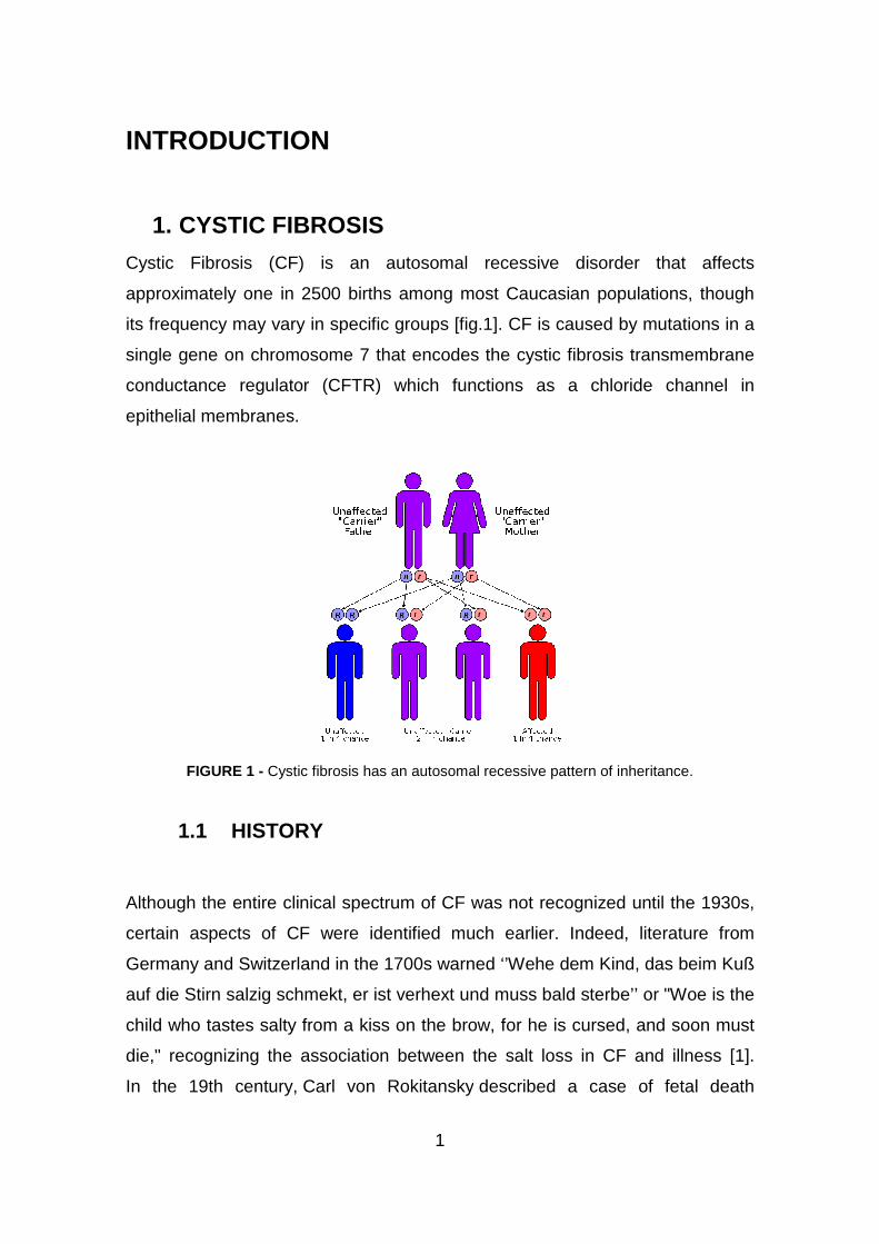
Cystic Fibrosis (CF)
approximately one in 2500 births among most Caucasian population
its frequency may vary in specific groups
single gene on chromosome 7 that encodes the cystic fibrosis transmembran
conductance regulator (CFTR
epithelial membranes.
FIGURE 1 - Cystic fibrosis has an autosomal recessive pattern of inheritance
1.1 HISTORY
Although the entire clinical spectrum of CF was not recognized until the 1930s,
certain aspects of CF were identified much e
Germany and Switzerland in the 1700s warned ‘’Wehe dem Kind, das beim Kuß
auf die Stirn salzig schmekt, er ist verhext und muss bald sterbe’’ or "Woe is the
child who tastes salty from a kiss on the brow, for he is cursed, and soon must
die," recognizing the association between
In the 19th century, Carl von Rokitansky
1
INTRODUCTION
CYSTIC FIBROSIS
(CF) is an autosomal recessive disorder that affects
500 births among most Caucasian population
its frequency may vary in specific groups [fig.1]. CF is caused by mutations in a
single gene on chromosome 7 that encodes the cystic fibrosis transmembran
conductance regulator (CFTR) which functions as a chloride channel in
Cystic fibrosis has an autosomal recessive pattern of inheritance
HISTORY
Although the entire clinical spectrum of CF was not recognized until the 1930s,
certain aspects of CF were identified much earlier. Indeed, literature from
Germany and Switzerland in the 1700s warned ‘’Wehe dem Kind, das beim Kuß
auf die Stirn salzig schmekt, er ist verhext und muss bald sterbe’’ or "Woe is the
child who tastes salty from a kiss on the brow, for he is cursed, and soon must
he association between the salt loss in CF and illness [1
Carl von Rokitansky described a case of fetal death
is an autosomal recessive disorder that affects
500 births among most Caucasian populations, though
CF is caused by mutations in a
single gene on chromosome 7 that encodes the cystic fibrosis transmembrane
) which functions as a chloride channel in
Cystic fibrosis has an autosomal recessive pattern of inheritance.
Although the entire clinical spectrum of CF was not recognized until the 1930s,
arlier. Indeed, literature from
Germany and Switzerland in the 1700s warned ‘’Wehe dem Kind, das beim Kuß
auf die Stirn salzig schmekt, er ist verhext und muss bald sterbe’’ or "Woe is the
child who tastes salty from a kiss on the brow, for he is cursed, and soon must
the salt loss in CF and illness [1].
described a case of fetal death

2
with meconium peritonitis, a complication of meconium ileus associated with
cystic fibrosis. Meconium ileus was first described in 1905 by Karl Landsteiner
[1]. In 1936, Guido Fanconi published a paper describing a connection
between celiac disease, cystic fibrosis of the pancreas, and bronchiectasis [2].
In 1938 Dorothy Hansine Andersen published an article, "Cystic Fibrosis of the
Pancreas and Its Relation to Celiac Disease: a Clinical and Pathological Study,"
in the American Journal of Diseases of Children. She was the first to describe
the characteristic cystic fibrosis of the pancreas and to correlate it with the lung
and intestinal disease prominent in CF [3]. She also first hypothesized that CF
was a recessive disease and first used pancreatic enzyme replacement to treat
affected children. In 1952 Paul di Sant' Agnese discovered abnormalities
in sweat electrolytes; a sweat test was developed and improved over the next
decade [4].
In 1988 the first mutation for CF, ∆F508 was discovered by Francis
Collins, Lap-Chee Tsui and John R. Riordan on the seventh chromosome.
Subsequent research has found over 1,000 different mutations that cause CF.
Because mutations in the CFTR gene are typically small, classical
genetics techniques had been unable to accurately pinpoint the mutated gene
[5]. Using protein markers, gene-linkage studies were able to map the mutation
to chromosome 7. Chromosome-walking and -jumping techniques were then
used to identify and sequence the gene [6]. In 1989 Lap-Chee Tsui led a team
of researchers at the Hospital for Sick Children in Toronto that discovered the
gene responsible for CF in 1989. Cystic fibrosis represents the first genetic
disorder elucidated strictly by the process of reverse genetics.
1.2 CLINICAL ASPECTS
Expression of the cystic fibrosis transmembrane conductance regulatory gene is
found in areas that are lined with epithelial tissue. Some of these places include
ciliated epithelium of high airways, the gastrointestinal tract, salivary and sweat
glands, cervix, uterus, fallopian tubes, epididymis and the vas deferens. These
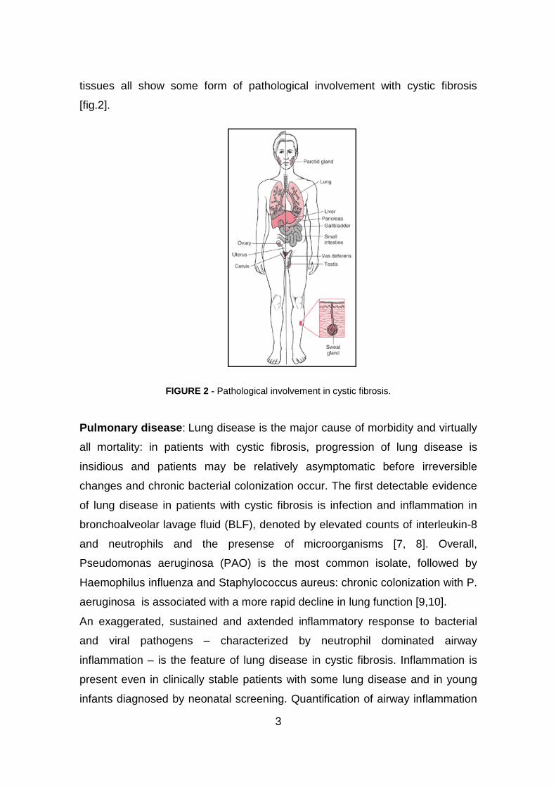
3
tissues all show some form of pathological involvement with cystic fibrosis
[fig.2].
FIGURE 2 - Pathological involvement in cystic fibrosis.
Pulmonary disease : Lung disease is the major cause of morbidity and virtually
all mortality: in patients with cystic fibrosis, progression of lung disease is
insidious and patients may be relatively asymptomatic before irreversible
changes and chronic bacterial colonization occur. The first detectable evidence
of lung disease in patients with cystic fibrosis is infection and inflammation in
bronchoalveolar lavage fluid (BLF), denoted by elevated counts of interleukin-8
and neutrophils and the presense of microorganisms [7, 8]. Overall,
Pseudomonas aeruginosa (PAO) is the most common isolate, followed by
Haemophilus influenza and Staphylococcus aureus: chronic colonization with P.
aeruginosa is associated with a more rapid decline in lung function [9,10].
An exaggerated, sustained and axtended inflammatory response to bacterial
and viral pathogens – characterized by neutrophil dominated airway
inflammation – is the feature of lung disease in cystic fibrosis. Inflammation is
present even in clinically stable patients with some lung disease and in young
infants diagnosed by neonatal screening. Quantification of airway inflammation

4
is necessary to monitor its evolution over time and the effect of anti-
inflammatory treatment. This monitoring remains a difficult task, since reliable
non invasive markers of airway inflammation are not available.
Gastrointestinal, liver and pancreatic disease : Prior to prenatal and newborn
screening, cystic fibrosis was often diagnosed when a newborn infant failed to
pass feces (meconium). Meconium may completely block the intestines and
cause serious illness. This condition, called meconium ileus, occurs in 10% of
newborns with CF [11]. In addition, protrusion of internal rectal membranes
(rectal prolapse) is more common in CF because of increased fecal volume,
malnutrition, and increased intra–abdominal pressure due to coughing [12]. The
thick mucus seen in the lungs has a counterpart in thickened secretions from
the pancreas, an organ responsible for providing digestive juices that help break
down food. These secretions block the movement of the digestive enzymes into
the duodenum and result in irreversible damage to the pancreas, often with
painful inflammation (pancreatitis) [13]. The lack of digestive enzymes leads to
difficulty absorbing nutrients with their subsequent excretion in the feces, a
disorder known as malabsorption. Malabsorption leads to malnutrition and poor
growth and development because of caloric loss. Individuals with CF also have
difficulties absorbing the fat-soluble vitamins A, D, E, and K. In addition to the
pancreas problems, CF patients experience more heartburn, intestinal blockage
by intussusception, and constipation [14]. Older CF patients may also
develop distal intestinal obstruction syndrome when thickened feces cause
intestinal blockage [15]. Thickened secretions also may cause liver problems in
patients with CF. Bile secreted by the liver to aid in digestion may block the bile
ducts, leading to liver damage. Over time, this can lead to cirrhosis, in which the
liver fails to rid the blood of toxins and does not produce important proteins such
as those responsible for blood clotting [16-17].
Endocrine disease : the pancreas contains the islets of Langerhans, which are
responsible for making insulin, a hormone that helps regulate blood glucose.
Damage of the pancreas can lead to loss of the isletcells, leading to a type of
diabetes that is unique to those with the disease [18]. This Cystic Fibrosis
5
Related Diabetes (CFRD) shares characteristics that can be found in Type
1 and Type 2 diabetics and is one of the principal non-pulmonary complications
of CF [19].
Osteoporosis : Vitamin D is involved in calcium and phosphorus regulation.
Poor uptake of vitamin D from the diet because of malabsorption can lead to the
bone disease osteoporosis in which weakened bones are more susceptible
to fractures [20]. In addition, CF patients often develop clubbing of their fingers
and toes due to the effects of chronic illness and low oxygen in their tissues.
Infertility : Infertility affects both men and women. At least 97 percent of men
with cystic fibrosis are infertile but are not sterile and can have children with
assisted reproductive techniques [21]. These men make normal sperm but are
missing the tube (vas deferens), which connects the testes to the ejaculatory
ducts of the penis [22]. Many men found to have congenital absence of the vas
deferens during evaluation for infertility have a mild, previously undiagnosed
form of CF [23]. Some women have fertility difficulties due to thickened cervical
mucus or malnutrition. In severe cases, malnutrition disrupts ovulation and
causes amenorrhea [24].
1.3 DIAGNOSIS AND MONITORING
Cystic fibrosis may be diagnosed by many different categories of testing
including those such as, newborn screening, sweat testing, or genetic testing.
The newborn screen initially measures for raised blood concentration
of immunoreactive trypsinogen [25]. Infants with an abnormal newborn screen
need a sweat test in order to confirm the CF diagnosis. Trypsinogen levels can
be increased in individuals who have a single mutated copy of the CFTR gene
(carriers) or, in rare instances, even in individuals with two normal copies of the
CFTR gene. Due to these false positives, CF screening in newborns is
somewhat controversial [26-27]. Therefore, most individuals are diagnosed after
symptoms prompt an evaluation for cystic fibrosis. The most commonly used
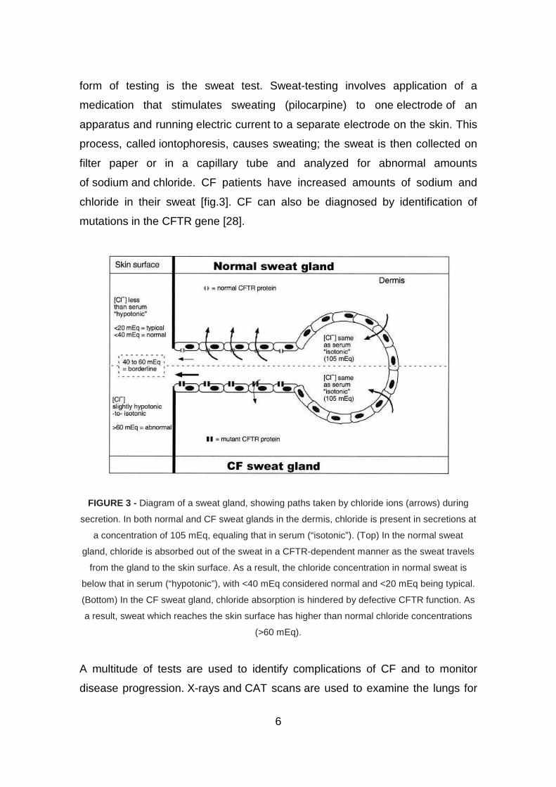
6
form of testing is the sweat test. Sweat-testing involves application of a
medication that stimulates sweating (pilocarpine) to one electrode of an
apparatus and running electric current to a separate electrode on the skin. This
process, called iontophoresis, causes sweating; the sweat is then collected on
filter paper or in a capillary tube and analyzed for abnormal amounts
of sodium and chloride. CF patients have increased amounts of sodium and
chloride in their sweat [fig.3]. CF can also be diagnosed by identification of
mutations in the CFTR gene [28].
FIGURE 3 - Diagram of a sweat gland, showing paths taken by chloride ions (arrows) during
secretion. In both normal and CF sweat glands in the dermis, chloride is present in secretions at
a concentration of 105 mEq, equaling that in serum (“isotonic”). (Top) In the normal sweat
gland, chloride is absorbed out of the sweat in a CFTR-dependent manner as the sweat travels
from the gland to the skin surface. As a result, the chloride concentration in normal sweat is
below that in serum (“hypotonic”), with <40 mEq considered normal and <20 mEq being typical.
(Bottom) In the CF sweat gland, chloride absorption is hindered by defective CFTR function. As
a result, sweat which reaches the skin surface has higher than normal chloride concentrations
(>60 mEq).
A multitude of tests are used to identify complications of CF and to monitor
disease progression. X-rays and CAT scans are used to examine the lungs for
7
signs of damage or infection. The examination of the sputum is required to
isolate organisms which may be causing an infection or colonising the lower
respiratory tract so that effective antimicrobial therapy can be provided. Culture
for organisms such as Burkholderia cepacia (previously Pseudomonas) is
required for candidates of lung transplantation as persisant bacterial
colonisation reduces the chances of survival.
Pulmonary function tests measure how well the lungs are functioning, and are
used to measure the need for and response to antibiotic therapy. Blood
tests can identify liver abnormalities, vitamin deficiencies, and the onset of
diabetes. DEXA scans can screen for osteoporosis and testing for fecal
elastase can help diagnose insufficient digestive enzymes.
1.4 PRENATAL DIAGNOSIS
Couples who are pregnant or who are planning a pregnancy can themselves be
tested for CFTR gene mutations to determine the degree of risk that their child
will be born with cystic fibrosis. Testing is typically performed first on one or
both parents and, if the risk of CF is found to be high, testing on the fetus can
then be performed. Because development of CF in the fetus requires each
parent to pass on a mutated copy of the CFTR gene and because CF testing is
expensive, testing is often performed on just one parent initially. If that parent is
found to be a carrier of a CFTR gene mutation, the other parent is then tested to
calculate the risk that their children will have CF. CF can result from more than
a thousand different mutations and it is not possible to test for each one.
Testing analyzes the blood for the most common mutations such as ∆F508 -
most commercially available tests look for 32 or fewer different mutations. If a
family has a known uncommon mutation, specific screening for that mutation
can be performed. Because not all known mutations are found on current tests,
a negative screen does not guarantee that a child will not have CF [29]. In
addition, because the mutations tested are necessarily those most common in
the highest risk groups, testing in lower risk ethnicities is less successful
because the mutations commonly seen in these groups are less common in the
8
general population. Couples who are at high risk for having a child with CF will
often opt to perform further testing before or during pregnancy. In vitro
fertilization with preimplantation genetic diagnosis offers the possibility to
examine the embryo prior to its placement into the uterus. The test, performed
three days after fertilization, looks for the presence of abnormal CF genes. If
two mutated CFTR genes are identified, the embryo is not used for embryo
transfer and an embryo with at least one normal gene is implanted. During
pregnancy, testing can be performed on the placenta (chorionic villus sampling)
or the fluid around the fetus (amniocentesis).
1.5 THERAPY
Although no definite cure has been discovered for cystic fibrosis as of yet, many
treatments have been developed to improve the quality of life for those who
suffer from this disorder. Since CF causes a build-up of mucus in the lung
tissue, the respiratory diseases which result are often the actual cause of death,
not CF itself.
1.5.1 ANTI-INFLAMMATORY THERAPY
Inflammation plays a major role in the pathophysiology of lung disease in CF.
This respose is probably triggered primarily as a reaction to the inability of the
affected lung to resist the invasion of the most common bacterial pathogens
seen in this disease.
Anti-inflammatoty therapy can be provided in various forms: these include the
use of oral corticosteroids which are potentially highly effective but which carry
with them the risk of long-term systemic side effects.
Inhaled corticosteroids also have considerable potential because of their local
action within the lung. Their potential therapeutic disadvantage is the difficulty of
penetrating the viscid mucus which lines the airway in CF patients particularly
as the disease progresses.
Other anti-inflammatory agents such as ibuprofen have considerable potential
and have been the subject of various studies over the years.
9
More recently macrolides have come forward as potent anti-inflammatory
agents and are beginning to have an established place in the therapeutic
regimen for patients with long-term Pseudomonas aeruginosa infection. Other
agents which have been used include immunoglobulins and dornase alpha
(DNase) that may have an anti-inflammatory role as well as being mucolytic.
1.5.2 CHEMOTERAPY
Many CF patients are on one or more antibiotic at all times, even when they are
considered healthy, in order to prophylacticly suppress infection. Antibiotics are
absolutely necessary whenever pneumonia is suspected or a lung function
decline has been noticeable, and are usually chosen based on the results of a
sputum analysis and the patient's past response. Many bacteria common in
cystic fibrosis are resistant to multiple antibiotics and require weeks of treatment
with Intravenous antibiotics such as vancomycin, tobramycin, meropenem,
ciprofloxacin and piperacillin. This prolonged therapy often necessitates
hospitalization and insertion of a more permanent IV such as a peripherally
inserted central catheter (PICC) line or Port-a-Cath. Inhaled therapy with
antibiotics such as tobramycin and colistin is often given for months at a time in
order to improve lung function by impeding the growth of colonized bacteria [30,
31]. Inhaled antibiotic therapy with aztreonam is also being developed and
clinical trials are underway [32]. Oral antibiotics such as ciprofloxacin
or azithromycin are given to help prevent infection or to control ongoing
infection [33]. Several of the antibiotics commonly used to treat CF patients,
such as tobramycin and vancomycin, can cause hearing loss, damage to
the balance system in the inner ear or kidney problems with long-term use. In
order to prevent these side effects, the amount of antibiotics in the blood are
routinely measured and adjusted accordingly.
1.5.3 OTHER METHODS TO TREAT LUNG DISEASE
Several mechanical techniques are used to dislodge sputum and encourage its
expectoration. In the hospital setting, chest physiotherapy (CPT) is utilized; a
respiratory therapist percusses an individual's chest with his or her hands

10
several times a day, to loosen up secretions. Devices that recreate this
percussive therapy include the ThAIRapy Vest and the intra-pulmonary
percussive ventilator (IPV).
Newer methods such as biphasic cuirass ventilation (BCV), and associated
clearance mode available in such devices, integrate a cough assistance phase,
as well as a vibration phase for dislodging secretions. Biphasic Cuirass
Ventilation is also shown to provide a bridge to transplantation.These are
portable and adapted for home use [34].
Physiotherapy is essential to help manage an individual’s chest on a long term
basis, and can also teach techniques for the older child and teenager to
manage themselves at home. Aerobic exercise is of great benefit to people with
cystic fibrosis. Not only does exercise increase sputum clearance but it also
improves cardiovascular and overall health.Aerosolized medications that help
loosen secretions include dornase alfa and hypertonic saline [35]. Dornase is
a recombinant human deoxyribonuclease, which breaks down DNA in the
sputum, thus decreasing its viscosity [36]. N-Acetylcysteine may also decrease
sputum viscosity, but research and experience have shown its benefits to be
minimal. Albuterol and ipratropium bromide are inhaled to increase the size of
the small airways by relaxing the surrounding muscles.
As lung disease worsens, mechanical breathing support may become
necessary. Individuals with CF may need to wear special masks at night that
help push air into their lungs. These machines, known as bilevel positive airway
pressure (BiPAP) ventilators, help prevent low blood oxygen levels during
sleep. BiPAP may also be used during physical therapy to improve sputum
clearance [37]. During severe illness, a tube may be placed in the throat (a
procedure known as a tracheostomy) to enable breathing supported by
a ventilator.
1.5.4 TREATMENT OF OTHER ASPECTS
Newborns with meconium ileus (bowel obstruction) typically require surgery,
whereas adults with distal intestinal obstruction syndrome typically do not.
Treatment of pancreatic insufficiency by replacement of missing digestive
11
enzymes allows the duodenum to properly absorb nutrients and vitamins that
would otherwise be lost in the feces. Even so, most individuals with CF are
advised take additional amounts of vitamins A, D, E, and K and eat high-calorie
meals. So far, no large-scale research involving the incidence
of atherosclerosis and coronary heart disease in adults with cystic fibrosis has
been conducted. This is likely due to the fact that the vast majority of people
with cystic fibrosis do not live long enough to develop clinically significant
atherosclerosis or coronary heart disease.
Diabetes is the most common non-pulmonary complication of CF. It mixes
features of type 1 and type 2 diabetes, and is recognized as a distinct entity,
cystic fibrosis-related diabetes (CFRD) [38,39]. While oral anti-diabetic
drugs are sometimes used, the only recommended treatment is the use
of insulin injections or an insulin pump [40], and, unlike in type 1 and 2 diabetes,
dietary restrictions are not recommended [38].
Development of osteoporosis can be prevented by increased intake of vitamin D
and calcium, and can be treated by bisphosphonates, although adverse
effects can be an issue [41]. Poor growth may be avoided by insertion of
a feeding tube for increasing calories through supplemental feeds or by
administration of injected growth hormone [42].
Sinus infections are treated by prolonged courses of antibiotics. The
development of nasal polyps or other chronic changes within the nasal
passages may severely limit airflow through the nose, and over time reduce the
patient's sense of smell. Sinus surgery is often used to alleviate nasal
obstruction and to limit further infections. Nasal steroids such as fluticasone are
used to decrease nasal inflammation [43]. Female infertility may be overcome
by assisted reproduction technology, particularly embryo transfer techniques.
Male infertility may be overcome with intracytoplasmic sperm injection
[44]. Third party reproduction is also a possibility for women with CF.
1.5.5 GENE THERAPY
Cystic fibrosis should be an ideal candidate for gene therapy, for four main
reasons: (1) it is a single gene defect; (2) it is a recessive condition, with
12
heterozygotes being phenotypically normal (suggesting gene dosage effects are
not critical); (3) the main pathology is in the lung, which is accessible for
treatment; and (4) it is a progressive disease with a virtually normal phenotype
at birth, offering a therapeutic window.
It has been suggested that only 5–10% of normal CFTR function is required to
reverse the chloride channel defect [45], although it is not clear whether this has
to be achieved in the majority of the airway epithelial cells, or whether a minority
of cells expressing much higher levels would suffice. In clinical trials to date, two
main vector systems have been harnessed to deliver the CFTR cDNA with
appropriate promoter into host cells [46,47]. First, viral vectors with the CFTR
cDNA incorporated into the viral genome exploit the efficiency of viruses to
enter host cells and achieve relatively high levels of gene expression. Secondly,
cationic liposomes mixed with plasmid DNA encoding CFTR enhance the
transport of the DNA into host cells. Although cationic liposomes seem to
generate a lower immune response than current viral vector systems, the levels
of CFTR expression using this delivery system have been relatively poor.
The ideal vector system would have the following characteristics: (1) an
adequate carrying capacity; (2) to be undetectable by the immune system; (3) to
be non-inflammatory; (4) to be safe to the patients with pre-existing lung
inflammation; (5) to have an efficiency sufficient to correct the cystic fibrosis
phenotype; and (6) to have long duration of expression and/or the ability to be
safely re-administered.
In-vivo gene therapy trials in patients with cystic fibrosis have been done with
viral vectors and cationic lipids, [48, 49] however, long-term effects were not
achieved. Repeat administration of adenovirus vectors reduces efficacy of
transfection because of formation of specific antibodies, whereas lipids might
not specifically target CFTR-expressing cells. Therefore, although much
progress has been made in gene therapy, it is presently not a treatment option
for patients with cystic fibrosis.
13
1.5.6 STEM CELLS
CF is a potential model disease for stem cell therapy because of the continuing
lung inflammation and infection leading to damage, which could promote
engraftment of stem cells. It is likely that infants and children with CF would be
the best candidates for this sort of therapy, when they have faster cell turnover
and before they have established lung damage: the ultimate therapy will be
gene therapy to CF babies by utilizing their own stored umbilical cord blood
stem cells [50].
Nevertheless, experiments to elucidate possible lung stem cell candidates are
difficult because of the slow turnover of such cells, and therefore most animal
studies use an injury model which promotes cell turnover. Moreover, basic
questions remain unanswered. It is unknown which cell type should be targeted;
current topical therapy targets the abundant surface epithelium, but it is the
submucosal glands which expresses the highest CFTR in the lung.
1.5.7 LUNG TRANSPLANTATION
Double lung or heart-lung transplantation is a treatment option for patients with
cystic fibrosis and end-stage lung disease. Overall survival of lung-transplant
patients is poorer than for other organ transplantation, with 3-year survival of
about 60% in patients with cystic fibrosis [51]. Generally, survival is better for
adults than for children [52], but some centres have reported a survival benefit
in children [53].

14
2. CFTR: CYSTIC FIBROSIS TRANSMEMBRANE CONDUCTANCE REGULATOR
2.1 CFTR PROTEIN
Cystic fibrosis transmembrane conductance regulator (CFTR) is a
phosphorylation-dependent epithelial Cl- channel. It is located primarily in the
apical membrane, where it provides a pathway for Cl- movement across
epithelia and regulates the rate of Cl- flow. Thus CFTR is central in determining
transepithelial salt transport, fluid flow, and ion concentrations. In the intestine,
pancreas, and sweat gland secretory coil, CFTR plays a key role in fluid and
electrolyte secretion, and in sweat gland duct and airway epithelia, it
participates in fluid and electrolyte absorption.
CFTR can also regulate other membrane proteins [54]. The most thoroughly
documented regulatory role for CFTR is its negative regulation of the amiloride-
sensitive epithelial Na+ channel (ENaC). CFTR decreases ENaC's open
probability (Po) and reverses its usual increases to elevations of [cAMP]i [55].
When CFTR function is lost, the Na+ conductance is markedly increased in CF
human airways [56] and CF mouse nasal epithelia [57], but apparently not in
human sweat ducts [58]. CFTR's channel and regulatory [59] functions require
that it has to be phosphorylated by various kinases, especially cAMP-dependent
protein kinase (PKA).
To summarize, CFTR is an anion channel and a channel regulator that plays
multiple roles in epithelial transport. Although other functions have been
proposed for CFTR, most present hypotheses of CF disease emphasize
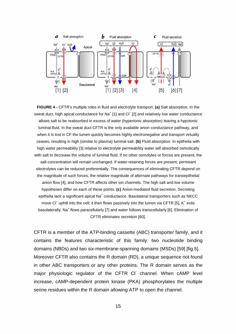
CFTR's roles in ion transport [fig.4].
15
FIGURE 4 - CFTR's multiple roles in fluid and electrolyte transport. (a) Salt absorption. In the
sweat duct, high apical conductance for Na+ [1] and Cl– [2] and relatively low water conductance
allows salt to be reabsorbed in excess of water (hypertonic absorption) leaving a hypotonic
luminal fluid. In the sweat duct CFTR is the only available anion conductance pathway, and
when it is lost in CF the lumen quickly becomes highly electronegative and transport virtually
ceases, resulting in high (similar to plasma) luminal salt. (b) Fluid absorption. In epithelia with
high water permeability [3] relative to electrolyte permeability water will absorbed osmotically
with salt to decrease the volume of luminal fluid. If no other osmolytes or forces are present, the
salt concentration will remain unchanged. If water-retaining forces are present, permeant
electrolytes can be reduced preferentially. The consequences of eliminating CFTR depend on
the magnitude of such forces, the relative magnitude of alternate pathways for transepithelial
anion flow [4], and how CFTR affects other ion channels. The high salt and low volume
hypotheses differ on each of these points. (c) Anion-mediated fluid secretion. Secreting
epithelia lack a significant apical Na+ conductance. Basolateral transporters such as NKCC
move Cl– uphill into the cell; it then flows passively into the lumen via CFTR [5], K+ exits
basolaterally, Na+ flows paracellularly [7] and water follows transcellularly [6]. Elimination of
CFTR eliminates secretion [60].
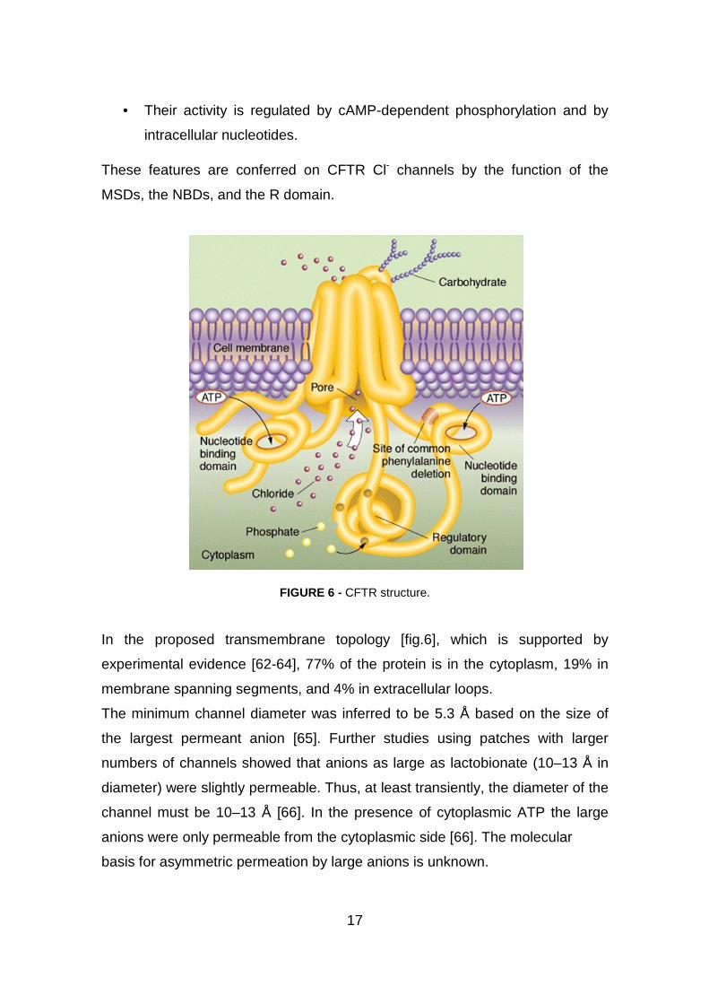
CFTR is a member of the ATP-binding cassette (ABC) transporter family, and it
contains the features characteristic of this family: two nucleotide binding
domains (NBDs) and two six-membrane-spanning domains (MSDs) [59] [fig.5].
Moreover CFTR also contains the R domain (RD), a unique sequence not found
in other ABC transporters or any other proteins. The R domain serves as the
major physiologic regulator of the CFTR Cl- channel. When cAMP level
increase, cAMP-dependent protein kinase (PKA) phosphorylates the multiple
serine residues within the R domain allowing ATP to open the channel.

16
PKA is the primary kinase that phosphorylates CFTR, although protein kinase
C, cGMP-dependent protein kinase, and tyrosine phosphorylation can also
stimulate channel activity [61].
FUGURE 5 - Model showing proposed domain structure of cystic fibrosis transmembrane
conductance regulator (CFTR). MSD, membrane-spanning domain; NBD, nucleotide-binding
domain; R, regulatory domain; PKA, cAMP-dependent protein kinase.
Once the R domain is phosphorylated, channel gating is regulated by a cycle of
ATP hydrolysis at the NBDs. Finally, protein phosphatases dephosphorylate the
R domain and return the channel to its quiescent state. At the moment, how
phosphorylation activates the channel is not well understood. Some models
propose that the R domain prevents the channel from opening and that
phosphorylation relieves this inhibition. Other models suggest that
phosphorylation of the R domain stimulates activity.
These channels have several distinguishing characteristics:
• They have a small single-channel conductance (6–10 pS).
• The current-voltage (I-V) relationship is linear.
• They are selective for anions over cations.
• The anion permeability sequence is Br- ≥ Cl- > I-.
• They show time- and voltage-independent gating behavior.
17
• Their activity is regulated by cAMP-dependent phosphorylation and by
intracellular nucleotides.
These features are conferred on CFTR Cl- channels by the function of the
MSDs, the NBDs, and the R domain.
FIGURE 6 - CFTR structure.
In the proposed transmembrane topology [fig.6], which is supported by
experimental evidence [62-64], 77% of the protein is in the cytoplasm, 19% in
membrane spanning segments, and 4% in extracellular loops.
The minimum channel diameter was inferred to be 5.3 Å based on the size of
the largest permeant anion [65]. Further studies using patches with larger
numbers of channels showed that anions as large as lactobionate (10–13 Å in
diameter) were slightly permeable. Thus, at least transiently, the diameter of the
channel must be 10–13 Å [66]. In the presence of cytoplasmic ATP the large
anions were only permeable from the cytoplasmic side [66]. The molecular
basis for asymmetric permeation by large anions is unknown.
18
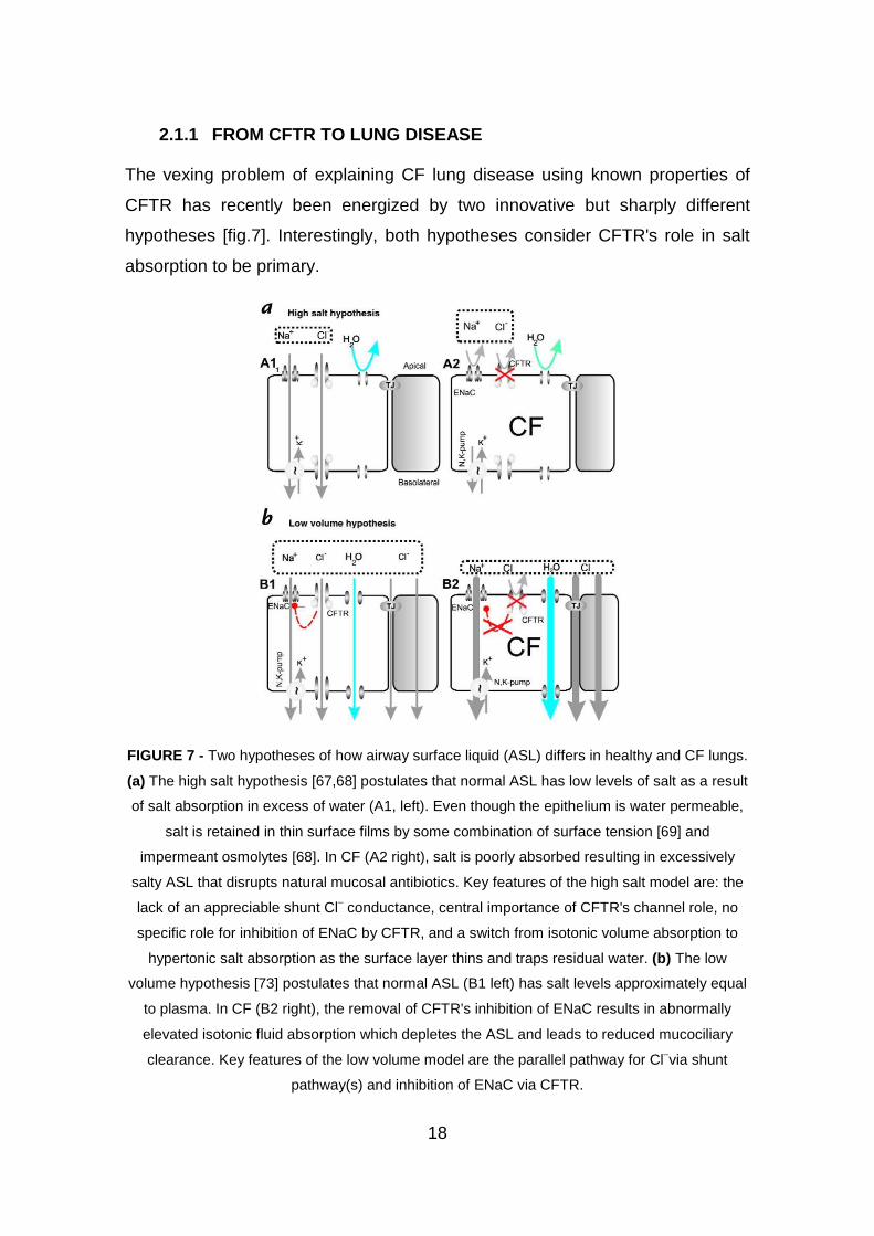
2.1.1 FROM CFTR TO LUNG DISEASE
The vexing problem of explaining CF lung disease using known properties of
CFTR has recently been energized by two innovative but sharply different
hypotheses [fig.7]. Interestingly, both hypotheses consider CFTR's role in salt
absorption to be primary.
FIGURE 7 - Two hypotheses of how airway surface liquid (ASL) differs in healthy and CF lungs.
(a) The high salt hypothesis [67,68] postulates that normal ASL has low levels of salt as a result
of salt absorption in excess of water (A1, left). Even though the epithelium is water permeable,
salt is retained in thin surface films by some combination of surface tension [69] and
impermeant osmolytes [68]. In CF (A2 right), salt is poorly absorbed resulting in excessively
salty ASL that disrupts natural mucosal antibiotics. Key features of the high salt model are: the
lack of an appreciable shunt Cl– conductance, central importance of CFTR's channel role, no
specific role for inhibition of ENaC by CFTR, and a switch from isotonic volume absorption to
hypertonic salt absorption as the surface layer thins and traps residual water. (b) The low
volume hypothesis [73] postulates that normal ASL (B1 left) has salt levels approximately equal
to plasma. In CF (B2 right), the removal of CFTR's inhibition of ENaC results in abnormally
elevated isotonic fluid absorption which depletes the ASL and leads to reduced mucociliary
clearance. Key features of the low volume model are the parallel pathway for Cl–via shunt
pathway(s) and inhibition of ENaC via CFTR.

19
The high salt hypothesis [67-68] emphasizes CFTR's function as an anion
channel. According to this hypothesis, missing or defective CFTR causes
reduced transepithelial Cl- conductance, and by analogy with the sweat duct
[70-71] this allows salt levels in the apical surface liquid (ASL) to remain at
levels similar to those in plasma [fig.7]. The high salt in the ASL interferes with
natural antibiotics such as defensins [72] and lysozyme.
In marked contrast, the low volume hypothesis [73] is based on CFTR's function
as a regulator of other channels - in this case ENaC. According to this
hypothesis, both normal and CF ASL have plasma-like levels of salt. CFTR
mutations eliminate CFTR's inhibition of ENaC, and because there are
significant shunt pathways for Cl– in the airways, increased Na+ transport drives
increased absorption of Cl– and water. Thus CF airways display accelerated
isotonic fluid absorption that depletes ASL volume and dehydrates mucus,
leading to obstruction and infection [74].
2.2 CFTR GENE
The gene responsible for CFTR was identified to reside on the long arm of
chromosome 7 (7q.31.2) and subsequently isolated by positional cloning in
1989 [3-5,75] [fig.8].
FIGURE 8 - The location of the CFTR gene on chromosome 7.
20
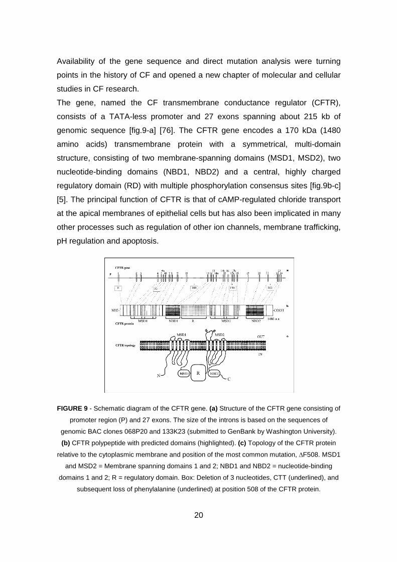
Availability of the gene sequence and direct mutation analysis were turning
points in the history of CF and opened a new chapter of molecular and cellular
studies in CF research.
The gene, named the CF transmembrane conductance regulator (CFTR),
consists of a TATA-less promoter and 27 exons spanning about 215 kb of
genomic sequence [fig.9-a] [76]. The CFTR gene encodes a 170 kDa (1480
amino acids) transmembrane protein with a symmetrical, multi-domain
structure, consisting of two membrane-spanning domains (MSD1, MSD2), two
nucleotide-binding domains (NBD1, NBD2) and a central, highly charged
regulatory domain (RD) with multiple phosphorylation consensus sites [fig.9b-c]
[5]. The principal function of CFTR is that of cAMP-regulated chloride transport
at the apical membranes of epithelial cells but has also been implicated in many
other processes such as regulation of other ion channels, membrane trafficking,
pH regulation and apoptosis.
FIGURE 9 - Schematic diagram of the CFTR gene. (a) Structure of the CFTR gene consisting of
promoter region (P) and 27 exons. The size of the introns is based on the sequences of
genomic BAC clones 068P20 and 133K23 (submitted to GenBank by Washington University).
(b) CFTR polypeptide with predicted domains (highlighted). (c) Topology of the CFTR protein
relative to the cytoplasmic membrane and position of the most common mutation, ∆F508. MSD1
and MSD2 = Membrane spanning domains 1 and 2; NBD1 and NBD2 = nucleotide-binding
domains 1 and 2; R = regulatory domain. Box: Deletion of 3 nucleotides, CTT (underlined), and
subsequent loss of phenylalanine (underlined) at position 508 of the CFTR protein.

21
2.3 CFTR MUTATIONS
The first, and as it turned out, the most common CFTR defect identified among
Caucasians was the ∆F508 mutation [fig.11], a 3-bp deletion in exon 10 causing
a loss of phenylalanine at the amino acid position 508 of the protein product [fig.
9c][75]. Presently, 10 years after the discovery of the CFTR gene, more than
850 different alleles have been reported as proven or putative disease causing
mutations (CFGAC website: http://www.genet.sickkids.on.ca/cftr/).
Feature How may it affect a phenotype?
A - Type of m utation Mutations that are predicted to prevent CFTR biosynthesis or produce grossly changed, unstable or
nonfunctional proteins tend to have severe phenotypic consequences (type: nonsense, frameshift, splice,
large in-frame deletion or insertion); effects of mutations that cause minor, local changes in the protein
may range from mild to severe depending on other factors such as its molecular mechanism, amino acid
change or location (type: missense, small inframe deletion or insertion)
B – Molecular mechanism Immediate consequence of the mutation for CFTR biosynthesis, processing, trafficking to
the membrane, stability and function (classes of mutations); the mechanisms of mutations
are evaluated empirically on the molecular and cellular levels
C - Position in the gene
(protein)
Applies particularly to minor, local changes (missense); mutations in structurally or functionally
critical regions of the protein tend to correlate with more severe phenotypes when compared
with mutations in less important regions; amino acid substitutions in highly conserved regions of
CFTR protein may also have more severe phenotypic consequences than mutations from
unconserved regions
D - Net molecular effect The net amount of the functional CFTR present in the apical membrane of secretory epithelial
cells, irrespective of type mutation or its mechanism; production of even a small amount of functional
CFTR may be sufficient to prevent a severe disease (class IV and V mutations)
E - Intragenic modulators
(complex alleles)
In some cases, a second change in the same allele may modulate the phenotypic effect of the
primary mutation; for example, the missense mutation R117H associated with the 5T variant
in the T-tract of the acceptor splice site of intron 8 is typically associated with the pancreatic sufficient
form of CF but only with infertility (males) or asymptomatic presentation (females) when on the 7T
background
F - Impact of second
mutation
As a recessive disease, CF requires the presence of mutations in each allele; therefore, the
type and molecular consequences of the second mutation in the genotype may be critical for
the clinical outcome; for example, a severe allele is associated with PI only if the second mutation is
severe; conversely, one mild mutation is sufficient to preserve pancreatic function, irrespective of the type
of the second allele
G - Site of
expression/organ
pathophysiology/
secondary modulation
A phenotypic impact of a mutation also depends on where the mutation is expressed and organ-specific
pathophysiology; in some organs like the pancreas, a CFTR genotype closely correlates with the severity
of its phenotype (PI vs. PS); in lungs, with complex pathophysiology, the primary effect of a particular
genotype may be considerably modulated by secondary genetic factors and environment
Table 1 - What determines a phenotypic effect of a mutation?
22
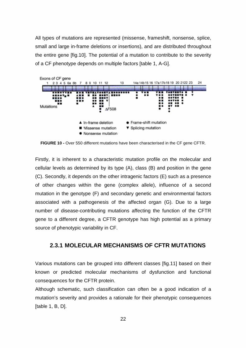
All types of mutations are represented (missense, frameshift, nonsense, splice,
small and large in-frame deletions or insertions), and are distributed throughout
the entire gene [fig.10]. The potential of a mutation to contribute to the severity
of a CF phenotype depends on multiple factors [table 1, A-G].
FIGURE 10 - Over 550 different mutations have been characterised in the CF gene CFTR.
Firstly, it is inherent to a characteristic mutation profile on the molecular and
cellular levels as determined by its type (A), class (B) and position in the gene
(C). Secondly, it depends on the other intragenic factors (E) such as a presence
of other changes within the gene (complex allele), influence of a second
mutation in the genotype (F) and secondary genetic and environmental factors
associated with a pathogenesis of the affected organ (G). Due to a large
number of disease-contributing mutations affecting the function of the CFTR
gene to a different degree, a CFTR genotype has high potential as a primary
source of phenotypic variability in CF.
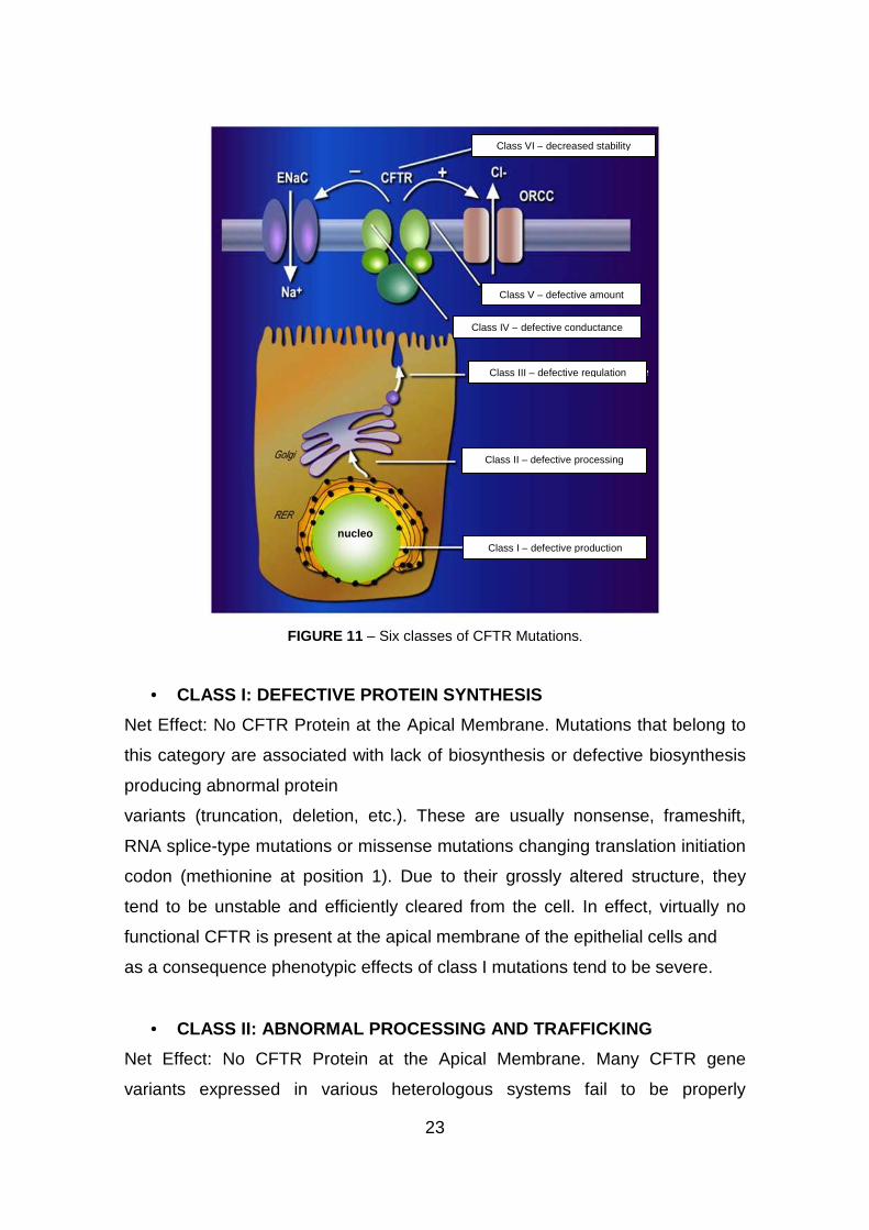
2.3.1 MOLECULAR MECHANISMS OF CFTR MUTATIONS
Various mutations can be grouped into different classes [fig.11] based on their
known or predicted molecular mechanisms of dysfunction and functional
consequences for the CFTR protein.
Although schematic, such classification can often be a good indication of a
mutation’s severity and provides a rationale for their phenotypic consequences
[table 1, B, D].
23
FIGURE 11 – Six classes of CFTR Mutations.
• CLASS I: DEFECTIVE PROTEIN SYNTHESIS
Net Effect: No CFTR Protein at the Apical Membrane. Mutations that belong to
this category are associated with lack of biosynthesis or defective biosynthesis
producing abnormal protein
variants (truncation, deletion, etc.). These are usually nonsense, frameshift,
RNA splice-type mutations or missense mutations changing translation initiation
codon (methionine at position 1). Due to their grossly altered structure, they
tend to be unstable and efficiently cleared from the cell. In effect, virtually no
functional CFTR is present at the apical membrane of the epithelial cells and
as a consequence phenotypic effects of class I mutations tend to be severe.
• CLASS II: ABNORMAL PROCESSING AND TRAFFICKING
Net Effect: No CFTR Protein at the Apical Membrane. Many CFTR gene
variants expressed in various heterologous systems fail to be properly
Classe 1: mancata sintesi
Classe 2: difetto di maturazione
Classe 3: difetto di attivazione
Classe 4: difetto di conduttanza
Classe 5: difetto di sintesi
Classe 6: difetto di regolazione
nucleoClasse 1: mancata sintesi
Classe 2: difetto di maturazione
Classe 3: difetto di attivazione
Classe 4: difetto di conduttanza
Classe 5: difetto di sintesi
Classe 6: difetto di regolazione
Classe 1: mancata sintesi
Classe 2: difetto di maturazione
Classe 3: difetto di attivazione
Classe 4: difetto di conduttanza
Classe 5: difetto di sintesi
Classe 6: difetto di regolazione
nucleoClass I – defective production
Class II – defective processing
Class III – defective regulation
Class IV – defective conductance
Class V – defective amount
Class VI – decreased stability

24
processed to a mature glycosylated form and transported to the apical
membrane. Because of their absence in the membrane, these mutant CFTR
variants are typically associated with severe CF phenotypes. Interestingly,
some of the class II mutations – such as the most common ∆F508 mutation
[fig.12] – if correctly processed, possess residual Cl channel activity and may
lead to a sustained normal or only mildly affected phenotype. For this reason,
mutations in this group – in particular the common ∆F508 deletion – are the
targets of potential therapies, aimed at correcting the processing and delivery of
a mutated CFTR protein to the apical membrane.
FIGURE 12 - The ∆F508 deletion is the most common cause of cystic fibrosis.
Recent studies of endogenous CFTR expression in CF-relevant epithelial
tissues from patients homozygous for the ∆F508 mutation demonstrated
however that immunolocalized distribution of CFTR in certain tissues
(respiratory and intestinal) is indistinguishable from that observed in the
corresponding wild-type cells [77]. The ∆F508 CFTR protein was absent only in
sweat gland ducts of those patients. It is still possible that the ∆F508 CFTR is
compartmentalized in vesicles in close proximity to the cell membrane and
therefore gives the appearance of being embedded in the membrane. It should
be noted, however, that the efficiency of processing and trafficking of the ∆F508
CFTR protein may vary conside ably between different epithelial cells.
Therefore, different intracellular conditions may result in variable severity of
tissue- or organ-specific pathophysiology.
25
• CLASS III: DEFECTIVE REGULATION
Net Effect: Normal Amount of Nonfunctional CFTR at the Apical Membrane.
Mutations in this class affect the regulation of CFTR function by preventing ATP
binding and hydrolysis at the nucleotide binding domains (NBD1; NBD2)
required for the channel activation. Alterations within NBD1 (such as missense
mutation G551D) may also affect CFTR regulation of other channels such as
the outwardly rectifying chloride channel [78] or ROMK2 potassium channel
[79]. This is an example of a mutation mechanism which affects some
regulatory functions of CFTR in addition to its chloride transporting function.
• CLASS IV: DECREASED CONDUCTANCE
Net Effect: Normal Amount of CFTR with Some Residual Function at the Apical
Membrane. Several mutations (R117H, R334W, R347P) were shown to affect
the properties of CFTR single-channel conductance [80]. Interestingly, these
mutations are located within MSD1 which is implicated in forming the pore of
the channel [81] and the corresponding CFTR variants retain residual function.
Alleles in this class are typically associated with a milder pancreatic phenotype:
this includes patients with pancreatic sufficency (PS).
• CLASS V: REDUCED SYNTHESIS/TRAFFICKING
Net Effect: Reduced Amount of Functional CFTR at the Apical Membrane.
Various mutations may be associated with reduced biosynthesis of fully active
CFTR due to partially aberrant splicing (3849+10kbC→T) [82], promoter
mutations or inefficient trafficking (A455E) [83, 84]. These mutations result in
reduced expression of functional CFTR channels in the apical membrane. Class
V mutations are associated with a milder CF phenotype.
• CLASS VI: DECREASED STABILITY
Net Effect: Functional but Unstable CFTR Present at the Apical Membrane.
According to a new study, truncation of the C-terminus of CFTR leads to the
marked instability of an otherwise fully processed and functional variant. These
are usually nonsense or frameshift mutations (Q1412X, 4326delTC, 4279insA)
26
causing a 70- to 100-bp truncation of the Cterminusof the CFTR [85] and
associated with severe CF presentation. The above classification categorizes
CFTR mutations according to their molecular mechanisms and consequences
for different aspects of CFTR biogenesis, metabolism and function but it does
not exclude an association of a mutation with more than one mechanism (e.g.
mislocalization and decreased function).
27
3. INFECTION AND INFLAMMATION IN CF
Although the cystic fibrosis transmembrane ion receptor (CFTR) gene and
protein have been identified since seventeen years, it remains enigmatic how
abnormalities in CFTR can cause chronic and persistent pulmonary infection
and inflammation that lead to bronchiectasis and end-stage lung disease [86].
3.1 LUNG INFECTION
Healthy airways are sterile below the first bronchial division. Sterility is
maintained, despite constant challenge from viruses and bacteria in the air that
we breathe, by an elaborate hierarchy of defenses. Lung defenses are
understood only in outline. The airway surfaces are covered with a thin film
(∼30 µm) of airway surface liquid (ASL) consisting of a periciliary sol and a
mucus gel that are propelled toward the mouth by coordinated ciliary beating.
Thus, mucociliary clearance, aided by cough, cleans the airways mechanically.
The ASL is not simply saltwater, but is instead a rich broth of
proteases/antiproteases, oxidants/antioxidants, antibiotics, and antibodies that
work together to inactivate or destroy pathogens without undue collateral
damage to the lungs. These mucosal mechanisms are backstopped by cellular
immune mechanisms involving dendritic cells, neutrophils, and macrophages
that are recruited and coordinated by signaling molecules in the ASL.
Although the complex pulmonary defense system is understood in outline,
details are lacking because of technical impediments that, together with the lack
of appropriate animal models, help to explain why we are still struggling to
understand CF lung disease. The pattern of CF lung disease is unlike that of
any other lung disease.
In patients with cystic fibrosis, progression of lung disease is insidious and
patients may be relatively asymptomatic before irreversible changes and
chronic bacterial colonization occur.
Cough is the predominant symptom in the early stages of cystic fibrosis,
occurring in as many as 50% o
many patients do not have pulmonary symptoms. The first detectable evidence
of lung disease in patients with cystic fibrosis is infection and/or inflammation in
bronchoalveolar lavage fluid, denoted by elevated counts of interleukin
neutrophils and the presence of microorganisms [88
influenza, Staphylococcus aureus
most prevalent early pathogens, and
least 1 of these bacteria by 1 year of age
be transient, and approximately half clear spontaneously
Chronic colonization with
function [97, 98], especially if the isolate becomes mucoid
most patients are initially infected with nonmucoid
transitions to a mucoid state. Mucoid
treat and eradicate because it lives in a defensive mode of growth called biofilm
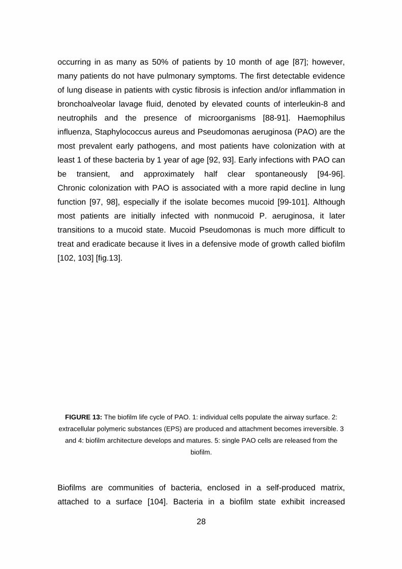
[102, 103] [fig.13].
FIGURE 13: The biofilm life cycle
extracellular polymeric substances (
and 4: biofilm architecture develops and matures. 5: single
Biofilms are communities of bacteria, enclosed in a self
attached to a surface
28
occurring in as many as 50% of patients by 10 month of age [87]
many patients do not have pulmonary symptoms. The first detectable evidence
of lung disease in patients with cystic fibrosis is infection and/or inflammation in
bronchoalveolar lavage fluid, denoted by elevated counts of interleukin
the presence of microorganisms [88-91].
Staphylococcus aureus and Pseudomonas aeruginosa
most prevalent early pathogens, and most patients have colonization with at
least 1 of these bacteria by 1 year of age [92, 93]. Early infections with
be transient, and approximately half clear spontaneously
Chronic colonization with PAO is associated with a more rapid decline in lung
, especially if the isolate becomes mucoid [99-
most patients are initially infected with nonmucoid P. aeruginosa
transitions to a mucoid state. Mucoid Pseudomonas is much more difficult t
treat and eradicate because it lives in a defensive mode of growth called biofilm
The biofilm life cycle of PAO. 1: individual cells populate the airway
extracellular polymeric substances (EPS) are produced and attachment becomes irreversible. 3
and 4: biofilm architecture develops and matures. 5: single PAO cells are released from the
biofilm.
Biofilms are communities of bacteria, enclosed in a self-produced matrix,
attached to a surface [104]. Bacteria in a biofilm state exhibit increase
f patients by 10 month of age [87]; however,
many patients do not have pulmonary symptoms. The first detectable evidence
of lung disease in patients with cystic fibrosis is infection and/or inflammation in
bronchoalveolar lavage fluid, denoted by elevated counts of interleukin-8 and
91]. Haemophilus
nosa (PAO) are the
most patients have colonization with at
. Early infections with PAO can
be transient, and approximately half clear spontaneously [94-96].
more rapid decline in lung
-101]. Although
P. aeruginosa, it later
is much more difficult to
treat and eradicate because it lives in a defensive mode of growth called biofilm
airway surface. 2:
produced and attachment becomes irreversible. 3
cells are released from the
produced matrix,
Bacteria in a biofilm state exhibit increased
29
resistance to antibiotics [105] and host defense factors [106]. Therefore,
clinically attainable antibiotic concentrations may not adequately clear biofilm
infections, allowing the bacterial population to recover, persist and spread.
Microscopic and physiologic evidence supports biofilm formation by P.
aeruginosa in sputum from patients with cystic fibrosis [107]. Antibiotic-resistant,
biofilm-forming mucoid P. aeruginosa are believed to play a dominant role in
the progression of lung disease in patients with cystic fibrosis [99-101].
3.2 LUNG INFLAMMATION
The lungs, including mucus glands, are structurally normal at birth [108]. Soon
after birth excessive endobronchial inflammation in the small airways has been
demonstrated in both sputum culture positive and sputum culture negative
patients. Broncho-alveolar lavage (BAL) fluid shows a predominantly neutrophil
inflammation with elevated interleukin (IL)-8 and neutrophil elastase [109-112].
This persistent inflammation is the major cause of progressive lung injury and
destruction leading to a decrease in lung function. This progressive loss of lung
function is the main reason for the limited life expectancy of most patients with
CF.
Virtually all patients with CF are chronically infected with one ore more bacterial
species. The inflammatory response to infection appears to be more intense in
patients with CF compared to non-CF patients. Additionally, it has been shown
that the number of colony forming units in BAL fluid is directly related to the
intensity of the inflammatory response with a significant increase in the number
of inflammatory cells and an increase in IL-8 concentrations [113].
Acquisition and consequent chronic infection with mucoid strains of P.
aeruginosa leads to an increase in the endobronchial inflammatory response to
infection. It has been shown that CF cell lines produce more pro-inflammatory
cytokines than normal cell lines in response to P. aeruginosa [114]. This
“overproduction” of pro-inflammatory cytokines can be found in the ELF of CF
airways [115]. Also significantly lower levels of the anti-inflammatory cytokine
30
IL-10 , which inhibits the production of pro-inflammatory cytokines as IL8 and
IL6, are found. This imbalance of anti-inflammatory and pro-inflammatory
cytokines results in an excessive and persistent inflammation in the CF airways.
As a result lung function deteriorates more rapidly in P. aeruginosa positive
patients compared with CF patients negative for P. aeruginosa [116, 117].
For all these reasons, understanding the pathophysiology of lung inflammation
and thereby the pathogenesis of lung disease in CF is needed to improve
current therapies and develop new therapeutic strategies.
3.3 CYTOKINES AND INFLAMMATORY MEDIATORS IN
CYSTIC FIBROSIS
Airway disease in cystic fibrosis (CF) is characterised by chronic infection and
an inflammatory response dominated by a neutrophilic infiltrate. There is
incomplete understanding of the relationship between the abnormal CFTR gene
product and the development of inflammation and progression of lung disease
in CF [118].
Evidence suggests that airway inflammation in CF is associated with increased
production of pro-inflammatory cytokines in the lung. Airway epithelial cells,
macrophages, and neutrophils are all capable of producing cytokines.
Several studies have found elevated concentrations of proinflammatory
cytokines such as interleukin-1 (IL-1), IL-6, IL-8, and tumour necrosis factor-
alpha (TNF-α) in the sputum and bronchoalveolar lavage fluid (BALF) of
patients with CF [118]. Their synthesis is promoted by the transcription factor
nuclear factor-kB (NF-kB), which plays an important role in intracellular
signalling for the production of pro-inflammatory cytokines [119]. IL-10, IL-1
receptor antagonist protein (IRAP), and soluble TNF-α receptor (TNFsR) are
anti-inflammatory cytokines that are relatively down-regulated in CF airway cells
[120]. The principle action of IL-10 is to increase the synthesis of I-kB, the
inhibitor of NF-kB. Downregulation of IL-10 leads to increased proinflammatory
cytokines due to less inhibition of NF-kB actions [119]. Furthermore we
31
investigated on MCP1 (Monocyte Chemotactic Protein-1, a member of the small
inducible gene -SIG- family, that plays a role in the recruitment of monocytes to
sites of injury and infection), RANTES (Regulated on Activation Normal T
Expressed and Secreted, a protein member of the interleukin-8 superfamily of
cytokines, that is a selective attractant for memory T lymphocytes and
monocytes), and G-CSF (Granulocyte Colony-Stimulating Factor, a colony-
stimulating factor that stimulates the production of neutrophils). Table 2 lists the
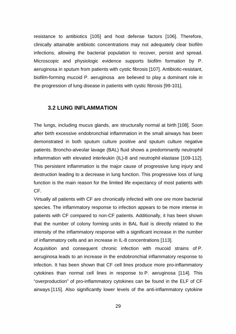
actions of both the proinflammatory and anti-inflammatory cytokines.
cytokines Action
IL1 Primes neutrophils
Increases adhesion of neutrophils to endothelium
IL8 Increases chemotaxis of neutrophils to site of inflammation
Activates neutrophils
Increases expression of adhesion molecules
TNFα Increases chemotaxis of neutrophils to site of inflammation
Increases adhesion of neutrophils to endothelium
Induces synthesis of chemoattractant neutrophils
Increases intermediary metabolism
IL6 Mediates acute-phase reaction
Matures B-lymphocytes
Activates T-lymphocytes
IL10 Inhibits secretion of TNFa and other cytokines
Inhibits antigen presentation
IRAP Inhibits IL-1 receptor binding
Antagonises activities of IL-1
IL-1, interleukin -1; IL-8, interleukin -8; TNFa, tumour necrosis factoralpha;
IL-6, interleukin-6; IL-10, interleukin-10; IRAP, i nterleukin-1
receptor antagonist protein.
Table 2 – Actions of cytokines.
32
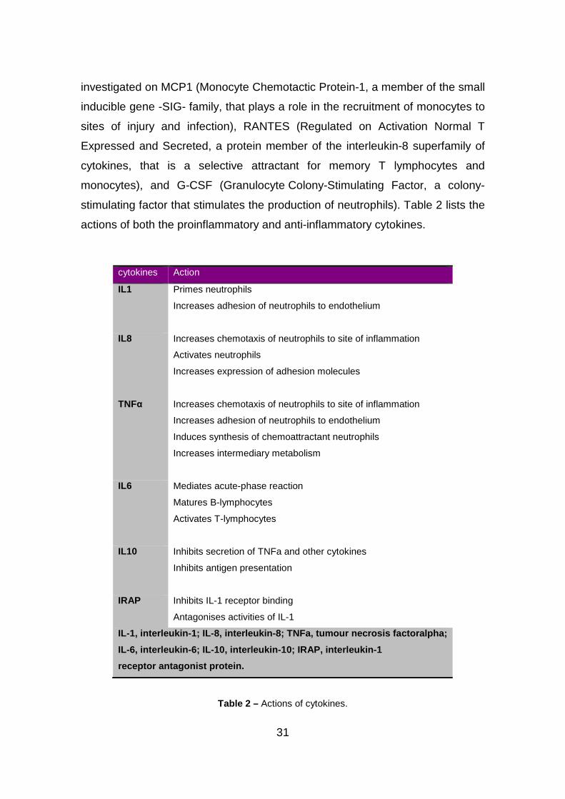
Airway epithelial cells may be involved directly in the excess inflammation by
several mechanisms. Pro-inflammatory cytokines arise from airway epithelial
cells, as well as from macrophages and infiltrating neutrophils [Table 3].
Neutrophils Epithelial cells Macrophages Lymphocytes
IL-1 β + + +++
IL-6 + + +++ +
IL-8 +++ ++ ++ +
IL-10 ++ + ++
TNFα ++ + +++ +
IRAP + +++
TNFsR ++ + +
IL-1β, interleukin -1beta; IL -6, interleukin -6; IL-8, interleukin -8; IL-10,
interleukin-10; TNF α, tumour necrosis factor-alpha; IRAP, interleukin-1
receptor antagonist protein; TNFsR, tumour necrosis factor soluble
receptor
Table 3 – Cellular source of cytokines produced in human airways.
Airway epithelial cells also express large numbers of the important pro-
inflammatory adhesion molecule, ICAM-1. This adhesion molecule is a ligand
for neutrophils, and adhesion is thought to result in increased IL-8 production,
leading to persistence of neutrophils in the airway [121]. The fact that epithelial
cells themselves are involved in cytokine release and are thought to play a
major role in the local inflammation leads to further speculation that defective
CFTR function (expressed most importantly in the epithelial cells) may be
directly related to excessive inflammation.
The cytokines tumor necrosis factor-alpha (TNF-α), interleukin-8 (IL-8),
interleukin-6 (IL-6) and intercellular adhesion molecule-1 (ICAM-1) have
important roles in regulating neutrophil migration and the inflammatory response
[122]:
33
• ICAM-1
Intercellular Adhesion Molecule-1 (ICAM-1, CD54) is a transmembrane
glycoprotein molecule of the immunoglobulin superfamily. The protein is a type
of intercellular adhesion molecule continuously present in low concentrations in
the membranes of leukocytes and endothelial cells. Upon cytokine stimulation,
the concentrations greatly increase. ICAM-1 can be induced by interleukin-1 (IL-
1) and tumor necrosis factor alpha (TNFα) and is expressed by the vascular
endothelium, macrophages, and lymphocytes.
In cystic fibrosis, neutrophil migration from the bloodstream into the airway
occurs through interactions between adhesion molecules on neutrophils and
ligands on the vascular endothelium. One ligand for the CD11/CD18 leukocyte
adhesion complex is the intercellular adhesion molecule-l which is expressed by
endothelial cells [123] as well as by bronchial epithelial cells and alveolar type I
cell [124-126]. A soluble form of ICAM- 1 (sICAM-1) circulates in the blood and
is present in the bronchoalveolar lining fluid [127]. sICAM- 1 is often increased
during inflammation; in particular, its concentration is elevated in tracheal
aspirates of infants with bronchopulmonary dysplasia [128], as well as in the
plasma or bronchoalveolar lavage (BAL) fluid of patients with idiopathic
pulmonary fibrosis, asthma, and sarcoidosis [129-134].
• IL8
Interleukin-8 (IL-8) is a protein member of the α-chemokine family. This
chemokine is one of the major mediators of the inflammatory response and it is
produced by macrophages and other cell types such as epithelial cells.
There are more receptors of the surface membrane capable to bind IL-8. The
most frequently studied types are the G protein coupled serpentine
receptors CXCR1 and CXCR2. Expression and affinity to IL-8 is different in the
two receptors (CXCR1 > CXCR2). Toll-like receptors are the receptors of the
innate immune system. These receptors recognize antigen patterns (like
lipopolysaccharides -LPS- in gram negative bacteria). Through a chain of
biochemical reactions IL-8 is secreted and is an important mediator of the
immune reaction in the innate immune system response.
34
Persistent infection with Pseudomonas aeruginosa increases interleukin-8
levels and causes dense neutrophil infiltrations in the airways of patients with
chronic airway diseases, such as cystic fibrosis. Recently, it has been reported
that nitrite reductase from P. aeruginosa enhances IL-8 gene promoter activity.
Nitrite reductase enhanced nuclear localization of the NF-kB (nuclear factor
kappa-light-chain-enhancer of activated B cells) binding complex. Furthermore,
nitrite reductase induced the degradation of IkBα, the major cytoplasmic
inhibitor of NF-kB, and the expression of IkBα mRNA. These data support the
critical role of the activation of NF-kB in nitrite reductase-induced IL- 8 gene
expression in CF airway epithelium [135,136].
• IL6
IL-6 is an interleukin that acts as both a pro-inflammatory and anti-
inflammatory cytokine. It is secreted by T cells and macrophages to stimulate
immune response to trauma, especially burns or other tissue damage leading to
inflammation. IL-6's role as an anti-inflammatory cytokine is mediated through
its inhibitory effects on TNF-alpha and IL-1, and activation of IL-1ra and IL-10.
In CF patients IL-6 gene is up regulated following infection of CF bronchial
epithelial cells with P. aeruginosa. It has been demonstrated the ability of
the P. aeruginosa mitogen, exoenzyme S, to induce proinflammatory and
immunoregulatory cytokines and chemokines, via NF-kB activation. Exoenzyme
S strongly induced transcription of proinflammatory cytokines and
chemokines,such as IL-6 and IL-8, and modest transcription of
immunoregulatory cytokines. The response occurred early and subsided
without evolving over time. These data suggest that cells responding
to exoenzyme S would rapidly express proinflammatory cytokines and
chemokines that may contribute to pulmonary inflammation in cystic fibrosis
[137].
• TNFα
Tumor necrosis factor-α is a cytokine involved in systemic inflammation and is a
member of a group of cytokines that stimulate the acute phase reaction.
35
The primary role of TNF-α is in the regulation of immune cells. TNF-α is also
able to induce apoptotic cell death, to induce inflammation, and to
inhibit tumorigenesis and viral replication, via NF-kB activation. Dysregulation of
TNF-α production has been implicated in a variety of human diseases, such as
cystic fibrosis and cancer [138]. Recombinant TNF is used as
an immunostimulant.
Norman et al. have demonstrated that concentrations of TNF-α are higher in
plasma from patients with cystic fibrosis than in normal controls and that its
concentration is increased during episodes of acute respiratory infection. It has
been found TNF-α in cystic fibrosis sputa, during times of clinical stability, at
concentrations that have been shown to produce neutrophil migration,
respiratory burst, and degranulation in vitro [139]. This provides evidence in
favour of the hypothesis that lung damage, due to chronic infection, is occurring
during periods of apparent wellbeing.
3.4 NF-kB IN DEFENSE AND DISEASES
NF-κB (nuclear factor kappa-light-chain-enhancer of activated B cells) is a
protein complex that controls the transcription of DNA.
3.4.1 NF-kB: A KEY ROLE IN INFLAMMATION
NF-κB regulates host inflammatory and immune responses and cellular growth
properties [140] by increasing the expression of specific cellular genes. These
include genes encoding at least 27 different cytokines and chemokines,
receptors involved in immune recognition such as members of the MHC,
proteins involved in antigen presentation, and receptors required for neutrophil
adhesion and migration [141]. Cytokines that are stimulated by NF-κB, such as
IL-1β and TNF-α, can also directly activate the NF-κB pathway, thus
establishing a positive autoregulatory loop that can amplify the inflammatory
response and increase the duration of chronic inflammation. NF-κB also
stimulates the expression of enzymes whose products contribute to the

36
pathogenesis of the inflammatory process, including the inducible form of nitric
oxide synthase (iNOS), which generates nitric oxide (NO), and the inducible
cyclooxygenase (COX-2), which generates prostanoids [142]. The NF-κB
pathway is likewise important in the control of the immune response. It
modulates B-lymphocyte survival, mitogen-dependent cell proliferation, and
isotype switching, which lead to the differentiation of B lymphocytes into plasma
cells [143]. In addition, NF-κB regulates IL-2 production, which increases the
proliferation and differentiation of T lymphocytes [144]. Thus, activation of NF-
κB leads to the induction of multiple genes that regulate the immune and the
inflammatory response.
In addition to activating the expression of genes involved in the control of the
immune and inflammatory response, the NF-κB pathway is also a key mediator
of genes involved in the control of the cellular proliferation and apoptosis.
Antiapoptotic genes that are directly activated by NF-κB include the cellular
inhibitors of apoptosis (c-IAP1, c-IAP2, and IXAP), the TNF receptor–associated
factors (TRAF1 and TRAF2), the Bcl-2 homologue A1/Bfl-1, and IEX-IL [145].
3.4.2 NF-kB FUNCTION AND REGULATION
NF-κB is composed of homo- and hetero-dimeric complexes of Rel family
polypeptides, which are characterized by an N-terminal RHD (Rel homology
domain), which mediates DNA binding, nuclear localization and subunit
dimerization [140]. Mammals express five NF-κB proteins, namely RelA (p65),
RelB, c-Rel, NF-κB1 p50 and NF-κB2 p52 [fig.14A]. NF-κB1 and NF-κB2 are
synthesized as large precursors of 105 (p105) and 100 kDa (p100) respectively.
These are partially proteolysed by the proteasome (termed processing), which
removes their C terminal halves, to produce the active NF-κB1 p50 and NF-κB2
p52 subunits. RelA and c-Rel both have C-terminal transactivation domains in
their RHDs and can strongly activate transcription from NF-κB binding sites in
target genes. RelB also contains a transactivation domain in its RHD and can
function as an NF-kB activator when complexed with p50 or p52. However,
RelB/RelA heterodimeric complexes are inhibitory, since they cannot bind DNA

37
[146]. NF-κB1 p50 and NF-κB2 p52 lack a transactivation domain, and can only
promote transcription when heterodimerized with a transactivating Rel subunit
[147]. Homodimers of p50 and p52 function as transcriptional repressors, but
can stimulate transcription when bound to the IκB-like nuclear protein BCL-3
[148-150].
FIGURE 14 - Schematic representation of members of the Rel/NF-κB (A) and
IκB (B) families of proteins Rel/NF-κB proteins are characterized by their conserved N-terminal
RHDs (blue) which mediate DNA binding, nuclear localization and subunit dimerization.
IκB proteins each contain ankyrin repeat regions (red), which interact with RHDs of NF-κB
subunits to prevent nuclear translocation. The numbers of amino acids in each protein are
shown on the right. In (A) arrowheads point to the C-terminal residues of p50 and p52, which
are produced by proteolytic processing of the C-terminal halves p105 and p100 respectively
(grey shading). In (B) the positions of the IKK phosphorylation sites, which regulate the
inducible proteolysis of IκB proteins, are shown. LZ, leucine zipper; TD, transactivation domain.
In unstimulated cells, NF-κB dimers are inactive, since they are sequestered in
the cytoplasm by interaction with inhibitory proteins termed IκBs (inhibitors of
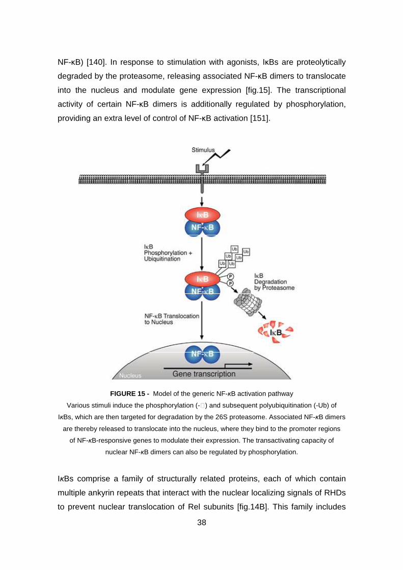
38
NF-κB) [140]. In response to stimulation with agonists, IκBs are proteolytically
degraded by the proteasome, releasing associated NF-κB dimers to translocate
into the nucleus and modulate gene expression [fig.15]. The transcriptional
activity of certain NF-κB dimers is additionally regulated by phosphorylation,
providing an extra level of control of NF-κB activation [151].
FIGURE 15 - Model of the generic NF-κB activation pathway
Various stimuli induce the phosphorylation (-℗) and subsequent polyubiquitination (-Ub) of
IκBs, which are then targeted for degradation by the 26S proteasome. Associated NF-κB dimers
are thereby released to translocate into the nucleus, where they bind to the promoter regions
of NF-κB-responsive genes to modulate their expression. The transactivating capacity of
nuclear NF-κB dimers can also be regulated by phosphorylation.
IκBs comprise a family of structurally related proteins, each of which contain
multiple ankyrin repeats that interact with the nuclear localizing signals of RHDs
to prevent nuclear translocation of Rel subunits [fig.14B]. This family includes
39
IκBα, IκBβ and IκBε, which are composed of a central ankyrin repeat region and
an N-terminal regulatory domain, which controls their inducible degradation.
The precursor NF-κB proteins, NF-κB1 p105 and NF-κB2 p100, also function as
IκBs as a result of ankyrin repeat regions in their C-terminal halves. Alternate
promoter usage of the NF-κB1 gene can generate an mRNA encoding only the
C-terminal IκB-like portion of p105, termed IκBγ, but this has so far only been
reported in murine cells [152, 153].
3.4.3 INHIBITION OF NF-kB: A STRATEGY IN ANTI-
INFLAMMATORY THERAPIES
The induction of proinflammatory genes usually results from the increased
activity of a subset of transcription factors that have thus become attractive
targets in the development of novel anti-inflammatory drugs. Among the
inducible transcription factors that control inflammatory gene expression,
nuclear factor NF-kB plays a central and evolutionarily conserved role in
coordinating the expression of various soluble proinflammatory mediators (as
cytokines and chemokines) and leukocyte adhesion molecules [140].
A wide range of genes involved in inflammation contain functional kB-sites
within their promoters and are induced by NF-kB [154]: thus, NF-kB could be
proposed as one target to down-modulate the immune processes involved in
different pulmonary diseases, including CF [155], by using of drugs, synthetic
molecules, natural products and normal or recombinant proteins that could act
at several steps to interfere with NF-kB activation [fig.16].
Various classes of NF-kB inhibitors are discussed below:
• NONSTEROIDAL ANTI-INFLAMMATORY DRUGS (NSAIDs)
Acetylsalicylic acid (ASA) and sodium salycilate function as an anti-
inflammatory agents by inhibiting cyclooxygenase (COX), an enzyme activity
essential to the production of prostaglandins (PGs) [156]. More recent studies
demonstrate that the mechanism of inhibition by both ASA and sodium
40
salycilate occurs by means of binding and blocking the ATP binding site of IKKβ
[157]. Intriguingly, expression of the COX-2 encoding gene, believed to be
responsible for the massive production of PGs at inflammatory sites, is
transcriptionally regulated by NF-kB [158, 159]. The ability of ASA and sodium
salicylate to inhibit NF-kB activity and kB-dependent gene expression has been
tested experimentally in a wide range of cell types and conditions. For example,
pretreatment of primary synovial fibroblasts or lung epithelial cells with ASA
blocks IL-1β and TNF-α – induced NF-kB activation so that the subsequent
expression of IL-6 and IL-8 is inhibited [160, 161].
Furthermore Ibuprofen inhibits NF-kB activation in T cells [162] as well as COX-
2 expression and PGE2 production in murine macrophages. Among the other
NSAIDs that have been investigated, acetaminophen inhibits the binding of NF-
kB to DNA in LPS- and interferon-γ – treated macrophages and thus precludes
expression of inducible nitric oxide synthase [163]. Moreover, sulindac, an
NSAID that is both structurally and pharmacologically related to indomethacin,
decreases IKKβ kinase activity and consequently inhibits NF-kB activation
[164]. Lastly, tepoxalin, which inhibits both COX and 5-lipoxygenase, blocks NF-
kB activation in Jurkat T-cells and HeLa cells.
Thus, the findings that salicylates and glucocorticoids can inhibit NF-kB activity
are among a growing body of data indicating that drugs specifically designed to
target NF-kB activation might be clinically useful for the treatment of diseases
that involve inflammation, as cystic fibrosis [165-169].
• GLUCOCORTICOIDS
Glucocorticoids (GCs) and other corticosteroids are the most widely used anti-
inflammatory, immunosuppressive drugs. GCs function to downregulate the
expression of genes involved in inflammation [170]. Previous studies
demonstrated that GCs increase the expression of an IkB molecule, named
IkBα, so that NF-kB is retained in the cytoplasm and thus rendered inactive
[165, 167].
41
The notion that NF-kB inhibition underlies the anti-inflammatory capacity of GCs
is also supported by animal studies. In a mouse model of acute inflammation
(carrageenin - induced air pouch inflammation), treatment with the synthetic
steroid dexamethasone reduces the number of cells that stain positively for
activated NF-kB [171]. In rats, dexamethasone not only inhibits TNF-α
production and hepatic NF-kB activation associated with peritoneal sepsis [172],
but also inhibits LPS-induced activation of NF-kB, restores myocardial
contractility, and increases myocardial IkBα protein levels [173]. Finally, a
recent study of normal human subjects demonstrates that intravenous injection
of hydrocortisone increases IkBα levels and reduces the translocation of NF-kB
into the nuclei of peripheral mononuclear cells [174], which supports the
experimental model originally proposed for the molecular action of GCs [165,
167].
• ANTIOXIDANTS
The generation of reactive oxygen species (ROS) by phagocytic leukocytes
(neutrophils, monocytes, macrophages, and eosinophils) is one of the most
important hallmarks of the inflammatory process. By oxidizing essential cellular
components of invading pathogens, reactive radicals and oxidants also
represent the first line of defense against microorganisms [175]. In addition to
promoting general cytotoxicity, ROS may also act to upregulate proinflammatory
gene expression by activating NF-kB, a process that is itself sensitive to the
cellular redox state [176].
Diverse agents that cause oxidative stress can activate NF-kB [177], and
numerous stimuli that activate NF-kB, including cytokines, phorbol esters, LPS,
and CD3 engagement, increase the levels of intracellular ROS [178]. This
generation of ROS, however, is cell- and stimulus-specific. Thus, whereas
stimulation with the proinflammatory cytokines IL-1 and TNF-α leads to
substantial intracellular ROS in lymphoid and monocytic cell lines, no such
increases have been observed in epithelial cell lines derived from ovaries,
colon, breast, or cervix [178].
42
• ANTISENSE OLIGODEOXYNUCLEOTIDES
Antisense oligodeoxynucleotides constitute a potentially important family of
therapeutic compounds for the treatment of a range of diseases [179]. The use
of antisense technology to inhibit expression of NF-kB proteins has been
evaluated both in vitro and in vivo in a variety of experimental systems.
Antisense targeting of the NF-kB p50 subunit can reduce IgM and IgG synthesis
in B cells [180], and can significantly reduce maturation and proliferation of
CD25/IL-2R T-cells [181]. Antisense strategies to reduce expression of the p65
subunit greatly inhibits the expression of cell adhesion molecules in endothelial
and smooth muscle cells in vitro [182]. In vivo, antisense inhibition of p65 blocks
tumor growth in nude mice, prolongs allo- and xenograft survival [183, 184], and
alleviates septic shock in LPS-treated animals [185, 186]. Clearly, the
therapeutic potential of antisense molecules directed against NF-kB and the
components required for its expression and activation warrants further
investigation.
• PROTEASOME INHIBITORS
Signal-induced phosphorylation and ubiquitination of IκB and its degradation by
the 26S proteasome precede NF-κB nuclear translocation. Inhibitors of
proteasome function reduce the degradation of IκB to prevent activation of the
NF-κB pathway. A variety of peptide aldehydes, including MG101, MG132, and
MG115, make up one class of agents that inhibit the protease activity of the
proteasome [187, 188].
Proteasome inhibitors of another class, including lactacystin, block protein
degradation activity by acylating a threonine residue in one of the key
proteasome subunits. Finally, a group of boronic acid peptides, including PS-
341, are extremely potent inhibitors of proteasome function [189, 190].
Recently, PS-341 has shown promise as an adjunct to cancer chemotherapy by
inhibiting activation of the NF-κB pathway. It is also possible that inhibitors of
the ubiquitin ligase that mediates IκB ubiquitination may be a useful target in
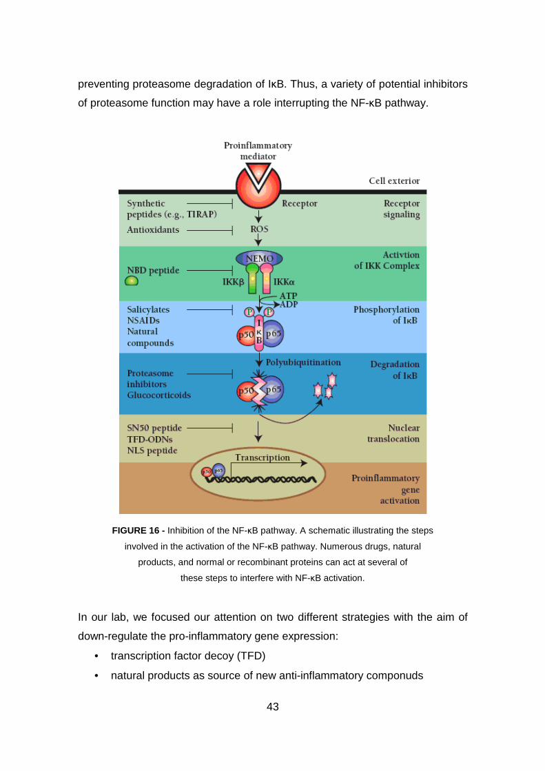
43
preventing proteasome degradation of IκB. Thus, a variety of potential inhibitors
of proteasome function may have a role interrupting the NF-κB pathway.
FIGURE 16 - Inhibition of the NF-κB pathway. A schematic illustrating the steps
involved in the activation of the NF-κB pathway. Numerous drugs, natural
products, and normal or recombinant proteins can act at several of
these steps to interfere with NF-κB activation.
In our lab, we focused our attention on two different strategies with the aim of
down-regulate the pro-inflammatory gene expression:
• transcription factor decoy (TFD)
• natural products as source of new anti-inflammatory componuds
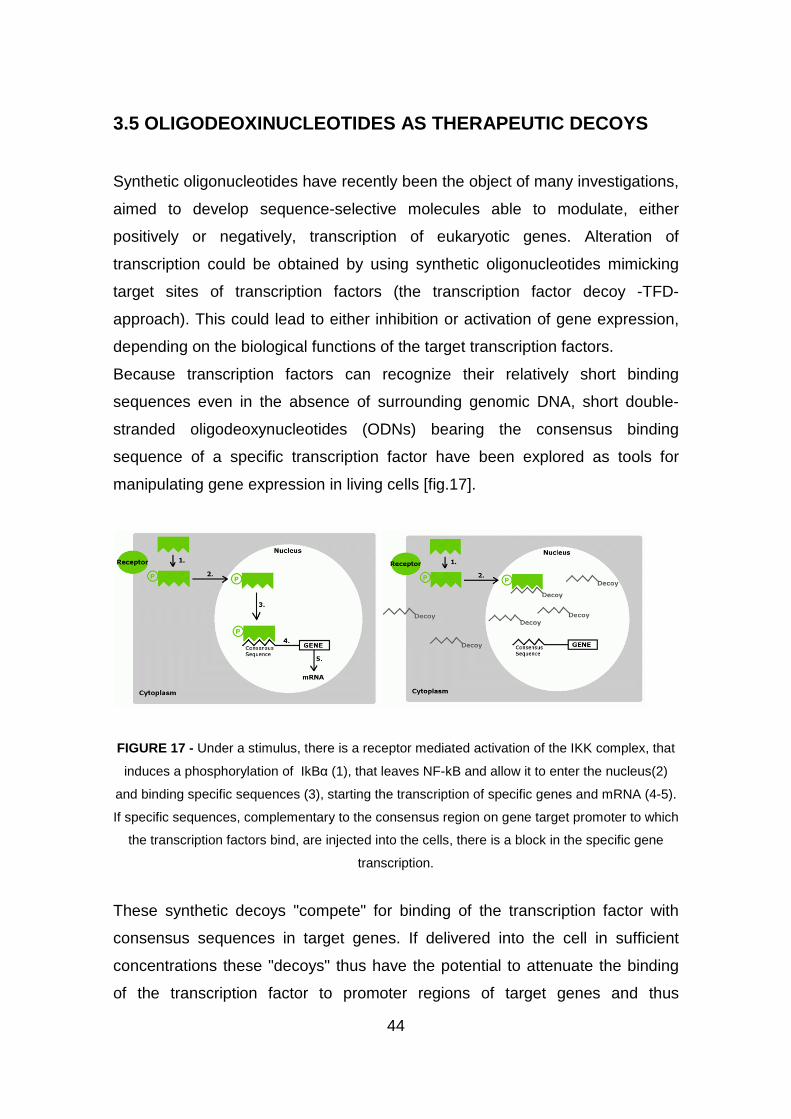
44
3.5 OLIGODEOXINUCLEOTIDES AS THERAPEUTIC DECOYS
Synthetic oligonucleotides have recently been the object of many investigations,
aimed to develop sequence-selective molecules able to modulate, either
positively or negatively, transcription of eukaryotic genes. Alteration of
transcription could be obtained by using synthetic oligonucleotides mimicking
target sites of transcription factors (the transcription factor decoy -TFD-
approach). This could lead to either inhibition or activation of gene expression,
depending on the biological functions of the target transcription factors.
Because transcription factors can recognize their relatively short binding
sequences even in the absence of surrounding genomic DNA, short double-
stranded oligodeoxynucleotides (ODNs) bearing the consensus binding
sequence of a specific transcription factor have been explored as tools for
manipulating gene expression in living cells [fig.17].
FIGURE 17 - Under a stimulus, there is a receptor mediated activation of the IKK complex, that
induces a phosphorylation of IkBα (1), that leaves NF-kB and allow it to enter the nucleus(2)
and binding specific sequences (3), starting the transcription of specific genes and mRNA (4-5).
If specific sequences, complementary to the consensus region on gene target promoter to which
the transcription factors bind, are injected into the cells, there is a block in the specific gene
transcription.
These synthetic decoys "compete" for binding of the transcription factor with
consensus sequences in target genes. If delivered into the cell in sufficient
concentrations these "decoys" thus have the potential to attenuate the binding
of the transcription factor to promoter regions of target genes and thus

45
attenuate the function of the transcription factor to regulate the expression of its
target gene(s). Transfected at high concentrations these decoys have been
reported in the literature to completely block transcription factor function. Clearly
they represent powerful research tools for studying gene regulation both in vitro
and also more recently in vivo [191, 192].
Yoshimura et al. reported the successful in vivo transfer of NF-kB decoy ODN to
reduce neointimal thickening in rat carotid artery model, providing a new
therapeutic strategy for restenosis after angioplasty. Since a rapid and transient
activation of NF-kB after balloon injury plays a crucial role in the formation of
neointima, they demonstrated that inhibition of transient NF-kB activation should
be a new therapeutic tool. To block NF-kB activation, they utilized a ‘decoy’ cis
element, since decoy ODN can bind NF-kB and thereby block its activation.
Using this technology, they already reported the successful inhibition of NF-kB
activation in a rat myocardial infarction model [193]. Therefore, they examined
the inhibition of NF-kB activation after balloon injury by in vivo transfection of
NFkB decoy ODN into balloon-injured blood vessels. As expected, it was
observed that activation of NF-kB in the injured vessels was successfully
inhibited by NF-kB, but not scrambled, decoy ODN in vivo. These studies reveal
the importance of NFkB activation in neointimal formation after vascular injury,
since transfection of NFkB decoy ODN inhibited neointimal formation
accompanied by a decrease in NFkB activity.
Furthermore, Penolazzi et al. reported the in vivo effects of a decoy
oligonucleotide targeting the nuclear factor κB (NF-κB) on osteoclasts during
forced orthodontic tooth movement in rats [194]. Wistar rats were subjected to
orthodontic forces, in the absence or presence of treatment with a decoy
molecule mimicking a nonsymmetric NF-κB binding site (5'-CGC TGG GGA
CTT TCC ACG G-3'). TUNEL staining of fragmented DNA revealed that
treatment with NF-κB decoy but not with scramble double-stranded
oligodeoxynucleotides (ODN) induced a high level of osteoclast apoptosis in
vivo. Immunohystochemical analysis for death receptor Fas revealed strong
positivity only in samples treated with NF-κB decoys, demonstrating that
osteoclasts are sensitive to death induction via Fas signaling. Induction of

46
apoptosis in osteoclasts could be a strategy for treatment of excessive
osteoclast activity in pathologic conditions such as osteoporosis, peri-articular
osteolysis, inflammatory arthritis, Paget's syndrome and tumour-associated
osteolytic metastases.
Moreover, the lung Infection with P. aeruginosa is known to induce expression
of genes of the innate immunity by activating different TFs, such as NF-kB, AP-
1, Elk-1, and NF–IL-6 [195-197]. Since NF-kB plays a well known pivotal role in
inflammatory processes, we choose it as the first molecular target for the TF
decoy approach.
3.6 NATURAL PRODUCTS
Classic therapies based on corticosteroids [170] and ibuprofen [162], as well as
chemiotherapy, have been employed in CF patients, but they have
demonstrated limited efficacy or occurrence of undesired effects, suggesting the
importance of investigations aimed at identifying alternative therapeutic targets
and novel anti-inflammatory molecules [198, 199, 200-203].
In this respect, we are studying natural products as sources for the discovery of
new drugs on the basis of their widespread application in ethnic and traditional
medicine to cure several human diseases, such as muscular and neuronal
diseases, mycosis, heart and respiratory diseases, atherosclerosis [204-206].
As an activator of many pro-inflammatory cytokines and inflammatory
processes the modulation of the NF-kB transduction pathway is a principal
target to alleviate the symptoms of such diseases as arthritis, inflammatory
bowel disease and asthma. Natural products and some extracts are reviewed
and assessed for their activity and potency as NF-kB inhibitors. A large number
of compounds are currently known as NF-kB modulators and include the
isoprenoids, most notably kaurene diterpenoids and members of the
sesquiterpene lactones class, several phenolics including curcumin and
flavonoids such as silybin. Additional data on cellular toxicity are also
highlighted as an exclusion principle for pursuing such compounds in clinical
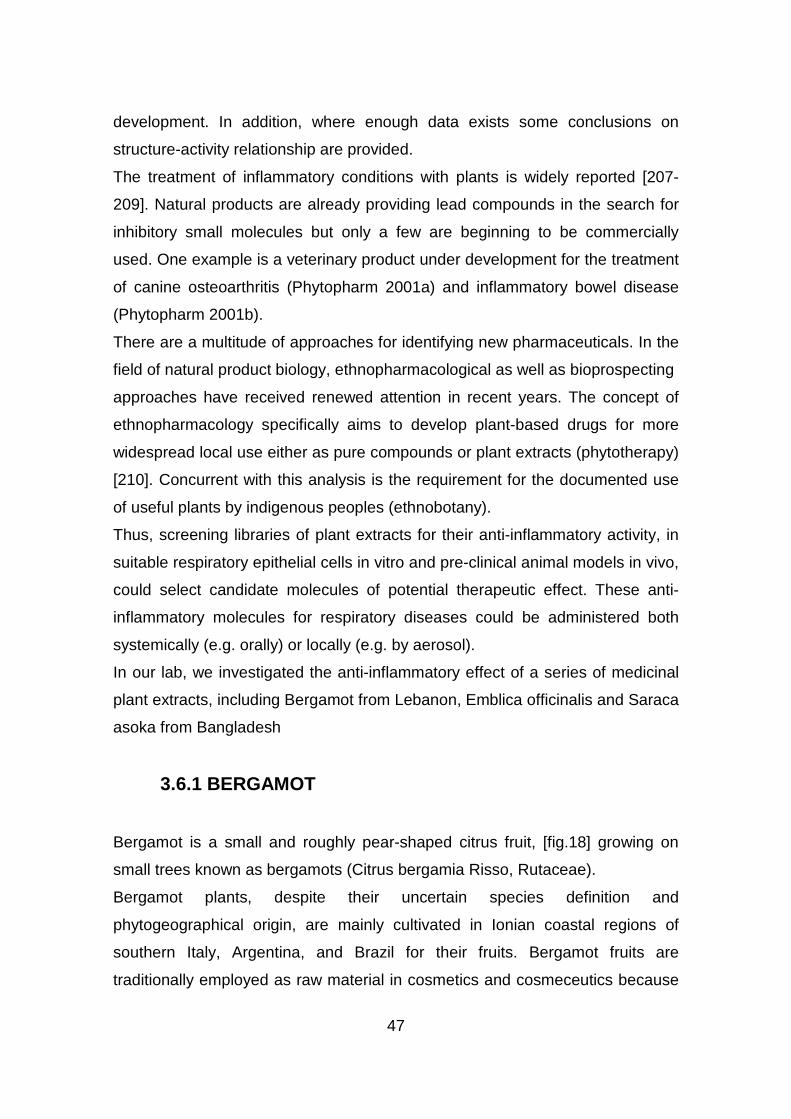
47
development. In addition, where enough data exists some conclusions on
structure-activity relationship are provided.
The treatment of inflammatory conditions with plants is widely reported [207-
209]. Natural products are already providing lead compounds in the search for
inhibitory small molecules but only a few are beginning to be commercially
used. One example is a veterinary product under development for the treatment
of canine osteoarthritis (Phytopharm 2001a) and inflammatory bowel disease
(Phytopharm 2001b).
There are a multitude of approaches for identifying new pharmaceuticals. In the
field of natural product biology, ethnopharmacological as well as bioprospecting
approaches have received renewed attention in recent years. The concept of
ethnopharmacology specifically aims to develop plant-based drugs for more
widespread local use either as pure compounds or plant extracts (phytotherapy)
[210]. Concurrent with this analysis is the requirement for the documented use
of useful plants by indigenous peoples (ethnobotany).
Thus, screening libraries of plant extracts for their anti-inflammatory activity, in
suitable respiratory epithelial cells in vitro and pre-clinical animal models in vivo,
could select candidate molecules of potential therapeutic effect. These anti-
inflammatory molecules for respiratory diseases could be administered both
systemically (e.g. orally) or locally (e.g. by aerosol).
In our lab, we investigated the anti-inflammatory effect of a series of medicinal
plant extracts, including Bergamot from Lebanon, Emblica officinalis and Saraca
asoka from Bangladesh
3.6.1 BERGAMOT
Bergamot is a small and roughly pear-shaped citrus fruit, [fig.18] growing on
small trees known as bergamots (Citrus bergamia Risso, Rutaceae).
Bergamot plants, despite their uncertain species definition and
phytogeographical origin, are mainly cultivated in Ionian coastal regions of
southern Italy, Argentina, and Brazil for their fruits. Bergamot fruits are
traditionally employed as raw material in cosmetics and cosmeceutics because
48
of their essential oil flavor and in dietary and herbal products as crude drugs
because of the eupeptic properties and therapeutic activities against digestive
disorders [211].
FIGURE 18 – Citrus Bergamia.
In the past, Bergamot essential oil has been used in the folk medicine as
antiseptic, to facilitate wound healing and as antihelminthic. Actually, likewise
other essential oils, Bergamot essential oil is widely used in aromatherapy, a
complementary medicine highly diffused in the industrialized countries to
improve mood and mild symptoms of stress-induced disorders such as
anxiety [212], depression [213], behavioural disturbances in dementia [214] and
chronic pain [215, 216]. Individual components of essential oils reach the blood,
cross the blood-brain barrier and enter the central nervous system (CNS)
following inhalation, dermal application [217], intraperitoneal (i.p.) or
subcutaneous (s.c.) injection and oral administration [218]. Physiological and
pharmacological studies, combined with phytochemical analyses, indicate that a
variety of essential oils exert specific effects on the CNS [219].
Recently, Hiroyasu Inoue et al. have released a study confirming the
inflammation reducing power of many aromatherapy essential oils. The oils
shown to reduce inflammation through suppression of the COX-2 pro-

49
inflammatory enzyme included herbs, like thyme, clove and fennel; rose;
eucalyptus; and even the citrus bergamot. They suppress the inflammation
COX-2 mediated in a manner similar to resveratrol, the chemical linked with the
health benefits of red wine. The oils considered to have the strongest anti-
inflammation activity used in aromatherapy were not even examined, which can
mean that there are naturally a great many compounds found in essential oils
that have an anti-inflammatory effect [220].
3.6.2 EMBLICA OFFICINALIS
Emblica officinalis is commonly known as Amla or the Indian gooseberry
[fig.19], which is a member of a small genus Emblica (Family Euphorbiaceae)
and is extensively found all over India, as well as Sri Lanka, Malaysia, China,
Pakistan and Bangladesh.
FIGURE 19 – Emblica Officinalis.
The tree is small to medium sized, reaching 8 to 18 m in height, with a crooked
trunk and spreading branches. The branchlets are glabrous or finely pubescent,
10–20 cm long, usually deciduous; the leaves simple, subsessile and closely
50
set along branchlets, light green, resembling pinnate leaves. The flowers are
greenish-yellow. The fruit is nearly spherical, light greenish yellow, quite smooth
and hard on appearance, with 6 vertical stripes or furrows.
The fruits of Emblica officinalis have been reported to contain constituents with
variable biological activity: they have been used for thousands of years in the
traditional Indian medicine for the treatment of several diseases. For many
years the therapeutic potential of the fruits was attributed to their high content of
ascorbic acid: about 1 g of vitamin C per 100 ml of fresh juice.
In 1996 professor Shibhnath Ghosal of Banaras Hindu University discovered
that Emblica fruits do not contain ascorbic acid neither in free nor in conjugated
form, but it contains two new hydrolysable tannins with low molecular weight
(B1000), called emblicanin A (2,3-di-o-galloyl-4,6-(S)-hexahydroxydiphenoyl-2-
keto-glucono-d-lactone) and emblicanin B (2,3,4,6-bis-(S)-
hexahydroxydiphenoyl-2-keto-glucono-d-lactone) and other tannins like
punigluconino (2,3-di-O-galloyl-4,6-(S)-hexahydroxydiphenoylgluconicacid) and
pedunclagin (2,3,4,6-bis-(S)-hexahydroxydiphenoyl-D-glucose) already isolated
in other species, Punica granatum in particular. These two new tannins have a
very strong antioxidant action. The two emblicanins A and B have been found to
preserve erythrocytes against oxidative stress induced by asbestos, generator
of superoxide radical. Emblicanin A oxidates when put in contact with asbestos
becoming emblicanin B and together they have a stronger protective action to
erythrocytes than vitamin C. Moreover they improve the efficacy of vitamin C in
reducing dihydroascorbic acid to ascorbic acid.
The same recycling process has been observed in the rutin-vitamin C
combination. Fruit extract has been shown to have an antimutagenic activity in
Ames test. Recently fruits have been tested for their antiviral activity, particularly
for inhibiting reverse transcriptase in the replication of retroviruses like HIV-1.
Some active ingredients inhibit the replication of the virus through a non
competitive action with respect to the dTTP but competitive action with respect
to the template primer.
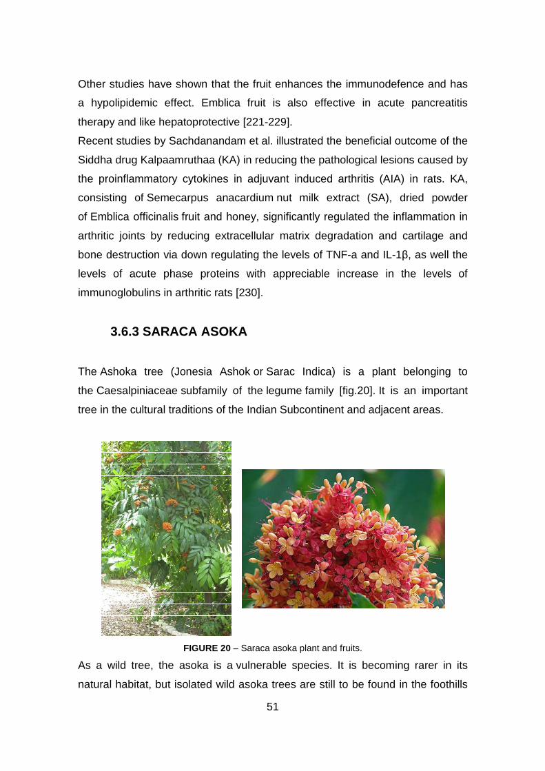
51
Other studies have shown that the fruit enhances the immunodefence and has
a hypolipidemic effect. Emblica fruit is also effective in acute pancreatitis
therapy and like hepatoprotective [221-229].
Recent studies by Sachdanandam et al. illustrated the beneficial outcome of the
Siddha drug Kalpaamruthaa (KA) in reducing the pathological lesions caused by
the proinflammatory cytokines in adjuvant induced arthritis (AIA) in rats. KA,
consisting of Semecarpus anacardium nut milk extract (SA), dried powder
of Emblica officinalis fruit and honey, significantly regulated the inflammation in
arthritic joints by reducing extracellular matrix degradation and cartilage and
bone destruction via down regulating the levels of TNF-a and IL-1β, as well the
levels of acute phase proteins with appreciable increase in the levels of
immunoglobulins in arthritic rats [230].
3.6.3 SARACA ASOKA
The Ashoka tree (Jonesia Ashok or Sarac Indica) is a plant belonging to
the Caesalpiniaceae subfamily of the legume family [fig.20]. It is an important
tree in the cultural traditions of the Indian Subcontinent and adjacent areas.
FIGURE 20 – Saraca asoka plant and fruits.
As a wild tree, the asoka is a vulnerable species. It is becoming rarer in its
natural habitat, but isolated wild asoka trees are still to be found in the foothills
52
of central and eastern Himalayas, in scattered locations of the northern plains of
India as well as on the west coast of the Subcontinent near Mumbai.
The asoka tree is considered sacred throughout the Indian subcontinent,
especially in India and Sri Lanka. This tree has many folklorical, religious and
literary associations in the region. Highly valued as well for its handsome
appearance and the color and abundance of its flowers, the asoka tree is often
found in royal palace compounds and gardens as well as close to temples
throughout India. The asoka tree has a symbolic importance
in Buddhism. Queen Māyā of Sakya is said to have given birth to
the Buddha under an asoka tree in a garden in Lumbini.
The earliest chronicled mention is in the Ayurvedic treatise, the Charka Samhita
(100 A.D.), in which Asoka is recommended in formulations for the
management pain with relation to uterus (gynecological) as anodynes. The
Bhavprakasha Nighantu, commonly known as the Indian Materia Medica (1500
A.D.), cites the plant as a uterine tonic: actually Saraca asoka is very famous for
its use in treating gynecological disorders, it is known as an astringent to treat
excessive uterine bleeding from various causes (including hormone disorders
and fibroids), but also for regulating the menstrual cycle and, in various complex
formulas, as a tonic for women. The bark, rich in tannins and cyanidins (red
colored compounds), is the primary medicinal part [231-233].
Chloroform, methanol, aqueous and ethanolic extracts of the stem bark of
Saraca indica were investigated for their antibacterial and antifungal activity
against standard strains of Staphylococcus aureus, Escherichia coli,
Pseudomonas aeruginosa, Bacillus cereus, Klebsiella pneumoniae, Proteus
mirabilis, Salmonella typhimurium and Streptococcus pneumoniae and the
fungi: Candida albicans and Cryptococcus albidus. Methanolic and aqueous
extract exhibited antimicrobial activity with MIC ranging from 0.5-2% and 1-3%
respectively. Methanolic extract exhibited the strongest activity against both
bacteria and fungi [234].
Different forms of uses of Asoka herb are described:

53
• Asoka fresh or dried bark: it is bitter, astringent and sweet in taste. It has
stimulating effect on endometrial and the ovarian tissue. It is useful in
internal bleeding, hemorrhoids, ulcers, uterine affections, menorrhagia
especially due to uterine fibroids, meno-metrorrhagia, leucorrhoea and
pimples.
• Asoka fresh flower: it is an excellent uterine tonic and is used in cervical
adenitis, biliousness, syphilis, hyperdipsia, burning sensation,
hemorrhagic dysentery, piles, scabies in children and inflammation.
• Asoka dried flowers: they are used in diabetes.
• Asoka seeds: they are used in treating bone fractures, strangury and
vesicle calculi.
We suggest that Saraca asoka, with its lengthy history of human use in
traditional medicine, deserves consideration as a candidate drug for
suppressing IL-8-dependent lung inflammation in CF.

54

54
AIM OF THESIS
Cystic fibrosis is an autosomal recessive genetic disorder common in
Caucasian population caused by mutations in the CFTR gene and
characterized by extensive lung inflammation. One of the hallmarks in CF is a
chronic infection sustained by strikingly few pathogens including
Staphylococcus aureus and Haemophilus influenzae, whereas in the course of
their lifetime CF patients become progressively colonized by Pseudomonas
aeruginosa. Elevated concentrations of pro-inflammatory cytokines and
chemokines have been found in the bronchoalveolar fluid of patients with CF
[118, 119]. Consistent with these findings, experimental models support the
concept that interaction of P.aeruginosa with CF respiratory epithelial cells
promotes PMN recruitment, mainly by releasing the chemokine interleukin 8 (IL-
8) [116, 117]. Within the lumen of CF airways, nucleic acids derived from PMNs
further reduce mucociliary clearance, and proteases released from PMNs
mediate epithelium injury, progressive bronchial wall damage, bronchiectasis,
and peribronchiolar fibrosis. Therefore, the exaggerated, PMN-dominated
inflammatory process is thought to critically contribute to the gradual decline in
CF lung function [113, 114].
Novel pharmacological correctors and potentiators of mutated CFTR protein
leave many unanswered questions concerning their application in CF, including
the degree of correction of the defect that can be obtained, the safety in long
term administration and the efficacy in reverting the extensive lung inflammatory
pathology [86]. Although anti-bacterial therapy has been shown to be more
promising since early eradication therapy has been established in some
centers, it remains questionable whether all patients will profit from this strategy
[30-33]. In addition, besides the fact that anti-inflammatory molecules have
demonstrated potential benefits in CF patients, enthusiasm has been tempered
either by the limited efficacy or the occurrence of undesired effects.
Therefore considering that at present no fully satisfactory anti-inflammatory
treatment is available for clinical use in CF patients, investigation on novel anti-
55
inflammatory molecules for the treatment of lung inflammation is considered a
major priority in the CF community (www.cff.org).
With respect to pro-inflammatory mechanisms, P. aeruginosa is known to
stimulate surface respiratory epithelial cells to express and release IL-8 through
activation of the transcription factor NF-kB [114, 115]. Besides IL-8, a large
number of other genes contain kB sites in their 5’-upstream untranslated
regions, including genes encoding cytokines, chemokines, and adhesion
molecules that orchestrate PMN transbronchial migration, such as TNF-α, IL-1,
GRO-γ, and intercellular adhesion molecule (ICAM)-1, together with different
other genes regulating innate immunity. Thus, NF-kB has been proposed as
one target to down-modulate the immune processes involved in different
pulmonary diseases, including CF.
The transcription factor (TF) decoy approach has been proposed to modulate
gene expression in vitro. This approach is based on the intracellular delivery of
double-stranded oligodeoxynucleotides causing inhibition of the binding of TF
related proteins to the different consensus sequences in the promoter of
specific genes. Several decoy oligodeoxynucleotides have been studied to
pursue the modulation of expression of genes relevant to human diseases. Due
to its central role in immune response and cell cycle regulation, TF decoy
oligodeoxynucleotides targeted to NF-kB have been studied in other and our
laboratories [118-121].
To date, the inhibition of IL-8 expression by NF-kB decoy molecules has been
limited both conceptually, by the potential lack of specificity related to the wide
role of NF-kB in regulating expression of several genes, and practically, by the
limited efficiency obtained in the preliminary experiments [235-237]. Thus, we
addressed the issues of efficiency and specificity by testing the effect of novel
NF-kB decoy sequences on the expression of different genes induced by P.
aeruginosa in human bronchial epithelial cells carrying either the wild type or
the mutant CFTR gene.
An other approach has been studied in our lab to modulate the pro-
inflammatory gene expression in vitro. This second approach is based on
extracts from plants used in ethnic medicine that are of great interest, as many
56
of them are known to retain anti inflammatory properties. This was reviewed by
Darshan and Doreswamy [238], who reported about the anti inflammatory
activity of drugs derived from 38 medicinal plants. In this review a clear role of
botanical plants (including polysaccharides, terpenes, curcuminoids and
alkaloids) was reported as alleviating inflammatory diseases, such as arthritis,
rheumatism, acne skin allergy and ulcers. The most potent in curing
inflammatory diseases were found to be plants containing polysaccharides
[238]. In the present study, we analyzed the effects of Bergamot, Emblica
officinalis and Saraca asoka extracts on the production of cytokines and
chemokines in cystic fibrosis IB3-1 cells [239-241] induced to hyper secretion of
inflammatory cytokines and chemokines by TNF-α [242-244].
In our studies we investigated the ability of the plant extracts and their main
constituent drugs derived, to interfere with specific bindings between target
DNA sequences and Rel/NF-kB proteins by EMSA assays (Electrophoretic
Mobility Shift Assay). To use this compound for in vitro experiments, anti-
proliferative assays have been made in cystic fibrosis IB3-1 cells model.
After determining the IC50 values (concentrations of extracts leading to 50%
inhibition of IB3-1 cell growth), we analyzed the inhibitory effect of these
compounds on gene expression of interleukin 8 (IL8) that is directly involved in
the inflammation pathway via NF-kB activation. In particular, IB3-1 cells have
been treated with different concentration of Bergamot, Emblica officinalis,
Saraca asoka plant extracts and with increasing concentration of their main
identified constituents bergamottin, bergapten, citropten, pyrogallol, 5-OH-
isoquinoline, digitoxin; the isolated RNA has been retro-transcribed (Retro-
Transcription PCR) and amplified for the gene of interest IL-8 and the
housekeeping GAPDH (glyceraldehyde-3-phosphate dehydrogenase).
The plant extracts resulted more interesting have been used on IB3-1 cells
induced with TNF-α (Tumor Necrosis Factor-α) to evaluate the inhibitory activity
in this inflamed cellular model, since previous studies demonstrated that TNF-α
cytokine, present in high concentration in CF patients, induces inflammatory
mediators expression. Finally, Bio-Plex technology has been used to analyze
the potential inhibitory effect of plant extracts on the expression of cytokines in

57
IB3-1 cells induced by TNF-α. The Bio-Plex suspension array system helps to
reveal a more complete view of the effects of candidate drugs on the biology of
cells.
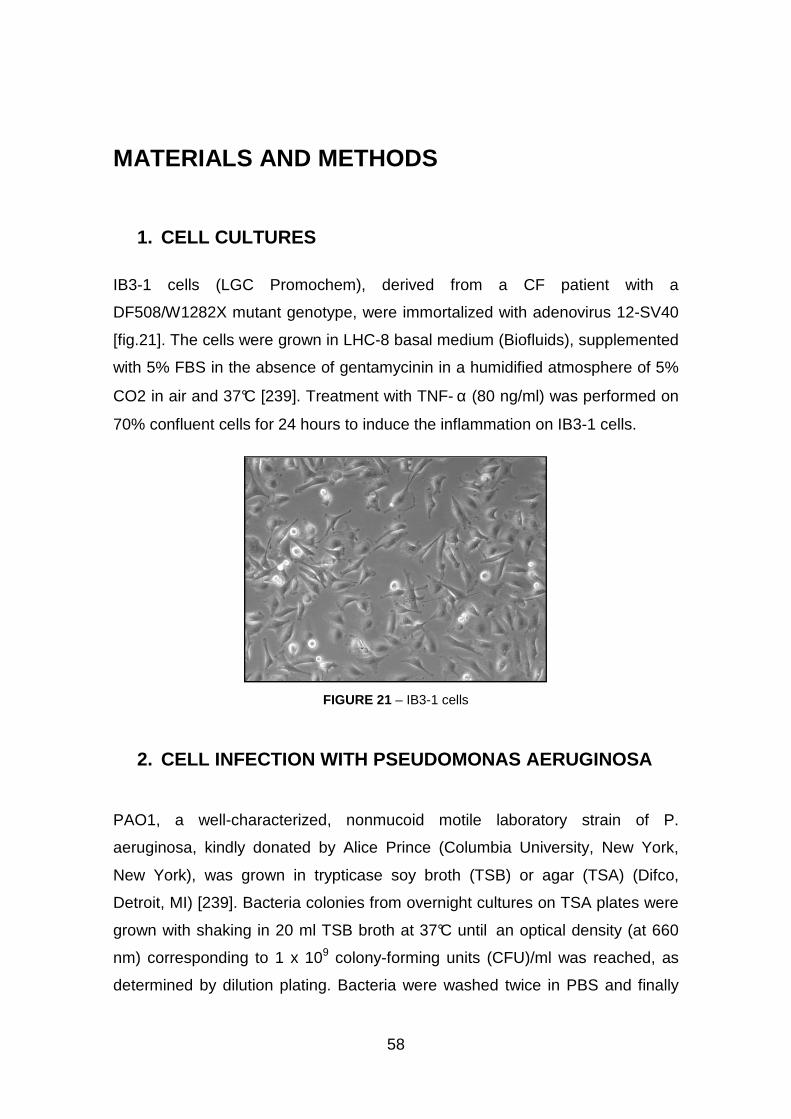
58
MATERIALS AND METHODS
1. CELL CULTURES
IB3-1 cells (LGC Promochem), derived from a CF patient with a
DF508/W1282X mutant genotype, were immortalized with adenovirus 12-SV40
[fig.21]. The cells were grown in LHC-8 basal medium (Biofluids), supplemented
with 5% FBS in the absence of gentamycinin in a humidified atmosphere of 5%
CO2 in air and 37°C [239]. Treatment with TNF- α (80 ng/ml) was performed on
70% confluent cells for 24 hours to induce the inflammation on IB3-1 cells.
FIGURE 21 – IB3-1 cells
2. CELL INFECTION WITH PSEUDOMONAS AERUGINOSA
PAO1, a well-characterized, nonmucoid motile laboratory strain of P.
aeruginosa, kindly donated by Alice Prince (Columbia University, New York,
New York), was grown in trypticase soy broth (TSB) or agar (TSA) (Difco,
Detroit, MI) [239]. Bacteria colonies from overnight cultures on TSA plates were
grown with shaking in 20 ml TSB broth at 37°C until an optical density (at 660
nm) corresponding to 1 x 10⁹ colony-forming units (CFU)/ml was reached, as
determined by dilution plating. Bacteria were washed twice in PBS and finally
59
diluted in each specific serum-free culture medium before infection. All cell lines
were infected with ranging doses of PAO1 at 37°C fo r 4 hours.
3. DECOY OLIGODEOXYNUCLEOTIDES
Decoy oligodeoxynucleotides (ODNs) mimicking the NF-kB consensus
sequences identified in the HIV-1 long terminal repeat (LTR) of the promoter
region of HIV-1 gene (sense: 5’-CGC TGG GGA CTT TCC ACG G-3’) were
used. A ‘‘scrambled’’ ODN sequence (sense: 5’-CAC AAA GTG TAA CAG TCT-
3’) was used in each experiment. Synthesis and high-performance liquid
chromatography–grade purification was obtained from Sigma. Sense and anti-
sense freeze-dried ODNs were resuspended in DNAse-free sterile water at the
stock concentration 5 mg/ml. Annealing to obtain double-stranded ODNs was
performed by mixing equimolar concentrations of both forward and reverse
strands (1 mg/ml in 150 mM NaCl, final concentration), incubating the solution
at 100°C for 5 minutes, then leaving it overnight a t room temperature.
4. TRANSFECTION OF CELLS WITH DECOY ODNS
IB3-1 cells have been transfected with ODNs by mixing the double-stranded
ODNs with the cationic liposome Lipofectamine 2000 (Invitrogen, Carlsbad,
CA). Lipofectamine 2000 (4ml) was diluted in 1ml LHC-8 serum-free cell culture
medium (Biofluids), according to the manufacturer’s instructions. Double-
stranded decoy or scrambled ODNs (2 mg) were added and incubated for 10
minutes. Liposome:DNA complexes in LHC-8 serum-free medium were added
to IB3-1cells and incubated at 37°C and 5% CO 2 for 6 hours. The cells were
washed twice with culture serum-free medium and left at 37°C and 5% CO 2 for
a further 18 hours before infection with P. aeruginosa.
60
5. PLANT EXTRACTS AND CHEMICAL
Bergamot oil was extracted by cold pressing of peels, and it is made up of 93-
96% volatile compounds high quantities of oxygenated compounds.
The dried fruits of Emblica officinalis were extracted with absolute ethanol and
the yield was 9.33%.
The stem barks of Saraca asoka were also extracted with absolute ethanol and
the obtained yields were 14.16%.
Bergamottin, bergapten and citropten were purchased from Extrasynthese
(Lyon, France) and resuspended in ethanol and 3% dimethil-sulfoxide (DMSO).
Pyrogallol, 5-OH-hydroquinoline, Digoxin were furnished from Sigma-Aldrich
(Milwaukee, WI, USA) and resuspended in water (pyrogallol) or methanol (5-
OH-hydroquinoline and digoxin). All the resuspended compounds were
conserved at -20°C protected from light.
6. ASSAYS OF IN VITRO ANTIPROLIFERATIVE ACTIVITY
In vitro antiproliferative activity was assayed as follows. Cells were seeded at
the initial concentration of 30.000 cells/cm2 and then cultured in the absence or
in the presence of increasing concentration of plants extracts or purchased
chemical drugs. The medium was not changed during the induction period. For
the IC50 (concentrations of extracts leading to 50% inhibition of IB3-1 cell
growth), we determined the values of cell number/ml after 3 days of culture,
when untreated cells are in the log phase of cell growth.
7. PREPARATION OF NUCLEAR EXTRACTS
Nuclear extracts were prepared from IB3-1 cells as described [246]. Cell were
washed twice with PBS and detached by trypsinization. Nuclear proteins were
obtained by hypotonic lysis, followed by high-salt extraction treatment of nuclei
61
Protein concentration was determined using Bio-Rad protein assay. Nuclear
extracts were brought to a concentration of 0.5 mg/ml for electrophoretic
mobility shift assay (EMSA) experiments.
8. ELECTROPHORESIS MOBILITY SHIFT ASSAY (EMSA)
Electrophoresis mobility shift assay (EMSA) was performed by using double-
stranded 32P-labelled oligonucleotides as target DNA [247, 248].
Oligodeoxynucleotides were labeled with γ32-P-ATP using 10 Units of T4-
polinucleotidekinase (MBI Fermentas, St. Leon-Rot, Germany) in 500 mM Tris-
HCl, pH 7.6, 100 mM MgCl2, 50 mM DTT, 1 mM spermidine, 1 mM EDTA in the
presence of 50 mCi γ32-P-ATP) in a volume of 20 µl for 45 minutes at 37°C .
Reaction was brought to 150 mM NaCl, and 150 ng complementary
oligodeoxynucleotide was added. Reaction temperature was increased to
100°C for 5 minutes and left diminishing to room te mperature overnight.
Binding reactions were set up as described elsewhere in binding buffer (10%
glycerol, 0.05% NP-40, 10 mM Tris-HCl pH 7.5, 50 mM NaCl, 0.5 mM DTT, 10
mM MgCl2), in the presence of poly(dI:dC).poly(dI:dC) (Pharmacia, Uppsala,
Sweden), 2-5 µg of crude nuclear extracts [246] and 0.25 ng of labeled
oligonucleotide, in a total volume of 20 µl [247, 248]. After 30 minutes of binding
at room temperature samples were electrophoresed at constant voltage (200 V
for 1 hr) through a low ionic strength (0.25x TBE buffer) (1x TBE = 0.089 M Tris-
borate, 0.002 M EDTA) on 6% polyacrylamide gels until tracking dye
(bromophenol blue) reached the end of a 16 cm slab. Gels were dried and
exposed for autoradiography with intensifying screens at -80°C. In these
experiments, DNA/protein complexes migrate through the gel with slower
efficiency.
To study inhibition of protein/DNA interactions, addition of the reagents was as
follows: (a) poly(dI:dC).poly(dI:dC); (b) labelled oligonucleotides mimicking the
binding sites for transcription factors to be modulated; (c) Saraca asoka,
Emblica officinalis or Bergamot extracts; (d) binding buffer; (e) nuclear factors.
62
The nucleotide sequences of double-stranded target DNA utilized in these
experiments were 5’-CGC FGG GGA CTT TCC ACG G-3’ (sense strand, NF-
kB) (Sigma Genosys, Cambs, UK).
To study the activation of NF-kB in the inflammatory cascade, we exposed our
model system of IB3-1 cells to the P. aeruginosa laboratory strain PAO1
infection: EMSA was perfomed as previously described with double-stranded
synthetic oligodeoxynucleotides mimicking the NF-kB–binding site present in
the promoter of the IL-8 gene (IL-8 NF-kB, sense: 5’-AAT CGT GGA ATT TCC
TCT-3’) incubated with nuclear extracts from IB3-1 cells or purified NF-kB p50
dimer protein (Promega, Madison, WI) were used at the specified
concentrations and poly(dI:dC) (1 mg per reaction) was also added to abolish
nonspecific binding.
For quantitative determinations, the bands corresponding to protein-DNA
complexes were analyzed using the BIO-RAD Gel Doc 2000 (Bio-Rad
Laboratories). The values obtained in the reactions performed in the presence
of plant extracts or purified NF-kB p50 dimer protein were compared to control
untreated reactions.
9. QUANTIFICATION OF IL-8 TRANSCRIPTS AND OTHER PRO-INFLAMMATORY GENES
Total RNA was isolated (Trizol reagent, Invitrogen), retro transcribed (Reverse
Transcription System, Promega Corporation, Medison, USA) and the resulting
cDNA was quantified by relative quantitative real-time PCR [245]. The
sequences of the oligonucleotides used for amplification of IL-8 mRNA were: 5’-
GTG CAG TTT TGC CAA GGA GT-3’ (forward) and 5’-TTA TGA ATT CTC
AGC CCT CTT CAA AAA CTT CTC-3’ (reverse); for β-actin mRNA: 5’-TGA
CGG GGT CAC CCA CAC TGT GCC CAT CTA-3’ (forward); 5’-CTA GAA GCA
TTT GCG GTG GAC GAT GGA GGG-3’ (reverse). PCR was performed in a
final volume of 50 µl containing 50 mM KCl, 10 mM TRIS-HCl pH 8.8, 1.5 mM
MgCl2 by using 1U/reaction of Taq DNA polymerase, 100 µM dNTPs, 0.5 µM
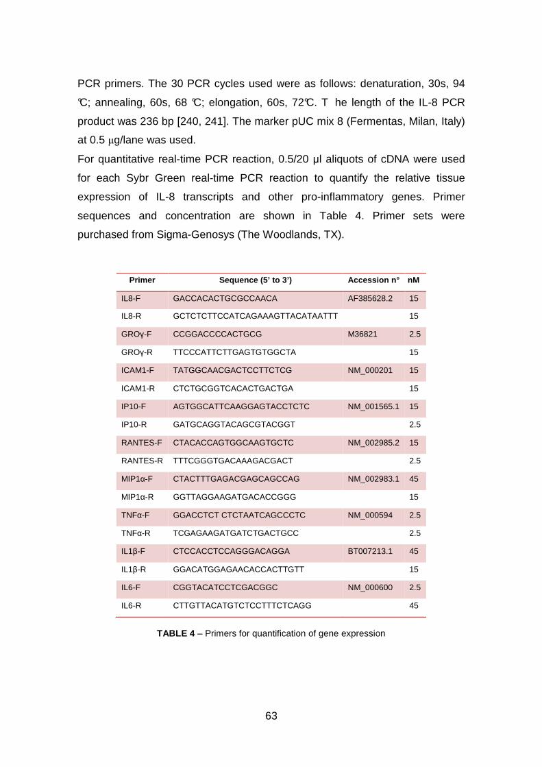
63
PCR primers. The 30 PCR cycles used were as follows: denaturation, 30s, 94
°C; annealing, 60s, 68 °C; elongation, 60s, 72°C. T he length of the IL-8 PCR
product was 236 bp [240, 241]. The marker pUC mix 8 (Fermentas, Milan, Italy)
at 0.5 µg/lane was used.
For quantitative real-time PCR reaction, 0.5/20 µl aliquots of cDNA were used
for each Sybr Green real-time PCR reaction to quantify the relative tissue
expression of IL-8 transcripts and other pro-inflammatory genes. Primer
sequences and concentration are shown in Table 4. Primer sets were
purchased from Sigma-Genosys (The Woodlands, TX).
Primer Sequence (5’ to 3’) Accession n° nM
IL8-F GACCACACTGCGCCAACA AF385628.2 15
IL8-R GCTCTCTTCCATCAGAAAGTTACATAATTT 15
GROγ-F CCGGACCCCACTGCG M36821 2.5
GROγ-R TTCCCATTCTTGAGTGTGGCTA 15
ICAM1-F TATGGCAACGACTCCTTCTCG NM_000201 15
ICAM1-R CTCTGCGGTCACACTGACTGA 15
IP10-F AGTGGCATTCAAGGAGTACCTCTC NM_001565.1 15
IP10-R GATGCAGGTACAGCGTACGGT 2.5
RANTES-F CTACACCAGTGGCAAGTGCTC NM_002985.2 15
RANTES-R TTTCGGGTGACAAAGACGACT 2.5
MIP1α-F CTACTTTGAGACGAGCAGCCAG NM_002983.1 45
MIP1α-R GGTTAGGAAGATGACACCGGG 15
TNFα-F GGACCTCT CTCTAATCAGCCCTC NM_000594 2.5
TNFα-R TCGAGAAGATGATCTGACTGCC 2.5
IL1β-F CTCCACCTCCAGGGACAGGA BT007213.1 45
IL1β-R GGACATGGAGAACACCACTTGTT 15
IL6-F CGGTACATCCTCGACGGC NM_000600 2.5
IL6-R CTTGTTACATGTCTCCTTTCTCAGG 45
TABLE 4 – Primers for quantification of gene expression
64
Each 25 µl of total reaction volume contained 0.5 µl of cDNA, 10 pmol of
primers, 1x iQ™ SYBR® Green Supermix (Bio-Rad Laboratories, Hercules, CA).
Real-time PCR reactions were performed for a total of 40 cycles using an
iCycler IQ® (Bio-Rad Laboratories, Hercules, CA). The relative proportions of
each template amplified were determined based on the threshold cycle (Tc)
value for each PCR reaction. The ∆∆Ct method was used to compare gene
expression data. Each sample was quantified in duplicate from at least two
independent experiments. Mean ± S.D. values were determined for each fold
difference. Amplification of glyceraldehyde-3-phosphate dehydrogenase
(GAPDH) cDNA served as internal standards (housekeeping gene). Duplicate
negative controls (no template cDNA) were also run with every experimental
plate to assess specificity and indicate potential contamination.
10. CYTOKINE PROFILES
Cytokines in tissue culture supernatants released from the cells under analysis,
were measured by Bio-Plex cytokine assay (Bio-Rad Laboratories, Hercules,
CA) [249, 250] as described by the manufacturer.
The Bio-Plex system is a flexible, easy-to-use multiplex analysis system that
permits the simultaneous analysis of up to 100 different biomolecules (proteins,
peptides, or nucleic acids) in a single microplate well. By multiplexing with the
Bio-Plex system, researchers can dramatically increase the amount of useful
information from rare or volume-limited samples such as cell colture
supernatants, and decipher complex interrelationships among proteins involved
in signal transduction pathways. The microplate platform allows the automated
analysis of 96-well plates with a throughput of more than 1,800 assay points (for
a 23-plex assay) in 30 minutes.
The Bio-Plex suspension array is built around three core technologies. The first
is the family of fluorescently dyed microspheres, or beads, from Luminex Corp.
The second is a flow cytometer with two lasers and associated optics to
measure biochemical reactions that occur on the surface of the microspheres,
65
and the third is a high-speed digital signal processor to efficiently manage the
fluorescent output. These technologies are brought together in the Bio-Plex
workstation [fig.22].
Figura 22 - (A) 100-Plex LuminexTM; (B) schematic sandwich immunoassay; (C) Red classify laser e Green reporter laser; (D) Bio-Plex workstation (BIORAD).
The Bio-Plex suspension array system uses the multiplexing technology of
Luminex Corp. to enable the simultaneous quantitation of up to 100 analytes.
This technology uses polystyrene beads internally dyed with differing ratios of
two spectrally distinct fluorophores. Each fluorophore can have any of 10
possible levels of fluorescent intensity, thereby creating a family of 100
spectrally addressed bead sets [fig.22 (A)].
Bio-Plex assays contain dyed beads conjugated with monoclonal antibodies
specific for a target protein or peptide such as a cytokine or a phosphoprotein.
Each of the 100 spectrally addressed bead sets can contain a capture antibody
specific for a unique target protein. The antibody-conjugated beads are allowed
to react with sample and a secondary, or detection, antibody in a microplate
well to form a capture sandwich immunoassay. Multiplex assays can be created
A
DC
B
66
by mixing bead sets with different conjugated antibodies to simultaneously test
for many analytes in one sample [fig.23].
FIGURE 23 - Bio-Plex technology
The assay solution is drawn into the Bio-Plex array reader, which illuminates
and reads the sample. When a red diode "classification" laser (635 nm) in the

67
Bio-Plex array reader illuminates a dyed bead, the bead's fluorescent signature
identifies it as a member of one of the 100 possible sets. Bio-Plex Manager
software correlates each bead set to the assay reagent that has been coupled
to it (for example, an IL-2 capture antibody coupled to bead #36). In this way the
Bio-Plex system can distinguish between the different assays combined within a
single microplate well. A green "reporter" laser (532 nm) in the array reader
simultaneously excites a fluorescent reporter tag (phycoerythrin, or PE) bound
to the detection antibody in the assay. The amount of green fluorescence is
proportional to the amount of analyte captured in the immunoassay.
Extrapolating to a standard curve allows quantitation of each analyte in the
sample. Patented digital signal-processing algorithms provide simultaneous
real-time acquisition of classification and reporter signal output from thousands
of beads per second, supporting up to 100 x 96 = 9,600 analyte measurements
from each 96-well plate.
In our experiments, the custom-made Bio-Plex human cytokine Human 7-Plex
Panel, which included seven cytokines [IL-1rα, IL-6, IL-8, G-CSF, MCP-1
(MCAF), RANTES, VEGF] were used. 50 µl of cytokine standards or samples
(supernatants from treated cells) were incubated with 50 µl of anti-cytokine
conjugated beads in 96-well filter plates for 30 min at room temperature with
shaking. Plates were then washed by vacuum filtration three times with 100 µl
of Bio-Plex wash buffer, 25 µl of diluted detection antibody were added, and
plates were incubated for 30 min at room temperature with shaking. After three
filter washes, 50 µl of streptavidin-phycoerythrin was added, and the plates
were incubated for 10 min at room temperature with shaking. Finally, plates
were washed by vacuum filtration three times, beads were suspended in Bio-
Plex assay buffer, and samples were analyzed on a Bio-Rad 96-well plate
reader using the Bio-Plex Suspension Array System and Bio-Plex Manager
software (Bio-Rad Laboratories, Hercules, CA).

68
11. GC-FID AND GC-MS ANALYSES
The sample extracts were analyzed and the relative peak areas for different
components weighted by gas chromatography-flame ionization detection (GC-
FID). The relative percentages were determined using GC TRACE
Thermoquest, equipped with autosampler Triplus Thermo Electron Corporation.
The column was VF-5ms, 30 m x 0.25 mm. Flow rate was 1.0 mL/min He, and
split 1:50. Injector temperature, 300 °C; detector temperature, 350 °C. Oven
temperature was initially 55 °C, raised to 100 °C a t a rate of 1 °C/min, and then
raised to 250 °C at a rate of 5 °C/min and finally held at that temperature for 15
min. One µl of each sample (15 mg/mL CH2Cl2) was injected. The percentage
composition of different components was computed by the normalization
method from the GC peak areas (data integration software: Jasco-Borwin
version 1.5, JMBS Developments, Fontaine, France), without correction factors.
Identification was performed by a Varian GC-3800 gas chromatograph
equipped with a Varian MS-4000 mass spectrometer with electron impact and
hooked to a NIST (National Institute of Standards and Technology) library. A
Varian FactorFour VF-5ms poly-5% phenyl-95%-dimethyl-siloxane bonded
phase column (i.d., 0.25 mm; length, 30 m; film thickness, 0.15 µm) was used.
Operating conditions were as follows: injector temperature 300 °C; FID
temperature 300 °C, Carrier (Helium) flow rate 1.0 mL/min and split ratio 1:50.
The MS (mass spectrometry) conditions were as follows: ionization voltage, 70
eV; emission current, 10 µAmp; scan rate, 1 scan/s; mass range, 29-400 Da;
trap temperature, 150 °C, transfer line temperature , 300 °C. The main
compounds were identified by comparing their relative retention time, KI (Kovats
Index) and the MS fragmentation pattern with pure compounds and by matching
the MS fragmentation patterns with the above mentioned mass spectra libraries
and with those in the literature [251]. To determine the Kovats index of the
components, an alkenes mixture (C8–C24) was added to the sample extracts
before injecting in the GC–MS equipment and analyzed under the same
conditions as above.

69
12. HPLC ANALYSIS
High-pressure liquid chromatography (HPLC) analysis was performed to
quantify the main constituents. Therefore, pure commercial standards were
used as external standards to set up and calculate appropriate calibration
curves. The experimental conditions were performed using a Jasco modular
HPLC (Tokyo, Japan, Model PU 2089) coupled to a Diode Array apparatus (MD
2010 Plus) linked to an injection valve with a 20 µL sampler loop. The column
used was a Tracer extrasil ODS2 25x0.46 cm, with a flow rate of 1.0 mL/min.
The mobile phase employed consisted of the solvent solution B (methanol) and
A (water/ formic acid = 95/5). The gradient system adopted was characterized
by 4 steps: 1. isocratic, with solvent solution B/A=40/60 (%), for 2 min; 2. the
solvent solution B raised progressively from 40 to 60% in 18 min (from min 2 to
min 20) until reaching the ratio B/A=60/40 (%); 3. the solvent solution B then
raised 100% in 4 min (from min 20 to min 24); 4. the solvent solution ratio
reached B/A=40/60 in 4 min (from min 24 to min 28). Injection volume, 40.0 µl.
Chromatograms were recorded and peaks from sample extracts were identified
by comparing their spectra with spectra obtained with pure standards. Peak
area was determined by integration using dedicated Borwin software (Borwin
ver. 1.22, JMBS Developments, Grenoble, France). The qualitative and
quantitative analysis of each extract was performed 3 times.
13. 1H NMR FINGERPRINTING ANALYSIS
Nuclear magnetic resonance (NMR) spectra were recorded on a Varian Gemini-
400 spectrometer operating at 399.97 MHz and at temperature of 303 K.
Sample extracts (10 mg) were dissolved in deuterated chloroform (0.8 mL) in a
5mm NMR tube; solvent signal was used for spectra calibration (1H 7.26 ppm). 1H spectra were run using a standard pulse sequence “s2pul”. 1H NMR spectra
in CDCl3 of the main constituents of plant extracts were compared with spectra
obtained at the Spectral Database for Organic Compounds (SDBS free site
70
organized by National Institute of Advanced Industrial Science and Technology
-AIST- Japan).
14. STATISTICAL ANALYSIS
Data are presented as the mean ± SEM from at least three independent
experiments. Statistical analysis was performed by one-way analysis of
variance followed by the Student's t-test. A P value < 0.005 was considered
statistically significant.
71
RESULTS
1. INDUCTION OF PRO-INFLAMMATORY GENES BY P. AERUGINOSA IN IB3-1 CELLS
Respiratory epithelial cells paving the conductive airways are known to play a
key role as early sensors of the presence of pathogens by expressing a series
of soluble molecules aimed at recruiting and activating immune cells. P.
aeruginosa, a critical pathogen in CF chronic lung pathology, is known to induce
a proinflammatory response in respiratory epithelial cells. Therefore, we
exposed the bronchial epithelial cell line IB3-1, which expresses the
DF508/W1282X-mutated CFTR protein, to the P. aeruginosa laboratory strain
PAO1 for 4 hours, and total RNA was extracted to quantify transcripts. As
shown in figure 24, PAO1 induced a very strong up-modulation of (1) IL-8 and
GRO-γ, which recruit polymorphonuclear neutrophils (PMNs); (2) of ICAM-1,
which cooperates as an adhesion molecule in driving the migration of
leukocytes through both endothelial and epithelial monolayers; and (3) of the
pro-inflammatory cytokines IL-1β and IL-6 in IB3-1 cells. These data confirm the
role of bronchial epithelial cells in the early induction of pro-inflammatory stimuli
upon exposure to P. aeruginosa.
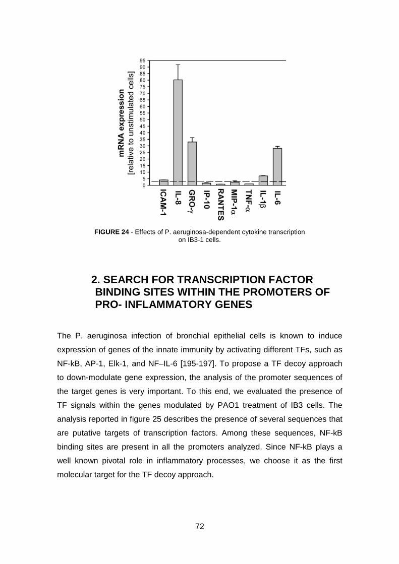
72
FIGURE 24 - Effects of P. aeruginosa-dependent cytokine transcription
on IB3-1 cells.
2. SEARCH FOR TRANSCRIPTION FACTOR BINDING SITES WITHIN THE PROMOTERS OF PRO- INFLAMMATORY GENES
The P. aeruginosa infection of bronchial epithelial cells is known to induce
expression of genes of the innate immunity by activating different TFs, such as
NF-kB, AP-1, Elk-1, and NF–IL-6 [195-197]. To propose a TF decoy approach
to down-modulate gene expression, the analysis of the promoter sequences of
the target genes is very important. To this end, we evaluated the presence of
TF signals within the genes modulated by PAO1 treatment of IB3 cells. The
analysis reported in figure 25 describes the presence of several sequences that
are putative targets of transcription factors. Among these sequences, NF-kB
binding sites are present in all the promoters analyzed. Since NF-kB plays a
well known pivotal role in inflammatory processes, we choose it as the first
molecular target for the TF decoy approach.
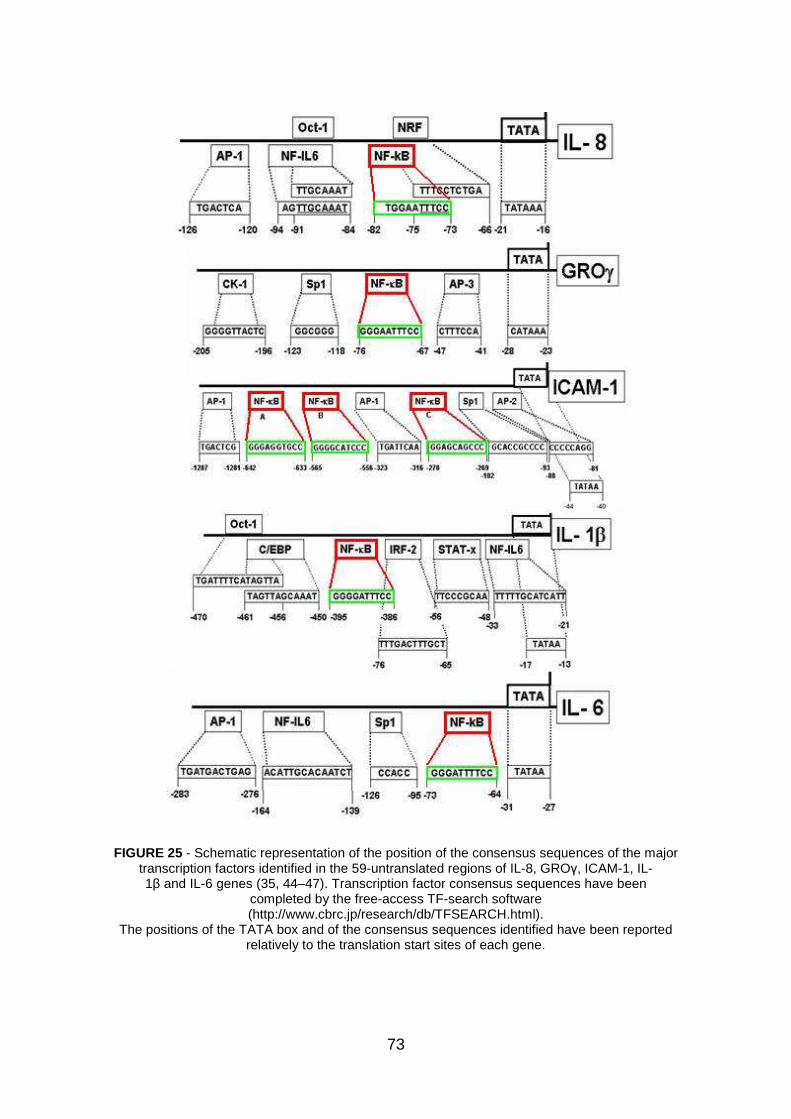
73
FIGURE 25 - Schematic representation of the position of the consensus sequences of the major transcription factors identified in the 59-untranslated regions of IL-8, GROγ, ICAM-1, IL- 1β and IL-6 genes (35, 44–47). Transcription factor consensus sequences have been
completed by the free-access TF-search software (http://www.cbrc.jp/research/db/TFSEARCH.html).
The positions of the TATA box and of the consensus sequences identified have been reported relatively to the translation start sites of each gene.
74
3. TRANSCRIPTION FACTOR DECOY
3.1 EFFECT OF NF-kB DECOY ODNs ON NUCLEAR EXTRACTS
Due to its role in recruiting PMNs in the bronchial wall and lumen of the
patients, down-modulation of IL-8 is considered a key target to reduce the
damage of the CF airway tract. Since consensus sequence for the transcription
factor NF-kB is contained in the promoter of IL-8 gene, the CF bronchial IB3-1
cells were transfected with TF decoy ODNs that have been previously designed
to mimic the DNA binding site of NF-kB and tested in their capacity to interfere
with the NF-kB dependent transcription [235]. A double-stranded ODN
homologous to the NF-kB binding site present in the LTR of the
immunodeficiency type I virus (HIV-1 LTR NF-kB decoy ODN) has been
previously demonstrated by our research group to efficiently interact with NF-kB
p52 transcription factor [252]. Moreover, a new decoy ODN homologous to the
NF-kB consensus sequence identified in the promoter of the IL-8 gene (IL-8 NF-
kB decoy ODN) was designed and prepared for this purpose. We exposed our
model system of cells to the P. aeruginosa laboratory strain PAO1 and studied
the activation of NF-kB by EMSA. As shown in figure 26-A, P. aeruginosa
increases NF-kB TF, as expected from previously reported data.
To test the ability of the decoy ODNs to compete for the binding of NF-kB TF
with the sequence contained in the promoter of IL-8, all the decoy ODNs were
incubated with nuclear extracts from IB3-1 cells in the presence of a
radiolabeled probe 100% homologous with the IL-8 NF-kB sequence, and
EMSA was performed. Complete inhibition of interaction of the 32P labeled IL-8
NF-kB probe with NF-kB proteins present in the nuclear extracts (NF-kB:DNA
complex) was obtained with very low concentrations of the IL-8 NF-kB cold
probe and both HIV-1 LTR 5 and 10 ng, as shown in figure 26-B. To further
validate the specificity of the competition, the same ODNs were used in the
binding experiments of the 32P-labeled IL-8 NF-kB probe with purified TF NF-kB
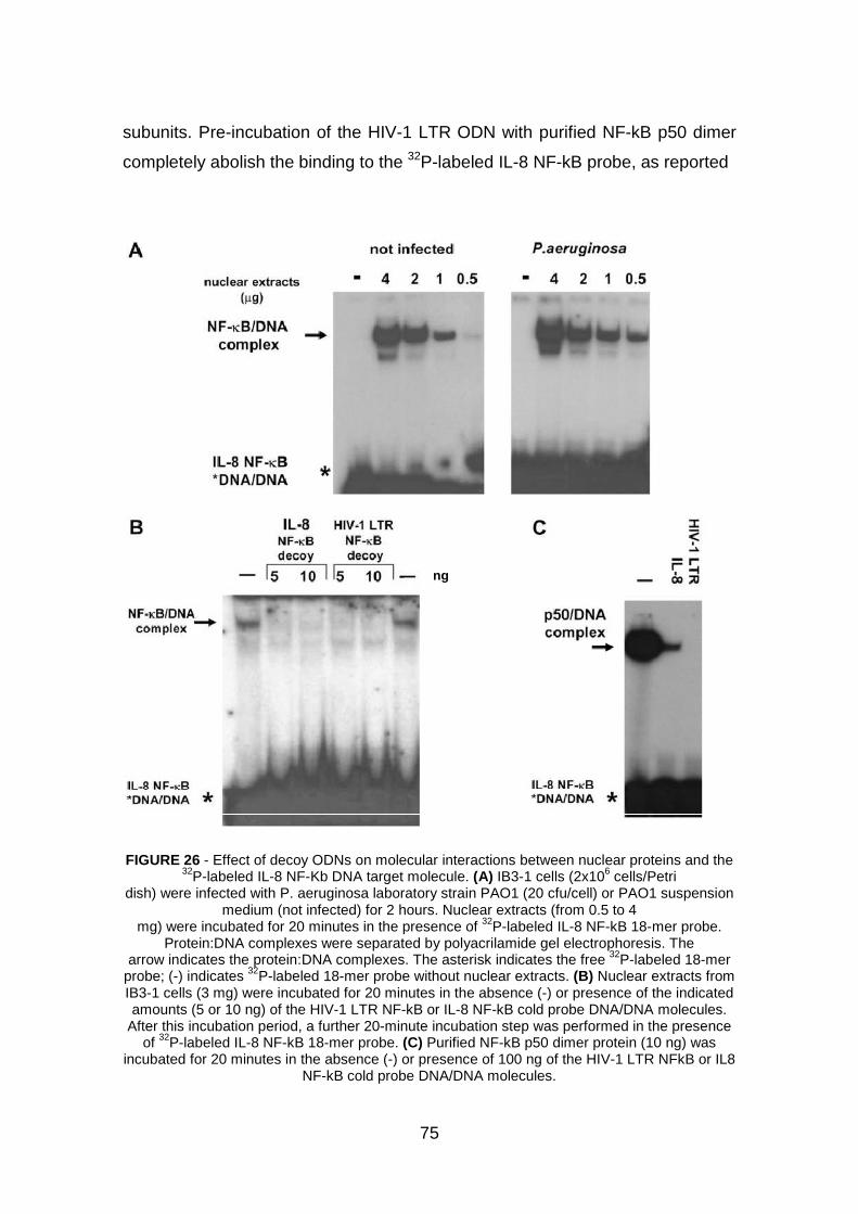
75
subunits. Pre-incubation of the HIV-1 LTR ODN with purified NF-kB p50 dimer
completely abolish the binding to the 32P-labeled IL-8 NF-kB probe, as reported
FIGURE 26 - Effect of decoy ODNs on molecular interactions between nuclear proteins and the 32P-labeled IL-8 NF-Kb DNA target molecule. (A) IB3-1 cells (2x10⁶ cells/Petri
dish) were infected with P. aeruginosa laboratory strain PAO1 (20 cfu/cell) or PAO1 suspension medium (not infected) for 2 hours. Nuclear extracts (from 0.5 to 4
mg) were incubated for 20 minutes in the presence of 32P-labeled IL-8 NF-kB 18-mer probe. Protein:DNA complexes were separated by polyacrilamide gel electrophoresis. The
arrow indicates the protein:DNA complexes. The asterisk indicates the free 32P-labeled 18-mer probe; (-) indicates 32P-labeled 18-mer probe without nuclear extracts. (B) Nuclear extracts from IB3-1 cells (3 mg) were incubated for 20 minutes in the absence (-) or presence of the indicated amounts (5 or 10 ng) of the HIV-1 LTR NF-kB or IL-8 NF-kB cold probe DNA/DNA molecules.
After this incubation period, a further 20-minute incubation step was performed in the presence of 32P-labeled IL-8 NF-kB 18-mer probe. (C) Purified NF-kB p50 dimer protein (10 ng) was
incubated for 20 minutes in the absence (-) or presence of 100 ng of the HIV-1 LTR NFkB or IL8 NF-kB cold probe DNA/DNA molecules.
ng
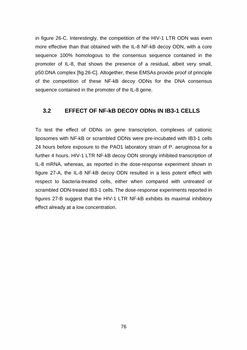
76
in figure 26-C. Interestingly, the competition of the HIV-1 LTR ODN was even
more effective than that obtained with the IL-8 NF-kB decoy ODN, with a core
sequence 100% homologous to the consensus sequence contained in the
promoter of IL-8, that shows the presence of a residual, albeit very small,
p50:DNA complex [fig.26-C]. Altogether, these EMSAs provide proof of principle
of the competition of these NF-kB decoy ODNs for the DNA consensus
sequence contained in the promoter of the IL-8 gene.
3.2 EFFECT OF NF-kB DECOY ODNs IN IB3-1 CELLS
To test the effect of ODNs on gene transcription, complexes of cationic
liposomes with NF-kB or scrambled ODNs were pre-incubated with IB3-1 cells
24 hours before exposure to the PAO1 laboratory strain of P. aeruginosa for a
further 4 hours. HIV-1 LTR NF-kB decoy ODN strongly inhibited transcription of
IL-8 mRNA, whereas, as reported in the dose-response experiment shown in
figure 27-A, the IL-8 NF-kB decoy ODN resulted in a less potent effect with
respect to bacteria-treated cells, either when compared with untreated or
scrambled ODN-treated IB3-1 cells. The dose-response experiments reported in
figures 27-B suggest that the HIV-1 LTR NF-kB exhibits its maximal inhibitory
effect already at a low concentration.
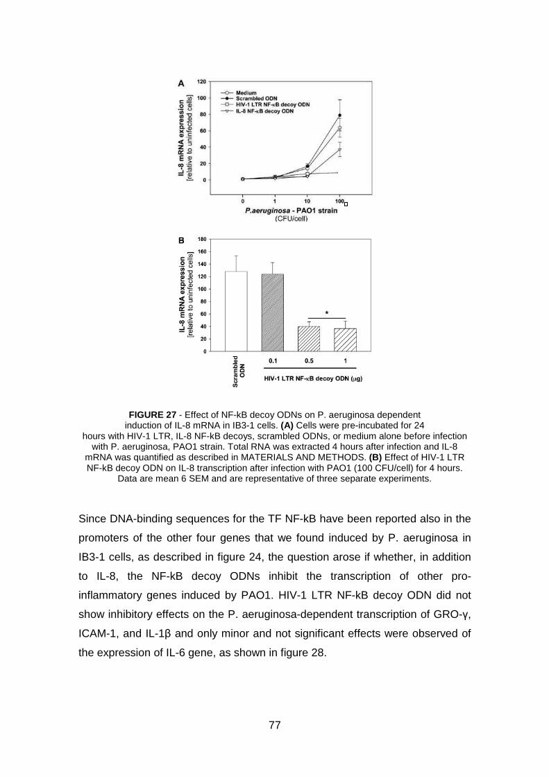
77
FIGURE 27 - Effect of NF-kB decoy ODNs on P. aeruginosa dependent induction of IL-8 mRNA in IB3-1 cells. (A) Cells were pre-incubated for 24
hours with HIV-1 LTR, IL-8 NF-kB decoys, scrambled ODNs, or medium alone before infection with P. aeruginosa, PAO1 strain. Total RNA was extracted 4 hours after infection and IL-8
mRNA was quantified as described in MATERIALS AND METHODS. (B) Effect of HIV-1 LTR NF-kB decoy ODN on IL-8 transcription after infection with PAO1 (100 CFU/cell) for 4 hours.
Data are mean 6 SEM and are representative of three separate experiments.
Since DNA-binding sequences for the TF NF-kB have been reported also in the
promoters of the other four genes that we found induced by P. aeruginosa in
IB3-1 cells, as described in figure 24, the question arose if whether, in addition
to IL-8, the NF-kB decoy ODNs inhibit the transcription of other pro-
inflammatory genes induced by PAO1. HIV-1 LTR NF-kB decoy ODN did not
show inhibitory effects on the P. aeruginosa-dependent transcription of GRO-γ,
ICAM-1, and IL-1β and only minor and not significant effects were observed of
the expression of IL-6 gene, as shown in figure 28.
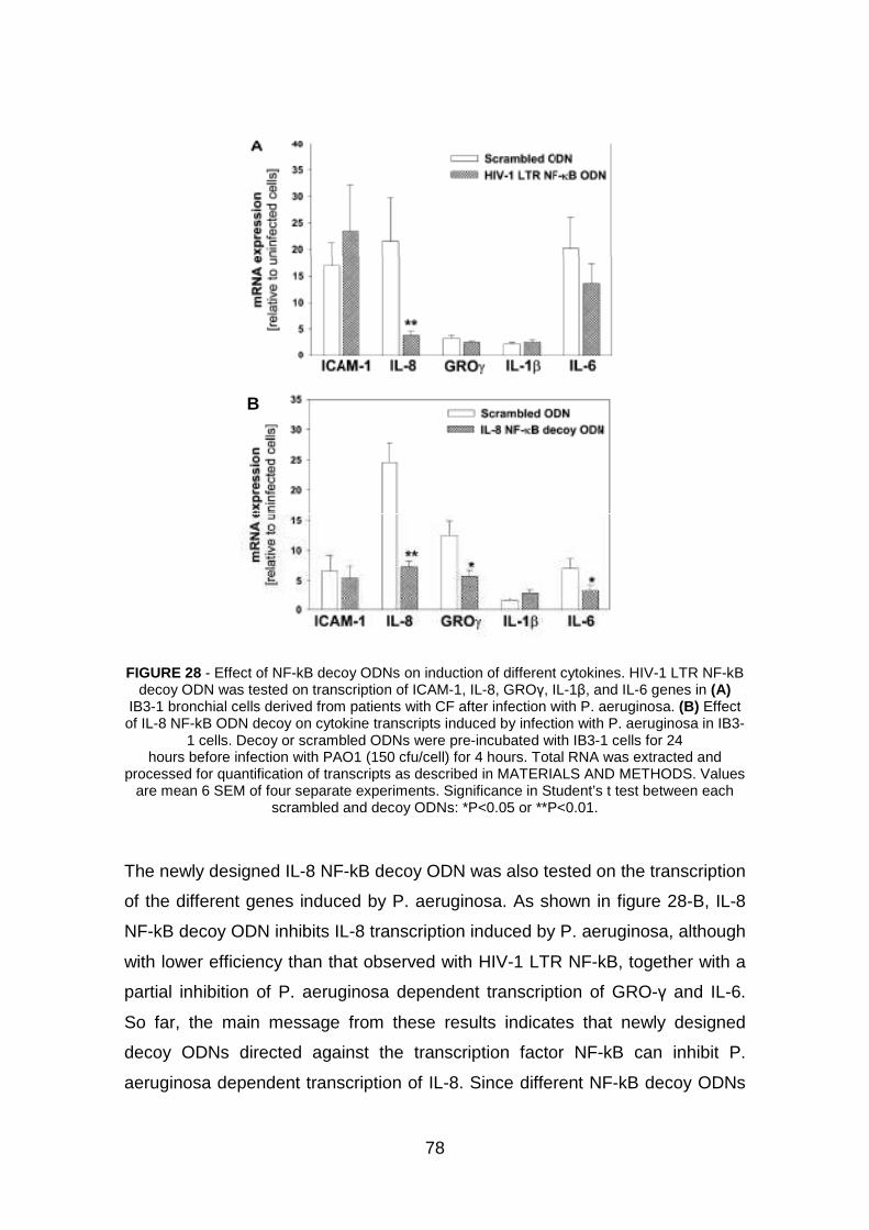
78
FIGURE 28 - Effect of NF-kB decoy ODNs on induction of different cytokines. HIV-1 LTR NF-kB decoy ODN was tested on transcription of ICAM-1, IL-8, GROγ, IL-1β, and IL-6 genes in (A)
IB3-1 bronchial cells derived from patients with CF after infection with P. aeruginosa. (B) Effect of IL-8 NF-kB ODN decoy on cytokine transcripts induced by infection with P. aeruginosa in IB3-
1 cells. Decoy or scrambled ODNs were pre-incubated with IB3-1 cells for 24 hours before infection with PAO1 (150 cfu/cell) for 4 hours. Total RNA was extracted and
processed for quantification of transcripts as described in MATERIALS AND METHODS. Values are mean 6 SEM of four separate experiments. Significance in Student’s t test between each
scrambled and decoy ODNs: *P<0.05 or **P<0.01.
The newly designed IL-8 NF-kB decoy ODN was also tested on the transcription
of the different genes induced by P. aeruginosa. As shown in figure 28-B, IL-8
NF-kB decoy ODN inhibits IL-8 transcription induced by P. aeruginosa, although
with lower efficiency than that observed with HIV-1 LTR NF-kB, together with a
partial inhibition of P. aeruginosa dependent transcription of GRO-γ and IL-6.
So far, the main message from these results indicates that newly designed
decoy ODNs directed against the transcription factor NF-kB can inhibit P.
aeruginosa dependent transcription of IL-8. Since different NF-kB decoy ODNs
B
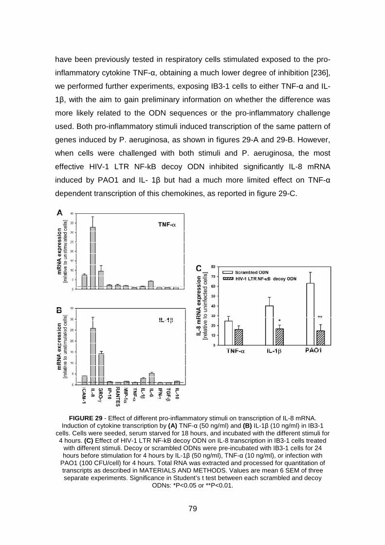
79
have been previously tested in respiratory cells stimulated exposed to the pro-
inflammatory cytokine TNF-α, obtaining a much lower degree of inhibition [236],
we performed further experiments, exposing IB3-1 cells to either TNF-α and IL-
1β, with the aim to gain preliminary information on whether the difference was
more likely related to the ODN sequences or the pro-inflammatory challenge
used. Both pro-inflammatory stimuli induced transcription of the same pattern of
genes induced by P. aeruginosa, as shown in figures 29-A and 29-B. However,
when cells were challenged with both stimuli and P. aeruginosa, the most
effective HIV-1 LTR NF-kB decoy ODN inhibited significantly IL-8 mRNA
induced by PAO1 and IL- 1β but had a much more limited effect on TNF-α
dependent transcription of this chemokines, as reported in figure 29-C.
FIGURE 29 - Effect of different pro-inflammatory stimuli on transcription of IL-8 mRNA. Induction of cytokine transcription by (A) TNF-α (50 ng/ml) and (B) IL-1β (10 ng/ml) in IB3-1
cells. Cells were seeded, serum starved for 18 hours, and incubated with the different stimuli for 4 hours. (C) Effect of HIV-1 LTR NF-kB decoy ODN on IL-8 transcription in IB3-1 cells treated
with different stimuli. Decoy or scrambled ODNs were pre-incubated with IB3-1 cells for 24 hours before stimulation for 4 hours by IL-1β (50 ng/ml), TNF-α (10 ng/ml), or infection with
PAO1 (100 CFU/cell) for 4 hours. Total RNA was extracted and processed for quantitation of transcripts as described in MATERIALS AND METHODS. Values are mean 6 SEM of three separate experiments. Significance in Student’s t test between each scrambled and decoy
ODNs: *P<0.05 or **P<0.01.
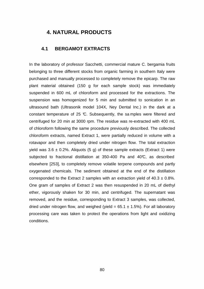
80
4. NATURAL PRODUCTS
4.1 BERGAMOT EXTRACTS
In the laboratory of professor Sacchetti, commercial mature C. bergamia fruits
belonging to three different stocks from organic farming in southern Italy were
purchased and manually processed to completely remove the epicarp. The raw
plant material obtained (150 g for each sample stock) was immediately
suspended in 600 mL of chloroform and processed for the extractions. The
suspension was homogenized for 5 min and submitted to sonication in an
ultrasound bath (Ultrasonik model 104X, Ney Dental Inc.) in the dark at a
constant temperature of 25 °C. Subsequently, the sa mples were filtered and
centrifuged for 20 min at 3000 rpm. The residue was re-extracted with 400 mL
of chloroform following the same procedure previously described. The collected
chloroform extracts, named Extract 1, were partially reduced in volume with a
rotavapor and then completely dried under nitrogen flow. The total extraction
yield was 3.6 ± 0.2%. Aliquots (5 g) of these sample extracts (Extract 1) were
subjected to fractional distillation at 350-400 Pa and 40°C, as described
elsewhere [253], to completely remove volatile terpene compounds and partly
oxygenated chemicals. The sediment obtained at the end of the distillation
corresponded to the Extract 2 samples with an extraction yield of 40.3 ± 0.8%.
One gram of samples of Extract 2 was then resuspended in 20 mL of diethyl
ether, vigorously shaken for 30 min, and centrifuged. The supernatant was
removed, and the residue, corresponding to Extract 3 samples, was collected,
dried under nitrogen flow, and weighed (yield = 65.1 ± 1.5%). For all laboratory
processing care was taken to protect the operations from light and oxidizing
conditions.
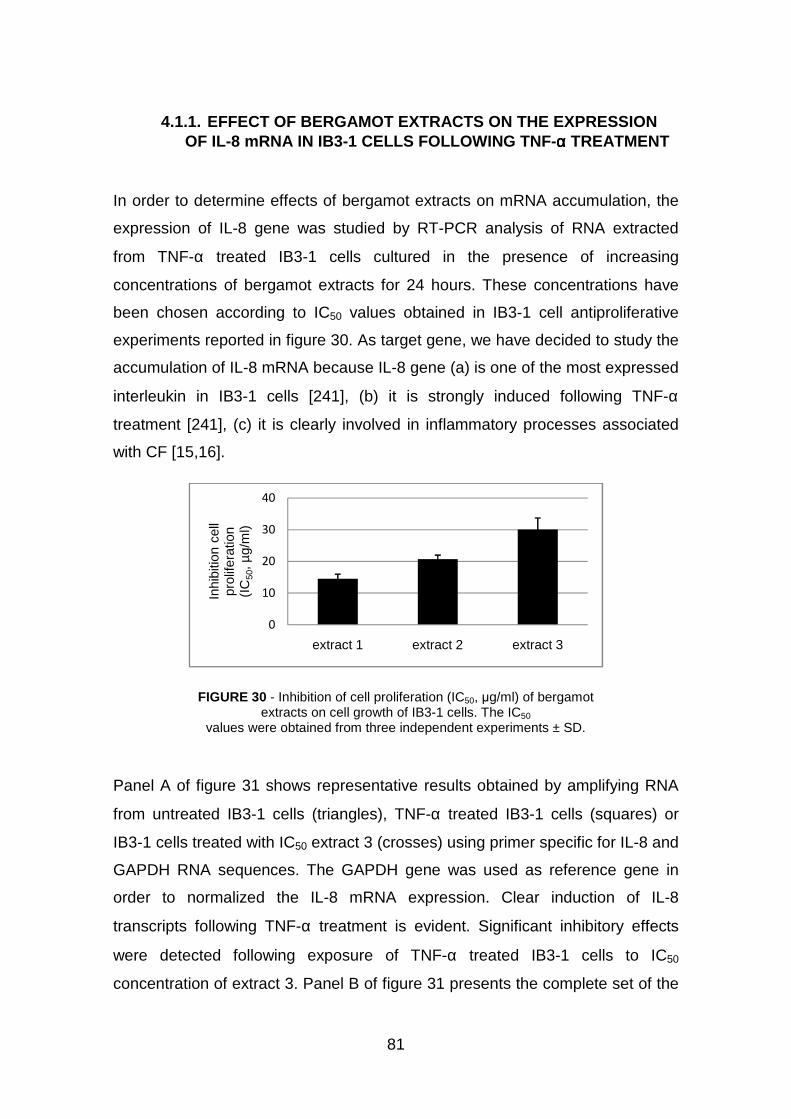
81
4.1.1. EFFECT OF BERGAMOT EXTRACTS ON THE EXPRESSION OF IL-8 mRNA IN IB3-1 CELLS FOLLOWING TNF-αααα TREATMENT
In order to determine effects of bergamot extracts on mRNA accumulation, the
expression of IL-8 gene was studied by RT-PCR analysis of RNA extracted
from TNF-α treated IB3-1 cells cultured in the presence of increasing
concentrations of bergamot extracts for 24 hours. These concentrations have
been chosen according to IC50 values obtained in IB3-1 cell antiproliferative
experiments reported in figure 30. As target gene, we have decided to study the
accumulation of IL-8 mRNA because IL-8 gene (a) is one of the most expressed
interleukin in IB3-1 cells [241], (b) it is strongly induced following TNF-α
treatment [241], (c) it is clearly involved in inflammatory processes associated
with CF [15,16].
FIGURE 30 - Inhibition of cell proliferation (IC50, µg/ml) of bergamot extracts on cell growth of IB3-1 cells. The IC50
values were obtained from three independent experiments ± SD.
Panel A of figure 31 shows representative results obtained by amplifying RNA
from untreated IB3-1 cells (triangles), TNF-α treated IB3-1 cells (squares) or
IB3-1 cells treated with IC50 extract 3 (crosses) using primer specific for IL-8 and
GAPDH RNA sequences. The GAPDH gene was used as reference gene in
order to normalized the IL-8 mRNA expression. Clear induction of IL-8
transcripts following TNF-α treatment is evident. Significant inhibitory effects
were detected following exposure of TNF-α treated IB3-1 cells to IC50
concentration of extract 3. Panel B of figure 31 presents the complete set of the
0
10
20
30
40
extract 1 extract 2 extract 3
Inhi
bitio
n ce
ll pr
olife
ratio
n (I
C50
, µg/
ml)

82
results obtained by the real-time quantitative RT-PCR data on the effects of
bergamot extracts on IL-8 mRNA accumulation with respect to GAPDH
transcripts. The first set of results consistently obtained is the strong increase
of IL-8 mRNA transcript in TNF-α treated IB3-1 cells (average, 34 fold, p <
0.01). In addition, the results obtained indicate that IL-8 mRNA expression is
much lower in TNF-α treated IB3-1 cells cultured in the presence of bergamot
extracts. More in detail, extract 1 and extract 3, even when used at IC25
concentrations, display high level of inhibition of TNF-α induced IL-8 mRNA
accumulation. IC50 concentrations are necessary to obtain similar inhibitory
effects using extract 2.
FIGURE 31 – Effects of bergamot extracts on IL-8 mRNA accumulation. IB3-1 were treated with 80 ng/ml TNF-α in the presence of increasing concentrations of bergamot extracts for 24 hours,
RNA was extracted and RT-PCR performed with primers specific for IL-8 and β-actin RNA sequences. (A) Representative quantitative RT-PCR analysis. Triangles: RNA from untreated IB3-1 cells; squares: TNF-α treated IB3-1 cells; crosses: IB3-1 cells treated with IC50 extract 3. (B) Effects of bergamot extracts on IL-8 mRNA accumulation with respect to β-actin transcripts.
(a): IC25 of extract; (b): IC50 of extract.
83
4.1.2. EFFECT OF BERGAMOT EXTRACTS ON THE EXPRESSION OF IL-8 GENES INDUCED IN IB3-1 CELLS FOLLOWING TNF-αααα TREATMENT: A BIO-PLEX ANALYSIS
In consideration of the inhibitory activity on IL-8 mRNA accumulation, Bio-plex
experiments were performed to verify the possible effects of bergamot extracts
on the release of this protein. IB3-1 cells were treated with 80 ng/ml of TNF-α
and increasing amounts (IC25-IC50-IC75) of bergamot extracts. After 24 hours,
recovered supernatants were analyzed using the Bio-Plex human cytokine IL-8
single-plex (Bio-Rad). When IB3-1 cells were induced with TNF-α and cultured
in the presence of bergamot extracts, some important differences in the release
of IL-8 occurred, as reported in figure 32. As it is evident the TNF-α induced
release of IL-8 (pg/ml) is strongly reduced using extract 1 and 3 at IC50
concentrations. The effect was found to be concentration-dependent. The
results obtained indicate a strong inhibition of release of IL-8 using extracts 3,
even when it was used at IC25 concentration, while extract 2 showed inhibitory
activity active only at IC75 concentration. These data suggest that bergamot
extracts might specifically inhibit the expression of the IL-8 pro-inflammatory
gene. These data are in agreement with the results obtained using the RT-PCR
approach [fig.31], and sustain the concept that bergamot extracts are potent
inhibitors of the TNF-α induced expression of the IL-8 gene in IB3-1 cells.
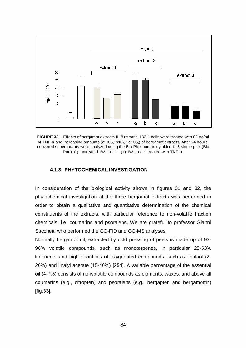
FIGURE 32 – Effects of bergamot extracts ILof TNF-α and increasing amounts (a: IC
recovered supernatants were analyzed using the BioRad). (-): untreated IB3
4.1.3. PHYTOCHEMICAL INVESTIGATION
In consideration of the biolog
phytochemical investigation of the three bergamot extra
order to obtain a qualitative and quantitative determination of the chemical
constituents of the extracts, with particular reference to non
chemicals, i.e. coumarins and psoralens.
Sacchetti who performed the
Normally bergamot oil, extracted by cold pressing of peels is made up of 93
96% volatile compounds, such as monoterpenes, in particular 25
limonene, and high quantities of oxygenated compounds, such as linalool (2
20%) and linalyl acetate (15
oil (4-7%) consists of nonvolatile compounds as pigments, waxes, and above all
coumarins (e.g., citropten) and psoralens (e.g., bergapten and bergamottin)
[fig.33].
84
Effects of bergamot extracts IL-8 release. IB3-1 cells were treated withand increasing amounts (a: IC25; b:IC50; c:IC75) of bergamot extracts. After 24 hours,
recovered supernatants were analyzed using the Bio-Plex human cytokine IL-8 single): untreated IB3-1 cells; (+):IB3-1 cells treated with TNF
PHYTOCHEMICAL INVESTIGATION
In consideration of the biological activity shown in figures 31
phytochemical investigation of the three bergamot extracts was performed in
obtain a qualitative and quantitative determination of the chemical
constituents of the extracts, with particular reference to non-volatile fraction
chemicals, i.e. coumarins and psoralens. We are grateful to professor Gianni
Sacchetti who performed the GC-FID and GC-MS analyses.
extracted by cold pressing of peels is made up of 93
96% volatile compounds, such as monoterpenes, in particular 25
limonene, and high quantities of oxygenated compounds, such as linalool (2
) and linalyl acetate (15-40%) [254]. A variable percentage of the essential
7%) consists of nonvolatile compounds as pigments, waxes, and above all
coumarins (e.g., citropten) and psoralens (e.g., bergapten and bergamottin)
1 cells were treated with 80 ng/ml ) of bergamot extracts. After 24 hours,
8 single-plex (Bio-with TNF-α.
1 and 32, the
cts was performed in
obtain a qualitative and quantitative determination of the chemical
volatile fraction
We are grateful to professor Gianni
extracted by cold pressing of peels is made up of 93-
96% volatile compounds, such as monoterpenes, in particular 25-53%
limonene, and high quantities of oxygenated compounds, such as linalool (2-
. A variable percentage of the essential
7%) consists of nonvolatile compounds as pigments, waxes, and above all
coumarins (e.g., citropten) and psoralens (e.g., bergapten and bergamottin)
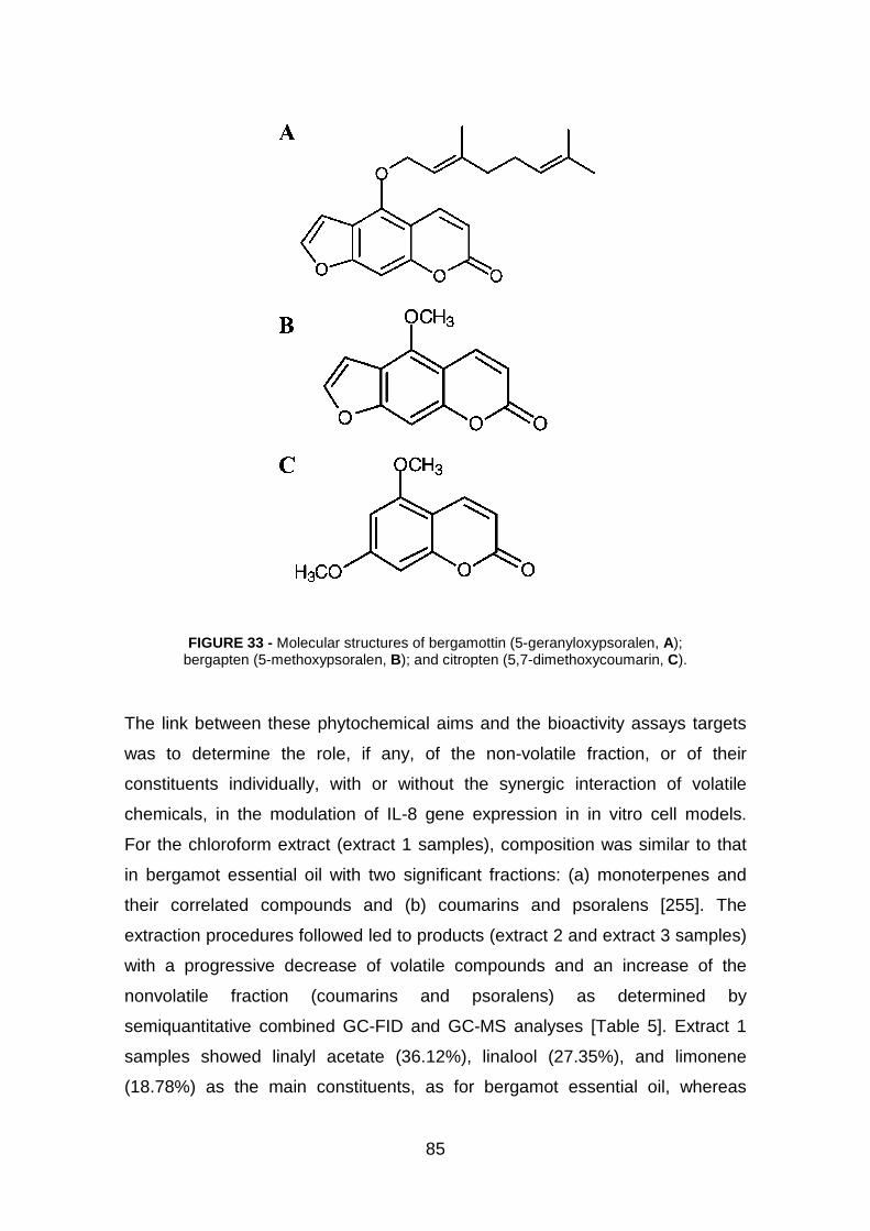
85
FIGURE 33 - Molecular structures of bergamottin (5-geranyloxypsoralen, A); bergapten (5-methoxypsoralen, B); and citropten (5,7-dimethoxycoumarin, C).
The link between these phytochemical aims and the bioactivity assays targets
was to determine the role, if any, of the non-volatile fraction, or of their
constituents individually, with or without the synergic interaction of volatile
chemicals, in the modulation of IL-8 gene expression in in vitro cell models.
For the chloroform extract (extract 1 samples), composition was similar to that
in bergamot essential oil with two significant fractions: (a) monoterpenes and
their correlated compounds and (b) coumarins and psoralens [255]. The
extraction procedures followed led to products (extract 2 and extract 3 samples)
with a progressive decrease of volatile compounds and an increase of the
nonvolatile fraction (coumarins and psoralens) as determined by
semiquantitative combined GC-FID and GC-MS analyses [Table 5]. Extract 1
samples showed linalyl acetate (36.12%), linalool (27.35%), and limonene
(18.78%) as the main constituents, as for bergamot essential oil, whereas

86
coumarins and psoralens made up 4.64% [Table 1]. Extract 2 samples showed
an important reduction of linalyl acetate (27.45%) and a dramatic decrease of
other terpene compounds. Many minority chemicals were not detectable,
whereas others, such as limonene, showed a reduction of 93.2%. Extract 3
samples gave an even more noticeable reduction of terpene compounds, with
linalyl acetate (0.43%), γ-terpinene (0.17%), and β-myrcene (0.53%) as the only
volatile fraction chemicals, against an increase of coumarins and psoralens of
45.5 and 95.2% with respect to extract 2 and 3 samples, respectively [table 5].
TABLE 5 - Composition of Bergamot Extracts Determined by GC-MS and GC-FID
HPLC analyses to detect and quantify coumarins and psoralens reputed to be
representative of the nonvolatile fraction and of the bioactivity of bergamot
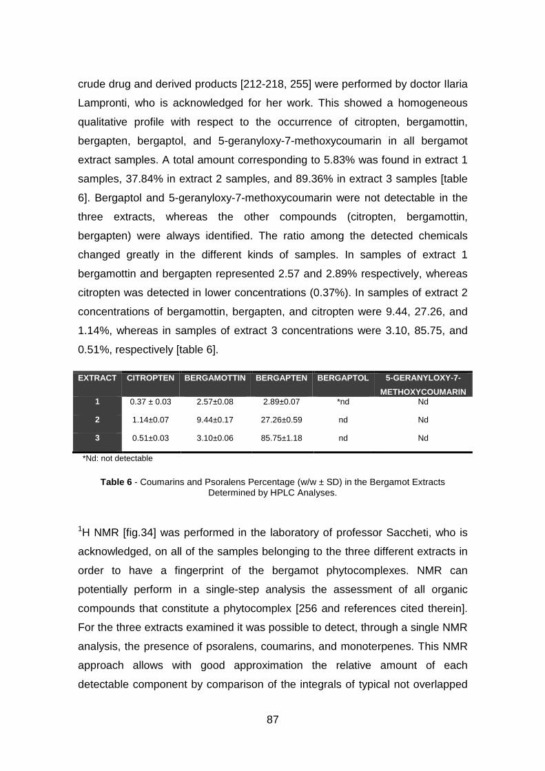
87
crude drug and derived products [212-218, 255] were performed by doctor Ilaria
Lampronti, who is acknowledged for her work. This showed a homogeneous
qualitative profile with respect to the occurrence of citropten, bergamottin,
bergapten, bergaptol, and 5-geranyloxy-7-methoxycoumarin in all bergamot
extract samples. A total amount corresponding to 5.83% was found in extract 1
samples, 37.84% in extract 2 samples, and 89.36% in extract 3 samples [table
6]. Bergaptol and 5-geranyloxy-7-methoxycoumarin were not detectable in the
three extracts, whereas the other compounds (citropten, bergamottin,
bergapten) were always identified. The ratio among the detected chemicals
changed greatly in the different kinds of samples. In samples of extract 1
bergamottin and bergapten represented 2.57 and 2.89% respectively, whereas
citropten was detected in lower concentrations (0.37%). In samples of extract 2
concentrations of bergamottin, bergapten, and citropten were 9.44, 27.26, and
1.14%, whereas in samples of extract 3 concentrations were 3.10, 85.75, and
0.51%, respectively [table 6].
EXTRACT CITROPTEN BERGAMOTTIN BERGAPTEN BERGAPTOL 5-GERANYLOXY-7-
METHOXYCOUMARIN 1 0.37 ± 0.03 2.57±0.08 2.89±0.07 *nd Nd
2 1.14±0.07 9.44±0.17 27.26±0.59 nd Nd
3 0.51±0.03 3.10±0.06 85.75±1.18 nd Nd
*Nd: not detectable
Table 6 - Coumarins and Psoralens Percentage (w/w ± SD) in the Bergamot Extracts Determined by HPLC Analyses.
1H NMR [fig.34] was performed in the laboratory of professor Saccheti, who is
acknowledged, on all of the samples belonging to the three different extracts in
order to have a fingerprint of the bergamot phytocomplexes. NMR can
potentially perform in a single-step analysis the assessment of all organic
compounds that constitute a phytocomplex [256 and references cited therein].
For the three extracts examined it was possible to detect, through a single NMR
analysis, the presence of psoralens, coumarins, and monoterpenes. This NMR
approach allows with good approximation the relative amount of each
detectable component by comparison of the integrals of typical not overlapped

signals assigned to these chemicals to be obtained
case we considered two allyl protons
proton on C2 on linalool (5.89 ppm) and linalyl acetate (5.97 ppm), doublet of
proton onC3 on citropten (7.95 ppm), and doublet of proton near the oxygen on
the furanic ring of bergapten (7.01 ppm) and bergamottin
FIGURE 34 - 1H NMR spectra of samples belonging to Extract 1 (a),
In consideration of the fact that extract 3 was the most active on inhibition of
TNF-α induced IL-8 mRNA accumulation and IL
cells, we hypothesized that the coumarin/psoralen fractions of the extracts were
among those responsible for the biological activity. Therefore, the activity
citropten, bergapen and bergamottin was further analyzed.
88
signals assigned to these chemicals to be obtained [256, 257]. In this specific
case we considered two allyl protons on limonene (4.72 ppm), double doublet of
proton on C2 on linalool (5.89 ppm) and linalyl acetate (5.97 ppm), doublet of
proton onC3 on citropten (7.95 ppm), and doublet of proton near the oxygen on
the furanic ring of bergapten (7.01 ppm) and bergamottin (6.93 ppm).
H NMR spectra of samples belonging to Extract 1 (a),Extract 2 (b), and Extract 3 (c).
In consideration of the fact that extract 3 was the most active on inhibition of
8 mRNA accumulation and IL-8 secretion by
cells, we hypothesized that the coumarin/psoralen fractions of the extracts were
among those responsible for the biological activity. Therefore, the activity
citropten, bergapen and bergamottin was further analyzed.
. In this specific
on limonene (4.72 ppm), double doublet of
proton on C2 on linalool (5.89 ppm) and linalyl acetate (5.97 ppm), doublet of
proton onC3 on citropten (7.95 ppm), and doublet of proton near the oxygen on
(6.93 ppm).
H NMR spectra of samples belonging to Extract 1 (a),
In consideration of the fact that extract 3 was the most active on inhibition of
8 secretion by treated IB3-1
cells, we hypothesized that the coumarin/psoralen fractions of the extracts were
among those responsible for the biological activity. Therefore, the activity of
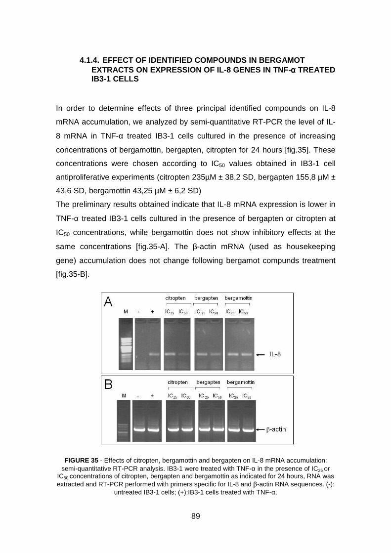
89
4.1.4. EFFECT OF IDENTIFIED COMPOUNDS IN BERGAMOT EXTRACTS ON EXPRESSION OF IL-8 GENES IN TNF-αααα TREATED IB3-1 CELLS
In order to determine effects of three principal identified compounds on IL-8
mRNA accumulation, we analyzed by semi-quantitative RT-PCR the level of IL-
8 mRNA in TNF-α treated IB3-1 cells cultured in the presence of increasing
concentrations of bergamottin, bergapten, citropten for 24 hours [fig.35]. These
concentrations were chosen according to IC50 values obtained in IB3-1 cell
antiproliferative experiments (citropten 235µM ± 38,2 SD, bergapten 155,8 µM ±
43,6 SD, bergamottin 43,25 µM ± 6,2 SD)
The preliminary results obtained indicate that IL-8 mRNA expression is lower in
TNF-α treated IB3-1 cells cultured in the presence of bergapten or citropten at
IC50 concentrations, while bergamottin does not show inhibitory effects at the
same concentrations [fig.35-A]. The β-actin mRNA (used as housekeeping
gene) accumulation does not change following bergamot compunds treatment
[fig.35-B].
FIGURE 35 - Effects of citropten, bergamottin and bergapten on IL-8 mRNA accumulation: semi-quantitative RT-PCR analysis. IB3-1 were treated with TNF-α in the presence of IC25 or
IC50 concentrations of citropten, bergapten and bergamottin as indicated for 24 hours, RNA was extracted and RT-PCR performed with primers specific for IL-8 and β-actin RNA sequences. (-):
untreated IB3-1 cells; (+):IB3-1 cells treated with TNF-α.

90
On the basis of this first set of results, showing that bergapten and citropten
exhibit an interesting inhibitory activity on mRNA accumulation, to better
quantify inhibitory effects of these two identified compounds, we performed
real-time quantitative RT-PCR analysis on RNA extracted from IB3-1 cells
induced with TNF-α and treated with IC25 and IC50 concentrations of citropten or
bergapten, comparing IL-8 mRNA accumulation with that of the internal controls
β-actin mRNA. Panel A of figure 36 shows the representative results obtained
by amplifying RNA from untreated (triangles), TNF-α (squares), citropten
(diamonds, IC50) and bergapten (crosses, IC50) treated IB3-1 cells using primer
specific for IL-8 RNA. Clear induction of IL-8 transcripts, following TNF-α
treatment is evident, but this increase is reduced after treatment with citropten
or bergapten. Particularly citropten is more efficient than bergapten to decrease
IL-8 mRNA accumulation. This result is more evident in panel B of figure 36,
showing a summary of the data obtained by real-time quantitative RT-PCR were
performed to investigate effects of bergapten and citropten at two different
concentrations (IC25 and IC50) on IL-8 mRNA accumulation. Significant inhibitory
effects on IL-8 mRNA expression normalized to β-actin transcripts were
detected following exposure of TNF-α induced IB3-1 cells to citropten and
bergapten at IC25 concentrations yet. Moreover, citropten was found to be more
active than bergapten at the same concentrations. These data are in agreement
with data obtained by semi-quantitative RT-PCR [fig.35], and confirm the potent
inhibitory activity of citropten and bergapten on expression of the IL-8 gene in
IB3-1 cells.
The results shown in figure 36-B have been also confirmed using Bioplex
technology to analyze the release of IL-8 protein (pg/ml) in supernatants
recovered from TNF-α induced IB3-1 cells treated with citropten and bergapten
at IC50 and IC25 concentrations. These supernatants were collected before to
perform the RNA extraction for real time PCR analysis. The results, shown in
figure 36-C, confirmed the inhibitory activity of citropten and bergapten
(citropten>bergapten) on expression of IL-8 protein in IB3-1 cell model.
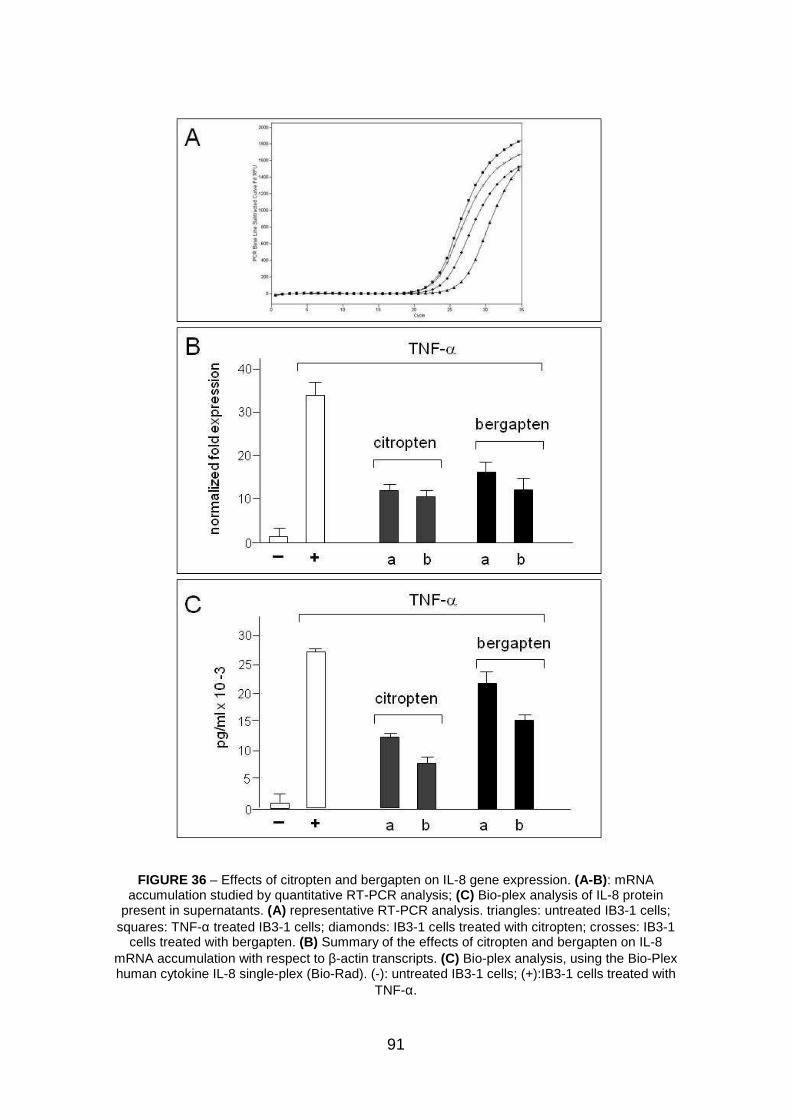
91
FIGURE 36 – Effects of citropten and bergapten on IL-8 gene expression. (A-B): mRNA accumulation studied by quantitative RT-PCR analysis; (C) Bio-plex analysis of IL-8 protein
present in supernatants. (A) representative RT-PCR analysis. triangles: untreated IB3-1 cells; squares: TNF-α treated IB3-1 cells; diamonds: IB3-1 cells treated with citropten; crosses: IB3-1
cells treated with bergapten. (B) Summary of the effects of citropten and bergapten on IL-8 mRNA accumulation with respect to β-actin transcripts. (C) Bio-plex analysis, using the Bio-Plex human cytokine IL-8 single-plex (Bio-Rad). (-): untreated IB3-1 cells; (+):IB3-1 cells treated with
TNF-α.

92
4.2 EMBLICA OFFICINALIS
The dried fruits of Emblica officinalis were extracted with absolute ethanol and
the yield was 9.33%. This ethanolic extract of Emblica officinalis was defatted
with petroleum ether and the defatted extract was successively fractionated with
different solvent systems on the polarity basis. The solvents were 100%
dichloromethane, 25% ethylacetate-dichloromethane, 50%
ethylacetatedichloromethane, 75% ethylacetate-dichloromethane, 100%
ethylacetate, butanol and acetone and the remaining aqueous portions were
separated.
4.2.1 GAS CHROMATOGRAPHY-MASS SPECTROMETRY ANALYSIS OF THE FRACTIONS OF EMBLICA OFFICINALIS EXTRACTS
In order to identify the putative antiproliferative compound present within
unfractionated extracts and n-butanol fraction from Emblica officinalis, gas
chromatography-mass spectrometry (GC-MS) analysis was performed by
doctor Ilaria Lampronti, who is acknowledged. Gas chromatography coupled to
mass spectrometry allows purification and identification of single components
from biological extracts. Emblica officinalis extracts and the respective n-butanol
fraction samples were diluted to a final volume of 1 ml with methanol
(approximately 1 mg/ml). One µl of each solution was injected into the gas
chromatograph with an appropriate microsyringe, chromatographed using a
fused-silica capillary column and analyzed with a quadrupole mass
spectrometric detector. The resulting chromatograms are shown in figure 37-A
and C, and demonstrate that in Emblica officinalis extracts 3 peaks with
different retention times (r.t., 13.4, 20.9, 22.3 min) are present. The mass
spectrometric analysis [fig.37-B, 37-D, and data not shown] demonstrated that
these 3 peaks correspond to 3 derivatives: a) pyrogallol (or 1,2,3-benzenetriol);
b) tetradecahydro-1,4-dimethyl-7- (1- methylethyl)-1-phenantrene

FIGURE 37 - (A, C, E), Ion chromatograms of unfractibutanol fraction of
(B, D, F), Mass spectrum of peak 1 of unfractionated n-butanol fraction of
93
Ion chromatograms of unfractionated Emblica officinalis butanol fraction of Emblica officinalis extracts (C), pyrogallol (E)
, Mass spectrum of peak 1 of unfractionated Emblica officinalis extracts butanol fraction of Emblica officinalis extracts (D) and
pyrogallol (F).
Emblica officinalis extracts (A), n-(E).
extracts (B), peak 1 of
94
methyl ester; and c) decahydro-4H-cyclopentacycloocten-4-one respectively,
identified by using the NIST library (Mass Spectrometry Data Handling System,
version 1.12, Fisons, Thermo Finnigan, San Jose, CA). In the chromatogram of
the n-butanol fraction of Emblica officinalis extracts [fig.37-C] we can observe
only one major peak at 13.3 min (r.t.) corresponding to pyrogallol [fig.37-D], also
present in Emblica officinalis. The retention time (r.t.) and the fragmentation
pathways are the same as shown in figure 37-A and B. Moreover, we analyzed
commercial pyrogallol for comparison with the two peaks found in Emblica
officinalis extracts and the respective n-butanol fraction samples. The r.t. and
the fragmentation pathways corresponded perfectly [fig. 37-E and 37-F].
Accordingly, we hypothesized that the antiproliferative activity of Emblica
officinalis extracts was due to pyrogallol. M/z values: a) pyrogallol (Emblica
officinalis extracts): 80.0, 96.8, 107.9, 125.8, 126.9; pyrogallol (Emblica
officinalis, n-butanol fraction): 80.0, 96.8, 107.9, 125.8, 126.9; pyrogallol
(Aldrich): 79.9, 96.9, 107.9, 125.9, 127.0; b) 81.0, 99.0, 121.0, 123.0, 150.0,
164.0, 186.9, 207.0, 279.2, 306.1, 324.2; c) 95.0, 96.0, 98.0, 123.0, 124.0,
130.8, 166.0, 195.0, 212.1, 241.1, 279.1.
4.2.2 EFFECTS OF EMBLICA OFFICINALIS EXTRACTS ON IN VITRO PROLIFERATION OF IB3-1 CELLS
In order to obtain preliminary information on the biological properties of the
employed extracts, their effects on cell growth were examined. The extracts
isolated from Emblica officinalis were first tested on IB3-1 cells. Figure 38
summarizes the data derived from three independent experiments. IB3-3 cells
were seeded at the initial concentration of 30.000 cells/cm2 and then cultured in
the absence or in the presence of 0.05-500 µg/ml of Emblica officinalis extracts
or purchased pyrogallol. For the IC50 (concentrations of extracts leading to 50%
inhibition of IB3-1 cell growth), we determined the values of cell number/ml after
3 days of culture, when untreated cells are in the log phase of cell growth.

95
FIGURE 38 - Inhibition of cell proliferation (IC50, µg/mL or µM) of Emblica officinalis extracts and identified compound pyrogallol on cell growth of IB3-1 cells. The IC50
values were obtained from three independent experiments ± SD.
4.2.3 EFFECT OF EMBLICA OFFICINALIS EXTRACTS ON THE EXPRESSION OF PRO-INFLAMMATORY GENES INDUCED IN IB3-1 CELLS BY TNF-αααα TREATMENT
To verify the potential anti-inflammatory activity of Emblica officinalis extracts
and its main constituent pyrogallol, Bio-plex experiments were performed
evaluating the release of pro-inflammatory cytokines and chemokines on
supernatant of IB3-1 cell model. Accordingly with the results of proliferation
activity assays [fig.38], IB3-1 cells were treated with increasing amounts (IC12,5-
IC50) of Emblica officinalis extracts or (IC25-IC50) pyrogallol and with or without
80 ng/ml of TNF-α. After 24 hours, recovered supernatants were analyzed using
the Bio-Plex human cytokine 7-plex (Bio-Rad). The results shown in table 7
demonstrated that since the amount of 50 µg/ml Emblica officinalis is able to
reduce the release of VEGF (Vascular Endothelial Growth Factor) and IL-8
proteins, by 20 and 37% respectively. Less relevant is the activity of Emblica
officinalis on down regulation of IL-12, G-CSF, MCP-1, RANTES e IL-10 gene
expression. Moreover, pyrogallol seems to down regulate gene transcription of
G-CSF, RANTES, VEGF, by 44, 73 and 63% respectively, and especially of IL8
by 51%.
0
20
40
60
80
100
120
140
160
emblica officinalis pyrogallol
Inhi
bitio
n ce
ll pr
olife
ratio
n (I
C50
, µg/
ml,
µM
)
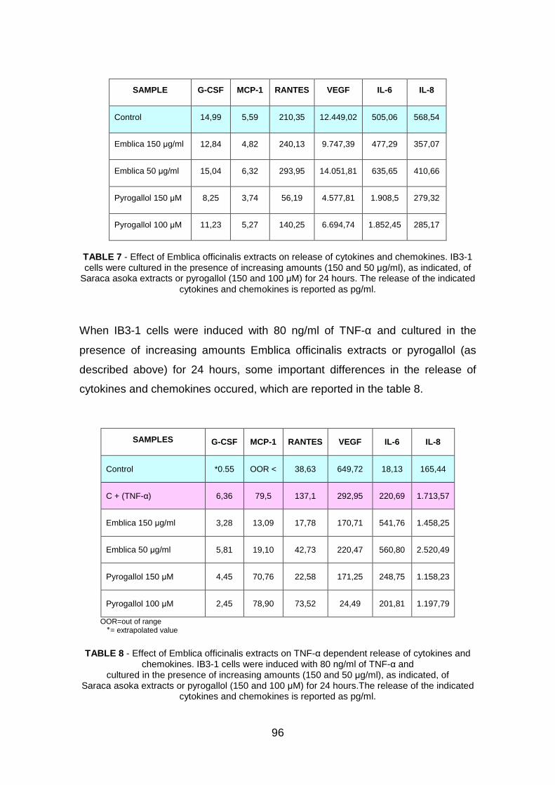
96
SAMPLE G-CSF MCP-1 RANTES VEGF IL-6 IL-8
Control 14,99 5,59 210,35 12.449,02 505,06 568,54
Emblica 150 µg/ml 12,84 4,82 240,13 9.747,39 477,29 357,07
Emblica 50 µg/ml 15,04 6,32 293,95 14.051,81 635,65 410,66
Pyrogallol 150 µM 8,25 3,74 56,19 4.577,81 1.908,5 279,32
Pyrogallol 100 µM 11,23 5,27 140,25 6.694,74 1.852,45 285,17
TABLE 7 - Effect of Emblica officinalis extracts on release of cytokines and chemokines. IB3-1 cells were cultured in the presence of increasing amounts (150 and 50 µg/ml), as indicated, of
Saraca asoka extracts or pyrogallol (150 and 100 µM) for 24 hours. The release of the indicated cytokines and chemokines is reported as pg/ml.
When IB3-1 cells were induced with 80 ng/ml of TNF-α and cultured in the
presence of increasing amounts Emblica officinalis extracts or pyrogallol (as
described above) for 24 hours, some important differences in the release of
cytokines and chemokines occured, which are reported in the table 8.
SAMPLES G-CSF MCP-1 RANTES VEGF IL-6 IL-8
Control *0.55 OOR < 38,63 649,72 18,13 165,44
C + (TNF-α) 6,36 79,5 137,1 292,95 220,69 1.713,57
Emblica 150 µg/ml 3,28 13,09 17,78 170,71 541,76 1.458,25
Emblica 50 µg/ml 5,81 19,10 42,73 220,47 560,80 2.520,49
Pyrogallol 150 µM 4,45 70,76 22,58 171,25 248,75 1.158,23
Pyrogallol 100 µM 2,45 78,90 73,52 24,49 201,81 1.197,79
OOR=out of range extrapolated value = ٭
TABLE 8 - Effect of Emblica officinalis extracts on TNF-α dependent release of cytokines and chemokines. IB3-1 cells were induced with 80 ng/ml of TNF-α and
cultured in the presence of increasing amounts (150 and 50 µg/ml), as indicated, of Saraca asoka extracts or pyrogallol (150 and 100 µM) for 24 hours.The release of the indicated
cytokines and chemokines is reported as pg/ml.
97
The positive control of the experiment is represented by IB3-1 cells induced with
TNF-α without Emblica officinalis extracts or components treatment. As it is
evident the TNF-α induced release of the cytokines and chemokines of the
panel. These data suggest that Emblica officinalis extracts inhibit especially the
expression of MCP-1 (-83%), RANTES (-87%), VEGF (-41%), IL-8 (-14%), and
the pyrogallol the expression of RANTES genes.
In consideration of the inhibitory activity on IL-8 release, RT-PCR experiments
were performed to verify the possible effects of Emblica officinalis extracts on
the expression of this gene.
4.2.4 EFFECT OF EMBLICA OFFICINALIS EXTRACTS AND IDENTIFIED COMPOUND PYROGALLOL ON EXPRESSION OF IL-8 IN TNF-αααα TREATED IB3-1 CELLS
In order to determine the effects of Emblica officnalis extracts and pyrogallol on
mRNA accumulation, the expression of IL-8 gene was analyzed by RT-PCR
analysis of RNA extracted from TNF-α treated IB3-1 cells cultured in the
presence of increasing concentration of Emblica officinalis extracts for 24 hours
[fig.39]. These concentrations were chosen according to IC50 values obtained in
IB3-1 cell antiproliferative experiments reported in figure 38.
The preliminary results obtained indicate that IL-8 mRNA expression is lower in
TNF-α treated IB3-1 cells cultured in the presence of Emblica offcinalis at IC25
(100µg/ml) concentrations, while pyrogallol does not show inhibitory effects at
the higher concentrations IC75 (150µM) [fig.39-A]. The β-actin mRNA (used as
housekeeping gene) accumulation does not change following Emblica officinalis
and pyrogallol treatment [fig.39-B].
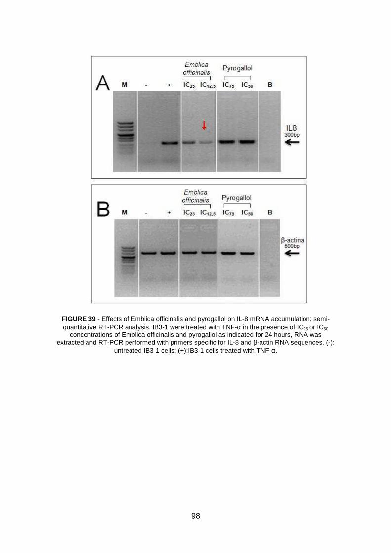
98
FIGURE 39 - Effects of Emblica officinalis and pyrogallol on IL-8 mRNA accumulation: semi-quantitative RT-PCR analysis. IB3-1 were treated with TNF-α in the presence of IC25 or IC50
concentrations of Emblica officinalis and pyrogallol as indicated for 24 hours, RNA was extracted and RT-PCR performed with primers specific for IL-8 and β-actin RNA sequences. (-):
untreated IB3-1 cells; (+):IB3-1 cells treated with TNF-α.
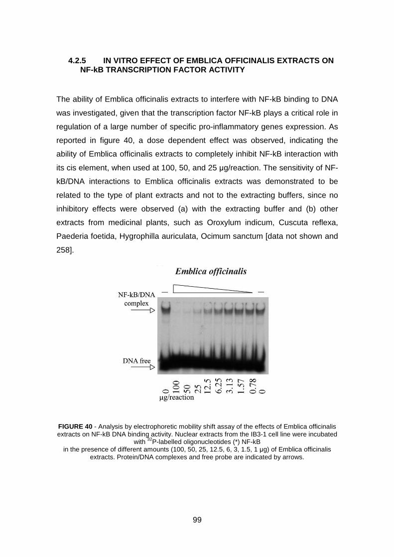
99
4.2.5 IN VITRO EFFECT OF EMBLICA OFFICINALIS EXTRACTS ON NF-kB TRANSCRIPTION FACTOR ACTIVITY
The ability of Emblica officinalis extracts to interfere with NF-kB binding to DNA
was investigated, given that the transcription factor NF-kB plays a critical role in
regulation of a large number of specific pro-inflammatory genes expression. As
reported in figure 40, a dose dependent effect was observed, indicating the
ability of Emblica officinalis extracts to completely inhibit NF-kB interaction with
its cis element, when used at 100, 50, and 25 µg/reaction. The sensitivity of NF-
kB/DNA interactions to Emblica officinalis extracts was demonstrated to be
related to the type of plant extracts and not to the extracting buffers, since no
inhibitory effects were observed (a) with the extracting buffer and (b) other
extracts from medicinal plants, such as Oroxylum indicum, Cuscuta reflexa,
Paederia foetida, Hygrophilla auriculata, Ocimum sanctum [data not shown and
258].
FIGURE 40 - Analysis by electrophoretic mobility shift assay of the effects of Emblica officinalis extracts on NF-kB DNA binding activity. Nuclear extracts from the IB3-1 cell line were incubated
with 32P-labelled oligonucleotides (*) NF-kB in the presence of different amounts (100, 50, 25, 12.5, 6, 3, 1.5, 1 µg) of Emblica officinalis
extracts. Protein/DNA complexes and free probe are indicated by arrows.
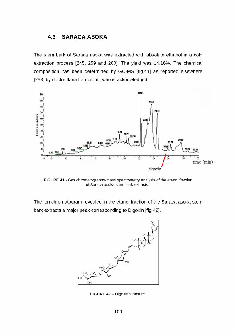
4.3 SARACA
The stem bark of Saraca asoka
extraction process [245, 259 and 260
composition has been determined by GC
[258] by doctor Ilaria Lampronti,
FIGURE 41 - Gas chromatography
The ion chromatogram revealed in the etanol fraction of the
bark extracts a major peak corresponding to Digoxin
100
SARACA ASOKA
Saraca asoka was extracted with absolute ethanol in a cold
245, 259 and 260]. The yield was 14.16%. The chemical
been determined by GC-MS [fig.41] as reported elsewhere
by doctor Ilaria Lampronti, who is acknowledged.
Gas chromatography-mass spectrometry analysis of the etanol fraction
of Saraca asoka stem bark extracts.
The ion chromatogram revealed in the etanol fraction of the Saraca asoka
bark extracts a major peak corresponding to Digoxin [fig.42].
FIGURE 42 – Digoxin structure.
digoxin
was extracted with absolute ethanol in a cold
]. The yield was 14.16%. The chemical
reported elsewhere
mass spectrometry analysis of the etanol fraction
Saraca asoka stem
101
4.3.1. INDUCTION OF CYTOKINE AND CHEMOKINE RELEASE IN IB3-1 CELLS TREATED WITH TNF-α
In order to determine the effects of TNF-α treatment on the release of
chemokines and cytokines, cystic fibrosis IB3-1 cells were treated with 80 ng/ml
of TNF-α for 24 hours and the supernatants derived from a same number of
TNF-α treated IB3-1 cells was analyzed using the Bio-Plex human cytokine
Human 7-Plex Panel (Bio-Rad). The results obtained are shown in figure 43.
FIGURE 43 - Effect of TNF-α on cytokine and chemokine release in IB3-1 cells. IB3-1 cells (50,000/cm2) were seeded and incubated in the absence (white boxes) or in the presence (grey boxes) of 80 ng/ml of TNF-α for 24 hours. Supernatants of same numbers of TNF-α treated IB3-
1 cells were analyzed using the Bio-Plex Human 7-Plex Panel (Bio-Rad). The results are presented as pg/ml (upper panel). In the lower panel the data are presented as fold increase of
cytokine and chemokine release in TNF-α treated cells in respect to untreated IB3-1 cells.
An increase of IL-6, IL-8, G-CSF, MCP-1 and RANTES was appreciable. VEGF
is secreted at high levels in IB3-1 cells, but its release was not further induced
by TNF-α. The highest fold induction was observed for MCP-1; the highest
absolute levels of release (pg/ml) were observed for IL-8. These data are in
agreement with results showing a close relationship between TNF-α stimulation
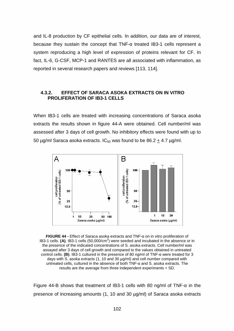
and IL-8 production by CF epithelial c
because they sustain the concept that TNF
system reproducing a high level of expression of proteins relevant for CF. In
fact, IL-6, G-CSF, MCP-1 and RANTES
reported in several research papers and reviews [
4.3.2. EFFECT OF PROLIFERATION OF IB3
When IB3-1 cells are treated with increasing concentrations of
extracts the results shown in figure 4
assessed after 3 days of cell growth. No inhibitory effects were found with up to
50 µg/ml Saraca asoka extracts. IC
FIGURE 44 - Effect of IB3-1 cells. (A). IB3-1 cells (50,000/cm
the presence of the indicated concentrations of assayed after 3 days of cell gro
control cells. (B). IB3-1 cultured in the presence of 80 ng/ml of TNFdays with S. asoka extracts (1, 10 and 30 untreated cells, cultured in
results are the average from three independent experiments + SD.
Figure 44-B shows that treatment of IB3
presence of increasing amounts (1, 10 and 30
102
8 production by CF epithelial cells. In addition, our data are of
because they sustain the concept that TNF-α treated IB3-1 cells
system reproducing a high level of expression of proteins relevant for CF. In
1 and RANTES are all associated with inflammation, as
several research papers and reviews [113, 114].
OF SARACA ASOKA EXTRACTS ON IN VITROPROLIFERATION OF IB3-1 CELLS
1 cells are treated with increasing concentrations of
results shown in figure 44-A were obtained. Cell number/ml was
assessed after 3 days of cell growth. No inhibitory effects were found with up to
extracts. IC50 was found to be 86.2 + 4.7 µ
Effect of Saraca asoka extracts and TNF-α on in vitro proliferation of1 cells (50,000/cm2) were seeded and incubated in the absence or in
the presence of the indicated concentrations of S. asoka extracts. Cell number/ml wasassayed after 3 days of cell growth and compared to the values obtained in untreated
1 cultured in the presence of 80 ng/ml of TNF-α were treated for 3extracts (1, 10 and 30 µg/ml) and cell number compared with
untreated cells, cultured in the absence of both TNF-α and S. asoka extracts. results are the average from three independent experiments + SD.
B shows that treatment of IB3-1 cells with 80 ng/ml of TNF
presence of increasing amounts (1, 10 and 30 µg/ml) of Saraca asoka
ells. In addition, our data are of interest,
1 cells represent a
system reproducing a high level of expression of proteins relevant for CF. In
ssociated with inflammation, as
IN VITRO
1 cells are treated with increasing concentrations of Saraca asoka
A were obtained. Cell number/ml was
assessed after 3 days of cell growth. No inhibitory effects were found with up to
µg/ml.
proliferation of ) were seeded and incubated in the absence or in
extracts. Cell number/ml was wth and compared to the values obtained in untreated
were treated for 3 g/ml) and cell number compared with
extracts. The results are the average from three independent experiments + SD.
1 cells with 80 ng/ml of TNF-α in the
aca asoka extracts
103
does not cause significant alteration in the cell growth rate. Accordingly, no
significant increase in the proportion of trypan-blue excluded cells was observed
following treatment with either TNF-α (80 ng/ml), Saraca asoka extracts (1, 10
and 30 µg/ml), or both (not shown). These data demonstrate that TNF-α and
Saraca asoka extracts are not cytotoxic on IB3-1 cells under the experimental
condition employed.
4.3.3. EFFECT OF SARACA ASOKA EXTRACTS ON THE EXPRESSION OF PRO-INFLAMMATORY GENES INDUCED IN IB3-1 CELLS BY TNF-αααα TREATMENT
When IB3-1 cells were induced with 80 ng/ml of TNF-α and cultured in the
presence of increasing amounts (1, 10 and 30 µg/ml) of Saraca asoka extracts
for 24 hours, some important differences in the release of cytokines and
chemokines occured, which are reported in the representative experiment
shown in figure 45. As it is evident [see also the summary Table included in
figure 45] the release of some TNF-α induced cytokines/chemokines is strongly
reduced. The effect was found to be concentration-dependent. The results
obtained indicate a strong inhibition of release of IL-8, MCP-1 and RANTES.
Low level of inhibition was on the contrary detected analyzing the release of
VEGF. No inhibitory effects on IL-6 and G-CSF release were found. These data
suggest that Saraca asoka extracts might inhibit the expression of pro-
inflammatory genes, the most important of which is IL-8. Considering the cell
number in each well, in a representative experiment untreated IB3-1 secreted
an average of 0.16 ± 0.02 pg of IL-8/106 cells/24 hours. This value increased to
2.8 ± 0.6 when IB3-1 cells were treated with TNF-α, and was found to be 3.0 ±
0.6, 1.8 ± 0.4 and 0.3 ± 0.15 if the cells were cultured with TNF-α in the
presence of 1, 10 and 30 µg/ml extracts from Saraca asoka, respectively [the
summary of the results obtained is included in figure 44]. In consideration of the
inhibitory activity on IL-8 release, RT-PCR experiments were performed to verify
the possible effects of Saraca asoka extracts on the expression of this gene.
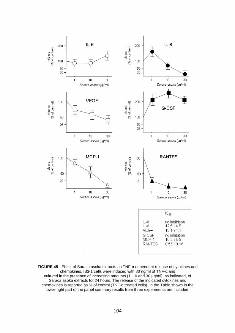
FIGURE 45 - Effect of Saraca asoka chemokines. IB3
cultured in the presence of iSaraca asoka extracts for 24 hours. The release of the indicated cytokines and
chemokines is reported as % of control (TNFlower-right part of the panel
104
Saraca asoka extracts on TNF-α dependent release of cytokines and IB3-1 cells were induced with 80 ng/ml of TNF-α and
cultured in the presence of increasing amounts (1, 10 and 30 µg/ml), as indicated, ofextracts for 24 hours. The release of the indicated cytokines and
chemokines is reported as % of control (TNF-α treated cells). In the Table shown in theright part of the panel summary results from three experiments are included.
dependent release of cytokines and and
g/ml), as indicated, of extracts for 24 hours. The release of the indicated cytokines and
treated cells). In the Table shown in the three experiments are included.
105
4.3.4. SARACA ASOKA EXTRACTS INHIBIT IL-8 mRNA EXPRESSION
In order to determine the effects of Saraca asoka extracts on mRNA
accumulation, the expression of IL-8 gene was analyzed by RT-PCR analysis of
RNA extracted from TNF-α treated IB3-1 cells cultured in the presence of 1 and
10 µg/ml of Saraca asoka extracts. The expression of IL-8 was chosen for the
following reasons: (a) IL-8 is one of the most expressed interleukin in IB3-1 cells
[fig.43]; (b) it is strongly induced following TNF-α treatment [fig.43]; (c) it is
clearly involved in inflammatory processes associated with CF [261,262].
Interestingly, IL-8 exhibit the highest release also in CF cells infected with
Pseudomonas aeruginosa [240].
The results obtained clearly indicate that IL-8 mRNA expression is much lower
in TNF-α treated IB3-1 cells cultured in the presence of 10 µg/ml Saraca asoka
extracts. No inhibitory effects were found on IL-8 mRNA accumulation in TNF-α
treated IB3-1 cells cultured with 1 µg/ml Saraca asoka extracts.
In figure 46-A the preliminary results are shown obtained by semi-quantitative
RT-PCR, indicating that (a) IL-8 mRNA expression is induced by TNF-α
treatment and (b) much lower accumulation of IL-8 mRNA is detectable in RNA
from TNF-α treated IB3-1 cells cultured in the presence of 10 µg/ml Saraca
asoka extracts. In order to better quantify the effects of Saraca asoka extracts
real-time quantitative RT-PCR reactions were also performed, comparing IL-8
mRNA accumulation with that of the internal controls β-actin and GAPDH
mRNA sequences. Panel B of figure 46 shows the representative results
obtained by amplifying RNA from untreated (open symbols) or TNF-α treated
(closed symbols) IB3-1 cells using primer specific for IL-8 and GAPDH RNA
sequences as indicated. Clear induction of IL-8 transcripts, and no changes of
GAPDH mRNA accumulation following TNF-α treatment is evident.
Significant inhibitory effects were detected following exposure of TNF-α treated
IB3-1 cells to 10 µg/ml Saraca asoka extracts, while low effects were found with
1 µg/ml Saraca asoka extracts. These data are in agreement with data obtained
using the Bioplex approach [fig.45], and sustain the concept that extracts from
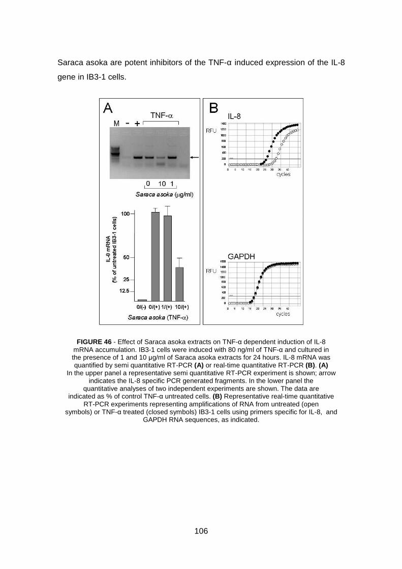
Saraca asoka are potent inhibitors of the TNF
gene in IB3-1 cells.
FIGURE 46 - Effect of Saraca asoka mRNA accumulation. IB3the presence of 1 and 10 quantified by semi quantitative RT
In the upper panel a representative semi quantitative RTindicates the IL-8 specific PCR generated fragments. In the lower panel the
quantitative analyses of two independent experiments are shindicated as % of control TNF
RT-PCR experiments representing amplifications of RNA from untreated (opensymbols) or TNF-α treated (closed symbols) IB3
GAPDH RNA sequences, as indicated.
106
are potent inhibitors of the TNF-α induced expression of the IL
Saraca asoka extracts on TNF-α dependent induction of ILIB3-1 cells were induced with 80 ng/ml of TNF-α and cultured in
the presence of 1 and 10 µg/ml of Saraca asoka extracts for 24 hours. IL-quantified by semi quantitative RT-PCR (A) or real-time quantitative RT-PCR
In the upper panel a representative semi quantitative RT-PCR experiment is shown;8 specific PCR generated fragments. In the lower panel the
quantitative analyses of two independent experiments are shown. The data areindicated as % of control TNF-α untreated cells. (B) Representative real-time quantitative
PCR experiments representing amplifications of RNA from untreated (opentreated (closed symbols) IB3-1 cells using primers specific for IL
GAPDH RNA sequences, as indicated.
induced expression of the IL-8
dependent induction of IL-8 and cultured in
-8 mRNA was PCR (B). (A)
PCR experiment is shown; arrow 8 specific PCR generated fragments. In the lower panel the
own. The data are time quantitative
PCR experiments representing amplifications of RNA from untreated (open cific for IL-8, and

107
4.3.5. SARACA ASOKA EXTRACTS INHIBIT INTERACTIONS BETWEEN NF-kB TRANSCRIPTION FACTOR AND TARGET DNA SEQUENCES
In order to determine the effects of Saraca asoka extracts on possible molecular
targets important for the onset of inflammatory state in CF cells, the possible
effects on the interactions between the nuclear transcription factor kB (NF-kB)
and target DNA sequences were analyzed by EMSA. NF-kB is indeed involved
in the inflammatory state of CF [263-265]. Moreover NF-kB is induced, together
with IL-8, in Pseudomonas aeruginosa infected CF cells. The results, shown in
figure 47, clearly demonstrate that increasing amounts of Saraca asoka extracts
fully suppress the interactions between NF-kB and target DNA elements.
FIGURE 47 - Effect of Saraca asoka extracts on molecular interactions between nuclear proteins and 32P-labeled NF-kB DNA target molecules. Protein/DNA
complexes were separated by polyacrylamide gel electrophoresis. The arrow indicates the protein/DNA complexes. * indicates the free 32P-labeled probe. A. Nuclear extracts from IB3-1 cells (4 µg) were incubated for 20 min in the absence (-) or presence of the indicated amounts of Saraca asoka extracts. After this incubation period, a further 20- min incubation step was performed in the presence of 32P-labeled NF-kB HIV-1 DNA
sequences.

108
DISCUSSION AND FUTURE PERSPECTIVES
One of the clinical feature of cystic fibrosis (CF) is a deep inflammatory process,
which is characterized by production and release of cytokines and chemokines,
among which interleukin 8 (IL-8) represent one of the most important [109-112].
In fact, high levels of IL-8 are exhibited in CF cells infected with Pseudomonas
aeruginosa or induced with TNF-α [118]. Accordingly, there is a growing interest
in developing therapies against CF in order to reduce the excessive
inflammatory response in the airways [99, 100] of CF patients. In this respect,
among the inducible transcription factors that control inflammatory gene
expression, nuclear factor NF-kB plays a central role.
In this thesis, we investigated on two different strategies with the aim of down-
regulate the pro-inflammatory gene expression interfering with NF-kB activation
pathway: (a) the transcription factor decoy approach; (b) screening of new
therapeutic molecules identified in extracts from medicinal plants, such as
Bergamot, Saraca asoka [259, 260] and Emblica officinalis, since they have
been reported to exhibit anti-inflammatory activities in several studies [221-229].
1. TRANSCRIPTION FACTOR DECOY
In the past few years, the transcription factor decoy approach has been applied
to modulate gene expression both in vitro [191, 192] and in vivo [193, 194],
suggesting that this approach has potential value in non viral gene therapy.
In this thesis, we applied a TF decoy strategy aimed at down-modulating the
expression of pro-inflammatory genes in wild-type and CF bronchial epithelial
cells infected with P. aeruginosa. Transfection of bronchial cells with decoy
ODNs mimicking the NFkB sequences contained in the HIV-1 LTR or IL-8
genes clearly inhibited P.aeruginosa–dependent expression of IL-8.
A TF decoy strategy to down-modulate IL-8 expression was first proposed by

109
Cooper and colleagues in human monocytes stimulated with IL-1β [235]. In that
experimental model, transfection of a 10-nucleotide decoy ODN 100%
homologous to the core sequence of NF-kB identified in IL-8 promoter produced
an average 60% inhibition of the IL-1β - dependent expression of IL-8 gene.
Application of TF decoy ODNs has been tried by Griesenbach and coworkers
with a different 20-nucleotide NFkB decoy ODN in CF tracheal epithelial cells
stimulated with TNF-α, as they obtained only an average 40% inhibition of IL-8
secretion [236]. The same decoy ODN was unable to inhibit IL-6 secretion after
bleomycin-induced pneumonia in murine lungs [237]. How can we reconcile
these previous results with the highly efficient inhibition of IL-8 expression we
report in the present thesis? Different decoy ODN sequences have been used
in our experiments with respect to all the previous studies, which could, by itself,
explain the discrepancies. In addition, Cooper and colleagues [235] used decoy
ODNs without flanking regions, which could be more easily degraded by
nucleases cleaving at the 5’ or 3’ ends of the ODNs, thus reducing their
effectiveness in competing for active NF-kB. Also, the application of different
pro-inflammatory stimuli could have impact on the efficiency of the NF-kB decoy
ODNs, a stimulus that we chose considering this pathogen particularly relevant
to study anti-inflammatory approaches for CF lung pathology. From this point of
view, we report here that the HIV-1 LTR NF-kB decoy ODN has a lower potency
of inhibition when IB3-1 cells have been stimulated with TNF-α instead of IL-1β
and P. aeruginosa [fig.29-C]. This is in agreement with the weak inhibition that
Griesenbach and coworkers [236] observed after using the same stimulus. This
can be partly explained considering that TNF-α activates a panel of receptors,
adapters, and kinases [266], which is quite different than those induced by P.
aeruginosa, thus suggesting that transcription factors other than NF-kB could
have a predominant role. Thus differences in the pro-inflammatory stimuli used
could have an important role besides that played by the choice of the
sequences of the decoy ODNs.
NF-kB proteins comprise a family of transcription factors involved in the control
of a large number of genes related to cellular growth, developmental processes,
apoptosis, and immune and inflammatory responses (for synopsis see http://
110
www.NF-kB.org). As far as our bronchial epithelial model system is concerned,
we found that P. aeruginosa induces transcription of IL-8, GROγ, ICAM-1 IL-1β,
and IL-6 genes, all of them containing NF-kB consensus sequences in their
promoters, as summarized also in figure 25. How can we possibly explain that
the HIV-1 LTR NF-kB decoy ODN strongly inhibits the P. aeruginosa -
dependent transcription of IL-8, but not that of the other four genes induced in
parallel in IB3-1 cells? We initially considered that the consensus sequences for
NF-kB are not 100% homologous in the genes regulated by this transcription
factor. Many nucleotide variations could be present, as summarized in the
general NF-kB consensus usually reported (5’-GGG[A/G]NN[C/T][C/T]CC- 3'),
which in principle accounts for the possibility of 27 different variants. In our
specific case, none of the seven NF-kB consensus sequences identified in the
promoters of the five genes that we found induced by P. aeruginosa are
perfectly homologous with at least one of the others. Consequently, different
degrees of homology between the HIV-1 LTR NF-kB decoy ODN and the
consensus sequences in the promoters can be observed. Thus we
hypothesized that an important parameter modulating the efficacy the NF-kB
decoy ODNs in inhibiting transcription of IL-8, GRO-γ, ICAM-1, IL- 1β, and IL-6
should be the degree of homology between decoy ODN and consensus
sequences.
For instance, the three consensus sequences identified within the promoter of
ICAM-1 gene present the highest number of mismatches with the HIV-1 LTR
NF-kB decoy ODN that prompted us to test the effect of new decoy ODNs
designed with 100% homology. Disappointingly, none of the three ICAM-1 NF-
kB decoy ODNs inhibited P. aeruginosa - dependent ICAM-1 transcription, even
after transfecting the three decoy ODNs together [data not shown 267].
Although this unsuccessful attempt could be related to different parameters
(e.g., length of the ODN, choice of the 5’- and 3’-flanking nucleotides,
secondary structure, etc.), overall these negative results seem to basically
disprove the hypothesis. Second, it should be noted that the HIV-1 LTR NF-kB
decoy ODN is even more homologous to the consensus sequences reported in
the GRO-γ and IL-6 than in the IL-8 promoter, whereas we proved the decoy
111
ODN being effective in inhibiting transcription of the latter but not of the former
genes [fig.28]. Third, the IL-8 NF-kB decoy ODN, which is much more
homologous than the HIV-1 LTR NF-kB decoy ODN with the consensus
sequence in the promoter of IL-8, is less efficient in inhibiting IL-8 expression
than the HIV-1 LTR NF-kB decoy ODN and inhibits partially also the
transcription of GRO-γ and IL-6. Therefore it becomes apparent that the extent
of homology between decoy ODN and consensus sequences is important but
not sufficient to explain all the differences in effectiveness of each NF-kB decoy
ODN in inhibiting the transcription of the five genes that we observed induced
by P. aeruginosa in bronchial epithelial cells. Extensive analyses of a large
series of ODNs mutated in their core consensus and/or flanking regions need
future investigation to thoroughly answer these questions. A further explanation
of the different efficiency of NF-kB decoy ODNs could be related to the complex
interplay of transcription factors in the expression of the different genes of
interest in the present study. Surface structures of P. aeruginosa, like flagellum
and pili, interact with different receptors expressed on respiratory epithelial
cells, leading eventually to activation of transcription factors such as NF-kB, AP-
1, Elk-1, and NF–IL- 6, as already reported [195-197]. Interestingly, all the five
genes we found induced by P. aeruginosa contain in their promoters one or
more consensus sequences for NF-kB and some of them also for the
transcription factors AP-1, NF-IL-6, and Sp-1 [fig.25]. Therefore, we hypothesize
that the competition of the decoy ODN for the binding of NF-kB to its consensus
sequence is not sufficient to inhibit drastically GRO-γ, ICAM-1, IL-1β, and IL-6
transcription also because of the hierarchical roles of AP-1, NF-IL-6, and Sp-1
in respect to NFkB for the transcription of some of those genes.
As far as the mechanism of action of the decoy ODNs is concerned, we can
infer various suggestions. The degree of homology between specific decoy
ODNs and the consensus sequences for the corresponding TF is important but
not sufficient to predict the efficiency in inhibiting gene transcription. Efficiency
of each decoy ODN could depend on the secondary structure and the kind of
flanking regions of the ODNs, and on the hierarchy of the different TFs in
regulating expression of specific genes. Thus, from a theoretical point of view,
112
our results strengthen the utility of the TF decoy strategy to provide useful
insights on the issue of regulation of gene transcription. Conversely, as a
practical consequence useful to applications in human diseases, decoy ODNs
appear to present the advantage of a much higher gene specificity than
previously expected. For instance, one may have anticipated a broad inhibitory
effect of our NF-kB decoy ODNs on all the five genes containing NF-kB
consensus sequences, whereas experimental results showed that only IL-8 is
strongly inhibited with selected decoy ODNs. Considering the therapeutic
applications of TF decoy ODNs to lung disease of patients with CF and/or to
other human lung diseases in which the innate immunity is involved, we
propose here a hint for inhibiting master genes in CF pulmonary pathology. This
means that novel anti-inflammatory approaches focused on specific target
genes relevant to specific diseases, without having too broad and undesired
effects on the innate immune response, could be seriously taken into
consideration. On the opposite side, knowing the redundancy of this response,
a disease-oriented intervention should be tailored, focusing on the inhibition of
the relevant pro-inflammatory genes with complementary TF decoy molecules.
Different molecular approaches can be investigated to obtain a novel anti-
inflammatory intervention. Strictly single-gene-related inhibitory approaches
could be devised considering the application of anti-sense and Silencing
inhibitory RNA (SiRNA) nucleotides. However, due to the redundancy of the
inflammatory response to P. aeruginosa observed in CF lungs, inhibition of one
chemokine (e.g., IL-8) could be bypassed by other potent chemoattractants
(e.g., Gro-γ). A much broader inhibitory approach such as the modulation of the
receptors for pathogens [268] would present the disadvantage of reducing the
wide array of genes induced by pathogen-associated molecular patterns that
are critical to maintain the proper anti-infective defenses against pathogens. In
terms of receptor blockade, those for chemokines could also be a theoretically
interesting target [269]. However, since chemokine receptors are expressed on
circulating immune cells, the systemic delivery of antagonist or competing
molecules could raise safety concerns. All this considered, TF decoy ODNs
directed toward NF-kB regulating pro-inflammatory genes transcribed in CF

113
respiratory epithelial cells exposed to P. aeruginosa could present the
advantage of extending or reducing the range of target genes by changing the
ODN sequence, as observed also in the results presented here. In the
perspective of application in the respiratory tract, NF-kB decoy ODNs
conjugated with novel lipoplexes have been successfully utilized in murine
lungs, obtaining effective inhibition of endotoxic shock (39). However, we are
perfectly aware that the delivery of decoy ODNs in the conductive airways still
requires extensive research and development, in particular to ensure sufficient
stability in respect to degradation in the biological fluids. Recent progress
obtained in other and our laboratories with PNA-chimeric TF decoy molecules
that are significantly more permeable to the plasma membrane and resistant to
the nucleases contained in serum and other biological fluids [247, 248, 252,
271-273 ] are rendering closer the possibility of extending the TF decoy strategy
to in vivo pre-clinical experiments.
2. NATURAL PRODUCTS
• EMBLICA OFFICINALIS AND SARACA ASOKA
Since extracts from medicinal plants have been reported in literature [275-278]
to exhibit anti-inflammatory activities, it could be interesting to analyze the basis
of this activity and the possible applications in biomedicine.
In this respect Emblica offcinalis and Saraca asoka [231, 233, 279] deserves
great attention. These medicinal plants have been reported to be useful in
traditional indian medicine for the treatment of several diseases. Fruits of
Emblica offcinalis are used for the treatment of a number of diseases, such as
dyslipidemia and atherosclerosis, as hepatoprotective, antibacterial and anti-
inflammatory agent [221-229]. In many cases, Emblica officinalis has been
shown to be a potent free radical scavenging agent thereby preventing
carcinogenesis and mutagenesis [227]. Whereas, Saraca asoka is especially
relied upon as an astringent to treat excessive uterine bleeding of different
114
origin, but also for regulating the menstrual cycle and, in various complex
formulas, as a tonic for women.
In spite of these promising features, the reports on the biological effects of
extracts from Emblica officinalis and Saraca asoka are few.
In this thesis we studied the effects of Emblica officinalis and Saraca asoka
extracts on TNF-α mediated induction of interleukin and chemokines in cystic
fibrosis IB3-1 cells, considering that: (a) in cystic fibrosis epithelium high levels
of interleukin (for instance IL-6) [280, 281] and chemokines (for instance IL-8)
[261, 262] are found; (b) this effect is thought to be mediated, at least in part, by
TNF-α [243, 282] and (c) targeting IL-8 is considered of great interest in the
anti-inflammatory treatment of CF patients [198, 201, 241, 245].
The first conclusion of the results presented in this thesis is that both Emblica
offcinalis and Saraca asoka extracts are strong inhibitors of IL-8 expression,
measured at the mRNA (using semi-quantitative as well as real-time
quantitative RT-PCR assays) and protein level. This was determined in cystic
fibrosis IB3-1 cells, either TNF-α-untreated (data not shown), or stimulated with
TNF-α.
To validate these results, cytokines and chemokines in culture supernatants
released from cystic fibrosis IB3-1 cells treated with TNF-α were measured by
Bio-Plex cytokine assay. When IB3-1 cells were induced with TNF-α and
cultured in the presence of Emblica officinalis extracts, some important
differences in the release of MCP-1 (-83%), RANTES (-87%), VEGF (-41%), IL-
8 (-14%) occurred compare to the control (IB3-1 cells induced with TNF-α
without plant extracts treatment). Similar effects were found after the treatment
of induced cells with increasing amount of Saraca asoka extracts: the results
show a down-regulation in MCP-1, RANTES, VEGF and IL-8 expression, by
92%, 99%, 54% and 96% respectively. These data suggest that Emblica
offcinalis and Saraca asoka extracts might inhibit the expression of the IL-8 pro-
inflammatory gene. These data are in agreement with the results obtained using
the RT-PCR experiments, and sustain the concept that these extracts could be
inhibitors of the TNF-α induced expression of the IL-8 gene in IB3-1 cells.
All these factors are important in the onset of the inflammatory state in CF
115
patients.
With respect to the anti-inflammatory activity of Emblica officinalis extracts, the
identification of bioactive compounds is of interest to restrict biopharmaceutical
drug design to single or few compounds. Gas chromatography-mass
spectrometry (GC-MS) analyses showed pyrogallol as the common compound
present both in unfractionated and n-butanol fraction of Emblica officinalis
extracts. The Bio-plex analysis on supernatant from TNF-α induced IB3-1 cells
suggest that pyrogallol inhibits especially the expression of RANTES (-83%)
gene, but no significant data were obtained on regulation of IL-8 expression.
These results were confirmed by RT-PCR analysis of RNA extracted from TNF-
α treated IB3-1 cells cultured in the presence of increasing concentration of
pyrogallol: IL-8 mRNA expression does not show inhibitory effects at the higher
pyrogallol concentrations.
The data obtained indicate that Emblica offcinalis and Saraca asoka extracts
inhibit NF-kB/DNA interactions. Nevertheless it is not clear the mechanism of
action of Emblica officinalis and Saraca asoka extracts although an attempt was
made to determine whether the plant extracts retain an inhibitory activity on the
transcription factor NF-kB, which is critical for IL-8 gene expression and
involved in CF inflammation. Phytochemical analysis will be performed to
understand the main contituents of the extracts in order to evaluate which
molecules are responsible for the downregulation on IL-8 expression.
Further experiments to determine other biochemical targets of Saraca asoka
and Emblica offcinalis extracts and the inhibitory effects on IL-8 gene
expression in other cellular pathological systems and murine models are
needed.
• BERGAMOT
The major result of this thesis is the finding that extracts obtained from
bergamot (Citrus bergamia Risso) epicarps, contain components displaying an
inhibitory activity on IL-8. This effect has been confirmed both at the mRNA

116
levels and the protein release in the CF cellular model IB3-1, induced with TNF-
α and treated with low concentrations of bergamot extracts. The qualitative and
quantitative determination of the chemical constituents of the extracts, with
particular reference to non-volatile fraction chemicals, has demonstrated the
presence of citropten, bergamottin and bergapten in all bergamot extract
samples and with different amounts (%w/w) [table 5 and 6]. In particular the
coumarin/psoralen portion was the major constituent of the most active among
the extracts (extracts 3). Accordingly, we hypothesized that the
coumarin/psoralen portion of these extracts were among the biological active
components. On the other hand, the data reported do not clarify why extract 2
exhibited the lower biological activity. It should be noted that the composition of
extract 1 and extracts 2 is complex and, therefore, the presence of compounds
exhibiting opposite activity or interfering with the coumarin/psoralen compounds
cannot be excluded. A second achievement of the results here presented is the
demonstration that, among the preminent coumarin/psoralen compounds,
bergapten and citropten were the most active molecules to reduce mRNA level
of IL-8 in TNF-α treated IB3-1 cells at IC50 concentrations, while bergamottin did
not show inhibitory effects at the same [fig.35] and at higher doses (data not
shown). These results have been confirmed by real-time quantitative RT-PCR
and Bio-plex analyses. In particular citropten is more efficient than bergapten in
decreasing IL-8 mRNA/protein accumulation in IB3-1 cells.
These results clearly indicate that bergapten and citropten are strong inhibitors
of IL-8 expression and could be proposed as potential anti-inflammatory
molecules to reduce lung inflammation in CF patients. Further experiments are

117
required to determine: (a) other biochemical targets of citropten and bergapten
as well as whether the inhibitory effects on IL-8 gene expression are
reproducible in other cellular pathological systems in which this chemokine
plays a role; (b) safety, potential toxicity, in vivo bioavailability, stability and
whether suitable concentrations can be reached following in vivo administration.
Moreover, it should be considered that coumarin compounds, as reported in
literature, display anti-proteinase activity [274]. For example, serin proteinase is
capable to degradate the matrix proteins and is involved in several inflammatory
respiratory diseases as CF. It could interesting to investigate and correlate the
anti-inflammatory effect of bergapten and citropten with their possible anti-
proteinase activity. This double role of these comppunds could be useful for the
treatment of lung inflammation in CF patients.
Last but not least, it would be of interest to see whether the effect of bergamot
and its extracts could play the same role with different inflammation stimuli,
such as IL-1β or PAO-1.