Unit 3: The Muscular System Lab 3: Muscle Physiology Jessica Radke-Snead, RD, MS Bio 241 Anatomy and...
25
Unit 3: The Muscular System Lab 3: Muscle Physiology Jessica Radke-Snead, RD, MS Bio 241 Anatomy and Physiology
-
Upload
earl-marsh -
Category
Documents
-
view
218 -
download
1
Transcript of Unit 3: The Muscular System Lab 3: Muscle Physiology Jessica Radke-Snead, RD, MS Bio 241 Anatomy and...
Unit 3: The Muscular SystemLab 3: Muscle Physiology
Jessica Radke-Snead, RD, MSBio 241 Anatomy and Physiology
Reminders
• Cadaver Lab– Ensure that you are familiarized with the location
of each muscle listed on your labs 1 & 2– Groups of 4-5 groups of about 4-6 people
• Optional– Complete and submit the optional lab assignment
(see assignment posted on faculty website)
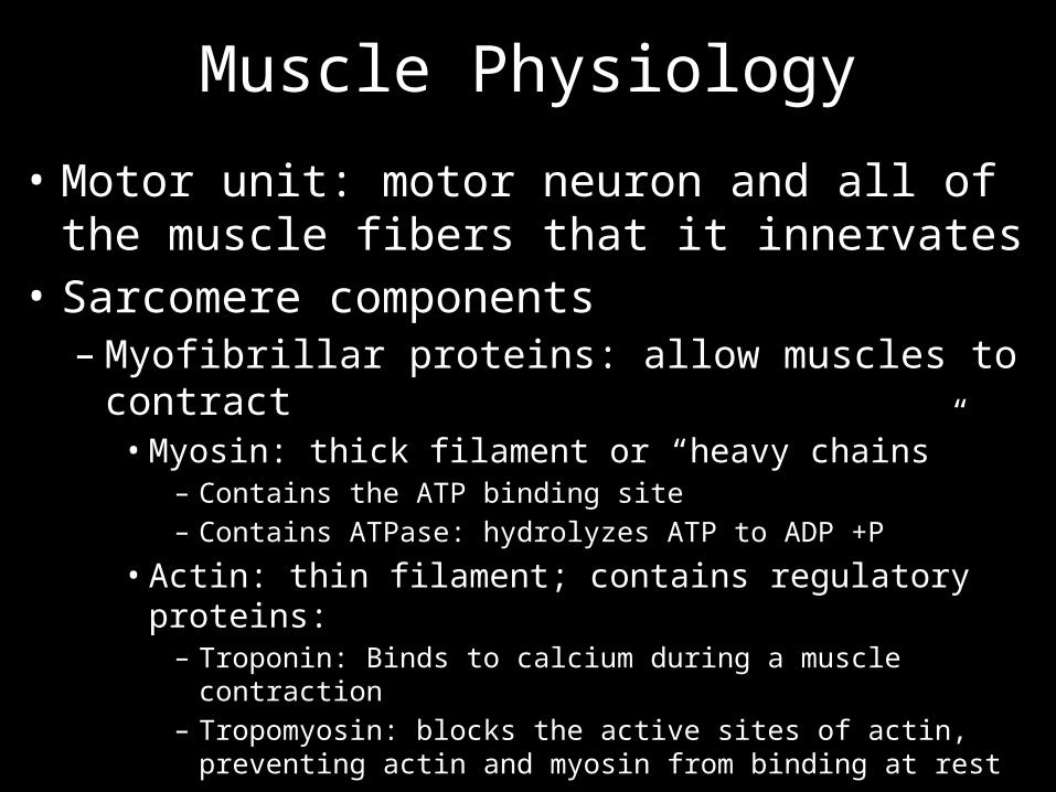
Muscle Physiology
• Motor unit: motor neuron and all of the muscle fibers that it innervates
• Sarcomere components– Myofibrillar proteins: allow muscles to contract• Myosin: thick filament or “heavy chains”
– Contains the ATP binding site– Contains ATPase: hydrolyzes ATP to ADP +P
• Actin: thin filament; contains regulatory proteins:– Troponin: Binds to calcium during a muscle contraction– Tropomyosin: blocks the active sites of actin, preventing actin and
myosin from binding at rest
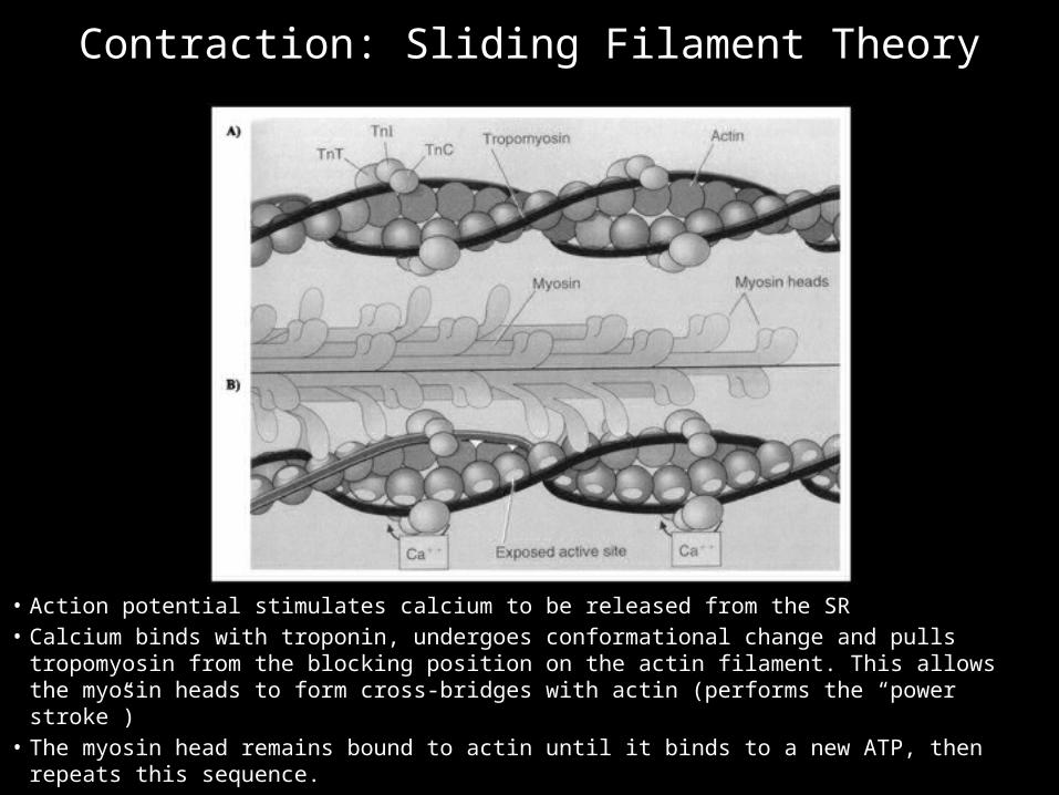
• Action potential stimulates calcium to be released from the SR• Calcium binds with troponin, undergoes conformational change and pulls tropomyosin from
the blocking position on the actin filament. This allows the myosin heads to form cross-bridges with actin (performs the “power stroke”)
• The myosin head remains bound to actin until it binds to a new ATP, then repeats this sequence.
Contraction: Sliding Filament Theory

As long as calcium and ATP are present, the myosin heads will attach to the actin molecules,
pull the actin, release and reattach
The speed at which cross-bridge cycling can occur is limited predominantly by the rate that the ATPase of the myosin head can hydrolyze
ATP
Contraction: Sliding Filament Theory
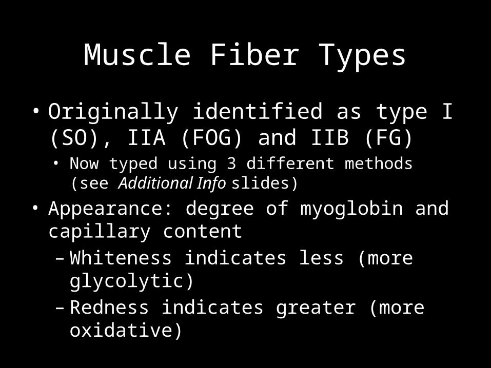
Muscle Fiber Types
• Originally identified as type I (SO), IIA (FOG) and IIB (FG)• Now typed using 3 different methods (see Additional Info
slides)
• Appearance: degree of myoglobin and capillary content– Whiteness indicates less (more glycolytic)– Redness indicates greater (more oxidative)
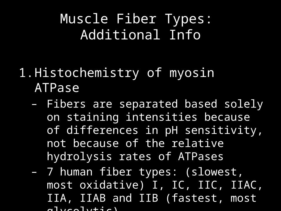
1. Histochemistry of myosin ATPase – Fibers are separated based solely on staining
intensities because of differences in pH sensitivity, not because of the relative hydrolysis rates of ATPases
– 7 human fiber types: (slowest, most oxidative) I, IC, IIC, IIAC, IIA, IIAB and IIB (fastest, most glycolytic)
Muscle Fiber Types: Additional Info
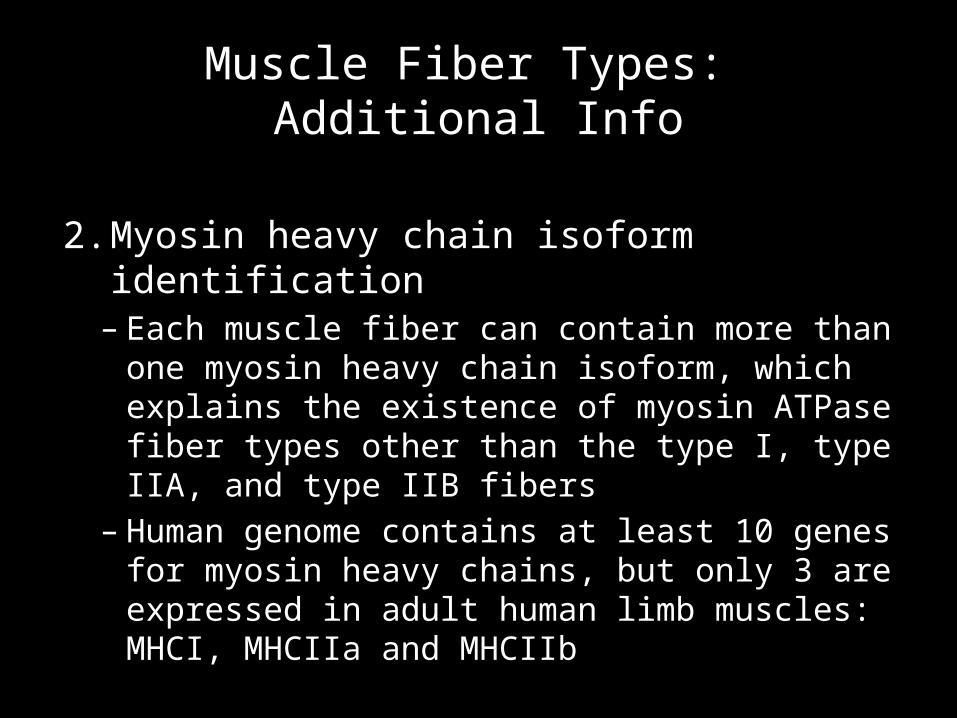
2. Myosin heavy chain isoform identification – Each muscle fiber can contain more than one
myosin heavy chain isoform, which explains the existence of myosin ATPase fiber types other than the type I, type IIA, and type IIB fibers
– Human genome contains at least 10 genes for myosin heavy chains, but only 3 are expressed in adult human limb muscles: MHCI, MHCIIa and MHCIIb
Muscle Fiber Types: Additional Info
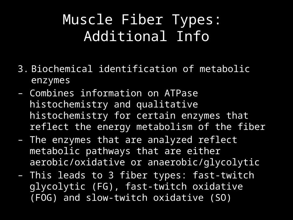
3. Biochemical identification of metabolic enzymes– Combines information on ATPase histochemistry and
qualitative histochemistry for certain enzymes that reflect the energy metabolism of the fiber
– The enzymes that are analyzed reflect metabolic pathways that are either aerobic/oxidative or anaerobic/glycolytic
– This leads to 3 fiber types: fast-twitch glycolytic (FG), fast-twitch oxidative (FOG) and slow-twitch oxidative (SO)
Muscle Fiber Types: Additional Info
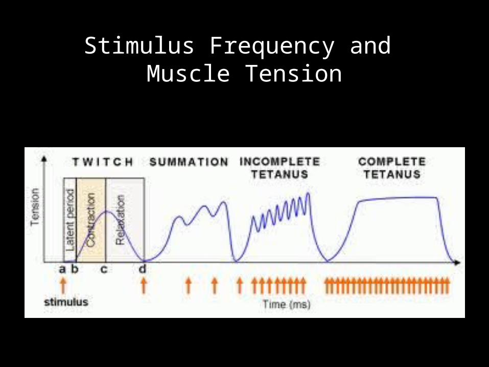
Myogram: Timing and Strength of a Contraction
• Threshold: minimum voltage necessary to generate an AP in the muscle fiber AND produce a contraction
• The AP triggers the release of a pulse of calcium into the cytosol and activates the sliding filament mechansim
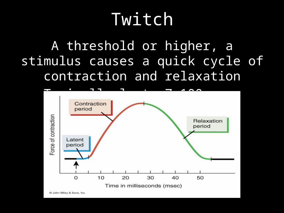
TwitchA threshold or higher, a stimulus causes a quick
cycle of contraction and relaxationTypically lasts 7-100 msec
Latent Period
• The delay between the onset of the stimulus and the onset of the twitch
• Time required for excitation, excitation-contraction coupling and tensing of the elastic components of the muscle
• Force generated during this period is internal tension (not visible on the myogram—no shortening of the muscle)
Contraction phase
• The muscle begins to produce external tension and move a resisting object or load
• Calcium and ATP are present = myosin and actin cross-bridges intact = sliding filament mechanism active
• Short, time-wise because the SR quickly reabsorbs calcium before the muscle develops maximal force
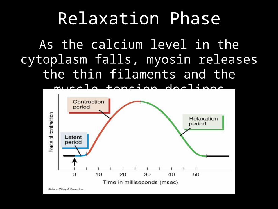
Relaxation PhaseAs the calcium level in the cytoplasm falls, myosin releases the thin filaments and the
muscle tension declines

Contraction Strength of Twitches
• Twitch strength varies with– Stimulation frequency: stimuli arriving close
together produce stronger twitches (tetanus)– Concentration of calcium in the sarcoplasm – Degree muscle stretched just before it was
stimulated (length-tension relationship)– Temperature: warm contracts stronger because
enzymes work more quickly– pH of the sarcoplasm: subnormal pH results in
weaker twitches (fatigue)– State of hydration: affects overlap between thick
and thin filaments and the ability of myosin and actin to cross-bridge
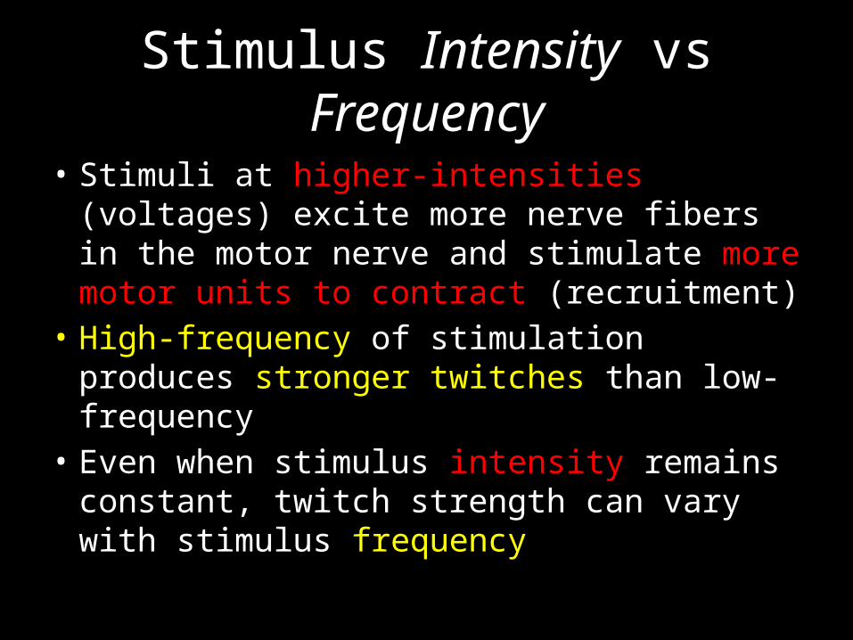
Stimulus Intensity vs Frequency
• Stimuli at higher-intensities (voltages) excite more nerve fibers in the motor nerve and stimulate more motor units to contract (recruitment)
• High-frequency of stimulation produces stronger twitches than low-frequency
• Even when stimulus intensity remains constant, twitch strength can vary with stimulus frequency

Stimulus Frequency and Muscle Tension
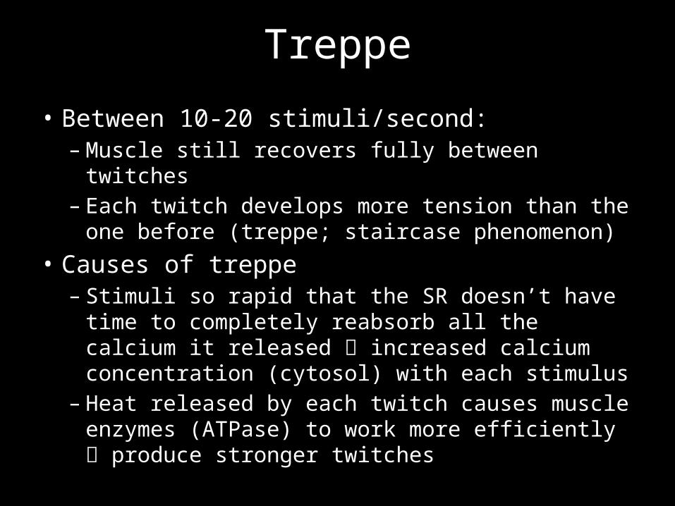
Treppe
• Between 10-20 stimuli/second:– Muscle still recovers fully between twitches – Each twitch develops more tension than the one
before (treppe; staircase phenomenon)• Causes of treppe– Stimuli so rapid that the SR doesn’t have time to
completely reabsorb all the calcium it released increased calcium concentration (cytosol) with each stimulus
– Heat released by each twitch causes muscle enzymes (ATPase) to work more efficiently produce stronger twitches
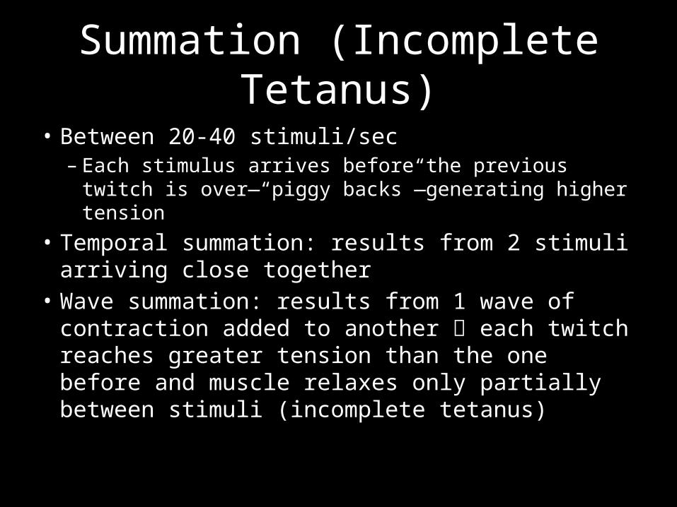
Summation (Incomplete Tetanus)
• Between 20-40 stimuli/sec– Each stimulus arrives before the previous twitch is over
—“piggy backs”—generating higher tension• Temporal summation: results from 2 stimuli
arriving close together• Wave summation: results from 1 wave of
contraction added to another each twitch reaches greater tension than the one before and muscle relaxes only partially between stimuli (incomplete tetanus)
Stimulus Frequency and Muscle Tension
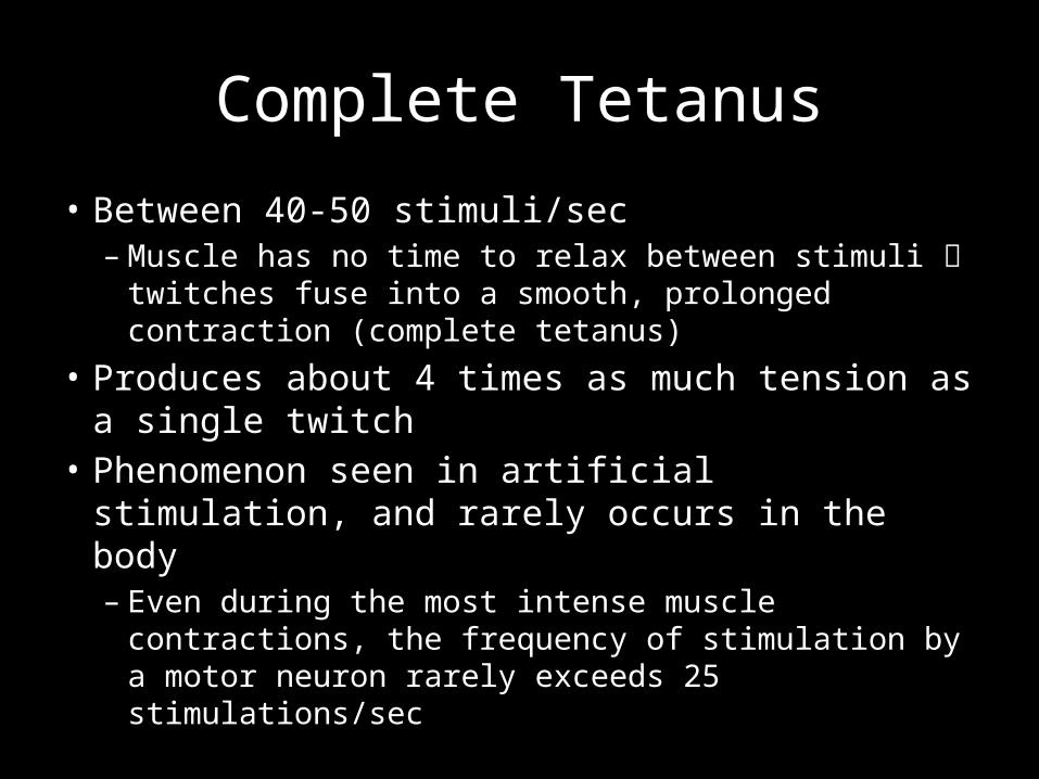
Complete Tetanus
• Between 40-50 stimuli/sec– Muscle has no time to relax between stimuli
twitches fuse into a smooth, prolonged contraction (complete tetanus)
• Produces about 4 times as much tension as a single twitch
• Phenomenon seen in artificial stimulation, and rarely occurs in the body– Even during the most intense muscle contractions, the
frequency of stimulation by a motor neuron rarely exceeds 25 stimulations/sec
Types of Contractions: Isometric
• Contraction without a change in length– Internal movement: Contraction at the cellular
level– Tension absorbed by the elastic components– Muscle as a whole is not producing external
movement– Isometric contraction of antagonistic muscle at a
single joint is important in maintaining joint stability
Types of Contractions:Isotonic
• Contraction with a change in length but no change in tension– Internal tension builds to the point that it
overcomes the resistance– Muscle shortens, moves the load and maintains
essentially the same tension throughout
Types of Contractions:Concentric vs Eccentric
• Concentric: Muscle shortens as it maintains tension
• Eccentric: Muscles lengthens as it maintains tension
• Example: Bicep Curls
Lab Objectives
• Cadaver Lab: Groups of 4-6 by table• Work on Lab 3 Assignment (separate
document)• Continue to work on your muscle list
Please let me know if you need assistance—have fun!