Unit 3 - Genetics Topic 3 – Genetics and Biotechnology.
85
Unit 3 - Genetics Topic 3 – Genetics and Biotechnology
-
Upload
theodora-townsend -
Category
Documents
-
view
234 -
download
0
Transcript of Unit 3 - Genetics Topic 3 – Genetics and Biotechnology.
Unit 3 - Genetics
Topic 3 – Genetics and Biotechnology
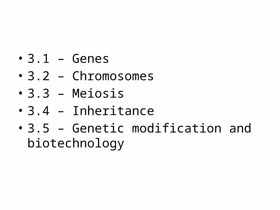
• 3.1 – Genes• 3.2 – Chromosomes• 3.3 – Meiosis• 3.4 – Inheritance• 3.5 – Genetic modification and biotechnology
3.1 - GENES
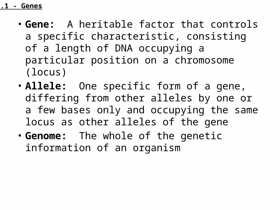
• Gene: A heritable factor that controls a specific characteristic, consisting of a length of DNA occupying a particular position on a chromosome (locus)
• Allele: One specific form of a gene, differing from other alleles by one or a few bases only and occupying the same locus as other alleles of the gene
• Genome: The whole of the genetic information of an organism
3.1 - Genes
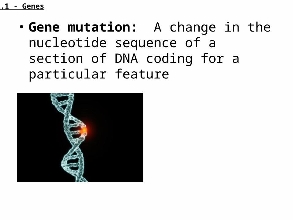
• Gene mutation: A change in the nucleotide sequence of a section of DNA coding for a particular feature
3.1 - Genes
• TYPES OF MUTATIONS• http://highered.mcgraw-hill.com/sites/007255
6781/student_view0/chapter11/animation_quiz_4.html
3.1 - Genes

Sickle Cell Anemia• Cause of Sickle Cell Anemia• A base substitution mutation is the change of a single base in a
sequence of DNA, resulting in a change to a single mRNA codon during transcription
• In the case of sickle cell anemia, the 6th codon for the beta chain of hemoglobin is changed from GAG to GTG (on the non-coding strand)
• This causes a change in the mRNA codon (GAG to GUG), resulting in a single amino acid change of glutamic acid to valine (Glu to Val)• DNA: GAG to GTG (non-coding strand) • mRNA: GAG to GUG • Amino Acid: Glu to Val
• The amino acid change alters the structure of hemoglobin, causing it to form fibrous, insoluble strands
• This causes the red blood cell to adopt a sickle shape
3.1 - Genes
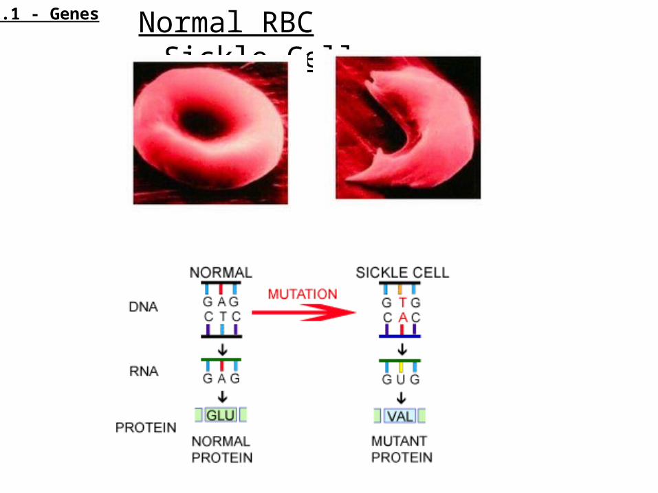
Normal RBC Sickle Cell3.1 - Genes
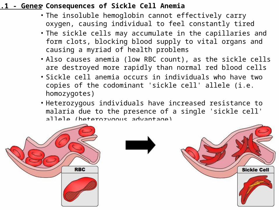
• Consequences of Sickle Cell Anemia• The insoluble hemoglobin cannot effectively carry oxygen, causing
individual to feel constantly tired• The sickle cells may accumulate in the capillaries and form clots,
blocking blood supply to vital organs and causing a myriad of health problems
• Also causes anemia (low RBC count), as the sickle cells are destroyed more rapidly than normal red blood cells
• Sickle cell anemia occurs in individuals who have two copies of the codominant 'sickle cell' allele (i.e. homozygotes)
• Heterozygous individuals have increased resistance to malaria due to the presence of a single 'sickle cell' allele (heterozygous advantage)
3.1 - Genes
Application and Skills
1) Application 1: The causes of sickle cell anemia, including a base substitution mutation, a change to the base sequence or mRNA transcribed from it and a change to the sequence of a polypeptide in hemoglobin
2) Application 2: Comparison to the number of genes in humans with other species (pick at least 5 different species)
3) Skill 1: Use of a database to determine differences in the base sequence of a gene in two species.
3.1 - Genes
3.2 - CHROMOSOMES
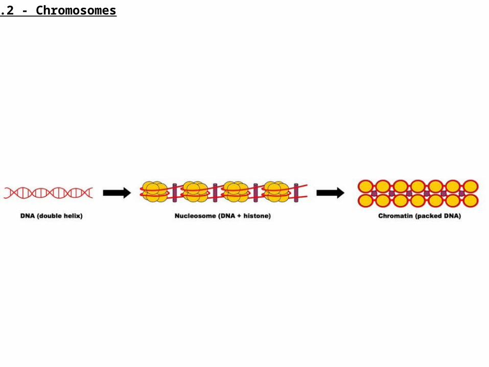
Eukaryotic chromosomes are made of DNA and protein
• Eukaryotic chromosomes consist of DNA wrapped around histone proteins
• This forms the basic structure of the nucleosome, which is packed together to form chromatin (in a 'beads on a string' arrangement)
• Chromatin will supercoil and condense during prophase to form chromosomes that can be visualised under a light microscope
• Prokaryotic DNA is not wrapped around proteins and is thus considered to be 'naked'
3.2 - Chromosomes
3.2 - Chromosomes

Homologous chromosomes• Homologous chromosomes are chromosomes that
share: • The same structural features (e.g. same size, same
banding pattern, same centromere position)• The same genes at the same loci positions (while
genes are the same, alleles may be different)
3.2 - Chromosomes
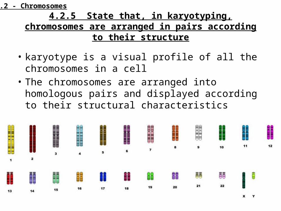
4.2.5 State that, in karyotyping, chromosomes are arranged in pairs according to their structure
• karyotype is a visual profile of all the chromosomes in a cell
• The chromosomes are arranged into homologous pairs and displayed according to their structural characteristics
3.2 - Chromosomes
Karyotype
• Karyotyping involves:• Harvesting cells (usually from fetus or white blood
cells of adults)• Chemically inducing cell division, then halting it
during mitosis when chromosomes are condensed and thus visible– The stage during which mitosis is halted will determine
whether chromosomes appear with sister chromatids• Staining and photographing chromosomes, before
arranging them according to structure
3.2 - Chromosomes
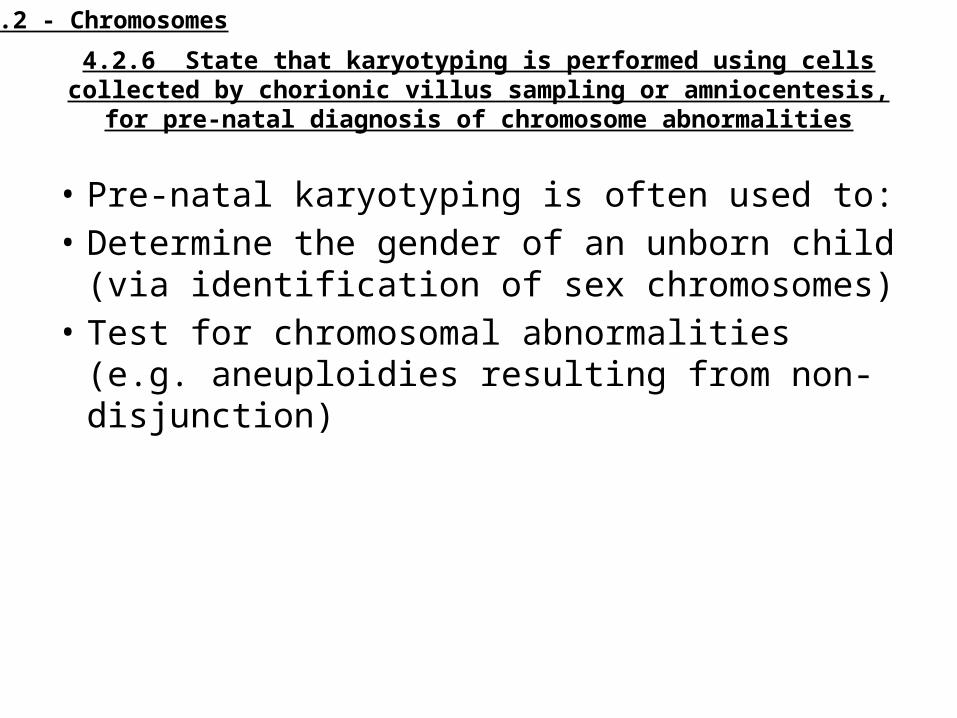
4.2.6 State that karyotyping is performed using cells collected by chorionic villus sampling or amniocentesis, for pre-natal diagnosis of chromosome
abnormalities
• Pre-natal karyotyping is often used to:• Determine the gender of an unborn child (via
identification of sex chromosomes)• Test for chromosomal abnormalities (e.g.
aneuploidies resulting from non-disjunction)
3.2 - Chromosomes
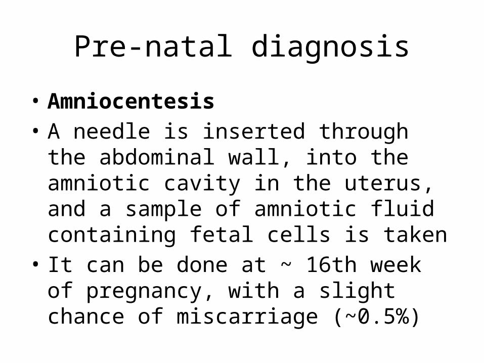
Pre-natal diagnosis
• Amniocentesis• A needle is inserted through the abdominal
wall, into the amniotic cavity in the uterus, and a sample of amniotic fluid containing fetal cells is taken
• It can be done at ~ 16th week of pregnancy, with a slight chance of miscarriage (~0.5%)
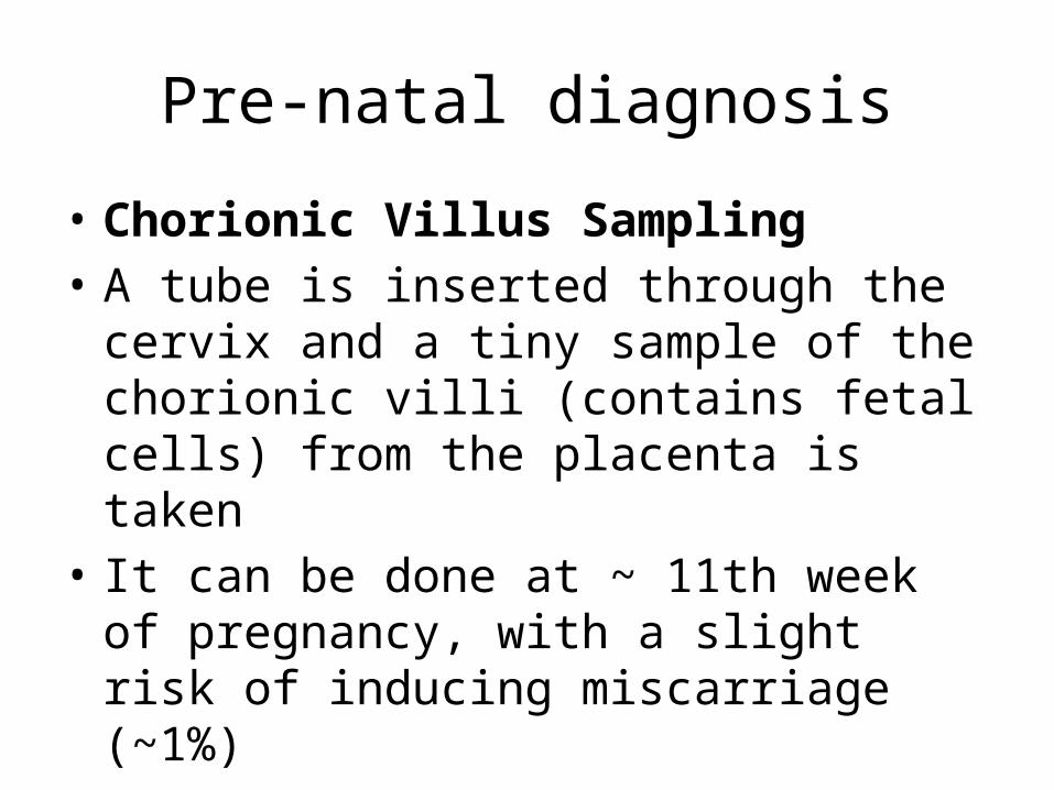
Pre-natal diagnosis
• Chorionic Villus Sampling• A tube is inserted through the cervix and a
tiny sample of the chorionic villi (contains fetal cells) from the placenta is taken
• It can be done at ~ 11th week of pregnancy, with a slight risk of inducing miscarriage (~1%)
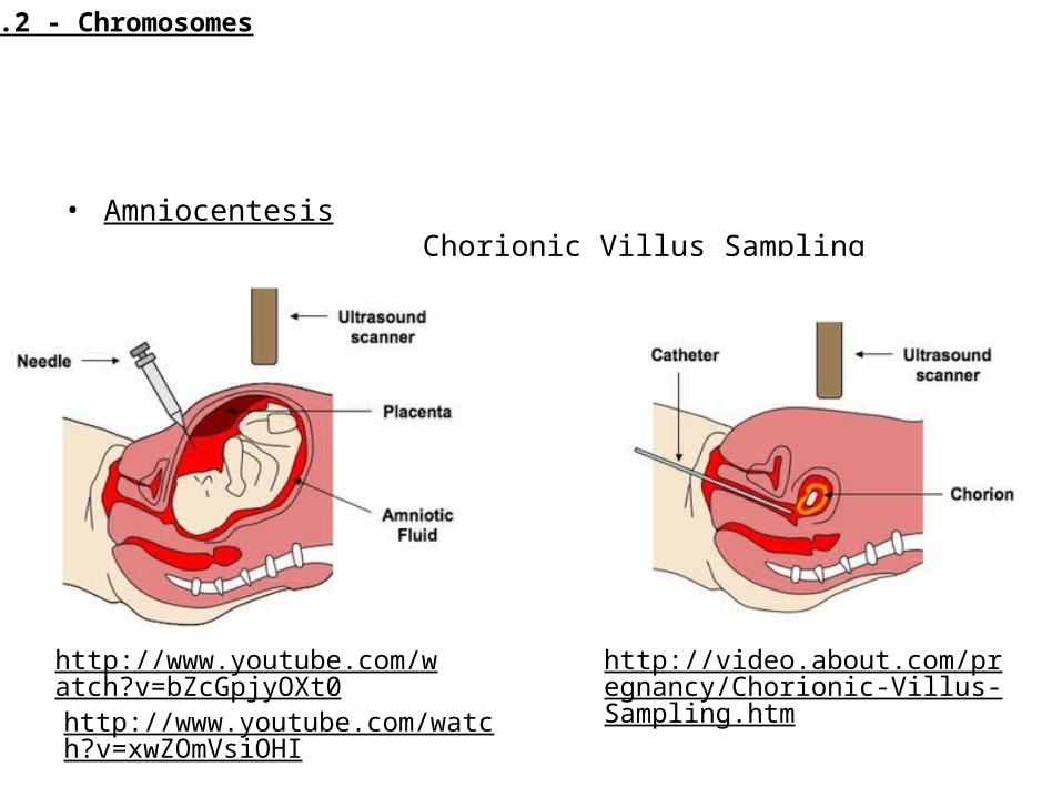
• Amniocentesis Chorionic Villus Sampling
http://www.youtube.com/watch?v=bZcGpjyOXt0
http://video.about.com/pregnancy/Chorionic-Villus-Sampling.htm http://www.youtube.com/watch?v=xwZ
OmVsiOHI
3.2 - Chromosomes
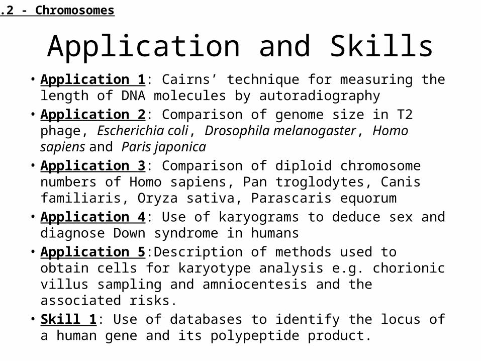
Application and Skills• Application 1: Cairns’ technique for measuring the length of DNA
molecules by autoradiography• Application 2: Comparison of genome size in T2 phage,
Escherichia coli, Drosophila melanogaster, Homo sapiens and Paris japonica
• Application 3: Comparison of diploid chromosome numbers of Homo sapiens, Pan troglodytes, Canis familiaris, Oryza sativa, Parascaris equorum
• Application 4: Use of karyograms to deduce sex and diagnose Down syndrome in humans
• Application 5:Description of methods used to obtain cells for karyotype analysis e.g. chorionic villus sampling and amniocentesis and the associated risks.
• Skill 1: Use of databases to identify the locus of a human gene and its polypeptide product.
3.2 - Chromosomes
3.3 - MEIOSIS
Meiosis is a reduction division of a diploid nucleus to form haploid nuclei
Meiosis is the process by which sex cells (gametes) are made in the reproductive organs:
• Most sexually reproducing animals are diploid - meaning they have two copies of every chromosome (one of maternal origin, one of paternal origin)
• In order to reproduce, these organisms need to make gametes that are haploid (have only one copy of each chromosome)
• Fertilization of two haploid gametes (egg + sperm) will result in the formation of a diploid zygote that will grow into a new organism
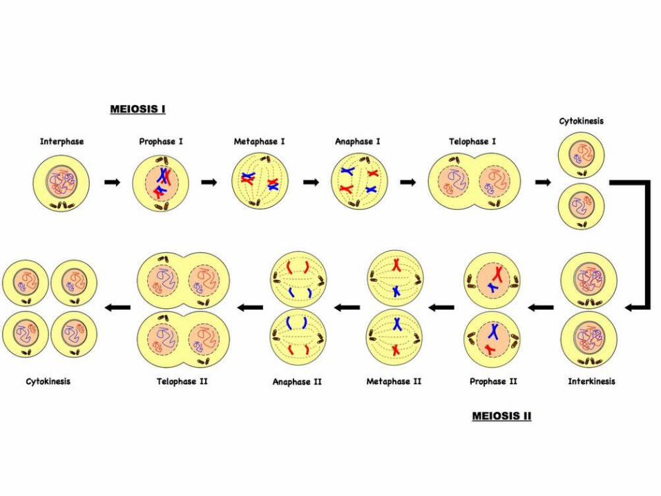
Meiosis consists of two cell divisions:1) The first division is a reduction division of the diploid nucleus to form haploid nuclei2) The second division separates sister chromatids (this division is necessary because meiosis is preceded by interphase, wherein DNA is replicated)
Phases of Meiosis
• Interphase: Cell growth and DNA replication (duplication of DNA creates sister chromatid chromosomes)
• Meiosis I• Prophase I: DNA supercoils and chromosomes condense, nuclear
membrane dissolves, homologous pairs form bivalents, crossing over occurs• Metaphase I: Spindle fibers from centrioles (at poles) attach to
centromeres of bivalent, bivalents line up along the equator of the cell• Anaphase I: Spindle fibers contract and split the bivalent, homologous
chromosomes move to opposite poles of the cell• Telophase I: Chromosomes decondense, nuclear membranes may reform,
cell divides (cytokinesis) forming two haploid daughter cells
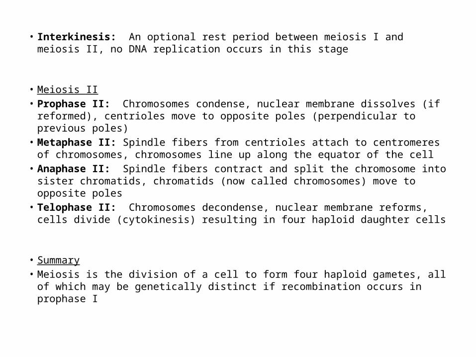
• Interkinesis: An optional rest period between meiosis I and meiosis II, no DNA replication occurs in this stage
• Meiosis II• Prophase II: Chromosomes condense, nuclear membrane dissolves (if
reformed), centrioles move to opposite poles (perpendicular to previous poles)• Metaphase II: Spindle fibers from centrioles attach to centromeres of
chromosomes, chromosomes line up along the equator of the cell• Anaphase II: Spindle fibers contract and split the chromosome into sister
chromatids, chromatids (now called chromosomes) move to opposite poles• Telophase II: Chromosomes decondense, nuclear membrane reforms, cells
divide (cytokinesis) resulting in four haploid daughter cells
• Summary• Meiosis is the division of a cell to form four haploid gametes, all of which may
be genetically distinct if recombination occurs in prophase I
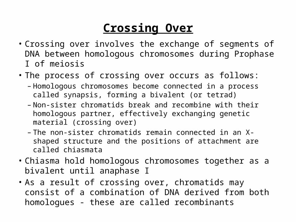
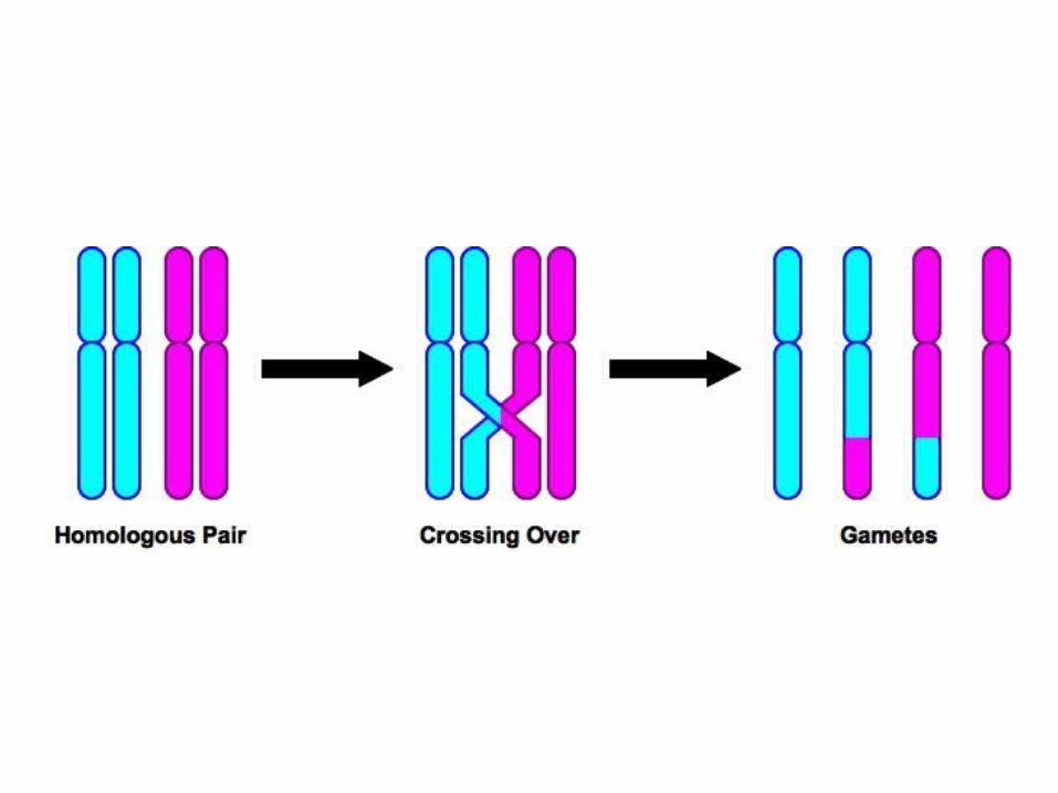
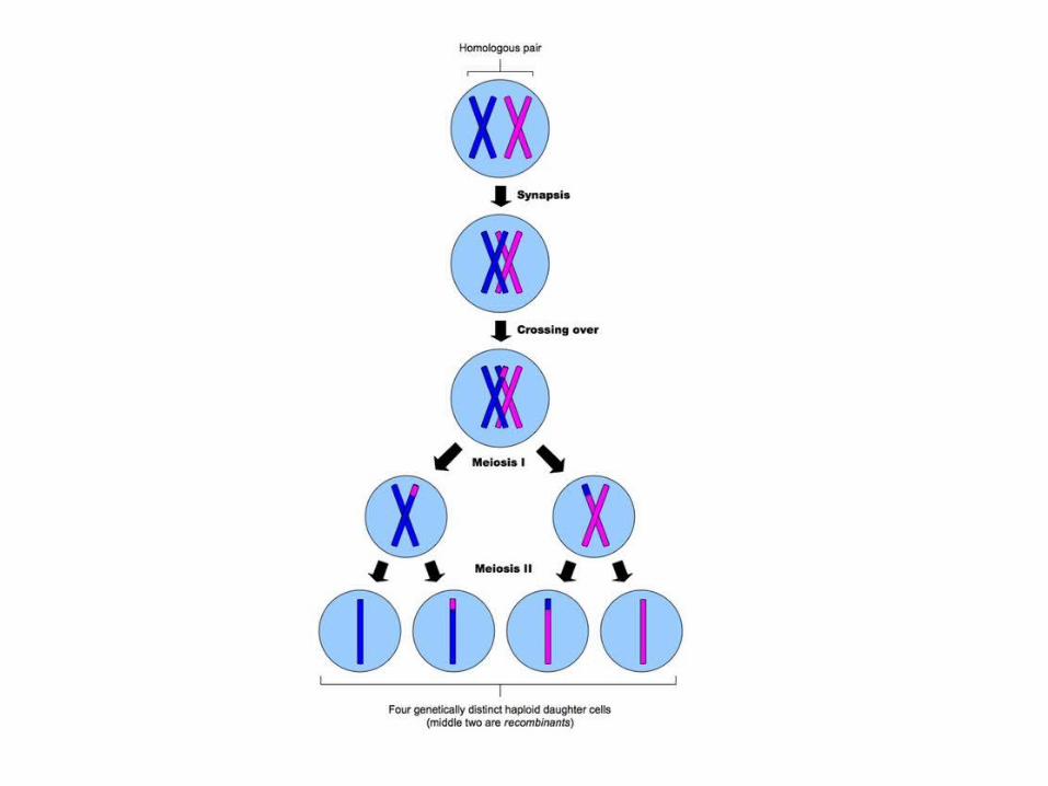
Crossing Over• Crossing over involves the exchange of segments of DNA
between homologous chromosomes during Prophase I of meiosis
• The process of crossing over occurs as follows:– Homologous chromosomes become connected in a process called
synapsis, forming a bivalent (or tetrad)– Non-sister chromatids break and recombine with their homologous
partner, effectively exchanging genetic material (crossing over)– The non-sister chromatids remain connected in an X-shaped
structure and the positions of attachment are called chiasmata• Chiasma hold homologous chromosomes together as a
bivalent until anaphase I• As a result of crossing over, chromatids may consist of a
combination of DNA derived from both homologues - these are called recombinants
Meiosis video
• Animation - http://highered.mcgraw-hill.com/sites/0072495855/student_view0/chapter28/animation__how_meiosis_works.html
• http://www.youtube.com/watch?v=qCLmR9-YY7o – 11 min
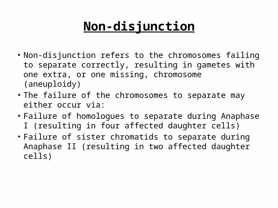
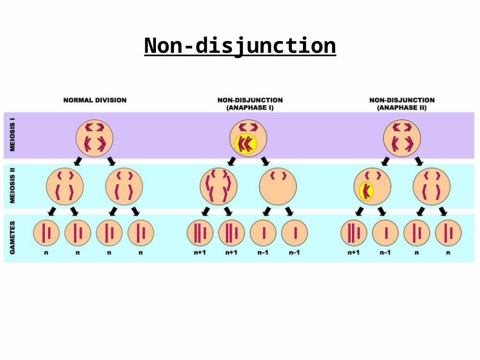
Non-disjunction
• Non-disjunction refers to the chromosomes failing to separate correctly, resulting in gametes with one extra, or one missing, chromosome (aneuploidy)
• The failure of the chromosomes to separate may either occur via:
• Failure of homologues to separate during Anaphase I (resulting in four affected daughter cells)
• Failure of sister chromatids to separate during Anaphase II (resulting in two affected daughter cells)
Non-disjunction
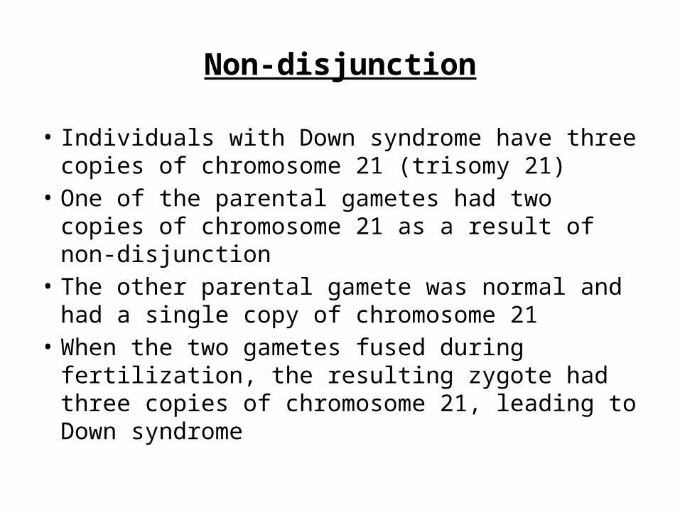
• Individuals with Down syndrome have three copies of chromosome 21 (trisomy 21)
• One of the parental gametes had two copies of chromosome 21 as a result of non-disjunction
• The other parental gamete was normal and had a single copy of chromosome 21
• When the two gametes fused during fertilization, the resulting zygote had three copies of chromosome 21, leading to Down syndrome
Non-disjunction
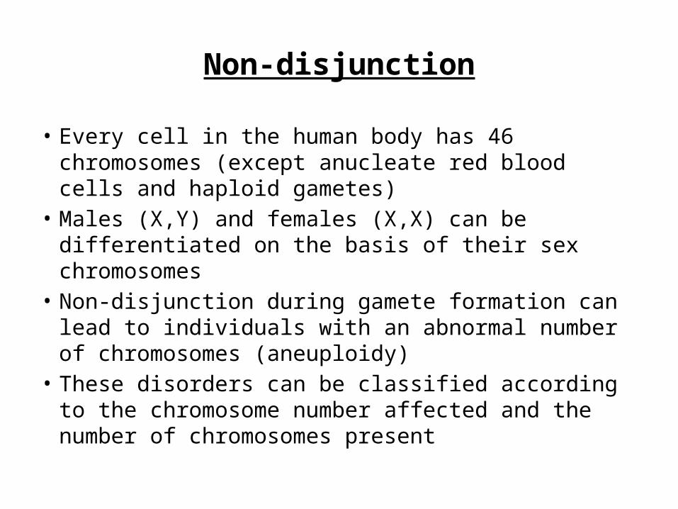
• Every cell in the human body has 46 chromosomes (except anucleate red blood cells and haploid gametes)
• Males (X,Y) and females (X,X) can be differentiated on the basis of their sex chromosomes
• Non-disjunction during gamete formation can lead to individuals with an abnormal number of chromosomes (aneuploidy)
• These disorders can be classified according to the chromosome number affected and the number of chromosomes present
Non-disjunction
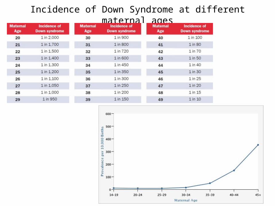
Incidence of Down Syndrome at different maternal ages
Application and Skills
• Application 1: Non-disjunction can cause Down syndrome and other chromosome abnormalities
• Application 2: Studies showing age of parents influences chances of non-disjunction
• Skill 1: Drawing diagrams to show the stages of meiosis resulting in the formation of four haploid cells.

3.4 - INHERITANCE
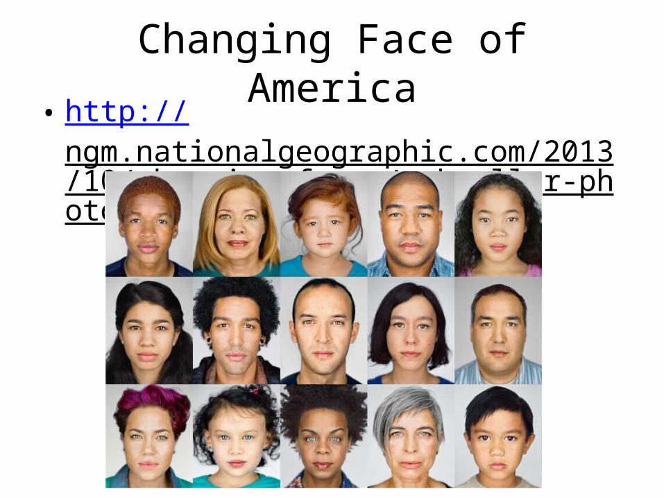
Changing Face of America• http://
ngm.nationalgeographic.com/2013/10/changing-faces/schoeller-photography
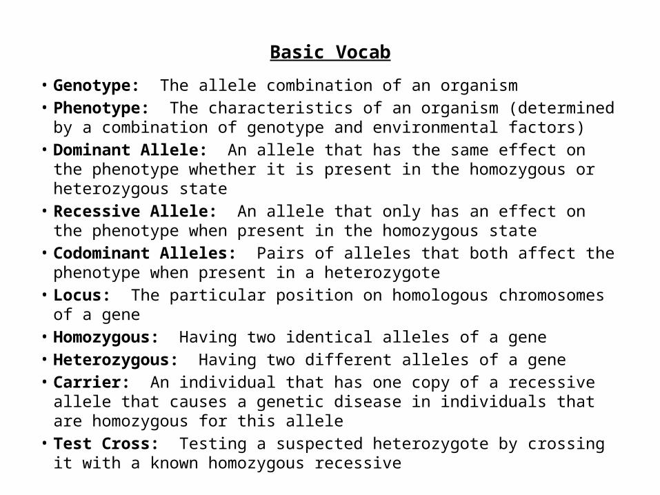
Basic Vocab
• Genotype: The allele combination of an organism• Phenotype: The characteristics of an organism (determined by a
combination of genotype and environmental factors)• Dominant Allele: An allele that has the same effect on the phenotype
whether it is present in the homozygous or heterozygous state• Recessive Allele: An allele that only has an effect on the phenotype
when present in the homozygous state• Codominant Alleles: Pairs of alleles that both affect the phenotype
when present in a heterozygote• Locus: The particular position on homologous chromosomes of a gene• Homozygous: Having two identical alleles of a gene• Heterozygous: Having two different alleles of a gene• Carrier: An individual that has one copy of a recessive allele that
causes a genetic disease in individuals that are homozygous for this allele
• Test Cross: Testing a suspected heterozygote by crossing it with a known homozygous recessive
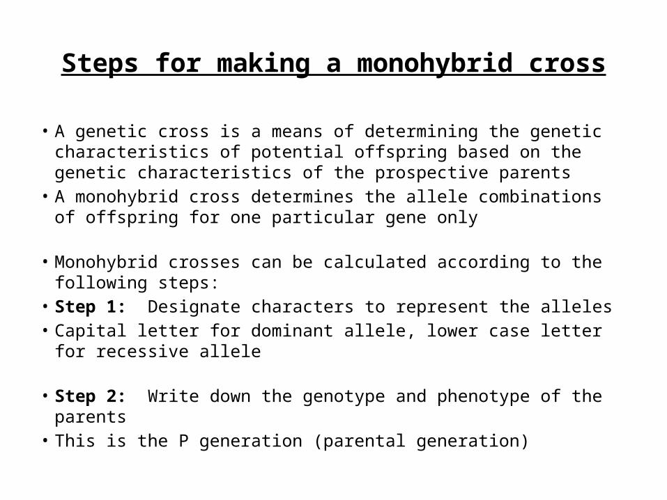
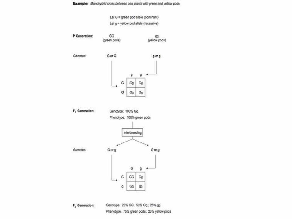
Steps for making a monohybrid cross
• A genetic cross is a means of determining the genetic characteristics of potential offspring based on the genetic characteristics of the prospective parents
• A monohybrid cross determines the allele combinations of offspring for one particular gene only
• Monohybrid crosses can be calculated according to the following steps:
• Step 1: Designate characters to represent the alleles • Capital letter for dominant allele, lower case letter for recessive allele
• Step 2: Write down the genotype and phenotype of the parents • This is the P generation (parental generation)
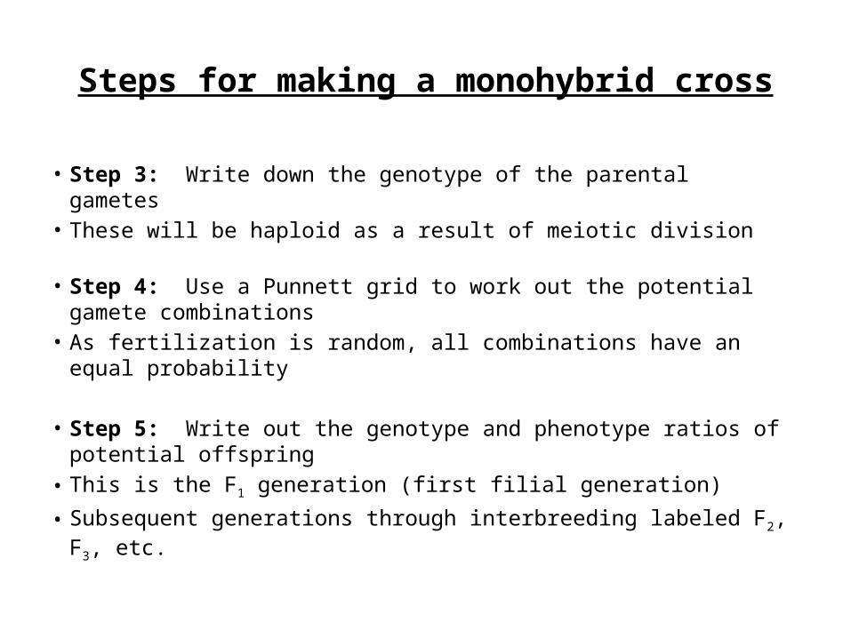
• Step 3: Write down the genotype of the parental gametes • These will be haploid as a result of meiotic division
• Step 4: Use a Punnett grid to work out the potential gamete combinations
• As fertilization is random, all combinations have an equal probability
• Step 5: Write out the genotype and phenotype ratios of potential offspring
• This is the F1 generation (first filial generation)
• Subsequent generations through interbreeding labeled F2, F3, etc.
Steps for making a monohybrid cross
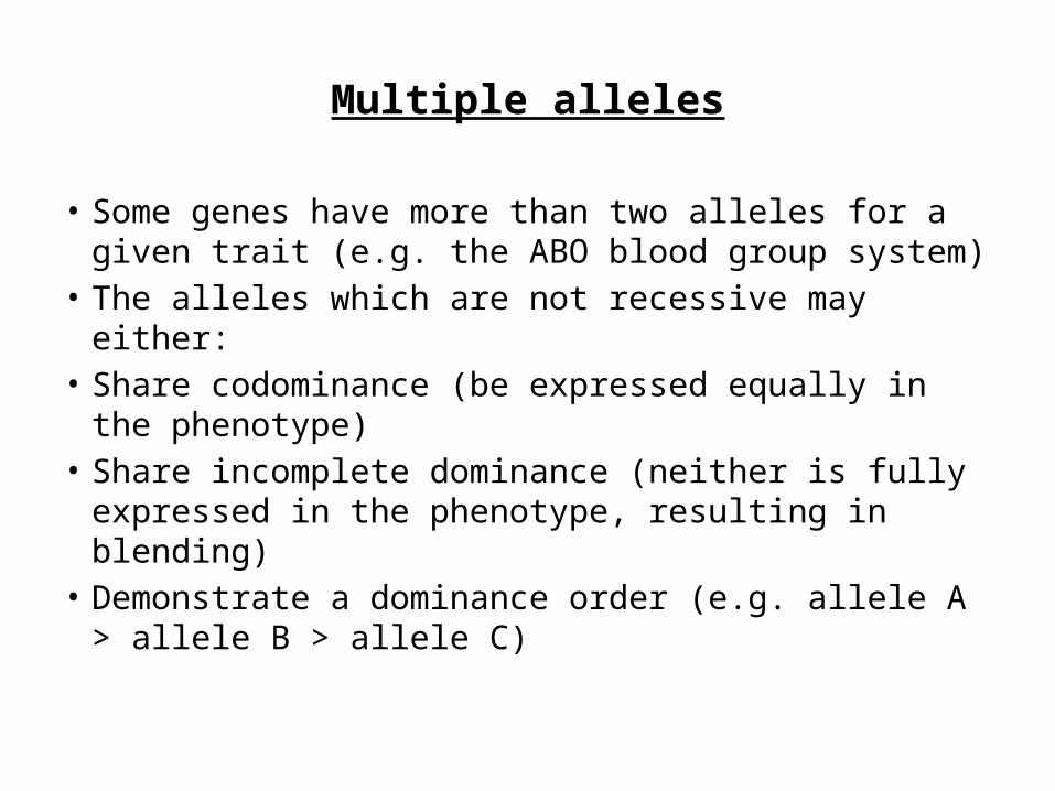
• Some genes have more than two alleles for a given trait (e.g. the ABO blood group system)
• The alleles which are not recessive may either: • Share codominance (be expressed equally in the
phenotype) • Share incomplete dominance (neither is fully
expressed in the phenotype, resulting in blending) • Demonstrate a dominance order (e.g. allele A >
allele B > allele C)
Multiple alleles
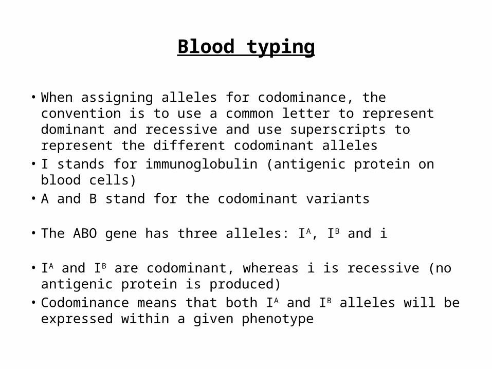
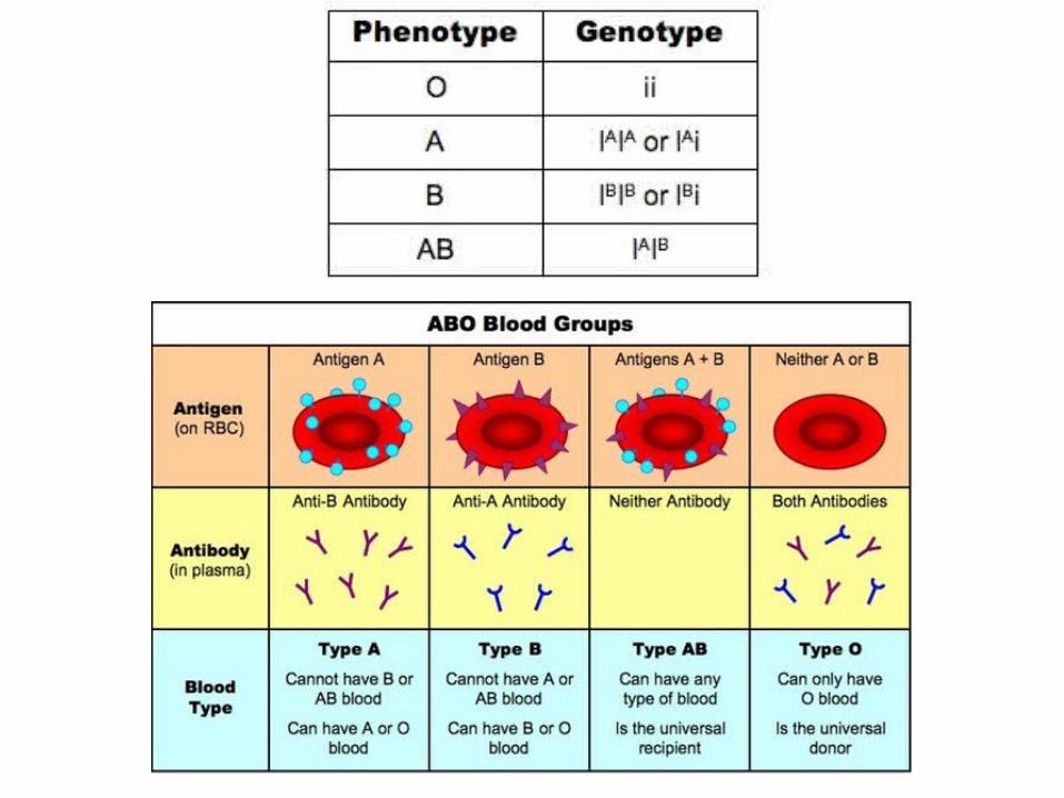
Blood typing
• When assigning alleles for codominance, the convention is to use a common letter to represent dominant and recessive and use superscripts to represent the different codominant alleles
• I stands for immunoglobulin (antigenic protein on blood cells)• A and B stand for the codominant variants
• The ABO gene has three alleles: IA, IB and i
• IA and IB are codominant, whereas i is recessive (no antigenic protein is produced)
• Codominance means that both IA and IB alleles will be expressed within a given phenotype
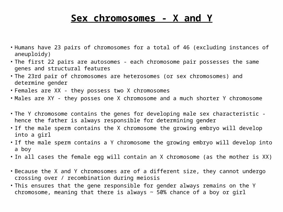
Sex chromosomes - X and Y
• Humans have 23 pairs of chromosomes for a total of 46 (excluding instances of aneuploidy)• The first 22 pairs are autosomes - each chromosome pair possesses the same genes and
structural features• The 23rd pair of chromosomes are heterosomes (or sex chromosomes) and determine
gender• Females are XX - they possess two X chromosomes• Males are XY - they posses one X chromosome and a much shorter Y chromosome
• The Y chromosome contains the genes for developing male sex characteristic - hence the father is always responsible for determining gender
• If the male sperm contains the X chromosome the growing embryo will develop into a girl• If the male sperm contains a Y chromosome the growing embryo will develop into a boy• In all cases the female egg will contain an X chromosome (as the mother is XX)
• Because the X and Y chromosomes are of a different size, they cannot undergo crossing over / recombination during meiosis
• This ensures that the gene responsible for gender always remains on the Y chromosome, meaning that there is always ~ 50% chance of a boy or girl
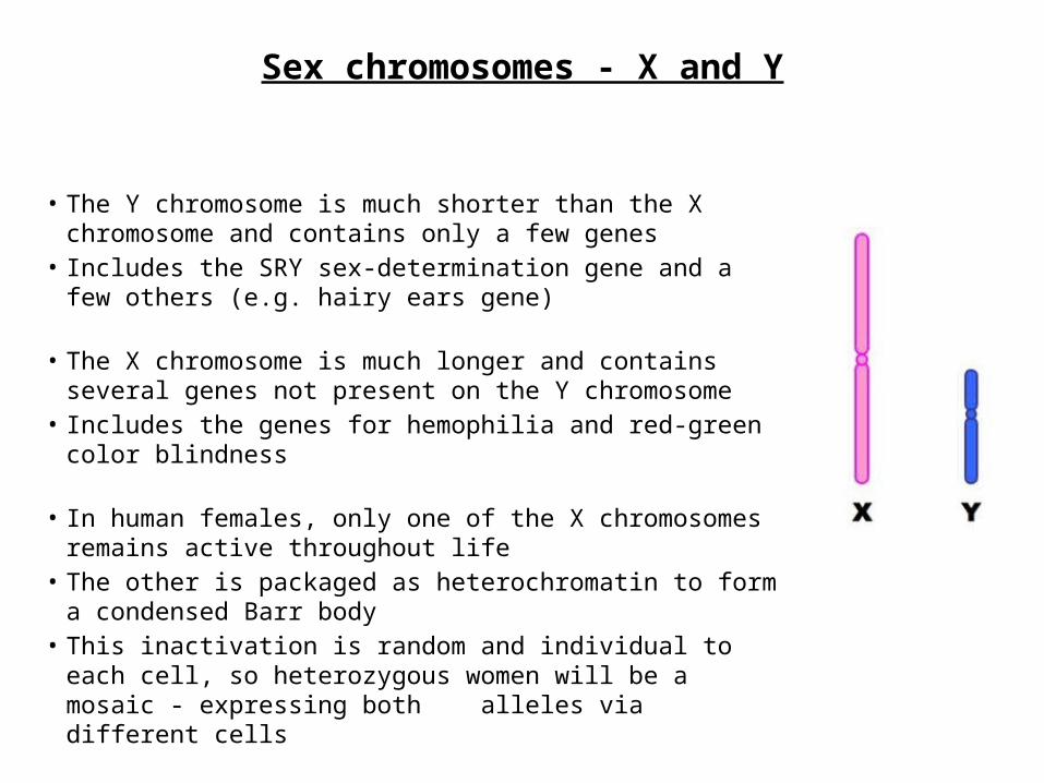
• The Y chromosome is much shorter than the X chromosome and contains only a few genes
• Includes the SRY sex-determination gene and a few others (e.g. hairy ears gene)
• The X chromosome is much longer and contains several genes not present on the Y chromosome
• Includes the genes for hemophilia and red-green color blindness
• In human females, only one of the X chromosomes remains active throughout life
• The other is packaged as heterochromatin to form a condensed Barr body
• This inactivation is random and individual to each cell, so heterozygous women will be a mosaic - expressing both alleles via different cells
Sex chromosomes - X and Y
Sex-linked traits
• Sex linkage refers to when a gene controlling a characteristic is found on a sex chromosome (and so we associate the trait with a predominant gender)
• Sex-linked conditions are usually X-linked, as very few genes exist on the shorter Y chromosome
• Color blindness and hemophilia are both examples of X-linked recessive conditions• The gene loci for these conditions are found on the non-homologous region of the
X chromosome (they are not present of the Y chromosome)• As males only have one allele for this gene they cannot be a carrier for the
condition • This means they have a higher frequency of being recessive and expressing the
trait• Males will always inherit an X-linked recessive condition from their mother• Females will only inherit an X-linked recessive condition if they receive a recessive
allele from both parents
• When assigning alleles for sex-linked traits the convention is to write the allele as a superscript to the sex chromosome (usually X)
• Hemophilia: XH = unaffected ; Xh = affected• Color Blindness: XA = unaffected ; Xa = affected
Sex-linked traits

Sex-linked traits
• As human females have two X chromosomes (and therefore two alleles for any given X-linked gene), they can be either homozygous or heterozygous
• Males only have one X chromosome (and therefore only one allele) and are hemizygous
Sex-linked traits
• An individual with a recessive allele for a disease condition that is masked by a normal dominant allele is said to be a carrier
• Carriers are heterozygous and can potentially pass the trait on to the next generation, but do not suffer from the defective condition themselves
• Females can be carriers for X-linked recessive conditions because they have two X chromosomes - males (XY) cannot be carriers
• Because a male only inherits an X chromosome from his mother, his chances of inheriting the disease condition from a carrier mother is greater
Sex-linked traits
Types of inheritance
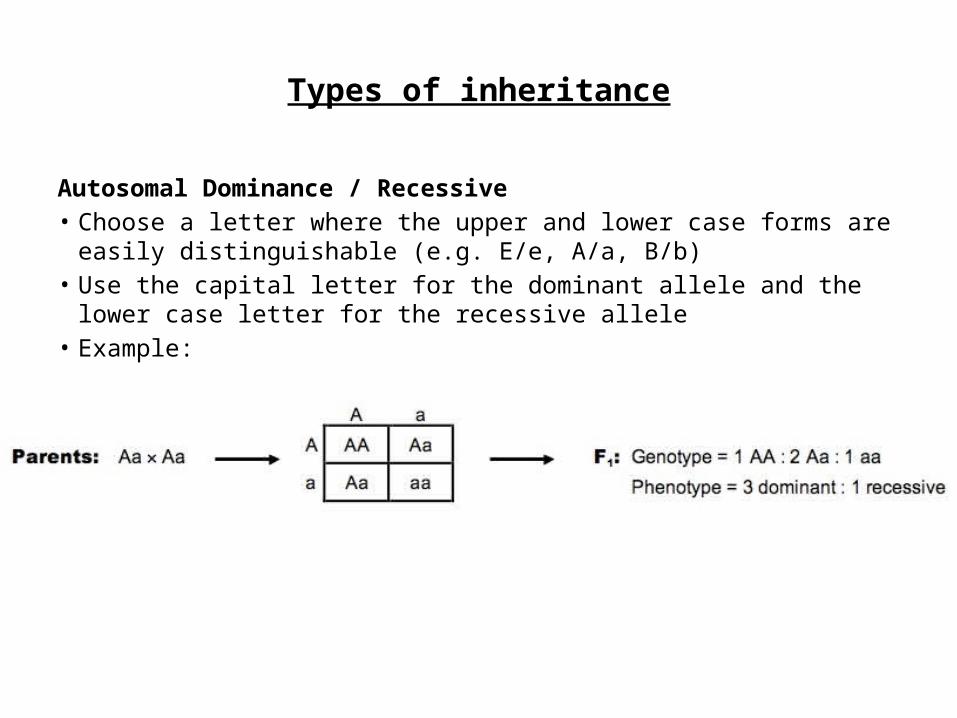
Autosomal Dominance / Recessive• Choose a letter where the upper and lower case forms are easily
distinguishable (e.g. E/e, A/a, B/b)• Use the capital letter for the dominant allele and the lower case letter for the
recessive allele• Example:
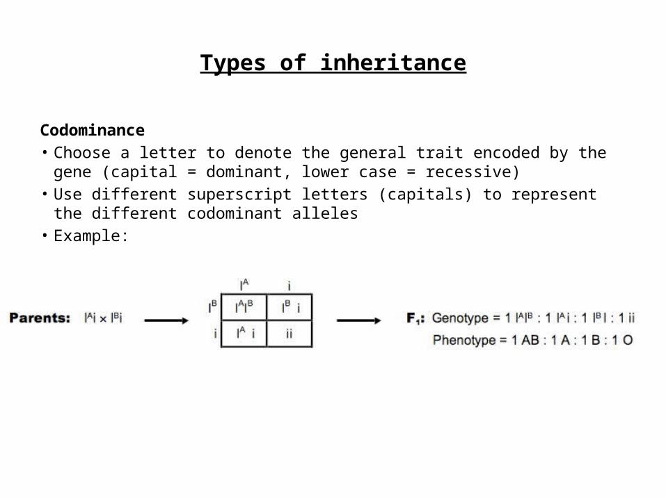
Codominance• Choose a letter to denote the general trait encoded by the gene (capital =
dominant, lower case = recessive)• Use different superscript letters (capitals) to represent the different codominant
alleles• Example:
Types of inheritance
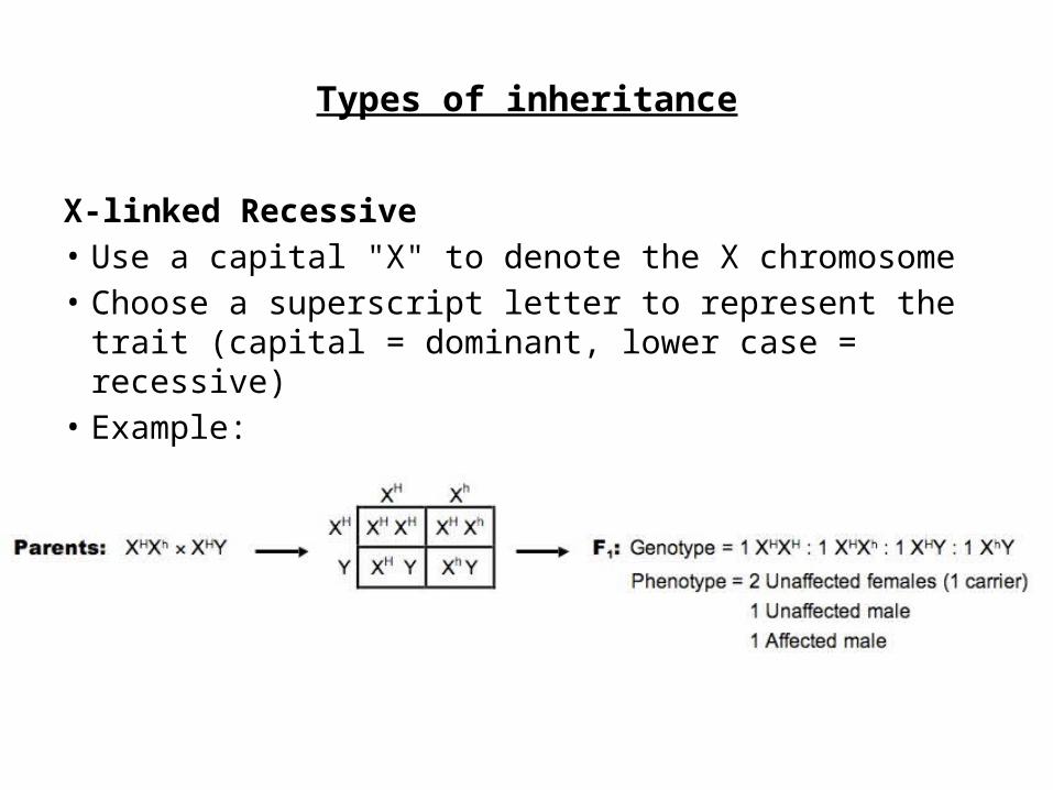
X-linked Recessive• Use a capital "X" to denote the X chromosome• Choose a superscript letter to represent the trait
(capital = dominant, lower case = recessive)• Example:
Types of inheritance
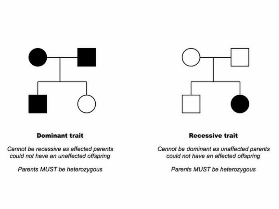
Pedigree
• A pedigree is a chart of the genetic history of a family over several generations
• Males are represented as squares, while females are represented as circles
• Shaded symbols means an individual is affected by a condition, while an unshaded symbol means they are unaffected
• A horizontal line between a man and woman represents mating and resulting children are shown as offshoots to this line

Autosomal Dominance• All affected individuals must have at least one
affected parent• If two parents are unaffected, all offspring must
be unaffected (homozygous recessive)• If two parents are affected, they may have
offspring who are unaffected (if parents are heterozygous)
• Often, more visible in a pedifree
Pedigree
Autosomal Recessive• If two parents show a trait, all children must
also show the trait (homozygous recessive)• An affected individual may have two normal
parents (if parents are both heterozygous carriers)
• Often less visible in a pedigree (can skip generations)
Pedigree
• X-Linked Recessive• If a female shows the trait, so must all sons as
well as her father• The disorder is more common in males
Pedigree
Application and Skills
• Application 1: Inheritance of ABO blood groups• Application 2: Red-green color blindness and hemophilia
as examples of sex-linked inheritance• Application 3: Consequences of radiation after nuclear
bombing of Hiroshima and accident at Chernobyl• Skill 1: Construction of Punnett grids for predicting the
outcomes of monohybrid genetic crosses• Skill 2: Comparison of predicted and actual outcomes of
genetic crosses using real data• Skill 3: Analysis of pedigree charts to deduce the pattern
of inheritance of genetic diseases.
3.5 – GENETIC MODIFICATION AND BIOTECHNOLOGY
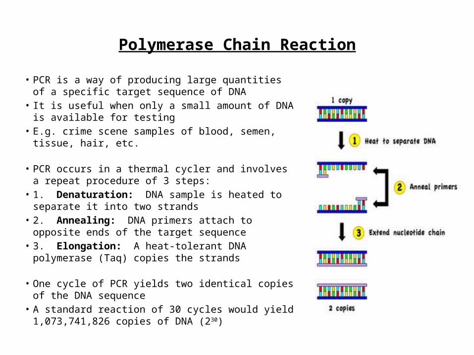
Polymerase Chain Reaction
• PCR is a way of producing large quantities of a specific target sequence of DNA
• It is useful when only a small amount of DNA is available for testing
• E.g. crime scene samples of blood, semen, tissue, hair, etc.
• PCR occurs in a thermal cycler and involves a repeat procedure of 3 steps:
• 1. Denaturation: DNA sample is heated to separate it into two strands
• 2. Annealing: DNA primers attach to opposite ends of the target sequence
• 3. Elongation: A heat-tolerant DNA polymerase (Taq) copies the strands
• One cycle of PCR yields two identical copies of the DNA sequence
• A standard reaction of 30 cycles would yield 1,073,741,826 copies of DNA (230)
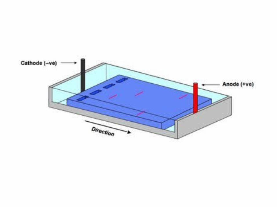
Gel electrophoresis
• Gel electrophoresis is a technique which is used to separate fragments of DNA according to size
• Samples of fragmented DNA are placed in the wells of an agarose gel• The gel is placed in a buffering solution and an electrical current is
passed across the gel• DNA, being negatively charged (due to phosphate), moves to the
positive terminus (anode)• Smaller fragments are less impeded by the gel matrix and move
faster through the gel• The fragments are thus separated according to size • Size can be calculated (in kilobases) by comparing against a known
industry standard
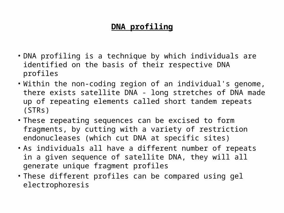
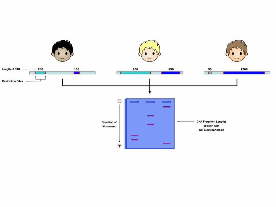
DNA profiling
• DNA profiling is a technique by which individuals are identified on the basis of their respective DNA profiles
• Within the non-coding region of an individual's genome, there exists satellite DNA - long stretches of DNA made up of repeating elements called short tandem repeats (STRs)
• These repeating sequences can be excised to form fragments, by cutting with a variety of restriction endonucleases (which cut DNA at specific sites)
• As individuals all have a different number of repeats in a given sequence of satellite DNA, they will all generate unique fragment profiles
• These different profiles can be compared using gel electrophoresis
DNA profiling
• A DNA sample is collected (blood, saliva, semen, etc.) and amplified using PCR• Satellite DNA (non-coding) is cut with specific restriction enzymes to generate
fragments• Individuals will have unique fragment lengths due to the variable length of
their short tandem repeats (STR)• The fragments are separated with gel electrophoresis (smaller fragments move
quicker through the gel)• The DNA profile can then be analyzed according to need
• Two applications of DNA profiling are:• Paternity testing (comparing DNA of offspring against potential fathers)• Forensic investigations (identifying suspects or victims based on crime-scene
DNA)
DNA profiling
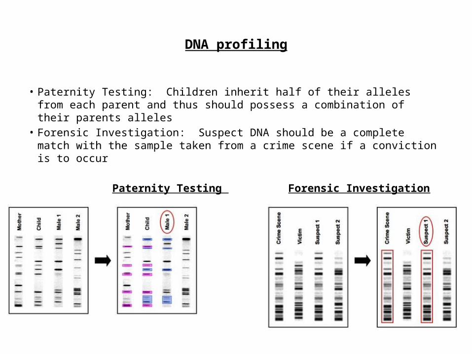
• Paternity Testing: Children inherit half of their alleles from each parent and thus should possess a combination of their parents alleles
• Forensic Investigation: Suspect DNA should be a complete match with the sample taken from a crime scene if a conviction is to occur
Paternity Testing Forensic Investigation
Human genome project
• The Human Genome Project (HGP) was an international cooperative venture established to sequence the 3 billion base pair (~25,000 genes) in the human genome
• The outcomes of this project include:• Mapping: We now know the number, location and basic sequence of human genes• Screening: This has allowed for the production of specific gene probes to detect
sufferers and carriers of genetic disease conditions• Medicine: With the discovery of new proteins and their functions, we can develop
improved treatments (pharmacogenetics and rational drug design)• Ancestry: It will give us improved insight into the origins, evolution and historical
migratory patterns of humans
• With the completion of the Human Genome Project in 2003, researcher have begun to sequence the genomes of several non-human organisms
• In Situ Hybridization
Genetic code
• The genetic code is universal, meaning that for every living organism the same codons code for the same amino acids (there are a few rare exceptions)
• This means that the genetic information from one organism could be translated by another (i.e. it is theoretically transferable)
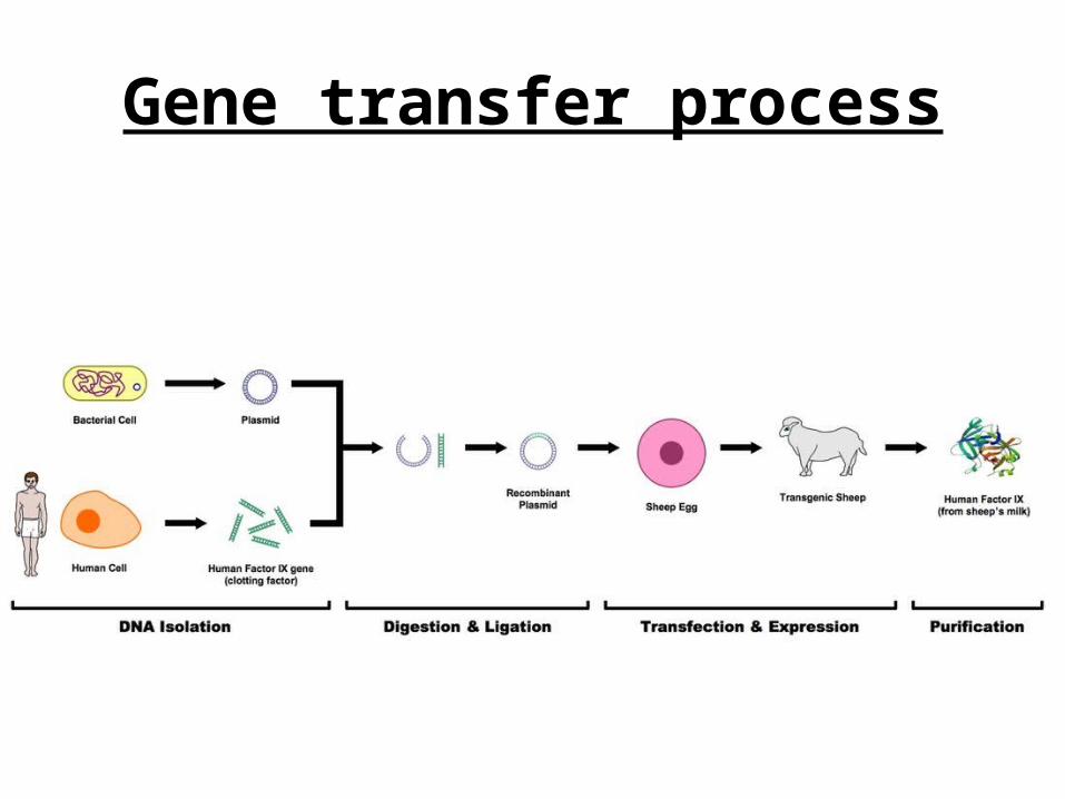
1. DNA Extraction• A plasmid is removed from a bacterial cell
(plasmids are small, circular DNA molecules that can exist and replicate autonomously)
• A gene of interest is removed from an organism's genome using a restriction endonuclease which cut at specific sequences of DNA
• The gene of interest and plasmid are both amplified using PCR technology
Gene transfer process
Gene transfer process
2. Digestion and Ligation• The plasmid is cut with the same restriction enzyme
that was used to excise the gene of interest• Cutting with certain restriction enzymes may
generate short sequence overhangs ("sticky ends") that allow the the two DNA constructs to fit together
• The gene of interest and plasmid are spliced together by DNA ligase creating a recombinant plasmid
Gene transfer process
3. Transfection and Expression• The recombinant plasmid is inserted into the desired
host cells (this is called transfection for eukaryotic cells and transformation for prokaryotic cells)
• The transgenic cells will hopefully produce the desired trait encoded by the gene of interest (expression)
• The product may need to subsequently be isolated from the host and purified in order to generate sufficient yield
Gene transfer process
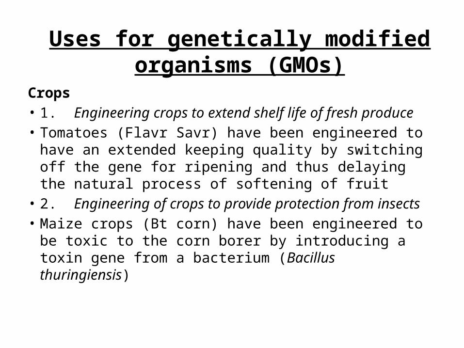
Crops• 1. Engineering crops to extend shelf life of fresh produce• Tomatoes (Flavr Savr) have been engineered to have an
extended keeping quality by switching off the gene for ripening and thus delaying the natural process of softening of fruit
• 2. Engineering of crops to provide protection from insects• Maize crops (Bt corn) have been engineered to be toxic to
the corn borer by introducing a toxin gene from a bacterium (Bacillus thuringiensis)
Uses for genetically modified organisms (GMOs)
Uses for genetically modified organisms (GMOs)
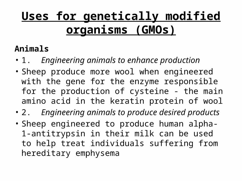
Animals• 1. Engineering animals to enhance production• Sheep produce more wool when engineered with the
gene for the enzyme responsible for the production of cysteine - the main amino acid in the keratin protein of wool
• 2. Engineering animals to produce desired products• Sheep engineered to produce human alpha-1-
antitrypsin in their milk can be used to help treat individuals suffering from hereditary emphysema
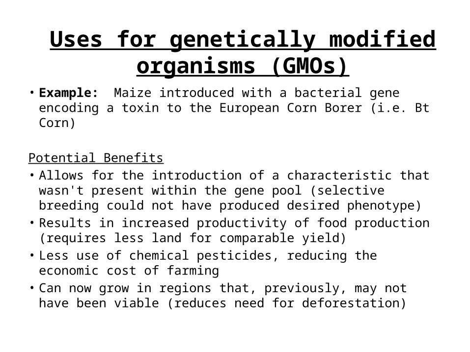
• Example: Maize introduced with a bacterial gene encoding a toxin to the European Corn Borer (i.e. Bt Corn)
Potential Benefits• Allows for the introduction of a characteristic that wasn't present
within the gene pool (selective breeding could not have produced desired phenotype)
• Results in increased productivity of food production (requires less land for comparable yield)
• Less use of chemical pesticides, reducing the economic cost of farming
• Can now grow in regions that, previously, may not have been viable (reduces need for deforestation)
Uses for genetically modified organisms (GMOs)
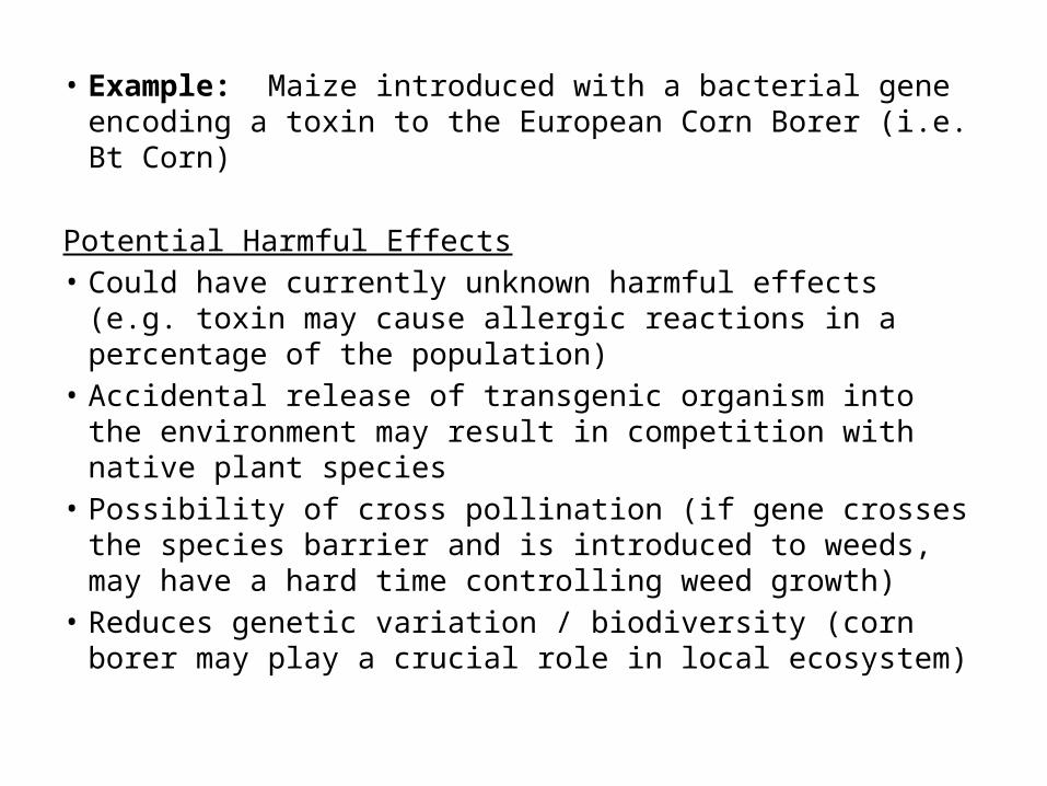
• Example: Maize introduced with a bacterial gene encoding a toxin to the European Corn Borer (i.e. Bt Corn)
Potential Harmful Effects• Could have currently unknown harmful effects (e.g. toxin
may cause allergic reactions in a percentage of the population)
• Accidental release of transgenic organism into the environment may result in competition with native plant species
• Possibility of cross pollination (if gene crosses the species barrier and is introduced to weeds, may have a hard time controlling weed growth)
• Reduces genetic variation / biodiversity (corn borer may play a crucial role in local ecosystem)
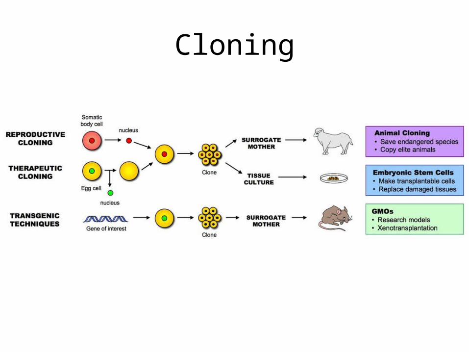
• A clone is a group of genetically identical organisms or a group of cells derived from a single parent cell
Cloning
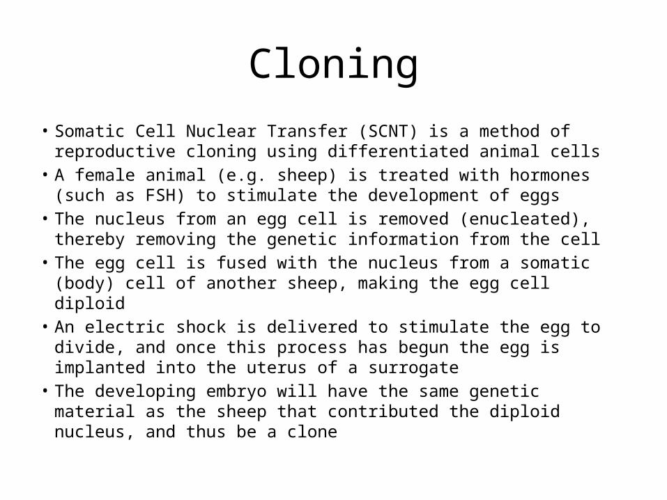
• Somatic Cell Nuclear Transfer (SCNT) is a method of reproductive cloning using differentiated animal cells
• A female animal (e.g. sheep) is treated with hormones (such as FSH) to stimulate the development of eggs
• The nucleus from an egg cell is removed (enucleated), thereby removing the genetic information from the cell
• The egg cell is fused with the nucleus from a somatic (body) cell of another sheep, making the egg cell diploid
• An electric shock is delivered to stimulate the egg to divide, and once this process has begun the egg is implanted into the uterus of a surrogate
• The developing embryo will have the same genetic material as the sheep that contributed the diploid nucleus, and thus be a clone
Cloning
Cloning
• Arguments for Therapeutic Cloning• May be used to cure serious diseases or disabilities with
cell therapy (replacing bad cells with good ones)• Stem cell research may pave the way for future
discoveries and beneficial technologies that would not have occurred if their use had been banned
• Stem cells can be taken from embryos that have stopped developing and would have died anyway (e.g. abortions)
• Cells are taken at a stage when the embryo has no nervous system and can arguably feel no pain
Cloning

• Arguments Against Therapeutic Cloning• Involves the creation and destruction of human embryos (at
what point do we afford the right to life?)• Embryonic stem cells are capable of continued division and
may develop into cancerous cells and cause tumors• More embryos are generally produced than are needed, so
excess embryos are killed• With additional cost and effort, alternative technologies may
fulfill similar roles (e.g. nuclear reprogramming of differentiated cell lines)
Cloning
Application and Skills
• Application 1: Use of DNA profiling in paternity and forensic investigations
• Application 2: gene transfer to bacteria using plasmids makes use of restriction endonucleases and DNA ligase.
• Application 3: Assessment of the potential risks and benefits associated with genetic modification of crops
• Application 4: Production of cloned embryos produced by somatic-cell nuclear transfer