Dr. Ahmed El-Bialy, Dr. Sahar Fawzy Combinational Circuits Dr. Ahmed El-Bialy Dr. Sahar Fawzy.
Upload
blake-thompsonCategory
view
221download
0
Unit 2 – Digestion ModuleHistology of the pancreas
and biliary tract
Safaa El Bialy (MD,PhD)Ottawa University
Objectives Outline the normal basic histology of the
pancreas, gall bladder and biliary ducts
Describe the histology of the exocrine pancreas and the function of the exocrine glands
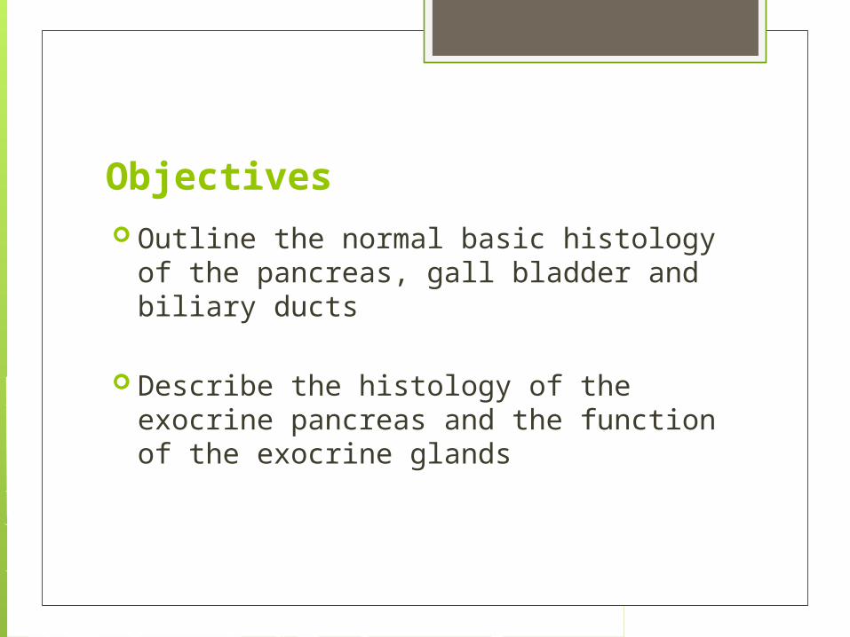
duodenum
liver
stomach
main pancreatic duct (duct of
Wirsung)
pancreas
gall bladder
Pancreas
The pancreas extends transversely from the duodenum to the spleen and lies behind the stomach .
Located in the upper abdomen , pancreas is a deep organ explaining the difficulties in early diagnosis of its pathologic conditions .
It consists of 4 parts : the head and neck that fit within the duodenum, the body and tail ( intraperitoneal ) that extends to the edge of the spleen.
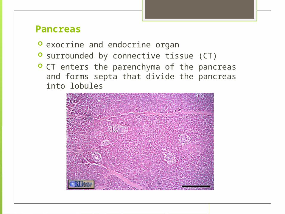
Pancreas exocrine and endocrine organ surrounded by connective tissue (CT) CT enters the parenchyma of the pancreas and forms
septa that divide the pancreas into lobules
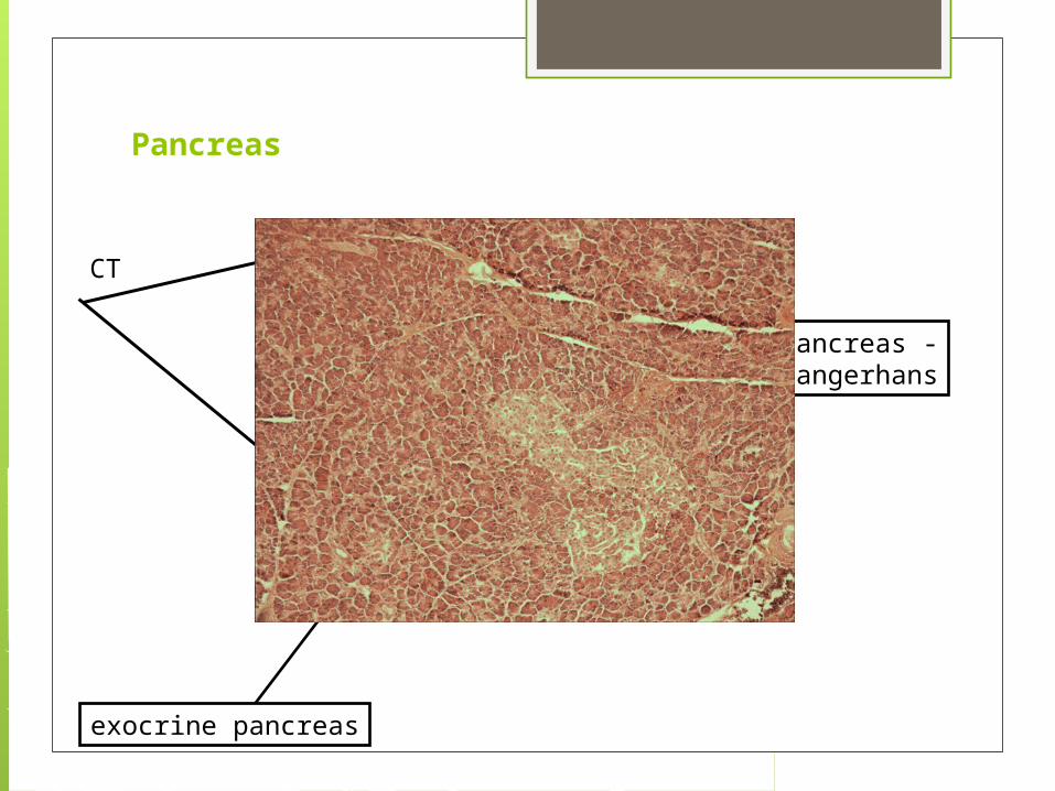
Pancreas
endocrine pancreas -island of Langerhans
exocrine pancreas
CT
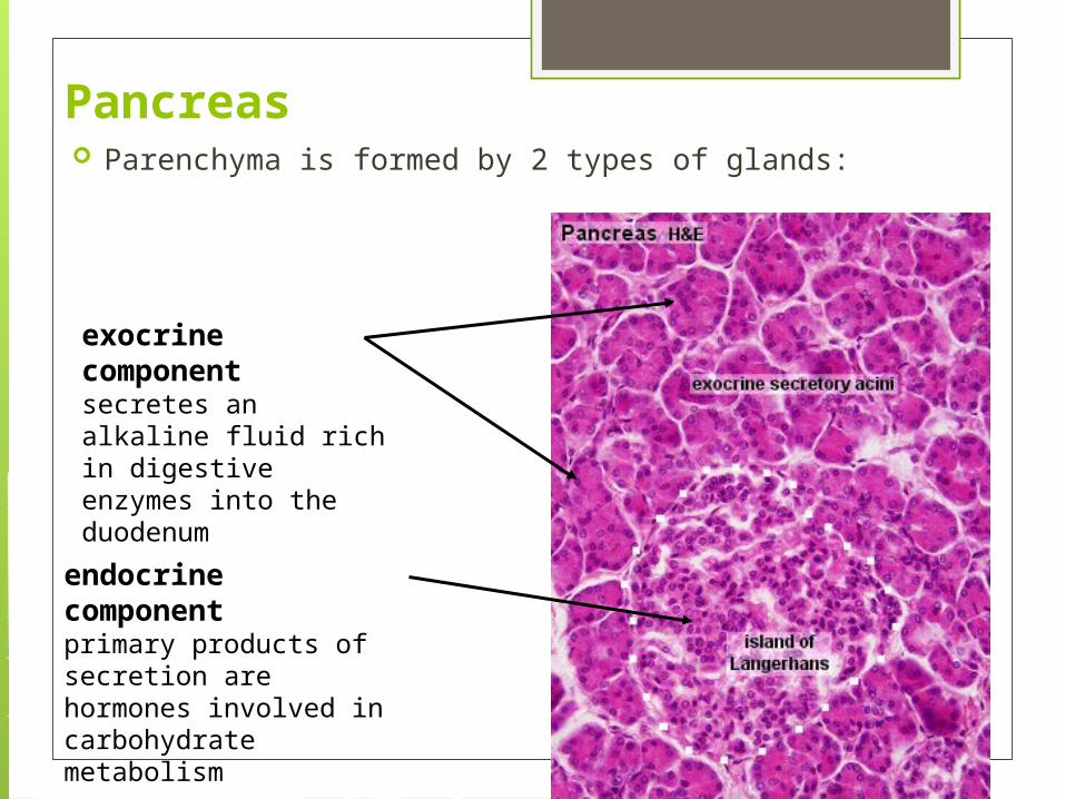
Pancreas Parenchyma is formed by 2 types of glands:
exocrine componentsecretes an alkaline fluid rich in digestive enzymes into the duodenum
endocrine componentprimary products of secretion are hormones involved in carbohydrate metabolism

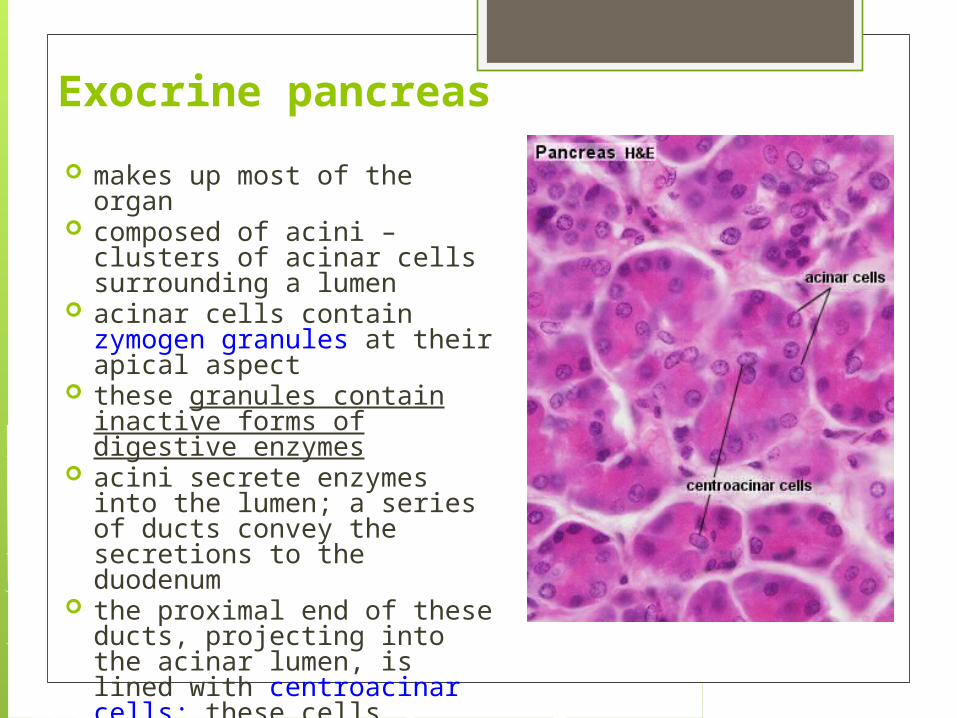
Exocrine pancreas
makes up most of the organ composed of acini – clusters of
acinar cells surrounding a lumen acinar cells contain zymogen
granules at their apical aspect these granules contain inactive
forms of digestive enzymes acini secrete enzymes into the
lumen; a series of ducts convey the secretions to the duodenum
the proximal end of these ducts, projecting into the acinar lumen, is lined with centroacinar cells; these cells secrete bicarbonate
Exocrine pancreas
centroacinar cells
acinar cells
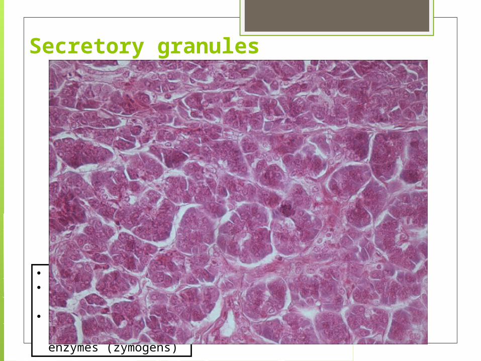
Secretory granules
• apical• acidophilic (proteinic)• contain precursors of
digestive enzymes (zymogens)
Pancreatic digestive enzymes
carbohydrate digestion - amylase digestion of proteins (proteases) - trypsin,
chymotrypsin, etc. lipid digestion - lipase, phospholipase, etc.
Control of pancreatic secretion secretion of digestive enzymes stimulated
primarily by: cholecystokinin (secreted by endocrine cells of the
mucosa of the duodenum and proximal jejunum) acetylcholine (secreted by parasympathetic fibres
of the vagus nerve and by nerves of the enteric nervous system)
secretion of bicarbonate and water stimulated primarily by: secretin (secreted by endocrine cells of the mucosa
of the duodenum and proximal jejunum)

Exocrine pancreas
This section shows several acini (A). Acinar cells contain acidophilic granules. The base of these acinar cells is basophilic due to the abundance of ribosomes. Some acini show in their center pale centroacinar cells ( blue arrows) .
In the center of the field we observe a longitudinal section of a small intercalated duct . The canal is lined with flattened epithelial cells ( arrowheads)
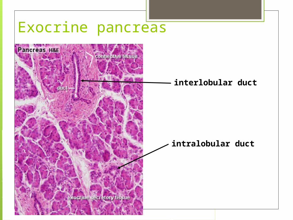
Exocrine pancreas
interlobular duct
intralobular duct
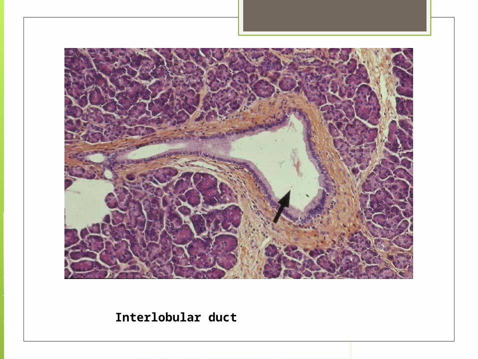
Interlobular duct
“Acute pancreatitis is the most terrible of all the calamities that occur in connection with the viscera, The suddenness of its onset , the illimitable agony which accompanies it, and the mortality attendant upon it, all render it the most formidable of catastrophes” Lord Moynihan
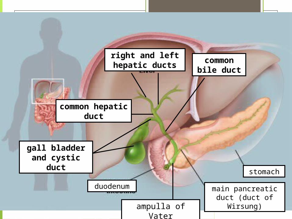
duodenum
right and left hepatic ducts
stomach
main pancreatic duct (duct of Wirsung)
gall bladder and cystic duct
ampulla of Vater
common hepatic duct
common bile duct
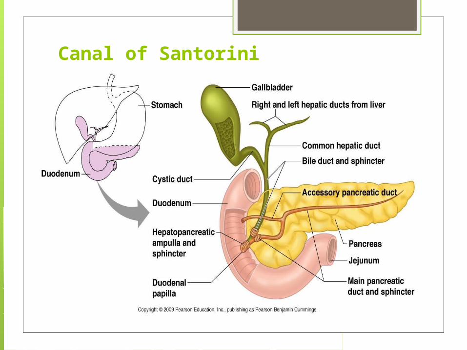
Canal of Santorini

Extrahepatic bile ducts
the intrahepatic bile ducts unite to form the right and left hepatic ducts, which anastomose to form the common hepatic duct, which becomes the common bile duct (draining into the duodenum)
the extrahepatic bile ducts are lined by a simple columnar epithelium supported by dense (fibrous) connective tissue
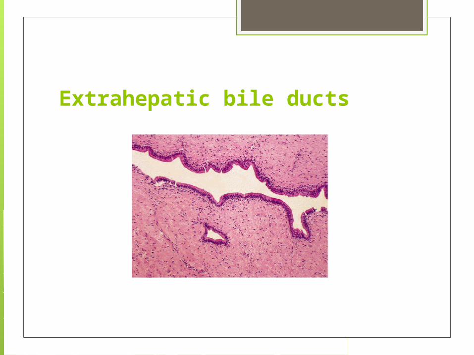
Extrahepatic bile ducts

Gall bladder Functions:
storage site for bile concentration of bile
wall is formed by a mucosa, a muscularis and an adventitia/serosa (no submucosa or muscularis mucosa)
the mucosa has numerous folds and is lined by simple columnar epithelium
the muscularis is a layer of smooth muscle containing receptor cells for cholecystokinin (CCK) – main stimulus for contraction of the gall bladder
Bile
secreted by the liver composed of: water, electrolytes, bile acids (salts),
bilirubin, cholesterol, lecithin, etc. bile acids are synthesized by smooth ER of
hepatocytes and undergo enterohepatic recirculation bile acids emulsify food lipids, facilitate their digestion
by pancreatic enzymes, and assist in lipid absorption
by the small intestine
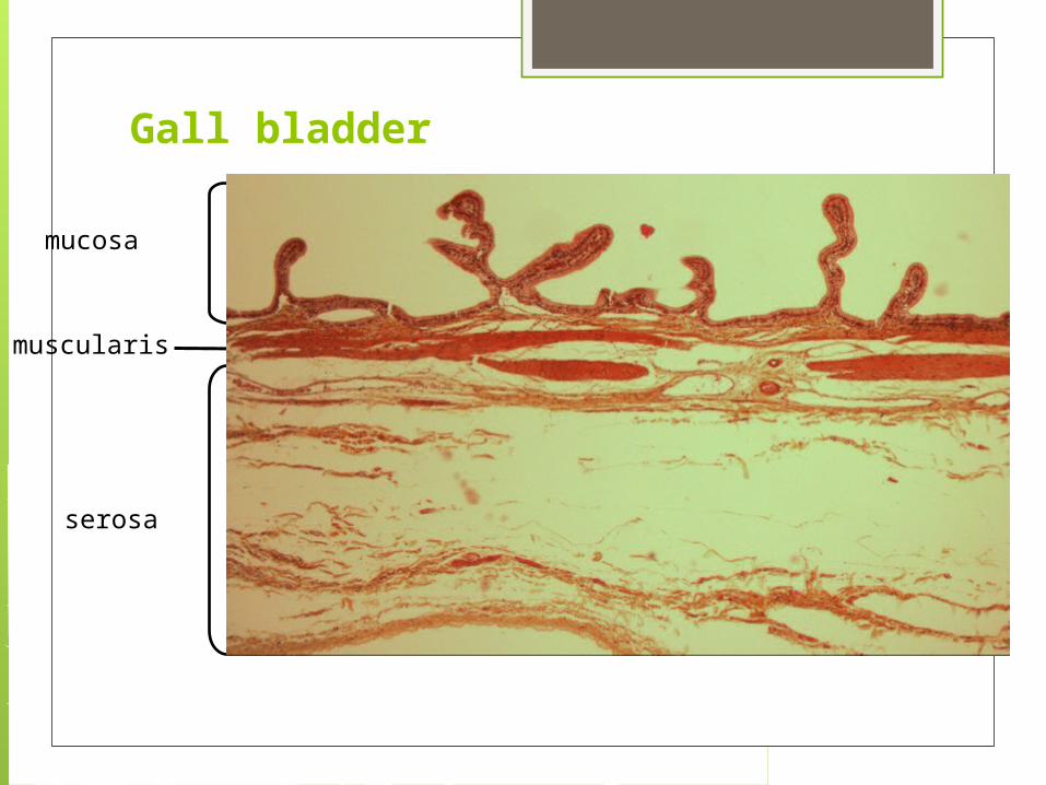
Gall bladder
mucosa
muscularis
serosa

lamina propria
Gall bladder mucosa• simple columnar
epithelium– appearance of
cells opening into the lumen that are covered by microvilli
– no goblet cells
epithelium
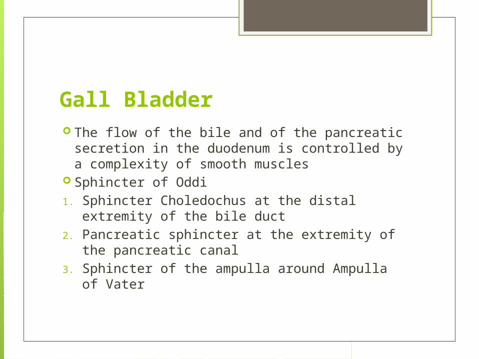
Gall Bladder The flow of the bile and of the pancreatic secretion
in the duodenum is controlled by a complexity of smooth muscles
Sphincter of Oddi
1. Sphincter Choledochus at the distal extremity of the bile duct
2. Pancreatic sphincter at the extremity of the pancreatic canal
3. Sphincter of the ampulla around Ampulla of Vater

Sphincter of Oddi
Gall bladder stones
There are two types of stones: cholesterol stones , represent about 80%
of the stones, and pigment stones , which are composed of
bilirubin and constitute the remaining 20% Gallbladder stones are more common in
women and in the elderly and in certain groups such as Americans of Indian origin and overweight people
4 F’s rule: female, fair, fatty, forty