Unit #1 Gross Practice Questions - University of Utah 01 Questions.pdfAnswer= The right lung has...
28
ANAT 6010 – Unit #1 Practice Problems The following are a sampling of exam style questions on material covered in Unit #1. Here are the disclaimers: • These questions have been reviewed but some mistakes may still appear. The definitive source for anatomical material is the textbook “Gray’s Anatomy for Students” not from these practice questions. • These practice questions do not provide an equal representation of all of the content covered thus far in the course. These are simply a sampling of question-types. • The purpose of these questions is to help provide examples of the types of questions you can expect on Monday’s exam as well as future lecture exams in ANAT 6010. • Please do not hesitate to email Dr. Morton if you have question. Dr. Weyrich and I wish the best on Monday’s (as well as Tuesday and Wedensday’s) examinations. Sincerely, Dr. Morton
Transcript of Unit #1 Gross Practice Questions - University of Utah 01 Questions.pdfAnswer= The right lung has...
ANAT 6010 – Unit #1 Practice Problems
The following are a sampling of exam style questions on material covered in Unit #1. Here are the
disclaimers:
• These questions have been reviewed but some mistakes may still appear. The definitive source
for anatomical material is the textbook “Gray’s Anatomy for Students” not from these practice
questions.
• These practice questions do not provide an equal representation of all of the content covered
thus far in the course. These are simply a sampling of question-types.
• The purpose of these questions is to help provide examples of the types of questions you can
expect on Monday’s exam as well as future lecture exams in ANAT 6010.
• Please do not hesitate to email Dr. Morton if you have question. Dr. Weyrich and I wish the
best on Monday’s (as well as Tuesday and Wedensday’s) examinations.
Sincerely,
Dr. Morton
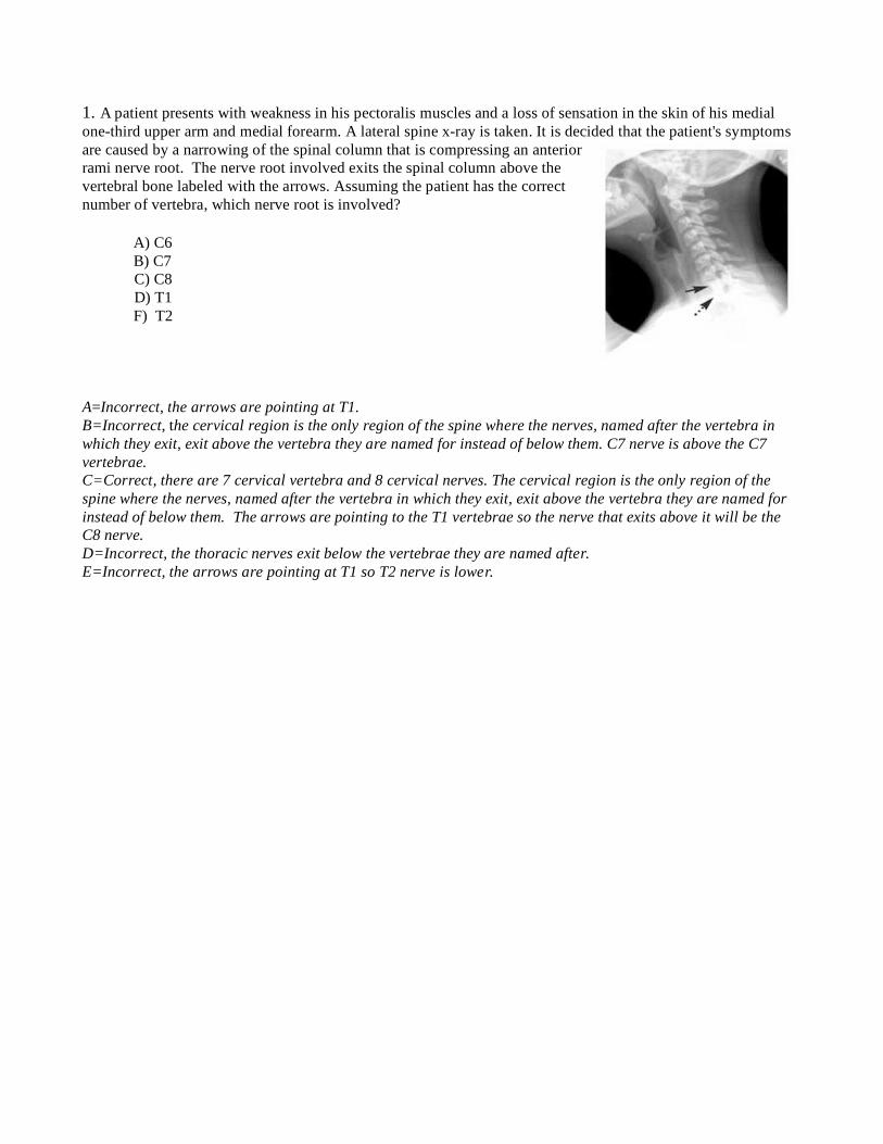
1. A patient presents with weakness in his pectoralis muscles and a loss of sensation in the skin of his medial
one-third upper arm and medial forearm. A lateral spine x-ray is taken. It is decided that the patient's symptoms
are caused by a narrowing of the spinal column that is compressing an anterior
rami nerve root. The nerve root involved exits the spinal column above the
vertebral bone labeled with the arrows. Assuming the patient has the correct
number of vertebra, which nerve root is involved?
A) C6
B) C7
C) C8
D) T1
F) T2
A=Incorrect, the arrows are pointing at T1.
B=Incorrect, the cervical region is the only region of the spine where the nerves, named after the vertebra in
which they exit, exit above the vertebra they are named for instead of below them. C7 nerve is above the C7
vertebrae.
C=Correct, there are 7 cervical vertebra and 8 cervical nerves. The cervical region is the only region of the
spine where the nerves, named after the vertebra in which they exit, exit above the vertebra they are named for
instead of below them. The arrows are pointing to the T1 vertebrae so the nerve that exits above it will be the
C8 nerve.
D=Incorrect, the thoracic nerves exit below the vertebrae they are named after.
E=Incorrect, the arrows are pointing at T1 so T2 nerve is lower.

2. A patent with a surgically fixed spine is x-rayed. The normal number of vertebral bones and ribs are seen
along with a metal fixation device (arrows are pointing to the metal fixation device). On the AP view the metal
device covers which three vertebrae?
A) T11, T12, T13
B) T12, L1, L2
C) T10, T11, L1
D) T11, T12, L1
A=Incorrect, the thoracic spine only has 12 vertebrae.
B=Correct, the image on the left is the AP. T12 is determined from the last rib. The lumbar spine has 5 vertebra
so you could count up from the bottom of the lumbar spine.
C=Incorrect, the thoracic spine has 12 vertebrae not 11.
D=Incorrect, the last rib is on T12 not T11.
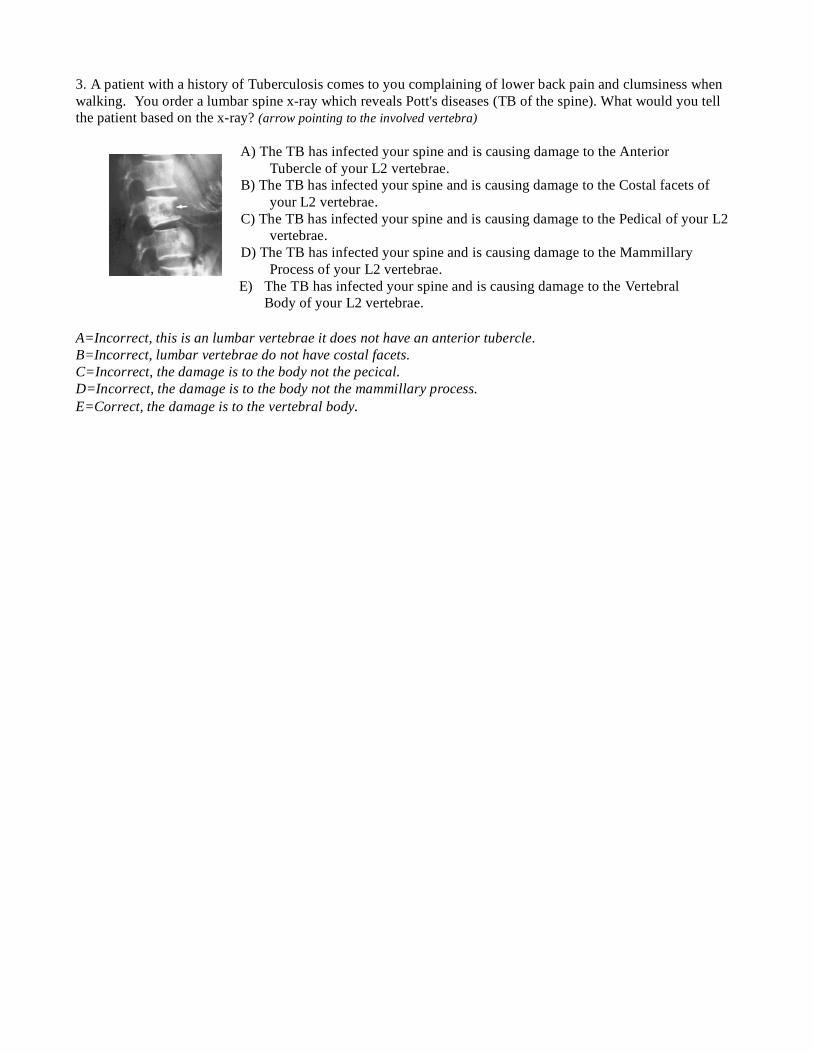
3. A patient with a history of Tuberculosis comes to you complaining of lower back pain and clumsiness when
walking. You order a lumbar spine x-ray which reveals Pott's diseases (TB of the spine). What would you tell
the patient based on the x-ray? (arrow pointing to the involved vertebra)
A) The TB has infected your spine and is causing damage to the Anterior
Tubercle of your L2 vertebrae.
B) The TB has infected your spine and is causing damage to the Costal facets of
your L2 vertebrae.
C) The TB has infected your spine and is causing damage to the Pedical of your L2
vertebrae.
D) The TB has infected your spine and is causing damage to the Mammillary
Process of your L2 vertebrae.
E) The TB has infected your spine and is causing damage to the Vertebral
Body of your L2 vertebrae.
A=Incorrect, this is an lumbar vertebrae it does not have an anterior tubercle.
B=Incorrect, lumbar vertebrae do not have costal facets.
C=Incorrect, the damage is to the body not the pecical.
D=Incorrect, the damage is to the body not the mammillary process.
E=Correct, the damage is to the vertebral body.
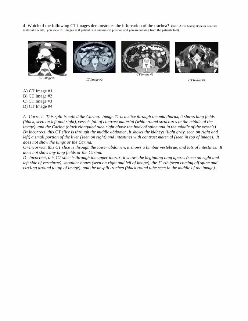
4. Which of the following CT images demonstrates the bifurcation of the trachea? (hint: Air = black; Bone or contrast
material = white; you view CT images as if patient is in anatomical position and you are looking from the patients feet)
A) CT Image #1
B) CT Image #2
C) CT Image #3
D) CT Image #4
A=Correct. This split is called the Carina. Image #1 is a slice through the mid thorax, it shows lung fields
(black, seen on left and right), vessels full of contrast material (white round structures in the middle of the
image), and the Carina (black elongated tube right above the body of spine and in the middle of the vessels).
B=Incorrect, this CT slice is through the middle abdomen, it shows the kidneys (light gray, seen on right and
left) a small portion of the liver (seen on right) and intestines with contrast material (seen in top of image). It
does not show the lungs or the Carina.
C=Incorrect, this CT slice is through the lower abdomen, it shows a lumbar vertebrae, and lots of intestines. It
does not show any lung fields or the Carina.
D=Incorrect, this CT slice is through the upper thorax, it shows the beginning lung apexes (seen on right and
left side of vertebrae), shoulder bones (seen on right and left of image), the 1st rib (seen coming off spine and
circling around to top of image), and the unsplit trachea (black round tube seen in the middle of the image).
CT Image #1
CT Image #2 CT Image #3
CT Image #4
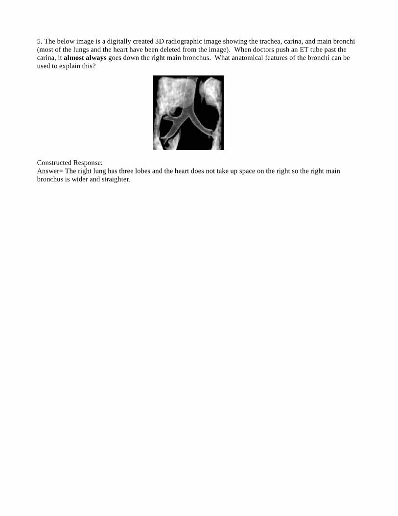
5. The below image is a digitally created 3D radiographic image showing the trachea, carina, and main bronchi
(most of the lungs and the heart have been deleted from the image). When doctors push an ET tube past the
carina, it almost always goes down the right main bronchus. What anatomical features of the bronchi can be
used to explain this?
Constructed Response:
Answer= The right lung has three lobes and the heart does not take up space on the right so the right main
bronchus is wider and straighter.
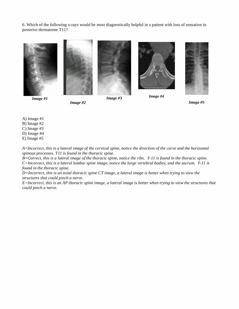
6. Which of the following x-rays would be most diagnostically helpful in a patient with loss of sensation in
posterior dermatome T11?
A) Image #1
B) Image #2
C) Image #3
D) Image #4
E) Image #5
A=Incorrect, this is a lateral image of the cervical spine, notice the direction of the curve and the horizontal
spinous processes. T11 is found in the thoracic spine.
B=Correct, this is a lateral image of the thoracic spine, notice the ribs. T-11 is found in the thoracic spine.
C=Incorrect, this is a lateral lumbar spine image, notice the large vertebral bodies, and the sacrum. T-11 is
found in the thoracic spine.
D=Incorrect, this is an axial thoracic spine CT image, a lateral image is better when trying to view the
structures that could pinch a nerve.
E=Incorrect, this is an AP thoracic spine image, a lateral image is better when trying to view the structures that
could pinch a nerve.
Image #1
Image #2
Image #3
Image #4
Image #5
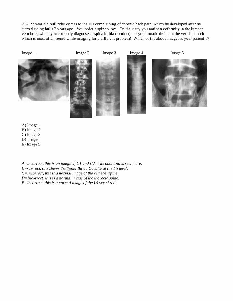
7. A 22 year old bull rider comes to the ED complaining of chronic back pain, which he developed after he
started riding bulls 3 years ago. You order a spine x-ray. On the x-ray you notice a deformity in the lumbar
vertebrae, which you correctly diagnose as spina bifida occulta (an asymptomatic defect in the vertebral arch
which is most often found while imaging for a different problem). Which of the above images is your patient’s?
Image 1 Image 2 Image 3 Image 4 Image 5
A) Image 1
B) Image 2
C) Image 3
D) Image 4
E) Image 5
A=Incorrect, this is an image of C1 and C2. The odontoid is seen here.
B=Correct, this shows the Spina Bifida Occulta at the L5 level.
C=Incorrect, this is a normal image of the cervical spine.
D=Incorrect, this is a normal image of the thoracic spine.
E=Incorrect, this is a normal image of the L5 vertebrae.
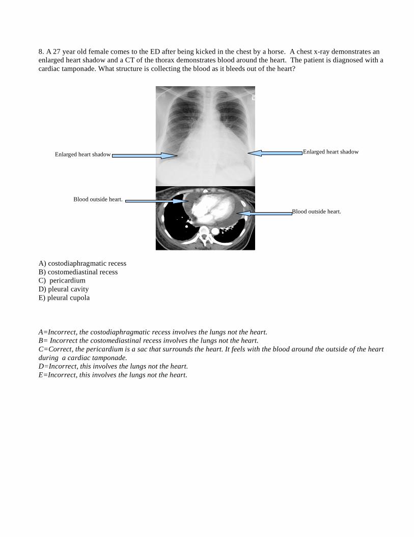
8. A 27 year old female comes to the ED after being kicked in the chest by a horse. A chest x-ray demonstrates an
enlarged heart shadow and a CT of the thorax demonstrates blood around the heart. The patient is diagnosed with a
cardiac tamponade. What structure is collecting the blood as it bleeds out of the heart?
A) costodiaphragmatic recess
B) costomediastinal recess
C) pericardium
D) pleural cavity
E) pleural cupola
A=Incorrect, the costodiaphragmatic recess involves the lungs not the heart.
B= Incorrect the costomediastinal recess involves the lungs not the heart.
C=Correct, the pericardium is a sac that surrounds the heart. It feels with the blood around the outside of the heart
during a cardiac tamponade.
D=Incorrect, this involves the lungs not the heart.
E=Incorrect, this involves the lungs not the heart.
Enlarged heart shadow Enlarged heart shadow
Blood outside heart.
Blood outside heart.
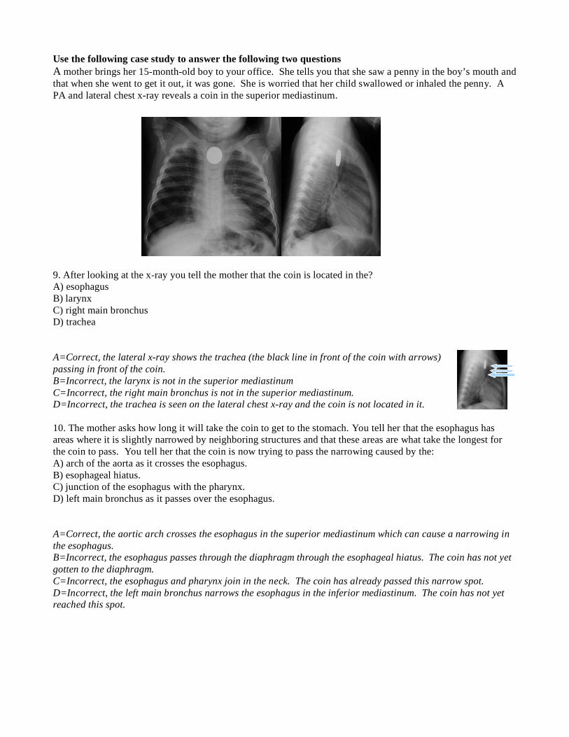
Use the following case study to answer the following two questions
A mother brings her 15-month-old boy to your office. She tells you that she saw a penny in the boy’s mouth and
that when she went to get it out, it was gone. She is worried that her child swallowed or inhaled the penny. A
PA and lateral chest x-ray reveals a coin in the superior mediastinum.
9. After looking at the x-ray you tell the mother that the coin is located in the?
A) esophagus
B) larynx
C) right main bronchus
D) trachea
A=Correct, the lateral x-ray shows the trachea (the black line in front of the coin with arrows)
passing in front of the coin.
B=Incorrect, the larynx is not in the superior mediastinum
C=Incorrect, the right main bronchus is not in the superior mediastinum.
D=Incorrect, the trachea is seen on the lateral chest x-ray and the coin is not located in it.
10. The mother asks how long it will take the coin to get to the stomach. You tell her that the esophagus has
areas where it is slightly narrowed by neighboring structures and that these areas are what take the longest for
the coin to pass. You tell her that the coin is now trying to pass the narrowing caused by the:
A) arch of the aorta as it crosses the esophagus.
B) esophageal hiatus.
C) junction of the esophagus with the pharynx.
D) left main bronchus as it passes over the esophagus.
A=Correct, the aortic arch crosses the esophagus in the superior mediastinum which can cause a narrowing in
the esophagus.
B=Incorrect, the esophagus passes through the diaphragm through the esophageal hiatus. The coin has not yet
gotten to the diaphragm.
C=Incorrect, the esophagus and pharynx join in the neck. The coin has already passed this narrow spot.
D=Incorrect, the left main bronchus narrows the esophagus in the inferior mediastinum. The coin has not yet
reached this spot.
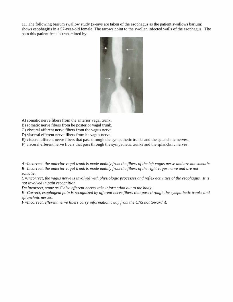
11. The following barium swallow study (x-rays are taken of the esophagus as the patient swallows barium)
shows esophagitis in a 57-year-old female. The arrows point to the swollen infected walls of the esophagus. The
pain this patient feels is transmitted by:
A) somatic nerve fibers from the anterior vagal trunk.
B) somatic nerve fibers from he posterior vagal trunk.
C) visceral afferent nerve fibers from the vagus nerve.
D) visceral efferent nerve fibers from he vagus nerve.
E) visceral afferent nerve fibers that pass through the sympathetic trunks and the splanchnic nerves.
F) visceral efferent nerve fibers that pass through the sympathetic trunks and the splanchnic nerves.
A=Incorrect, the anterior vagal trunk is made mainly from the fibers of the left vagus nerve and are not somatic.
B=Incorrect, the anterior vagal trunk is made mainly from the fibers of the right vagus nerve and are not
somatic.
C=Incorrect, the vagus nerve is involved with physiologic processes and reflex activities of the esophagus. It is
not involved in pain recognition.
D=Incorrect, same as C also efferent nerves take information out to the body.
E=Correct, esophageal pain is recognized by afferent nerve fibers that pass through the sympathetic trunks and
splanchnic nerves.
F=Incorrect, efferent nerve fibers carry information away from the CNS not toward it.
12. Referred pain is a condition where pain is felt in one part of the body but is actually coming from a
different part of the body. This phenomenon is based on visceral sensory neurons and somatic sensory
neurons converging at the same vertebral level and confusing the brain of the pain’s origin. An
inflamed appendix, for instance, can cause painful stimulations in the umbilicus region [Snell, Richard.
Clinical Anatomy by Regions. 8th edition]. What nerve is responsible for carrying the visceral
sensation in the case of an inflamed appendix as described above?
A. Intercostal nerve of spinal level T4
B. Intercostal nerve of spinal level T10
C. Least splanchnic nerve
D. Lesser splanchnic nerve
E. Phrenic nerve
F. Vagus nerve
Answer:
A. Incorrect. The intercostal nerves are responsible for general sensory, not visceral sensory.
B. Incorrect. The intercostal nerves are responsible for general sensory, not visceral sensory.
C. Incorrect. The least splanchnic nerve arises from the sympathetic trunk at the spinal level T12, and
the umbilicus is innervated by dermatome level T10.
D. Correct. The dermatome level of the umbilicus is T10, and the lesser splanchnic nerve originates
from the sympathetic trunk at levels T10-11.
E. Incorrect. The phrenic nerve innervates the diaphragm and is unrelated in this case.
F. Incorrect. The vagus nerve originates from the brainstem, so it doesn’t cause referred pain.
13. Referred pain is a condition where pain is felt in one part of the body but is actually coming from a
different part of the body. This phenomenon is based on visceral sensory neurons and somatic sensory
neurons converging at the same vertebral level and confusing the brain of the pain’s origin. Describe
the referred pain that would be felt during ischemic (oxygen deficient) damage to the myocardial
tissue.
Answer: The visceral sensory to the heart is carried in sympathetic neurons arising from spinal levels
of T1-T4. The referred pain is felt at the dermatomes of these levels, which turns out to be the chest
and medial portion of the arm.
14. A patient presents with labored breathing. It is determined that he has a nerve paralysis due to
metastatic lung cancer that has invaded the parietal pleura where it comes in contact with the
pericardium (Chest. 2003;123:97S-104S). Which nerve is most likely lesioned?
A. Intercostal nerves
B. Greater splanchnic nerve
C. Left recurrent laryngeal nerve
D. Phrenic nerve
E. Vagus nerve
Answer:
A. Incorrect. The area where the parietal pleura and pericardium come in contact is deep within
the chest cavity, away from the chest wall and intercostal nerves.
B. Incorrect. The greater splanchnic nerve runs laterally to the spinal column and doesn’t come in
contact with pericardium.
C. Incorrect. The left recurrent laryngeal nerve dives under the arch of the aorta on its way to the
larynx, but it doesn’t come in contact with the pericardium.
D. Correct. The phrenic nerve runs between the pericardium and parietal pleura on its way to the
diaphragm.
E. Incorrect. The vagus nerve runs along the esophagus and therefore deep to the pericardium and
parietal pleura.
15. Which artery could also become obstructed in the case above?
A. Internal thoracic artery
B. Musculophrenic artery
C. Pericardiacophrenic artery
D. Right bronchial artery
Answer:
A. Incorrect. The internal thoracic runs in the anterior thoracic wall and is not located anywhere
in-between the parietal pleura and pericardium.
B. Incorrect. The musculophrenic artery is a continuation of the internal thoracic as it course
superior to the diaphragm.
C. Correct. The pericardiacophrenic artery runs alongside the phrenic nerve on its course to the
diaphragm.
D. Incorrect. The right bronchial artery runs to the hilum, but does not accompany the phrenic
nerve.
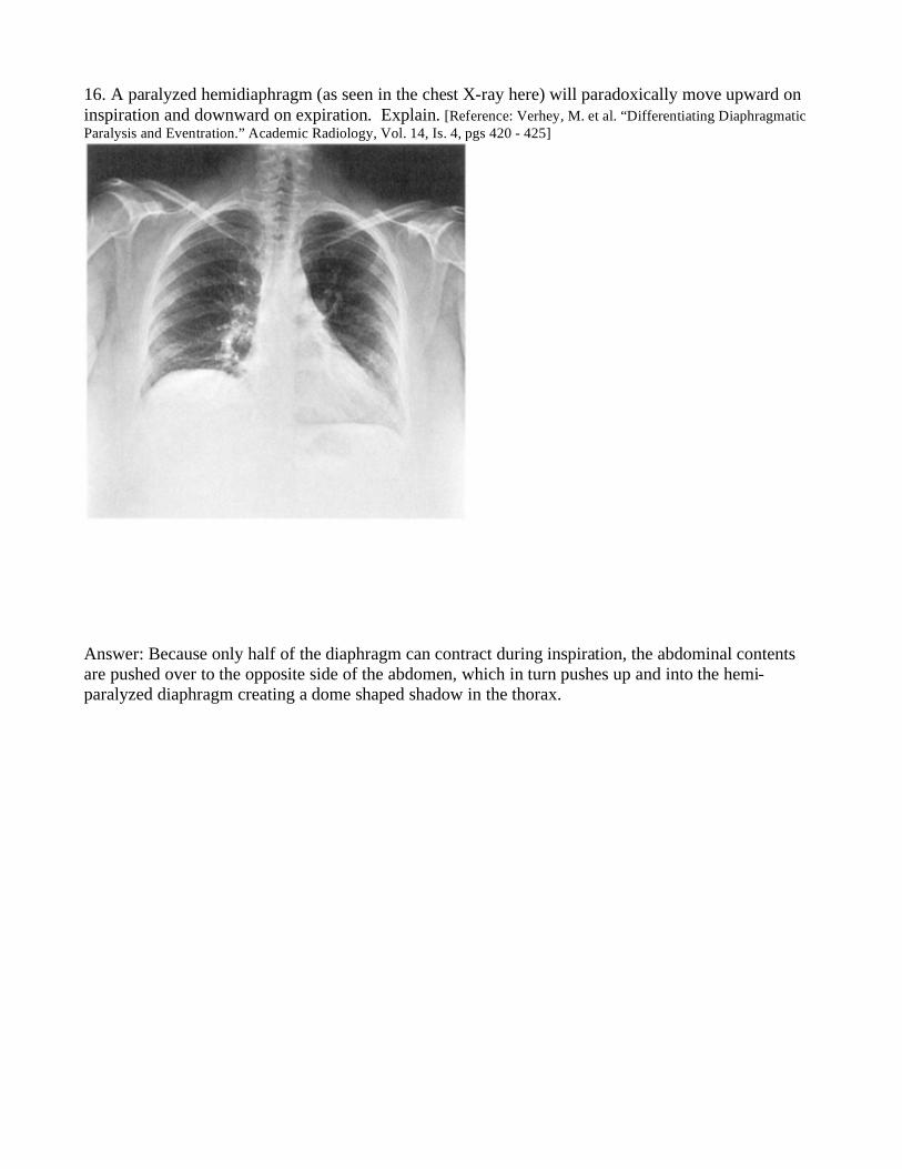
16. A paralyzed hemidiaphragm (as seen in the chest X-ray here) will paradoxically move upward on
inspiration and downward on expiration. Explain. [Reference: Verhey, M. et al. “Differentiating Diaphragmatic
Paralysis and Eventration.” Academic Radiology, Vol. 14, Is. 4, pgs 420 - 425]
Answer: Because only half of the diaphragm can contract during inspiration, the abdominal contents
are pushed over to the opposite side of the abdomen, which in turn pushes up and into the hemi-
paralyzed diaphragm creating a dome shaped shadow in the thorax.
17. Pleurisy (pleuritis) is a painful condition where the visceral and parietal pleura become inflamed. It
can be caused by a number of things, including influenza, tuberculosis, pneumonia or autoimmune
disease. Symptoms include shortness of breath, chest pain during breathing and dry cough. Some
patients suffering from pleurisy find temporary relief from holding their breath. What is occurring in
pleurisy that causes this phenomenon?
A. Increased resistance of blood flow through the pulmonary system
B. The intercostal nerves become pinched during inspiration
C. The negative pressure in the pleural space is compromised
D. The pleura are no longer frictionless against each other
Answer:
A. Incorrect. Blood flow is more or less constant whether breathing or not.
B. Incorrect. It is very unlikely anatomically that the lungs or pleura could compress the
intercostal nerves due to the costal groove and protection from the innermost intercostal
muscles.
C. Incorrect. Negative pressure is a constant regardless of breathing or holding one’s breath.
D. Correct. The purpose of the parietal and visceral pleura is to facilitate the expansion of the
lungs by using negative pressure during muscle contraction. The change in size and position of
the lungs during respiration causes a significant amount of movement between the visceral and
parietal pleura—it is essential that these two surfaces remain frictionless. In the case of
pleurisy, the inflammation causes more friction between pleura, causing pain during breathing.
18. A bronchoscopy is a medical procedure where a tube is inserted into the airways, usually through
the nose or mouth. This allows the practitioner to examine inside a patient's airway for abnormalities
such as foreign bodies, bleeding, tumors, or inflammation. There are currently two general types of
bronchoscopes, rigid and flexible. The rigid bronchoscope is the preferable choice for removing
foreign objects. Which bronchi would be more difficult for a rigid instrument to access? Why?
A. Left bronchi
B. Right bronchi
Answer: A
Explanation: The left bronchi is skinnier, longer and runs more laterally.
19. Pulmonary edema is a life-threatening condition in which the alveoli of the lung fill with fluid. It is
most often caused by pressure building up in the pulmonary vessels due to congestive heart failure.
Which chamber of the heart, when it is pathologic, is most likely to cause this condition?
A. Left atrium
B. Left ventricle
C. Right atrium
D. Right ventricle
Answer:
A. Incorrect. The left atrium is tempting because it is the first chamber en route from the lungs,
but with a working left ventricle, a dysfunctional left atrium could be mostly overcome since
the atrium’s role in cardiac output is minimal in comparison to the ventricle.
B. Correct. The main reason that fluid from the pulmonary system can get into alveoli is because
of pulmonary hypertension. The high pressure pushes plasma out of the pulmonary capillaries
into the lower pressured space of the alveoli. If the left ventricle starts failing, the blood gets
backed up in the pulmonary system, causing pulmonary edema.
C. And D. Incorrect. The right side of the heart receives systemic blood, so when it is
dysfunctional it causes systemic hypertension, not pulmonary
Use the following case study to answer the following two questions
In pulmonary atresia, a rare congenital heart disease, the pulmonary valve doesn’t develop and a wall
of tissue lies in its place, blocking off blood flow in that direction. Most treatment options for this
condition involve keeping two embryological structures open—the foramen ovale and the patent ductus
arteriosus (http://www.mayoclinic.org/pulmonary-atresia/treatment.html).
20. Trace a drop of blood from the superior vena cava to the lungs (to be oxygenated) back to the heart
and then to the aorta in a patient with pulmonary atresia (assume the two embryological structures
mentioned are patent (open)).
Answer:
SVC
Right atrium
Foramen ovale
Left atrium
Mitral valve
Left ventricle
Aortic valve
Ascending aorta
Patent ductus arteriosus
Pulmonary trunk to arteries to capillaries to veins
Left atrium
Mitral valve
Left ventricle
Aortic valve
Aorta
21. The body has a tendency to increase in size (hypertrophy) structures or tissues being over used and
decrease in size (atrophy) structures or tissues being under used. Assuming an open foramen ovale and
patent ductus arteriosus, which structure is most likely to atrophy in a patient with pulmonary atresia?
(American Heart Journal. 1987 Dec;114(6):1415-20)
A. Left atrium
B. Left ventricle
C. Pulmonary arteries
D. Pulmonary veins
E. Right atrium
F. Right ventricle
Answer:
A. Incorrect. The left atrium would be actively used in these cases (see trace).
B. Incorrect. The left ventricle would hypertrophy if anything, because it is doing the job of both
ventricles.
C. Incorrect. The pulmonary arteries would still be used because of the patent ductus arteriosus.
D. Incorrect. The pulmonary veins are also actively used in these cases (see trace).
E. Incorrect. The right atrium is still receiving blood from the superior and inferior vena cava.
F. Correct. The right ventricle is totally bypassed in cases of pulmonary atresia (which was
demonstrated by the previous trace). Therefore, this portion of the heart is underused and
therefore atrophies.
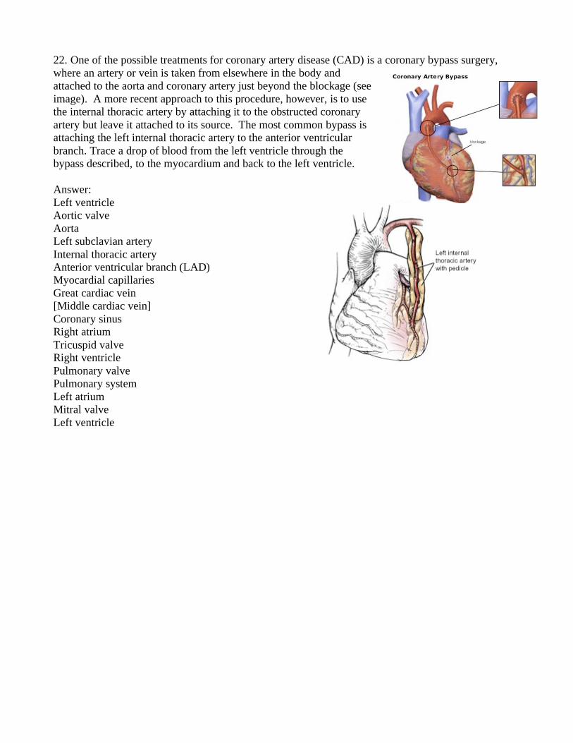
22. One of the possible treatments for coronary artery disease (CAD) is a coronary bypass surgery,
where an artery or vein is taken from elsewhere in the body and
attached to the aorta and coronary artery just beyond the blockage (see
image). A more recent approach to this procedure, however, is to use
the internal thoracic artery by attaching it to the obstructed coronary
artery but leave it attached to its source. The most common bypass is
attaching the left internal thoracic artery to the anterior ventricular
branch. Trace a drop of blood from the left ventricle through the
bypass described, to the myocardium and back to the left ventricle.
Answer:
Left ventricle
Aortic valve
Aorta
Left subclavian artery
Internal thoracic artery
Anterior ventricular branch (LAD)
Myocardial capillaries
Great cardiac vein
[Middle cardiac vein]
Coronary sinus
Right atrium
Tricuspid valve
Right ventricle
Pulmonary valve
Pulmonary system
Left atrium
Mitral valve
Left ventricle
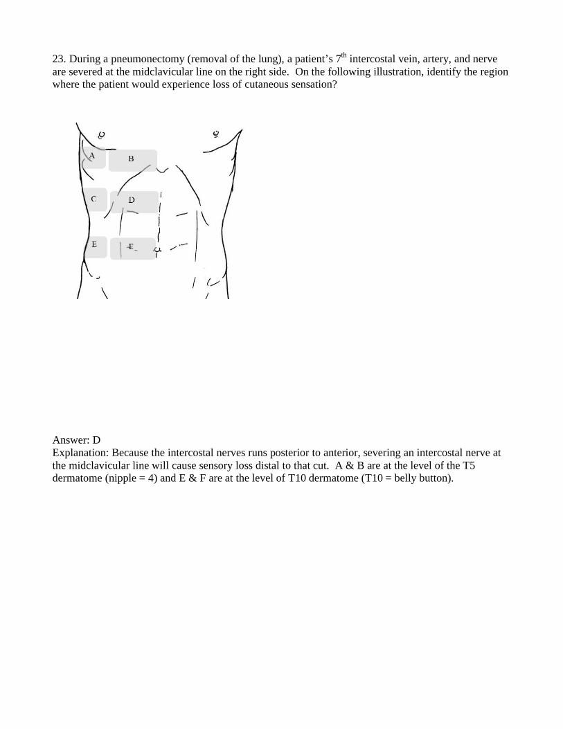
23. During a pneumonectomy (removal of the lung), a patient’s 7th intercostal vein, artery, and nerve
are severed at the midclavicular line on the right side. On the following illustration, identify the region
where the patient would experience loss of cutaneous sensation?
Answer: D
Explanation: Because the intercostal nerves runs posterior to anterior, severing an intercostal nerve at
the midclavicular line will cause sensory loss distal to that cut. A & B are at the level of the T5
dermatome (nipple = 4) and E & F are at the level of T10 dermatome (T10 = belly button).
24. In a radical mastectomy, most of the chest muscles and all of the axillary lymph nodes are removed
in addition to the breast tissue. A latissimus dorsi flap is advantageous in post radical mastectomy
patients because it replaces a portion of the muscle that was removed. Which of the following options
is the best reason that the latissimus dorsi is a good candidate to replace the chest wall muscles?
A. It is similar in shape to one of the chest wall muscles.
B. It shares a common attachment site with one of the chest wall muscles
C. It shares a common blood supply with one of the chest wall muscles.
D. It helps stabilize the scapula in the absence of the chest wall muscles
Answer:
A. Incorrect. False statement.
B. Correct. The latissimus dorsi inserts in the intertubercular groove of the humerus, which is
same location as the insertion of the pectoralis major.
C. Incorrect. False statement.
D. Incorrect. False statement.
25. In the labor and delivery room, a 29-year-old woman who has been in labor for 9 hours asks for an
epidural. When the anesthesiologist arrives he asks the woman to lie down on her side, and he begins
locating the appropriate landmarks that will allow him to administer the epidural in the safest place
possible. He wants to access the subarachnoid space without the risk of puncturing the spinal cord.
Which vertebral levels would provide the most appropriate range?
A T12-L2
B L1-L3
C L2- L4
D L3-L5
Answer
A,B,C the spinal cord can extend as low as the disk between L2 and L3, so all of these options would
include spaces that might include spinal cord therefore, they are incorrect.
D provides the only range where the procedure would avoid an area where the spinal cord might be
present. Either between L3 and L4, or between L4 and L5 is the most common place to insert the
needle.
The following questions deal with this scenario:
Cameron, a 23-year-old male and well conditioned athlete has finally reached one of his goals of
running the Boston Marathon. The following questions deal with the functions of his nervous system
during the race.
26. Just after the race gets underway, the sun peaks over the trees raising the air temperature
dramatically. Cameron begins to sweat more profusely. Starting in the correct horn of the gray matter
of the spinal cord, describe the initial pathway of the nerves responsible for his sweating.
A anterior horn, ant. root and ramus, gray ramus communicans, prevertebral ganglion
B anterior horn, ant. root and ramus, white ramus communicans, paravertebral ganglion
C anterior horn, ant. root and ramus, white ramus communicans, prevertebral ganglion
D lateral horn, ant. root and ramus, gray ramus communicans, prevertebral ganglion
E lateral horn, ant root and ramus, gray ramus communicans, paravertebral ganglion
F lateral horn, ant. root and ramus, white ramus communicans, paravertebral ganglion
Answer:
A, B, C, all the sympathetics originate in the lateral horn of the gray matter.
D and E the preganglionic sympathetics are myelinated and enter the paravertebral ganglion via a white
ramus communicans.
F correct. The paravertebral ganglia make up the sympathetic trunk. This is the initial pathway of the
sympathetic neurons which are responsible for sweating. After arrival in the sympathetic trunk the
continuing pathway varies.
27. Along with sweating, which other physiological event would be stimulated by nerves with the same
initial pathway?
A constriction of the bronchioles
B dilation of the coronary arteries
C increased salivation
D pupilary constriction
Answer:
A this is a function of the parasympathetic division of the ANS and thus the neurons would not follow
the sympathetic pathway outlined above.
B correct. the sympathetics dilate the coronary arteries so that the heart can receive more oxygenated
blood as it increases rate and strength of contraction.
C and D Both of these physiological functions are stimulated by the parasympathetics.
28. Cameron experiences every marathoner’s worst fear, the need to defecate during the race, and
heads for a portable toilet. Cameron is hoping this unfortunate break will be as short as possible. A
parasympathetic motor impulse will travel along two neurons from the CNS to the internal anal
sphincter, an involuntary muscle. The pre-synaptic neurons responsible for relaxing the internal anal
sphincter originate from what spinal nerve levels?
A L4-L5
B L5-S1
C S2-S4
D T1-L2
Answer:
The correct answer is C. This is the level of origin of the parasympathetic nerves (locations in the
brain stem are associated with cranial nerves III, VII, IX and X).
29. Where would you most likely find the synapse between the pre-synaptic and post-synaptic
parasympathetic neurons involved in the relaxation of the internal anal sphincter?
A dorsal root ganglion
B intramural ganglion
C paravertebral ganglion
D prevertebral ganglion
Answer:
A this ganglion contains cell bodies of sensory neurons
B correct. The parasympathetic branch of the ANS often has very long pre-synaptic neurons which
will extend all the way to the organ of interest. Within the organ there are ganglia called intramural
ganglia where the synapse takes place followed by an obviously very short post synaptic neuron.
C and D both house cell bodies of sympathetic neurons. Again, defecation is a function of the
parasypathetics.
30. Between mile 20 and 21 Cameron has to ascend “heartbreak hill”. After continually contracting for
over two and a half hours, the muscles in his calves and thighs are exhausted. Starting with the cell
bodies in the gray matter of the spinal cord, what is the initial pathway of the neurons responsible for
the contraction of Cameron’s leg muscles.
A anterior horn, anterior root, anterior ramus
B anterior horn, anterior root, posterior ramus
C lateral horn, anterior root, anterior ramus
D lateral horn, posterior root, posterior ramus
E posterior horn, posterior root, anterior ramus
F posterior horn, posterior root, posterior ramus
Answer:
A correct
B the only muscles the posterior rami innervate are the deep muscles of the back
C and D, the lateral horn houses the cell bodies of visceral motor neurons (the sympathetic division of
the ANS)
E and F, the posterior horn is where only sensory neurons enter the spinal cord.
31. Following “heartbreak hill” Cameron suddenly feels a severe cramp in his calf. He hasn’t been
drinking enough or replenishing the electrolytes he is losing in sweat. Where would you most likely
find the cell body of the neurons responsible for the sensation of pain in Cameron’s calf?
A anterior horn of the gray matter
B dorsal root (spinal) ganglion
C lateral horn of the gray matter
D paravertebral ganglion
E prevertebral ganglion
F posterior horn of the gray matter
Answer:
A This contains cells bodies of somatic motor neurons
B correct
C Contains cell bodies of visceral motor neurons
D and E these would both contain cell bodies of postsynaptic sympathic neurons.
F this is where the nerve fibers of sensory nerves enter the spinal cord, but the cell bodies of the
sensory nerves are found in the dorsal root ganglia.
32. During the repair of a thoracic aortic aneurysm, (severely dilated aorta) the blood supply to the
posterior intercostal arteries, and subsequently to parts of the spinal cord is lost. (J Vasc Surg. 2007
Sep; 46(3):421-6.) One potential technique for the revascularization of posterior intercostal muscles
is to transfer the thoracodorsal artery from its normal destination and graft it to the posterior intercostal
arteries mentioned above . (Ann Vasc Surg. 2002 Nov; 16(6):723-9.) When this artery is used, what
action might be compromised in this patient in the future?
A abducting the arm
B a traditional pull-up
C a traditional push-up
D extending the head
E raising the arm above the head
Answer:
A this is accomplished by the deltoid and supraspinatus muscles
B correct. The thoracodorsal artery provides blood supply to the latissimus dorsi muscle, which is
largely responsible for the action of pulling ones body upward in climbing or during a pull up. If the
arterial supply was sacrificed the muscle would bcome ischemic, and die. This muscle is not essential
to every day life and therefore is often a candidate for muscle grafts as well.
C the pectoralis muscles would be responsible for this.
D the splenius muscles and suboccipital muscles are largely responsible for this acion
E this action is hindered upon damage to the trapezius muscle.
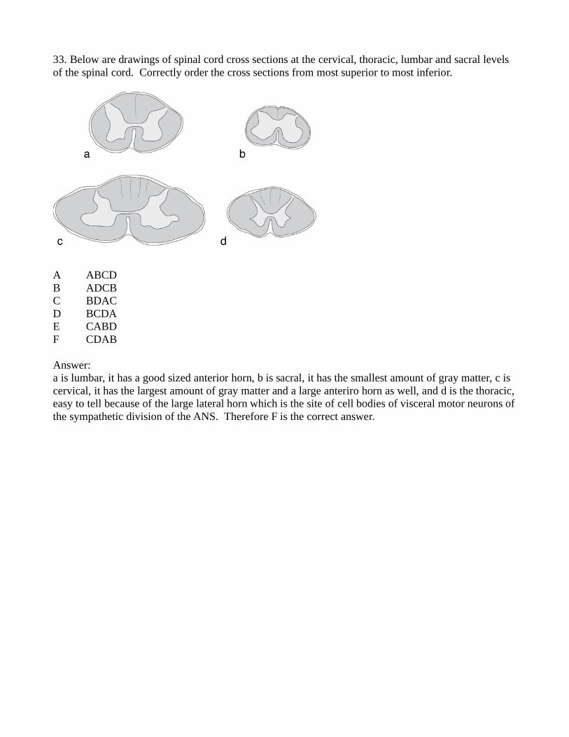
33. Below are drawings of spinal cord cross sections at the cervical, thoracic, lumbar and sacral levels
of the spinal cord. Correctly order the cross sections from most superior to most inferior.
A ABCD
B ADCB
C BDAC
D BCDA
E CABD
F CDAB
Answer:
a is lumbar, it has a good sized anterior horn, b is sacral, it has the smallest amount of gray matter, c is
cervical, it has the largest amount of gray matter and a large anteriro horn as well, and d is the thoracic,
easy to tell because of the large lateral horn which is the site of cell bodies of visceral motor neurons of
the sympathetic division of the ANS. Therefore F is the correct answer.