UNILATERAL PARALYSIS OF THE DIAPHRAGM AND LARYNX ... ·
14
Thorax (1950), 5, 169. UNILATERAL PARALYSIS OF THE DIAPHRAGM AND LARYNX ASSOCIATED WITH INFLAMMATORY LUNG DISEASE BY BERNARD FREEDMAN From St. Giles' and Dulwich Hospitals, London Unilateral paralysis of the diaphragm, not the result of a deliberate operative procedure, is uncommon. It may have a variety of causes, but so far as I am aware pneumonia* has never previously been included amongst them. The commoner causes of unilateral paralysis are diseases of the cervical spinal cord affecting the anterior horn cells of the fourth segment, such as poliomyelitis, tumour, progressive muscular atrophy, myelitis, haemorrhage, and injuries and caries of the cervical vertebrae. Peripheral lesions of the phrenic nerve or its roots include neck wounds, polyneuritis (usually diphtheritic), destruction by neoplastic masses (usually near the hilum of the lung), and avulsion from birth injury as an extension of Erb's palsy, of which about 20 cases have been described. A number of cases occurring during the course of pulmonary tuberculosis have been ascribed to involve- ment of the phrenic nerve in scar tissue. Two instances in newborn infants, not associated with Erb's palsy, were attributed to intrauterine malposition (Blattner, 1942; Light, 1944). An aortic aneurysm may rarely be responsible (Sanguinetti and Galzerano, 1943). Eventration of the diaphragm, a condition in which the affected dome is much raised and thinned with fibrous replacement, may in a sense be regarded as a form of phrenic palsy. Though usually regarded as congenital, it may in some instances be the atrophic result of permanent interruption of the nerve supply. Diseases of the diaphragm itself, acute diaphragmitis (Hedblom's syndrome), trichiniasis, and the degenerations that may result from prolonged contact with purulent effusions, severe pneumonia and asphyxial states, all of course impair diaphragmatic function, which would imply impaired descent on inspiration. Whether they would cause actual paralysis is open to question. A search of the literature has failed to reveal any previous record of unilateral paralysis of the diaphragm due to pneumonia. Upward displacement of the diaphragm in pneumonia has been reported by Wu (1932), but such elevation is apparent only, being in fact a failure to descend during deep inspiration as much as the dome of the sound side. It is due to a loss of expansibility of the lung result- ing from the consolidation, and is apparent only in radiographs taken in full inspira- tion. The extensive reviews by Graeser, Wu, and Robertson (1934), Davies, Hodgson, and Whitby (1935), and Heffron (1939) of the radiological findings in pneumonia likewise mention this apparent elevation during the acute phase, but *Throughout this article the term " pneumonia " is used to cover a variety of inflammations affecting the lung.-EDITOR. M on April 9, 2020 by guest. Protected by copyright. http://thorax.bmj.com/ Thorax: first published as 10.1136/thx.5.2.169 on 1 June 1950. Downloaded from
Transcript of UNILATERAL PARALYSIS OF THE DIAPHRAGM AND LARYNX ... ·

Thorax (1950), 5, 169.
UNILATERAL PARALYSIS OF THE DIAPHRAGMAND LARYNX ASSOCIATED WITHINFLAMMATORY LUNG DISEASE
BY
BERNARD FREEDMANFrom St. Giles' and Dulwich Hospitals, London
Unilateral paralysis of the diaphragm, not the result of a deliberate operativeprocedure, is uncommon. It may have a variety of causes, but so far as I am awarepneumonia* has never previously been included amongst them.
The commoner causes of unilateral paralysis are diseases of the cervical spinalcord affecting the anterior horn cells of the fourth segment, such as poliomyelitis,tumour, progressive muscular atrophy, myelitis, haemorrhage, and injuries and cariesof the cervical vertebrae. Peripheral lesions of the phrenic nerve or its roots includeneck wounds, polyneuritis (usually diphtheritic), destruction by neoplastic masses(usually near the hilum of the lung), and avulsion from birth injury as an extensionof Erb's palsy, of which about 20 cases have been described. A number of casesoccurring during the course of pulmonary tuberculosis have been ascribed to involve-ment of the phrenic nerve in scar tissue. Two instances in newborn infants, notassociated with Erb's palsy, were attributed to intrauterine malposition (Blattner,1942; Light, 1944). An aortic aneurysm may rarely be responsible (Sanguinetti andGalzerano, 1943). Eventration of the diaphragm, a condition in which the affecteddome is much raised and thinned with fibrous replacement, may in a sense beregarded as a form of phrenic palsy. Though usually regarded as congenital, it mayin some instances be the atrophic result of permanent interruption of the nervesupply.
Diseases of the diaphragm itself, acute diaphragmitis (Hedblom's syndrome),trichiniasis, and the degenerations that may result from prolonged contactwith purulent effusions, severe pneumonia and asphyxial states, all of courseimpair diaphragmatic function, which would imply impaired descent on inspiration.Whether they would cause actual paralysis is open to question.A search of the literature has failed to reveal any previous record of unilateral
paralysis of the diaphragm due to pneumonia. Upward displacement of thediaphragm in pneumonia has been reported by Wu (1932), but such elevation isapparent only, being in fact a failure to descend during deep inspiration as muchas the dome of the sound side. It is due to a loss of expansibility of the lung result-ing from the consolidation, and is apparent only in radiographs taken in full inspira-tion. The extensive reviews by Graeser, Wu, and Robertson (1934), Davies,Hodgson, and Whitby (1935), and Heffron (1939) of the radiological findings inpneumonia likewise mention this apparent elevation during the acute phase, but
*Throughout this article the term " pneumonia " is used to cover a variety of inflammationsaffecting the lung.-EDITOR.
M
on April 9, 2020 by guest. P
rotected by copyright.http://thorax.bm
j.com/
Thorax: first published as 10.1136/thx.5.2.169 on 1 June 1950. D
ownloaded from

BERNARD FREEDMAN
none mentions paralysis. Leitner (1938) described diaphragmatic palsy followingartificial pneumothorax due to mediastinal pleurisy. His diagnosis may be ques-tioned, as the diaphragm, though raised, was fixed.
CRITERIA OF UNILATERAL DIAPHRAGMATIC PARALYSISClinically unilateral diaphragmatic paralysis may be suspected but cannot be
diagnosed with certainty. Physical signs, occasionally striking, are more often soinsignificant as to be easily missed or overshadowed by coexisting processes, andwhen present are not usually diagnostic. The following criteria were thereforeadopted in the cases described. (1) Elevation of the diaphragm 1.5 in. (about 4 cm.)or more above the normal position: the extent of the elevation was determined bycomparison with the sound side. For purposes of measurement the right dome wasarbitrarily taken as being normally I in. (1.25 cm.) higher than the left. (2) Para-doxical movement, especially noticeable on sniffing: the sound side descends oninspiration, while the paralysed side rises by a smaller amount. This phenomenonis rendered more obvious when the subject sniffs sharply. The latter test is a severeone, and may be positive with incompletely paralysed diaphragms. A- positiveresponse to the sniff test was not therefore taken by itself as the only criterion.
Direct stimulation of the phrenic nerve with simultaneous radiographic controlwould have been crucial, but was scarcely practicable under circumstances of acuteillness.
DIFFERENTIAL DIAGNOSISElevation of the diaphragm may result from its being pushed up as, for example,
by a subphrenic abscess. It may also result from the diaphragm being pulled up byadhesions. In both these circumstances the diaphragm is fixed; there is no para-doxical movement. However, an apparently paradoxical movement may occur witha fixed diaphragm, especially in breathers of the " thoracic" type. This is due toelevation of the anterior part of the thoracic cage during inspiration and with it theanterior diaphragmatic attachments. Careful inspection of films taken in both phaseswill demonstrate failure to rise relatively to the front of the thoracic cage. This isunlikely to cause ccnfusion cn fluoroscopy, as the elevation that occurs is slight andoccurs towards the end of inspiration.
Apparent elevation may result from the inspiratory descent on the affected sidebeing less than that on the sound side. This factor will operate in films taken infull inspiration, as is the universal routine. The phenomenon occurs reflexly in painfulconditions, such as pleurisy and fibrositis of the chest-wall (pleurodynia), and inpneumonia, due to loss of expansibility in the consolidated portion as previouslymentioned (Wu, 1932).
Elevation of the diaphragm may be associated with the Kienbock (1898)phenomenon.* In hydropneumothorax the fluid level may be seen to rise oninspiration. The diaphragm is masked by the fluid in the erect posture. Kienbockattributed the phenomenon to phrenic palsy, but this may be absent. In the latterevent the rise in fluid level is caused by movement of the mediastinum towards theaffected side.
*The term Kienb6ck phenomenon is often, but erroneously, used to describe the para-doxical movement of phrenic paralysis. Kienbock's description referred to the occurrence ofthis movement in the fluid level of hydropneumothorax.
170
on April 9, 2020 by guest. P
rotected by copyright.http://thorax.bm
j.com/
Thorax: first published as 10.1136/thx.5.2.169 on 1 June 1950. D
ownloaded from

UNILATERAL PARALYSIS OF DIAPHRAGM AND LARYNX 171
FIG. 1. FIG. 2.
FIG. 1.-Case 1 twenty-one daysafter operation. Post-operativepneumonia has progressed toan abscess with fluid level inthe subapical segment of theleft lower lobe.
FIG. 2.-Case 1. The left dome ofthe diaphragm is raised 2.5 in.(6.3 cm.) above its normallevel.
FIG. 3.-Case 1. Complete recoveryof diaphragmatic mobility;residual fibrosis.
FKG. 3.
on April 9, 2020 by guest. P
rotected by copyright.http://thorax.bm
j.com/
Thorax: first published as 10.1136/thx.5.2.169 on 1 June 1950. D
ownloaded from

BERNARD FREEDMAN
In positive-pressure pneumothorax the diaphragm on the same side may riseslightly during inspiration (Udaondo and Vadone, 1929; Stivelman, 1935).
In eventration of the diaphragm the criteria laid down are fulfilled. Whateverthe aetiology may be, it can therefore be regarded functionally as a unilateralparalysis with atrophy and replacement fibrosis of the muscle fibres. In diagnosinga case of phrenic palsy of recent onset it is therefore desirable if possible to excludeeventration by the provision of evidence of previously normal function. It isdistinctly rare.
It must also be said that pneumonia associated with unilateral phrenic palsy maybe a sign of bronchial carcinoma, and the latter condition should be excluded beforethe palsy is regarded as resulting from pneumonia.
CASE REPORTSSix cases of unilateral diaphragmatic paralysis following pneumonia are pre-
sented; in one of these there was an ipsilateral vocal cord paralysis.Case 1.-A street trader, aged 40 years, gave a two years' history of pain due to
proved gastric ulcer, for which he had received medical treatment elsewhere. Hewas admitted on October 16, 1947, suffering from perforation of a gastric ulcer,which was found high on the anterior surface of the stomach near the lesser curve.It was treated by operat:ve closure (Mr. J. L. Stephen). The pyrexia settled duringthe next few days.
Owing to persistent pain during the next five weeks, he was given a barium meal.This showed that a large gastric ulcer was still present. Partial gastrectomy wastherefore performed, with drainage of a pelvic abscess that was found (Mr. J. L.Stephen), the anaesthetic being "pentothal," nitrous oxide, oxygen, and " myanesin ";the operation lasted 95 minutes. The post-operative pyrexia failed to fall duringthe next few days, during which time a cough developed and persisted in spite ofintramuscular penicillin. Eleven days after the second operation the pyrexia increasedto 101.20 F. (38.40 C.), and physical and radiological signs of bronchopneumoniawith early abscess formation were present in the left lower lobe. Oral sulphadimidine(" sulphamezathine ") 1 g. 4-hourly for one week was given without effect.
On the 21st post-operative day radiography (Fig. 1) showed an abscess with fluidlevel in the sub-apical segment of the left lower lobe, with slight elevation of theposterior part of the diaphragm. The sputum amounted to 10 oz. (280 ml.) daily ofwhich one-third was pus. Numerous pneumococci and a few Staph. albus were grownfrom it; no tubercle bacilli were found.
I first saw the patient on the 32nd post-operative day, when, in addition tothe physical signs in the left lower lobe, I noted a peculiar bitonal quality in hisvoice, subsequently shown to be due to paralysis of the left vocal cord. For tendays he had been given postural drainage and penicillin (500,000 units twice daily),the pyrexia had settled, the general condition was improved, and the pneumonitisaround the abscess showed corresponding clearing radiologically, but the abscess cavityitself was larger, the sputum had increased to 1 pint (560 ml.) a day, and a leucocytosiswas present (20,800 per c.mm., of which 74% were polymorphs).
By the 43rd day the left dome of the diaphragm was considerably raised, being2.5 in. (6.3 cm.) above normal (Fig. 2), and on fluoroscopy it exhibited paradoxicalmovement. The abscess was now less in evidence. By the 60th day the left diaphragmwas slightly lower, and a little opacity only was present at the site of the abscess,which had almost healed. The bitonal voice noted on the 32nd day was unchanged,
172
on April 9, 2020 by guest. P
rotected by copyright.http://thorax.bm
j.com/
Thorax: first published as 10.1136/thx.5.2.169 on 1 June 1950. D
ownloaded from

UNILATERAL PARALYSIS OF DIAPHRAGM AND LARYNX 173
and indirect laryngoscopy disclosed complete paralysis of the left vocal cord, whichwas in the cadaveric position, the right cord crossing the midline to meet it onphonation. The patient was discharged 67 days after the onset of the bronchopneumonia.
Subsequent follow-up showed on the 84th day returning though incomplete con-tractility of the left diaphragm, and fibrosis at the site of the abscess. The vocalcord paralysis persisted. The general condition was fairly good, only a moderateamount of mucopurulent sputum being produced. The erythrocyte sedimentation rate(E.S.R.) was 20 mm. in one hour (Westergren). Incomplete closure of the abscesswas suspected, but the patient declined bronchography to settle this point. He wastherefore given inhalations of nebulized penicillin solution and more postural drainage.By the 112th day the chest radiograph (Fig. 3) was clear except for left basal fibrosis.The left vocal cord was now fixed in the midline, indicating recovery of the adductorgroup of muscles only, but four and a half months after operation it had returnedto the cadaveric position. After 20 months the cord showed a little movement amountingto about one-fifth of normal, and was atrophied ; the voice retained its peculiar timbre.
Though possibly irrelevant to the condition of his phrenic and recurrent laryngealnerves, it must be added that the patient developed -hepatitis with jaundice on March 3.Its onset was 135 days after his first operation, when he received two pints of plasma.
The course and diagnosis of this patient's illness may be summarized as perforatedgastric ulcer with operative closure, and partial gastrectomy five weeks later followedby pneumonia and abscess in the sub-apical segment of the left lower lobe. Laterserum hepatitis developed. There was paralysis of the left vocal cord, first noticedfour weeks after the onset of pneumonia and lasting over four and a half months.Twenty months later there was a very slight recovery of movement.
Paralysis of the left dome of the diaphragm, present six weeks after the onsetof pneumonia (possibly beginning at the third week) and lasting for 10 weeks, was noted.
Case 2.-A shoe repairer, aged 29 years, had an acute illness beginning with arigor, followed shortly by stabbing pain in the right loin, worse on breathing. Onthe third day he coughed up a few small blood clots, and was admitted to hospitalon June 14, 1947. There had been no cough or sputum. On admission his temperaturewas 102.60 F. (39.20 C.), pulse rate 130, respiration rate 28. At the base of theright lung there were impaired percussion note, diminished breath sounds, and a fewcrepitations. He was treated with oral sulphadimidine (sulphamezathine) 1.5 g. 6-hourly,and intramuscular penicillin 20,000 units, increased after four days to 50,000 units,3-hourly for a total of seven days. The patient failed to respond to this treatment,his pyrexia remaining at 1010 F. (38.30 C.) MDtil the 11th day of his illness, thereafterby lysis reaching normal on the 18th day. No tubercle bacilli were found in the sputum.
On the sixth day radiography (Fig. 4) showed a small right-sided pleural effusion,opacity in the lower zone suggesting consolidation, and slight elevation of the rightdome of the diaphragm. Signs of effusion increased; a specimen of the fluid wasclear yellow, and contained lymphocytes and polymorphs in approximately equalnumbers; no tubercle bacilli were seen in the deposit, and no organisms were grownon culture. By the 19th day the effusion was absorbing, friction being audible overthe lower part of the right side of the chest, with whispering pectoriloquy, thoughthe percussion note was still very dull. Although the temperature had reached normal,the pulse rate was 120, and the E.S.R. 53 mm. in one hour (Wintrobe). On the28th day radiography (Fig. 5) showed clearing of the effusion and of the lung fieldsbut for a very little opacity at the right base and a thickened fissure; the right domeof the diaphragm was further elevated, being 2.1 in. (5.3 cm.) above normal, andthe costo-phrenic angle was obliterated. Gastric lavage, in the absence of sputum,
on April 9, 2020 by guest. P
rotected by copyright.http://thorax.bm
j.com/
Thorax: first published as 10.1136/thx.5.2.169 on 1 June 1950. D
ownloaded from

FIG. 4. Case 2 sixth day of illness; a small effusion is FIG. 5. Case 2 twenty-eighth day. The right dome ot thepresent in the right pleural cavity, and pneumonic diaphragm is raised 2.1 in. (5.3 cm.) above its normalopacity is present centrally. level. The effusion has absorbed.
FIG. 6. Case 2 fortieth day. Recrudescence ofpneumonia; opacity in apical segment of right lowerlobe.
FiG. 7.-Case 2 forty-ninth day. Bronchogram. Sub-pleural abscess cavity filled with oil communicates withposterior basal branch of right lower lobe bronchus.
on April 9, 2020 by guest. P
rotected by copyright.http://thorax.bm
j.com/
Thorax: first published as 10.1136/thx.5.2.169 on 1 June 1950. D
ownloaded from

UNILATERAL PARALYSIS OF DIAPHRAGM AND LARYNX
yielded no tubercle bacilli.Leucocyte count was 18,000 perc.mm., with 81% polymorphs.The Mantoux test, 1:1,000, waspositive.On the 40th day radiography
(Fig. 6) showed opacity of theapical segment of the right lowerlobe. The diaphragmatic shadow.though still elevated on the rightside, had now regained its clear.rounded contour. It exhibitedparadoxical movement on fluoro-scopy.
On the 49th day bronchography(Fig. 7) showed a small cavity atthe right base posteriorly, levelwith the 11th interspace, situatedwithin the chest wall, and com-municating with the posterior-basal branch of the lower lobebronchus. This might have beena small empyema or a subpleuralabscess originally, though at thisstage no distinction was possibleThe apical segment of the lowerlobe did not fill well, thoughthe bronchus supplying it did so.Bronchoscopy showed no abnor-mality. No tubercle bacilli ormalignant cells were found inthe bronchial mucus, and nopathogenic organisms weregrown from it.On the 60th day there was
evidence that the sepsis wassubsiding. The leucocyte countwas 10,000 per c.mm.,with 78%polymorphs, and the E.S.R. 24mm. in 1 hour, and the tem-perature had been normal for42 days, though moderate tachy-cardia persisted.On the 73rd day radiography
showed moderate descent of theright dome from its elevatedposition, and the patient wastherefore discharged. He failedto attend again until sevenmonths later, when he was foundto be in good health, free from
FIG. 8.-Case 3 thirty-sixth day after onset. Opacity slightlylarger. Left dome of diaphragm peaked.
FIG. 9.-Case 3 sixty-third day. The left dome of thediaphragm is elevated 1.8 in. (4.6 cm.) above itsnormal level. The opacity in the lung field persists.
175
on April 9, 2020 by guest. P
rotected by copyright.http://thorax.bm
j.com/
Thorax: first published as 10.1136/thx.5.2.169 on 1 June 1950. D
ownloaded from

BERNARD FREEDMAN
symptoms, and to possess anormal chest on physical andradiological examination.The diagnosis was pneu-
monia involving the base of theright lower lobe with formationof a subpleural abscess and aserous pleural effusion. Therewas a recrudescence of pneu-monia in the apical segment ofthe right lower lobe on the40th day. Paralysis of the rightdome of the diaphragm waspresent on the 28th day of theillness, possibly beginning at theend of the first week. Theduration of paralysis was uncer-tain, but probably a little overseven weeks.
Case 3.-An electrician's mate.aged 19 years, had had no rele-vant previous illness, but had feltout of sorts for three months.with irritability and slight loss ofweight, when he developed slightcough, pain in the left side of thechest, and blood-stained sputum.He was admitted to hospital onthe third day of illness (March21, 1946). His temperature was99.00 F. (37.20 C.), pulse rate80, and respiration rate 22.The patient was thin and sallow.but his general condition wasgood, and there were no abnor-mal physical signs in the chest orelsewhere. A radiograph showed
FIG. 10.-Case 4 third day of illness.Opacity due to pneumoniaaffecting the left lower lobe. Theleft dome of the diaphragm israised 2.6 in. (6.6 cm.) above itsnormal level.
FIG. 11.-Case 4. Apparently per-manent elevation of the affecteddome. The pneumonia hadresolved after three weeks. Thisradiograph was taken sevenweeks after the onset, and is notessentially different from otherstaken during the next two years.
riu. IU.
FIG. II.
176
on April 9, 2020 by guest. P
rotected by copyright.http://thorax.bm
j.com/
Thorax: first published as 10.1136/thx.5.2.169 on 1 June 1950. D
ownloaded from

UNILATERAL PARALYSIS OF DIAPHRAGM AND LARYNX 177
an opacity, presumably pneumonic, in the left mid-zone. It was at no time of sufficientdensity to be visible in lateral views, and its exact location is therefore not known.The temperature was 99.6° F. (37.60 C.) on the fourth day, 1030 F. (39.4° C.) onthe fifth, but thereafter it was normal. Pain persisted until the sixth day, by whichtime there was slight impairment of the percussion note, with diminution of breathsounds at the base of the left lung. No tubercle bacilli were seen in the sputum.Thereafter he made an uninterrupted recovery with clearing of the clinical signs, andwas discharged on the 22nd day. As his disease was mild and progress appeared tobe satisfactory, no specific treatment was given.
Two days after discharge the pain recurred, and the patient presented himself afortnight later (36th day). His general condition was good; temperature, pulse andrespiration rates normal ; coarse friction was now present over the greater part ofthe left chest with some impairment of percussion note ; there were no crepitationsor rhonchi. The E.S.R. was 11 mm. in one hour (Wintrobe); Mantoux test 1: 1,000positive. Radiography (Fig. 8) showed increase in the extent, though not in the density,of the opacity seen five weeks previously, and very slight elevation with peaking ofthe left dome of the diaphragm.
By the ninth week slight pain in the left chest was still occasionally present, butthe general condition remained good with a pulse rate of 100 and temperature andrespiration rate normal. Friction, though present, was much diminished, and therewere impairment of percussion note and diminished breath sounds on the affectedside. The E.S.R. was 22 mm. in 1 hour. Radiography showed considerable elevationof the left dome of the diaphragm which was now 1.8 in. (4.6 cm.) above its normallevel on inspiration, and on fluoroscopy exhibited paradoxical movement; the shadowin the left mid-zone persisted unchanged (Fig. 9), but now appeared in the lowerzone owing to ascent of the diaphragm. By the 10th week the diaphragm was stillelevated and paralysed, and the opacity, now clearing, was gone a week later.
By the 16th week the position and movements of the diaphragm had returnedto normal, the lung fields remained clear, and the peaking had gone. There wereno symptoms or signs and the E.S.R. was 5 mm. in one hour.
The illness was diagnosed as pneumonitis of the mid-zone of the left lung withdry pleurisy lasting 10 weeks. Paralysis of the left dome of the diaphragm was firstdetected at the fifth week and lasted for 11 weeks.
Case 4.-A woodworker, aged 63 years, had had pneumonia 25 years previously,and was subject to mild chronic bronchitis during the winter months. His illnessstarted with a rigor, cough, pain in left side of the chest, and pink sputum. Hewas admitted to hospital on the second day (February 19, 1947). His general conditionwas good and there was no respiratory distress. Temperature 99.00 F. (37.2° C.),pulse rate 120, respiration rate 24 per minute. Over the apical segment of the left lowerlobe crepitations with tubular breathing and whispering pectoriloquy were present;over the rest of the lower lobe there were crepitations with impaired percussion noteand diminished breath sounds. The leucocyte count was 22,000 per c.mm. with98% neutrophils. He was treated with sulphadimidine (sulphamezathine) 1 g. 6-hourlyfor six days.
Radiography two days after the onset showed pneumonic opacity at the base ofthe left lung, with elevation of the left dome of the diaphragm to 2.6 in. (6.6 cm.)above normal (Fig. 10). The evening temperature remained at 99.00 F. (37.20 C.)for the first week, apart from an unexplained rise to 101.40 F. (38.60 C.) on the sixthday; thereafter it remained normal.
At three weeks the patient was clinically well. Some diminution of the breathsounds at the left base was attributable to elevation of the diaphragm.
on April 9, 2020 by guest. P
rotected by copyright.http://thorax.bm
j.com/
Thorax: first published as 10.1136/thx.5.2.169 on 1 June 1950. D
ownloaded from
BERNARD FREEDMAN
Radiographs at intervals during the next two and a half years showed persistentelevation of the left dome of the diaphragm by 2.6 in. (6.6 cm.), with paradoxicalmovement (Fig. 11).
A bronchogram at the seventh week outlined a normal bronchial tree on the leftside. In view of the persistent diaphragmatic paralysis bronchoscopy was performed sixmonths after the onset, and this too was normal, except for a slight excess of muCusin the left main bronchus consistent with diminished expulsive power on that side.
The patient's general condition remains excellent and there is no sign of respiratorydisability. A trace of iodized oil was still visible in the left lung field two years later.
The diagnosis was acute lobar pneumonia involving the left lower lobe, andparalysis of the left dome of diaphragm, probably occurring at the onset of acuteillness, and apparently permanent.
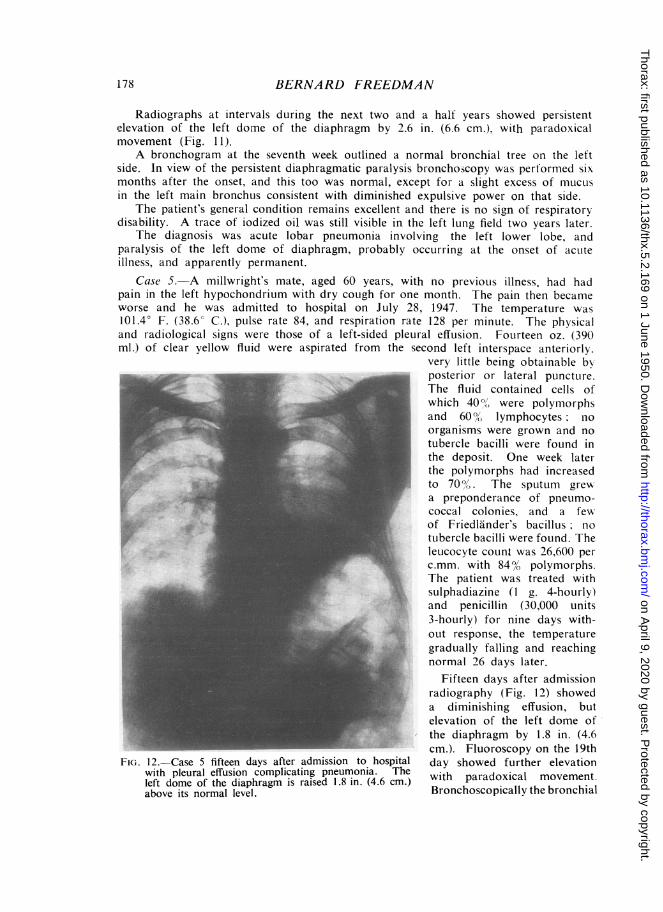
Case 5.-A millwright's mate, aged 60 years, with no previous illness, had hadpain in the left hypochondrium with dry cough for one month. The pain then becameworse and he was admitted to hospital on July 28, 1947. The temperature was101.4° F. (38.6° C.), pulse rate 84, and respiration rate 128 per minute. The physicaland radiological signs were those of a left-sided pleural effusion. Fourteen oz. (390ml.) of clear yellow fluid were aspirated from the second left interspace anteriorly.
very little being obtainable byposterior or lateral puncture.The fluid contained cells ofwhich 400, were polymorphsand 600/. lymphocytes nioorganisms were grown and notubercle bacilli were found inthe deposit. One week laterthe polymorphs had increasedto 70O/ The sputum grew\a preponderance of pneumo-coccal colonies, and a fewof Friedlander's bacillus notubercle bacilli were found. Theleucocyte count was 26,600 per
i .mm. with 84o% polymorphs.The patient was treated withsulphadiazine (1 g. 4-hourly)and penicillin (30,000 units3-hourly) for nine days with-
LZ out response, the temperaturegradually falling and reachingnormal 26 days later.
Fifteen days after admissionradiography (Fig. 12) showeda diminishing effusion, butelevation of the left dome ofthe diaphragm by 1.8 in. (4.6cm.). Fluoroscopy on the 19th
FIG. 12.-Case 5 fifteen days after admission to hospital day showed further elevationwith pleural effusion complicating pneumonia. The h paradoxileft dome of the diaphragm is raised 1.8 in. (4.6 cm.) wit cal movement.above its normal level. Bronchoscopically the bronchial
178
on April 9, 2020 by guest. P
rotected by copyright.http://thorax.bm
j.com/
Thorax: first published as 10.1136/thx.5.2.169 on 1 June 1950. D
ownloaded from

UNILATERAL PARALYSIS OF DIAPHRAGM AND LARYNX 179
tree was normal. By the 30th day after admission the effusion had absorbed, andthe patient was clinically well. As he was not under my immediate care, I did notsee him again until five months later when clinically and radiologically the lungswere clear and diaphragmatic function normal.
In view of the patient's history on admission the onset of the illness may probablybe taken at 28 days before that date. The leucocytosis favours a pyogenic infectionand virtually excludes a primary tuberculous effusion. It is usual to find a fairproportion of lymphocytes in pneumonic effusions if the latter are non-purulent.It may be taken that his illness was a lobar pneumonia, of which the physical andradiological signs were masked by the presence of a sterile effusion. Paralysis ofthe left dome of the diaphragm occurred six weeks after the onset and persisted foran unknown period, but less than five months.
Case 6.-A housewife, aged 23 years, had suffered from recurrent bronchitis since1942, and from cystitis in 1947. A primigravida, she was admitted to hospital onDecember 31, 1948, for delivery of twins. The progress of labour was slow, andowing to the death of one foetus she was delivered with forceps 44 hours after thebeginning of labour. Premedication was atropine 1/100 grain (0.6 mg.); the anaes-thetic was "kemithal," cyclopropane, and oxygen, and its administration lasted for40 minutes. Bleeding from an upper vaginal tear was controlled with a pack. Apartfrom this delivery was straightforward. Morphine, I grain (15 mg.), was given post-operatively. There was no vomiting during or after anaesthesia. There were nosymptoms or signs of respiratory or cardiac disease on admission.
On the day following delivery, the patient complained of pain in the right scapularregion. Her temperature was 101.00 F. (38.30 C.), pulse rate 130, respiration rate40 per min., and there were signs of consolidation at the base of the right lung.She was given penicillin 60,000 units and sulphadimidine (sulphamezathine) I g. 4-hourly for five days. On thesecond day sonorous rhonchiwere also present at the rightbase. The sputum was coloured _a faint green.On the fourth day breath
sounds at the right base weremuch diminished, the percus-sion note was impaired, andrhonchi persisted. Radiography(Fig. 13) showed 1.8 in. (4.6cm.) elevation of the right domeof the diaphragm, but clearlung fields. There was someprominence of the conus. Thetemperature and the pulse andrespiratory rates fell steadily.,reaching normal on the fourthday, but a leucocytosis was stillpresent (total 17,000 per c.mm.of which polymorphs were70%). No pneumonic shadow FIG. 13.-Case 6. Post-operative pneumonia on fourth daywas seen in the postero-anterior of illness. The right dome of the diaphragm is raisedradiograph-; the situation of 1.8 in. (4.6 cm.) above its normal level. Lung fieldsradiographthe situation of appear clear in this view, as the affected tissue wasthe physical signs suggested situated behind the dome.
on April 9, 2020 by guest. P
rotected by copyright.http://thorax.bm
j.com/
Thorax: first published as 10.1136/thx.5.2.169 on 1 June 1950. D
ownloaded from

BERNARD FREEDMAN
0~~~~U
~ ~ ~ 0
to E
~~~~~0
E_E 80
CO
E0)1
l l001v
0)I -I
00:-
_*
0
00
V
to<)00
*
A
0N-
A0
A vN A
-It o o
I 0)
CO CZ C
0 io 0 0 0
8°OE, E E E E
0 ONCi4
ON O) en(-
W.c
ci
u
0)
Ct
0)
el
Ct
0
0
CO
0)
0)
0):
0)
N^
00
0)0t._
0u
30
2
0)
0)
2
180
0
z
0
z
<C
¾;Z-
0z
Cd.
0
X
Vx0
HK
a
U
0)
r
I~~~~~~~~~~~~~~~~~~~~~
,t
on April 9, 2020 by guest. P
rotected by copyright.http://thorax.bm
j.com/
Thorax: first published as 10.1136/thx.5.2.169 on 1 June 1950. D
ownloaded from

UNILATERAL PARALYSIS OF DIAPHRAGM AND LARYNX 181
that the affected lung tissue lay in the lowest part of the posterior basal segmentwhere it would be obscured by the shadow of the diaphragm.
Fluoroscopy confirmed elevation of the right diaphragm, which exhibited para-doxical movement. By the ninth day the affected dome was lower, but movementwas still paradoxical, and it has remained so until the time of writing, 10 monthslater. There is very slight breathlessness on exertion, and moderate diminution ofbreath sounds at the right base; otherwise the patient remains well.
The diagnosis may be summed up as post-operative pneumonia affecting the rightlower lobe, with paralysis of the right dome of the diaphragm beginning at the onsetof the pneumonia, or shortly after, and improving after one week, but with weaknessstill present 10 months later
DISCUSSIONThe tabulated findings reveal certain features in common as well as certain
features of difference between the cases. A lower lobe was concerned in five of thesix cases. Pleurisy was present in three cases, two of which were accompanied byeffusion, the third being "dry." In two of the six cases diaphragmatic paralysisprobably occurred at the onset of the acute illness. In the four other cases theparalysis occurred on an average five-and-a-quarter weeks after the onset of theacute illness. There was a similar latent period in the onset of the vocal cordparalysis in Case 1, namely four weeks. The duration of the diaphragmatic paralysisis known for certain in only two cases, 10 and 11 weeks respectively. In a third(Case 2) it was probably a little over seven weeks. In another two cases it is stillpresent after 44 and 130 weeks. Owing to follow-up difficulties the duration inthe remaining case is not certainly known, but it was less than 22 weeks.
Pneumococci were grown from the sputum of two cases. No sputum was pro-duced by two others, and none was sent for culture from the remaining two. Thepleural effusions were sterile.
Two cases were post-operative, and two were associated with lung abscess.There seem to be two possible ways in which pneumonia may bring about
paralysis of the diaphragm and, on occasion, the ipsilateral vocal cord (left). Oneis a toxic or inflammatory neuritis of the phrenic and recurrent laryngeal nerves.The other is a toxic or inflammatory diaphragmitis. The evidence in support of thelatter explanation is not strong. Long-standing empyemata may cause degenerationof the fibres of the diaphragm. Waxy degeneration is sometimes seen in fatal casesof pneumonia and anaphylaxis ; it is due to overwork of the diaphragm or to asphyxia(Wells, 1927). In none of these conditions is there evidence that the diaphragm isparalysed. Norris and Landis (1938) state that pleural effusion may cause " cripplingof the function of the diaphragm . . . if the effusion has been present some time";these authors make no mention of paralysis. Only two of the six cases described hadpleural effusion, and one had dry pleurisy. The following facts are against thisexplanation in the cases described. It does not account for the four cases withouteffusion, nor for the laryngeal palsy. The effusions were not purulent, and grew noorganisms; a toxic or bacterial diaphragmitis therefore seems unlikely. In myopinion a toxic or inflammatory neuritis of the phrenic and left recurrent laryngealnerves was the operative factor, for this would fully explain the clinical phenomena.Whenever a motor nerve is situated in a toxic medium, loss of function may occur,and the longer the nerve the greater is the length exposed to noxious influences.
on April 9, 2020 by guest. P
rotected by copyright.http://thorax.bm
j.com/
Thorax: first published as 10.1136/thx.5.2.169 on 1 June 1950. D
ownloaded from

BERNARD FREEDMAN
The vulnerability of the cranial nerves, especially the sixth, in meningitis is anexample.
The phrenic nerves take a long course through the thorax and throughout thatcourse they are situated immediately under the mediastinal pleura, where they areliable to damage by toxins present in the pleural cavity. There is also a possibilitythat, where the nerves pass in front of the respective lung hila, they would be exposedto toxins travelling in the bronchial lymphatics.
The left recurrent laryngeal nerve fibres before they leave the vagus trunk arelikewise situated subpleurally. Their shorter subpleural course corresponds to thelower incidence of paralysis (1 in 6).
Unilateral diaphragmatic paralysis in itself causes very little disability. Therewould appear to be no practicable form of treatment for the one patient withapparently permanent paralysis, nor is it necessary, since he is not conscious ofany disability.
These six cases were encountered in the medical wards of a general hospital inthe course of three years. It is surprising therefore that no previous record of thiscondition exists in the literature. I believe that the condition may be relativelycommon, but that it has hitherto escaped notice, first because the onset of theparalysis is delayed until after the patient has been discharged from hospital, andsecondly because the paralysis itself gives rise to no symptoms that would causethe patient to return for medical advice.
SUMMARYSix cases of unilateral paralysis of the diaphragm in association with pneumonia
are described. The paralyses occurred on the same side as the pneumonia.In one of these there was also ipsilateral vocal cord paralysis.The onset of paralysis was probably coincidental with that of the pneumonia, or
only slightly delayed in two cases; in the remaining four there was a latent intervalof over five weeks. The paralysis lasted many weeks, and in one case is probablypermanent.
Two cases were associated with pleural effusion, one with dry pleurisy, and twowith lung abscess. Two were post-operative.
Reasons are given for the opinion that the paralyses were the result of toxic orinflammatory neuritis involving the phrenic nerves and the left recurrent laryngealnerve.
REFERENCES
Blattner, R. J. (1942). J. Pediat., 20, 225.Davies, D. T., Hodgson, H. G., and Whitby, L. E. H. (1935). Lancet, 1, 919.Graeser, J. B., Wu, C., and Robertson, 0. H. (1934). Arch. intern. Med., 53, 249.Heifron, R. (1939). Pneumonia. The Commonwealth Fund, New York; Oxford University Press.Kienb6ck, R. (1898). Wien. klin. Wschr., 11, 538 and 1178.Leitner, St. J. (1938). Schweiz. med. Wschr., 19, 747.Light, J. S. (1944). J. Pediat., 24, 627 (Case l).Norris, G W., and Landis, H. R. M. (1938). Diseases of the Chest, p. 781. Saunders, Philadelphia.Sanguinetti, A. A., and Galzerano, D. A. (1943). Rev. Asoc. med. argent., 57, 413.Stivelman, B. P. (1935). Amer. J. med. Sci., 190, 256.Udaondo, C. B., and Vadone, A. (1929). Amer. Rev. Tuberc., 20, 741.Wells, H. G. (1927). Arch. Path. Lab. Med., 4, 681.Wu, C. (1932). Radiology, 19, 215.
182
on April 9, 2020 by guest. P
rotected by copyright.http://thorax.bm
j.com/
Thorax: first published as 10.1136/thx.5.2.169 on 1 June 1950. D
ownloaded from